Embed Size (px)
Citation preview

I
Heinrich Klefenz
Industrial Pharmaceutical Biotechnology
Industrial Pharmaceutical Biotechnology. Heinrich KlefenzCopyright © 2002 Wiley-VCH Verlag GmbH
ISBNs: 3-527-29995-5 (Hardcover); 3-527-60012-4 (Electronic)

III
Heinrich Klefenz
Industrial PharmaceuticalBiotechnology
Industrial Pharmaceutical Biotechnology. Heinrich KlefenzCopyright © 2002 Wiley-VCH Verlag GmbH
ISBNs: 3-527-29995-5 (Hardcover); 3-527-60012-4 (Electronic)
IV
Dr. Heinrich KlefenzHauptstr. 35D-76879 BornheimGermany
This book was carefully produced. Nevertheless, author and publisher do not warrant the informa-tion contained therein to be free of errors. Readers are advised to keep in mind that statements, data,illustrations, procedural details or other items may inadvertently be inaccurate.
Cover illustration: Design by ‘das trio kommunikation und marketing gmbh; Mannheim, München’
Copyright of and reprint permissions granted byAmerican Society for Microbiology (Tables 7.1, 7.2; ref. 502)American Association for the Advancement of Science (Tables: 4.7, ref. 219; 5.1, ref. 224; Figures: 4.1,ref. 154; 4.2, ref. 510; 6.1, ref. 301)Nature Publishing Group (Fig. 1.3, ref. 432; Tables: 1.6, ref. 432; 1.7, ref. 433; 1.8, ref. 436; 1.9, ref.437; 1.10, ref. 439).
Library of Congress Card No.: Applied for.
British Library Cataloguing-in-Publication Data:A catalogue record for this bookis available from the British Library
Die Deutsche Bibliothek Cataloguing-in-Publication Data:A catalogue record for this publication is available from Die Deutsche BibliothekISBN 3-527-29995-5
© WILEY-VCH Verlag GmbH, Weinheim (Federal Republic of Germany), 2002
Printed on acid-free paper.
All rights reserved (including those of translation into other languages). No part of this book may bereproduced in any form – by photoprinting, microfilm, or any other means – nor transmitted or translatedinto a machine language without written permission from the publishers. Registered names, trademarks,etc. used in this book, even when not specifically marked as such, are not to be considered unprotected bylaw.
Composition: Manuela Treindl, RegensburgPrinting: Strauss Offsetdruck GmbH, MörlenbachBookbinding: J. Schäffer GmbH & Co. KG, Grünstadt
Printed in the Federal Republic of Germany.
Industrial Pharmaceutical Biotechnology. Heinrich KlefenzCopyright © 2002 Wiley-VCH Verlag GmbH
ISBNs: 3-527-29995-5 (Hardcover); 3-527-60012-4 (Electronic)
V
Preface
Biotechnology and its applications in medicine, pharma, and related industries representone of the most influential developments and pose one of the greatest challenges of the 21st
century, both with respect to its political, societal, and ethical implications and in the searchfor the fulfillment of its promises for health.
Biotechnology is stepping beyond previously insurmountable boundaries in understandingand manipulating life, in the efforts to understand biology, to eradicate disease, to maintainhealth and vigor, and to endow humans and life forms with desired properties.
This book aims to describe a fast-moving subject (or rather a whole interconnected sys-tem of subjects) and, like in optics, some parts of the picture may be blurred and willrequire further refining. It pulls together topics, which are essential for the realization ofthe promises of biomedicine – the repertoire of genomics, proteomics, cytomics, bio-informatics, and the interaction of networks – and combines with pertinent methods innanotechnologies, such as engineering tools to design and construct devices, artificial in-telligence and vision processing for nano-devices, implantates, and for the envisioned swarmsof remedial nano-robots.
Crucial topics for future therapies are regenerative medicine and the cultivation of tis-sues and organs as well as the underlying genetics and regulatory, developmental, bio-chemical networks.
Complex traits, critical in multifactor and degenerative diseases, are being dealt with,with a focus on senescence which forms the background against which numerous degen-erative and acute diseases develop, the elucidation of which will facilitate the strengtheningof immune responses, the maintenance of homeostasis and biochemical networks, the pres-ervation of the integrity of genetic and cellular structures.
Drug discovery encompasses the identification of molecular structures, the creation ofactive molecules, and the development of novel comprehensive therapies like immuno-therapy and cellular or organismal therapy with genetically engineered cells.
Biotechnology, chemistry, physics provide the tools for target identification, for the cre-ation of new molecular structures, and for the recovery of biologically active moleculesprovided by the biosphere and efficiency-honed during continuous evolutionary processes.
The huge amounts of data and information alone will not be sufficient to lead to newmolecular entities and novel therapies, since synthesizing millions of compounds will nei-ther fill the universe of potential molecular structures nor allow the identification of thosethree-dimensional structures specifically interacting with targets. The knowledge of thebiological processes and structures as the templates and targets for the identification ofactive molecules is indispensable.
Biological plus chemical functional information and knowledge of interactions and net-works will be the foundation to which the essential components of creativity and innova-
Industrial Pharmaceutical Biotechnology. Heinrich KlefenzCopyright © 2002 Wiley-VCH Verlag GmbH
ISBNs: 3-527-29995-5 (Hardcover); 3-527-60012-4 (Electronic)
VI
tion (and chance) are to be added as keys for the successful application of the pertinenttechnologies.
The reference list of more than 700 literature citations is meant to underpin the contentsand the conclusions of the book’s theme, and to serve as a starting point for delving deeperinto individual subjects.
Special thanks go to Dr. Hovsep Sarkissian for his support in layout, in the production offigures and tables, in proofreading and the generation of a readable manuscript. Thanks arealso due to the staff of Wiley-VCH for their organization, continuous encouragement, andstimulation; and to the ‘Muttersprachler’ who critically read the English manuscript pro-vided and contributed to the professionalism of the writing.
Utmost to be thankful for is the patience, understanding, and support of my family andour two children who have tolerated extended periods of negligence.
Undoubtedly the rapid development in biotechnology, biomedicine, and supporting tech-nologies, will affect many topics of the book’s field and will necessitate modifying, chang-ing, or complementing the subjects.
I have no doubt that in our efforts to fulfill the potential of pharmaceutical biotechnol-ogy, we are on a steep uphill slope and the top of the mountain (control of health, disease,and desired properties) is in the clouds at incalculable but reachable distance.
I welcome critical comments or suggestions about the book, proposals for areas to bedealt with in the future, and I am ready to provide further details, information, or referencesabout the various topics upon request.
H. Klefenz Bornheim, December 2001e-mail address: [email protected]
Preface
VII
Contents
Preface V
1 Introduction to Functional Biotechnology 1
1.1 Scientific and Technological Foundations 11.2 Genomics 21.3 Proteomics 141.4 Cytomics 301.5 Micro- and Nanotechnology 321.6 Cellular Cloning 421.7 Tissue Engineering (Organ Cultivation) 501.8 Micro- and Nanotechnologies for Medicine 621.9 Structural Genomics 66
2 Organizational Structures 73
2.1 Virtual and Real Enterprises 732.2 R & D Networks 742.3 Outsourcing 752.4 Registrations/Permissions 76
3 Markets and Factors 77
3.1 Products and Services 773.2 Economies 773.3 Manpower 783.4 Resources 78
4 Biotechnology and Medicine 79
4.1 Diagnostics 804.2 Therapeutics 804.3 Gene Therapy 824.4 Implantates 924.5 Medical Devices and Technology 934.6 Complex Traits 96
Industrial Pharmaceutical Biotechnology. Heinrich KlefenzCopyright © 2002 Wiley-VCH Verlag GmbH
ISBNs: 3-527-29995-5 (Hardcover); 3-527-60012-4 (Electronic)
VIII Contents
5 Drug Discovery 117
5.1 Substances Derived from Bacteria, Plants, Insects, and Animals 1345.2 Sources of Active Principles 1355.3 Assay Systems and Models (e.g., Knock-out Mice) 140
6 Molecular Pharma-Biotechnology 145
6.1 Bioinformatics 1466.2 Biological Systems and Models 1626.3 Assay Systems 1676.4 High-throughput Screening 1706.5 Automation 1726.6 Combinatorial Synthesis: Chemistry, Biology, and Biotechnology 1736.7 Genotyping: Genetic Pre-Disposition, and Heterogeneity 1776.8 Sequencing 1816.9 Pharmaco-Genomics 184
7 Research and Development 195
7.1 Biology, Medicine, and Genetics 1957.2 Pre-clinical and Clinical Development 1957.3 Processes 1957.4 Pilot Plants 1967.5 Engineering 1967.6 Fermentation Process Development 196
8 Pharmaceutical Production 201
8.1 GenePharming (Animals and Plants) 2038.2 Vitamins 2088.3 Amino Acids 2088.4 Proteins 2098.5 Antibiotics 2128.6 Biocatalysis 2198.7 Natural Compounds 2298.8 Recovery/(Bio-) Processing 2298.9 Chemical–Biotechnological Syntheses 2318.10 Gene Therapy Vectors/Systems 2358.11 Production: Safety, Efficacy, Consistency, and Specificity 2358.12 Registration 238
IXContents
9 Safety 239
9.1 Medical Safety 2399.2 Biological Safety 2399.3 Chemical Safety 2399.4 Equipment Safety 240
10 Environment 241
10.1 Pharmaceuticals and the Environment 24110.2 Biological Containment 24110.3 Physical/Chemical Containment 24210.4 Process-Integrated Environmental Protection 24210.5 Waste/Effluent Treatment and Recycling 242
11 Ethics 245
12 Companies, Institutes, Networks, and Organizations 247
References 263
Index 303
303Index
AAAV vectors 92active principles 76aeropyrum pernix 147ageing 99, 102Agrobacterium tumefaciens 212algorithm 138amplified fragment length polymorphism (AFLP)
4angiogenesis 45angiopoietin-1 (Ang-1) 83animal biotechnology 1anti-infective therapies 145anti-infectives 134antibiotics 212, 213antibody diversity 142antibody libraries 19apoptosis 138, 157Aspergillus awamori 212Aspergillus terreus 217assay systems 167atherosclerotic vascular disease 53atomic force microscopy (AFM) 176autoimmune diseases 142automation 15
Bbait 18biocatalysis 223, 227biocavity lasers 93biochips 1, 20bioengineering 207bioinformatics 2, 20, 188biological systems 19biomaterials 50biopharmaceuticals 2biosensors 20, 138biosynthesis 137birth defects 44bone marrow cell 62
Index
CC. elegans 96Caenorhabditis elegans 96caloric restriction 97cancer 2, 102cancer therapy 70cardiac hypertrophy 5cardiovascular diseases 82caspase 123catalysis 63catalysts 2, 64cDNA libraries 9cellular cloning 42cellular signaling pathway 118channelopathies 167chemoinformatics 160chiral 174chromatin 10, 184chromosomal architecture 49chromosomal position 87clinical studies 91coagulation factors 93combinatorial chemistry 172combinatorial synthesis 172complex traits 155complexity 159computational biology 2crystal growth 39crystal structure 49cystic fibrosis (CF) 89cytomics 2
DDamID 10database 3, 160Deinocaccus radiodurans 149denaturing gradient gel electrophoresis (DGGE)
178diabetes 80, 177diagnostics 1, 80differential display 186
Industrial Pharmaceutical Biotechnology. Heinrich KlefenzCopyright © 2002 Wiley-VCH Verlag GmbH
ISBNs: 3-527-29995-5 (Hardcover); 3-527-60012-4 (Electronic)
304 Index
differential expression profiles 7differential gene expression 97differentially expressed genes 101differentiation 102, 103digital organisms 158dimerization 216diploid 207direct-write technology 33discovery genetics 189discovery genomics 189disruption phenotypes 7DNA chips 63DNA computers 41DNA detection 65DNA methylation 10DNA repair 97DNA sequences 3DNA sequencing 88dopamine 18, 27droplet micro-dispensing 33Drosophila 10Drosophila melanogaster 105drug design 124drug discovery 131Duchenne muscular dystrophy 90dynamic allele-specific hybridization 13
Ee-beam 33effectors 86electrokinetic flow 33embryonic carcinoma (EC) 45embryonic stem cells (ES) 43, 48enantioselectivity 208endoscopes 93endostatin 85endothelial growth factor (VEGF) 83energetic field 40enhancers 85enzymes 2, 94epigenomics 2Escherichia coli 10ethics 48, 245eukaryotic genomes 108expressed sequence tag (EST) 5expression profiles 2expression profiling 6
FFabry disease 84
factor VII 92factor VIIa 92factor VIII 92factor IX 92factor Xa 92factor XIIa 92fiber optics 32fibrin 82field-effect transistors 95filamentous fungi 211fluorescence-activated cell sorter (FACS) 7fluorescence resonance energy transfer 228fluorescent proteins 167focused ion beam 33functional genomics 88functional interactions 2
GGaAs 93galactose utilization 202gene analysis 3gene arrays 3gene calling 6gene clusters 212gene expression 3, 6gene expression analysis 15gene expression profiling 27gene inactivation 61gene networks 6gene profiling 118gene sequencing 3gene silencing 124gene therapy 88gene transcription 31gene-based diagnostics 2genepharming 203genetic ablation 163genetic engineering 88genetic testing 178genomics 2genomic imprinting 187genotyping 146Girardia tigrina 156glycoconjugates 127growth factor 45, 49
HHaematococcus pluvialis 201Haemophilus influenzae 4Helicobacter pylori 30
305Index
hematopoietic stem cell 49hemophilia A 93hemophilia B 93high-throughput 3, 8, 30high-throughput screening 170higher-order chromatin 11histocompatibility proteins 128histone acetylation 43HLA-DR 9homeogenes 155homeoproteins 155hormones 2, 56human corneas 53human immunodeficiency virus 31human leukocyte antigen group DR 9human telomerase reverse transcriptase (hTERT)
50–62human transcriptome map 30hybridization 3hydrogel 40
Iimmune rejection 50immune response 89immune system 93immunity 60immunotherapy 80implantates 95implantation 54imprinting 2, 63in silico 132in utero fetal gene transfer 83in vitro 132in vitro culture 43in vivo 132ink jet 33interactions 38introns 88islet cell transplantation 53, 56isopenicillin N synthase (IPNS) 218isotope-coded affinity tags (ICATs) 19
Llaboratory automation 174laser chemical vapor deposition 33lead compounds 135leukemia 14life span 96linkage studies 115lysosomal storage disorders (LSDs) 84
Mmagnetic resonance imaging (MRI) 45major histocompatibility complex (MHC) 3, 9MALDI MS 26MALDI time-of-flight (TOF) MS 15MALDI-TOF 15mammalian chromosome 85mammalian retina 54MAPREC 27medical devices 73membrane proteins 130Mendelian inheritance 188metabolic engineering 196metabolic networks 2, 199metabolic profiling 203metabolism 205metabolome 153metamorphosis 101Methanococcus jannaschii 147methylation 3, 187MHC 88MHC class I molecules 31micro-machining 32micro-pen 33microarrays 7microbead 7microbial biotechnology 201microdevice 35microelectromechanical structures (MEMS) 32microfluidic biosensor arrays 33microfluidic systems 40microfluidics 32microinjection 36micromachining 32microorganisms 33microPET 96microrobot 35mitochondrial DNA (mtDNA) 87MobyDick 152molecular electronics 36molecular imprinting 63molecular interactions 71molecular machine 37molecular scanner 28motor disorders 95mRNA 3mRNA profiling technique 6multidimensional protein identification
technology 28multifactorial diseases 96
306 Index
Mycobacterium tuberculosis 10Mycoplasma genitalium 147myoseverin 175
Nnanocomposites 33nanodevices 35nanofluidics 182nanomechanics 40nanorobots 35nanosensors 36nanotechnology 40nanotube 37nanotubes 36natural products 173networks 184neural processes 95neural stem cells 49neurotoxins 167neurotransmitter 27NMR spectroscopy 69nuclear magnetic resonance (NMR) 15, 71nuclear transfer 112
Ooligonucleotide 112oocyte 42oral drug delivery 94organ cultivation 50organ replacement 54osteoporosis 82osteoprotegerin 143outsourcing 75
Ppathogen 4, 30peptidomimetics 174Pfiesteria piscicida 183pharmaceutical companies 170pharmaco-genomics 2, 185, 186phenotype-based screens 133phenotypes 47, 133phenotypic variation 100Phormidium laminosum 36phosphatase 55photolithography 34pig cloning 47plasmon-resonant particles (PRP) 64pluripotent ES cells 45pluripotent human stem cells 58
pluripotential cells 45Podospora anserina 105polyketide 215polymerase chain reaction (PCR) 3, 6polymorphism 179population doubling level (PDL) 59porcine endogenous retroviruses (PERV) 58positron emission tomography (PET) 96primate cloning 42programmed cell death 157promoter 86protein chips 62protein folding 27protein localization 7proteome 153proteome analysis 28proteomics 2pulmonary fibrosis 6Pyrobaculum aerophilum 4
Qquantitative trait loci (QTL) 100
Rrapid-prototyping 38rare diseases 87recombinant proteins 121regenerative medicine 2regulatory elements 83regulatory networks 124replication moulding 32representational differential analysis 186
SSaccharomyces cerevisiae 7, 196SAGE 186Schizosaccharomyces pombe 11screening 146self-assembly 34senescence 59senescent cells 103sensors 32sequence-specific binding 15sequencing 3, 90SEQUEST 17SEQUEST algorithm 28serial analysis of gene expression (SAGE) 3seven transmembrane receptors (7TMRs) 130seven-transmembrane proteins 164
307Index
severe combined immunodeficiency mice (SCID)59
signal transduction 71signaling pathway 121silencers 86single-nucleotide polymorphism (SNP) 2, 27single-strand conformation polymorphism
(SSCP) 178single-walled carbon nanotubes 182small molecules 133soft lithography 32solid-state fermentation 235somatic mutations 102somatostatin 27stem cells 49Streptomyces collinus 215Streptomyces griseus 217structural biology 2, 131structural genomics 3structural studies 30structure determination 66subtractive hybridization 4supramolecular structures 11surface plasmon resonance (SPR) 80
Ttag sequences 7tagging 21telomerase 59telomerase ribonucleoprotein complex (TERT)
59telomere 60, 98tethering 124therapeutic cloning 2therapeutic targets 3Thermotoga maritima 147three-dimensional structures 159TILLING 13tissue 43tissue engineering 1total gene expression analysis (TOGA) 3, 186toxicology 132toxins 136trait analysis 3transactions 12transcript 3, 15transcription factors 155
transcriptional silencing 85transcriptome 153transfection 89transgenes 89transgenic animals 144transgenic mouse 102transgenic plants 202translational 203translocation 131transmembrane domains 18transmembrane receptors 164transplantation 43, 162transplants 44transposon-tagged proteins 7transposon-tagging 7triple-helix-forming oligonucleotides (TFO) 124trophectoderm (TE) 48Trypanosoma brucei 126tumor genotyping 118tumor growth 136two-dimensional gel electrophoresis 16two-hybrid 16two-hybrid-system 16tyrosine kinase 118
Vvaccines 10vaccinia virus 18visualization 3vitamin 208
WWerner syndrome (WS) 97wound healing 114
XX-ray crystallography 66Xenopus laevis 96xenotransplantation 57
Yyeast 85yeast mutants 7yeast two-hybrid 15
Zzebra fish 125
1
1 Introduction to Functional Biotechnology
1.1 Scientific and Technological Foundations
Pharmaceutical biotechnology focuses on biotechnology with pharmaceutical relevance asthe central science and technology of the ‘Life Sciences’ with its fundamentals, develop-ments, influences and effects.
This monograph demonstrates the paradigmatic changes effected by biotechnology incombination with pharmaceutical science, cell biology, chemistry, electronics, materialsscience and technology, plus organizational changes on pharmaceutical research, develop-ment and industry as well as pharmaceutical-related animal and plant biotechnology (‘LifeSciences’).
Pharmaceutical biotechnology exemplifies the transformation towards a knowledge-basedsociety with innovation as the essential basis of activity in an age of globalization, in-creased competition, and accelerated speed of development, changes and decisions.
The total spectrum of concepts, processes and technologies of biotechnology, chemistryand electronics is being applied in modern industrial pharmaceutical research, develop-ment and production.
In pharmaceutical and medical research, diagnostics, production and therapy, the resultsof genome sequencing and studies of biological–genetic function (functional genomics)are combined with chemical, microelectronic and micro system technologies to producemedical devices, known as diagnostic ‘Biochips’.
In chemical, pharmaceutical and biotechnological production processes the multitude ofbiologically active molecules is expanded by additional novel structures created with newlyarranged ‘gene clusters’ and (bio-) catalytic chemical processes.
Materials synthesized with chemical and biotechnological processes support novelimplantates, tissue engineering and even competitors to silicon-based computing, as wellas analytics, diagnostics, medical devices, electronics, data processing and energy conver-sion.
New organizational structures in the cooperation of institutes, companies and networksenable faster knowledge and product development, and immediate application of scientificresearch and process developments.
Target groups of readers are biotechnologists, pharmaceutical scientists, biochemists,biologists, physicians, pharmacologists, chemists, reproductive biologists, genetic engi-neers, agro-scientists, and animal and plant breeders.
Organizationally, this monograph is addressed to scientists, technicians and managers ofbiotechnology, pharmaceutical and chemical companies, research institutes, and biotechventures, and decision makers in industry, science, venture capital/finance and politics.
Industrial Pharmaceutical Biotechnology. Heinrich KlefenzCopyright © 2002 Wiley-VCH Verlag GmbH
ISBNs: 3-527-29995-5 (Hardcover); 3-527-60012-4 (Electronic)
2 1 Introduction to Functional Biotechnology
This monograph aims to present an integrated view of the manifold and diverse develop-ments and their impact on the discovery of new drugs and therapies. Specifically, the topicsdeal with:
• The integration of genomics, proteomics, cytomics, structural and functional biology.• Studies of networks and multi-gene traits at the molecular, genetic, biochemical, cellu-
lar and organism levels.• Micro- and nanotechnologies for R & D and therapy.• Stem cell research, therapeutic cloning and regenerative medicine.• Drug discovery and therapy development from genomics, proteomics to small molecules,
biopharmaceuticals to systems.• Organizational solutions and core competencies for the pharmaceutical industry.• Bioinformatics, functional genomic, structural analysis and computational biology.• Scientific and technological foundations.
1.2 Genomics
Functional genomics is the scientific field dealing with extracting or synthesizing biologi-cally relevant and therapeutically useful information from sequences, genomics, proteomics,expression profiles and linkage studies. The analysis of genomic, expression and proteomicdata produces networks of functional interactions and linkages between proteins, cells,tissues and organs.
Proteins are the main catalysts, structural elements, signaling messengers and molecularmachines of biological tissues. Phylogenetic profile generation and two-hybrid screen meth-ods are the major techniques used to study protein–protein interactions.[1]
Gene-based diagnostics is rapidly expanding in the medical/industrial sector. It involvesthe study of DNA and RNA as compared to ‘classical’ medical diagnostics, which dealswith enzymes, hormones, proteins and metabolic intermediates. The total business volumein medical diagnostics is about US$ 18 billion (1998), out of which gene-based diagnosticscomprises US$ 500–700 million, with annual growth rates of 25%.
The pharmaco-genomics market (products and services) is estimated to grow from US$47 million in 1998 to US$ 795 million in 2005, with the major areas being cardiovasculardiseases (US$ 139 million), infectious diseases (US$ 123 million), central nervous system(CNS)-related disorders (US$ 72 million) and cancer (US$ 41 million). In 1999, 28pharmaco-genomic collaborations had been formed, 20 concerning the application ofpharmaco-genomics to drug development; seven were involved in drug discovery and fourin marketed drugs.
There are conceptual and real developments aimed at bringing the fields of genomics,functional genomics, pharmaco-genomics, single-nucleotide polymorphism (SNP) stud-ies, imprinting, metabolic networks, genetic hierarchies in embryonic development andepigenetic mechanisms of cancer together under the conceptual umbrella of ‘epigenomics’,studying complex phenotypes from the genomic level down. The focus of scientific efforts
31.2 Genomics
is genome-scale mapping of the methylation status of CpG dinucleotides, the identificationand analysis of epigenomic loci in the major histocompatibility complex (MHC), and thecomparative analysis of epigenomic information from different organisms.[2]
The flow of novel genes from efforts in genomics provides the opportunity to greatlyexpand the number of therapeutic targets – the limited resource in drug discovery. Strate-gies to accelerate the evaluation of candidate molecules as disease-relevant targets involvethe establishment of pertinent models (e.g., mice, cells, organs, zebra fish, nematodes andyeast).
The challenge of transforming DNA sequences into disease-relevant targets will con-tinue to be a major requirement in drug discovery.[3] Genomics stretches from gene se-quencing, gene analysis and trait analysis via structural genomics to functional genomics.
Structural genomics aims to experimentally determine the structures of all possible pro-tein folds. Such efforts entail a conceptual shift from traditional structural biology in whichstructural information is obtained on known proteins to one in which the structure of aprotein is determined first and the function assigned later. Whereas the goal of convertingprotein structure into function can be accomplished by traditional sequence motif-basedapproaches, recent studies have shown that assignment of a protein’s biochemical functioncan also be achieved by scanning its structure for a match to the geometry and chemicalidentity of a known active site. This approach can use low-resolution structures providedby contemporary structure prediction methods. When applied to genomes, structural infor-mation (either experimental or predicted) is likely to play an important role in high-through-put function assignment.
Sequence genomics is the starting point for structural and functional genomics whichprovide the experimental structural data for the molecular design of antagonists, agonistsand biologically (respectively, pharmacologically) active substances.[4]
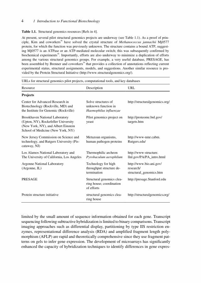
Table 1.1 shows a compilation of projects, sources and databases for structural data tofacilitate access to these fundamental sources for pharmaceutical development.
Genomics, the study of the whole genome, requires ever-increasing efficiency in themethods used for gene analysis.
An automated, high-throughput, systematic cDNA display method called total gene ex-pression analysis (TOGA) was developed. TOGA utilizes 8-nucleotide sequences, com-prised of a 4-nucleotide restriction endonuclease cleavage site and adjacent 4-nucleotideparsing sequences, and their distances from the 3′ ends of mRNA molecules to give eachmRNA species in an organism a single identity. The parsing sequences are used as parts ofprimer-binding sites in 256 polymerase chain reaction (PCR)-based assays performedrobotically on tissue extracts to determine simultaneously the presence and relative con-centration of nearly every mRNA in the extracts, regardless of whether the mRNA has beendiscovered previously. Visualization of the electrophoretically separated fluorescent assayproducts from different extracts displayed via a Netscape browser-based graphical userinterface allows the status of each mRNA to be compared among samples and its identity tobe matched with sequences of known mRNAs compiled in databases.[5]
Methods for gene expression analysis include transcript sampling by sequencing or byhybridization signature, transcript amplification and imaging, and hybridization to genearrays. Serial analysis of gene expression (SAGE), one of the most effective methods, is
4 1 Introduction to Functional Biotechnology
limited by the small amount of sequence information obtained for each gene. Transcriptsequencing following subtractive hybridization is limited to binary comparisons. Transcriptimaging approaches such as differential display, partitioning by type IIS restriction en-zymes, representational difference analysis (RDA) and amplified fragment length poly-morphism (AFLP) are rapid and theoretically comprehensive since they use fragment pat-terns on gels to infer gene expression. The development of microarrays has significantlyenhanced the capacity of hybridization techniques to identify differences in gene expres-
Table 1.1. Structural genomics resources [Refs in 4].
At present, several pilot structural genomics projects are underway (see Table 1.1). As a proof of prin-ciple, Kim and coworkers58 have solved the crystal structure of Methanococcus jannachii Mj0577 protein, for which the function was previously unknown. The structure contains a bound ATP, suggest-ing Mj0577 is an ATPase or an ATP-mediated molecular switch; this was subsequently confirmed by biochemical experiments58. Importantly, efforts are also underway to minimize a duplication of efforts among the various structural genomics groups. For example, a very useful database, PRESAGE, has been assembled by Brenner and coworkers59 that provides a collection of annotations reflecting current experimental status, structural assignments, models, and suggestions. Another similar resource is pro-vided by the Protein Structural Initiative (http://www.structuralgenomics.org/).
URLs for structural genomics pilot projects, computational tools, and key databases
Resource Description URL
Projects
Center for Advanced Research in Biotechnology (Rockville, MD) and the Institute for Genomic (Rockville)
Solve structures of unknown function in Haemophilus influenzae
http://structuralgenomics.org/
Brookhaven National Laboratory (Upton, NY), Rockefeller University (New York, NY), and Albert Einstein School of Medicine (New York, NY)
Pilot genomics project on yeast
http://proteome.bnl.gov/ targets.htm
New Jersey Commission on Science and technology, and Rutgers University (Pis-cataway, NJ)
Metazoan organisms, human pathogen proteins
http://www-nmr.cabm. Rutgers.edu/
Los Alamos National Laboratory and The University of California, Los Angeles
Thermophilic archeon Pyrobaculum aerophilum
http://www-structure. llnl.gov/PA/PA_intro.html
Argonne National Laboratory (Argonne, IL)
Technology for high throughput structure de-termination
http://www.bio.ani.gov/ research/ structural_genomics.htm
PRESAGE Structural genomics clea-ring house; coordination of efforts
http://presage.Stanford.edu
Protein structure initiative structural genomics clea-ring house
http://structuralgenomics.org/
5
sion. In practice, however, hybridization methods are limited by an inability to detect geneswith no expressed sequence tag (EST) representation.
A methodological variation to expression analysis was developed which provides rapid,comprehensive sampling of cDNA populations together with sensitive detection of differ-ences in mRNA abundance for both known and novel genes. By using this method, thegene expression in a rat model of pressure overload-induced cardiac hypertrophy was ana-lyzed.
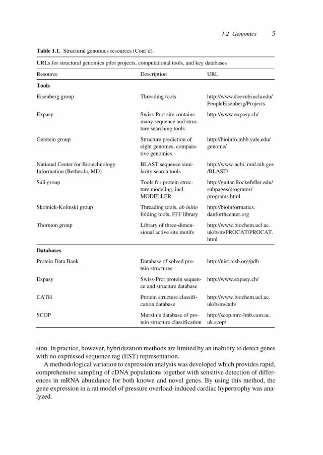
1.2 Genomics
Table 1.1. Structural genomics resources (Cont’d).
URLs for structural genomics pilot projects, computational tools, and key databases
Resource Description URL
Tools
Eisenberg group Threading tools http://www.doe-mbi.ucla.edu/ PeopleEisenberg/Projects
Expasy Swiss-Prot site contains many sequence and struc-ture searching tools
http://www.expasy.ch/
Gerstein group Structure prediction of eight genomes, compara-tive genomics
http://bioinfo.mbb.yale.edu/ genome/
National Center for Biotechnology Information (Bethesda, MD)
BLAST sequence simi-larity search tools
http://www.ncbi..nml.nih.gov/BLAST/
Sali group Tools for protein struc-ture modeling, incl. MODELLER
http://guitar.Rockefeller.edu/ subpages/programs/ programs.html
Skolnick-Kolinski group Threading tools, ab initio folding tools, FFF library
http://bioinformatics. danforthcenter.org
Thornton group Library of three-dimen-sional active site motifs
http://www.biochem.ucl.ac. uk/bsm/PROCAT/PROCAT. html
Databases
Protein Data Bank Database of solved pro-tein structures
http://nist.rcsb.org/pdb
Expasy Swiss-Prot protein sequen-ce and structure database
http://www.expasy.ch/
CATH Protein structure classifi-cation database
http://www.biochem.ucl.ac. uk/bsm/cath/
SCOP Murzin’s database of pro-tein structure classification
http://scop.mrc-lmb.cam.ac. uk.scop/
6 1 Introduction to Functional Biotechnology
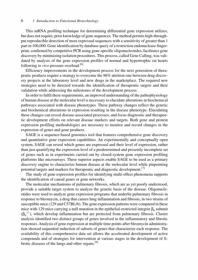
This mRNA profiling technique for determining differential gene expression utilizes,but does not require, prior knowledge of gene sequences. The method permits high-through-put reproducible detection of most expressed sequences with a sensitivity of greater than 1part in 100,000. Gene identification by database query of a restriction endonuclease finger-print, confirmed by competitive PCR using gene-specific oligonucleotides, facilitates genediscovery by minimizing isolation procedures. This process, called Gene Calling, was vali-dated by analysis of the gene expression profiles of normal and hypertrophic rat heartsfollowing in vivo pressure overload.[6]
Efficiency improvements in the development process for the next generation of thera-peutic products require a strategy to overcome the 96% attrition rate between drug discov-ery projects at the laboratory level and new drugs in the marketplace. The required newstrategies need to be directed towards the identification of therapeutic targets and theirvalidation while addressing the milestones of the development process.
In order to fulfil these requirements, an improved understanding of the pathophysiologyof human disease at the molecular level is necessary to elucidate alterations in biochemicalpathways associated with disease phenotypes. These pathway changes reflect the geneticand biochemical alterations in expression resulting in the disease phenotype. Elucidatingthese changes can reveal disease-associated processes, and focus diagnostic and therapeu-tic development efforts on relevant disease markers and targets. Both gene and proteinexpression profiling methodologies are necessary to monitor and record changes in theexpression of genes and gene products.
SAGE is a sequence-based genomics tool that features comprehensive gene discoveryand quantitative gene expression capabilities. An experimentally and conceptually opensystem, SAGE can reveal which genes are expressed and their level of expression, ratherthan just quantifying the expression level of a predetermined and presently incomplete setof genes such as in experiments carried out by closed-system gene expression profilingplatforms like microarrays. These superior aspects enable SAGE to be used as a primarydiscovery engine to characterize human disease at the molecular level while pinpointingpotential targets and markers for therapeutic and diagnostic development.[7]
The study of gene expression profiles for identifying multi-effect phenomena supportsthe identification of causal genes or gene networks.
The molecular mechanisms of pulmonary fibrosis, which are as yet poorly understood,provide a suitable target system to analyze the genetic basis of the disease. Oligonucle-otides were used to analyze gene expression programs that underlie pulmonary fibrosis inresponse to bleomycin, a drug that causes lung inflammation and fibrosis, in two strains ofsusceptible mice (129 and C57BL/6). The gene expression patterns were compared in thesemice with 129 mice carrying a null mutation in the epithelial-restricted integrin β6 subunit(β6
–/–), which develop inflammation but are protected from pulmonary fibrosis. Clusteranalysis identified two distinct groups of genes involved in the inflammatory and fibroticresponses. Analysis of gene expression at multiple time points after bleomycin administra-tion showed sequential induction of subsets of genes that characterize each response. Theavailability of this comprehensive data set allows the accelerated development of activecompounds and of strategies for intervention at various stages in the development of fi-brotic diseases of the lungs and other organs.[8]
7
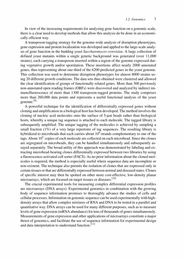
In view of the increasing requirements for analyzing gene function on a genomic scale,there is a clear need to develop methods that allow this analysis do be done in an economi-cally efficient way.
A transposon-tagging strategy for the genome-wide analysis of disruption phenotypes,gene expression and protein localization was developed and applied to the large-scale analy-sis of gene function in the budding yeast Saccharomyces cerevisiae. A large collection ofdefined yeast mutants within a single genetic background was generated (over 11,000strains), each carrying a transposon inserted within a region of the genome expressed dur-ing vegetative growth and/or sporulation. These insertions affect nearly 2000 annotatedgenes, thus representing about one-third of the 6200 predicted genes in the yeast genome.This collection was used to determine disruption phenotypes for almost 8000 strains us-ing 20 different growth conditions. The data sets thus obtained were clustered and allowedthe clear identification of groups of functionally related genes. More than 300 previouslynon-annotated open reading frames (ORFs) were discovered and analyzed by indirect im-munofluorescence of more than 1300 transposon-tagged proteins. The study comprisesmore than 260,000 data points and represents a useful functional analysis of the yeastgenome.[9]
A powerful technique for the identification of differentially expressed genes withoutcloning and amplification in a biological host has been developed. The method involves thecloning of nucleic acid molecules onto the surface of 5-µm beads rather than biologicalhosts, whereby a unique tag sequence is attached to each molecule. The tagged library issubsequently amplified. The unique tagging of the molecules is achieved by sampling asmall fraction (1%) of a very large repertoire of tag sequences. The resulting library ishybridized to microbeads that each carries about 106 strands complementary to one of thetags. About 105 copies of each molecule are collected on each microbead. Since the clonesare segregated on microbeads, they can be handled simultaneously and subsequently as-sayed separately. The broad utility of this approach was demonstrated by labeling and ex-tracting microbead-bearing clones differentially expressed between two libraries by usinga fluorescence-activated cell sorter (FACS). As no prior information about the cloned mol-ecules is required, the method is especially useful where sequence data are incomplete ornon-existent. The technique also permits the isolation of clones that are expressed only incertain tissues or that are differentially expressed between normal and diseased states. Clonesof specific interest may then be spotted on other more cost-effective, low-density planarmicroarrays, which are focused on target tissues or diseases.[10]
The crucial experimental tools for measuring complex differential expression profilesare microarrays (DNA arrays). Experimental genomics in combination with the growingbody of sequence information promises to thoroughly advance the studies of cells andcellular processes. Information on genomic sequence can be used experimentally with high-density arrays that allow complex mixtures of RNA and DNA to be tested in a parallel andquantitative way. DNA arrays can be used for many different purposes, such as to measurelevels of gene expression (mRNA abundance) for tens of thousands of genes simultaneously.Measurements of gene expression and other applications of microarrays constitute a majorthrust of genomics, and facilitate the use of sequence information for experimental designand data interpretation to understand function.[11]
1.2 Genomics
8 1 Introduction to Functional Biotechnology
The high-throughput technologies enable researchers to study gene expression for thou-sands of genes simultaneously, thus involving a huge repertoire of data. The resulting out-put of microarray studies is subject to experimental bias and substantial variability, thusrequiring statistical analysis and the replication of studies.
Statistical methods for analyzing replicated cDNA microarray expression data and re-sults of controlled experiments have provided valuable arguments for statistically controlledand validated experimentation. A study was conducted to investigate inherent variability ingene expression data, and the extent to which replication in an experiment produces moreconsistent and reliable findings. A statistical model was applied that describes the probabil-ity that mRNA is contained in the target sample tissue, subsequently converted to probe andultimately detected on the slide. An analysis of the combined data from all replicates wasalso carried out. Of the 288 genes studied in this controlled experiment, 32 would be ex-pected to produce strong hybridization signals because of the known presence of repetitivesequences within those genes. Results based on individual replicates show that there are 55,36 and 58 highly expressed genes in replicates 1, 2 and 3, respectively. An analysis usingthe combined data from all three replicates reveals that only two of the 288 genes areincorrectly classified as expressed. The experiment demonstrates that any single microarrayoutput is subject to substantial variability. By pooling data from replicates, a more reliableanalysis of gene expression data can be achieved. Thus, designing experiments with repli-cations will greatly reduce misclassification rates. At least three replicates should be usedin designing experiments when using cDNA microarrays, particularly when gene expres-sion data from single specimens are being analyzed.[12]
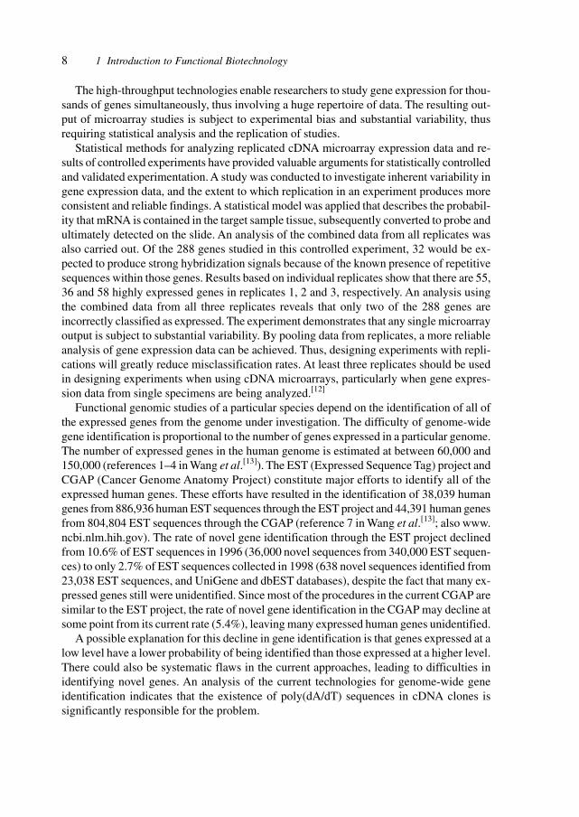
Functional genomic studies of a particular species depend on the identification of all ofthe expressed genes from the genome under investigation. The difficulty of genome-widegene identification is proportional to the number of genes expressed in a particular genome.The number of expressed genes in the human genome is estimated at between 60,000 and150,000 (references 1–4 in Wang et al.[13]). The EST (Expressed Sequence Tag) project andCGAP (Cancer Genome Anatomy Project) constitute major efforts to identify all of theexpressed human genes. These efforts have resulted in the identification of 38,039 humangenes from 886,936 human EST sequences through the EST project and 44,391 human genesfrom 804,804 EST sequences through the CGAP (reference 7 in Wang et al.[13]; also www.ncbi.nlm.hih.gov). The rate of novel gene identification through the EST project declinedfrom 10.6% of EST sequences in 1996 (36,000 novel sequences from 340,000 EST sequen-ces) to only 2.7% of EST sequences collected in 1998 (638 novel sequences identified from23,038 EST sequences, and UniGene and dbEST databases), despite the fact that many ex-pressed genes still were unidentified. Since most of the procedures in the current CGAP aresimilar to the EST project, the rate of novel gene identification in the CGAP may decline atsome point from its current rate (5.4%), leaving many expressed human genes unidentified.
A possible explanation for this decline in gene identification is that genes expressed at alow level have a lower probability of being identified than those expressed at a higher level.There could also be systematic flaws in the current approaches, leading to difficulties inidentifying novel genes. An analysis of the current technologies for genome-wide geneidentification indicates that the existence of poly(dA/dT) sequences in cDNA clones issignificantly responsible for the problem.
9
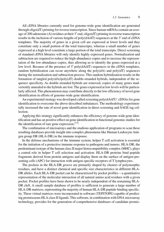
All cDNA libraries currently used for genome-wide gene identification are generatedthrough oligo(dT) priming for reverse transcription. Since human mRNAs contain an aver-age of 200 adenosine (A) residues at their 3′ end, oligo(dT) priming in reverse transcriptionresults in the inclusion of various lengths of poly(dA/dT) sequences at the 3′ end of cDNAtemplates. The majority of genes in a given cell are expressed at lower levels and theyconstitute only a small portion of the total transcripts, whereas a small number of genesexpressed at a high level constitute a large portion of the total transcripts. Direct screeningof standard cDNA libraries will only identify highly expressed genes. Normalization andsubtraction are required to reduce the high-abundance copies and to increase the represen-tation of the low-abundance copies, thus allowing us to identify the genes expressed at alow level. Because of the presence of 3′ poly(dA/dT) sequences in the cDNA templates,random hybridization can occur anywhere along the poly(dA) and poly(dT) sequencesduring the normalization and subtraction process. This random hybridization results in theformation of tangled poly(dA)/poly(dT) double-stranded hybrids, independent of the se-quence specificity. As double-stranded hybrids are removed, copies of many genes inad-vertently annealed to the hybrids are lost. The genes expressed at low levels will be particu-larly affected. This phenomenon may contribute directly to the low efficiency of novel geneidentification in efforts of genome-wide gene identification.
An experimental strategy was developed called screening poly(dA/dT)– cDNAs for geneidentification to overcome the above-described imbalances. The methodology experimen-tally increased the rate of novel gene identification in direct screening and SAGE tag col-lection.
Applying this strategy significantly enhances the efficiency of genome-wide gene iden-tification and has an positive effect on gene identification in functional genomic studies forthe identification of rare gene expression.[13]
The combination of microarrays and the studious application of programs to scan theseresulting databases provide insight into complex phenomena like Human Leukocyte Anti-gen group DR (HLA-DR) in the immune response.
In the defense mechanisms of the immune system, helper T cell activation is essentialfor the initiation of a protective immune response to pathogens and tumors. HLA-DR, thepredominant isotype of the human class II major histocompatibility complex (MHC), playsa central role in helper T cell selection and activation. HLA-DR proteins bind peptidefragments derived from protein antigens and display them on the surface of antigen-pre-senting cells (APC) for interaction with antigen-specific receptors of T lymphocytes.
The pockets in the HLA-DR groove are primarily shaped by clusters of polymorphicresidues, and have a distinct chemical and specific size characteristics in different HLA-DR alleles. Each HLA-DR pocket can be characterized by pocket profiles – a quantitativerepresentation of the molecular interaction of all natural amino acid residues with a givenpocket. Pocket profiles have been shown to be nearly independent of the remaining HLA-DR cleft. A small sample database of profiles is sufficient to generate a large number ofHLA-DR matrices, representing the majority of human HLA-DR peptide-binding specific-ity. These virtual matrices were incorporated in software (TEPITOPE) capable of predict-ing promiscuous HLA class II ligands. This software, in combination with DNA microarraytechnology, provides for the generation of comprehensive databases of candidate promis-
1.2 Genomics
10 1 Introduction to Functional Biotechnology
cuous T cell epitopes in human disease tissues. DNA microarrays are used to reveal genesthat are specifically expressed or up-regulated in disease tissues. Subsequently, the predic-tion software enables the scanning of these genes for promiscuous HLA-DR binding sites.Starting from nearly 20,000 genes, a database of candidate colon cancer-specific and pro-miscuous T cell epitopes could be fully populated within a matter of days. The approachhas provided directions for the development of epitope-based vaccines.[14]
DNA microarrays have the ability to analyze the expression of thousands of the same setof genes under at least two different experimental conditions. DNA microarrays requiresubstantial amounts of RNA to generate the probes, especially when bacterial RNA is usedfor hybridization (50 µg of bacterial RNA contains approximately 2 µg of mRNA). Acomputer-based algorithm was developed for the prediction of the minimal number of prim-ers to specifically anneal to all genes in a given genome. The algorithm predicts that 37oligonucleotides should prime all genes in the Mycobacterium tuberculosis genome. Theusefulness of the genome-directed primers (GDPs) was demonstrated in comparison torandom primers for gene expression profiling using DNA microarrays. Both types of prim-ers were used to generate fluorescent-labeled probes and to hybridize to an array of 960mycobacterial genes. The GDP probes were more sensitive and more specific than therandom-primer probes, especially when mammalian RNA samples were spiked with my-cobacterial RNA. The GDPs were used for gene expression profiling of mycobacterialcultures grown to log or stationary growth phases. This approach is useful for accurategenome-wide expression analysis, in particular for in vivo gene expression profiling, aswell as directed amplification of sequenced genomes.[15]
Interactions between protein complexes and DNA are at the core of essential cellularprocesses such as transcription, DNA replication, chromosome segregation and genomemaintenance. Techniques are therefore needed to identify DNA loci that interact in vivowith specific proteins. A limited repertoire of techniques is presently available.[16,17]
One method involves in situ cross-linking followed by purification of protein–DNAcomplexes. This technique does have the inherent risk of artifacts induced by the cross-linking agent, but it requires specific antibodies against each protein of interest as well as alarge number of cells. Another method employs in vivo targeting of a nuclease to markbinding sites of a specific protein. Induction of protein breaks is, however, likely to causemajor changes in chromatin structure and activation of DNA damage checkpoint pathways– both being distinct disadvantages.
A novel technique was developed, named DamID, for the identification of DNA locithat interact in vivo with specific nuclear proteins in eukaryotes. By tethering Escherichiacoli DNA adenine methyltransferase (Dam) to a chromatin protein, Dam can be targeted invivo to native binding sites of this protein, resulting in local DNA methylation. Sites ofmethylation can subsequently be mapped using methylation-specific restriction enzymesor antibodies. The successful application of DamID both in Drosophila cell cultures and inwhole flies was demonstrated. When Dam is tethered to the DNA-binding domain of GAL4,targeted methylation is limited to a region of a few kilobases surrounding a GAL4 bindingsequence. By using DamID, a number of expected and unexpected target loci for Droso-phila heterochromatin protein 1 were identified. DamID has usefulness for the genome-wide mapping of in vivo targets of chromatin proteins in various eukaryotes.[17]
11
The number of targets for therapeutic intervention is assessed by considering the num-ber of genes, the different splicing of the RNAs, the resulting larger number of proteins,and the numerous processes involved in generating membranes, complexes and supramo-lecular structures.
Higher-order chromatin is essential for epigenetic gene control and for the functionalorganization of chromosomes. Differences in higher-ordered chromatin structure are linkedwith distinct covalent modifications of histone tails that regulate transcriptional ‘on’ or‘off’ states, and influence chromosome condensation and segregation. Post-translationalmodifications of histone N-termini, particularly of H4 and H3, are well documented andhave
functionally been characterized as changes in acetylation, phosphorylation and, mostrecently, methylation. In contrast to the large number of histone acetyltransferases (HATs)and histone deacetylases (HDACs) described, genes encoding enzymes that regulate phos-phorylation or methylation of histone N-termini are only now being identified. The interde-pendence of the different histone tail modifications for the integration of transcriptionaloutput or higher-order chromatin organization is as yet not fully understood.
Human SUV39H1 and murine Suv39h1 – mammalian homologs of Drosophila Su(var)3-9 and of Schizosaccharomyces pombe clr4 – encode histone H3-specific methyltransferasesthat selectively methylate Lys9 of the N-terminus of histone H3 in vitro. The catalytic motifwas mapped to the evolutionarily conserved SET domain, which requires adjacent cys-teine-rich regions to confer histone methyltransferase activity. Methylation of Lys9 inter-feres with phosphorylation of Ser10, but is also influenced by pre-existing modifications inthe N-terminus of H3. In vivo, deregulated SUV39H1 or disrupted Suv39h1 activity modu-late H3 Ser10 phosphorylation in native chromatin and induce aberrant mitotic divisions.The data demonstrate a functional interdependence of site-specific H3 tail modificationsand propose a dynamic mechanism for the regulation of higher-order chromatin.[18]
Transcription is controlled in part by the dynamic acetylation and deacetylation of his-tone proteins. The latter process is mediated by HDACs. Analysis of the regulation of HDACactivity in transcription has focused primarily on the recruitment of HDAC proteins tospecific promoters or chromosomal domains by association with DNA-binding proteins.To characterize the cellular function of the identified HDAC4 and HDAC5 proteins, com-plexes were isolated by immunoprecipitation. Both HDACs were found to interact with 14-3-3 proteins at three phosphorylation sites. The association of 14-3-3 with HDAC4 andHDAC5 results in the sequestration of these proteins in the cytoplasm. Loss of this interac-tion allows HDAC4 and HDAC5 to translocate to the nucleus, interact with HDAC3 andrepress gene expression. Regulation of the cellular localization of HDAC4 and HDAC5represents a mechanism for controlling the transcriptional activity of these class II HDACproteins.[19]
In Drosophila, compensation for the reduced dosage of genes located on the single maleX chromosome involves doubling their expression in relation to their counterparts on thefemale X chromosomes. Dosage compensation is an epigenetic process involving the spe-cific acetylation of histone H4 at lysine 16 by the histone acetyltransferase MOF. AlthoughMOF is expressed in both sexes, it only associates with the X chromosome in males. Itsabsence causes male-specific lethality. MOF is part of a chromosome-associated complex
1.2 Genomics
12 1 Introduction to Functional Biotechnology
comprising male-specific lethal (MSL) proteins and at least one non-coding roX RNA. Theintegration of MOF into the dosage compensation complex is still not understood. Theassociation of MOF with the male X chromosome depends on its interaction with RNA.MOF binds specifically through its chromodomain to roX2 RNA in vivo. In vitro analysesof the MOF and MSL-3 chromodomains indicate that these chromodomains may functionas RNA interaction modules. Their interaction with non-coding RNA may target regulatorsto specific chromosomal sites.[20]
The structural and functional organization of chromatin needs to be considered in stud-ies of gene function, gene expression and molecular interaction in pharmaceutical inter-ventions.
The functional regulation of chromatin is closely related to its spatial organization withinthe nucleus. In yeast, perinuclear chromatin domains constitute areas of transcriptionalrepression. These silent domains are defined by the presence of perinuclear telomere clus-ters. The only protein found to be involved in the peripheral localization of telomeres isYku70/Yku80. This conserved heterodimer can bind telomeres and functions in both repairof DNA double-strand breaks and telomere maintenance. These findings do not describethe underlying structural basis of perinuclear silent domains. Nuclear pore complex exten-sions formed by the conserved TPR homologs Mlp1 and Mlp2 are responsible for thestructural and functional organization of perinuclear chromatin. Loss of MLP2 results in asevere deficiency in the repair of double-stranded breaks. Double deletions of MLP1 andMLP2 disrupt the clustering of perinuclear telomeres and releases telomeric gene expres-sion. These effects are probably mediated through the interaction with Yku70. Mlp2 physi-cally tethers Yku70 to the nuclear periphery, thus forming a link between chromatin and thenuclear envelope. This structural link is docked to nuclear pore complexes through a cleav-able nucleoporin, Nup145. Through these interactions, nuclear pore complexes organize anuclear subdomain that is intimately involved in the regulation of chromatin metabolism.[21]
The packaging of the eukaryotic genome in chromatin presents barriers that restrict theaccess of enzymes that process DNA. To overcome these barriers, cells possess a number ofmulti-protein, ATP-dependent chromatin remodeling complexes, each containing an AT-Pase subunit from the SNf2/SW12 superfamily. Chromatin remodeling complexes func-tion by increasing nucleosome mobility and are clearly implicated in transcription. SNF2/SW12- and ISWI-related proteins were analyzed to identify remodeling complexes thatpotentially assist other DNA transactions. A complex from S. cerevisiae was purified thatcontains the Ino80 ATPase. The Ino80 complex contains about 12 polypeptides includingtwo proteins related to the bacterial RuvB DNA helicase, which catalyzes branch migrationof Holliday junctions. The purified complex remodels chromatin, facilitates transcriptionin vitro and displays 3′ to 5′ DNA helicase activity. Mutations of Ino80 show hypersensitiv-ity to agents that cause DNA damage, in addition to defects in transcription. Chromatinremodeling driven by the Ino80 ATPase may be connected to transcription as well as DNAdamage repair.[22]
SNPs are point mutations that constitute the most common type of genetic variation andare found at a rate of 0.5–10 per 1000 base pairs within the human genome. SNPs are stablemutations that can be contributory factors for human disease and can also serve as geneticmarkers. The complex interaction between multiple genes and the environment necessi-
13
tates the tracking of SNPs in large populations in order to elucidate their contribution todisease development and progression. Several projects are intensively pursuing the identi-fication of human SNPs through large-scale mapping projects with high-density arrays,mass spectrometry (MS), molecular beacons, peptide nucleic acids and the 5′ nucleaseassay. A study has integrated microelectronics and molecular biology for the discrimina-tion of SNPs, and a rapid assay for SNP detection was developed that utilizes electroniccircuitry on silicon microchips. The method was validated by the accurate discriminationof blinded DNA samples for the complex quadra-allelic SNP of mannose-binding protein.The microchip directed the transport, concentration and attachment of amplified patientDNA to selected electrodes (test sites), creating an array of DNA samples. Through controlof the electric field, the microchip enabled accurate genetic identification of these samplesusing fluorescent-labeled DNA reporter probes. The accuracy was established by internalcontrols of dual-labeled reporters and by using mismatched sequences in addition to thewild-type and variant reporter sequences to validate the SNP genotype. The ability to cus-tomize this assay for multiple genes offers advantages for bringing the assay to the clinicallaboratory.[23]
Dynamic allele-specific hybridization, a method to detect SNPs, is based on dynamicheating and coincident monitoring of DNA denaturation and avoids the use of additionalenzymes or reaction steps.[24]
The most common DNA sequence variations, SNPs, are stable and widely scatteredacross the chromosome. Once constructed, a high-density SNP map of several hundredthousand markers will be an indispensable tool for genome-wide association studies toidentify genes that contribute to disease risk and individual differences in drug response. Tofacilitate large-scale SNP identification, new technologies are being developed to replacegel-based resequencing. Highly redundant, sequence-specific oligonucleotide arrays werehybridized against fluorescent-labeled DNA targets. The hybridization patterns are scannedfor possible mismatches in sequences (references 2–5 in Tang et al.[25]).
A different experimental approach to SNP detection combines mass spectrometric detec-tion with enzymatic extension of primers hybridized to immobilized DNA target arrays. Theadvantage of this combination is high specificity and high accuracy of allele identification.
Silicon chips with immobilized target DNAs were used for accurate genotyping by MS.Genomic DNAs were amplified with PCR and the amplified products were covalently at-tached to chip wells via N-succinimidyl(4-iodoacetyl)amino benzoate (SIAB) chemistry.
Primer annealing, extension and termination were performed on at the microliter scaledirectly in the chip wells in parallel. Diagnostic products thus generated were detected insitu by using matrix-assisted laser desorption ionization (MALDI)-MS. This miniaturizedmethod has applicability for accurate, high-throughput, low-cost identification of geneticvariations.[25] With the accumulation of large-scale sequence data, emphasis in genomics isshifting from determining gene structure to testing gene function, relying on reverse ge-netic methodology. The feasibility of screening for chemically induced mutations in targetsequences in Arabidopsis thaliana was explored. The TILLING (Targeted Induced LocalLesions In Genomes) method combines the efficiency of ethyl methanesulfonate (EMS)-induced mutagenesis with the ability of denaturing high-performance liquid chromatogra-phy (DHPLC) to detect base pair changes by heteroduplex analysis. This method generates
1.2 Genomics
14 1 Introduction to Functional Biotechnology
a wide range of mutant alleles, is fast and automatable, and is applicable to any organismthat can be chemically mutagenized.[26]
Strategies to experimentally detect translocations are important because of the numer-ous cases of genes in leukemia-associated translocations. Such methods include Southernblot analysis, which is not as sensitive as PCR, karyotype analysis and fluorescence in situhybridization (FISH) with specific probes. Reverse transcriptase (RT)-PCR with gene-spe-cific primers detects only a fraction of translocations because there are no primers availablefor many of the genes involved.
Translocations of the MLL gene at chromosome band 11q23 occur in leukemias of in-fants and in leukemias associated with DNA topoisomerase II inhibitors. The ability torapidly identify MLL translocations, whether by cytogenetic or molecular approaches, isrelevant for diagnosis, prognosis, and treatment. MLL is an example of a gene involved intranslocations with numerous different partner genes and the specific partner gene withwhich MLL is fused may have an impact on the clinical response.
Identifying translocations of the MLL gene at chromosome band 11q23 is important forthe characterization and treatment of leukemia. Cytogenetic analysis does not always findthe translocations and the many partner genes of MLL make molecular detection difficult.cDNA panhandle PCR was developed to identify der(11) transcripts regardless of the part-ner gene. By reverse transcribing first-strand cDNAs with oligonucleotides containing codingsequence from the 5′ MLL breakpoint cluster region at the 5′ ends and random hexamers atthe 3′ ends, the known MLL sequence was attached to the unknown partner sequence. Thisenabled the formation of stem–loop templates with the fusion point of the chimerical tran-script in the loop and the use of MLL primers in two-sided PCR. The assay was validated bydetection of the known fusion transcript and the transcript from the normal MLL allele inthe cell line MV4-11. cDNA panhandle PCR then was used to identify the fusion tran-scripts in two cases of treatment-related acute myeloid leukemia where the karyotypeswere normal and the partner genes unknown. cDNA panhandle PCR revealed a fusion ofMLL with AF-10 in one case and a fusion of MLL with ELL in the other. Spliced transcriptsand exon scrambling were detectable by the method. Leukemias with normal karyotypesmay contain cryptic translocations of MLL with a variety of partner genes. cDNA pan-handle PCR is useful for identifying MLL translocations and determining unknown partnersequences in the fusion transcripts.[27]
An efficient and rapid subtraction hybridization technique (RaSH) allows the identifica-tion and cloning of differentially expressed genes[688].
1.3 Proteomics
Proteomics is the large-scale analysis of proteins and constitutes a valuable tool for under-standing gene function. Proteomics deals mainly with protein microcharacterization forlarge-scale identification of proteins and their post-translational modifications, differen-tial-display proteomics for comparison of protein levels with potential application in awide range of diseases and studies of protein–protein interactions using techniques such as
15
MS or the yeast two-hybrid system. Due to the difficulty in predicting the function of aprotein based on homology to other proteins or even their three-dimensional structure, thedetermination of components of a protein complex or of a cellular structure is central tofunctional analysis.
Proteomics provides a powerful set of tools for the large-scale study of gene function atthe protein level. In particular, the MS studies of gel-separated proteins are leading to a re-emphasis of biochemical studies of protein function. Protein characterization continues toimprove in terms of throughput, sensitivity and completeness. Post-translational modifica-tions are increasingly being studied.[28]
Proteomics is the linguistic equivalent to genomics (from genome) and refers to theconcept of the whole set of expressed proteins – the proteome. It involves research into theproteome using the technologies of protein separation (e.g., by two-dimensional electro-phoresis) plus identification.[29]
Genome sequencing projects are only the starting point for understanding the structureand, in particular, the function of proteins. A major challenge is the study of the co-expres-sion of thousands of genes under physiological and pathophysiological conditions, and thedefinition of an organism by this pattern of gene expression. To define protein-based geneexpression analysis, the concept of the proteome and the field of proteomics (studies of theproteome) were defined as the proteome being the entire PROTEin complement expressedby a genOME.[30]
The field of proteomics is rapidly expanding towards increases in the number of proteinsstudied, automation of separation and subsequent structural analyses, studies of protein–protein interactions, applications of automated MS analyses, and development of softwareto process the resulting data.[31] Further to the structural identification of proteins, the pro-tein interactions are crucial to understanding the cellular system. Protein interactions areanalyzed by biochemical, physical, cellular and genetic means.
A substantial number of proteins involved in transcriptional regulation have been iden-tified, but the majority are probably still unknown. Genetic strategies such as the one-hybrid assay and phage-display techniques suffer from the inability to detect proteins whosespecific binding to a DNA element is dependent upon accessory proteins. An approachrelying on MALDI time-of-flight (TOF) MS identifies DNA-binding proteins isolated fromcell extracts by virtue of their interaction with double-stranded DNA probes immobilizedonto small, paramagnetic particles.
This method enables the rapid identification of DNA-binding proteins. ImmobilizedDNA probes harboring a specific sequence motif are incubated with cell or nuclear extract.Proteins are analyzed directly off the solid support by MALDI-TOF. The determined mo-lecular masses are often sufficient for identification. If not, the proteins are subject to MSpeptide mapping followed by database searches. Apart from protein identification, the pro-tocol also yields information on post-translational modifications. The protocol was vali-dated by the identification of known prokaryotic and eukaryotic DNA-binding proteins,and is use provided evidence that poly(ADP-ribose) polymerase exhibits DNA sequence-specific binding to DNA.[32]
A method for solving the three-dimensional structures of protein–protein complexes insolution on the basis of experimental nuclear magnetic resonance (NMR) restraints pro-
1.3 Proteomics
16 1 Introduction to Functional Biotechnology
vides requisite translational [i.e. intermolecular nuclear Overhauser enhancement (NOE)data] and orientational (i.e. backbone 1H–15N dipolar couplings and intermolecular NOEs)information. Providing high-resolution structures of the proteins in the unbound states areavailable and no significant backbone conformational changes occur upon complexation(which can readily be assessed by analysis of dipolar couplings measured on the complex),accurate and rapid docking of the two proteins can be achieved. The method, which isdemonstrated for the 40 kDa complex of enzyme I and the histidine phosphocarrier pro-tein, involves the application of rigid body minimization using a target function comprisingonly three terms, i.e. experimental NOE-derived intermolecular interproton distance anddipolar coupling restraints, and a simple intermolecular van der Waals’ repulsion potential.This approach promises to dramatically reduce the amount of time and effort required tosolve the structures of protein–protein complexes by NMR and to extend the capabilities ofNMR to larger protein–protein complexes, possibly up to molecular masses of 100 kDaand more.[33]
The genomics revolution has changed the paradigm for the comprehensive analysis ofbiological processes and systems. Genetic, biochemical and physiological biological pro-cesses and systems may be described by comparison of global, quantitative gene expres-sion patterns from cells or tissues representing different states. For these comparisons,applicable methods for the precise measurement of gene expression are being developedand applied.
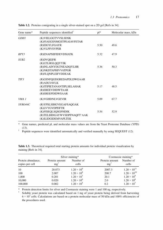
Proteome analysis is most commonly accomplished by a combination of two-dimen-sional gel electrophoresis to separate and visualize proteins, and MS for protein identifica-tion. This technique is powerful, mature and sensitive, but challenges remain concerningthe characterization all of the elements of a proteome. More than 1500 features were visu-alized by silver staining a narrow pH range (4.9–5.7) two-dimensional gel in which 0.5 mgof total soluble yeast protein was separated. Fifty spots migrating to a region of 4 cm2 weresubjected to MS protein identification. Despite the high sample load and extended electro-phoretic separation, proteins from genes with codon bias values of <0.1 (lower abundanceproteins) were not found, even though fully one-half of all yeast genes fall into that range.Proteins from genes with codon bias values of <0.1 were found, however, if protein amountsexceeding the capacity of two-dimensional gels were fractionated and analyzed. The largerange of protein expression levels limits the ability of the two-dimensional gel/MS ap-proach to analyze proteins of medium to low abundance, and thus the potential of thistechnique for total proteome analysis is limited.[34]
Table 1.2 points to another difficulty, co-migration, in identifying proteins from two-dimensional gels.
Table 1.3 lists the theoretical amounts of starting protein to visualize individual proteinsof different abundances.
Protein–protein interactions are studied, for example, by the yeast two-hybrid-system, agenetic technique designed to identify novel protein–protein interactions that were previ-ously detected by biochemical studies. All two-hybrid screening systems rely on the factthat transcriptional activation and DNA-binding domains of transcription factors are modularin nature. In these systems, the coding sequence for the DNA-binding domain of a tran-scription factor such as Gal4 or LexA is fused to the cDNA of a protein of interest, termed
17
Table 1.2. Proteins comigrating in a single silver-stained spot on a 2D gel [Refs in 34].
Gene name* Peptide sequences identified† pl* Molecular mass, kDa
GDH1 (K)VIELGGTVVSLSDSK(K)FIAEGSNMGSTPEAIAVFETAR(R)EIGYLFGAYR 5.50 49.6(K)VLPIVSVPER
RPT3 (R)ENAPSIIFIDEVDSIATK 5.32 47.9
SUB2 (R)DVQEIFR(K)LTLHGLQQYYIK(R)INLAINYDLTNEADQYLHR 5.36 50.3(K)NKDTAPHIVVATPGR(R)FLQNPLEIFVDDEAK
TIF3 (R)GSNFQGDGREDAPDLDWGAAR(R)ADLVAVLK(K)ITIPIETANANTIPLSELAHAK 5.17 48.5(R)EREEVDIDWTAAR(R)EREEPDIDWSAAR
VMA 1 (K)VGHDNLVGEVIR 5.09 67.7
YFRO44C (R)YPSLSIHGVEGAFSAQGAK(K)LVYGVDPDFTR(K)FISEQLSQSGFHDIK 5.54 52.9(R)TELIHDGAYWVSDPFNAQFT’AAK(K)ILIDGIDEMVAPLTEK
* Gene names, predicted pl, and molecular mass values are from the Yeast Proteome Database (YPD)(13).
† Peptide sequences were identified automatically and verified manually by using SEQUEST (12).
Table 1.3. Theoretical required total starting protein amounts for individual protein visualization bystaining [Refs in 34].
Silver staining* Coomassie staining*Protein abundance, Protein amount Number of Protein amount Number ofcopies per cell mg† cells mg† cells
10 20.073 1.20 × 10⁹ 2007.3 1.20 × 10¹¹
100 2.007 1.20 × 10⁸ 200.7 1.20 × 10¹⁰
1,000 0.201 1.20 × 10⁷ 20.1 1.20 × 10⁹
10,000 0.020 1.20 × 10⁶ 2.0 1.20 × 10⁸
100,000 0.002 1.20 × 10⁵ 0.2 1.20 × 10⁷
* Protein detection limits for silver and Coomassie staining were 1 and 100 ng, respectively.† Soluble yeast protein was calculated based on 1 mg of yeast protein being derived from harvesting
6 × 10⁷ cells. Calculations are based on a protein molecular mass of 50 kDa and 100% efficiencies ofthe procedures used.
1.3 Proteomics
18 1 Introduction to Functional Biotechnology
the bait. The fusion protein thus encoded tethers the bait to the promoter region of a re-porter gene. A second fusion of a cDNA library with the coding sequence of a transcrip-tional activation domain is termed the prey. Functional reconstitution of transcription fac-tor activity occurs upon association of the bait and prey protein domains. This interaction isdetected by expression of reporter genes that are dependent upon the bait’s DNA-bindingdomain. The two-hybrid system is a powerful tool for screening libraries for novel protein–protein interactions and for the isolation of factors that promote or disrupt protein interac-tions. A differential two-hybrid yeast system can screen for interactions between prey pro-teins and two different bait proteins through the activation of bait-specific reporters. Itallows the identification of proteins that interact differentially with one bait tethered to theGal4 DNA-binding domain and another bait tethered to the LexA DNA-binding domain.[35]
To detect interactions between proteins of vaccinia virus, a two-hybrid analysis wascarried out to assay every pair wise combination. An array of yeast transformants thatcontained each of the 266 predicted viral ORFs as Gal4 activation domain hybrid proteinswas constructed. The array was individually mated to transformants containing each ORFas a Gal4 DNA-binding domain hybrid and diploids expressing the two-hybrid reportergene were identified. Of the 70,000 combinations, 37 protein–protein interactions werefound, including 28 that were previously unknown. In some cases, e.g., late transcriptionfactors, both proteins were known to have related roles although there was no prior evi-dence of physical associations. For some other interactions, neither protein had a knownrole. In the majority of cases, one of the interacting proteins was known to be involved inDNA replication, transcription, virion structure or host evasion, thereby providing a clue tothe role of the other uncharacterized protein in a specific process.[36]
Direct interaction between proteins is an important means of relaying information in anetwork or chain of signaling molecules.
Of the numerous classes of cell-surface-receptor signaling molecules, synaptic trans-mission between individual neurons is mediated largely by two major structurally and func-tionally distinct neurotransmitter receptor families, i.e. ligand-gated channels and G pro-tein-coupled receptors (GPCRs). Although both are integral membrane proteins, ligand-gatedreceptors modulate synaptic neurotransmission directly through the formation and openingof an inherent ion channel, whereas GPCRs are single-polypeptide proteins containingseven hydrophobic transmembrane domains that transduce extracellular neurotransmittersignals into the cell interior by interacting with heterotrimeric G proteins. These in turnmodulate a diverse array of cellular effectors to produce changes in cellular second-mes-senger systems and/or ionic conductance, and ultimately physiological responsiveness.
GABA(A) (γ-aminobutyric-acid A) and dopamine D1 and D5 receptors represent twostructurally and functionally divergent families of neurotransmitter receptors. The formercomprises a class of multi-subunit ligand-gated channels mediating fast interneuronal syn-aptic transmission, whereas the latter belongs to the seven-transmembrane-domain single-polypeptide receptor super family that exerts its biological effects, including the modula-tion of GABA(A) receptor function, through the activation of second-messenger signalingcascades by G proteins. GABA(A)-ligand-gated channels complex selectively with D5 re-ceptors through the direct binding of the D5 C-terminal domain with the second intracellu-lar loop of the GABA(A) γ2(short) receptor subunit. This physical association enables
19
mutually inhibitory functional interactions between these receptor systems. The data high-light a new signal transduction mechanism whereby subtype-selective GPCRs dynamicallyregulate synaptic strength independently of classically defined second-messenger systemsand provide a heuristic framework in which to view these receptor systems in the mainte-nance of psychomotor disease states.[37]
With the completion of an increasing number of genomic sequences, attention is focus-ing on the interpretation of the data contained in sequence databases in terms of structure,function and control of biological systems. Approaches for global profiling of gene expres-sion analysis at the mRNA level are identifying clusters of genes for which the expressionis idiotypic for a specific state. These sensitive methods of profiling do not indicate changesin protein expression. Quantitative proteome analysis, the global analysis of protein ex-pression, is a complementary method to study steady-state gene expression and perturba-tion-induced changes. In a further step towards functional studies, proteome analysis pro-vides more accurate information about biological systems and pathways because themeasurements directly focus on the actual biological effector molecules.
Quantitative protein analysis is accomplished by combining protein separation, mostcommonly by high-resolution two-dimensional polyacrylamide gel electrophoresis (PAGE),with MS-(mass spectrometry) based or tandem MS (MS/MS)-based sequence identifica-tion of selected, separated protein species. This method is sequential, labor intensive anddifficult to automate. It selects against specific classes of proteins, such as membrane pro-teins, very large and small proteins, and extremely acidic or basic proteins. The techniquehas a bias toward highly abundant proteins, as lower abundance regulatory proteins (e.g.,transcription factors and protein kinases) are rarely detected when total-cell lysates areanalyzed.
A method has been developed for the accurate quantification and concurrent sequenceidentification of the individual proteins within complex mixtures. The method is based ona class of new chemical reagents termed isotope-coded affinity tags (ICATs) and tandemMS. With this strategy, protein expression in the yeast S. cerevisiae was compared, usingeither ethanol or galactose as a carbon source. The measured differences in protein expres-sion correlated with known yeast metabolic function under glucose-repressed conditions.The method is redundant if multiple cysteinyl residues are present and the relative quanti-fication is highly accurate because it is based on stable isotope dilution techniques. TheICAT approach provides a broadly applicable means to compare quantitatively global pro-tein expression in cells and tissues.[38]
Phage antibody libraries provide a source of binders to almost any antigen, includingmany that were previously considered difficult targets, such as self-antigens or cell-surfaceproteins. Phage selection involves repeated rounds of growth, panning and infection, whichselects both for binding and for antibody fragments that are well expressed on phages.When selecting against highly complex targets, there is often a strong bias for antibodiesdirected against immunodominant epitopes and abundant proteins.
A technique for high-throughput screening of recombinant antibodies, based on the cre-ation of antibody arrays, uses robotic picking and high-density gridding of bacteria con-taining antibody genes followed by filter-based enzyme-linked immunosorbent assay(ELISA) screening to identify clones that express binding antibody fragments. By elimi-
1.3 Proteomics
20 1 Introduction to Functional Biotechnology
nating the need for liquid handling, up to 18,342 different antibody clones can be screenedat a time, and, because the clones are arrayed from master stocks, the same antibodies canbe double-spotted and screened simultaneously against 15 different antigens. The tech-nique was applied in several different applications, including the isolation of antibodiesagainst impure proteins and complex antigens, where several rounds of phage display oftenfail. The results indicate that antibody arrays can be used to identify differentially expressedproteins.[39]
The array format for analyzing peptide and protein function offers an attractive experi-mental alternative to traditional library screens. Approaches range from synthetic peptidearrays to whole proteins expressed in living cells. Comprehensive sets of purified peptidesand proteins permit high-throughput screening for discrete biochemical properties, whereasformats involving living cells facilitate large-scale genetic screening for novel biologicalactivities. Three major genome-scale studies using yeast as a model organism have investi-gated different aspects of protein function, including biochemical activities, gene disrup-tion phenotypes and protein–protein interactions. Such studies show that protein arrays canbe used to examine in parallel the functions of thousands of proteins previously knownonly by their DNA sequence.[40]
The systematic approach of arrays towards the simultaneous studies of structure andfunction is applied to proteins, promising advances in the research of protein interactionson a large scale.
Systematic efforts are currently under way to construct defined sets of cloned genes forhigh-throughput expression and purification of recombinant proteins. To facilitate subse-quent studies of protein function, miniaturized assays were developed that accommodateextremely low sample volumes and enable the rapid, simultaneous processing of thousandsof proteins. A high-precision robot designed to manufacture complementary DNA arrayswas used to spot proteins onto chemically derivatized glass slides at extremely high spatialdensities. The proteins attached covalently to the slide surface yet retained their ability tointeract specifically with other proteins, or with small molecules, in solution. Three appli-cations for protein microarrays were demonstrated: screening for protein–protein interac-tions, identifying the substrates of protein kinases and identifying protein targets of smallmolecules.[41]
Protein–DNA interactions are crucial processes in gene regulation and gene function.Study of complexes, receptors and assemblies in cells, organs and organisms is necessaryto understand and manipulate the functions.
The study of components of transcriptional complexes and the effects of ligands in alter-ing the pattern of association has been studied with µESI-MS. This allows the screening oftranscriptionally active libraries and enables rapid data acquisition, especially data on themolecular masses of complexes. These methods and data have potential for automation indrug discovery efforts by the identification of non-covalent ligand interactions with associ-ating molecules and the identification of specific ligands that effect changes in transcrip-tion. The technique is useful for studying functional and regulatory protein–DNA and pro-tein–DNA complexes.[42]
The new sciences (genomics and proteomics) and the new technologies (combinatorialchemistry, bioinformatics, biochips and biosensors) are increasing the speed of drug dis-
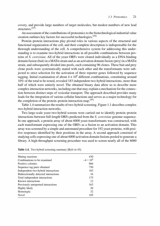
21
covery, and provide large numbers of target molecules, but modest numbers of new leadstructures.[43]
An assessment of the contributions of proteomics to the biotechnological industrial valuecreation outlines key factors for successful technologies.[44]
Protein–protein interactions play pivotal roles in various aspects of the structural andfunctional organization of the cell, and their complete description is indispensable for thethorough understanding of the cell. A comprehensive system for addressing this under-standing is to examine two-hybrid interactions in all possible combinations between pro-teins of S. cerevisiae. All of the yeast ORFs were cloned individually as a DNA-bindingdomain fusion (bait) in a MATα strain and as an activation domain fusion (prey) in a MATαstrain, and subsequently divided into pools, each containing 96 clones. These bait and preyclone pools were systematically mated with each other and the transformants were sub-jected to strict selection for the activation of three reporter genes followed by sequencetagging. Initial examination of about 4 × 10⁶ different combinations, constituting around10% of the total to be tested, revealed 183 independent two-hybrid interactions, more thanhalf of which were entirely novel. The obtained binary data allow us to describe morecomplex interaction networks, including one that may explain a mechanism for the connec-tion between distinct steps of vesicular transport. The approach described provides manyleads for the integration of various cellular functions and serves as a major technology forthe completion of the protein–protein interaction map.[45]
Table 1.4 summarizes the results of two-hybrid screening. Figure 1.1 describes complextwo-hybrid interaction networks.
Two large-scale yeast two-hybrid screens were carried out to identify protein–proteininteractions between full-length ORFs predicted from the S. cerevisiae genome sequence.In one approach, a protein array of about 6000 yeast transformants was constructed, witheach transformant expressing one of the ORFs as a fusion to an activation domain. Thisarray was screened by a simple and automated procedure for 192 yeast proteins, with posi-tive responses identified by their positions in the array. A second approach consisted ofstudying cells expressing one of about 6000 activation domain fusions pooled to generate alibrary. A high-throughput screening procedure was used to screen nearly all of the 6000
Table 1.4. Two-hybrid screening summary [Refs in 45].
Mating reactions 430Combinations to be examined ~4 × 10⁶
Positive colonies 866Sequence tag pairs obtained 750Independent two-hybrid interactions 183Bidirectionally detected interactions 16Total independent interactions 175Known interactions 12Previously unreported interactions 163Highly likely 26Homotypic 32Novel 105
1.3 Proteomics
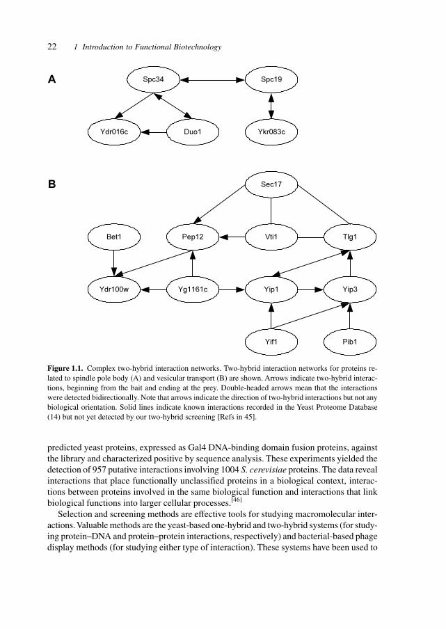
22 1 Introduction to Functional Biotechnology
Spc34
Ydr016c Duo1 Ykr083c
Spc19A
B
Bet1 Pep12 Vti1 Tlg1
Sec17
Ydr100w Yg1161c Yip1 Yip3
Yif1 Pib1
predicted yeast proteins, expressed as Gal4 DNA-binding domain fusion proteins, againstthe library and characterized positive by sequence analysis. These experiments yielded thedetection of 957 putative interactions involving 1004 S. cerevisiae proteins. The data revealinteractions that place functionally unclassified proteins in a biological context, interac-tions between proteins involved in the same biological function and interactions that linkbiological functions into larger cellular processes.[46]
Selection and screening methods are effective tools for studying macromolecular inter-actions. Valuable methods are the yeast-based one-hybrid and two-hybrid systems (for study-ing protein–DNA and protein–protein interactions, respectively) and bacterial-based phagedisplay methods (for studying either type of interaction). These systems have been used to
Figure 1.1. Complex two-hybrid interaction networks. Two-hybrid interaction networks for proteins re-lated to spindle pole body (A) and vesicular transport (B) are shown. Arrows indicate two-hybrid interac-tions, beginning from the bait and ending at the prey. Double-headed arrows mean that the interactionswere detected bidirectionally. Note that arrows indicate the direction of two-hybrid interactions but not anybiological orientation. Solid lines indicate known interactions recorded in the Yeast Proteome Database(14) but not yet detected by our two-hybrid screening [Refs in 45].
23
identify interaction partners for particular DNA or protein targets and they were used incombination with mutagenesis or randomization strategies to study the details of biologi-cally important interactions (reviewed in 1–5 in Joung et al.[47]).
A bacterial ‘two-hybrid’ system that readily allows selection from libraries larger than108 in size was developed. The bacterial system may be used to study either protein–DNAor protein–protein interactions, and it offers a number of potentially significant advantagesover existing yeast-based one-hybrid and two-hybrid methods. The system was tested byselecting zinc finger variants from a large randomized library that bind tightly and specifi-cally to desired DNA target sites. The method allows sequence-specific zinc fingers to beisolated in a single selection step. Thus it is faster than phage display strategies that typi-cally require multiple enrichment/amplification cycles. Given the large library sizes thatcan be handled with the bacterial-based selection system, this method is an efficient toolfor the identification and optimization of protein–DNA and protein–protein interactions.[47]
Many intracellular processes are mediated through inducible protein–protein interac-tions. Methods that allow such processes to be manipulated are powerful tools for under-standing and controlling cellular activities. The use of chemical inducers of dimerization(dimerizers) is a versatile approach. Cells are engineered to express chimeric proteins com-prising a signaling domain fused to a drug-binding domain; treatment with bivalent ligandscross-links the proteins and initiates signaling. This strategy has been used to create induc-ible alleles of numerous signaling proteins that are activated by dimerization. A strategy forachieving the opposite mode of control, wherein proteins are constitutively associated untiladdition of a drug, is valuable for probing the consequences of rapidly abolishing oligo-merization events inside cells. A point mutant was discovered and characterized that hasthe unusual property of forming discrete dimers that can be dissociated by ligand. This isthe basis of a reverse dimerization system that is applicable as a disaggregation switch forintracellular processes.
Chemically induced dimerization provides a general way to gain control over intracellu-lar processes. Typically, FK506-binding protein (FKBP) domains are fused to a signalingdomain of interest, allowing cross-linking to be initiated by addition of a bivalent FKBPligand. In the course of protein engineering studies on human FKBP, a single point muta-tion was discovered in the ligand-binding site (Phe36 → Met) that converts the normallymonomeric protein into a ligand-reversible dimer. Two-hybrid, gel filtration, analyticalultra-centrifugation and X-ray crystallographic studies show that the mutant (FM9) formsdiscrete homodimers with micromolar affinity that can be completely dissociated withinminutes by addition of monomeric synthetic ligands. These unexpected properties form thebasis for a ‘reverse dimerization’ regulatory system involving FM fusion proteins, in whichassociation is the ground state and addition of ligand abolishes interactions. This strategywas used to rapidly and reversibly aggregate fusion proteins in different cellular compart-ments, and to provide an off switch for transcription. Reiterated FM domains should begenerally useful as conditional aggregation domains (CADs) to control intracellular eventswhere rapid, reversible dissolution of interactions is required. Dimerization is apparently alatent property of the FKBP fold. The crystal structure reveals a remarkably complemen-tary interaction between the monomer binding sites, with only subtle changes in side chaindisposition accounting for the dramatic change in quaternary structure.[48]
1.3 Proteomics
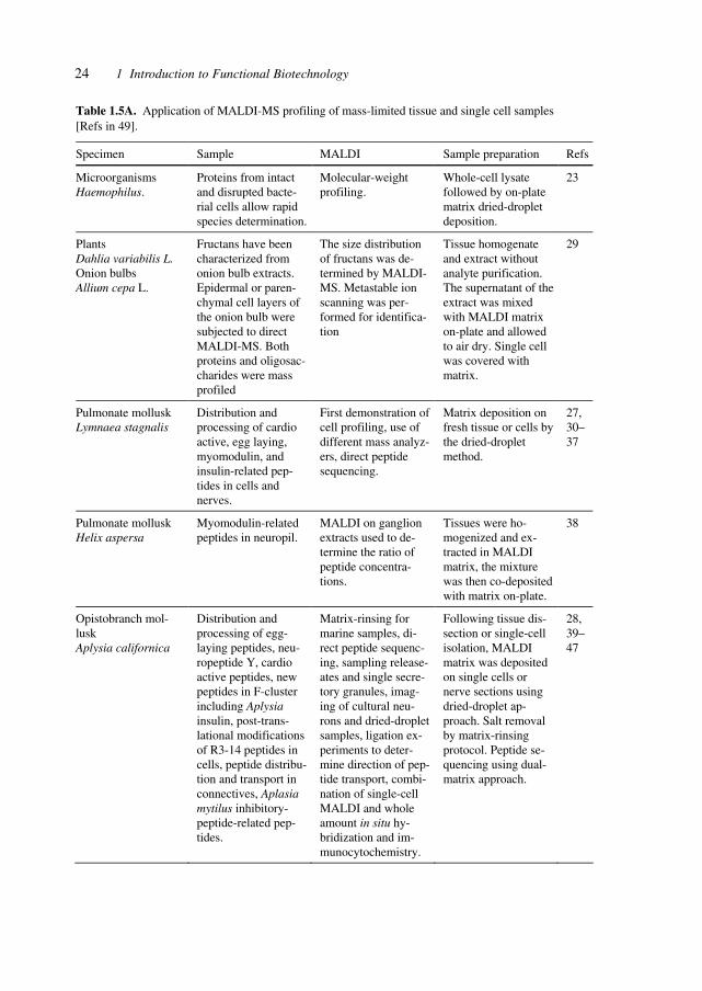
24 1 Introduction to Functional Biotechnology
Table 1.5A. Application of MALDI-MS profiling of mass-limited tissue and single cell samples [Refs in 49].
Specimen Sample MALDI Sample preparation Refs
Microorganisms Haemophilus.
Proteins from intact and disrupted bacte-rial cells allow rapid species determination.
Molecular-weight profiling.
Whole-cell lysate followed by on-plate matrix dried-droplet deposition.
23
Plants Dahlia variabilis L. Onion bulbs Allium cepa L.
Fructans have been characterized from onion bulb extracts. Epidermal or paren-chymal cell layers of the onion bulb were subjected to direct MALDI-MS. Both proteins and oligosac-charides were mass profiled
The size distribution of fructans was de-termined by MALDI-MS. Metastable ion scanning was per-formed for identifica-tion
Tissue homogenate and extract without analyte purification. The supernatant of the extract was mixed with MALDI matrix on-plate and allowed to air dry. Single cell was covered with matrix.
29
Pulmonate mollusk Lymnaea stagnalis
Distribution and processing of cardio active, egg laying, myomodulin, and insulin-related pep-tides in cells and nerves.
First demonstration of cell profiling, use of different mass analyz-ers, direct peptide sequencing.
Matrix deposition on fresh tissue or cells by the dried-droplet method.
27, 30–37
Pulmonate mollusk Helix aspersa
Myomodulin-related peptides in neuropil.
MALDI on ganglion extracts used to de-termine the ratio of peptide concentra-tions.
Tissues were ho-mogenized and ex-tracted in MALDI matrix, the mixture was then co-deposited with matrix on-plate.
38
Opistobranch mol-lusk Aplysia californica
Distribution and processing of egg-laying peptides, neu-ropeptide Y, cardio active peptides, new peptides in F-cluster including Aplysia insulin, post-trans-lational modifications of R3-14 peptides in cells, peptide distribu-tion and transport in connectives, Aplasia mytilus inhibitory-peptide-related pep-tides.
Matrix-rinsing for marine samples, di-rect peptide sequenc-ing, sampling release-ates and single secre-tory granules, imag-ing of cultural neu-rons and dried-droplet samples, ligation ex-periments to deter-mine direction of pep-tide transport, combi-nation of single-cell MALDI and whole amount in situ hy-bridization and im-munocytochemistry.
Following tissue dis-section or single-cell isolation, MALDI matrix was deposited on single cells or nerve sections using dried-droplet ap-proach. Salt removal by matrix-rinsing protocol. Peptide se-quencing using dual-matrix approach.
28, 39–47
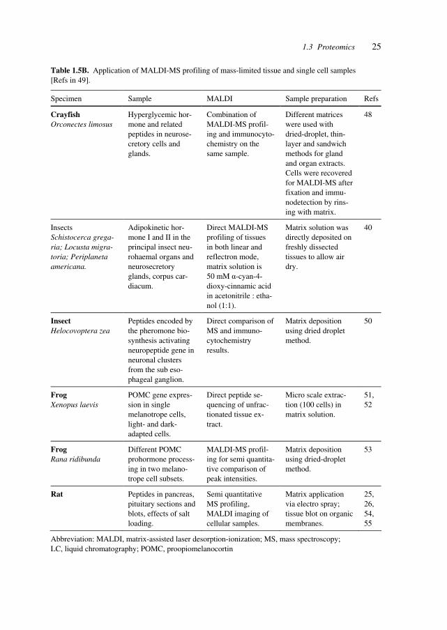
251.3 Proteomics
Table 1.5B. Application of MALDI-MS profiling of mass-limited tissue and single cell samples [Refs in 49].
Specimen Sample MALDI Sample preparation Refs
Crayfish Orconectes limosus
Hyperglycemic hor-mone and related peptides in neurose-cretory cells and glands.
Combination of MALDI-MS profil-ing and immunocyto-chemistry on the same sample.
Different matrices were used with dried-droplet, thin-layer and sandwich methods for gland and organ extracts. Cells were recovered for MALDI-MS after fixation and immu-nodetection by rins-ing with matrix.
48
Insects Schistocerca grega-ria; Locusta migra-toria; Periplaneta americana.
Adipokinetic hor-mone I and II in the principal insect neu-rohaemal organs and neurosecretory glands, corpus car-diacum.
Direct MALDI-MS profiling of tissues in both linear and reflectron mode, matrix solution is 50 mM α-cyan-4-dioxy-cinnamic acid in acetonitrile : etha-nol (1:1).
Matrix solution was directly deposited on freshly dissected tissues to allow air dry.
40
Insect Helocovoptera zea
Peptides encoded by the pheromone bio-synthesis activating neuropeptide gene in neuronal clusters from the sub eso-phageal ganglion.
Direct comparison of MS and immuno-cytochemistry results.
Matrix deposition using dried droplet method.
50
Frog Xenopus laevis
POMC gene expres-sion in single melanotrope cells, light- and dark-adapted cells.
Direct peptide se-quencing of unfrac-tionated tissue ex-tract.
Micro scale extrac-tion (100 cells) in matrix solution.
51, 52
Frog Rana ridibunda
Different POMC prohormone process-ing in two melano-trope cell subsets.
MALDI-MS profil-ing for semi quantita-tive comparison of peak intensities.
Matrix deposition using dried-droplet method.
53
Rat Peptides in pancreas, pituitary sections and blots, effects of salt loading.
Semi quantitative MS profiling, MALDI imaging of cellular samples.
Matrix application via electro spray; tissue blot on organic membranes.
25, 26, 54, 55
Abbreviation: MALDI, matrix-assisted laser desorption-ionization; MS, mass spectroscopy; LC, liquid chromatography; POMC, proopiomelanocortin
26 1 Introduction to Functional Biotechnology
The quest for large-scale automated proteomic studies is based on the desire to increasethe sensitivity of detection, in combination with powerful two-dimensional resolution.
MALDI MS is an analytical approach suitable for obtaining molecular weights of pep-tides and proteins from complex samples. MALDI MS can profile the peptides and proteinsfrom single-cell and small tissue samples without the need for extensive sample prepara-tion, except for cell isolation and matrix preparation. Strategies for peptide identificationand characterization of post-translational modifications are versatile and broadly appli-cable.[49]
Mass spectrometry is also a useful technique for the analysis of nucleic acids. Matrix-assisted laser adsorption/ionization time of flight (MALDI-TOF) mass spectrometry servesto analyze genetic variations such as micro satellites, insertions/deletions, and especiallysingle-nucleotide polymorphisms (SNPs). The ability to resolve oligonucleotides varyingin mass by less than a single nucleotide makes MALDI-TOF mass spectrometry an appli-cable technology for SNP and mutant analysis. The technique was used for quantitativeanalysis of mutations in attenuated mumps virus vaccines and the results showed excellentcorrelation with data from mutant analysis by PCR and restriction enzyme cleavage(MAPREC)[49].
The protein-folding enigma, the shaping of linear protein-sequences into three-dimen-sional structures, is addressed both experimentally and by computational methods.
Membrane proteins acquire their unique functions through specific folding of theirpolypeptide chains stabilized by specific interactions in the membrane. Classical methodsto study their stability or resistance to unfolding are usually investigated by chemical andthermal denaturation. Atomic force microscopy and single-molecule force spectroscopywere combined to image and manipulate purple membrane patches from Halobacteriumsalinarum. Individual bacteriorhodopsin molecules were first localized and then extractedfrom the membrane; the remaining vacancies were imaged again. Anchoring forces be-tween 100 and 200 pN for the different helices were found. Upon extraction, the heliceswere found to unfold. The force spectra revealed the individuality of the unfolding path-ways. Helices G and F as well as helices E and D unfolded pair wise, whereas helices B andC occasionally unfolded one after the other. Experiments with cleaved loops revealed theorigin of the individuality: stabilization of helix B by neighboring helices.[50]
The studies of protein folding show that the mechanisms and principles of protein fold-ing may be simpler than the complexity of the process and proteins indicate, and may bedetermined by the topology of the native state.[51]
Cellular processes as different as growth factor signaling and transcription depend oninteractions between proteins. Large-scale protein–protein interaction screens in modelsystems together with global gene expression profiling after receptor activation provide theidentification of many unexpected interactions between GPCRs and other proteins.[52]
Somatostatin and dopamine are two major neurotransmitter systems that share a numberof structural and functional characteristics. Somatostatin receptors and dopamine receptorsare co localized in neuronal subgroups, and somatostatin is involved in modulating dopam-ine-mediated control of motor activity. The molecular basis for such interaction betweenthe two systems is unclear. It was shown that dopamine receptor D2R and somatostatinreceptor SSTR5 interact physically through hetero-oligomerization to create a novel recep-
27
tor with enhanced functional activity. The results provide evidence that receptors fromdifferent GPCR families interact through oligomerization. Such direct intramembrane as-sociation defines a new level of molecular cross talk between related GPCR families.[53]
The identification and characterization of all proteins expressed by a genome in biologi-cal samples is the major challenge in proteomics. The high-throughput approaches com-bine two-dimensional electrophoresis with peptide mass finger-printing (PMF) analysis.Automation is often possible but a number of limitations still adversely affect the rate ofprotein identification and annotation in two-dimensional gel electrophoresis databases: thesequential excision process of pieces of gel containing protein; the enzymatic digestionstep; the interpretation of mass spectra (reliability of identifications); and the manual up-dating of two-dimensional gel electrophoresis databases. A highly automated method hasbeen developed that generates a fully annotated two-dimensional gel electrophoresis map.Using a parallel process, all proteins of a two-dimensional gel electrophoresis are firstsimultaneously digested proteolytically and electro-transferred onto a poly(vinylidenedifluoride) membrane. The membrane is then directly scanned by MALDI-TOF MS. Afterautomated protein identification from the obtained peptide mass fingerprints using PeptIdentsoftware (from www.expasy.ch), a fully annotated two-dimensional map is created on-line.It is a multidimensional representation of a proteome that contains interpreted PMF data inaddition to protein identification results. This MS imaging method represents a major steptoward the development of a clinical molecular scanner.[54]
The analysis of multi-protein complexes is essential for analyzing the biological activi-ties of proteins, since most cellular functions are performed by protein assemblies or multi-protein complexes rather than by individual proteins.
The identity of the members of such complexes can now be determined by mass spec-trometry (MS). MS can also be used to define the spatial organization of these complexes.Thus, components of a protein complex are purified via molecular interactions using anaffinity tagged member and the purified complex is then partially cross-linked. The prod-ucts are separated by gel electrophoresis and their constituent components identified byMS, yielding nearest-neighbor relationships. A member of the yeast nuclear pore complex(Nup85p) was tagged and a six-member subcomplex of the pore was cross-linked and ana-lyzed by one-dimensional sodium dodecylsulfate (SDS)–PAGE. Cross-linking reactionswere optimized for yield and number of products. Analysis by MALDI MS resulted in theidentification of protein constituents in the cross-linked bands even at a level of a fewhundred femtomoles. Based on those results, a model of the spatial organization of thecomplex was derived that was supported by biological experiments. The use of MS is themethod of choice for analyzing cross-linking experiments aimed at nearest neighbor rela-tionships.[55]
A large-scale protein-protein interaction map of the human gastric pathogen Helicobacterpylori was constructed by using a high-throughput strategy of the yeast two-hybrid assay toscreen 261 H. pylori proteins against a highly complex library of genome-encoded polypep-tides. More than 1,200 interactions were identified between H. pylori proteins, connecting46.6% of the proteome. This technology is applicable to higher eukaryotic organisms aswell and enables the generation of proteomes for studies of diseaes, for the construction ofmutants, and the development of screening assays for drug discovery[507].
1.3 Proteomics
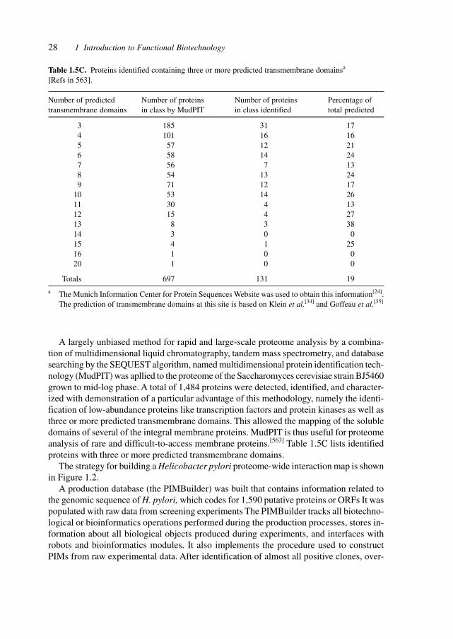
28 1 Introduction to Functional Biotechnology
A largely unbiased method for rapid and large-scale proteome analysis by a combina-tion of multidimensional liquid chromatography, tandem mass spectrometry, and databasesearching by the SEQUEST algorithm, named multidimensional protein identification tech-nology (MudPIT) was apllied to the proteome of the Saccharomyces cerevisiae strain BJ5460grown to mid-log phase. A total of 1,484 proteins were detected, identified, and character-ized with demonstration of a particular advantage of this methodology, namely the identi-fication of low-abundance proteins like transcription factors and protein kinases as well asthree or more predicted transmembrane domains. This allowed the mapping of the solubledomains of several of the integral membrane proteins. MudPIT is thus useful for proteomeanalysis of rare and difficult-to-access membrane proteins.[563] Table 1.5C lists identifiedproteins with three or more predicted transmembrane domains.
The strategy for building a Helicobacter pylori proteome-wide interaction map is shownin Figure 1.2.
A production database (the PIMBuilder) was built that contains information related tothe genomic sequence of H. pylori, which codes for 1,590 putative proteins or ORFs It waspopulated with raw data from screening experiments The PIMBuilder tracks all biotechno-logical or bioinformatics operations performed during the production processes, stores in-formation about all biological objects produced during experiments, and interfaces withrobots and bioinformatics modules. It also implements the procedure used to constructPIMs from raw experimental data. After identification of almost all positive clones, over-
Table 1.5C. Proteins identified containing three or more predicted transmembrane domainsa
[Refs in 563].
Number of predicted Number of proteins Number of proteins Percentage oftransmembrane domains in class by MudPIT in class identified total predicted
3 185 31 174 101 16 165 57 12 216 58 14 247 56 7 138 54 13 249 71 12 17
10 53 14 2611 30 4 1312 15 4 2713 8 3 3814 3 0 015 4 1 2516 1 0 020 1 0 0
Totals 697 131 19
a The Munich Information Center for Protein Sequences Website was used to obtain this information[24].The prediction of transmembrane domains at this site is based on Klein et al.[34] and Goffeau et al.[35]
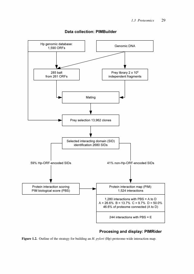
29
Hp genomic database:
1,590 ORFsGenomic DNA
Data collection: PIMBuilder
285 balt
from 261 ORFs
Prey library 2 x 10⁶
independent fragments
Mating
Prey selection 13,962 clones
Selected interacting domain (SID)
identification 2680 SIDs
Protein interaction scoring
PIM biological score (PBS)
Protein interaction map (PIM):
1,524 interactions
1,280 interactions with PBS = A to D
A = 26.6% B = 13.7% C = 9.7% D = 50.0%
46.6% of proteome connected (A to D)
244 interactions with PBS = E
41% non-Hp-ORF-encoded SIDs59% Hp-ORF-encoded SIDs
Procesing and display: PIMRider
Figure 1.2. Outline of the strategy for building an H. pylori (Hp) proteome-wide interaction map.
1.3 Proteomics

30 1 Introduction to Functional Biotechnology
lapping prey fragments were clustered into families to define SIDs. Those families that hadno biological coding capability (antisepses or intergenic region, out of frame fragmentsoccurring in a single frame) were discarded. A PIM biological score (PBS; see Methods)was then calculated for H. pyloriORF-encoded SIDs. Interactions were grouped into cat-egories A to D (from high to 10w heuristic values). The global connectivity of the PIM wasalso analysed to detect highly connected prey polypeptides. Those interactions were groupedin the E category. Processing of data and visualization of interactions were performed by anin-house bioinformatic platform (PIMRider) [Refs in 507].
The structures, the interactions, and the dynamics of proteins are fundamental to theirfunction. The dynamic properties of proteins in the nucleus are essential for their functionin nuclear architecture and gene expression. High mobility of proteins ensures their avail-ability throughout the nucleus and the dynamic interaction creates the architectural frame-work for nuclear processes. Thus, nuclear morphology is shaped by the functional interac-tions of nuclear protein components. Studies with fluorescently tagged protein moleculesreveal high mobility, compartmentation, and repeated transient interactions.[564]
A Human Transcriptome Map was generated by integration of the mapping data aboutthe chromosomal position of human genes with genome-wide messenger RNA expressionprofiles from serial analysis of gene expression (SAGE). Over 2.45 million SAGE tran-script tages, including 160,000 tags of neuroblastomas, known for 12 tissue types wereassigned by algorithms to UniGene clusters and their chromosomal position. The resultingexpression profiles for chromosomal regions in 12 normal and pathologic tissue types re-veal a clustering of highly expressed genes to specific chromosomal regions and providethe tool for identifying genes that are overexpressed or silenced in cancer.[636]
The technologies and applications in proteomic studies are expanding and include thegeneration of novel proteins from distantly related sequences by sequence homology-inde-pendent protein recomination (SHIPREC),[669] the directed evolution of proteins by exonshuffling to generate libraries of therapeutic proteins,[673] the creation of genome-wideprotein expression libraries by random activation of gene expression,[665] the developmentof multi-channel devices with electrospray tips for mass-spectrometry,[694] and make thestudy of the protein complement of the genome, the proteome, an essential basis for func-tional biology which requires the integration with DNA sequence data, mRNA profiles,metabolite con-centrations (the metabolome), structural studies, and biological models andsystems.[667]
1.4 Cytomics
The study of biochemical processes at the cellular level is yielding results integrating thelower level mechanisms into cellular and ultimately organismal processes.
Chemoselective ligation reactions designed to modify only one cellular component amongall others have provided insight into cellular processes. The goal in developing such trans-formations is to relate the selectivity of non-covalent recognition events, such as antibody–antigen binding, to biological processes.
31
Selective chemical reactions enacted within a cellular environment can be powerful toolsfor elucidating biological processes or engineering novel interactions. A chemical transfor-mation that permits the selective formation of covalent adducts among richly functionalizedbiopolymers within a cellular context has been developed. A ligation modeled after theStaudinger reaction forms an amide bond by coupling of an azide and a specifically engi-neered triarylphosphine. Both reactive partners are abiotic and chemically orthogonal tonative cellular components. Azides installed within cell-surface glycoconjugates by me-tabolism of a synthetic azidosugar were reacted with a biotinylated triarylphosphine to pro-duce stable cell-surface adducts. The selectivity of the transformation permits its executionwithin a cell’s interior, offering new possibilities for probing intracellular interactions.[56]
The level of complexity of biological studies and pharmaceutical intervention is in-creased by the necessity to take structures and modifications at the chromatin level intoaccount.
The manipulation of eukaryotic genomes within a chromatin environment is a crucialand fundamental issue in biology. The chromatin structure is composed of highly con-served histone proteins (H3, H4, H2A, H2B and H1) that function as building blocks topackage eukaryotic DNA into repeating nucleosomal units that are folded into higher-orderchromatin fibers. The histones are integral and dynamic components of the machinery re-sponsible for gene transcription. Distinct histone modifications, on one or more tails, actsequentially or in combination to form a ‘histone code’ that is read by other proteins tobring about distinct downstream events.[57]
The overall efficiency of protein biogenesis in cells has been the target of an investiga-tion focussing on proteasomes. MHC class I molecules function to present peptides eight to10 residues long to the immune system. These peptides originate primarily from a cytosolicpool of proteins through the actions of proteasomes and are transported into the endoplas-mic reticulum, where they assemble with nascent class I molecules. Most peptides aregenerated from proteins that are apparently metabolically stable. To explain this, it wasproposed that peptides arise from proteasomal degradation of defective ribosomal products(DRiPs). DRiPs are polypeptides that never attain native structures owing to errors in trans-lation or post-translational processes necessary for proper protein folding. DRiPs consti-tute upwards of 30% of newly synthesized proteins as determined in a variety of cell types.Some DRiPs represent ubiquinated proteins und ubiquinated proteins are formed from hu-man immunodeficiency virus Gag polyprotein, a long-lived viral protein that serves as asource of antigenic peptides.[59]
It is noteworthy that the 70% efficiency for protein synthesis is in line with the estimated50% efficiency of producing functional mRNA from heterogeneous nuclear RNA.
The cell being the basic unit of functional tissues, organs, and organisms, the under-standing and reconstruction of the whole-cell biology including genome, proteome, meta-bolome, and the interacting networks requires the description and simulation of cells. Insilico analysis, description, and simulation complements the in vitro and in vivo processesand the construction of minimal organisms and cells.[663]
The analysis of the intracellular metabolites, the metabolome, by means of comparisonof mutants‘ metabolic profiles (comparative metabolomics) aids in understanding the net-works of proteins and the presence of silent genes.[698]
1.4 Cytomics
32 1 Introduction to Functional Biotechnology
1.5 Micro- and Nanotechnology
Oligonucleotide microarrays, also called DNA chips, are currently made by a light-di-rected chemistry that requires a large number of photolithographic masks for each chip. Amaskless array synthesizer (MAS) was described that replaces the chrome masks with vir-tual masks generated on a computer, which are relayed to a digital micro-mirror array. A1:1 reflective imaging system forms an ultraviolet image of the virtual mask on the activesurface of the glass substrate, which is mounted in a flow cell reaction chamber connectedto a DNA synthesizer. Programmed chemical coupling cycles follow light exposure andthese steps are repeated with different virtual masks to grow desired oligonucleotides in aselected pattern. This instrument has been used to synthesize oligonucleotide microarrayscontaining more than 76,000 features measuring 16 µm2. The oligonucleotides were syn-thesized at high repetitive yield and, after hybridization, could readily discriminate single-base pair mismatches. The MAS is adaptable to the fabrication of DNA chips containingprobes for thousands of genes, as well as any other solid-phase combinatorial chemistry tobe performed in high-density microarrays.[60]
Nanotechnology involves the ability to arrange molecules and atoms into molecularstructures. This will have major impacts on computation (higher density of computationalpower), materials (properties of catalysts and strengths of structures), and the manufactureof tools and devices (surgical tools and instruments of molecular dimensions), capable ofintervening at the fundamental level of disease.[61]
The application of micro-machining techniques is growing rapidly and has applicationsin microfluidics (for labs-on-a-chip), in sensors as well as in fiber optics and displays. Thetwo most widespread methods for the production of microelectromechanical structures(MEMS) are bulk micromachining and surface micromachining. Bulk micromachining is asubtractive fabrication method whereby single-crystal silicon is lithographically patternedand then etched to form three-dimensional structures. Surface micromachining is an addi-tive method where layers of semiconductor-type materials (polysilicon, metals, silicon ni-tride, silicon dioxide, etc.) are sequentially added and patterned to make three-dimensionalstructures.
An alternative microfabrication technique is based on replication moulding. An elas-tomer is patterned by curing on a micromechanical mold. This technique is termed ‘softlithography’, and is used to make blazed grating optics, stamps for chemical patterning andmicrofluidics devices. Soft lithography is essentially a subtractive method.
A technique was developed called ‘multilayer soft lithography’ that combines soft li-thography with the capability to bond multiple patterned layers of elastomer.
Soft lithography is an alternative to silicon-based micromachining that uses replicamoulding of non-traditional elastomeric materials to fabricate stamps and microfluidic chan-nels. An extension to the soft lithography paradigm, multilayer soft lithography, is de-scribed, with which devices consisting of multiple layers may be fabricated from soft ma-terials. This technique was used to build active microfluidic systems containing on/off valves,switching valves and pumps entirely out of elastomer. The softness of these materials al-lows the device areas to be reduced by more than two orders of magnitude compared with
33
silicon-based devices. The other advantages of soft lithography, such as rapid prototyping,ease of fabrication and biocompatibility, are retained.[62]
Microscopic fluidic devices, ranging from surgical endoscopes and MEMS to the com-mercial ‘lab-on-a-chip’, allow chemical analysis and synthesis on scales unimaginable adecade ago. These devices transport miniscule quantities of liquid along networked chan-nels. Several techniques have been developed to control small-scale flow, includingmicromechanical and electrohydrodynamic pumping, electro-osmotic flow, electrowettingand thermocapillary pumping. Most of these schemes require micromachining of interiorchannels and kilovolt sources to drive electrokinetic flow. Recently the use of temperatureinstead of electric fields to derive droplet movement was suggested. A simple, alternativetechnique utilizing temperature gradients to direct microscopic flow on a selectively pat-terned surface (consisting of alternating stripes of bare and coated SiO2) was demonstrated.The liquid is manipulated by simultaneously applying a shear stress at the air–liquid inter-face and a variable surface energy pattern at the liquid–solid interface. To further advancethe technology, a theoretical estimate of the smallest feature size attainable with this tech-nique.[63]
Direct-write technologies are of increasing importance in materials processing. Struc-tures are built directly without the use of masks, allowing rapid prototyping. The tech-niques comprise plasma spray, laser particle guidance, matrix-assisted pulsed-laser evapo-ration, laser chemical vapor deposition, micro-pen, ink jet, e-beam, focused ion beam andseveral droplet micro-dispensing methods. Micrometer-scale patterns of viable cells arerequired for the next generation of tissue engineering, fabrication of cell-based microfluidicbiosensor arrays, and selective separation and culturing of microorganisms.
Patterns of viable E. coli bacteria have been transferred onto various substrates withlaser-based forward transfer technique. These tools can be used to create three-dimensionalmesoscopically engineered structures of living cells, proteins, DNA strands and antibodiesand to co fabricate electronic devices on the same substrate to generate cell-based biosensorsand bioelectronic interfaces and implantates.[64]
To allow researchers the design and construction of nanodevices with scale-up for largeproduct runs, a special serial scanning probe lithography method, called dip-pen nanolitho-graphy (DPN), an eight-pen nanoplotter capable of doing parallel DPN, for the depositionof organic molecules, has been developed.
Because line width and patterning speed in DPN are independent of contact force, onlyone of the tips in the parallel writing mode (the imaging tip) has a feedback system tomonitor tip position and to write the pattern. All other tips reproduce what occurs at theimaging tip in a passive fashion. Proof-of-concept experiments that demonstrate eight-penparallel writing, ink and rinsing wells, and ‘molecular corralling’ via a nanoplotter-gener-ated structure have been reported. This device enables research to do high-resolution andaligned patterning of nanostructures on a large scale in a fast and automated way.[65]
Work on supramolecular structures has opened access to useful nanocomposites by in-fluencing phase-separating fluids. Simulations show that when low-volume fractions ofnanoscale rods are immersed in a binary, phase-separating blend, the rods self-assembleinto needle-like, percolating networks. The interconnected network arises through the dy-namic interplay of phase-separation between the fluids, through preferential adsorption of
1.5 Micro- and Nanotechnology
34 1 Introduction to Functional Biotechnology
the minority component onto the mobile rods, and through rod-rod repulsion. Such coop-erative effects provide a means of manipulating the motion of nanoscopic objects and di-recting their association into supramolecular structures. Increasing the rod concentrationbeyond the effective percolation threshold drives the system to self-assemble into a lamel-lar morphology, with layers of wetted rods alternating with layers of the majority-compo-nent fluid. This approach can potentially yield organic/inorganic composites that are or-dered on a nanometer scale and exhibit electrical or structural integrity.[66]
Whereas most fabrication of microelectronic devices is carried out by photolithographyand thus is intrinsically two-dimensional, increasingly modern devices and micro- (respec-tively, nano-) structures for biomedicine require three-dimensional structures and manu-facturing methods.
Self-assembly is a demonstrated strategy to achieve interconnections and to constructthree-dimensional circuits and structures. Self-assembly of millimeter-scale polyhedra, withsurfaces patterned with solder dots, and light-emitting diodes, generated electrically func-tional, three-dimensional networks. The patterns of dots and wires controlled the structureof the networks formed. Both parallel and serial connections were formed. This self-as-sembly mechanism may be suitable for generating other structures for implantates andbiosensors.[67]
Self-assembly and controlled organization are fabrication methods of increasing impor-tance for nanodevices in order to achieve precision and economies-of-scale.
Colloidal inorganic nanoparticles have size-dependent optical, optoelectronic and mate-rial properties that are expected to lead to superstructures with a range of practical applica-tions. Discrete nanoparticles with controlled chemical composition and size distributionare readily synthesized using reverse micelles and microemulsions as confined reactionmedia, but their assembly into well-defined superstructures amenable to practical use re-mains a difficult and demanding task. This usually requires the initial synthesis of sphericalnanoparticles, followed by further processing such as solvent evaporation, molecular cross-linking or template patterning. The interfacial activity of reverse micelles and microemulsionscan be exploited to couple nanoparticle synthesis and self-assembly over a range of lengthscales to produce materials with complex organization arising from the interdigitation ofsurfactant molecules attached to specific nanoparticle crystal faces. This principle is dem-onstrated by producing three different barium chromate nanostructures – linear chains,rectangular super-lattices and long filaments – as a function of reactant molar ration, whichin turn is controlled by fusing reverse micelles and microemulsion droplets containingfixed concentrations of barium and chromate ions, respectively. If suitable soluble precur-sors and amphiphiles with head groups complementary to the crystal surface of the Nanparticle target are available, it should be possible to extend this approach to the facile pro-duction of one-dimensional wires and higher-order colloidal architectures made of metalsand semi-conductors.[68]
DNA microarrays can be manufactured by either synthesizing DNA on a substrate, ordelivering and covalently attaching presynthesized DNA to the microchips. Technologiessuch as photolithographic DNA synthesis allow the manufacture of high-density oligo-nucleotide microarrays. These methods, however, are costly, time consuming and not eas-ily amenable to custom-manufacture.
35
A method for fabricating DNA microarrays was developed which uses a bubble jet inkjet device to eject 5′-terminal-thiolated oligonucleotides to a glass surface. The oligonucle-otides are covalently attached to the glass surface by heterobifunctional cross-linkers thatreact with the amino group on the substrate and a thiol group on the oligonucleotide probe.Using this method, DNA microarrays were fabricated that carried 64 groups of 18meroligonucleotides encoding all possible three-base mutations in the mutational ‘hot spot’ ofthe p53 tumor suppressor gene. These were screened with a fluorescent labeled synthetic18mer oligonucleotide derived from the p53 gene, or segments of the p53 gene that hadbeen PCR amplified from genomic DNA of two cell lines of human oral squamous cellcarcinoma (SCC). This allowed to discriminate between matched hybrids and 1-bp mis-matched hybrids.[69]
Microrobots for fluid media have been developed that show potential for acting as tinymobile robots in the body fluids. Conducting polymers are excellent materials for actuatorsthat are operated in aqueous media. Microactuators based on polypyrrole-gold bilayersenable large movement of structures attached to these actuators and are of particular inter-est for the manipulation of biological objects, such as single cells. A fabrication method forcreating individually addressable and controllable polypyrrole-gold microactuators wasdeveloped. With these individually controlled microactuators, a micrometer-size manipu-lator (microrobotic arm) was fabricated. This microrobotic arm can pick up, lift, move andplace micrometer-size objects within an area of about 250 µm by 100 µm, making themicrorobot an excellent tool for single-cell manipulation.[70]
The application of micro- and nanorobots in the human body requires the developmentof co-operative mechanism for targeted action of the devices on desired phenomena, e.g.,removal of obstacles, construction of scaffolds, measurements of cellular parameters. Oneof the greatest challenges in robotics is to create machines that are able to interact with un-predictable environments in real time. A possible solution may be to use swarms of robotsbehaving in a self-organized manner, similar to workers in an ant colony. Efficient mecha-nisms of division of labor, in particular series–parallel operation and transfer of informa-tion among group members, are key components of the tremendous ecological success ofants. The general principles regulating division of labor in ant colonies allow the design offlexible, robust and effective robotic systems. Groups of robots using ant-inspired algo-rithms of decentralized control techniques foraged more efficiently and maintained higherlevels of group energy than single robots. However, the benefits of group living decreasedin larger groups, most probably because of interference during foraging. A similar relation-ship between group size and efficiency has been documented in social insects. When fooditems were clustered, groups where robots could recruit other robots in an ant-like mannerwere more efficient than groups without information transfer, suggesting that group dy-namics of swarms of robots may follow rules similar to those governing social insects.[71]
The construction and the evolutionary improvement of micro- and nanodevices may becarried out using evolving populations of artificial creatures. Biological life is in control ofits own means of reproduction, which generally involves complex, auto-catalyzing chemi-cal reactions. But this autonomy of design and manufacture has not yet been realized arti-ficially. Robots are still laboriously designed and constructed by teams of human engi-neers, usually at considerable expense. Few robots are available because these costs must
1.5 Micro- and Nanotechnology
36 1 Introduction to Functional Biotechnology
be absorbed through mass production, which is justified only for toys, weapons and indus-trial systems such as automatic teller machines. A combined computational and experi-mental approach in which simple electromechanical systems are evolved through simula-tions from basic building blocks (bars, actuators, and artificial neurons) were reported; the‘fittest’ machines (defined by their locomotive ability) are then fabricated robotically usingrapid manufacturing technology. Autonomy of design and construction are achieved usingevolution in a ‘limited universe’ physical simulation coupled to automatic fabrication.[72]
Microinjection of fluorochromes, antibodies, proteins and genetic material is widelypracticed by biologists despite negative effects on the target and disadvantages associatedwith insertion of microcapillaries.
A galinstan expansion femto-syringe enables femtoliters to attoliters to be introducedinto prokaryotes and subellular compartments of eukaryotes. The method uses heat-in-duced expansion of galinstan (a liquid metal alloy of gallium, indium and tin) within aglass syringe to expel samples through a tip diameter of about 0.1 µm. The narrow tipinflicts less damage than conventional capillaries and the heat-induced expansion of thegalinstan allows fine control over the rate of injection. Injection of Lucifer Yellow andLucifer Yellow–dextrin conjugates into cyanobacteria, and into nuclei and chloroplasts ofhigher organisms was demonstrated. Injection of a plasmid containing the bla gene into thecyanobacterium Phormidium laminosum resulted in transformed ampicillin-resistant cul-tures. Green fluorescent protein (GFP) was expressed in attached leaves of tobacco andVicia faba following injection of DNA containing its gene into individual chloroplasts.[73]
The study of individual cells or even compartments within a cell requires appropriatelysized nanosensors. Optical nanosensors are usually grouped into chemical and biologicalnanosensors, depending on the probe being used. Both types of sensors have been used tomeasure chemicals in microscopic environments and to detect different entities within singlecells.
The construction of nanosensors is crucially dependent on the construction of nanom-eter-sized tips on optical fibers. Near-field optical microscopy has spawned nanoscale op-tical fibers produced by either pulling with micropipette pullers or by chemical etching.[74]
The synthesis of materials that are ordered on all length scales, from molecular via nanoto meso (‘hierarchical materials’) is a major goal of materials science. In this way, theproperties of the large-scale structures and devices can be controlled by fabricating thedesired molecular structures and characteristics. The methods deployed include biomimeticmethods with polypeptides as building blocks as well as amphiphile and colloidal templatingwith appropriate mesophases as templates for inorganic mesophorous materials.[75]
The technologies and materials for creating nanodevices are increasingly applicable bothin the realm of microelectronic manufacturing and chemical as well as biotechnologicalsynthesis.
Carbon nanotubes find increasing application in fields as diverse as microelectronics,molecular electronics, nanodevices and actuators as well as sensors and chemical processes.
For constructing mobile mechanical devices, molecular surfaces need to be engineering.The controlled and reversible telescopic extension of multiwall nanotubes was demon-strated, realizing ultralow-friction nanoscale linear bearings and constant-force nanosprings.Measurements performed in situ on individual custom-engineered nanotubes inside a high-
37
resolution transmission electron microscope demonstrated the anticipated van der Waalsenergy-based retraction force and enabled the placement of quantitative limits on the staticand dynamic interwall frictional forces between nested nanotubes. Repeated extension andretraction of telescoping nanotube segments revealed no wear or fatigue on the atomicscale. These nanotubes may be almost perfect wear-free surfaces for nanodevices andnanomechanical apparatus.[76]
The versatility of carbon nanotubes in the field of molecular electronics was evidencedby a concept exploiting carbon nanotubes as both molecular device elements and molecularwires for reading and writing information. Each device element is based on a suspended,crossed nanotube geometry that leads to bistable, electrostatically switchable ON/OFF states.The device elements are naturally addressable in large arrays by the carbon nanotube mo-lecular wires making up the devices. These reversible, bistable device elements could beused to construct non-volatile random access memory and logic function tables at an inte-gration level approaching 1012 elements/cm2 and an element operation frequency in excessof 100 GHz. The viability of this concept was demonstrated by detailed calculations and byexperimental realization of a reversible, bistable nanotube-based bit.[77]
The construction of complicated three-dimensional structures on a sub-micrometer scalerequires increasingly sensitive and sophisticated methods amenable to both custom andmass production. Electrochemical machining is increasingly important (in addition to litho-graphy, direct writing, molecular assembly) to achieve the construction of micro- and nano-devices.
The application of ultra short voltage pulses between a tool electrode and a workpiece inan electrochemical environment allows the three-dimensional machining of conductingmaterials with sub-micrometer precision. The principle is based on the finite time constantfor double-layer charging, which varies linearly with the local separation between the elec-trodes. During nanosecond pulses, the electrochemical reactions are confined to electroderegions in close proximity. The technique was used for local etching of copper and siliconas well as for local copper deposition.[78]
A molecular machine was constructed which uses DNA both as structural material andas fuel. Molecular recognition between complementary strands of DNA allows construc-tion on a nanometer length scale. DNA tags may be used to organize the assembly ofcolloidal particles, and DNA templates can direct the growth of semiconductor nanocrystalsand metal wires. As a structural material in its own right, DNA can be used to make orderedstatic arrays of tiles, linked rings and polyhedra. The construction of active devices is alsopossible, such as a nanomechanical switch, whose conformation is changed by inducing atransition in the chirality of the DNA double helix. Melting of chemically modified DNAhas been induced by optical absorption, and conformational changes caused by the bindingof small groups have been shown to change the enzymatic activity of ribozymes. The con-struction of a DNA machine was described in which the DNA is used not only as a struc-tural material, but also as fuel. The machine, made from three strands of DNA, has the formof a pair of tweezers. It may be closed and opened by addition of auxiliary strands of fuelDNA; each cycle produces a duplex DNA waste product.[79]
The methods to produce nanoscale devices are developing rapidly and involve numer-ous physical and chemical methodologies.
1.5 Micro- and Nanotechnology
38 1 Introduction to Functional Biotechnology
The production of materials with micrometer- and sub-micrometer-scale patterns is ofimportance in a range of applications, such as photonic materials, high-density magneticstorage devices, microchip reactors and biosensors. One method of preparing such struc-tures is through the assembly of colloidal particles. Micropatterned colloidal assemblieshave been produced with lithographically patterned electrodes or micro-molds. Anothermethod combines the well-known photochemical sensitivity of semiconductors with elec-tric-field-induced assembly to create ordered arrays of micrometer-sized colloidal particleswith tunable patterns. Light affects the assembly process and patterns are produced usingelectrophoretic deposition in the presence of an ultraviolet illumination motif. The distri-bution of current across an indium tin oxide (ITO) electrode can be altered by varying theillumination intensity; during the deposition process, this causes colloidal particles to beswept from darkened areas into lighted regions. Illumination also assists in immobilizingthe particles on the electrode surface. The patterning effects of the ultraviolet light mayresult from alterations in the current density that affects particle assembly on an ITO elec-trode.[80]
A rapid-prototyping method that uses readily available equipment has been describedwhich will facilitate research and experimental construction of nanostructured devices.
Living systems exhibit form and function on multiple length scales and at multiple loca-tions.
In order to mimic such natural structures, it is necessary to develop efficient strategiesfor assembling hierarchical materials. Conventional photolithography, although ubiquitousin the fabrication of microelectronics and MEMS, is impractical for defining features be-low 0.1 µm and poorly suited to pattern chemical functionality. ‘Soft’ lithographic approacheshave been combined with surfactant and particulate templating procedures to create mate-rials with multiple levels of structural order. The materials thus formed have been limitedprimarily to oxides with no specific functionality and the associated processing times haveranged from hours to days. Using a self-assembling ‘ink’, silica surfactant self-assemblywas combined with three rapid printing procedures – pen lithography, ink-jet printing anddip-coating of patterned self-assembled monolayers – to form functional, hierarchicallyorganized structures in seconds. The described rapid-prototyping procedures are simple,employ readily available equipment, and provide a link between computer-aided designand self-assembled nanostructures. The ability to form arbitrary functional designs on arbi-trary surfaces will be of practical importance for directly writing sensor arrays and fluidicor photonic systems.[81]
Another production technology combines biomolecules and their molecular recognitionproperties with semiconductor materials.
In biological systems, organic molecules exert significant control over the nucleationand mineral phase of inorganic materials such as calcium carbonate and silica, and over theassembly of crystallites and other nanoscale building blocks into complex structures re-quired for biological function. This ability to direct the assembly of nanoscale componentsinto controlled and sophisticated structures has motivated intense efforts to develop assem-bly methods that mimic or exploit the recognition capabilities and interactions found inbiological systems. Of particular value would be methods that could be applied to materialswith interesting electronic or optical properties, but natural evolution has not selected for
39
interactions between biomolecules and such materials. Peptides with limited selectivity forbinding to metal surfaces and metal oxide surfaces have been selected. This approach wasextended to demonstrate that combinatorial phage display libraries can be used to evolvepeptides that bind to a range of semiconductor surfaces with high specificity, depending onthe crystallographic orientation and composition of the structurally similar materials used.As electronic devices contain structurally related materials in close proximity, such pep-tides may find use for the controlled placement and assembly of a variety of practicallyimportant materials, thus broadening the scope for bottom-up fabrication approaches.[82]
An approach which may lend itself to the mass production of organic–silicon structuresis based on self-directed chemical growth processes. Advances in techniques for the nanoscalemanipulation of matter are important for the realization of molecule-based miniature de-vices with new or advanced functions. A promising approach involves the construction ofhybrid organic molecule–silicon devices. However, challenges exist both in the formationof nanostructures that will constitute the active parts of future devices and in the construc-tion of commensurately small connecting wires. Atom-by-atom crafting of structures withscanning tunneling microscopes, although essential to fundamental advances, is too slowfor any practical fabrication process; self-assembly approaches may permit rapid fabrica-tion, but lack the ability to control growth location and shape. Furthermore, moleculardiffusion on silicon is greatly inhibited, thereby presenting a problem for self-assemblytechniques. An approach is described for fabricating nanoscale organic structures on sili-con surfaces, employing minimal intervention by the tip of a scanning tunneling micro-scope and a spontaneous self-directed chemical growth process. Growth of straight mo-lecular styrene lines is demonstrated – each composed of many organic molecules – and thecrystalline silicon substrate determines both the orientation of the lines and the spacingwithin these lines. The described process should in principle allow parallel fabrication ofidentical complex functional structures.[83]
Sol–gel synthesis is widely used for making transition metal oxide solids with fine-scaled microstructures. Pore and particle sizes no greater than a couple of nanometers caneasily be achieved in the freshly derived gels. Maintaining such microstructural dimen-sions when the fresh gels are subsequently crystallized at elevated temperatures is difficult.Freshly derived metal oxide gels have a hydrous solid skeleton that contains many hy-droxyl groups and is either amorphous or paracrystalline. A post-firing step is thereforeindispensable for dehydroxylation and for achieving a sufficient degree of crystallinity inorder to give the desired combination of mechanical, catalytic or optoelectronic propertiesfor their applications. Upon firing of gels, condensation among surface hydroxyl groups,nucleation of new oxide crystals and growth of existing crystals occur concurrently over afairly wide temperature range. Accordingly, some crystals grow extensively before the gelis fully dehydroxylated and crystallized, which reduces surface area, enlarges pores andincreases the difficulty of removing intracrystal defects.
Crystal growth upon firing of hydrous metal oxide gels can be effectively inhibited byreplacing the surface hydroxyl groups before firing with another functional group that doesnot condense and that can produce small, secondary-phase particles that restrict advancingof grain boundaries at elevated temperatures. Fully crystallized SnO2, TiO2 and ZrO2 mate-rials with mean crystallite sizes of around 20, 50 and 15 Å, respectively, were synthesized
1.5 Micro- and Nanotechnology
40 1 Introduction to Functional Biotechnology
by replacing the hydroxyl group with methyl siloxyl before firing at 500 °C. An ultrasensitiveSnO2-based chemical sensor resulting from the microstructural miniaturization was dem-onstrated.[84]
Micro-devices need power for operation and several approaches to supplying this powerrequirement exist, e.g., external power supply. An intriguing method for generating powerby stimuli-responsive hydrogels has been developed. Hydrogels have been developed torespond to a variety of stimuli, but their use in macroscopic systems has been hindered byslow response times (diffusion being the rate-limiting factor governing the swelling pro-cess). However, there are many natural examples of chemically driven actuation that relyon short diffusion paths to produce a rapid response. It is therefore expected that scalingdown hydrogel objects to the micrometer scale should greatly improve response times. Atthese scales, stimuli-responsive hydrogels could enhance the capabilities of microfluidicsystems by allowing self-regulated flow control. The fabrication of active hydrogel compo-nents inside microchannels via direct photo-patterning of a liquid phase was reported. Theapproach greatly simplifies system construction and assembly as the functional compo-nents are fabricated in situ, and the stimuli-responsive hydrogel components perform bothsensing and actuating functions. Significantly improved response times were demonstrated(less than 10 s) in hydrogel valves capable of autonomous control of local flow.[85]
Another phenomenon linking nano-sized structures and biochemical interaction has beenreported in nanomechanics. The specific transduction, via surface stress changes, of DNAhybridization and receptor–ligand binding into a direct nanomechanical response of micro-fabricated cantilevers was reported. Cantilevers in an array were functionalized with a se-lection of biomolecules. The differential deflection of the cantilevers was found to providea true molecular recognition signal despite large non-specific responses of individual can-tilevers. Hybridization of complementary oligonucleotides showed that a single base mis-match between two 12mer oligonucleotides is clearly detectable. Similar experiments onProtein A–immuno-globulin interactions demonstrate the wide-ranging applicability ofnanomechanical trans-duction to detect biomolecular recognition.[86]
Techniques have been described which allow interaction between energetic fields andmatter on a nanoscale, and may thus support motion and operate sensors in micro- ornanodevices. Analogs of mechanical devices that operate on the molecular level, such asshuttles, brakes, ratchets, turnstiles and unidirectional spinning motors, are current targetsof both synthetic chemistry and nanotechnology. These structures are designed to restrictthe degrees of freedom of submolecular components such that they can only move withrespect to each other in a predetermined manner, ideally under the influence of some exter-nal stimuli. Alternating current (a.c.) electric fields are commonly used to probe electronicstructure, but can also change the orientation of molecules (a phenomenon exploited inliquid crystal displays), or interact with large-scale molecular motions, such as the back-bone fluctuations of semi-rigid polymers. It was shown that modest a.c. fields can be usedto monitor and influence the relative motion within certain rotaxanes, molecules compris-ing a ring that rotates around a linear ‘thread’ carrying bulky ‘stoppers’ at each end. Strongbirefringence was observed at frequencies that correspond to the rate at which molecularring pirouettes about the thread, with the frequency of maximum birefringence, and byinference also the rate of pirouetting giving rise to it, changing as the electric field strength
41
is varied. Computer simulations and NMR spectroscopy show the ring rotation to be theonly dynamic process occurring on a timescale corresponding to the frequency of maxi-mum birefringence, thus confirming that mechanical motion within the rotaxanes can beaddressed, and to some extent controlled, by oscillating electric fields.[87]
Another observation concerning magnetic fields may contribute to sensors in micro- andnanodevices that allow directional motion in operation.
Animals can detect small changes in the Earth’s magnetic field by two distinct mecha-nisms, one using the mineral magnetite as the primary sensor and one using magneticallysensitive chemical reactions. Magnetite responds by physically twisting, or even reorient-ing the whole organism in the case of some bacteria, but the magnetic dipoles of individualmolecules are too small to respond in the same way. In order to asses whether reactionswhose rates are affected by the orientation of reactants in magnetic fields could form thebasis of a biological compass, a general model was used, incorporating biological compo-nents and design criteria, to calculate realistic constraints for such a compass. This modelcompares a chemical signal produced due to magnetic field effects with stochastic noiseand with changes due to physiological temperature variation. The analysis shows that achemically based biological compass is feasible, with its size, for any give detection limit,being dependent on the magnetic sensitivity of the rate constant of the chemical reaction.[88]
The combination of branched DNA molecules and sticky ends creates a powerful mo-lecular assembly kit for structural DNA nanotechnology. Polyhedra, complex topologicalobjects, a nanomechanical device and two-dimensional arrays with programmable surfacefeatures have already been produced in this way. Future applications range from macro-molecular crystallography and new materials to molecular electronics and DNA-based com-putation.[89]
The field of DNA computers has been extended to RNA and presents a general approachfor the solution of satisfiability problems. A variant of the ‘Knight problem’ was consid-ered, which asks generally what configurations of knights can one place on an n × n chessboard such that no knight is attacking any other knight on the board. Using specific ribonu-clease digestion to manipulate strands of a 10-bit binary RNA library, a molecular algo-rithm was developed and applied to a 3 × 3 chessboard as a 9-bit instance of this problem.The nine spaces on the board correspond to nine ‘bits’ or placeholders in a combinatorialRNA library. A set of winning molecules was recovered that describes solutions to thisproblem.[90]
Molecular computation (DNA computing) as a massively parallel type of computingcan complement rather than replace silicon computing as a computational medium in thefuture.[91]
Another mode of DNA computation is aiming at autonomous computing. Hairpin for-mation by single-stranded DNA molecules was exploited in DNA-based computation inorder to explore the feasibility of autonomous molecular computing. An instance of thesatisfiability problem, a famous hard combinatorial problem, was solved by using molecu-lar biology techniques. The satisfiability of a given Boolean formula was examined autono-mously, on the basis of hairpin formation by the molecules that represent the formula. Thiscomputation algorithm can test several clauses in the given formula simultaneously, whichcould reduce the number of laboratory steps required for computation.[92]
1.5 Micro- and Nanotechnology
42 1 Introduction to Functional Biotechnology
DNA computing was proposed as a means of solving a class of intractable computa-tional problems in which the computing time can grow exponentially with problem size(the ‘NP-complete’ or non-deterministic polynomial time complete problems). The prin-ciple of the technique has been demonstrated experimentally for a simple example of theHamiltonian path problem (in this case, finding an airline flight path between several cities,such that each city is visited only once). DNA computational approaches to the solution ofother problems have also been investigated. One technique involves the immobilizationand manipulation of combinatorial mixtures of DNA on a support. A set of DNA moleculesencoding all candidate solutions to the computational problem of interest is synthesizedand attached to the surface. Successive cycles of hybridization operations and exonucleasedigestion are used to identify and eliminate those members of the set that are not solutions.Upon completion of all the multistep cycles, the solution to the computational problem isidentified using PCR to amplify the remaining molecules, which are then hybridized to anaddressed array. The advantages of this approach are its scalability and potential to beautomated (the use of solid-phase formats simplifies the complex repetitive chemical pro-cesses, as has been demonstrated in DNA and protein synthesis). The use of this method tosolve a NP-complete problem is reported. A small example of the satisfiability problem(SAT) is considered, in which the values of a set of Boolean variables satisfying certainlogical constraints are determined.[93]
The future of such devices may lie in fully organic or organic–silicon computing devicesto be implanted in living bodies that can integrate signals from several sources and computea response in terms of an organic molecule delivery device for a drug or a signal.[94]
1.6 Cellular Cloning
Cellular cloning offers major advances in the breeding of higher organisms with specifictraits, in the field of therapeutic cloning, the generation of organs, tissues and cells withgenetic identity and immunological selfness,[95] and even primate cloning.
Significant efforts are necessary to turn the various techniques of generating transgenicanimals into a routine method.[607]
The automated sorting of living transgenic embryos, demonstrated with Drosophilamutants, facilitates research in generating and characterizing transgenics.[600]
Cloning of pigs is attracting major efforts because the pig is physiologically very closeto humans and there is interest in using pigs as donors for transplantation (xenotransplan-tation). There are two major problems facing pig cloners: activating development of theoocyte after nuclear transfer and the need for at least four pig embryos in the uterus of asurrogate mother sow for embryonic development to proceed normally.
One of the techniques used is piezo-actuated microinjection, whereby a vibrating needlepierces the donor cell and oocyte plasma membrane. This technique was used to directlyinject porcine fetal fibroblast donor nuclei into enucleated oocytes. The injection was fol-lowed by activation by means of an electric pulse after nuclear transfer by microinjec-tion.
43
Another method involves the fusion of porcine granulosa-derived donor cells with enucle-ated mature oocytes. After 18 h, the donor nucleus is removed from the first oocyte andtransferred to the cytoplasm of a fertilized egg. This double nuclear transfer strategy wasused to overcome the putatively insufficient activation stimulus provided after nuclear transferin the prior one-step method.
A key feature of cloning by nuclear transfer is that the donor nucleus must be repro-grammed by oocyte-specific factors so that it can direct the development of the embryo.The components within a somatic cell that are responsible for directing its differentiation,such as transcription factors, histones and nuclear lamins, are associated with chromatinand their composition and abundance changes with the differentiation status of the cell. Ifthe entire donor cell is fused with an enucleated oocyte, those cell-specific factors are alsotransferred into the cytoplasm of the recipient oocyte and can block the ability of oocyte-specific factors to reprogram the nucleus. If the nucleus alone is injected, then only thosefactors associated with chromatin are transferred, increasing the likelihood that oocyte-specific factors will be able to reprogram the donor nucleus. Another approach would bethe microinjection of donor cell metaphase chromosomes alone into the recipient enucle-ated oocyte. This would prevent the transfer of most donor cell-specific factors, and thusallow for a greater degree of nuclear remodeling and reprogramming.
In addition to retention of oocyte-specific factors to restructure the donor nucleus, otherfactors are important for efficient nuclear transfer and cloning nuclear reprogramming,such as retaining the correct pattern of histone acetylation and DNA methylation during invitro culture of the donor cell line. For successful reprogramming of the donor nucleus, themethylation pattern must be faithfully maintained during manipulations in vitro. Anotherimportant area of cloning concerns the development of defined culture media which shouldallow successful maturation of oocytes in vitro as well as early development of embryosbefore transfer to the uterus. A highly efficient, well-defined culture system has been devel-oped which enabled the successful maturation and fertilization of immature pig oocytes.[96]
ES cells, derived from the inner cell mass of developing blastocysts, can grow continu-ously in an undifferentiated state in culture, but have the potential to develop into all adulttissues and cell types. ES cells are thus widely regarded as important vehicles for tissuetransplantation and gene therapy. One approach is to generate lineage-committed progeni-tor cells from ES cells in vitro and then to transplant such cells so they can undergo furtherdifferentiation in vitro. Another approach is to generate from ES cells a differentiated celltype that can express cell lineage-specific function in vitro and then to transplant suchdifferentiated cells to restore such functional activity in vivo.
An important goal of tissue engineering is to achieve reconstitution of specific function-ally active cell types by transplantation of differentiated cell populations derived from nor-mal or genetically altered ES cells in vitro. Mast cells derived in vitro from wild-type orgenetically manipulated ES cells can survive and orchestrate immunologically specific IgE-dependent reactions after transplantation into mast cell-deficient Kitw/KitW–v mice. Thesefindings define a unique approach for analyzing the effects of mutations of any genes thatare expressed in mast cells, including embryonic lethal mutations, in vitro or in vivo.[97]
Stem cells, one source of cloning tissues and organs, hold a promise for contributing toa vast array of ailments, since they have the potential to produce various cell types and
1.6 Cellular Cloning

44 1 Introduction to Functional Biotechnology
could provide replacements for tissues damaged by age, trauma or disease. Stem cells dem-onstrate a remarkable variety and plasticity. They have been discovered in the adult CNS,epithelial tissues (requiring matrix structures for differentiation) and in bone marrow,whereby hematopoietic stem cells (HSC) may provide transplants for blood and immunecells. The other source of cells for generating tissues and organs, ES cells, may have awider range of applications and potential. The distinct advantage of ES cells lies in theirability to differentiate into any cell type.
Mouse ES cells have been shown to become glial cells producing the protective myelinsheath in vitro and when injected into the brains of myelin-deficient mice.[98] Spinal cordfunction could be partially restored by implanting immature nerve cells in rats.[99]
Purging of tumor cells and selection of stem cells are key technologies for enabling celltransplantation and stem cell gene therapy. A strategy was reported for cell selection basedon physical properties of the cells. Exposing cells to an external pulsed electric field (PEF)increases the natural potential difference across the cell membrane until a critical thresholdis reached and pore formation occurs, resulting in fatal perturbation of cell physiology.Attaining this threshold is a function of the applied field intensity and cell size, with largercells porated at lower field intensities than smaller cells. Since HSC are smaller than otherhematopoietic cells and tumor cells, the exposure of peripheral blood mononuclear cells(PBMCs) to PEFs caused stepwise elimination of monocytes without affecting the functionof smaller lymphocyte populations. Mobilized peripheral blood exposed to PEFs was en-riched for CD34+/CD38+ cells and stem cell function was preserved. Furthermore, PEFtreatment was able to selectively purge blood preparations of tumor cells and eradicatetransplantable tumor.[100]
The vast possibilities of organs and tissues derived from stem cells or ES cells are matchedby the tremendous demand building up in the population. From the standpoint of patientsor patients-to-be, stem cell research holds the promise of delivering health to millions ofsufferers. In the USA alone, out of a population of 265 million, an estimated 128 millionpatients may be helped by human pluripotent stem cell research for afflictions such ascardiovascular diseases, autoimmune diseases, diabetes, osteoporosis, cancer, Alzheimer’sdisease, Parkinson’s disease, severe burns, spinal cord injuries and birth defects.[101]
A key regulatory protein called Oct-3/4, a transcription factor protein, is involved in thedetermination of the development of ES cells.
Pluripotential cells in the embryo have the capacity to give rise to differentiated progenyrepresentative of all embryonic germ layers, as well as the extraembryonic tissues thatsupport development. In mammals the property of pluripotentiality is restricted to the oo-cyte, the zygote, early embryonic cells, primordial germ cells and the stem cells of tumorsderived from pluripotential cells (embryonic carcinomas). Under certain conditions,pluripotential stem cells can be propagated indefinitely in vitro and still maintain the ca-pacity for differentiation into a wide variety of somatic and extraembryonic tissues. Pio-neering work on mouse embryonic carcinoma (EC) cells led to the derivation of pluripotentdiploid ES cells directly from the mouse blastocysts. Since the first description of mouseES cells, it has been recognized that the derivation of human ES cells could provide aunique resource for the functional analysis of early human development. Human ES cellscould be used to identify polypeptide factors involved in differentiation and proliferation of
45
committed embryonic progenitor cells. Since ES cells can in principle serve as an unlim-ited source of any cell type in the body, human ES cells could yield highly effective in vitromodels for use in drug discovery programs and provide a renewable source of cells for usein transplantation therapy.
The derivation of pluripotent ES cells from human blastocysts was described. Two dip-loid ES cell lines have been cultivated in vitro for extended periods while maintainingexpression of markers characteristic of pluripotent primate cells. Human ES cells expressthe transcription factor Oct-4, essential for the development of pluripotential cells in themouse. When grafted into SCID mice, both lines give rise to teratomas containing deriva-tives of all three embryonic germ layers. Both cell lines differentiate in vitro into extraem-bryonic and somatic cell lineages. Neural progenitor cells may be isolated from differenti-ating ES cell cultures and induced to form mature neurons. ES cells provide a model tostudy early human embryology, an investigational tool for discovery of novel growth fac-tors and medicines, and a potential source of cells for use in transplantation therapy.[102]
The utilization of this regulatory protein or its gene under desired regulatory control willallow us to influence the direction of stem cells.
The primary role of cytokines in hemato-lymphopoiesis is assumed to be the regulationof cell growth and survival. A clonogenic common lymphoid progenitor, a bone marrow-resident cell that gives rise exclusively to lymphocytes (T, B and natural killer cells), can beredirected to the myeloid lineage by stimulation through exogenously expressed interleukin(IL-2) and granulocyte/macrophage colony-stimulating factor (GM-CSF) receptors. Analysisof mutants of the β-chain of the IL-2 receptor revealed that the granulocyte- and monocyte-differentiation signals are triggered by different cytoplasmic domains, showing that thesignaling pathway(s) responsible for these unique developmental outcomes are separable.The endogenous myelo-monocytic cytokine receptors for GM-CSF and macrophage colony-stimulating factor (M-CSF) are expressed at low to moderate levels on the more primitiveHSC, are absent on common lymphoid progenitors and are up-regulated after myeloidlineage induction by IL-2. Cytokine signaling can thus regulate cell-fate decisions. A criti-cal step in lymphoid commitment is down regulation of cytokine receptors that drive my-eloid cell development.[103]
The ability of cells to migrate is critical during developmental events in embryogenesis,in the function of mature cells, particularly those of the immune and hematopoietic sys-tems, in vascular remodeling during angiogenesis, and in most immune and infectious dis-eases. Signals affecting cell movements can be mediated by a number of gene families andare a defining characteristic of the chemokine family of cytokines. Significant advances inunderstanding these events can be gained by investigating cell migration in vivo. In particu-lar, the goal of non-invasive imaging of cell migration, and specifically of genetically engi-neered stem and progenitor cells has been sought. Developments in magnetic resonanceimaging (MRI) have enabled in vivo imaging at near microscopic resolution.
The ability to track the distribution and differentiation of progenitor and stem cells byhigh-resolution in vivo imaging has significant clinical and research applications. A cell-labeling approach was developed using short HIV-Tat peptides to derivatize superpara-magnetic nanoparticles. The particles are efficiently internalized into hematopoietic andneural progenitor cells in quantities up to 10–30 pg of superparamagnetic iron per cell.
1.6 Cellular Cloning
46 1 Introduction to Functional Biotechnology
Following intravenous injection into immunodeficient mice, 4% of magnetically CD34+
cells homed to bone marrow per gram of tissue, and single cells could be detected by MRIin tissue samples. In addition, magnetically labeled cells that had homed to bone marrowcould be recovered by magnetic separation columns. Localization and retrieval of cell popu-lations in vivo enable detailed analysis of specific stem cell and organ interactions criticalfor advancing the therapeutic use of stem cells.[104]
Understanding of the regulation of the quiescent state of stem cells and their release intodifferentiation and proliferation is crucial for controlled use of stem cells in therapy andregenerative medicine. Relative quiescence is a defining characteristic of HSC, while theirprogeny have dramatic proliferative ability and inexorably move toward terminal differen-tiation. The quiescence of stem cells is considered to be of critical biological importance inprotecting the stem cell compartment, which was directly assessed using mice engineeredto be deficient in the G1 checkpoint regulator, cyclin-dependent kinase inhibitor, p21cip1/waf1
(p21). In the absence of p21, hematopoietic stem cell proliferation and absolute numberwere increased under normal homeostatic conditions. Exposing the animals to cell cycle-specific myelotoxic injury resulted in premature death due to hematopoietic cell depletion.Self-renewal of primitive cells was impaired in serially transplanted bone marrow from p21−/−
mice, leading to hematopoietic failure. It is thus demonstrated that p21 is the molecular switchgoverning the entry of stem cells into the cell cycle, and, in its absence, increased cell cy-cling is crucial to prevent premature stem cell depletion and hematopoietic death.[105]
An intriguing prospect of the merger of tissue engineering and gene therapy is glimpsedfrom research on treating liver diseases. Liver diseases affect approximately 20 millionpeople in the USA alone and many of the 40,000 deaths attributable to liver failures couldbe prevented by treatments of liver damage.[106]
The restoration of liver function was achieved in rodents by using gene therapy thatprevents the withering of telomeres.[107]
In a step towards demonstrating the effectiveness of generating liver tissue for transplan-tation, researchers grew enough human liver cells in culture to help rats through an acuteliver failure. They used a transformation of human liver cells with a cancer gene to over-come previously poor growth characteristics of the cultured cells and, having achievedgrowth, cut the transforming gene out again.[108]
The first demonstration that mouse ES cells could be used to transfer a pre-determinedgenetic modification to a whole animal was published in 1989. The extension of this tech-nique to other mammalian species, particularly livestock, might bring numerous biomedi-cal benefits, e.g., ablation of xenoreactive transplantation antigens, inactivation of genesresponsible for neuropathogenic disease and precise placement of transgenes designed toproduce proteins for human therapy. Gene targeting has not yet been achieved in mammalsother than mice, because functional ES cells have not been derived. Nuclear transfer fromcultured somatic cells provides an alternative means of cell-mediated transgenesis. An effi-cient and reproducible gene targeting in fetal fibroblasts to place a therapeutic transgene atthe ovine α1(I) procollagen (COL1A1) locus and the production of live sheep by nucleartransfer was reported.[109]
Live births of cloned sheep, cattle and goats have been achieved by somatic cell transfer,in which a nucleus donor cell is fused with an enucleated oocyte. Clonal propagation of
47
selected porcine phenotypes is especially important in meat production. In addition, ge-netic modification could be combined with cloning in the provision of potential donors forxenotransplantation to humans.
Pig cloning will thus have a marked impact on the optimization of meat production andxenotransplantation. To clone pigs from differentiated cells, the nuclei of porcine (Sus scrofa)fetal fibroblasts were injected into enucleated oocytes, and the development was inducedby electro activation. The transfer of 110 cloned embryos to four surrogate mothers pro-duced an apparently normal female piglet. The clonal provenance of the piglet was indi-cated by her coat color and confirmed by DNA microsatellite analysis.[110]
Cloning has been extended to animals while using adult cells as nucleus donors. Cloningof whole animals with somatic cells as parents offers the possibility of targeted geneticmanipulations in vitro such as ‘gene knock-out’ by homologous recombination. Such ma-nipulation requires prolonged culture of nuclear donor cells. Previous successes in cloningwere limited to the use of cells collected either fresh or after short-term culture. Demon-stration of genetic totipotency of cells after prolonged culture is pivotal to combining site-specific genetic manipulations and cloning. The birth of six clones of an aged (17-year-old)Japanese Black Beef bull using ear skin fibroblast cells as nuclear donor cells after up to 3months of in vitro culture (10–15 passages) was reported. Higher developmental rates wereobserved for embryos derived from later passages (10 and 15) as compared with thoseembryos from an early passage. The four surviving clones are at the time of publication 10–12 months of age and appear normal, similar to their naturally reproduced peers. The datashow that fibroblasts of aged animals remain competent for cloning and prolonged culturedoes not affect the cloning competence of somatic adult donor cells.[111]
Eight calves had been derived from differentiated cells of a single adult cow, five fromcumulus cells and three from oviductal cells out of 10 embryos transferred to surrogatecows (80% success). All calves were visibly normal, but four died at or soon after birthfrom environmental causes and post-mortem analysis revealed no abnormality. These re-sults show that bovine cumulus and oviductal epithelial cells of the adult have the geneticcontent to direct the development of newborn calves.[112]
Since the first report of live mammals produced by nuclear transfer from a cultureddifferentiated population in 1995, successful development has been obtained in sheep, cattle,mice and goats using a variety of somatic cell types as nuclear donors. The methodologyused for embryo reconstruction in each of these species is essentially similar: diploid donornuclei have been transplanted into enucleated MII oocytes that are activated on, or after,transfer. Preactivated oocytes have also proved successful as cytoplast recipients in sheepand goats. The reconstructed embryos are then cultured and selected embryos transferredto surrogate recipients for development to term. In pigs, nuclear transfer has been signifi-cantly less successful; a single piglet was reported after transfer of a blastomere nucleusfrom a four-cell embryo to an enucleated oocyte; however, no live offspring were obtainedin studies using somatic cells such as diploid or mitotic fetal fibroblasts as nuclear donors.The development of embryos reconstructed by nuclear transfer is dependent upon a rangeof factors. Some of these factors were investigated and the successful production of clonedpiglets from a cultured adult somatic cell population using a new nuclear transfer proce-dure was reported.[113]
1.6 Cellular Cloning
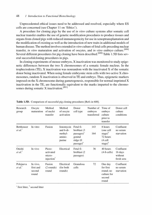
48 1 Introduction to Functional Biotechnology
Unprecedented ethical issues need to be addressed and resolved, especially where EScells are concerned (see Chapter 11 on ‘Ethics’).
A procedure for cloning pigs by the use of in vitro culture systems after somatic cellnuclear transfer enables the use of genetic modification procedures to produce tissues andorgans from cloned pigs with reduced immunogenicity for use in xenoptransplantation andthe modification of existing as well as the introduction of new traits in additional models ofhuman disease. The method involves extended in vitro culture of fetal cells preceding nucleartransfer, in vitro maturation and activation of oocytes, and in vitro embryo culture.[606]
Several different procedures for pig cloning have been described.[608] Table 1.5D lists sev-eral successfulcloning procedures in pigs.
In cloning experiments of mouse embryos, X inactivation was monitored to study epige-netic differences between the two X chromosomes of a somatic female nucleus. In thetrophectoderm (TE), X inactivation was nonrandom with the inactivated X of the somaticdonor being inactivated. When using female embryonic stem cells with two active X chro-mosomes, random X inactivation is observed in TE and embryo. Thus, epigenetic markersimposed on the X chromosome during gametogenesis, responsible for normal imprinted Xinactivation in the TE, are functionally equivalent to the marks imparted to the chromo-somes during somatic X inactivation.[601]
Table 1.5D. Comparison of successful pig cloning procedures [Refs in 608].
Research group
Oocyte maturation
Method of nuclei transfer
Method of oocyte activation
Donor cell type
Number of embryos transferred
Time of embryo culture prior to transfer
Donor cell culture conditions
Betthouser et al.3
In vitro Fusion Ionomycin and 6-di-methyl-amino-purine
Fetal fi-broblast (3 passages)a genital ridge (0 passage)b
143
164
8 hours (one cell stage)b 72 hours (4 cell stage)b
Confluent no serum starvation
Onishi et al.2
In vivo Piezo-activated micro-injection5
Electrical Fetal fi-broblast (2 passages)
36 40 hours (4-8-cells)
Confluent 16 days without fresh sera
Polejaeva et al.1
In vivo, first and second round
Fusion (2 rounds) round
Electrical (for both rounds)
Granulosa cells
72 One day for first round; no culture for second round
Confluent no serum starvation
a first litter, b second litter
49
Stem cell research is pivotal for understanding the development and function of organsin mammals and for the generation of tissues for regenerative medicine,[614] such as CNSstem cells, hepatocytes, and other differentiated cells of other organs, including muscle,blood, liver, and heart. Adult human liver cells can be derived from stem cells originating inthe bone marrow or circulating outside the liver, implying that blood-system stem cellsmight be used clinically to generate hepatocytes for replacement therapy.[613]
Human embryonic stem (ES) cells derived from the inner cell mass of in vitro fertilizedhuman blastocysts were subjected to eight growth factors (basic fibroblast growth factor,transforming growth factor β1, activin-A, bone morphogenetic protein 4, hepatocyte growthfactor, epidermal growth factor, β nerve growth factor, retinoic acid) to direct the differen-tiation of human ES-derived cells in vitro. Each factor has a unique effect and the resultsindicate the potential for directing differentiation and for the enrichment of human celltypes in vitro.[611]
Clonogenic proliferative cells were isolated from adult human dental pulp. These dentalpulp stem cells (DPSCs) produced sporadic densely calcified nodules. In immuno-compomised mice, DPSCs generated a dentin-like structure lined with human odontoblast-like cells surrounding a pulp-like interstitial tissue. The DPSCs may be used for dentinalrepair and, after proliferation ex vivo, for the fabrication of a viable dental implant.[602]
Epidermal stem cells offer the potential for research on and treatment of various skindiseases.[603] Bone-marrow-derived cells offer an alternative source of neurons in patientswith neurodegenerative disease.[605] A genome-wide expression analysis was performed todefine regulatory pathways in hematopoietic stem cells and their global genetic program.Subtracted complementary DNA libraries studied with array hybridization techniques en-abled the characterization of several thousand previously undescribed molecules with appar-ently regulatory properties, thus defing a molecular-genetic phenotype of the hematopoieticstem cell.[616] A clonogenic common myeloid progenitor giving rise to all myeloid lineageswas identified providing the basis for identifying determinants of lineage commitment.[612]
The remodeling of chromosomal architecture following transplantation of somatic nu-clei into unfertilized eggs is a key feature of cloning. The chromatin-remodeling nucleoso-mal adenosine triphosphatase (ATPase) ISWI erases the TATA binding protein from asso-ciation with the nuclear matrix. Dissecting the biochemical processes of nuclear remodelingwill improve the efficiency of cloning and the control of differention in stem cell develop-ment.[617] Murine GATA-2 and GATA-3 are specifically expressed during adipogenesis inwhite adipocyte precursors. Constitutive GATA- and GATA-3 expression suppressesadipocyte differentiation and traps cells at the preadipocyte stage. GATA-3-deficient embry-onic stem cells show an enhanced capacity for differentiation into adipocytes, and defectiveGATA-2 and GATA-3 expression is associated with obesity. GATA-2 and GATA-3 regulateadipocyte differentiation and provide targets for therapeutic intervention in differentiation-related diseases.[618] Stem cell factor (SCF) is influencing hematopiesis and the survival,proliferation, and differentiation of mast cells, melanocytes, and germ cells. The elucida-tion of the crystal structure provides the bassis for SCF-complex formation and function.[620]
Neural stem cells display a broad developmental capacity and may be used to generate anumber of cell types for transplantation in different diseases.[615] Neural stem cells (NSCs)may even be used as therapeutic agents to destroy migratory brain tumor cells plus provid-
1.6 Cellular Cloning
50 1 Introduction to Functional Biotechnology
ing repair of tumor-associate damage.[609] NSCs implanted into experimental intracranialgliomas in vivo in adult rodents, distributed quickly and extensively throughout the tumorbed, actively targeting the tumor cells (including human glioblastomas). NSCs can delivera therapeutically active molecule – cytosine deaminase – causing a quantifiable reductionin tumor burden. Inherently migratory NSCs may function as a delivery vehicle for thera-peutic genes and vectors to refractory, migratory, invasive brain tumors.[610]
Identifying the niches of stem cells in vivo and the requirements for stem cell viabilityand repopulation is essential for future therapies in regenerative medicine, possibly byactivating stem cells in situ. In Drosophila, three cell types act as germ line stem cell niche.[619]
The creation of complex biological structures such as limbs requires detailed knowledge ofthe genetic developmental program for sucessful tissue regeneration.[604]
1.7 Tissue Engineering (Organ Cultivation)
The ‘Holy Grail’ of tissue engineering, the generation of specific tissues and organs withimmunologic identity to the recipient and the conveyance of desired attributes, is gettingcloser with the possibilities of cloning without embryos, by using stem cells which directlydevelop into cells or tissues for the patient (without the need for embryos and the relatedethical obstacles).[114] The regeneration of tissues and organs – regenerative medicine –offers vastly expanded teatment opportunities in injury and disease.[566] Table 1.5E lists aselection of programs and companies in regenerative medicine, covering human organs,xenoptransplantation, growth factors, stem cells, and biomaterials.
The usefulness of genetic modification of cells which are to be used in engineeringtissue is evidenced by non-malignant transformation with human telomerase reverse tran-scriptase of adrenocortic bovine cells.[115]
The expanding list of tissues that use differentiation from stem to progenitor to maturecells includes blood cells, immune system cells, central and peripheral nervous tissues,skeletal muscles, and epithelial cells. The resulting clinical stem cell transplantation couldgreatly increase the physician’s armamentarium against degenerative diseases and the con-sequences of acute failures.
Due to the tremendous research effects, the large numbers of afflicted individuals andthe central role of the respective tissues, particularly pertinent areas are cells of the immunesystem, the pancreas, and the liver.
Fine-tuning of the immune rejection by co-transplantation of human stem cells and stemcells for other organs might be used for tolerance induction and cell- and organ-specificregeneration of diseased or damaged organs.
A crucial technique in therapeutic cloning is the mastery of nuclear transfer to endow therecipient cell, destined to become an organ, with a cell nucleus from the donor, i.e. the pa-tient.
The successful application of nuclear transfer techniques to a range of mammalian spe-cies has brought the possibility of human therapeutic cloning significantly closer. The ob-jective of therapeutic cloning is to produce pluripotent stem cells that carry the nuclear
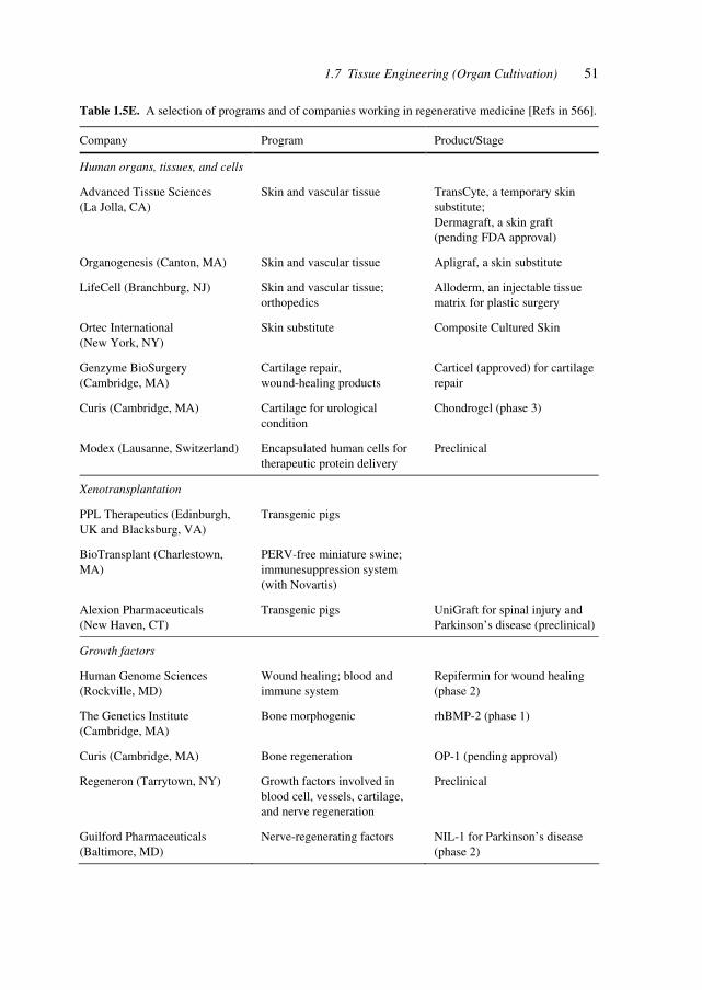
511.7 Tissue Engineering (Organ Cultivation)
Table 1.5E. A selection of programs and of companies working in regenerative medicine [Refs in 566].
Company Program Product/Stage
Human organs, tissues, and cells
Advanced Tissue Sciences (La Jolla, CA)
Skin and vascular tissue TransCyte, a temporary skin substitute; Dermagraft, a skin graft (pending FDA approval)
Organogenesis (Canton, MA) Skin and vascular tissue Apligraf, a skin substitute
LifeCell (Branchburg, NJ) Skin and vascular tissue; orthopedics
Alloderm, an injectable tissue matrix for plastic surgery
Ortec International (New York, NY)
Skin substitute Composite Cultured Skin
Genzyme BioSurgery (Cambridge, MA)
Cartilage repair, wound-healing products
Carticel (approved) for cartilage repair
Curis (Cambridge, MA) Cartilage for urological condition
Chondrogel (phase 3)
Modex (Lausanne, Switzerland) Encapsulated human cells for therapeutic protein delivery
Preclinical
Xenotransplantation
PPL Therapeutics (Edinburgh, UK and Blacksburg, VA)
Transgenic pigs
BioTransplant (Charlestown, MA)
PERV-free miniature swine; immunesuppression system (with Novartis)
Alexion Pharmaceuticals (New Haven, CT)
Transgenic pigs UniGraft for spinal injury and Parkinson’s disease (preclinical)
Growth factors
Human Genome Sciences (Rockville, MD)
Wound healing; blood and immune system
Repifermin for wound healing (phase 2)
The Genetics Institute (Cambridge, MA)
Bone morphogenic rhBMP-2 (phase 1)
Curis (Cambridge, MA) Bone regeneration OP-1 (pending approval)
Regeneron (Tarrytown, NY) Growth factors involved in blood cell, vessels, cartilage, and nerve regeneration
Preclinical
Guilford Pharmaceuticals (Baltimore, MD)
Nerve-regenerating factors NIL-1 for Parkinson’s disease (phase 2)
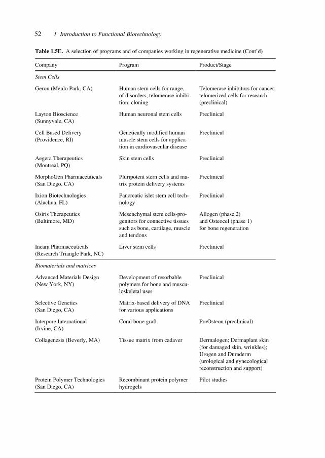
52 1 Introduction to Functional Biotechnology
Table 1.5E. A selection of programs and of companies working in regenerative medicine (Cont’d)
Company Program Product/Stage
Stem Cells
Geron (Menlo Park, CA) Human stern cells for range, of disorders, telomerase inhibi-tion; cloning
Telomerase inhibitors for cancer; telomerized cells for research (preclinical)
Layton Bioscience (Sunnyvale, CA)
Human neuronal stem cells Preclinical
Cell Based Delivery (Providence, RI)
Genetically modified human muscle stem cells for applica-tion in cardiovascular disease
Preclinical
Aegera Therapeutics (Montreal, PQ)
Skin stem cells Preclinical
MorphoGen Pharmaceuticals (San Diego, CA)
Pluripotent stern cells and ma-trix protein delivery systems
Preclinical
Ixion Biotechnologies (Alachua, FL)
Pancreatic islet stem cell tech-nology
Preclinical
Osiris Therapeutics (Baltimore, MD)
Mesenchymal stem cells-pro-genitors for connective tissues such as bone, cartilage, muscle and tendons
Allogen (phase 2) and Osteocel (phase 1) for bone regeneration
Incara Pharmaceuticals (Research Triangle Park, NC)
Liver stem cells Preclinical
Biomaterials and matrices
Advanced Materials Design (New York, NY)
Development of resorbable polymers for bone and muscu-loskeletal uses
Preclinical
Selective Genetics (San Diego, CA)
Matrix-based delivery of DNA for various applications
Preclinical
Interpore International (Irvine, CA)
Coral bone graft ProOsteon (preclinical)
Collagenesis (Beverly, MA) Tissue matrix from cadaver Dermalogen; Dermaplant skin (for damaged skin, wrinkles); Urogen and Duraderm (urological and gynecological reconstruction and support)
Protein Polymer Technologies (San Diego, CA)
Recombinant protein polymer hydrogels
Pilot studies
531.7 Tissue Engineering (Organ Cultivation)
genome of the patient and then induce them to differentiate into replacement cells, such ascardiomyocytes to replace damaged heart tissue or insulin-producing β cells for patientswith diabetes. Although cloning would eliminate the critical problem of immune incom-patibility, there is also the task of reconstituting the cells into more complex tissues andorgans in vitro.[116]
Liver transplants are the desired therapy for a number of conditions wherein the livertissue has been damaged by disease, infections, toxins, medications, drugs or metabolic(inborn or induced) defects.
Islet cell transplantation or extraneously cultivated organs would be preferable to mul-tiple daily insulin treatments. These cells or organs will sense the circulating levels ofglucose and respond appropriately and immediately by releasing the right dose of humancorrect insulin (glycosylation patterns) at the right dose and speed. The complications ofdiabetes, due to imperfect matching of the body’s demands with the substituting therapy,are frequent, life shortening and difficult to manage.[117]
The achievement of growing human corneas in the laboratory will impact the transplan-tation of damaged or impaired corneas, the studies of corneal wound healing and eye dis-eases, as well as the testing of household products for toxicity.[118]
Human corneal equivalents comprising the three main layers of the cornea (epithelium,stroma and endothelium) were constructed. Each cellular layer was fabricated from im-mortalized human corneal cells that were screened for use on the basis of morphological,biochemical and electrophysiological similarity to their natural counterparts. The resultingcorneal equivalents mimicked human corneas in key physical and physiological functions,including morphology, biochemical marker expression, transparency, ion and fluid trans-port, and gene expression. Morphological and functional equivalents to human corneas thatcan be produced in vitro have immediate applications in toxicity and drug efficacy testing,and form the basis for future development of implantable tissues.[119]
Atherosclerotic vascular disease, in the form of coronary artery and peripheral vasculardisease, is the largest cause of mortality in the USA. A tissue engineering approach wasdeveloped to produce arbitrary lengths of vascular graft material from smooth muscle andendothelial cells that were derived from a biopsy of vascular tissue. Bovine vessels culturedunder pulsatile conditions had rupture strengths greater than 2000 mmHg, suture retentionstrengths of up to 90 g and collagen contents of up to 50%. Cultured vessels also showedcontractile responses to pharmacological agents and contained smooth muscle cells thatdisplayed markers of differentiation such as calponin and myosin heavy chains. Tissue-engineered arteries were implanted in miniature swine, with patency documented up to 24days by digital angiography.[120]
Angiogenesis and vascular remodeling play significant roles in wound healing, tumorgrowth, cardiovascular disease and tissue transplantation. Significant effort is directed atdeveloping models to study and manipulate these processes. Angiogenesis is typified bythe elongation of thin-walled capillary tubes from existing vascular structures, followed bygrowth of mesenchymal cells such as pericytes and smooth-muscle cells during the forma-tion of mature stable vessels. Mechanisms were studied of early vascular remodeling inexperiments of suspending EC in three-dimensional culture, where they form tubular struc-tures resembling immature capillaries.
54 1 Introduction to Functional Biotechnology
Conditions for forming cultured human umbilical vein EC (HUVEC) into tubes withina three-dimensional gel that on implantation into immuno-incompetent mice undergo re-modeling into complex microvessels lined by human endothelium were identified. HUVECsuspended in mixed collagen/fibronectin gels organize into cords with early lumena by24 h and then apoptose. Twenty-hour constructs, s.c. implanted in immunodeficient mice,display HUVEC-lined thin-walled microvessels within the gel 31 days after implantation.Retroviral-mediated overexpression of a caspase-resistant Bcl-2 protein delays HUVECapoptosis in vitro for over 7 days. Bcl-2 transduced HUVEC produce an increased densityof HUVEC-lined perfused microvessels in vivo compared with untransduced or control-transduced HUVEC. Bcl-2, but not control-transduced HUVEC recruit an ingrowth ofperivascular smooth muscle α-actin-expressing mouse cells at 31 days, which organize by60 days into HUVEC-lined multilayered structures resembling true microvessels. This sys-tem provides an in vivo model for dissecting mechanisms of microvascular remodeling byusing genetically modified endothelium. Incorporation of such human endothelial-linedmicrovessels into engineered synthetic skin may improve graft viability, especially in re-cipients with impaired angiogenesis.[121]
The problems of organ replacements have been addressed by so far five approaches.Transplantation, which suffers from lack of donors and entails immune rejection and im-munosuppression problems; autografting, which uses parts of organs to restore organ func-tion in other body parts; replacement with an implantate or prothesis, often lasting insuffi-cient time before another surgery or treatment becomes necessary; in vitro synthesis, inwhich an organ is synthesized in culture and then implanted; and in vivo synthesis, whereonly a minimum of tissue is implanted to induce organ regeneration in situ. This combina-tion of in vitro and in vivo methodologies may advance some hitherto intractable problemsof transplantation, especially in older patients, which suffer from chronically open woundsresulting from lack of vascular supply to the skin. The reported enrichment of vascularstructures in host skin by implanting a skin substitute addresses these needs.
In vitro conditions appear to allow the synthesis of the avascular, epithelium-like tissueon one side of the basement membrane but not the synthesis of the vascularized supportingconnective tissue on the other side. Synthesis of supporting tissue requires the presence ofnon-diffusible regulators. The synthesis of a near-physiological peripheral nerve connect-ing two transected nerve stumps across a gap and synthesis of a physiological conjunctivain a conjunctiva-free bed, which had been prepared by excision to bare sclera, required forin vivo synthesis the presence of non-diffusible regulator. Studies of conditions requiredfor organ synthesis, in particular the requirements for extracellular matrix-like regulators,will advance the field of organ replacement.[122]
The search for stem cells has revealed such cells in tissues considered previously un-likely to harbor cells capable of regenerative capacities.
The mature mammalian retina is thought to lack regenerative capacity. The identifica-tion of a stem cell in the adult mouse eye has been reported, which represents a possiblesubstrate for retinal regeneration. Single pigmented ciliary margin cells clonally proliferatein vitro to form sphere colonies of cells that can differentiate into retinal-specific cell types,including rod photoreceptors, bipolar neurons and Müller glia. Adult retinal stem cells arelocalized to the pigmented ciliary margin and not to the central and peripheral retinal pig-
55
mented epithelium, indicating that these cells may be homologous to those found in the eyegerminal zone of other non-mammalian vertebrates.[123]
ES cells are clonal cell lines derived from the inner cell mass of the developing blasto-cyst that can proliferate extensively in vitro and are capable of adopting all the cell fates ina developing embryo. Clinical interest in the use of ES cells has been stimulated by studiesshowing that isolated human cells with ES properties from the inner cell mass or develop-ing germ cells can provide a source of somatic precursors. Previous studies have defined invitro conditions for promoting the development of specific somatic fates, specifically, he-matopoietic, mesodermal and neurectodermal. A method was presented for obtaining dopam-inergic (DA) and serotonergic neurons in high yield from mouse ES cells in vitro. Further-more, the ES cells can be obtained in unlimited numbers and these neuron types are generatedefficiently. CNS progenitor populations were generated from ES cells, these cells wereexpanded and their differentiation promoted into DA and serotonergic neurons in the pres-ence of mitogen and specific signaling molecules. The differentiation and maturation ofneuronal cells was completed after mitogen withdrawal from the growth medium. Thisexperimental system provides a powerful tool for analyzing the molecular mechanismscontrolling the functions of these neurons in vitro and in vivo, and potentially for under-standing and treating neurodegenerative and psychiatric diseases.[124]
The differentiation potential of stem cells in tissues of the adult has been thought to belimited to cell lineages present in the organ from which they were derived, but there isevidence that some stem cells may have a broader differentiation repertoire. It was shownthat neural stem cells from the adult mouse brain can contribute to the formation of chi-merical chick and mouse embryos, and give rise to cells of all germ layers. This demon-strates that an adult neural stem cell has a very broad developmental capacity and maypotentially be used to generate a variety of cell types for transplantation in different dis-eases.[125]
Tissue engineering of human bone is a complex process, as the functional developmentof bone cells requires that regulatory signals be temporally and spatially ordered. The roleof three-dimensional cellular interactions is well understood in embryonic osteogenesis,but in vitro correlates are lacking. In vitro serum-free transforming growth factor (TGF)-β1stimulation of osteogenic cells results immediately after passage in the formation of three-dimensional cellular condensations (bone cell spheroids) within 24–48 h. In turn, bone cellspheroid formation results in the up-regulation of several bone-related proteins (e.g., alka-line phosphatase, type I collagen and osteonectin) during days 3–7, and the concomitantformation of microcrystalline bone. This system of ex vivo bone formation should provideimportant information on the physiological, biological and molecular basis of osteogen-esis.[126]
Massive bone defects are a great challenge to reconstructive surgery. The preferred treat-ment is an autologous bone graft. Bone lesions above a critical size become scarred ratherthan re-generated, leading to non-union. A greater degree of regeneration was obtained byusing a resorbable scaffold with regeneration-competent cells to recreate an embryonicenvironment in injured adult tissues, and thus improve clinical outcome. A combination ofa coral scaffold with in vitro-expanded marrow stromal cells (MSC) was used to increaseosteogenesis more than that obtained with the scaffold alone or the scaffold plus fresh bone
1.7 Tissue Engineering (Organ Cultivation)
56 1 Introduction to Functional Biotechnology
marrow. The efficiency of the various combinations was assessed in a large segmental de-fect model in sheep. The tissue-engineered artificial bone underwent morphogenesis lead-ing to complete recorticalization and the formation of a medullary canal with mature lamellarcortical bone in the most favorable cases. Clinical union never occurred when the defectswere left empty of filled with the scaffolding alone. In contrast, clinical union was obtainedin three out of seven operated limbs when the defects were filled with the tissue-engineeredbone.[127]
Approximately 400 million persons worldwide suffer from bladder disease. Individualswith end-stage bladder disease often require bladder replacement or repair. The bladder isresponsible for being an elastic reservoir for the storage of urine, which maintains a lowintraluminal pressure as it fills. In general, human organ replacement is limited by a donorshortage, problems with tissue compatibility and rejection. Creation of an organ with au-tologous tissue would be advantageous. Transplantable urinary bladder neo-organs werereproducibly created in vitro from urothelial and smooth muscle cells grown in culturefrom canine native bladder biopsies and seeded onto preformed bladder-shaped polymers.The native bladders were subsequently excised from canine donors and replaced with thetissue-engineered neo-organs. In functional evaluations for up to 11 months, the bladderneo-organs demonstrated a normal capacity to retain urine, normal elastic properties, andhistological architecture. Successful re-constitution of an autologous hollow organ is pos-sible using tissue-engineering methods.[128]
One of the main obstacles to successful islet transplantation for both type 1 and 2 diabe-tes is the limitation of available insulin-producing tissue. Only about 3000 cadaver pancreasesbecome available in the USA each year, while about 35,000 new cases of type 1 diabetesare diagnosed each year. This lack of tissue has given a high priority to efforts to stimulatethe growth of new pancreatic islet tissue. The need for transplantable human islets hasstimulated efforts to expand existing pancreatic islets and to grow new ones. In order todemonstrate that human adult duct tissue could be expanded and differentiated in vitro toform islet cells, digested pancreatic tissue that is normally discarded from eight human isletisolations was cultured under conditions that allowed the expansion of the ductal cells as amonolayer where-upon the cells were overlaid with a thin layer of Matrigel. With thismanipulation, the mono-layer of epithelial cells formed three-dimensional structures ofductal cysts from which 50–150 µm diameter islet-like clusters of pancreatic endocrinecells budded. Over 3–4 weeks culture the insulin content per flask increased 10- to 15-foldas the DNA content increased up to 7-fold. The cultivated human islet buds were shown byimmunofluorescence to consist of cytokeratin 19-positive duct cells and hormone-positiveislet cells. Double staining of insulin and non-β cell hormones in occasional cells indicatedimmature cells still in the process of differentiation. Insulin secretion studies were doneover 24 h in culture. Compared with their basal secretion at 5 mM glucose, cysts/cultivatedhuman islet buds exposed to stimulatory 20 mM glucose had a 2.3-fold increase in secretedinsulin. Duct tissue from human pancreas can be expanded in culture and then be directedto differentiate into glucose responsive islet tissue in vitro. This approach may provide apotential new source of pancreatic islet cells for transplantation.[129]
Expression profiling was studied in pancreatic β cells that will contribute to under standingregulation of development, function and regeneration of islet tissue. The β cells of the
57
pancreatic islets of Langerhans are a primary component in vertebrate glucose hemostasis.Pancreatic β cells respond to changes in blood glucose by secreting insulin and increasinginsulin synthesis. To identify genes used in thee responses, expression profiling was carriedout of β cells exposed to high (25 mM) or low (5.5 mM) glucose by using oligonucleotidemicroarrays. Functional clustering of genes that averaged a 2.2-fold or greater change re-vealed large groups of secretory pathway components, enzymes of intermediate metabo-lism, cell-signaling components and transcription factors. Many secretory pathway geneswere up-regulated in high glucose, including seven members of the endoplasmic reticulumtranslocon. In agreement with array analysis, protein levels of translocon components wereincreased by high glucose. Most dramatically, the α subunit of the signal recognition par-ticle receptor was increased over 20-fold. These data indicate that the translocon and ribo-some docking are major regulatory targets of glucose in the β cell. Analysis of genes en-coding enzymes of intermediary metabolism indicated that low glucose brought about greaterutilization of amino acids as an energy source. This is supported by observations of in-creased urea production under low-glucose conditions. Genome-wide integration of β cellfunctions at the level of transcript abundance validates the efficacy of expression profilingin identifying genes in the β cell glucose response.[130]
An experiment in xenotransplantation and on porcine retrovirus is included in this chap-ter, in order to show the potential of grafts in axonal regeneration and in highlighting viraldangers lurking in xenografts.
Transplantation of olfactory ensheathing cells (OECs) or Schwann cells derived fromtransgenic pigs expressing the human complement inhibitory protein, CD59 (hCD59), intotransected dorsal column lesions of the spinal cord of the immunosuppressed rat to induceaxonal regeneration. Non-transplanted lesion-controlled rats exhibited no impulse conduc-tion across the transection site, whereas in animals receiving transgenic pig OECs or Schwanncells impulse conduction was restored across and beyond the lesion site for more than acentimeter. Cell labeling indicated that the donor cells migrated into the denervated hosttract. Conduction velocity measurements showed that the regenerated axons conductedimpulses faster than normal axons. By morphological analysis, the axons seemed thicklymyelinated with a peripheral pattern of myelin expected from the donor cell type. Theseresults indicate that xenotransplantation of myelin-forming cells from pigs genetically al-tered to reduce the hyperacute response in humans are able to induce elongated axonalregeneration and re-myelination and restore impulse conduction across the transected spi-nal cord.[131]
Oligodendrocyte precursor cells (OPCs) are the best-characterized precursors in the CNSof mammals. They arise from multi-potential cells in spatially restricted germinal zonesand then migrate through the developing CNS.
During animal development, cells become progressively more restricted in the cell typesto which they can give rise. In the CNS, multipotential stem cells produce various kinds ofspecified precursors that divide a limited number of times before they terminally differen-tiate into either neurons or glial cells. Certain extracellular signals can induce OPCs torevert to multipotential neural stem cells, which can self-renew and give rise to neurons andastrocytes, as well as to oligodendrocytes, thus demonstrating a great developmental po-tential.[132]
1.7 Tissue Engineering (Organ Cultivation)
58 1 Introduction to Functional Biotechnology
Animal donors such as pigs could provide an alternative source of organs for trans-plantation. The promise of xenotransplantation is offset by the possible public health riskof cross-species infection. All pigs contain several copies of porcine endogenous retroviruses(PERV), and at least three variants of PERV can infect human cell lines in vitro in co-culture, infectivity and pseudotyping experiments. If xenotransplantation of pig tissuesresults in PERV viral replication, there is a risk of spreading and adaptation of this retrovirusto the human host. C-type retroviruses related to PERV are associated with malignancies ofhematopoietic lineage cells in their natural hosts. Pig pancreatic islets were shown to pro-duce PERV and can infect human cells in culture. After transplantation into NOD/SCID(non-obese diabetic/severe combined immunodeficiency) mice, ongoing viral replicationwas detected and several tissue compartments became infected. PERV is thus demonstratedto be transcriptionally active and infectious cross-species in vivo after transplantation ofpig tissues. The concern for PERV infection risk associated with pig islet xenotransplantationin immunosuppressed human patients appears justified.[133]
The progress toward human therapeutic cloning requires the successful application ofnuclear transfer techniques to a range of mammalian species and the subsequent transfer oftechnologies to human cells. The objective of therapeutic cloning is to produce pluripotentstem cells that carry the nuclear genome of the patient and then induce them to differentiateinto replacement cells, such as cardiomyocytes to replace damaged heart tissue or insulin-producing β cells for patients with diabetes. Although cloning would eliminate the criticalproblem of immune incompatibility, there is also the task of reconstituting the cells intomore complex tissues and organs in vitro. The range of disorders where human therapeuticcloning may find application is impressive.[134]
The isolation of pluripotent ES cells opens the way to generate many different tissuesand organs for gene and tissue therapies.
Pluripotent human stem cells isolated from early embryos thus represent a potentiallyun-limited source of many different cell types for cell-based gene and tissue therapies. Ifthe full potential of cell lines derived from donor embryos is to be realized, the problem ofdonor-recipient tissue matching needs to be overcome. One approach, which avoids theproblem of transplant rejection, would be to establish stem cell lines from the patient’s owncells through therapeutic cloning. Recent studies have shown that it is possible to transferthe nucleus from an adult somatic cell to an unfertilized oocyte that is devoid of maternalchromosomes, and achieve embryonic development under the control of the transferrednucleus. Stem cells isolated from such a cloned embryo would be genetically identical tothe patient and pose no risk of immune rejection. The isolation of pluripotent murine stemcells was reported from reprogrammed adult somatic cell nuclei. Embryos were generatedby direct injection of mechanically isolated cumulus cell nuclei into mature oocytes. EScells isolated from cumulus-cell-derived blastocysts displayed the characteristic morphol-ogy and marker expression of conventional ES cells and underwent extensive differentia-tion into all three embryonic germ layers (endoderm, mesoderm, and ectoderm) in tumorsand in chimeric fetuses and pups. The ES cells were also shown to differentiate readily intoneurons and muscle in culture. Thus, pluripotent stem cells can be derived from nuclei ofterminally differentiated adult somatic cells and offer a model system for the developmentof therapies that rely on autologous, human pluripotent stem cells.[135]
59
The senescence of normal cells poses a barrier against their use in organ generation andtransplantation.
Most normal somatic cells from humans and other mammals cannot divide indefinitelybecause of the progressive loss of DNA from telomeres, the specialized ends of the linearchromosomes. The gradual erosion of telomeres during repeated cell division results fromthe combined effects of the inability of normal DNA replication to completely extendtelomeric DNA and the lack of expression of the reverse transcriptase component of thetelomerase ribonucleoprotein complex (TERT). Stem cells and germ line cells, which nor-mally express TERT and have telomerase activity, are able to fully replicate telomeric DNAand thereby are able to divide indefinitely. Cells that do not have telomerase activity areable to divide only a limited number of times before they senesce. In cells with forcedexpression of TERT, the progressive shortening of telomeres is prevented and these cellscontinue to proliferate rapidly at a cumulative population doubling level (PDL) greatlyexceeding the PDL at which they would otherwise senesce.
These results indicate that expression of TERT is sufficient to immortalize at least somecell types that normally undergo cellular senescence. Moreover, immortalization resultingfrom TERT expression does not appear to be accompanied by the acquisition of character-istics of cancer cells, such as chromosomal abnormalities, anchorage-independent growthin culture, loss of normal cell cycle checkpoints, or tumorigenicity in immunodeficientmice. Such findings encouraged speculation that cells expressing TERT could be used incell-based therapies, such as ex vivo gene therapy, tissue engineering, and transplantationof cells to correct metabolic or endocrine defects. The fulfillment of this promise dependson ectopic expression of TERT not affecting cellular regulatory systems such as those thatcontrol differentiation.
Human telomerase reverse transcriptase (hTERT) expression in experimental xenotrans-plantation was demonstrated. It had been shown that bovine adrenocortical cells can betransplanted into SCID mice and that these cells form functional tissue that replaces theanimals’ own adrenal glands. Primary bovine adrenocortical cells were co-transfected withplasmids encoding hTERT, SV40 T antigen, neo and GFP. These clones do not undergoloss of telomeric DNA and appear to be immortalized. Two clones were transplanted be-neath the kidney capsule of SCID mice. Animals that received cell transplants survivedindefinitely despite adrenalectomy. The mouse glucocorticoid, corticosterone, was replacedby the bovine glucocorticoid, cortisol, in the plasma of these animals. The tissue formedfrom the transplanted cells resembled that formed by transplantation of cells that were notgenetically modified and was similar to normal bovine adrenal cortex. The proliferationrate in tissues formed from these clones was low and there was no indication of malignanttransformation.[136]
Retroviral-mediated transduction of hTERT (human telomerase reverse transcriptase) inhuman dermal microvascular endothelial cells (EC) (HDMEC) results in cell lines thatform microvascular structures upon subcutaneous implantation in severe combined immu-nodeficiency mice (SCID) mice. Anti-human type IV collagen basement membrane immu-noreactivity and visualization of enhanced green fluorescent protein (eGFP)-labeledmicrovessels confirmed the human origin of these vessels. Primary HDMEC-derived ves-sel density decreased with time, while telomerized HDMEC maintained durable vessels six
1.7 Tissue Engineering (Organ Cultivation)
60 1 Introduction to Functional Biotechnology
weeks after xenografting. The modulation of implant vessel density by exposure to differ-ent angiogenic and angiostatic factors confirms the utility of the system for human mi-crovascular remodeling and pertinent in vivostudies.[565]
Slowing the rate of telomere shortening could have benefits in specific tissues wheretelomere-based growth arrest, i.e. senescence, occurs. The medical implications of thistechnology are profound. Some promising areas of cell engineering include rejuvenationof HSC for improving bone marrow transplants or enhancing general immunity for olderpatients. Other possibilities include an unlimited supply of skin cells for grafts in burnpatients and for treating ulcerated lesions that do not heal. The immortalization ofchondrocytes or osteoprogenitor cells to treat osteoarthritis or for bone grafts, and endot-helial cells for the generation of tissue-engineered blood vessels are other areas of inter-est. In the future we could try to grow cells in the laboratory for disorders for which thereare currently no cures, e.g., muscle satellite cells for muscular dystrophy, retinal cells forthe treatment of macular degeneration (a leading cause of age-related blindness) and im-mune cells for HIV patients. This could avoid immune rejections and could slow down orreverse some of the problems associated with these disorders. Proteins could also be pro-duced from normal human cell cultures to treat the donor patient. Engineered cells mayalso be useful for in vitro markets, as alternatives to animal testing or to produce prod-ucts for cosmetic applications. Of course, safety and efficacy standards, quality and con-trol assurances will need to be developed as well as preclinical and clinical evalua-tions.[137]
Gene therapy and tissue engineering are new concepts in the treatment of human dis-ease. Gene therapy uses DNA as a ‘super pharmaceutical’, an agent with the potential toalter cell function for an extended period of time in relation to more established therapeuticagents. Tissue engineering aims to regrow tissue structure lost as a result of trauma ordisease through the application of engineered materials. Both fields can demonstrate initialsuccess, such as the replacement of adenosine deaminase genes in children with severeimmunodeficiency and the use of synthetic materials to accelerate healing of burns andskin ulcers. Major limitations are the low level of expression in gene therapy and the tissueengineers have not yet learned to reproduce complex architecture, such as vascular net-works, which are essential for normal tissue function. A combination of both methods hasbeen used as a new strategy to overcome problems remaining.
DNA delivery has been described in situ from polymer coatings, microspheres and syn-thetic matrices. DNA–material hybrid systems promise to enable forms of gene therapy notpossible with other gene delivery systems, e.g., polymeric microspheres facilitate the ex-pression of orally administered genes.[138]
Loss or deficient tissue function leads to millions of surgical procedures each year andto a loss of, for example, the US economy alone of hundreds of billions of dollars. Tissueengineering has emerged as a potential means of growing new tissues and organs to treatthese patients, and several approaches are currently under investigation to engineer struc-tural tissues. One approach involves transplanting cells on biodegradable polymer matri-ces. Matrices serve to deliver cells to specific anatomical sites, create and maintain a spacefor tissue development, and guide tissue formation before being degraded. Limitations ofthis approach include the need to isolate and expand cells in vitro, and poor survival of
61
many cell types following tissue transplantation. A separate strategy involves the deliveryof tissue-inductive proteins [(e.g., bone morphogenetic proteins and platelet-derived growthfactor (PDGF)]. One important drawback of this approach is decreased protein stability inthe delivery system. Delivery of plasmid DNA encoding for inductive factors has beenproposed as a replacement to direct delivery of the protein. The delivery of plasmid DNA invivo is typically associated with low levels of gene transfer and cellular expression, perhapsdue to a limited exposure of cells to the plasmid.
The engineering of tissues is proposed by the incorporation and sustained release ofplasmids encoding tissue-inductive proteins from polymer matrices. Matrices of poly(lactide-co-glycolide) (PLG) were loaded with plasmid, which was subsequently released over aperiod ranging from days to a month in vitro. Sustained delivery of plasmid DNA frommatrices led to the transfection of large numbers of cells. Furthermore, in vivo delivery of aplasmid encoding PDGF enhanced matrix deposition and blood vessel formation in thedeveloping tissue. This contrasts with direct injection of the plasmids, which did not sig-nificantly affect tissue formation. This method of DNA delivery may find utility in tissueengineering and gene therapy applications.[139]
Biocompatible materials are commonly used in reconstructive medicine. One such ma-terial, CellFoam, has been used to repair bone defects in vivo; the open pore structurepermits infiltration and integration of cells into the surrounding engraftment site. CellFoamis fabricated by high-temperature precipitation of tantalum, a highly biocompatible metalwith an established history of in vivo medical use, as a thin layer onto a highly regularreticulated carbon skeleton. The resulting material is highly porous and has a heavily con-toured surface with nanostructure ridges that facilitate cell attachment. Three-dimensional-ity is advantageous for bone reconstruction and is critical for organ tissue function.
Biocompatible inorganic matrices have been used to enhance bone repair by integratingwith endogenous bone structure. Hypothesizing that a three-dimensional framework mightsupport reconstruction of other tissues as well, the capacity of tantalum-coated carbon matrixto support reconstitution of functioning thymic tissue was assessed. A thymic organoid wasan engineered by seeding matrices with murine thymic stoma. Co-culture of human bonemarrow-derived hematopoietic progenitor cells within this xenogenic environment gener-ated mature functional T cells within 14 days. The proportionate T cell yield from thissystem was highly reproducible, generating over 70% CD3+ T cells from either AC133+ orCD34+ progenitor cells. Cultured T cells expressed a high level of T cell receptor excisioncircles (TREC), demonstrating de novo T lymphopoiesis, and function of fully mature Tcells. This system not only facilitates analysis of the T lymphopoietic potential of progeni-tor cell populations; it also permits ex vivo genesis of T cells for possible applications intreatment of immunodeficiency.[140]
Cell lineage studies are essential basis for elucidating the genetic and biochemical path-ways and switches in stem cells and differentiation. Hematopoietic stem cells give rise toall blood cell lineages in mammals. Molecular decisions that differentiate one lineage fromanother are regulated by unique protein complexes constituted of general as well as lin-eage-specific transcription factors. Gene inactivation studies in mice have identified animportant role for a number of hematopoietic transcription factors, such as GATA-1 forerythroid development, GATA-2 for definitive hematopoiesis and GATA-3 for T cell devel-
1.7 Tissue Engineering (Organ Cultivation)
62 1 Introduction to Functional Biotechnology
opment. The Drosophila GATA homolog serpent (srp) is required for the embryonic bloodcell development. Another mammalian gene encoding the AML1 protein is the most fre-quent target of chromosomal translocations in acute myeloid leukemia.
Two major classes of cells observed within the Drosophila hematopoietic repertoire areplasmocytes/macrophages and crystal cells. The transcription factor Lz (Lozenge), whichresembles human AML1 (acute myeloid leukemia-1) protein, is necessary for the develop-ment of crystal cells during embryonic and larval hematopoiesis. Another transcriptionfactor, Gcm (glial cells missing), has previously been shown to be required for plasmocytesdevelopment. Misexpression of Gcm causes crystal cells to be transformed into plasmocytes.The Drosophila GATA protein Srp (Serpent) is required for both Lz and Gcm expressionand is necessary for the development of both classes of hemocytes, whereas Lz and Gcmare required in a lineage-specific manner. Given the similarities of Srp and Lz to mamma-lian GATA and AML1 proteins, observations in Drosophila are likely to have broad impli-cations for understanding mammalian hematopoiesis and leukemia.[141]
Therapeutic cloning describes the potential use of nuclear transfer to provide cells, tis-sues and organs for patients requiring the replacement or supplementation of diseased ordamaged tissues. The concept of using ES cells as a source of multiple cell types for use intissue repair is being developed towards biomedical application based on breakthroughs insomatic nuclear transfer and human ES cell derivation. ES cell lines that are customizedand genetically identical to those of the patient are becoming feasible.[142] Locally deliv-ered bone marrow cells can generate de novo myocardium, thus ameliorating the outcomeof coronary artery disease.[659] Cells amenable to regenerative tissue studies include neu-rons derived from human neurospheres[671] and insulin-secreting structures similar to pan-creatic islets.[672]
1.8 Micro- and Nanotechnologies for Medicine
The most profound effects of microtechnologies are evident in the development ofmicroarrays (biochips) for drug discovery, testing and genotyping.
Biological microchips (biochips) are revolutionizing gene expression analysis and clas-sical genotyping as well as diagnostics and testing. The worldwide markets for biochips areprimarily composed of DNA chips, protein chips and laboratory chips. Generally, arraysare differentiated from microfluidic systems, which can actively operate analysis, separa-tion and synthetic processes by means of microscopic capillary systems, mini-pumps andmini-valves. These are often called a ‘lab-on-a-chip’.
The total market volume for biochips was estimated for 1999 to be approximately US$180 million, whereby the percentage of DNA chips is above 90%. Annual growth of morethan 30% is expected for the years until 2005. In 2005, the total market volume is expectedto amount to US$ 950 million (US$ 725 million DNA chips, US$ 157 million for lab chipsand US$ 68 million for protein chips).
The market is segmented (pharmaceutical, biotechnology and academia), and out of atotal of anticipated US$ 660 million for 2003, the pharmaceutical industry will comprise
63
US$ 260 million, the biotechnology industry US$ 185 million and the academic institu-tions US$ 212 million.
DNA chips are provided by more than 20 companies (e.g., Affymetrix, Α Gene, AmershamPharmacia Biotech, Genometrix, Genomic Solutions, Hyseq Incyte Pharmaceuticals,Nanogen, etc.), protein chips by one company (Ciphergen) and microfluidic chips by sev-eral companies [Aclara Biosciences, Caliper (Cellomics), Cepheid and Orchid Bio-computer].[143]
The requirements for ever more discriminating methods of separating synthesized mol-ecules, especially during later stages of drug development, is facilitated by improved meth-ods in the design of separating phases. Biomimetic synthesis offers the opportunity to de-sign and synthesize specific inorganic structures to fit hand-in-glove with target molecules.Self-assembled block co-polypeptide architecture provides the matrix for silica shapes.The synthetic capability to control directly silica shape, hydrolysis and condensation rateby adjustment of block co-polypeptide composition offers a route to the environmentallybenign, biomimetic synthesis of inorganic materials.
In biological systems such as diatoms and sponges, the formation of solid silica struc-tures with precisely controlled morphologies is directed by proteins and polysaccharides,and occurs in water at neutral pH and ambient temperature. Laboratory methods, in con-trast, have to rely on extreme pH conditions and/or surfactants to induce the condensationof silica precursors into specific morphologies or patterned structures. This contrast inprocessing conditions and the growing demand for benign synthesis methods that mini-mize adverse environmental effects have spurred much interest in biomimetic approachesin materials science. The recent demonstration that silicatein – a protein found in the silicaspicules of the sponge Thethya aurantia – can hydrolyze and condense the precursor mol-ecule tetraethoxysilane to form silica structures with controlled shapes at ambient condi-tions seems particularly promising in this context. Synthetic cysteine–lysine blockcopolypeptides were described that mimic the properties of silicatein: the copolypeptidesself-assemble into structured aggregates that hydrolyze tetraethoxysilane while simulta-neously directing the formation of ordered silica morphologies. Oxidation of the cysteinsulfydryl groups, which is known to affect the assembly of the block co-polypeptide, al-lows to produce different structures: hard silica spheres and well-defined columns of amor-phous silica are produced using the fully reduced and the oxidized forms of the copolymer,respectively.[144]
Another example of molecular imprinting has been demonstrated by a generally appli-cable imprinting strategy which is used to synthesize bulk, amorphous, microporous silicaswith binding sites comprising either one, two or three propylamine groups that are co-valently linked to the silicon atoms of the silicate.
Molecular imprinting aims to create solid materials containing chemical functionalitiesthat are spatially organized by covalent or non-covalent interactions with imprint (or tem-plate) molecules during the synthesis process. Subsequent removal of the imprint mol-ecules leaves behind designed sites for the recognition of small molecules, making thematerial ideally suited for applications such as separations, chemical sensing and cataly-sis. The molecular imprinting of bulk polymers and polymer and silica surfaces has beenreported, but the extension of these methods to a wider range of materials remains prob-
1.8 Micro- and Nanotechnologies for Medicine
64 1 Introduction to Functional Biotechnology
lematic. For example, the formation of substrate-specific cavities within bulk silica, whileconceptually straightforward, has been difficult to accomplish experimentally. The imprint-ing of bulk amorphous silicas with single aromatic rings carrying up to three 3-amino-propyl-triethoxysilane side groups has been described. This generates and occupiesmicroporosity and attaches functional groups to the pore walls in a controlled fashion.The triethoxysilane part of the molecules’ side groups is incorporated into the silica frame-work during sol–gel synthesis and subsequent removal of the aromatic core creates a cav-ity with spatially organized aminopropyl groups covalently anchored to the pore walls.The imprinted silicas act as shape-selective base catalysts. The strategy can be extendedto imprint other functional groups, which should give access to a wide range of functional-ized materials.[145]
Progress towards analytical precision required in nanotechnology has been reported inthe search for optical reporters for diagnostic detection and labeling to be used in biomedi-cal research, testing and application.
The use of colloidal silver plasmon-resonant particles (PRPs) as optical reporters inbiological assays has been demonstrated. PRPs are ultrabright, nano-sized optical scatter-ers, which scatter light elastically and can be prepared with a scattering peak at any color inthe visible spectrum. PRPs are readily observed individually with a microscope configuredfor dark-field microscopy, with white-light illumination of typical power. The use of PRPs,surface coated with standard ligands, was demonstrated as target-specific labels in an insitu hybridization and an immunocytology assay. PRPs can replace or complement estab-lished labels, such as those based on radioactivity, fluorescence, chemiluminescence orenzymatic/colorimetric detection that are used routinely in biochemistry, cell biology andmedical diagnostic applications. Moreover, because PRP labels are non-bleaching and brightenough to be rapidly identified and counted, an ultrasensitive assay format based on single-target molecule detection is now practical. The results of a model sandwich immunoassayfor goat anti-biotin antibody used the number of PRP labels counted in an image to consti-tute the measured signal.[146]
Fiber optic chemical sensors and biosensors offer important advantages for in situ moni-toring applications because of the optical nature of the detection signal. Recent advances innanotechnology leading to the development of optical fibers with submicron-sized dimen-sions have opened up new horizons for intracellular measurements.
The application of an antibody-based nanoprobe for in situ measurements of a singlecell was reported. The nanoprobe employs antibody-based receptors targeted to a fluores-cent analyte, benzopyrene tetrol (BPT), a metabolite of the carcinogen benzo[a]pyrene(BaP) and of the BaP–DNA adduct. Detection of BPT is of great biomedical interest, sincethis species can serve as a biomarker for monitoring DNA damage due to BaP exposure andfor possible precancer diagnosis. The measurements were performed on the rat liver epithe-lial clone 9 cell line, which was used as the model cell system. Before making measure-ments, the cells were treated with BPT. Nanoprobes were inserted into individual cells,incubated 5 min to allow antigen-antibody binding, and then removed for fluorescencedetection. A concentration of 96 ± 0.2 × 10¹¹ M for BPT in the individual cells investigatedwas determined. The results demonstrate the possibility of in situ measurements inside asingle cell using an antibody-based nanoprobe.[147]
65
Sequence-selective DNA detection is becoming increasingly important to unravel thegenetic basis of disease and for the development of diagnostics and therapies. A DNA arrayprobing technique with increased sensitivity has been developed which uses oligonucle-otide-modified gold nanoparticle probes and a conventional flatbed scanner for analyzingcombinatorial DNA arrays. Labeling oligonucleotide targets with nanoparticle rather thanfluorophore probes substantially alters the melting profiles of the targets from an arraysubstrate. This difference permits the discrimination of an oligonucleotide sequence fromtargets with single nucleotide mismatches with a selectivity that is over three times thatobserved for fluorophore-labeled targets. In addition, when coupled with a signal amplifi-cation method based on nanoparticle-promoted reduction of silver (I), the sensitivity of thisscanometric array detection system exceeds that of the analogous fluorophore system bytwo orders of magnitude.[148]
Functional nanoelectromechanical systems (NEMS) require locomotive forces for powerto act as biomolecular motors. The integration of enzymes as biomolecular motors withnano-scale engineered systems generates hybrid organic-inorganic devices capable of uti-lizing ATP as an energy source. Such a biomolecular motor consisting of an endineeredsubstrate, an F1-ATPase biomolecular motor and fabricated nanopropellers was con-structed.[459] The molecular dynamics of the F1-ATP-ase at the molecular level were de-scribed[652] and the suitability of biomolecular devices as nanomolecular machines com-prehensively reviewed.[653]
The technological requirements for designing and constructing microfluidic devices formicro- and nano-technology are expanding. A microfluidic platform for the construction ofmicroscale components and autonomous systems consists of a combination of liquid-phasephotopolymerization, lithography, and laminar flow to generate channels, valves, actua-tors, sensors, and systems.[460]
Bioelectronics, the integration of biomaterials, cells and tissues or organs, electronics,optronics, electronic transducers, enables the electronic transduction of biorecognition events,biotransaformations and biosensing events.[666] Electrogenic cells can be coupled to elec-trodes and the resulting bio-electronic devices, such as cell-transistor hybrids, can be usedfor drug screening, the generation of biosensors, and implantates.[708] The capabilities tomanipulate single biomolecules will lead to molecular motors and devices.[668]
Nanotechnologies provide increasing potential for modifications of surfaces and theconstruction of biocompatible devices.[656] There is an expanding range of tools availablefor developing, engineering, and constructing micro- and nano-devices, such as thesubmicrometer patterning of thin-film electrets with trapped charge by using a flexible,electriclly conductive electrode,[562] by replica-molding techniques.[557,558] Robust chemi-cal methods allow the fabrication of ultrahigh-density arrays of nanopores with high aspectratios using the equi-librium self-assembled morphology of asymmetric diblock copoly-mers.[556] The direct printing of functional electronic materials provides possibilities forthe construction of polymer devices and integrated transistor circuits.[555] Sensor arrays,nanoreactors, photonic and fluidic devices can be constructed by optically defined multi-functional patterning of photo-sensitive thin-film silica mesophases.[554]
The required energy conversion in nanodevices may be achieved by constructing mo-lecular motors[552,553] and providing molecular switches[551,554] such as those consisting of
1.8 Micro- and Nanotechnologies for Medicine
66 1 Introduction to Functional Biotechnology
a metal cluster and redox-addressable groups.[550]. The integration of sensors, measure-ment and control will be faciliated by the development of mono-molecular electronics wheresingle molecules integrate elementary functions and interconnections.[549]. The rules andmethods as well as the design tools for nano-technology are rapidly spreading and develop-ing.[529–543]
The potential of electronics with organic molecules and the technologies employed intheir production, such as soft lithography and self-assembly of monolayers,[709,710] increasesthe feasibility of micro- and nano-devices for biomedical applications.
1.9 Structural Genomics
The rational design of drugs, therapies, agonists and antagonists requires knowledge of thestructure and function of target molecules such as receptors, enzymes, RNA, complexesand regulatory proteins. Following the elucidation of gene sequences and the analysis ofproteins and their interactions, the knowledge of the structure of target molecules becomeessential for further research into and development of the desired interactions. The struc-ture is expected to provide useful information about biological and biochemical functionsin order to facilitate the understanding of normal and disease phenomena, and the develop-ment of therapeutically active molecules.
The structure reveals the organization of the protein, RNA or complex in three dimen-sions, and thus allows the identification of the partial structures, domains, sequences and
interactions that determine the function(s) of the molecule. Protein–ligand complexesare especially useful providers of functional information as they define the location of theligand, the active site, the conformational effects and the mechanism of action of the mol-ecule.[428]
The choice of targets from the numerous biological molecules is determined by thefeasibility of isolating and purifying the molecules or complexes, as well as by the interestin the putative or known biological function or context. Structural genomics aims to deter-mine the structure of all proteins in order to provide targets for R & D, and to construct atrue structural and functional model of cells and organisms. The selected target moleculesare studied primarily by crystallography and NMR in order to elucidate their structure,followed by biochemical and biological studies and validation.[429]
The production of biological molecules, in particular proteins, for structural analysis isa major challenge primarily met by cloning and by purification.[430]
Automation will be a major key for the success of structural genomics. The develop-ment of effective macromolecular structure determination facilities using intense X-raysfrom synchroton sources and advances in protein expression, purification and crystalliza-tion are accelerating the process of determining the structures of target macromol-ecules.[431, 432]
Figure 1.3 shows the procedure for structure determination by X-ray crystallographyand Table 1.6 lists software used in structural analysis (data processing, molecular replace-ment, heavy atom site identification, model building, refinement and validation).
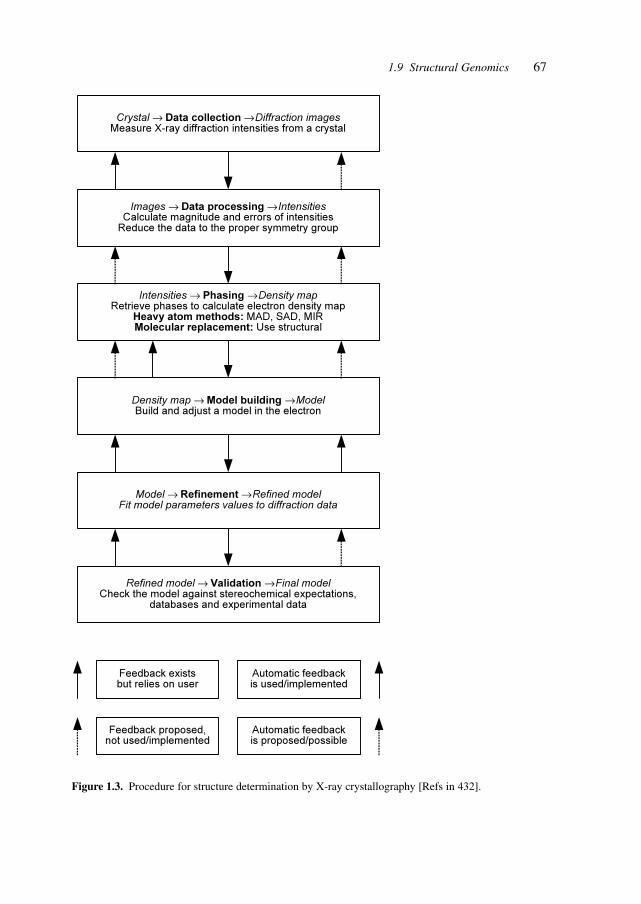
67
Crystal → Data collection → Diffraction imagesMeasure X-ray diffraction intensities from a crystal
Images → Data processing → IntensitiesCalculate magnitude and errors of intensities
Reduce the data to the proper symmetry group
Intensities → Phasing → Density mapRetrieve phases to calculate electron density map
Heavy atom methods: MAD, SAD, MIRMolecular replacement: Use structural
Density map → Model building → ModelBuild and adjust a model in the electron
Model → Refinement → Refined modelFit model parameters values to diffraction data
Refined model → Validation → Final modelCheck the model against stereochemical expectations,
databases and experimental data
Feedback existsbut relies on user
Automatic feedbackis used/implemented
Feedback proposed,not used/implemented
Automatic feedbackis proposed/possible
Figure 1.3. Procedure for structure determination by X-ray crystallography [Refs in 432].
1.9 Structural Genomics
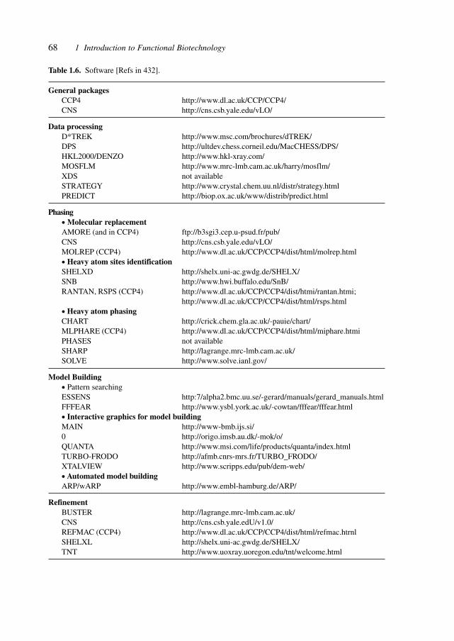
68 1 Introduction to Functional Biotechnology
Table 1.6. Software [Refs in 432].
General packagesCCP4 http://www.dl.ac.uk/CCP/CCP4/CNS http://cns.csb.yale.edu/vLO/
Data processingD*TREK http://www.msc.com/brochures/dTREK/DPS http://ultdev.chess.corneil.edu/MacCHESS/DPS/HKL2000/DENZO http://www.hkl-xray.com/MOSFLM http://www.mrc-lmb.cam.ac.uk/harry/mosflm/XDS not availableSTRATEGY http://www.crystal.chem.uu.nl/distr/strategy.htmlPREDICT http://biop.ox.ac.uk/www/distrib/predict.html
Phasing• Molecular replacementAMORE (and in CCP4) ftp://b3sgi3.cep.u-psud.fr/pub/CNS http://cns.csb.yale.edu/vLO/MOLREP (CCP4) http://www.dl.ac.uk/CCP/CCP4/dist/html/molrep.html• Heavy atom sites identificationSHELXD http://shelx.uni-ac.gwdg.de/SHELX/SNB http://www.hwi.buffalo.edu/SnB/RANTAN, RSPS (CCP4) http://www.dl.ac.uk/CCP/CCP4/dist/htmi/rantan.htmi;
http://www.dl.ac.uk/CCP/CCP4/dist/html/rsps.html• Heavy atom phasingCHART http://crick.chem.gla.ac.uk/-pauie/chart/MLPHARE (CCP4) http://www.dl.ac.uk/CCP/CCP4/dist/html/miphare.htmiPHASES not availableSHARP http://lagrange.mrc-lmb.cam.ac.uk/SOLVE http://www.solve.ianl.gov/
Model Building• Pattern searchingESSENS http:7/alpha2.bmc.uu.se/-gerard/manuals/gerard_manuals.htmlFFFEAR http://www.ysbl.york.ac.uk/-cowtan/fffear/fffear.html• Interactive graphics for model buildingMAIN http://www-bmb.ijs.si/0 http://origo.imsb.au.dk/-mok/o/QUANTA http://www.msi.com/life/products/quanta/index.htmlTURBO-FRODO http://afmb.cnrs-mrs.fr/TURBO_FRODO/XTALVIEW http://www.scripps.edu/pub/dem-web/• Automated model buildingARP/wARP http://www.embl-hamburg.de/ARP/
RefinementBUSTER http://lagrange.mrc-lmb.cam.ac.uk/CNS http://cns.csb.yale.edU/v1.0/REFMAC (CCP4) http://www.dl.ac.uk/CCP/CCP4/dist/html/refmac.htrnlSHELXL http://shelx.uni-ac.gwdg.de/SHELX/TNT http://www.uoxray.uoregon.edu/tnt/welcome.html
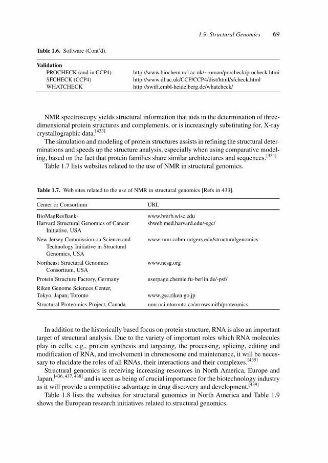
69
NMR spectroscopy yields structural information that aids in the determination of three-dimensional protein structures and complements, or is increasingly substituting for, X-raycrystallographic data.[433]
The simulation and modeling of protein structures assists in refining the structural deter-minations and speeds up the structure analysis, especially when using comparative model-ing, based on the fact that protein families share similar architectures and sequences.[434]
Table 1.7 lists websites related to the use of NMR in structural genomics.
Table 1.7. Web sites related to the use of NMR in structural genomics [Refs in 433].
Center or Consortium URL
BioMagResBank- www.bmrb.wisc.eduHarvard Structural Genomics of Cancer sbweb.med.harvard.edu/-sgc/
Initiative, USA
New Jersey Commission on Science and www-nmr.cabm.rutgers.edu/structuralgenomicsTechnology Initiative in StructuralGenomics, USA
Northeast Structural Genomics www.nesg.orgConsortium, USA
Protein Structure Factory, Germany userpage.chemie.fu-berlin.de/-psf/
Riken Genome Sciences Center,Tokyo, Japan; Toronto www.gsc.riken.go.jp
Structural Proteomics Project, Canada nmr.oci.utoronto.ca/arrowsmith/proteomics
In addition to the historically based focus on protein structure, RNA is also an importanttarget of structural analysis. Due to the variety of important roles which RNA moleculesplay in cells, e.g., protein synthesis and targeting, the processing, splicing, editing andmodification of RNA, and involvement in chromosome end maintenance, it will be neces-sary to elucidate the roles of all RNAs, their interactions and their complexes.[435]
Structural genomics is receiving increasing resources in North America, Europe andJapan,[436, 437, 438] and is seen as being of crucial importance for the biotechnology industryas it will provide a competitive advantage in drug discovery and development.[439]
Table 1.8 lists the websites for structural genomics in North America and Table 1.9shows the European research initiatives related to structural genomics.
Table 1.6. Software (Cont’d).
ValidationPROCHECK (and in CCP4) http://www.biochem.ucl.ac.uk/~roman/procheck/procheck.htmiSFCHECK (CCP4) http://www.dl.ac.uk/CCP/CCP4/dist/html/sfcheck.htmlWHATCHECK http://swift.embl-heidelberg.de/whatcheck/
1.9 Structural Genomics
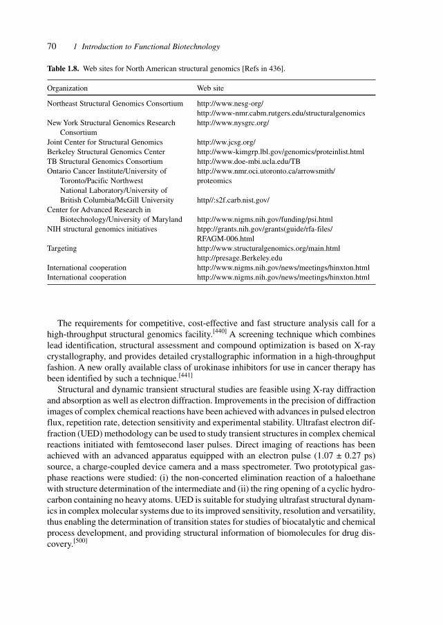
70 1 Introduction to Functional Biotechnology
The requirements for competitive, cost-effective and fast structure analysis call for ahigh-throughput structural genomics facility.[440] A screening technique which combineslead identification, structural assessment and compound optimization is based on X-raycrystallography, and provides detailed crystallographic information in a high-throughputfashion. A new orally available class of urokinase inhibitors for use in cancer therapy hasbeen identified by such a technique.[441]
Structural and dynamic transient structural studies are feasible using X-ray diffractionand absorption as well as electron diffraction. Improvements in the precision of diffractionimages of complex chemical reactions have been achieved with advances in pulsed electronflux, repetition rate, detection sensitivity and experimental stability. Ultrafast electron dif-fraction (UED) methodology can be used to study transient structures in complex chemicalreactions initiated with femtosecond laser pulses. Direct imaging of reactions has beenachieved with an advanced apparatus equipped with an electron pulse (1.07 ± 0.27 ps)source, a charge-coupled device camera and a mass spectrometer. Two prototypical gas-phase reactions were studied: (i) the non-concerted elimination reaction of a haloethanewith structure determination of the intermediate and (ii) the ring opening of a cyclic hydro-carbon containing no heavy atoms. UED is suitable for studying ultrafast structural dynam-ics in complex molecular systems due to its improved sensitivity, resolution and versatility,thus enabling the determination of transition states for studies of biocatalytic and chemicalprocess development, and providing structural information of biomolecules for drug dis-covery.[500]
Table 1.8. Web sites for North American structural genomics [Refs in 436].
Organization Web site
Northeast Structural Genomics Consortium http://www.nesg-org/http://www-nmr.cabm.rutgers.edu/structuralgenomics
New York Structural Genomics Research http://www.nysgrc.org/Consortium
Joint Center for Structural Genomics http://ww.jcsg.org/Berkeley Structural Genomics Center http://www-kimgrp.lbl.gov/genomics/proteinlist.htmlTB Structural Genomics Consortium http://www.doe-mbi.ucla.edu/TBOntario Cancer Institute/University of http://www.nmr.oci.utoronto.ca/arrowsmith/
Toronto/Pacific Northwest proteomicsNational Laboratory/University ofBritish Columbia/McGill University http//:s2f.carb.nist.gov/
Center for Advanced Research inBiotechnology/University of Maryland http://www.nigms.nih.gov/funding/psi.html
NIH structural genomics initiatives htpp://grants.nih.gov/grants(guide/rfa-files/RFAGM-006.html
Targeting http://www.structuralgenomics.org/main.htmlhttp://presage.Berkeley.edu
International cooperation http://www.nigms.nih.gov/news/meetings/hinxton.htmlInternational cooperation http://www.nigms.nih.gov/news/meetings/hinxton.html
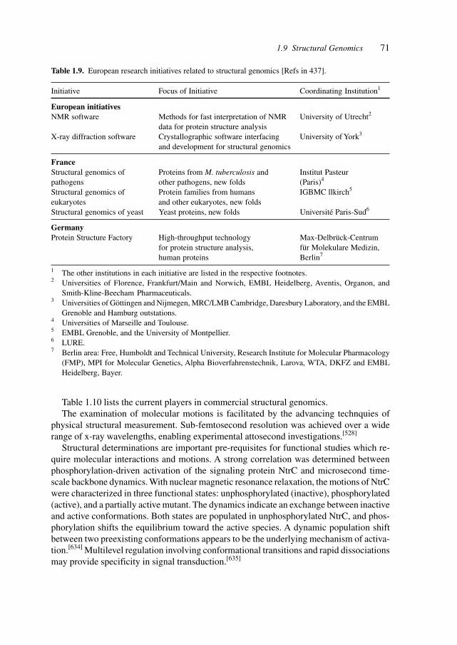
71
Table 1.10 lists the current players in commercial structural genomics.The examination of molecular motions is facilitated by the advancing technquies of
physical structural measurement. Sub-femtosecond resolution was achieved over a widerange of x-ray wavelengths, enabling experimental attosecond investigations.[528]
Structural determinations are important pre-requisites for functional studies which re-quire molecular interactions and motions. A strong correlation was determined betweenphosphorylation-driven activation of the signaling protein NtrC and microsecond time-scale backbone dynamics. With nuclear magnetic resonance relaxation, the motions of NtrCwere characterized in three functional states: unphosphorylated (inactive), phosphorylated(active), and a partially active mutant. The dynamics indicate an exchange between inactiveand active conformations. Both states are populated in unphosphorylated NtrC, and phos-phorylation shifts the equilibrium toward the active species. A dynamic population shiftbetween two preexisting conformations appears to be the underlying mechanism of activa-tion.[634] Multilevel regulation involving conformational transitions and rapid dissociationsmay provide specificity in signal transduction.[635]
Table 1.9. European research initiatives related to structural genomics [Refs in 437].
Initiative Focus of Initiative Coordinating Institution1
European initiativesNMR software Methods for fast interpretation of NMR University of Utrecht2
data for protein structure analysisX-ray diffraction software Crystallographic software interfacing University of York3
and development for structural genomics
FranceStructural genomics of Proteins from M. tuberculosis and Institut Pasteurpathogens other pathogens, new folds (Paris)4
Structural genomics of Protein families from humans IGBMC llkirch5
eukaryotes and other eukaryotes, new foldsStructural genomics of yeast Yeast proteins, new folds Université Paris-Sud6
GermanyProtein Structure Factory High-throughput technology Max-Delbrück-Centrum
for protein structure analysis, für Molekulare Medizin,human proteins Berlin7
1 The other institutions in each initiative are listed in the respective footnotes.2 Universities of Florence, Frankfurt/Main and Norwich, EMBL Heidelberg, Aventis, Organon, and
Smith-Kline-Beecham Pharmaceuticals.3 Universities of Göttingen and Nijmegen, MRC/LMB Cambridge, Daresbury Laboratory, and the EMBL
Grenoble and Hamburg outstations.4 Universities of Marseille and Toulouse.5 EMBL Grenoble, and the University of Montpellier.6 LURE.7 Berlin area: Free, Humboldt and Technical University, Research Institute for Molecular Pharmacology
(FMP), MPI for Molecular Genetics, Alpha Bioverfahrenstechnik, Larova, WTA, DKFZ and EMBLHeidelberg, Bayer.
1.9 Structural Genomics
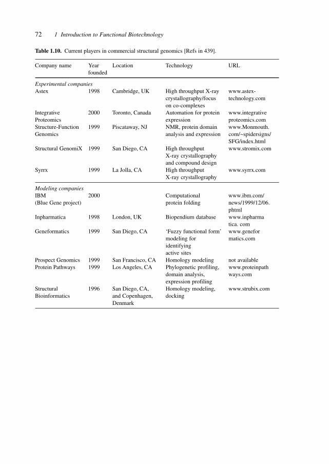
72 1 Introduction to Functional Biotechnology
Table 1.10. Current players in commercial structural genomics [Refs in 439].
Company name Year Location Technology URLfounded
Experimental companiesAstex 1998 Cambridge, UK High throughput X-ray www.astex-
crystallography/focus technology.comon co-complexes
Integrative 2000 Toronto, Canada Automation for protein www.integrativeProteomics expression proteomics.comStructure-Function 1999 Piscataway, NJ NMR, protein domain www.Monmouth.Genomics analysis and expression com/~spidersigns/
SFG/index.htmlStructural GenomiX 1999 San Diego, CA High throughput www.stromix.com
X-ray crystallographyand compound design
Syrrx 1999 La Jolla, CA High throughput www.syrrx.comX-ray crystallography
Modeling companiesIBM 2000 Computational www.ibm.com/(Blue Gene project) protein folding news/1999/12/06.
phtmlInpharmatica 1998 London, UK Biopendium database www.inpharma
tica. comGeneformatics 1999 San Diego, CA ‘Fuzzy functional form’ www.genefor
modeling for matics.comidentifyingactive sites
Prospect Genomics 1999 San Francisco, CA Homology modeling not availableProtein Pathways 1999 Los Angeles, CA Phylogenetic profiling, www.proteinpath
domain analysis, ways.comexpression profiling
Structural 1996 San Diego, CA, Homology modeling, www.strubix.comBioinformatics and Copenhagen, docking
Denmark
73
2 Organizational Structures
2.1 Virtual and Real Enterprises
The realm of healthcare covers prediction, prevention, treatment and follow-up; the phar-maceutical areas involved include diagnostics, pharmaceuticals, therapies and medical de-vices. The recognition of improved benefits for patients by combining several fields ofhealthcare services together, i.e. the ‘healthcare service provider’, leads to the formation ofintegrated organizations. Such integrated organizations aim to provide (usually focused ona therapy or disease area) diagnostics, treatment and follow-up.
The necessity to deliver state-of-the-art comprehensive care requires either the forma-tion of large vertically integrated health providers, e.g., in the dialysis field, or the creationof a federation of complementary companies (respectively, organizations) which togethercommand the range of competencies required.
The formation of such a federation of companies by contractual means can be describedas a virtual enterprise, i.e. an enterprise formed temporarily (although possibly for a longterm). Such a virtual enterprise consists of real components and real enterprises, which arecombined by contractual, organizational and technological means. It has a high flexibilityto adapt to changing circumstances and technology or market conditions, e.g., new thera-pies developed elsewhere may easily be integrated or component companies may be ex-changed for better performers.
The possibility of ‘telemedicine’, i.e. the provision of diagnosis and therapy at a dis-tance by use of telecommunication systems, and the access to databases for medical knowl-edge plus the existence of, for example, automated analytical devices and robot surgeons(obviating the physical presence of the surgeon in the operating theater) will facilitate theprovision of the best expertise anywhere, anytime.
In recent years (decades), the pharmaceutical industry has been (and still is) changingdramatically, primarily due to the developments in molecular biology and the surroundingdisciplines, grouped together as biotechnology. Observers have arranged the pharmaceuti-cal and biotechnology companies into categories such as FIPCOs (fully integrated pharma-ceutical companies), which span the business from research (discovery) to market. Otherswere categorized as VIPCOs (virtually integrated pharmaceutical companies) which spe-cialize in one area such as drug discovery and outsource all other activities. There areplatform technology companies evolving around a particular technology or data stream,and integrated platform technology companies which have assembled several hopefullysynergistic technologies. In the future, the main characterizing function between a pharma-ceutical or biotechnological company and the other participants in that industry, the CROs(contract research organizations), is having R & D as a main function.[149]
Industrial Pharmaceutical Biotechnology. Heinrich KlefenzCopyright © 2002 Wiley-VCH Verlag GmbH
ISBNs: 3-527-29995-5 (Hardcover); 3-527-60012-4 (Electronic)
74 2 Organizational Structures
Mastering the technologies is the task of the organizations having the core competenceof drug discovery and development as well as marketing.
It is crucial to start with the right approach, i.e. by defining the objectives to be achievedand deriving which technologies, resources, alliances, outsourcing, and financial means arerequired to achieve this objective. In the course of implementing the business strategy inthe technology-determined modes, it is necessary to weigh the risks and benefits in a quan-titative or semi-quantitative way.[150]
The complexity of technology management highlights the need for technology acquisi-tion professionals. Enabling technologies fuel the innovation engine, and generate oppor-tunities and information that improve decision making. Choosing the appropriate technol-ogy for pharmaceutical R & D demands full-time, professional attention. Sophisticatedorganizations accept this tenet and employ dedicated technology managers. Although mostorganizations recognize the importance of the aforementioned management practices, feworganizations employ all of them. The pharmaceutical industry lies within a complex mazeof social, economic, political and technical forces. Successfully managing this complexitymaze demands technology leadership.[151]
2.2 R & D Networks
The expanding knowledge base and the increasing multitude of sophisticated technologiesas well as the cost of acquiring and maintaining technological expertise and human re-sources are the major reasons for companies to enter into alliances with specialized part-ners, thus forming R & D networks.
The chain of required activities and competencies which generates a therapy, a pharma-ceutical, a diagnostic procedure, a medical device or an implantate or an organ ranges frombasic or fundamental research via discovery to (preclinical and clinical) development on tomanufacturing, marketing and sales. The discovery portion offers a high reward and lever-age in delivering new and better treatments to known diseases, treatments for previouslyintractable illnesses, and possibilities for diagnostics and prevention or remediation other-wise unattainable.
Over the last approximately 15 years, the biotechnology industry has delivered, togetherwith bio-oriented academia, the major biopharmaceutical discoveries and the technologieswhich shape the discovery process.[461]
This shift in the source areas for discovery and technologies away from the ‘big pharma’corporations (integrated global pharmaceutical companies) necessitates the creation of R & Dnetworks and alliances to capture those innovations and bring them through the develop-ment phase to the patient and market.[462]
Table 2.1 illustrates the number of transactions in pharmaceutical deal making betweenestablished pharmaceutical companies and biotech corporations plus the cumulative dealvalues.[462] The distribution of biotech corporations focus between disease research, drugdelivery and technologies is shown in Table 2.2 (based on revenue), whereas Table 2.3depicts the distribution between the various core technologies.
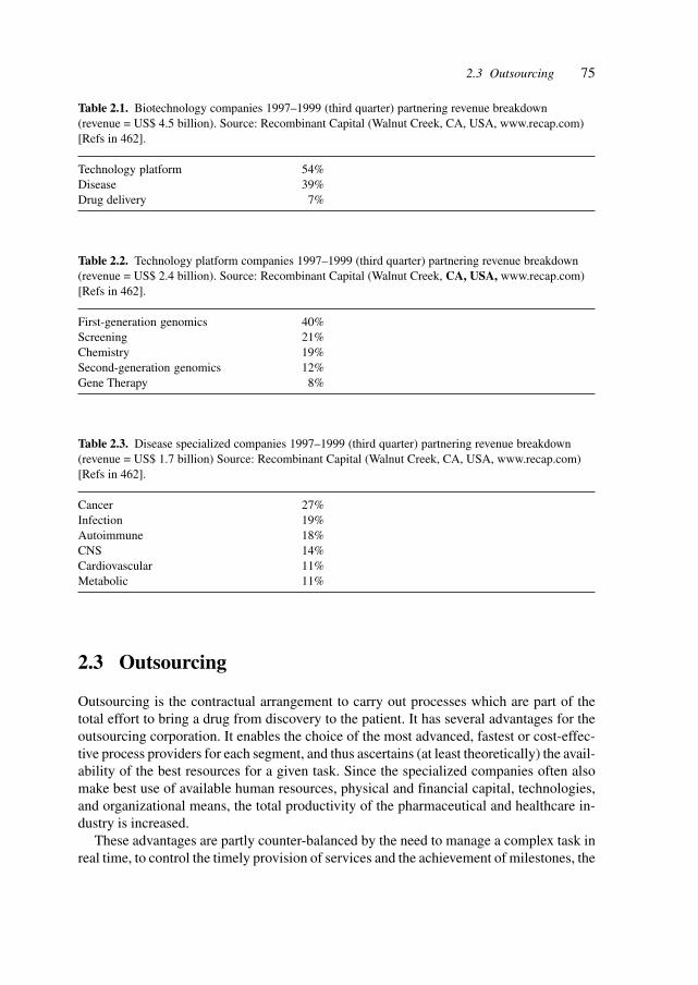
752.3 Outsourcing
2.3 Outsourcing
Outsourcing is the contractual arrangement to carry out processes which are part of thetotal effort to bring a drug from discovery to the patient. It has several advantages for theoutsourcing corporation. It enables the choice of the most advanced, fastest or cost-effec-tive process providers for each segment, and thus ascertains (at least theoretically) the avail-ability of the best resources for a given task. Since the specialized companies often alsomake best use of available human resources, physical and financial capital, technologies,and organizational means, the total productivity of the pharmaceutical and healthcare in-dustry is increased.
These advantages are partly counter-balanced by the need to manage a complex task inreal time, to control the timely provision of services and the achievement of milestones, the
Table 2.1. Biotechnology companies 1997–1999 (third quarter) partnering revenue breakdown(revenue = US$ 4.5 billion). Source: Recombinant Capital (Walnut Creek, CA, USA, www.recap.com)[Refs in 462].
Technology platform 54%Disease 39%Drug delivery 7%
Table 2.2. Technology platform companies 1997–1999 (third quarter) partnering revenue breakdown(revenue = US$ 2.4 billion). Source: Recombinant Capital (Walnut Creek, CA, USA, www.recap.com)[Refs in 462].
First-generation genomics 40%Screening 21%Chemistry 19%Second-generation genomics 12%Gene Therapy 8%
Table 2.3. Disease specialized companies 1997–1999 (third quarter) partnering revenue breakdown(revenue = US$ 1.7 billion) Source: Recombinant Capital (Walnut Creek, CA, USA, www.recap.com)[Refs in 462].
Cancer 27%Infection 19%Autoimmune 18%CNS 14%Cardiovascular 11%Metabolic 11%
76 2 Organizational Structures
partly simultaneous execution of subprocesses, and quality control of the services, prod-ucts, devices and results.
Various segments of the total effort to discover, develop, test, produce and market activeprinciples, medical devices and therapies are outsourced to specialized companies or otherorganizations, which cover a certain part of the discovery-to-patient chain. So-called con-tract research organizations (CROs) are used for toxicity testing, clinical studies and galenics.Even drug discovery is outsourced, as seen by the huge number of biotechnology compa-nies, which have drug discovery as their core competence. Marketing may be outsourced tospecialized marketing organizations or ‘big pharma’ companies with huge numbers of salesand marketing staff.
The concept of ‘virtual pharma’ carries the notion to the (present) extreme, where allactivities are outsourced except strategy and financing.[152]
The cost of calculating the discovery of a new drug is yielding astonishing figures, sinceeach successful drug has to cover the cost burden of the failures. Approximately 50 noveldrugs enter the market each year at a current combined cost of approximately US$ 60billion in R & D, resulting in a cost of approximately US$ 1.2 billion per drug. The timerequirement for development to bring the drug to market at present is on average in excessof 12 years.
These unsustainable figures require increases at every stage of drug discovery, drugdevelopment, clinical trials and marketing. Thus, strategic outsourcing is potentially a cost-effective solution for accelerating drug discovery.
Pharmaceutical research organizations can benefit from outsourcing discovery activitiesthat are not core competencies of the organization. The core competencies for a discoveryoperation are the expertise and systems that give the organization an advantage over itscompetitors. Successful outsourcing ventures result in cost reduction, increased operationefficiency and optimization of resource allocation. While there are pitfalls to outsourcing,including poor partner selection and inadequate implementation, outsourcing can be a pow-erful tool for enhancing drug discovery operations.[153]
2.4 Registrations/Permissions
The well-developed ways of validating R & D, preclinical studies and clinical studies of‘active pharmaceutical ingredients’ (APIs) are applied with the appropriate modificationsto the regulatory processes for small molecules, therapeutic proteins, proteins analogs, vec-tors for delivery and gene therapy, synthetic lipids, and other compounds derived frommolecular biology.
Special consideration is given to the effects on the increasingly better understood bio-logical, biochemical and genetic networks as well as the chemical and biotechnologicalsystems of manufacture, recovery and formulation.
The different glycosylation patterns (depending on the eukaryotic cell chosen for pro-ducing therapeutic or analytical proteins or protein analogs) are an example of the influ-ence of the manufacturing system on the structure of the product.
77
3 Markets and Factors
The demand for pharmaceutical intervention to achieve freedom from disease and the aimof delaying or preventing the onset of degenerative diseases are shaping the targets of drugdiscovery. Additionally, the patterns of diseases and the demands for therapies are influ-enced by the changing demographic composition of the population and the increasing af-fluence of the economically developing areas of the world.
The increasing prevalence (with age) of different cancers is prompting several pharma-ceutical companies to step up their R & D efforts in pertinent fields, such as the profiling ofdifferent tumor cells and the development of specific antibodies.
The competitive outcome of drug discovery is influenced by the size of the R & D bud-get (which is a major factor driving mergers in the pharmaceutical field) as well as by theavailability of human resources, the network of collaborative institutions and biotech com-panies, and the supportive regulatory environment (such as speedy registration processesand orphan drug regulation).
3.1 Products and Services
The pharmaceutical industry has long relied on the outsourcing of parts of the developmentand manufacturing process, e.g., clinical trials and contract manufacture. The increasingdiversity and multitude of competencies and technologies required have already led to theincreased outsourcing of functions like marketing, development, registration, manufactureand, most remarkably, the drug discovery process itself.
A vast industry, including CROs, fine chemicals manufacturers, biotechnology compa-nies, equipment suppliers, human resource providers, software developers and contractmanufacturers is providing an increasing variety and increasing share of the pharmaceuti-cal industry’s activities.
3.2 Economies
The increasing body of knowledge and the resulting requirements for generating, handling,managing and evaluating data plus the increasing need to take into account the effects ofAPIs and therapies on different parts of a biological system have increased the cost of drugdiscovery and drug development.
Industrial Pharmaceutical Biotechnology. Heinrich KlefenzCopyright © 2002 Wiley-VCH Verlag GmbH
ISBNs: 3-527-29995-5 (Hardcover); 3-527-60012-4 (Electronic)
78 3 Markets and Factors
3.3 Manpower
The rapidly expanding requirements of the pharmaceutical industry, and the broadeningareas of expertise contributing to the processes of R & D, development, manufacturing,marketing, validation, surveillance, and in particular its innovative potential are increasingthe demand for skilled manpower.
The educational systems of different countries are providing a crucial competitive ad-vantage (or disadvantage) for essentially R & D-based industries like the pharmaceuticaland the biotechnological industries.
3.4 Resources
The range of resources required for the pharmaceutical industry has expanded from itstraditional areas (chemists, physicians, pharmacists, epidemiologists, etc.) to includebiotechnologists, bioinformatics specialists, software developers, chip designers, and mi-cro- and nanotechnology experts.
The classical resources such as industrial enterprises to organize the R & D, trials, anddevelopment, manufacturing, and marketing are still necessary, but the providers of theseresources are becoming increasingly fragmented.
Another crucial resource is the financial base upon which to develop the API or therapy.In this field, too, the changing structures of markets, financial markets, and the industriallandscape have opened additional sources for drug discovery and development, such asdirect access to capital markets and alliances with larger companies.
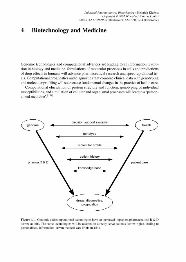
79
4 Biotechnology and Medicine
Genomic technologies and computational advances are leading to an information revolu-tion in biology and medicine. Simulations of molecular processes in cells and predictionsof drug effects in humans will advance pharmaceutical research and speed-up clinical tri-als. Computational prognostics and diagnostics that combine clinical data with genotypingand molecular profiling will soon cause fundamental changes in the practice of health care.
Computational elucidation of protein structure and function, genotyping of individualsusceptibilities, and simulation of cellular and organismal processes will lead to a ‘person-alized medicine’.[154]
Figure 4.1. Genomic and computational technologies have an increased impact on pharmaceutical R & D(arrow at left). The same technologies will be adapted to directly serve patients (arrow right), leading topersonalized, information-driven medical care [Refs in 154].
genome health
drugs, diagnostics,
prognostics
decision support systems
genotype
molecular profile
patient history
knowledge base
patient carepharma R & D
Industrial Pharmaceutical Biotechnology. Heinrich KlefenzCopyright © 2002 Wiley-VCH Verlag GmbH
ISBNs: 3-527-29995-5 (Hardcover); 3-527-60012-4 (Electronic)
80 4 Biotechnology and Medicine
4.1 Diagnostics
The expanding availability of information on genetic sequences, polymorphisms, SNPs,protein variations and interactions, metabolic and phenotypical variants as well as the sen-sor technologies plus micro- and nanotechnologies in combination with the communica-tion network spanning the globe will allow increasingly precise measurements of the:
• Susceptibilities for disease• Strengths of genetic and metabolic networks• Status of vital organs and tissues• Regimen of pharmaceuticals and therapies• Developments in surgery and implantation
Diagnostics is an essential tool for providing basic data for medical therapies, and biologi-cal and industrial processes, e.g., process control, and food and environmental monitoring.One of the first biomedical applications is the measurement of blood glucose in diabetics.Due to the expanding numbers of diabetics (projected to increase from 175 million personsin 2000 to 239 million afflicted in 2010 according to World Health Organization data), thisis a large and still growing diagnostic market segment.
The sensitivity and versatility of diagnostic devices is increasing due to refinements inestablished technologies such as enzymatic assays and enzyme-linked immunoassays, theongoing expansion of the analytical repertoire to include DNA and RNA diagnostics suchas PCR, and physico-chemical analytics such as MALDI-TOF, affinity sensors and tech-niques such as surface plasmon resonance (SPR). Neural network software is being appliedto assess complex mixtures such as in olfactory analyses. The technique of molecular im-printing is enabling the design of molecule-specific assays, separations and sensors.[501]
Table 4.1 lists typical examples of molecular imprinting in design and applications fromamino acids, pharmaceuticals, herbicides, and chemicals to proteins, steroids and alka-loids.
4.2 Therapeutics
Immunotherapy is the focus of significant efforts in the development of cancer therapies.Several approaches have tried to create tumor cell vaccines for cancer treatment. An effec-tive cancer cell vaccine is created by expressing MHC class II molecules without the invari-ant chain protein (Ii) that normally blocks the antigenic peptide-binding site of MHC classII molecules at their synthesis in the endoplasmic reticulum. Such cell–cell constructs arecreated either by the transfer of genes for MHC class II α and β chains or by the inductionof MHC class II molecules and Ii protein with a transacting factor, followed by Ii suppres-sion using antisense methods. Preclinical validation of this approach has been reviewedwith the goal of using this immunotherapy for metastatic human cancers.[155]
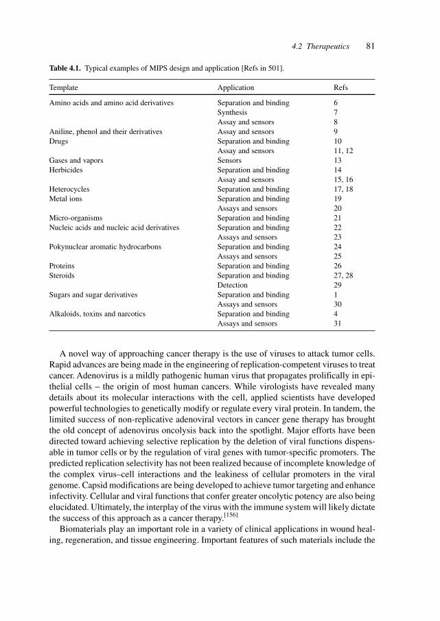
814.2 Therapeutics
A novel way of approaching cancer therapy is the use of viruses to attack tumor cells.Rapid advances are being made in the engineering of replication-competent viruses to treatcancer. Adenovirus is a mildly pathogenic human virus that propagates prolifically in epi-thelial cells – the origin of most human cancers. While virologists have revealed manydetails about its molecular interactions with the cell, applied scientists have developedpowerful technologies to genetically modify or regulate every viral protein. In tandem, thelimited success of non-replicative adenoviral vectors in cancer gene therapy has broughtthe old concept of adenovirus oncolysis back into the spotlight. Major efforts have beendirected toward achieving selective replication by the deletion of viral functions dispens-able in tumor cells or by the regulation of viral genes with tumor-specific promoters. Thepredicted replication selectivity has not been realized because of incomplete knowledge ofthe complex virus–cell interactions and the leakiness of cellular promoters in the viralgenome. Capsid modifications are being developed to achieve tumor targeting and enhanceinfectivity. Cellular and viral functions that confer greater oncolytic potency are also beingelucidated. Ultimately, the interplay of the virus with the immune system will likely dictatethe success of this approach as a cancer therapy.[156]
Biomaterials play an important role in a variety of clinical applications in wound heal-ing, regeneration, and tissue engineering. Important features of such materials include the
Table 4.1. Typical examples of MIPS design and application [Refs in 501].
Template Application Refs
Amino acids and amino acid derivatives Separation and binding 6Synthesis 7Assay and sensors 8
Aniline, phenol and their derivatives Assay and sensors 9Drugs Separation and binding 10
Assay and sensors 11, 12Gases and vapors Sensors 13Herbicides Separation and binding 14
Assay and sensors 15, 16Heterocycles Separation and binding 17, 18Metal ions Separation and binding 19
Assays and sensors 20Micro-organisms Separation and binding 21Nucleic acids and nucleic acid derivatives Separation and binding 22
Assays and sensors 23Pokynuclear aromatic hydrocarbons Separation and binding 24
Assays and sensors 25Proteins Separation and binding 26Steroids Separation and binding 27, 28
Detection 29Sugars and sugar derivatives Separation and binding 1
Assays and sensors 30Alkaloids, toxins and narcotics Separation and binding 4
Assays and sensors 31
82 4 Biotechnology and Medicine
potential to be remodeled and replaced by the proteolytic activity associated with cellsduring migration and invasion, as well as the ability to display a variety of adhesiveligands that directly bind to cell-surface receptors to provide adhesive and morphogeneticclues.
Fibrin plays an important role in wound healing and regeneration, and enjoys wide-spread use in surgery and tissue engineering. The enzymatic activity of Factor XIIIa wasemployed to covalently incorporate exogenous bioactive peptides within fibrin during co-agulation. Fibrin gels were formed with incorporated peptides from laminin and N-cadherinalone and in combination at concentrations up to 8.2 mol peptide/mol fibrinogen. Neuriteextension in vitro was enhanced when gels were augmented with exogenous peptide, withthe maximal improvement reaching 75%. When this particular fibrin derivative was evalu-ated in rats in the repair of the severed dorsal root within polymeric tubes, the number ofregenerated axons was enhanced by 85% relative to animals treated with tubes filled withunmodified fibrin. These results demonstrate that it is possible to enhance the biologicalactivity of fibrin by enzymatically incorporating exogenous oligopeptide domains ofmorphoregulatory proteins.[157]
4.3 Gene Therapy
Gene therapy promises to revolutionize medicine by treating the (genetic) causes of dis-eases rather than the symptoms. This radical improvement is possible because the gene-based approach can provide superior targeting and prolonged duration of action. Moreover,gene therapy is a platform technology applicable to a wide range of diseases. Ongoingclinical studies are addressing a wide range of diseases and target cells, such as cardiovas-cular diseases, inherited monogenic disorders, rheumatoid arthritis (RA), cancer and cubaltunnel syndrome. The vectors utilized include adeno-associated virus (AAV), pox viruses,Herpes simplex virus (HSV), naked DNA, naked RNA, adenoviruses and retroviruses.
The most promising gene-therapy concepts concern the direct killing of tumor cells withgenes delivered by adenovirus vectors, delivery of naked DNA for preventive vaccinationagainst infectious diseases, naked DNA delivery of genes promoting angiogenesis for car-diovascular disorders, and AAV delivery for chronic disorders such as hemophilia and ane-mia. The efficiency in clinical studies has been clearly demonstrated.[158]
The genomic technologies will provide an increasing range of genes and informationabout their causal relationship with diseases and the genetic susceptibility for diseases,rendering many more amenable to gene therapy.
Gene therapy may be segmented into somatic gene therapy and germline therapy. So-matic gene therapy promises to be more successful when targeting selected cell popula-tions rather than total changes.
Examples of cell populations are osteoblasts, which undergo apoptosis and are depletedwith increasing age. A directed change in that program will prevent osteoporosis by reduc-ing bone loss and increasing bone mass. The classical approach, i.e. application of parathy-roid hormone (PTH), will be superseded by gene therapy.
83
Pancreas failure, Alzheimer’s disease and Parkinson’s disease are urgent targets for phar-maceutical intervention, where tissue engineering and gene therapy offer the best solu-tions.
An intriguing example of gene therapy in cardiovascular diseases is the combined appli-cation of angiopoietin-1 (Ang-1) and vascular endothelial growth factor (VEGF), both ofwhich are endothelial cell-specific growth factors. Direct comparison of transgenic miceoverexpressing these factors in the skin revealed that VEGF-induced blood vessels wereleaky, whereas those induced by Ang-1 were non-leaky. Vessels in Ang-1-overexpressingmice were resistant to leaks caused by inflammatory agents. Co-expression of Ang-1 andVEGF had an additive effect on angiogenesis but resulted in leakage-resistant vessels typi-cal of Ang-1. Thus, Ang-1 may be useful in reducing microvascular leakage in diseases inwhich the leakage results from chronic inflammation or elevated VEGF and in combina-tion with VEGF for promoting the growth of non-leaky vessels. Since plasma leakage is akey pathophysiological feature of various diseases, such as chronic inflammatory, and de-generative and neoplastic diseases, inhibition of the leakage could have important thera-peutic benefits. Ang-1 has the potential to reduce plasma leakage in such conditions, whetherthe leak is due to inflammatory mediators or VEGF.[159]
Gene therapy requires the introduced genes to act in concert with the existing genomicenvironment without upsetting the genetic, transcriptional and metabolic balances and net-works. Regulated hormone expression has been demonstrated in pigs. Ectopic expressionof a new serum protease-resistant porcine growth hormone-releasing hormone, directed byan injectable muscle-specific synthetic promoter plasmid vector (pSP-HV-HGRH), elicitsgrowth in pigs. A single muscular injection of 10 mg followed by electroporation in 3-week-old piglets elevated serum GHRH levels by 2- to 4-fold, enhanced growth hormonesecretion and increased serum insulin-like growth factor I (IGF-I) by 3- to 6-fold overcontrol pigs. Evaluation of body composition indicated a uniform increase in mass, with noorganomegaly or associated pathology.[160]
In utero fetal gene transfer could provide an alternative to transgenic approaches forfunctional analysis of stage- and tissue-specific, cis-acting DNA regulatory elements. Stage-specific in utero gene transfer and expression might also prove useful for conditional res-cue of lethal targeted genes, either in utero or in the immediate postnatal period. Moreover,studies of fetal gene transfer may help in developing strategies to ameliorate or correcthuman genetic deficiencies in utero.
In utero injection of cationic liposome–DNA complexes (CLDCs) containing chloram-phenicol acetyltransferase, β-galactosidase (β-gal) or human granulocyte colony-stimulat-ing factor (hG-CSF) expression plasmids produced high-level gene expression in fetal rats.Tissues adjacent to the injection site exhibited the highest levels of gene expression. Chloram-phenicol acetyltransferase expression persisted for at least 14 days and was re-expressedfollowing postnatal re-injection of CLDCs. Intraperitoneal administration of the hG-CSFgene produced high serum hG-CSF levels. X-gal staining demonstrated widespread β-galexpression in multiple fetal tissues and cell types. No toxic or inflammatory responseswere observed nor was there evidence of fetal–maternal or maternal–fetal gene transfer,suggesting that CLDCs may provide a useful alternative to viral vectors for in utero genetransfer.[161]
4.3 Gene Therapy

84 4 Biotechnology and Medicine
Gene therapy of the brain is hindered by the presence of the blood–brain barrier (BBB),which prevents the brain uptake of blood-borne gene formulations. Exogenous genes havebeen expressed in the brain after invasive routes of administration, such as craniotomy orintra-carotid arterial infusion of noxious agents causing BBB disruption. Studies were pre-sented describing the expression of an exogenous gene in brain after non-invasive i.v. ad-ministration of a 6–7 kb expression plasmid encoding either luciferase or β-gal packaged inthe interior of neutral pegylated immunoliposomes. The latter are conjugated with the OX26mAb to the rat transferrin receptor, which enables targeting of the plasmid DNA to thebrain via the endogenous BBB transferrin receptor. Unlike cationic liposomes, this neutralliposome formulation is stable in blood and does not result in selective entrapment in thelung. Luciferase gene expression in the brain peaks at 48 h after a single i.v. administrationof 10 µg of plasmid DNA per adult rat, a dose that is 30- to 100-fold lower than that usedfor gene expression in rodents with cationic liposomes. β-Gal histochemistry demonstratedgene expression throughout the CNS, including neurons, choroid plexus epithelium andthe brain microvasculature. Widespread gene expression in the brain can be achieved byusing a formulation that does not employ viruses or cationic liposomes, but instead usesendogenous receptor-mediated transport pathways at the BBB.[162]
Lysosomal storage disorders (LSDs) constitute an important group of conditions in whichthe potential of gene manipulation as therapy can be assessed. They are monogenic defects,often with severe manifestations for which there are limited treatment options. Over-expression of the lysosomal hydrolase by gene-corrected cells results in secretion of someof the enzyme and its uptake by uncorrected bystander cells (metabolic cooperativity).Gene therapy strategies, enzyme therapy and bone marrow transplantation have all beendescribed.
Fabry disease is a compelling target for gene therapy as a treatment strategy. A defi-ciency in the lysosomal hydrolase α-galactosidase A (α-gal A; EC 3.2.1.22) leads to im-paired catabolism of α-galactosyl-terminal lipids such as globotriaosylceramide (b3). Pa-tients develop vascular occlusions that cause cardiovascular, cerebrovascular and renaldisease. Unlike for some lysosomal storage disorders, there is limited primary nervoussystem involvement in Fabry disease. The enzyme defect can be corrected by gene transfer.Overexpression of α-gal A by transduced cells results in secretion of this enzyme. Secretedenzyme is available for uptake by non-transduced cells, presumably by receptor-mediatedendocytosis. Correction of bystander cells may occur locally or systemically after circula-tion of the enzyme in the blood. A long-term genetic correction in an α-gal A-deficientmouse model of Fabry disease was reported. α-Gal A-deficient bone marrow mononuclearcells (BMMCs) were transduced with a retrovirus encoding α-gal A, and transplanted intosublethally and lethally irradiated α-gal A-deficient mice. α-gal A activity and Gb3 levelswere analyzed in plasma, peripheral blood mononuclear cells, BMMCs, liver, spleen, heart,lung, kidney and brain. Primary recipient animals were followed for up to 26 weeks. BMMCswere then transplanted into secondary recipients. Increased α-gal A activity and decreasedGb3 storage were observed in all recipient groups in all organs and tissues except the brain.These effects occurred even with a low percentage of transduced cells. Genetic correctionof bone marrow cells derived from patients with Fabry disease may have utility for pheno-typic correction of patients with this disorder.[163]
85
In vivo gene therapy may be a method of choice for compounds that are difficult toproduce or to deliver, such as endostatin. Tumors require ongoing angiogenesis to supporttheir growth. Inhibition of angiogenesis by production of angiostatic factors should be aviable approach for cancer gene therapy. Endostatin, a potent angiostatic factor, was ex-pressed in mouse muscle and secreted into the bloodstream for up to 2 weeks after a singleintramuscular administration of the endostatin gene. The biological activity of the expressedendostatin was demonstrated by its ability to inhibit systemic angiogenesis. Moreover, thesustained production of endostatin by intramuscular gene therapy inhibited both the growthof primary tumors and the development of metastatic lesions. These results demonstrate thepotential utility of intramuscular delivery of an antiangiogenic gene for the treatment ofdisseminated cancers.[164]
Gene therapy requires appropriate vectors with the necessary regulatory and codingregions for integration, regulation and synthesis of required genes (respectively, proteins).Artificial chromosomes are DNA molecules of defined structure, which are assembled invitro from defined constituents that behave with the properties of natural chromosomes.Artificial chromosomes were first assembled in budding yeasts and have proved useful inmany aspects of yeast genetics. Several attempts have been made to build artificial chromo-somes in mammals. Particularly, mini-chromosomes of defined structure have been devel-oped to address questions regarding mammalian chromosome function and for biotechno-logical applications.[165]
Major targets for gene therapy are the diseases involving mutated globins, such as β-thalassemia and sickle-cell disease. The stable introduction of a functional β-globin gene inHSC could be a powerful approach to treat β-thalassemia and sickle-cell disease. Geneticapproaches aiming to increase normal β-globin expression in the progeny of autologousHSC might circumvent the limitations and risks of allergenic cell transplants. Low-levelexpression, position effects and transcriptional silencing hampered the effectiveness of vi-ral transduction of the human β-globin gene when it was linked to minimal regulatorysequences. It was shown that the use of a recombinant lentivirus enables efficient transferand faithful integration of the human β-globin gene together with large segments of itslocus control region. In long-term recipients of unselected transduced bone marrow cells,tetramers of two murine α-globin and two human β(A)-globin molecules account for up to13% of total hemoglobin in mature red cells of normal mice. In β-thalassemic heterozy-gous mice higher percentages are obtained (17–24%), which are sufficient to ameliorateanemia and red cell morphology. Such levels should be of therapeutic benefit in patientswith severe defects in hemoglobin production.[166]
Gene silencing is defined as a state of complete transcriptional repression that is epige-netically inherited through subsequent cell divisions. This phenomenon has great impacton gene therapy, as transferred genes may become silenced to various degrees, dependingon the presence of cis-acting elements opposing the forces of silencing (e.g., enhancers,locus control regions, matrix attachment sites and insulators), the sites of chromosomalintegration, and the state of differentiation the cells initially transduced and of their subse-quent progeny. During development, a widespread wave of global silencing takes place atthe time of implantation. Subsequently, progressive tissue-specific gene activation occurs.In pre-implantation embryos, ES cells and HSC, transferred genes are exposed to a high
4.3 Gene Therapy
86 4 Biotechnology and Medicine
frequency of gene silencing. Irreversible HSC gene silencing remains one of the main ob-stacles to the gene therapy of hematological disorders.
Transcriptional silencing of genes transferred into HSC thus poses one of the most sig-nificant challenges to the success of gene therapy. If the transferred gene is not completelysilenced, a progressive decline in gene expression as the mice age often is encountered.These phenomena were observed to various degrees in mouse transplant experiments usingretroviral vectors containing a human β-globin gene, even when cis-linked to locus controlregion derivatives. Ex vivo preselection of retrovirally transduced stem cells was investi-gated on the basis of expression of the GFP driven by the CpG island phosphoglyceratekinase promoter being able to ensure subsequent long-term expression of a cis-linked β-globin gene in the erythroid lineage of transplanted mice. All mice (n = 7) engrafted withpre-selected cells concurrently expressed human β-globin and the GFP in 20–95% of theirred blood cells (RBCs) for up to 9.5 months post-transplantation, the longest time pointassessed. This expression pattern was successfully transferred to secondary transplant re-cipients. In the presence of β-locus control region hypersensitive site 2 alone, human β-globin mRNA expression levels ranged from 0.15 to 20% with human β-globin chainsdetected by HPLC. Neither the proportion of positive blood cells nor the average expres-sion levels declined with time in transplanted recipients. Although suboptimal expressionlevels and heterocellular position effects persisted, in vivo stem cell gene silencing and age-dependent extinction of expression were avoided, thus demonstrating the potential of thisvector for the gene therapy of human hemoglobinopathies.[167]
Most gene therapy strategies involving HSC require both a high level of gene transferand persistent transgene expression in specific target lineages. Gene transfer rates of ap-proximately 10% in reconstituting HSC can now be routinely achieved with virus vectorsbased on murine leukemia virus and related oncoretroviruses. Achieving persistent, uni-form gene expression from murine leukemia virus-based vectors remains a challenge. Fo-cus has been on the definition of elements of the virus long terminal repeat (LTR) that areresponsible for provirus silencing in vivo, and the identification of appropriate promotersand enhancers. Expression of integrated provirus is also affected by chromatin structure.Since the bulk of the mammalian genome is packaged into transcriptionally silent hetero-chromatin and murine leukemia virus-based vectors insert at random sites in the genome, alarge portion of murine leukemia virus insertions result in gene silencing. The progeny of asingle clone containing a unique integration event can also be affected by the surroundingchromatin to varying degrees, a phenomenon known as position effect variegation.
The mammalian chromosome is organized into discrete chromosomal domains, in partthrough the use of sequences termed chromatin insulators. These elements, first describedin Drosophila and also in several vertebrate species, help define the boundary betweendifferentially regulated loci and serve to shield promoters from the influence of neighbor-ing regulatory elements. Insulators function in a polar manner (e.g., they must be locatedbetween the cis effectors and promoter) and do not have stimulatory or inhibitory transcrip-tional effects on their own, distinguishing them from classical enhancers and silencers. Thebest characterized vertebrate chromatin insulator is located within the chicken β-globinlocus control region, and contains a DNase I hypersensitive site (cHS4) and appears toconstitute the 5′ boundary of the chicken β-globin locus. A 1.2-kb fragment containing the
87
cHS4 element displays classic insulator activities, including the ability to block the interac-tion of globin gene promoters and enhancers in cell lines, and the ability to protect expres-sion cassettes in Drosophila, transformed cell lines and transgenic mammals from positioneffects. Much of this activity is contained in a 250-bp fragment, which comprises a 49-bpcHS4 core that interacts with the zinc finger DNA-binding protein CTCF implicated inenhancer-blocking assays.
Recombinant murine retroviruses are widely used as delivery vectors for gene therapy.Once integrated into a chromosome, these vectors often suffer from profound position ef-fects, with vector silencing observed in vitro and in vivo. To overcome this problem, theHS4 chromatin insulator from the chicken β-globin locus control region was studied withrespect to protection of retrovirus vector from position effects. When used to flank a re-porter vector, this element significantly increased the fraction of transduced cells that ex-pressed the provirus in cultures and in mice transplanted with transduced marrow. A chro-matin insulator can improve the expression performance of a widely used class of genetherapy vectors by protecting these vectors from chromosomal position effects.[168]
A combination of gene therapy and ribozyme drug technology was reported which in-volved the generation of a novel ribozyme (‘maxizyme’) to specifically cleave the Bcr–AblmRNA thus inducing apoptosis in chronic myelogenous leukemia (CML) cells. CML is ahematopoietic malignant disease associated with the expression of a chimeric Bcr–Ablgene. A retroviral system was used to transduce a line of CML cells (BV173) which wereinjected into NOD-SCID mice. These mice are produced by crossing SCID and NOD mice,and are a valuable tool for growing human hematopoietic cells in vivo. As could be ob-served by examining the spleens of injected and control animals, the maxizyme functionssuccessfully in animals by cleaving Bcr–Abl mRNA with high efficiency. Maxizymes couldbe a new class of gene-inactivating agents that can cleave any type of chimeric mRNA.[169]
Mitochondrial DNA (mtDNA) mutations underlie many rare diseases and may contrib-ute to human ageing. Gene therapy is a tempting future possibility for intervening inmitochondriopathies. Expression of the 13 mtDNA-encoded proteins from nuclear transgenes(allotopic expression) might be the most effective gene-therapy strategy. Its only confirmeddifficulty is the extreme hydrophobicity of these proteins, which prevents their import intomitochondria from the cytosol. Inteins (self-splicing ‘protein introns’) might offer a solu-tion to this problem: their insertion into such transgenes could greatly reduce the encodedproteins’ hydrophobicity, enabling import, with post-import excision restoring the naturalamino acid sequence.[170]
Precise targeting of genetic modifications is a prerequisite for safe and successful genetherapy by modification of DNA sequences.
Mobile group II intron RNAs insert directly into DNA target sites and are then reversetranscribed into genomic DNA by the associated intron-encoded protein. Target site recog-nition involves modifiable base-pairing interactions between the intron RNA and a longerthan 14-nucleotide region of the DNA target site, as well as fixed interactions between theprotein and flanking regions. A highly efficient E. coli genetic assay was developed todetermine detailed target site recognition rules for Lactococcus lactis group II intron Ll.LtrBand to select introns that insert into desired target sites. Using HIV-1 proviral DNA and thehuman CCR5 gene as examples, it was shown that group II introns can be retargeted to
4.3 Gene Therapy
88 4 Biotechnology and Medicine
insert efficiently into virtually any target DNA and that the retargeted introns retain activityin human cells. The work provides the practical basis for potential applications of targetedgroup II introns in genetic engineering, functional genomics and gene therapy.[171]
Chimeric RNA–DNA oligonucleotides have formed the basis of genetic strategies forcorrecting mutations, while maintaining genomic organization important for the appropri-ate expression and regulation of genes. Visualization of pigmentation in a live animal andeasy accessibility makes skin an attractive system for in vivo testing of chimeric oligo-nucleotides as novel skin gene therapeutics.
An RNA–DNA oligonucleotide corrected a point mutation in the mouse tyrosinase gene,resulting in permanent and inheritable restoration of tyrosinase enzymatic activity, melaninsynthesis and pigmentation changes in cultured melanocytes. Gene correction was extendedfrom tissue culture to live animals, using a chimeric oligonucleotide designed to correct apoint mutation in the tyrosinase gene. Both topical applications and intradermal injectionof this oligonucleotide to albino BALB/c mouse skin resulted in dark pigmentation of sev-eral hairs in a localized area. The restored tyrosinase enzymatic activity was detected bydihydroxyphenylacetic acid (DOPA) staining of hair follicles in the treated skin. Tyrosi-nase gene correction was also confirmed by restriction fragment length polymorphism(RFLP) analysis, and DNA sequencing from skin that was positive for DOPA staining andmelanin synthesis. Localized gene correction was maintained 3 months after the last appli-cation of the chimeric oligonucleotides. These results demonstrated correcting of the tyro-sinase gene point mutation by chimeric oligonucleotides in vivo.[172]
The suitability of using the tRNA import pathway for the correction of respiratory defi-ciencies in the mitochondrial DNA and the applicability of this system for human therapeu-tic application were investigated. Mitochondrial import of nuclear encoded tRNAs has beendescribed in yeasts, plants and protozoans. The complexity of the imported tRNA poolvaries among organisms, from a complete set required for reading all codons of the mito-chondrial genetic code in trypanosomatids to a single tRNA in the yeast S. cerevisiae.
Mitochondrial import of a cytoplasmic tRNA in yeast requires the preprotein importmachinery and cytosolic factors. Cytoplasmic tRNAs with altered aminoacylation identitycan be specifically targeted to the mitochondria and participate in mitochondrial transla-tion. Human mitochondria, which do not normally import tRNAs, are able to internalizeyeast tRNA derivatives in vitro and this import requires an essential yeast import factor.[173]
Adoptive immunotherapy is developing into a tool for combating diseases like infec-tions and cancers. The infusion of antigen-specific T lymphocytes is a potential therapyagainst certain cancers and infectious diseases. One limitation to its broad usage is thegeneration of autologous T cells directed against well-defined epitopes. The induction andexpansion of antigen-specific cells require optimal antigen presentation and T cell co-stimu-lation. These requirements are met by antigen-presenting cells (APC) such as Epstein–Barrvirus-transformed B cells and dendrite cells (DC), which constitutively express high levelsof co-stimulatory, adhesion and MHC molecules. Despite a cumbersome process, the useof autologous cells to present cell-defined epitopes is mandated to obviate strong allergenicresponses.
The adoptive transfer of antigen-specific cytotoxic T lymphocytes (CTLs) is a promis-ing therapeutic approach for a number of diseases. To overcome the difficulty in generating
89
specific CTLs, stable artificial APCs (AAPCs) were established that can be used to stimu-late T cells of any patient of a given HLA type. Mouse fibroblasts were retrovirally trans-duced with a single HLA–peptide complex along with the human accessory moleculesB7.1, ICAM-1 and LFA-3. These AAPCs consistently elicit strong stimulation and expan-sion of HLA-restricted CTLs. Due to the high efficiency of retrovirus-mediated gene trans-fer, stable AAPCs can be readily engineered for any HLA molecule and any specific pep-tide.[174]
Activation of the immune system in response to challenge by a foreign organism is acomplex process. In the case of T-dependent antigens, naive T cells are activated in re-sponse to novel peptides presented by APCs. To activate T cells the APCs must acquireantigen, and then present the antigen in the context of self-MHC molecules and the appro-priate co-stimulatory signals. Tissue damage and/or cell death may facilitate antigen pre-sentation and may be necessary for activation of the immune system. Following cell death,dying cells transfer their antigens (by ingestion or otherwise) to ‘professional’ APCs, whichthen prime T cells. Antigen from necrotic cells was shown to prime MHC II-restricted Tcell responses in vitro. Antigen transfer and priming have been demonstrated by showingthat bone marrow-derived APCs can acquire viral antigens from apoptotic cells and primeantigen-specific CTLs. Thereby, DCs were observed to engulf intact macrophages as wellas their cellular fragments, suggesting that apoptosis can prime CTLs through DCs.
Immunity to tumors as well as to viral and bacterial pathogens is often mediated byCTLs. Thus, the ability to induce a strong cell-mediated immune response is an importantrequirement of novel immunotherapies. APCs, including DCs, are specialized in initiatingT cell immunity. Harnessing this innate ability of these cells to acquire and present anti-gens, antigen presentation was improved by targeting antigens directly to DCs in vivo throughapoptosis. Fas-mediated apoptotic death of antigen-bearing cells was engineered in vivo byco-expressing the immunogen and Fas in the same cell. The death of antigen-bearing cellsresults in an increased antigen acquisition by APCs including DCs. This in vivo strategy ledto enhanced antigen-specific CTLs, and the elaboration of T helper 1 (Th1)-type cytokinesand chemokines. This adjuvant approach has important implications for viral and non-viraldelivery strategies for vaccines or gene therapies.[175]
The topical delivery of transgenes to hair follicles has potential for treating disorders ofthe skin and hair. The topical administration of liposome–DNA mixtures (lipoplex) to mouseskin and to human skin xenografts resulted in efficient in vivo transfection of hair folliclecells. Transfection depended on liposome composition and occurred only at the onset of anew growing stage of the hair cycle. Manipulating the hair follicle cycle with depilationand retinoic acid treatment resulted in nearly 50% transfection efficiency – defined as theproportion of transfected, newly growing follicles within the xenograft. Transgenes admin-istered in this fashion are selectively expressed in hair progenitor cells and therefore havethe potential to affect the characteristics of the follicle. These findings form a foundationfor the future use of topical lipoplex applications to alter hair follicle phenotype, and treatdiseases of the hair and skin.[176]
Gene therapy for cystic fibrosis (CF) has focused on correcting electrolyte transport inairway epithelia. Success has been limited by the failure of vectors to attach to and enterinto airway epithelia, and may require redirecting vectors to targets on the apical mem-
4.3 Gene Therapy
90 4 Biotechnology and Medicine
brane of airway cells that mediate these functions. The G-protein-coupled P2Y2 receptor(P2Y2-R) is abundantly expressed on the airway lumenal surface and internalizes into coatedpits upon agonist activation. A small-molecule agonist (UTP) was tested whether it coulddirect vectors to P2Y2-R and mediate attachment, internalization, and gene transfer. Fluo-rescein–UTP studies demonstrated that P2Y2-R agonists internalized with their receptorand biotinylated UTP (BUTP) mediated P2Y2-R-specific internalization of fluorescentlylabeled streptavidin (SAF) or SAF conjugated to biotinylated Cy3 adenoviral vector (BCAV).BUTP conjugated to BACV-mediated P2Y2-R-specific gene transfer in (i) adenoviral-re-sistant A9 and polarized MDCK cells by means of heterologous P2Y2-R, and (ii) well-differentiated human airway epithelial cells by means of endogenous P2Y2-R. Targetingvectors with small-molecule ligands to apical membrane GPCRs may be a feasible ap-proach for successful CF gene therapy.[177]
In the canine model of Duchenne muscular dystrophy in golden retrievers (GRMD), apoint mutation within the splice acceptor site of intron 6 leads to deletion of exon 7 fromthe dystrophin mRNA and the consequent frameshift causes early termination of transla-tion. A DNA and RNA chimeric oligonucleotide was designed to induce host cell mis-match repair mechanisms and correct the chromosomal mutation to wild-type. Direct skel-etal muscle injection of the chimeric oligonucleotide into the cranial tibialis compartmentof a 6-week-old affected male dog, and subsequent analysis of biopsy and necropsy samples,demonstrated in vivo repair of the GRMD mutation that was sustained for 48 weeks. RT-PCR analysis of exons 5–10 demonstrated increasing levels of exon 7 inclusion with time.An isolated exon 7-specific dystrophin antibody confirmed synthesis of normal-sizeddystrophin product and positive localization to the sarcolemma. Chromosomal repair inmuscle tissue was confirmed by RFLP-PCR and sequencing the PCR product. This workprovides evidence for the long-term repair of a specific dystrophin point mutation in muscleof a live animal using a chimeric oligonucleotide.[178]
The use of neurotrophic viruses as vectors for targeted gene delivery to the CNS hasmany applications for the development of new therapies for neurological diseases and spi-nal cord trauma. Poliovirus is attractive for the development of such a gene delivery vectorbecause it has been established in humans that once poliovirus invades the CNS, infectionis restricted to the motor neurons of the hindbrain and the spinal cord. To exploit the uniquefeatures of poliovirus tropism, poliovirus genomes (referred to as replicons) were con-structed to encode foreign proteins in place of the capsid proteins. Because replicons do notencode capsid proteins, they undergo only a single round of infection, without spreading toneighboring cells. Since no infectious poliovirus is generated during production of repli-cons, the use of replicons for gene delivery purposes following worldwide poliovirus eradi-cation will not be a concern. Replicons maintain the tropism of poliovirus in the CNS andexclusively infect spinal cord and brainstem motor neurons. Replicons can mediate genedelivery in animals that have previously been immunized with poliovirus, indicating thatpre-existing immunity in humans from vaccination will not be a limitation for the use ofreplicons.
Poliovirus replicon vectors transiently express foreign proteins selectively in motor neu-rons of the anterior horn of the spinal cord. Mice transgenic for the poliovirus receptor(PVR) were intraspinally inoculated with replicons encoding murine tumor necrosis factor
91
(mTNF)-α. High-level expression of mTNF-α was detected in spinal cords of these ani-mals at 8–12 h post-inoculation and this returned to background by 72 h. The mice exhib-ited ataxia and tail atony, whereas animals given a replicon encoding GFP exhibited noneurological symptoms. Histology of spinal cords from mice given the replicon encodingmTNF-α revealed neuronal chromatolysis, reactive astrogliosis, decreased expression ofmyelin basic protein and demyelination. These animals recovered with only slight residualdamage. This showed replicon vectors to have potential for targeted delivery of therapeuticproteins to the CNS and provide a new approach for treatment of spinal cord trauma andneurological disease.[179]
Clinical trials of gene therapy for CF have demonstrated encouraging steady progress,indicating that transfer of the CF transmembrane conductance regulator (CFTR) gene canpartially correct the chloride transport defect in human subjects. Current levels of genetransfer are apparently too low for clinical effectiveness – in large part as a result of thebarriers faced by gene transfer vectors within the airways. Clinical studies of gene therapyfor CF suggest that the key problem is the efficiency of gene transfer to the airway epithe-lium. The availability of relevant vector receptors, the transient contact time between vec-tor and epithelium, and the barrier function of airway mucus contribute significantly to thisproblem. Recombinant Sendai virus (SeV) was developed as a new gene transfer agent.SeV produces efficient transfection throughout the respiratory tract of both mice and fer-rets in vivo, as well as in freshly obtained human nasal epithelial cells in vitro. Gene trans-fer efficiency was several log orders greater than with cationic liposomes or adenovirus.Even very brief contact time was sufficient to produce this effect and levels of expressionwere not significantly reduced by airway mucus. These investigations suggest that SeVmay provide a useful new vector for airway gene transfer.[180]
Retroviral vectors can insert genes stably into the chromosomes of mammalian cells andhave been used in clinical human gene therapy trials to introduce genes into a variety ofcells and tissues. All retroviral vectors currently used in such clinical trials have been de-rived from murine leukemia virus (MLV), a C-type retrovirus, which is considered non-pathogenic for humans. In contrast to lentiviruses, C-type retroviruses have a rather simplegenomic organization and contain only two gene units, which code for the inner core struc-tural proteins and the envelope protein. As safe gene delivery systems, they require only theretroviral vector, a genetically modified viral genome containing the gene of interest inplace of all retroviral protein coding sequences, and a helper cell that provides the retroviralproteins for the encapsidation of the vector genome into retroviral particles. Gene therapyapplications of retroviral vectors derived from C-type retroviruses have been limited tointroducing genes into dividing target cells. Genetically engineered C-type retroviral vec-tors were constructed by derivation from spleen necrosis virus (SNV), which are capable ofinfecting non-dividing cells. This was achieved by introducing a nuclear localization signal(NLS) sequence into the matrix protein (MA) of SNV by site-directed mutagenesis. Thisincreased the efficiency of infecting non-dividing cells, and was sufficient to endow thevirus with the capability to efficiently infect growth-arrested human T lymphocytes andquiescent primary monocyte-derived macrophages. This vector was demonstrated to ac-tively penetrate the nucleus of a target cell and has the potential for a gene therapy vector totransfer genes into non-dividing cells.[181]
4.3 Gene Therapy
92 4 Biotechnology and Medicine
AAV vectors have demonstrated considerable promise for gene therapy of inherited dis-eases. With a packaging size of less than 5 kb, applications have been limited to relativelysmall disease genes. Based on the finding that AAV genomes undergo inter-molecular cir-cular concatemerization after transduction in muscle, a paradigm was developed to increasethe size of delivered transgenes with this vector through trans-splicing between two indepen-dent vectors co-administered to the same tissue. When two vectors encoding either the 5′ or3′ portions of the erythropoietin genomic locus were used, functional erythropoietin proteinwas expressed in muscle subsequent to the formation of intermolecular circular concatemersin a head-to-tail orientation through trans-splicing between these two independent vectorgenomes. This allows the AAV technologies to be applied to a wider variety of diseases forwhich therapeutic transgenes exceed the packaging limitation of prior AAV vectors.[182]
An intriguing use of gene therapy by using genetically engineered cells in cancer therapywas demonstrated by applying encapsulated endostatin-secreting cells for the effective treat-ment of human glioblastoma xenografts[703] in mice and of BT4C glioma cells upon intrac-erebral implantation in rats.[704] The continuous release of endostatin, a sensitive anti-ang-iogenic protein, by implanted bioreactors, demonstrates an effective approach to tumortherapy with cellular and gene therapy.[705] The other extreme of application of gene therapy,oral delivery, is subject to intensive studies due to its advantages with respect to compli-ance, cost, and patient convenience.[706]
4.4 Implantates
Small implants acting as artificial neurons serve to stimulate muscles and thus preventthem from wasting away,[183] and offer the prospect of helping patients with stroke or spi-nal cord injury.
Biodegradable rods to stabilize bone fractures are absorbed once the bone has healed,whereby clearly the timing of resorption is crucial.[184]
An efficient porous chamber system to convert Factor VII to VIIa was developed andimplantation of this chamber, in vivo, was shown to be effective. The chamber containingimmobilized Factor XIIa was able to generate Factor VII-dependent bypass activity for atleast 3 days as a peritoneal implant in guinea pigs and for up to 1 month when tested inrhesus monkeys.
Hemophilia A and B coagulation defects, which are caused by deficiencies of FactorVIII and Factor IX, respectively, can be bypassed by administration of recombinant FactorVIIa. However, the short half-life of recombinant Factor VIIa in vivo negates its routineclinical use. An in vivo method for the continuous generation of Factor VIIa is reportedwhich depends on the implantation of a porous chamber that contains Factor Xa or XIIaand continuously generates Factor VIIa bypass activity from the subject’s own Factor VII,which enters the chamber by diffusion. Once inside, the Factor VII is cleaved to Factor VIIaby the immobilized Factor Xa or XIIa. The newly created Factor VIIa diffuses out of thechamber and back into circulation, where it can bypass the deficient Factors VIII or IX andenable coagulation to occur. In vitro, this method generates sufficient Factor VIIa to sub-
93
stantially correct Factor VIII-deficient plasma when assessed by the classical aPTT coagu-lation assay. In vivo, a Factor XIIa peritoneal implant generates bypass activity for up to 1month when tested in rhesus monkeys. Implantation of such a chamber in a patient withhemophilia A or B could eventually provide a viable alternative to replacement therapiesusing exogenous coagulation factors.[185]
4.5 Medical Devices and Technology
Medical devices include drug-delivery systems, neurological stimulators, cardiovasculardevices such as implantable cardiac defibrillators as well as monitoring devices for bloodglucose and heart function. Intelligent devices like the implantable cardiac defibrillatorsare endowed with sufficient intelligence to analyze the signals from the heart, and apply theright power and type of impulse for correction. Likewise, implantable devices for bloodconstituents are capable of monitoring, for example, glucose levels and blood factors, anddeliver the right amounts of insulin or therapeutic proteins. The market for medical devicesamounts to approximately US$ 160 billion per year and is thus about half the size of themarket for pharmaceuticals at around US$ 350 billion (in the year 2000).
The diagnosis of biological systems with lasers, such as blood disorders, cancer cells,and cellular constituents like proteins, RNA, DNA is becoming a reality with the availabil-ity of biocavity lasers.[477]
The development of MEMS enables the expansion of biomedical applications in chemi-cal and biochemical analysis, and molecular detection and health monitoring. The biologi-cal microcavity laser or biocavity laser is such a system on the nanometer scale. Nanolasershave numerous applications in fiber optic communications, optical computers, and nano-or microscale system sensors, actuators and systems-on-a-chip. The nanolaser can probethe cell and nuclear dimensions of lymphocytes and thus shed light on the status of thehuman immune system or it can diagnose genetic disorders by measuring, for example, redblood cell shapes and hemoglobin content. The semiconductor laser is based on silicon,which is a material suitable for microfabrication, microfluidics and sensors, thus allowingthe integrated design of test arrays on chips and implantable devices. Compound semicon-ductors based on III–V materials (from columns III and V of the periodic table), such asGaAs, are especially suited to the generation, modulation and transmission of light, andthus can combine illumination, data transmission, optical computing and optical analysisplus biomedical applications such as photodynamic therapy, optical tomography, cell mi-cro-manipulation and laser cytometry.
Table 4.2 correlates various materials and (bio-)medical products.
Microsystems in healthcare can generally be divided into• Sensor systems for diagnostics and patient monitoring.• Endoscopes and instrumentation for minimally invasive therapy.• Active implantable devices.• Systems for bioanalytics and pharmaceutical screening.
4.5 Medical Devices and Technology
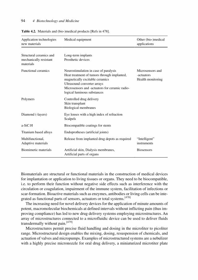
94 4 Biotechnology and Medicine
Biomaterials are structural or functional materials in the construction of medical devicesfor implantation or application to living tissues or organs. They need to be biocompatible,i.e. to perform their function without negative side effects such as interference with thecirculation or coagulation, impairment of the immune system, facilitation of infections orscar-formation. Bioactive materials such as enzymes, antibodies or living cells can be inte-grated as functional parts of sensors, actuators or total systems.[478]
The increasing need for novel delivery devices for the application of minute amounts ofpotent, macromolecular biochemicals at defined intervals without inflicting pain (thus im-proving compliance) has led to new drug delivery systems employing microstructures. Anarray of microstructures connected to a microfluidic device can be used to deliver fluidstransdermally without pain.[479]
Microstructures permit precise fluid handling and dosing in the microliter to picoliterrange. Microstructural design enables the mixing, dosing, resuspension of chemicals, andactuation of valves and micropumps. Examples of microstructured systems are a nebulizerwith a highly precise micronozzle for oral drug delivery, a miniaturized microtiter plate
Table 4.2. Materials and (bio-)medical products [Refs in 478].
Application technologies new materials
Medical equipment Other (bio-)medical applications
Structural ceramics and mechanically resistant materials
Long-term implants Prosthetic devices
Functional ceramics Neurostimulation in case of paralysis Heat treatment of tumors through implanted, magnetically excitable ceramics Ultrasound converter arrays Microsensors and -actuators for ceramic radio-logical luminous substances
Microsensors and -actuators Health monitoring
Polymers Controlled drug delivery Skin transplant Biological membranes
Diamond (-layers) Eye lenses with a high index of refraction Scalpels
a-SiC:H Biocompatible coatings for stents
Titanium based alloys Endoprotheses (artificial joints)
Multifunctional, Adaptive materials
Release from implanted drug depots as required “Intelligent” instruments
Biomimetic materials Artificial skin, Dialysis membranes, Artificial parts of organs
Biosensors
95
(laboratory-on-a-chip) for the determination of microbial sensitivities and a microspectro-meter for analysis and color measurement, e.g., in bilirubin analysis.[480]
Living cells acting as parts of bioelectronic hybrid systems offer intriguing avenues forthe development of biosensors, bioinformatics and implantable devices for the restorationof function. A crucial issue is the functional coupling of the output signal from the cellsystem to a micro-electronic or opto-electronic transducer unit. The coupling of excitablecells with an array of field-effect transistors (FETs) integrated into the bottom of a cellculture dish allows the measurement of action potentials.[481]
Controllable prosthetics are possible with the creation of interfaces between neural tis-sue and machines. Electrical signals from five regions in the cerebral cortex of monkeyshave already been used to drive the movement of robotic arms.[482]
The direct electrical interfacing of a recombinant ion channel to a field-effect transistoron a silicon chip has been reported. In the bio-electronic hybrid, an ion current throughactivated maxi-KCa channels in human embryonic kidney (HEK293) cells gives rise to anextracellular voltage between cell and silicon chip which controls the electronic source-drain current. The channels at the cell/chip interface are fully functional as is shown bypatch-clamp recording. Moreover, the channels are accumulated at the cell/chip interface.The direct coupling of potassium channels to a semiconductor on the level of an individualcell is a prototype for an iono-electronic interface of ligand-gated or G protein-coupled ionchannels. It allows the development of bio-electronic hybrids for implantates and ofbiosensors for biochemical or biopharmaceutical screening arrays with numerous cells ona silicon chip combined with an array of transistors.[506]
These developments will provide amputees and patients with a variety of motor disor-ders such as paralysis, amyotropic lateral sclerosis with the means to act and communicateby replacing the control of muscles with the control of artificial devices by brain activity.
The study of neural processes for the development of neural–robotic hybrid systems isfacilitated by experimental research on visual processing using functional MRI (fMRI).The fMRI method was used to test key predictions of the proposed object-based theorywherein pre-attentive mechanisms segment the visual array into discrete objects, groups orsurfaces serving as targets for visual attention. The magnetic resonance signal was recordedfrom subjects viewing stationary versus moving objects. The signals were recorded fromeach subject’s fusiform face area, parahippocampal place area and MT/MST area, provid-ing a measure of the processing of faces, houses and visual motion. Attending to one at-tribute of an object enhanced the neural representation of that attribute as well as of theother attribute of the same object. The experiments provide physiological evidence of theselection of whole objects with one relevant visual attribute and may lead to vision-di-rected steering of prosthetics.[483]
The involvement of the visual cortex in tactile discrimination of orientation was demon-strated experimentally,[484] thus pointing to the necessity to take the wider concept of per-ceptions into account when developing advanced hybrid interfaces for prosthetics.
Transcranial magnetic stimulation (TMS), whereby a pulsed magnetic field creates cur-rent flow in the brain and can temporarily excite or inhibit specific areas, is being develop-ing as an analytically and therapeutically useful non-invasive tool for studying the humanbrain. The application of TMS to the motor cortex can produce a muscle twitch or may
4.5 Medical Devices and Technology
96 4 Biotechnology and Medicine
block the movement of a muscle. TMS of the occipital cortex can produce visual phos-phenes or scotomas. Moreover, TMS can be used to alter the functioning of the brain be-yond the time of stimulation, and thus offers potential for the analysis of neural processes,and research in and therapy of neural disorders.[485]
TMS has been applied to studies of the cortical physiology, to elucidate the processesunderlying the mature brain’s plasticity such as those involved in repair, learning, memory,and in research on neurologic disorders such as epileptic seizures, Parkinson’s disease,Huntington’s disease and dystonia.
Figure 4.19 illustrates the physics and mechanism of action of TMS, and specificallyemphasizes adherence to safety guidelines in the application of this technique.
A microversion of a positron emission tomography (PET) scanner called microPET wasdeveloped and applied to studies of the metabolic activity in different regions of the con-scious rodent brain using [18F]fluorodeoxyglucose (FDG) as a tracer to monitor changes inneuronal activity. Limbic seizures result in significantly elevated metabolic activity in thehippocampus and vibrissal stimulation causes modest increases in FDG uptake in the con-tralateral neocortex. MicroPET is also useful for studying lesion-induced plasticity of thebrain, as shown by cerebral hemidecortication resulting in diminished relative glucosemetabolism in the neostriatum and thalamus ipsilateral to the lesion, with subsequent re-covery of metabolic function. Thus, microPET is useful for serial assessment of metabolicfunction of individual awake rats with minimal invasiveness, and is applicable to studies ofneural disorders and brain repair.[486]
The experimental tagging of expressed genes and the resulting observation of gene acti-vation and gene down-regulation enable the generation of a three-dimensional atlases forgene expression.[186]
In Xenopus laevis and Caenorhabditis elegans, organism-wide gene expression and down-regulation has been measured with a mutant (E5) of the red fluorescent protein drFP583which changes its fluorescence from green to red over time. The rate of color conversion isindependent of protein concentration and can be applied to trace time-dependent gene ex-pression. In vivo labeling with E5 was used to measure expression from the heat shock-dependent promoter in C. elegans and from the Otx-2 promoter in developing Xenopusembryos.[487]
4.6 Complex Traits
Increasingly, multifactorial diseases are the targets of medical and biotechnological inter-vention. A particularly important area is the study of the molecular, biochemical and ge-netic basis of senescence.
Senescence appears from certain studies to be a multigenetic trait. The mutated form ofa single protein, p66shc, which controls the oxidative stress response, also significantlyshapes the mammalian life span.[513] Ageing was studied in a mouse mutant klotho showingpremature ageing. The syndrome resembling human ageing includes reduced liefspan, de-creased activity, infertility, osteoporosis, arteriosclerosis is caused by the disruption of a
97
single gene, klotho. The klotho gene codes for a protein apparently functioning outside ofthe cells, exhibits homology to β-glucosidase enzymes, and may function through a signal-ing pathway involving circulating humoral factor(s).[516]
The evolutionary theory of ageing points to the complex cellular and molecular pro-cesses shaping senescence, in particular stress resistance phenomena.[519] A link is observedbetween advanced age and increased inciidence of cancer. Accumulated experimental evi-dence indicates that the cancer-prone phenotype is a result of the combined pathogeneticeffects of mutation load, epigenetic regulation, telomere dysfunction, and altered stromalmilieu.[521] Lessons may be drawn from the study of human progeroid syndromes,whereina number of genes have been identified in whichg mutations can lead to the acceleratedemergence of senescence.[523] The intervention in the ageing process will have profoundsocietal, biomedical, philosophical, psychological, and economic consequences.[524]
Profiling of gene expression is providing transcriptional patterns and pattern changes aswell as targets for identification of genes (respectively, gene sequences) for further studies.
Most multicellular organisms exhibit a progressive and irreversible physiological de-cline that characterizes senescence, the molecular basis of which remains unknown. Postu-lated mechanisms include cumulative damage to DNA leading to genomic instability, epi-genetic alterations that lead to altered gene expression patterns, telomere shortening inreplicative cells, oxidative damage to critical macromolecules by reactive oxygen species(ROS) and non-enzymatic glycation of long-lived proteins.
Genetic manipulation of the ageing process in multicellular organisms has been achievedin Drosophila through the overexpression of catalase and Cu/Zn superoxide dismutase, inthe nematode C. elegans through alterations in the insulin receptor (IR) signaling pathwayand through the selection of stress-resistant mutants in either organism. In mammals, mu-tations in the Werner syndrome (WS) locus (WRN) accelerate the onset of a subset ofageing-related pathology in humans, but the only intervention that appears to slow theintrinsic rate of ageing is caloric restriction.
The gene expression profile of the aging process was analyzed in skeletal muscle ofmice. Use of high-density oligonucleotide arrays representing 6347 genes revealed thatageing resulted in a differential gene expression pattern indicative of a marked stress re-sponse and lower expression of metabolic and biosynthetic genes. Most alterations wereeither completely or partially prevented by caloric restriction – the only intervention knownto retard ageing in mammals. Transcriptional patterns of calorie-restricted animals suggestthat caloric restriction retards the ageing process by causing a metabolic shift towards in-creased protein turnover and decreased macromolecular damage.[188]
DNA repair plays a crucial role in the maintenance of genetic integrity and thus the basisfor organismal homeostasis. Cellular DNA is under continuous attack by reactive speciesinside cells and by environmental agents. Toxic and mutagenic consequences are mini-mized by distinct repair pathways and the presently 130 known human DNA repair geneswere described. These include four enzymes that remove uracil from DNA, sevenrecomibantion genes related to RAD51, and several DNA polymerases that bypass dam-age. Only one system is described which removes the main DNA lesions induced by ultraviletlight. The number of DNA repair genes is likely to be extended by comparative studies ofdifferent organisms and the structural, functional protein and genomics studies. Modula-
4.6 Complex Traits
98 4 Biotechnology and Medicine
tion of DNA repair has potential clinical applications in radiotherapy, anticancer treatment,and understanding and possibly modulation of the aging process.[637]
WS is an inherited disease characterized by premature onset of ageing, increased cancerincidence and genomic instability. The WS gene encodes a 1432 amino acid polypeptide(WRN) with a central domain homologous to the RecQ family of DNA helicases. PurifiedWRN unwinds DNA with 3′–5′ polarity and also possesses 3′–5′ exonuclease activity. Elu-cidation of the physiologic function(s) of WRN may be aided by the identification of WRN-interacting proteins. WRN functionally interacts with DNA polymerase δ (pol δ), a eucha-ristic polymerase required for DNA replication and DNA repair. WRN increases the rate ofnucleotide incorporation by pol δ in the absence of proliferating cell nuclear antigen (PCNA)but does not stimulate the activity of eukaryotic DNA polymerases α or ε, or a variety ofother DNA polymerases. Functional interaction with WRN is mediated through the thirdsubunit of pol δ, i.e. Pol32p of S. cerevisiae, corresponding to the recently identified p66subunit of human pol δ. Absence of the third subunit abrogates stimulation by WRN andstimulation is restored by reconstituting the three-subunit enzyme. WRN may facilitate polδ-mediated DNA replication and/or DNA repair, and disruption of WRN-pol δ interactionin WS may contribute to the observed S-phase defects and/or the unusual sensitivity to alimited number of DNA damaging agents.[189]
An intriguing observation in yeast sheds some light on the effects of caloric restrictionon life span. Yeast silent information regulator 2 (Sir2) is a heterochromatin componentthat silences transcription at silent mating loci, telomeres and the ribosomal DNA, and alsosuppresses recombination in the rDNA and extends replicative life span. Mutational stud-ies indicate that lysine 16 in the N-terminal tail of histone H4 and lysines 9, 14 and 18 in H3are critically important in silencing, whereas lysines 5, 8 and 12 of H4 have more redun-dant functions. Lysines 9 and 14 of histone H3 and lysines 5, 8 and 16 of H4 are acetylatedin active chromatin and hypoacetylated in silenced chromatin, and overexpression of Sir2promotes global deacetylation of histones, indicating that Sir2 may be a histone deacetylase.Deacetylation of lysine 16 of H4 is necessary for binding the silencing protein, Sir3. It wasshown that yeast and mouse Sir2 proteins are nicotinamide adenine dinucleotide (NAD)-dependent histone deacetylases, which deacetylate lysines 9 and 14 of H3 and specificallylysine 16 of H4. The analysis of two Sir2 mutations supports the idea that this deacetylaseactivity accounts for silencing, recombination suppression and extension of life span invivo. These findings provide a molecular framework of NAD-dependent histone deacetylationthat connects metabolism, genomic silencing and ageing in yeast and, perhaps, in highereukaryotes.[425]
Yeast Sir2 protein functions in transcriptional silencing of the silent mating loci, telom-eres and rDNA. The Sir2 family of enzymes catalyze a NAD–nicotinamide exchange reac-tion that requires the presence of acetylated lysines such as are found in the N-termini ofhistones and there appears to be no evidence of ADP-ribosylation activity.[190]
Yeast Sir2 is a heterochromatin component that silences transcription at silent matingloci, telomeres and the ribosomal DNA, and that also suppresses recombination in therDNA and extends replicative life span. Mutational studies indicate that lysine 16 in theamino-terminal tail of histone H4 and lysines 9, 14 and 18 in H3 are critically important insilencing, whereas lysines 5,8 and 12 of H4 have more redundant functions. Lysines 9 and
99
14 of histone H3 and lysines 5,8 and 16 of H4 are acetylated in active chromatin andhypoacetylated in silenced chromatin, and overexpression of Sir2 promotes globaldeacetylation of histones, indicating that Sir2 may be a histone deacetylase. Deacetylationof lysine 16 of H4 is necessary for binding the silencing protein, Sir3. It was shown thatyeast and mouse Sir2 proteins are nicotinamide adenine dinucleotide (NAD)-dependenthistone deacetylases, which deacetylate lysines 9 and 14 of H3 and specifically lysine 16 ofH4. The analysis of two Sir2 mutations supports the idea that this deacetylase activityaccounts for silencing, recombination suppression and extension of life span in vivo. Thefindings provide a molecular framework of NAD-dependent histone deacetylation that con-nects metabolism, genomic silencing and ageing in yeast and possibly in higher eukary-otes.[204]
Severe dietary restriction, catabolic states and even short-term caloric deprivation im-pair fertility in mammals. Likewise, obesity is associated with infertile conditions such aspolycystic ovary syndrome. The reproductive status of lower organisms such as C. elegansis also modulated by availability of nutrients. Thus, fertility requires the integration ofreproductive and metabolic signals. Deletion of IR substrate-2 (IRS-2), a component ofthree insulin/IGF-1 signaling cascade, causes female infertility. Mice lacking IRS-2 havesmall, anovulatory ovaries with reduced numbers of follicles. Plasma concentrations ofluteinizing hormone, prolactin and sex steroids are low in these animals. Pituitaries aredecreased in size and contain reduced numbers of gonadotrophs. Females lacking IRS-2have increased food intake and obesity, despite elevated levels of leptin. Insulin, togetherwith leptin and other neuropeptides, may modulate hypothalamic control of appetite andreproductive endocrinology. Coupled with findings on the role of insulin-signaling path-ways in the regulation of fertility, metabolism and longevity in C. elegans and Drosophila,an evolutionarily conserved mechanism in mammals that regulates both reproduction andenergy homeostasis has been identified.[191]
Ageing in budding yeast is measured by the number of mother cell divisions beforesenescence. Genetic studies have linked ageing in this organism to the Sir genes whichmediate genomic silencing at telomeres, mating type loci and the repeated ribosomal RNA.Sir2p determines life span in a dose-dependent manner by creating silenced rDNA chroma-tin, thereby repressing recombination and the generation of toxic rDNA circles. Silencingis triggered by the deacetylation of certain lysines in the N-termini of histones H3 and H4.Sir2p has a NAD-dependent histone deacetylase activity that is conserved in Sir2p ho-mologs.
Caloric restriction extends life span in a wide variety of organisms. Although it has beensuggested that calorie restriction may work by reducing the level of ROS produced duringrespiration, the mechanism by which this regimen slows ageing is uncertain. Calorie re-striction was mimicked in yeast by physiological or genetic means and showed a substan-tial extension in life span. This extension was not observed in strains mutant for Sir2 (whichencodes the silencing protein Sir2p) or NPT1 (a gene in a pathway in the synthesis of NAD,the oxidized form of nicotinamide adenine dinucleotide). The increased longevity inducedby calorie restriction requires the activation of Sir2p by NAD.[192]
In view of the involvement of IRs in energy metabolism, ageing and homeostasis, thestudies of these IRs bears on several connected phenomena. IRs are expressed in most
4.6 Complex Traits
100 4 Biotechnology and Medicine
tissues of the body, including classic insulin-sensitive tissues (liver, muscle and fat), as wellas ‘insulin-insensitive’ tissue such as RBCs and the neuronal tissue of the CNS. In fact, IRand insulin signaling proteins are widely distributed throughout the CNS. To study thephysiological role of insulin signaling in the brain, mice were created with a neuron-spe-cific disruption of the IR gene (NIRKO mice). Inactivation of the IR had no impact on braindevelopment or neuronal survival. Female NIRKO mice showed increased food intake, andboth male and female mice developed diet-sensitive obesity with increases in body fat andplasma leptin levels, mild insulin resistance, elevated plasma insulin levels, and hyper-triglyceridemia. NIRKO mice also exhibited impaired spermatogenesis and ovarian fol-licle maturation because of hypothalamic dysregulation of luteinizing hormone. IR signal-ing in the CNS plays an important role in regulation of energy disposal, fuel metabolism,and reproduction.[193]
The study of quantitative trait loci (QTLs) requires several methodologies to study theconnection between genotype and phenotype associated with certain normal or aberrantconditions.[194]
Recent studies have intriguingly shown that complex traits may be controlled by fewgenes or even a single gene.
In natural populations, most phenotypic variation is continuous and is effected by allelesat multiple loci. Although this quantitative variation fuels evolutionary change and hasbeen exploited in the domestication and genetic improvement of plants and animals, theidentification and isolation of the genes underlying this variation have been difficult.
Domestication of many plants has correlated with dramatic increases in fruit size. Intomato, one QTL, fw2.2, was responsible for a large step in this process. When transformedinto large-fruited cultivars, a cosmid derived from the fw2.2 region of a small-fruited wildspecies reduced fruit size by the predicted amount and had the gene action expected forfw2.2. The cause of the QTL effect is a single gene, ORFX, that is expressed early in floraldevelopment, controls carpel cell number and has a sequence suggesting structural similar-ity to the human oncogene c-H-ras p21. Alterations in fruit size, imparted by fw2.2 alleles,are most likely due to changes in regulation rather than in the sequence and structure of theencoded protein.[195]
Key proteins and their genes may influence whole organisms and thus determine overallphysiological traits. Uncoupling protein-3 (UCP-3) is a member of the mitochondrial trans-porter superfamily that is expressed predominantly in skeletal muscle. Its close relativeUCP-1 is expressed exclusively in brown adipose tissue, a tissue whose main function is fatcombustion and thermogenesis. Studies on the expression of UCP-3 in animals and hu-mans in different physiological situations support a role for UCP-3 in energy balance andlipid metabolism. Transgenic mice were created that overexpress human UCP-3 in skeletalmuscle. These mice are hyperphagic but weigh less than their wild-type littermates. MRIanalysis shows a striking reduction in adipose tissue mass. The mice also exhibit lowerfasting plasma glucose and insulin levels, and an increased glucose clearance rate. Thisprovides evidence that skeletal muscle UCP-3 has the potential to influence metabolic rateand glucose homeostasis in the whole animal.[196]
This observation may have relevance for a systemic approach to deal with a multitude ofage-related pathological developments.
101
The reaction of glucose with the amino groups of proteins such a collagen and elastin toform advanced glycosylation end-products (AGEs) leads to gradual loss of elasticity in thecardiovascular system and plays a role in the ageing process as exemplified by atheroscle-rosis, stroke and heart failure. Cardiac stiffening, which is accelerated in diabetics, can beprevented by inhibitors of AGE formation. A new class of therapeutic agents has beendeveloped which can reverse the cross-linking process and restore the cardiovascular sys-tem to a more youthful state. A lead compound is ALT711 (4,5-dimethyl-3-(2-oxo-2-phenylethyl)-thiazolium chloride) which interacts with the cross-linked proteins, separat-ing them by cleaving the cross-link.
AGE cross-link breakers could also be beneficial for many other conditions such asnephropathy, retinopathy, neuropathy, and urinary elastic dysfunction.[197]
Another example where one gene influences the whole organism in an apparently con-certed fashion is the size of the whole organism which appears to be controlled by few keygenes and proteins. Suppressor of cytokine signaling-2 (SOCS-2) is a member of the sup-pressor of cytokine signaling family, a group of related proteins implicated in the negativeregulation of cytokine action through inhibition of the Janus kinase (JAK) signal transduc-ers and activators of transcription (STAT) signal-transduction pathway. Mice unable toexpress SOCS-2 were used to examine its function in vivo. SOCS-2–/– mice grew signifi-cantly larger than their wild-type littermates. Increased body weight became evident afterweaning, and was associated with significantly increased long bone lengths and the propor-tionate enlargement of most organs. Characteristics of deregulated growth hormone andIGF-I signaling, including decreased production of major urinary protein, increased localIGF-I production and collagen accumulation in the dermis, were observed in SOCS-2-deficient mice, indicating that SOCS-2 may have an essential negative regulatory role inthe growth hormone/IGF-I pathway.[198]
With the escalation of obesity-related disease, there is a great interest in defining themechanisms that control appetite and body weight. A link between anabolic energy me-tabolism and appetite control was identified. Both systemic and intracerebroventriculartreatment of mice with fatty acid synthase (FAS) inhibitors (cerulenin and a synthetic com-pound C75) led to inhibition of feeding and dramatic weight loss. C75 inhibited expressionof the prophagic signal neuropeptide Y in the hypothalamus and acted in a leptin-indepen-dent manner that appears to be mediated by malonyl-CoA. Fas may represent an importantlink in feeding regulation and may be a potential therapeutic target.[199]
The study of complex phenomena involving hundreds or thousands of genes is facili-tated by genome profiling (gene expression analysis). An example of a temporally andspatially complex process is metamorphosis. Metamorphosis is an integrated set of devel-opmental processes controlled by a transcriptional hierarchy that coordinates the action ofhundreds of genes. In order to identify and analyze the expression of these genes, high-density DNA microarrays containing several thousand D. melanogaster gene sequenceswere constructed. Many differentially expressed genes were assigned to developmentalpathways known to be active during metamorphosis, whereas others can be assigned topathways not previously associated with metamorphosis. Additionally, many genes of un-known function were identified that may be involved in control and execution of metamor-phosis. The utility of this genome-based approach is demonstrated for studying a set of
4.6 Complex Traits

102 4 Biotechnology and Medicine
complex biological processes in a multicellular organism, such as the coordination be-tween exdysone-regulated pathways and developmental pathways controlling differentia-tion of particular cell types and tissues.[200]
The genetic basis of senescence is the target of intense studies at the molecular, differen-tial gene expression level. Recent studies point towards the machinery of chromosomeseparation and repair as major contributors to senescence. Fibroblasts derived from donorsof different ages (normal young, normal middle and normal old) and from pathologicalsource (Hutchinson–Gilford progeria) were studied by examining their different mRNAtranscription profiles for significant expression changes. The comparative analysis of geneexpression in natural and accelerated human ageing at the fibroblast level revealed a lim-ited set of genes that are differentially expressed.
mRNA levels were measured in actively dividing fibroblasts isolated from human do-nors of various ages as well as from an individual suffering from progeria, a rare geneticdisorder of accelerated ageing. Genes whose expression is associated with age-related phe-notypic changes and with disease were identified. The results indicate that an underlyingmechanism of the ageing process involves increasing errors in the mitotic machinery ofdividing cells in the post-reproductive stage of life. This dysfunction may lead to chromo-somal pathologies resulting in misregulation of genes involved in the ageing process.[201]
Evidence that somatic mutations are causally related to the degenerative aspects of theageing process has been derived from human syndromes of accelerated ageing, such asWS. This disease is caused by a heritable mutation in the WRN gene, encoding both ahelicase and an exonuclease, and is considered to play a role in suppressing genomic insta-bility. Cultured somatic cells from patients with WS display an increased rate of somaticmutations and a variety of cytogenetic abnormalities, such as deletions and translocations.Other so-called progeroid syndromes, such as ataxia telangiectasia and Bloom syndrome,also show genomic instability.
Mouse models with inactivated genes involved in double-strand break repair show en-hanced chromosomal instability and prematurely exhibit symptoms of age-related degen-eration in various tissues. Telomere erosion in telomerase-deficient mice results in an earlyinitiation of genetic instability, accelerating the age-related loss of cell viability and in-creased tumor formation. Genetic defects promoting gross genomic instability are associ-ated with symptoms of accelerated ageing. Ageing affects both proliferative and post-mi-totic organs. Mutation accumulation in post-mitotic cells is difficult to study because mostmethods require cycling cells. With the development of transgenic mouse models harbor-ing bacterial transporter genes which can be retrieved from chromosomal DNA, mutationaccumulation in post-mitotic tissues can be quantitated and characterized.
Somatic mutation accumulation has been implicated as a major cause of cancer andageing. By using a transgenic mouse model with a chromosomally integrated lacZ reportergene, mutational spectra were characterized at young and old age in two organs greatlydiffering in proliferative activity, i.e. the heart and small intestine. At young age the spectrawere nearly identical, mainly consisting of GC → AT transitions and 1-bp deletions. At oldage, however, distinct patterns of mutations had developed. In the small intestine, onlypoint mutations were found to accumulate, including GC → TA, GC → CG and AT → CGtransversions and GC → AT transitions. In contrast, in heart about half of the accumulated
103
mutations appeared to be large genome arrangements, involving up to 34 cM of chromo-somal DNA. Virtually all other mutations accumulating in the heart appeared to be GC →AT transitions at CpG sites. Distinct mechanisms lead to organ-specific genome deteriora-tion and dysfunction at old age.[202]
Induction of the cyclin-dependent kinase (CDK) inhibitor p21Waf1/Cip1/Sdi1 is a commonmechanism of growth arrest in different physiological situations. p21 is transiently inducedin the course of replicative senescence, reversible and irreversible forms of damage-in-duced growth arrest, and terminal differentiation of post-mitotic cells. Its induction is regu-lated through p53-dependent and -independent mechanisms. Ectopic overexpression of p21leads to cell growth arrest in G1 and G2; this arrest is accompanied by phenotypic markersof senescence in some or all cells.
Induction of CDK inhibitor p21Waf1/Cip1/Sdi1 triggers cell growth arrest associated withsenescence and damage response. Overexpression of p21 from an inducible promoter in ahuman cell line induces growth arrest and phenotypic features of senescence. cDNA arrayhybridization showed that p21 expression selectively inhibits a set of genes involved inmitosis, DNA replication, segregation and repair. The kinetics of inhibition of these geneson p21 induction parallels the onset of growth arrest and their re-expression on releasefrom p21 precedes the re-entry of cells into the cell cycle, indicating that inhibition of cellcycle progression genes is a mechanism of p21-induced growth arrest. P21 also up-regu-lates multiple genes that have been associated with senescence or implicated in age-relateddiseases, including atherosclerosis, Alzheimer’s disease, amyloidosis and arthritis. Most ofthe tested p21-induced genes were not activated in cells that had been growth arrested byserum starvation, but some genes were induced in both forms of growth arrest. Several p21-induced genes encode secreted proteins with paracrine effects on cell growth and apoptosis.In agreement with the overexpression of such proteins, conditioned media from p21-in-duced cells were found to have anti-apoptotic and mitogenic activity. The effects of p21induction on gene expression in senescent cells may contribute to the pathogenesis of can-cer and age-related diseases.[203]
Studies of ageing involve numerous model systems, such as cell cultures, yeast, nema-todes (C. elegans), Drosophila and mice. Some revealing genetic studies in yeast haveprovided clues to the fundamental mechanisms.
Studies of the replicative potential of rat cells in vivo have shown that rat Schwann cellsand most oligodendrocyte precursor cell purified from postnatal rat optic nerve are capableof indefinite proliferation in culture.[508,509] The rat Schwann cells maintain checkpointsotherwise lost in the immortalization process. The rat oligodendrocyte precursor cells canproliferate indefinitely in serum-free culture provided they are prevented from differentiat-ing. The cells maintain high telomerase activity and p53- and Rb-dependent cell cyclecheckpoint responses. Serum or genotoxic drugs induce the cells to develop a senescence-like phenotype. The experiments suggest that for rat cells senescence is not inevitable dur-ing cultural proliferation and that some normal rodent precursor cells have an unlimitedproliferative capacity in conditions avoiding differentiation and the activation of cell-cyclearresting checkpoint responses.
Rodent cells thus appear not to have the cell counting mechanism of human cells, wherethe shortening of telomeres with each cell division counts the number of cell divisions they
4.6 Complex Traits
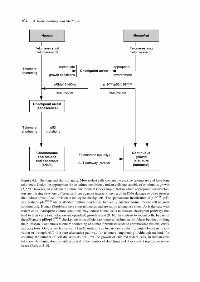
104 4 Biotechnology and Medicine
Human Mouse/rat
Checkpoint arrest
Chromosome
end fusions
and apoptosis
(crisis)
Continuous
growth
in culture
(immortal)
Telomerase (usually)
ALT pathway (rarely9
Checkpoint arrest
(senescence)
Telomere
shortening
p53
mutations
Telomere
shortening
p19ARF/p53/p16INK4a
inactivation
pRb/p16INK4a
inactivation
appropriate
environment
Telomeres short
Telomerase off
Telomeres long
Telomerase on
inadequate
growth conditions
Figure 4.2. The long and short of aging. Most rodent cells contain the enzyme telomerase and have longtelomeres. Under the appropriate tissue culture conditions, rodent cells are capable of continuous growth(1,2,8). However, an inadequate culture environment (for example, that in which appropriate survival fac-tors are missing or where different cell types cannot interact) may result in DNA damage or other stressesthat induce arrest of cell division at cell cycle checkpoints. The spontaneous inactivation of p19ARF, p53,and perhaps p16INK4a under standard culture conditions frequently enables normal rodent cell to growcontinuously. Human fibroblasts have short telomeres and are satiny telomerase silent. As is the case withrodent cells, inadequate culture conditions may induce human cells to activate checkpoint pathways thatlead to their early (and telomere-independent) growth arrest (9, 10). In contrast to rodent cells, bypass ofthe p53 and/or pRb/p16INK4a checkpoints is insufficient to immortalize human fibroblasts but does prolongtheir lifespan. Continuous telomere shortening of human fibroblasts leads to chromosome fusions, crisis,and apoptosis. Only a rare human cell (1 in 10 million) can bypass crisis either through telomerase reacti-vation or through ALT (the rare alternative pathway for telomere lengthening). Although methods forcounting the number of cell divisions do not limit the growth of cultured rodent cells, in human cellstelomere shortening does provide a record of the number of doublings and does control replicative senes-cence [Refs in 510].
105
have undergone.[510] The difference between human and rodent cellular aging is depicted inFigure 4.2.
Telomeres play a crucial role in the viability of cells. Experimental disruption oftelomerase action causes telomere shortening and cellular senescence[525] and genetic de-fects of telomerase have significant impact. The X-linked form of the human disease dys-keratosis congenita (DKC) is due to mutations in the gene coding for dyskerin and causesdefects in regenerative tissues such as skin and bone marrow, chromosome instability, anda predisposition towards certain malignancies. Dyskerin is associated with human telomeraseRNA which contains an H/ACA RNA motif and mutations lead to defective telomere main-tenance.[526]
In Caenorhabditis elegans, an insulinlike signaling pathway controls aging, metabo-lism, and development. Mutations in the daf-2 insulin receptor-like gene or the downstreamage-1 phosphoinositide 3-kinase extend adult life-span by two- to three-fold. In order toidentify the regulation of aging and metabolism by this pathway, the daf-2 pathway signal-ing was restored to only neurons, muscle, or intestine. Insulin like signaling in neuronsalone was sufficient to specify wild-type life span, whereas restoration of muscle or intes-tinal signaling was insufficient to do so. The restoration of the daf-2 pathway signaling inmuscle rescued metabolic defects, indicating a decoupling of life-span and metabolism.The nervous system appears to be a central regulator of longevity in C. elegans.[511] Thestudy of aging and developmental genetic regulation in C. elegans is supported by theavailability of genome-wide data on gene expression profiles.[588]
Research for genes involved in aging carried out in genetically tractable organisms suchas yeast, the nematode Caenorhabditis elegans, Drosophila melanogaster fruitflies, andmice has established that aging is regulated by specific genes. The pertinent pathways arelinking physiology, signaltransduction, and gene regulation. The mutations identified andthe correlations of phenotypes indicate that these genes are in control of aging in higherorganisms.[522]
Mutations in Caenorhabditis elegans affecting sensory cilia or their support cells, or insensory signal transduction, extend lifespan. C. elegans senses environmental signals throughciliated sensory neurons located primarily in specific sensory organs in head and tail. Thecilia are membrane-bound microtubule-based structures found at the dendritic endings ofsensory neurons in C. elegans. The findings of increased lifespan in mutants with defects insensory cilia implies that lifespan may be regulated by environmental signals.[512]
Biochemical and genetic studies in the long-lived Caenorhabditis clk-1 mutants demon-strated that these mutants are Q auxotrophs. CLK-1 is a mitochondrial polypeptide withsequence and functional conservation from human to yeast. The Saccharomyces cerevisiaehomolog Coq7p is essential for ubiquinone synthesis and thus respiration. Development ofthe clk-1 mutants requires a dietary source of Coenzyme Q.[527]
In the filamentous fungus Podospora anserina the impairment of the nuclear COX5gene encoding subunit V of the cytochrome c oxidase complex leads to the use of thealternative respiratory pathway and a decrease in production of reactive ion species with aconcomitant increase in lifespan associated with stabilization of the mitochondrial genome.This provides evidence for a causal link between mitochondrial metabolism and longevityin Podospora anserina.[518]
4.6 Complex Traits
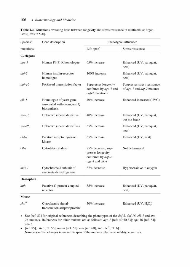
106 4 Biotechnology and Medicine
Table 4.3. Mutations revealing links between longevity and stress resistance in multicellular organ-isms [Refs in 520].
Species/ Gene description Phenotypic influence*
mutations Life span† Stress resistance
C. elegans
age-1 Human Pl (3) K homologue 65% increase Enhanced (UV, paraquat, heat)
daf-2 Human insulin-receptor homologue
100% increase Enhanced (UV, paraquat, heat)
daf-16 Forkhead transcription factor Suppresses longevity conferred by age-1 and daf-2 mutations
Suppresses stress resistance of age-1 and daf-2 mutants
clk-1 Homologue of yeast gene associated with coenzyme Q biosynthesis
40% increase Enhanced increased (UVC)
spe-10 Unknown (sperm defective 40% increase Enhanced (UV, paraquat, but not heat)
spe-26 Unknown (sperm defective) 65% increase Enhanced (UV, paraquat, heat)
old-1 Putative receptor tyrosine kinase
65% increase Enhanced (UV, heat)
ctl-1 Cytostatic catalase 25% decrease; sup-presses longevity conferred by daf-2, age-1 and clk-1
Not determined
mev-1 Cytochrome b subunit of succinate dehydrogenase
37% decrease Hypersensitive to oxygen
Drosophila
mth Putative G-protein-coupled receptor
35% increase Enhanced (UV, paraquat, heat)
Mouse
shc66 Cytoplasmic signal-transduction adaptor protein
30% increase Enhanced (UV, H2O2)
• See [ref. 83] for original references describing the phenotypes of the daf-2, daf-16, clk-1 and spe-26 mutants. References for other mutants are as follows: age-1 [refs 49,50,83]; spe-10 [ref. 84]; old-1
• [ref. 85]; ctl-1 [ref. 56]; mev-1 [ref. 55]; mth [ref. 60]; and shc66[ref. 6]. † Numbers reflect changes in mean life span of the mutants relative to wild-type animals.
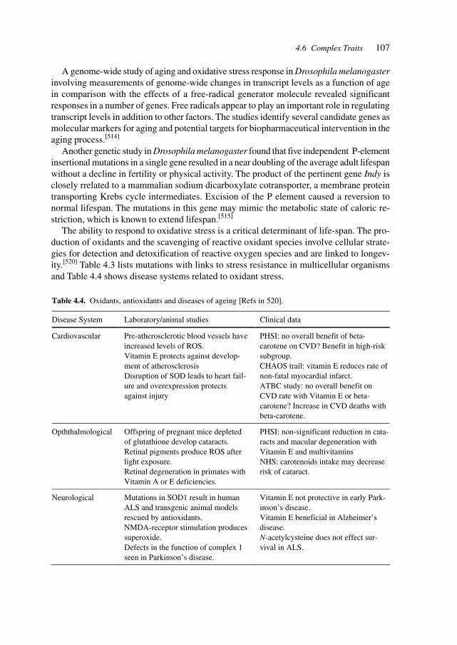
107
A genome-wide study of aging and oxidative stress response in Drosophila melanogasterinvolving measurements of genome-wide changes in transcript levels as a function of agein comparison with the effects of a free-radical generator molecule revealed significantresponses in a number of genes. Free radicals appear to play an important role in regulatingtranscript levels in addition to other factors. The studies identify several candidate genes asmolecular markers for aging and potential targets for biopharmaceutical intervention in theaging process.[514]
Another genetic study in Drosophila melanogaster found that five independent P-elementinsertional mutations in a single gene resulted in a near doubling of the average adult lifespanwithout a decline in fertility or physical activity. The product of the pertinent gene Indy isclosely rrelated to a mammalian sodium dicarboxylate cotransporter, a membrane proteintransporting Krebs cycle intermediates. Excision of the P element caused a reversion tonormal lifespan. The mutations in this gene may mimic the metabolic state of caloric re-striction, which is known to extend lifespan.[515]
The ability to respond to oxidative stress is a critical determinant of life-span. The pro-duction of oxidants and the scavenging of reactive oxidant species involve cellular strate-gies for detection and detoxification of reactive oxygen species and are linked to longev-ity.[520] Table 4.3 lists mutations with links to stress resistance in multicellular organismsand Table 4.4 shows disease systems related to oxidant stress.
Table 4.4. Oxidants, antioxidants and diseases of ageing [Refs in 520].
Disease System Laboratory/animal studies Clinical data
Cardiovascular Pre-atherosclerotic blood vessels have increased levels of ROS. Vitamin E protects against develop-ment of atherosclerosis Disruption of SOD leads to heart fail-ure and overexpression protects against injury.
PHSI: no overall benefit of beta-carotene on CVD? Benefit in high-risk subgroup. CHAOS trail: vitamin E reduces rate of non-fatal myocardial infarct. ATBC study: no overall benefit on CVD rate with Vitamin E or beta-carotene? Increase in CVD deaths with beta-carotene.
Opththalmological Offspring of pregnant mice depleted of glutathione develop cataracts. Retinal pigments produce ROS after light exposure. Retinal degeneration in primates with Vitamin A or E deficiencies.
PHSI: non-significant reduction in cata-racts and macular degeneration with Vitamin E and multivitamins. NHS: carotenoids intake may decrease risk of cataract.
Neurological Mutations in SOD1 result in human ALS and transgenic animal models rescued by antioxidants.
NMDA-receptor stimulation produces superoxide. Defects in the function of complex 1 seen in Parkinson’s disease.
Vitamin E not protective in early Park-inson’s disease. Vitamin E beneficial in Alzheimer’s disease. N-acetylcysteine does not effect sur-vival in ALS.
4.6 Complex Traits
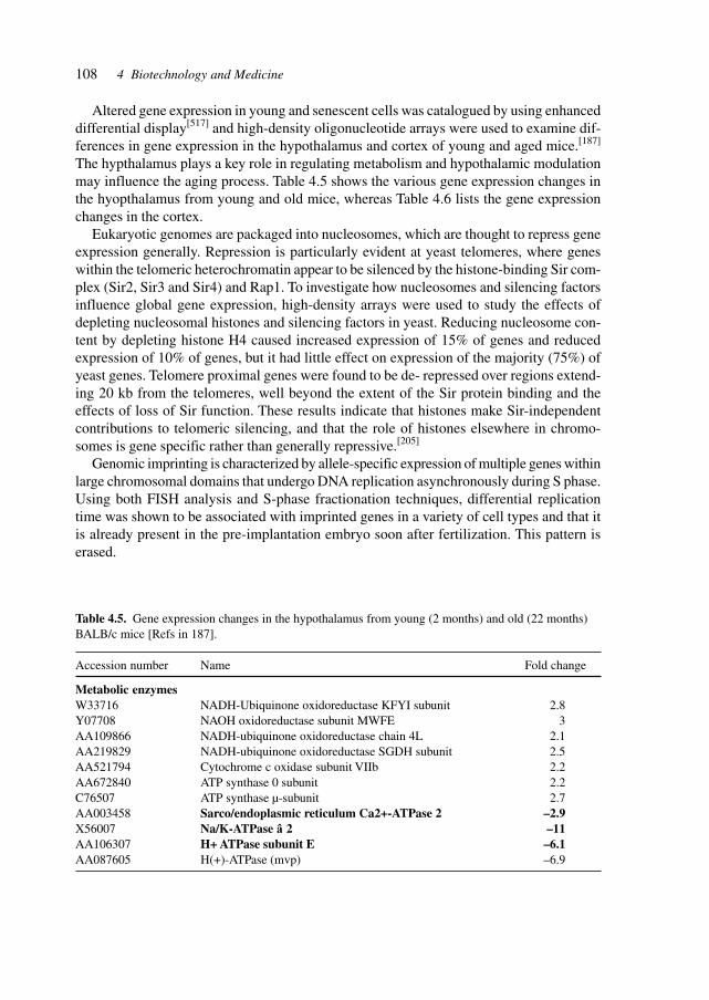
108 4 Biotechnology and Medicine
Altered gene expression in young and senescent cells was catalogued by using enhanceddifferential display[517] and high-density oligonucleotide arrays were used to examine dif-ferences in gene expression in the hypothalamus and cortex of young and aged mice.[187]
The hypthalamus plays a key role in regulating metabolism and hypothalamic modulationmay influence the aging process. Table 4.5 shows the various gene expression changes inthe hyopthalamus from young and old mice, whereas Table 4.6 lists the gene expressionchanges in the cortex.
Eukaryotic genomes are packaged into nucleosomes, which are thought to repress geneexpression generally. Repression is particularly evident at yeast telomeres, where geneswithin the telomeric heterochromatin appear to be silenced by the histone-binding Sir com-plex (Sir2, Sir3 and Sir4) and Rap1. To investigate how nucleosomes and silencing factorsinfluence global gene expression, high-density arrays were used to study the effects ofdepleting nucleosomal histones and silencing factors in yeast. Reducing nucleosome con-tent by depleting histone H4 caused increased expression of 15% of genes and reducedexpression of 10% of genes, but it had little effect on expression of the majority (75%) ofyeast genes. Telomere proximal genes were found to be de- repressed over regions extend-ing 20 kb from the telomeres, well beyond the extent of the Sir protein binding and theeffects of loss of Sir function. These results indicate that histones make Sir-independentcontributions to telomeric silencing, and that the role of histones elsewhere in chromo-somes is gene specific rather than generally repressive.[205]
Genomic imprinting is characterized by allele-specific expression of multiple genes withinlarge chromosomal domains that undergo DNA replication asynchronously during S phase.Using both FISH analysis and S-phase fractionation techniques, differential replicationtime was shown to be associated with imprinted genes in a variety of cell types and that itis already present in the pre-implantation embryo soon after fertilization. This pattern iserased.
Table 4.5. Gene expression changes in the hypothalamus from young (2 months) and old (22 months)BALB/c mice [Refs in 187].
Accession number Name Fold change
Metabolic enzymesW33716 NADH-Ubiquinone oxidoreductase KFYI subunit 2.8Y07708 NAOH oxidoreductase subunit MWFE 3AA109866 NADH-ubiquinone oxidoreductase chain 4L 2.1AA219829 NADH-ubiquinone oxidoreductase SGDH subunit 2.5AA521794 Cytochrome c oxidase subunit VIIb 2.2AA672840 ATP synthase 0 subunit 2.2C76507 ATP synthase µ-subunit 2.7AA003458 Sarco/endoplasmic reticulum Ca2+-ATPase 2 –2.9X56007 Na/K-ATPase â 2 –11AA106307 H+ ATPase subunit E –6.1AA087605 H(+)-ATPase (mvp) –6.9
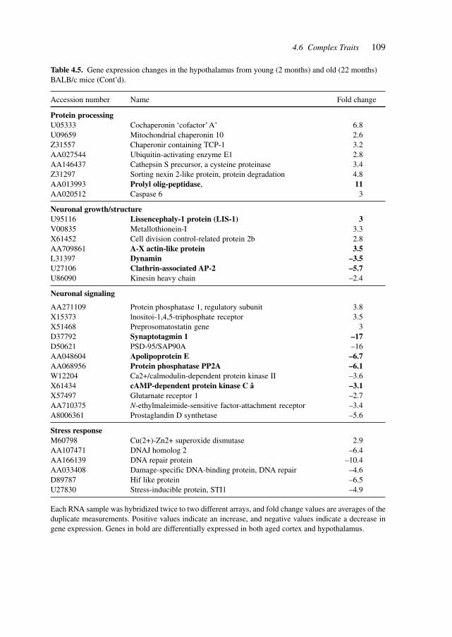
109
Table 4.5. Gene expression changes in the hypothalamus from young (2 months) and old (22 months)BALB/c mice (Cont’d).
Accession number Name Fold change
Protein processingU05333 Cochaperonin ‘cofactor’ A’ 6.8U09659 Mitochondrial chaperonin 10 2.6Z31557 Chaperonir containing TCP-1 3.2AA027544 Ubiquitin-activating enzyme E1 2.8AA146437 Cathepsin S precursor, a cysteine proteinase 3.4Z31297 Sorting nexin 2-like protein, protein degradation 4.8AA013993 Prolyl olig-peptidase, 11AA020512 Caspase 6 3
Neuronal growth/structureU95116 Lissencephaly-1 protein (LIS-1) 3V00835 Metallothionein-I 3.3X61452 Cell division control-related protein 2b 2.8AA709861 A-X actin-like protein 3.5L31397 Dynamin –3.5U27106 Clathrin-associated AP-2 –5.7U86090 Kinesin heavy chain –2.4
Neuronal signaling
AA271109 Protein phosphatase 1, regulatory subunit 3.8X15373 lnositoi-1,4,5-triphosphate receptor 3.5X51468 Preprosomatostatin gene 3D37792 Synaptotagmin 1 –17D50621 PSD-95/SAP90A –16AA048604 Apolipoprotein E –6.7AA068956 Protein phosphatase PP2A –6.1W12204 Ca2+/calmodulin-dependent protein kinase II –3.6X61434 cAMP-dependent protein kinase C â –3.1X57497 Glutarnate receptor 1 –2.7AA710375 N-ethylmaleimide-sensitive factor-attachment receptor –3.4A8006361 Prostaglandin D synthetase –5.6
Stress responseM60798 Cu(2+)-Zn2+ superoxide dismutase 2.9AA107471 DNAJ homolog 2 –6.4AA166139 DNA repair protein –10.4AA033408 Damage-specific DNA-binding protein, DNA repair –4.6D89787 Hif like protein –6.5U27830 Stress-inducible protein, STI1 –4.9
Each RNA sample was hybridized twice to two different arrays, and fold change values are averages of theduplicate measurements. Positive values indicate an increase, and negative values indicate a decrease ingene expression. Genes in bold are differentially expressed in both aged cortex and hypothalamus.
4.6 Complex Traits
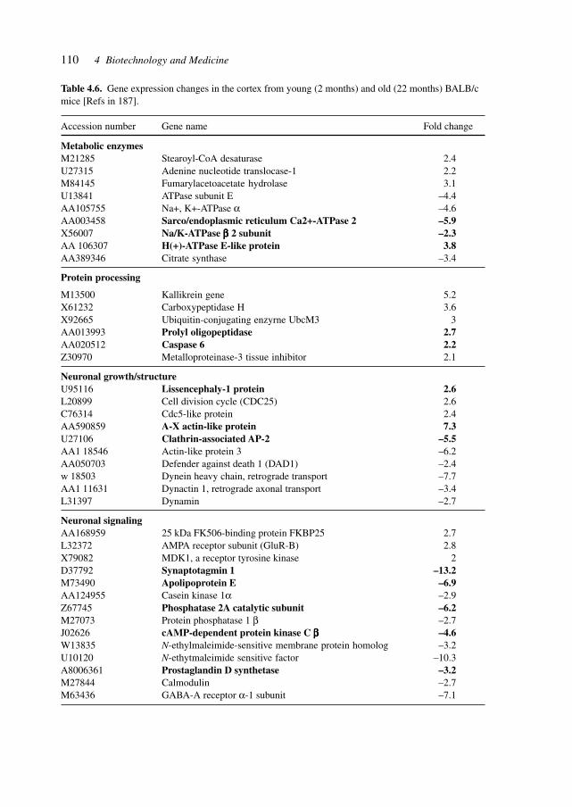
110 4 Biotechnology and Medicine
Table 4.6. Gene expression changes in the cortex from young (2 months) and old (22 months) BALB/cmice [Refs in 187].
Accession number Gene name Fold change
Metabolic enzymesM21285 Stearoyl-CoA desaturase 2.4U27315 Adenine nucleotide translocase-1 2.2M84145 Fumarylacetoacetate hydrolase 3.1U13841 ATPase subunit E –4.4AA105755 Na+, K+-ATPase α –4.6AA003458 Sarco/endoplasmic reticulum Ca2+-ATPase 2 –5.9X56007 Na/K-ATPase βββββ 2 subunit –2.3AA 106307 H(+)-ATPase E-like protein 3.8AA389346 Citrate synthase –3.4
Protein processing
M13500 Kallikrein gene 5.2X61232 Carboxypeptidase H 3.6X92665 Ubiquitin-conjugating enzyrne UbcM3 3AA013993 Prolyl oligopeptidase 2.7AA020512 Caspase 6 2.2Z30970 Metalloproteinase-3 tissue inhibitor 2.1
Neuronal growth/structureU95116 Lissencephaly-1 protein 2.6L20899 Cell division cycle (CDC25) 2.6C76314 Cdc5-like protein 2.4AA590859 A-X actin-like protein 7.3U27106 Clathrin-associated AP-2 –5.5AA1 18546 Actin-like protein 3 –6.2AA050703 Defender against death 1 (DAD1) –2.4w 18503 Dynein heavy chain, retrograde transport –7.7AA1 11631 Dynactin 1, retrograde axonal transport –3.4L31397 Dynamin –2.7
Neuronal signalingAA168959 25 kDa FK506-binding protein FKBP25 2.7L32372 AMPA receptor subunit (GluR-B) 2.8X79082 MDK1, a receptor tyrosine kinase 2D37792 Synaptotagmin 1 –13.2M73490 Apolipoprotein E –6.9AA124955 Casein kinase 1α –2.9Z67745 Phosphatase 2A catalytic subunit –6.2M27073 Protein phosphatase 1 β –2.7J02626 cAMP-dependent protein kinase C βββββ –4.6W13835 N-ethylmaleimide-sensitive membrane protein homolog –3.2U10120 N-ethytmaleimide sensitive factor –10.3A8006361 Prostaglandin D synthetase –3.2M27844 Calmodulin –2.7M63436 GABA-A receptor α-1 subunit –7.1
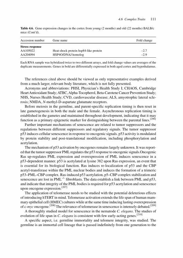
111
The references cited above should be viewed as only representative examples derivedfrom a much larger, relevant body literature, which is not fully presented.
Acronyms and abbreviations: PHSI, Physician’s Health Study I; CHAOS, CambridgeHeart Antioxidant Study; ATBC, Alpha-Tocopherol, Beta-Carotene Cancer Prevention Study;NHS, Nurses Health Study; CVD, cardiovascular disease; ALS, amyotrophic lateral scle-rosis; NMDA, N-methyl-D-aspartate glutamate receptors.
Before meiosis in the germline, and parent-specific replication timing is then reset inlate gametogenesis in both the male and the female. Asynchronous replication timing isestablished in the gametes and maintained throughout development, indicating that it mayfunction as a primary epigenetic marker for distinguishing between the parental lines.[206]
Further important mechanisms of senescence are related to tumor suppressors and theregulations between different suppressors and regulatory signals. The tumor suppressorp53 induces cellular senescence in response to oncogenic signals. p53 activity is modulatedby protein stability and post-translational modification, including phosphorylation andacetylation.
The mechanism of p53 activation by oncogenes remains largely unknown. It was report-ed that the tumor suppressor PML regulates the p53 response to oncogenic signals.OncogenicRas up-regulates PML expression and overexpression of PML induces senescence in ap53-dependent manner. p53 is acetylated at lysine 382 upon Ras expression, an event thatis essential for its biological function. Ras induces re-localization of p53 and the CBPacetyl-transferase within the PML nuclear bodies and induces the formation of a trimericp53–PML–CBP complex. Ras-induced p53 acetylation, p5–CBP complex stabilization andsenescence are lost in PML–/– fibroblasts. The data establish a link between PML and p53,and indicate that integrity of the PML bodies is required for p53 acetylation and senescenceupon oncogene expression.[207]
The application of telomerase needs to be studied with the potential deleterious effectsof introducing hTERT in mind. Telomerase activation extends the life span of human mam-mary epithelial cell (HMEC) cultures while at the same time inducing lasting overexpressionof c-myc oncogene.[208] The relevance of telomerase in senescence is intensely debated.[209]
A thoroughly studied model for senescence in the nematode C. elegans. The studies ofevolution of life span in C. elegans is consistent with few early-acting genes.[210]
A specific aspect, i.e. germline immortality and telomere integrity, was studied. Thegermline is an immortal cell lineage that is passed indefinitely from one generation to the
Table 4.6. Gene expression changes in the cortex from young (2 months) and old (22 months) BALB/cmice (Cont’d).
Accession number Gene name Fold change
Stress responseAA105022 Heat-shock protein hsp84-like protein –2.7AA204094 HSP4O/DNAJ homolog –2.9
Each RNA sample was hybridized twice to two different arrays, and fold change values are averages of theduplicate measurements. Genes in bold are differentially expressed in both aged cortex and hypothalamus.
4.6 Complex Traits
112 4 Biotechnology and Medicine
next. To identify the genes that are required for germline immortality, C. elegans mutantswith mortal germlines were isolated – worms that can reproduce for several healthy genera-tions but eventually become sterile. One of these mortal germline (mrt) mutants, mrt-2,exhibits progressive telomere shortening and accumulates end-to-end chromosome fusionsin later generations, indicating that the MRT-2 protein is required for telomere replication.In addition, the germline of mrt-2 is hypersensitive to X-rays and to transposon activity.Therefore, mrt-2 has defects in responding both to damaged DNA and to normal double-strand breaks present at telomeres. Mrt-2 encodes a homolog of a checkpoint gene that isrequired to sense DNA damage in yeast. The results indicate that telomeres may be identi-fied as a type of DNA damage and then repaired by the telomere-replication enzymetelomerase.[211]
The potential of cloning depends in part on whether the procedure can reverse cellularageing and restore somatic cells to a phenotypically youthful state. The birth of six healthycloned calves derived from populations of senescent donor somatic cells was reported.Nuclear transfer extended the replicative life span of senescent cells (zero to four popula-tion doublings remaining) to greater than 90 populations doublings. Early population dou-bling level complementary DNA-1 (EPC-1, an age-dependent gene) expression in cellsfrom the cloned animals was 3.5- to 5-fold higher than that in cells from age-matched(5−10 months old) controls. Southern blot and flow cytometric analyses indicated that thetelomeres were also extended beyond those of newborn (less than 2 weeks old) and age-matched control animals. The ability to regenerate animals and cells may have importantimplications for medicine and the study of mammalian ageing.[212]
Mice have been cloned by nuclear transfer into enucleated oocytes. In particular, thereiterative cloning of mice to four and six generations in two independent lines was re-ported. Successive generations show no signs of premature ageing, as judged by grossbehavioral parameters and there was no evidence of shortening of telomeres at the ends ofchromosomes, normally an indicator of cellular senescence. The telomeres actually in-creased slightly in length. This increase is astonishing, since the number of mitotic divi-sions greatly exceeds that of sexually produced animals and any deleterious effects of clon-ing might be amplified in sequentially cloned mice. These observations bear on studies oforganismal ageing.[213]
Telomerase is amenable to therapeutic intervention by small-molecule effectors and thussuitable therapeutics may be developed. Telomerase, a ribonucleoprotein up-regulated inmany types of cancers, possesses an RNA template necessary to bind and extend telomereends. The intrinsic accessibility of Telomerase to incoming nucleic acids makes the RNAtemplate an ideal target for inhibition by oligonucleotides. 2′-O-methyl RNA (2′-O-meRNA),an oligonucleotide known to exert sequence-specific effects in cell culture and animals,inhibits telomerase with potencies superior to those possessed by analogous peptide nucleicacids (PNAs). Potent inhibition relative to PNAs is surprising, because the binding affinityof 2′-O-meRNAs for complementary RNA is low relative to analogous PNAs. A 2′-O-meRNA oligomer with terminal phosphorothioate substitutions inhibits telomerase sequence-selectively within human-tumor-derived DU145 cells when delivered with cationic lipids.In contrast to the ability of 2′-O-meRNA oligomers to inhibit telomerase, the binding of a2′-O-meRNA to an inverted repeat within plasmid DNA was not detectable, whereas bind-
113
ing of PNA was efficient, suggesting that the relative accessibility of the telomerase RNAtemplate is essential for inhibition by 2′-O-meRNA. Inhibition of telomerase by 2′-O-meRNAwill facilitate probing the link between telomerase activity and sustained cell proliferation,and may provide a basis for the development of chemopreventive and chemotherapeuticagents.[214]
The importance of telomeres is underscored by studies of chromosomal rearrangementsand telomeres. Chromosomal rearrangement mechanisms are intimately linked to cancerdevelopment, and are thought to generate the numerous gains and losses of segments ofchromosomes needed for epithelial carcinogenesis. Aged humans sustain a high rate ofepithelial cancers such as carcinomas of the breast and colon, whereas mice carrying com-mon tumor suppressor gene mutations typically develop soft tissue carcinomas and lym-phomas. Among the many factors that may contribute to this species variance are differ-ences in telomere length and regulation. Telomeres comprise the nucleoprotein complexesthat cap the ends of eukaryotic chromosomes and are maintained by the reverse transcriptase,telomerase. In human cells, insufficient levels of telomerase lead to telomere attrition withcell division in culture, and possibly with ageing and tumorigenesis in vivo. In contrast,critical reduction in telomere length is not observed in the mouse owing to promiscuoustelomerase expression and long telomeres. Telomere attrition in ageing telomerase-defi-cient p53 mutant mice promotes the development of epithelial cancers by a process offusion-bridge breakage that leads to the formation of complex non-reciprocal transloca-tions – a classical cytogenetic feature of human carcinomas. Telomere dysfunction broughtabout by continual epithelial renewal during life may generate the massive ploidy changesassociated with the development of epithelial cancers.[215]
Life span is also influenced by external factors, such as ROS which primarily arise asby-products of normal metabolic activities and are thought to influence the etiology of age-related diseases. In order to test the theory that ROS cause ageing, the natural antioxidantsystems of C. elegans with small synthetic superoxide dismutase/catalase mimetics. Treat-ment of wild-type worms increased their mean life span by a mean of 44%, and treatmentof prematurely aging worms resulted in normalization of their life span (a 67% increase). Itappears that oxidative stress is a major determinant of life span and that it can be counter-acted by pharmacological intervention.[216]
Expression profiling allows the simultaneous identification of differentially expressedgenes, and by comparing different cells, tissues, disease states and metabolic states enablesthe pin-pointing of condition-associated and/or causative gene expression. In tumor therapy,the identification of tumor type and genetic make-up of the malignant cells allows the earlyand often decisive choice of therapy.
Diffuse large B cell lymphoma (DLBCL), the most common subtype of non-Hodgkin’slymphoma, is clinically heterogeneous: 40% of patients respond well to current therapyand have prolonged survival, whereas the remainder succumb to the disease. This variabil-ity in natural history may reflect unrecognized molecular heterogeneity in the tumors. Us-ing DNA microarrays, a systematic characterization of gene expression in B cell malignan-cies was conducted. It was shown that there is diversity in gene expression among thetumors of DLBCL patients, apparently reflecting the variation in tumor proliferation rate,host response and differentiation state of the tumor. Two molecularly distinct forms of
4.6 Complex Traits
114 4 Biotechnology and Medicine
DLBCL were identified which had gene expression patterns indicative of different stagesof B cell differentiation. One type expressed genes characteristic of germinal center B cells(‘germinal center B-like DLBCL’); the second type expressed genes normally induced dur-ing in vitro activation of peripheral blood B cells (‘activated B-like DLBCL’). Patients withgerminal center B-like DLBCL had a significantly better overall survival than those withactivated B-like DLBCL. The molecular classification of tumors on the basis of gene ex-pression can thus identify previously undetected and clinically significant subtypes of can-cer.[217]
Similarly, the technique of gene expression profiling was applied to another difficult tocategorize type of tumor, malignant melanoma. The most common human cancers are ma-lignant neoplasms of the skin. Incidence of cutaneous melanoma is rising especially steeply,with minimal progress in non-surgical treatment of advanced disease. Despite significanteffort to identify independent predictors of melanoma outcome, no accepted histopatho-logical, molecular or immunohistochemical marker defines subsets of this neoplasm. Ac-cordingly, although melanoma is thought to present different ‘taxonomic’ forms, these areconsidered part of a spectrum rather than discrete entities. The discovery of a subset ofmelanomas identified by mathematical analysis of gene expression in a series of sampleswas reported. Many genes underlying the classification of this subset are differentiallyregulated in invasive melanomas that form primitive tubular networks in vivo, a feature ofsome highly aggressive metastatic melanomas. Global transcript analysis can identify un-recognized subtypes of coetaneous melanoma and predict experimentally verifiable pheno-typic characteristics that may be of importance to disease progression and choice of treat-ment.[218]
To gain an understanding of the molecular basis of tumor angiogenesis, gene expressionpatterns were compared of endothelial cells derived from blood vessels of normal and ma-lignant colorectal tissues. Of over 170 transcripts predominantly expressed in the endothe-lium, 79 were differentially expressed, including 46 that were specifically elevated in tu-mor-associated endothelium. Several of these genes encode extracellular matrix proteins,but most are of unknown function. Most of these tumor endothelial markers were expressedin a wide range of tumor types, as well as in normal vessels associated with wound healingand corpus luteum formation. These studies demonstrated that tumor and normal endothe-lium are distinct at the molecular level – a finding that may have significant implicationsfor the development of antiangiogenic therapies.[219]
SAGE analysis reveals previously characterized and novel pan endothelial markers. Themost abundant characterized or novel tags derived by summing the tags from normal EC(N-ECs) and tumor EC (T-ECs). SAGE libraries are listed in descending order. For com-parison, the corresponding number of SAGE tags found in HUVEC and HMVEC endothe-lial cell cultures, and several nonendothelial cell lines (14), are shown. Tag numbers foreach group were normalized to 100.000 transcripts. A description of the gene product cor-responding to each tag is given, followed by alternative names in parentheses. Someuncharacterized genes have predicted full-length coding sequence. The sequence CATGprecedes all tags, and the 15th base (11th shown) was determined as described (38) [Refer-ences in 219].
115
Human breast tumors are diverse in their natural history and in their responsiveness totreatment. Variation in transcriptional programs accounts for much of the biological diver-sity of human cells and tumors. In each cell, signal transduction and regulatory systemstransduce information from the cell’s identity to its environmental status, thereby control-ling the level of expression of every gene in the genome. Variation was characterized ingene expression patterns in a set of 65 surgical specimens of human breast tumors from 42different individuals, using complementary DNA microarrays representing 8102 humangenes. These patterns provided a distinctive molecular portrait of each tumor. Twenty ofthe tumors were sampled twice, before and after a 16-week course of doxorubicin chemo-therapy, and two tumors were paired with a lymph node metastasis from the same patient.Gene expression patterns in two tumor samples from the same individual were almost al-ways more similar to each other than either was to any other sample. Sets of co-expressedgenes were identified for which variation in mRNA levels could be related to specific fea-tures of physiological variation. The tumors could be classified into subtypes distinguishedby pervasive differences in their gene expression patterns.[220]
The elucidation of genetic contributions to senescence in Drosophila melanogaster andCaenorhabditis elegans and their relationship points towards fundamental evolutionaryconserved genetic and biochemical mechanisms.[655]
The Drosophila melanogaster gene insulin-like receptor (InR) was found homologousto mammalian insulin receptors as well as to Caenorhabditis elegans daf-2, a signal trans-ducer regulating worm dauer formation and adult longevity. A heteroallelic, hypomorphicgenotype of mutant InR, which results in dwarf females with up to an 85% extension ofadult longevity and dwarf males with reduced late age-specific mortality. Treatment of thelong-lived InR dwarfs with a juvenile hormone analoge restores the life expectancy towardthat of wild-type controls thus demonstrating that juvenile hormone deficiency resultingfrom the InR signal pathway mutation is sufficient to extend life-span in flies. The resultsalso show that insulin-like ligands nonautonomously mediate aging in flies through retar-dation of growth or activation of specific endocrine tissue.[654]
The Drosophila melanogaster gene chico encoding an insulin receptor substrate whichfunctions in an insulin/insulin-like growth factor (IGF) signaling pathway was found toextend upon mutation the fruit fly median life-span by up to 48% in homozygotes and 36%in heterozygotes. The extension of life-span was neither a result of impaired oogenesis inchico females nor correlated with increased stress resistance nor was the dwarf phenotypeof chico homozygotes necessary for extension of life-span.[653]
Genetic linkage studies are required to identify the multiple genes involved in complextraits and population genetic studies involving single-nucleotide polymorphisms and othergenetic variations such as different alleles are the basis for pharmaco-genomics and theidentification of genetic disease associations.[691] [692]
Table 4.7 shows a listing of the pan-endothelial markers identified by SAGE analysis.
4.6 Complex Traits
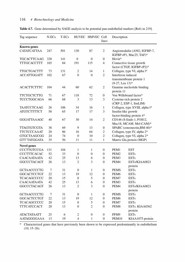
116 4 Biotechnology and Medicine
Table 4.7. Gene determined by SAGE analysis to be potential pan-endothelial markers [Refs in 219].
Tag sequence N-ECs T-ECs HUVEC HMVEC Cell Descriptionlines
Known genesCATATCATTAA 247 501 130 87 2 Angiomodulin (ANG, IGFBP-7,
IGFBP-rP1, Mac25, TAF)*TGCACTTCAAG 328 141 0 0 0 Hevin*TTTGCACCTTT 165 84 191 115 4 Connective tissue growth
factor (CTGF, IGFBP-rP2)*TTGCTGACTTT 73 131 2 14 1 Collagen, type VI, alpha l*ACCATTGGATT 102 67 0 0 2 Interferon induced
transmembrane protein 1(9-27, Leu 13)*
ACACTTCTTTC 104 44 60 62 2 Guanine nucleotide bindingprotein 11
TTCTGCTCTTG 71 67 118 72 0 Von Willebrand factor*TCCCTGGCAGA 66 68 3 13 3 Cysteine-rich protein 2
(CRP-2, ESP-1, SmLIM)TAATCCTCAAG 26 106 34 16 1 Collagen, type XVIII, alpha l*ATGTCTTTTCT 58 65 17 17 3 Insulin-like growth
factor-binding protein 4*GGGATTAAAGC 40 67 30 14 2 CD146 (S-Endo 1, P1H12,
Muc18, MCAM. Mel-CAM)*TTAGTGTCGTA 38 69 9 13 0 SPARC (osteonectin,BM-40)*TTCTCCCAAAT 20 86 16 64 2 Collagen, type IV, alpha 2*GTGCTAAGCGG 24 74 0 10 2 Collagen, type VI, alpha 2*GTT’TATGGATA 35 56 11 11 1 Matrix Gla protein (MGP)
Novel genesCCCTTGTCCGA 131 104 1 1 0 PEM1 ESTCCCTTTCACAC 52 33 0 0 0 PEM2 ESTsCAACAATAATA 42 25 13 6 0 PEM3 ESTsGGCCCTACAGT 26 13 2 3 0 PEM4 ESTs/KIAA0821
proteinGCTAACCCCTG 7 31 0 1 0 PEMS ESTsGGCACTCCTGT 22 13 19 12 0 PEM6 ESTsTCACAGCCCCC 20 15 8 5 0 PEM7 ESTsCAACAATAATA 42 25 13 6 0 PEM3 ESTsGGCCCTACAGT 26 13 2 3 0 PEM4 ESTs/KIAA0821
proteinGCTAACCCCTG 7 31 0 1 0 PEMS ESTsGGCACTCCTGT 22 13 19 12 0 PEM6 ESTsTCACAGCCCCC 20 15 8 5 0 PEM7 ESTsTTTCATCCACT 20 13 0 2 0 PEM8 ESTs. KIAA0362
proteinATACTATAATT 25 6 2 0 0 PFM9 ESTsAATAGGGGAAA 13 19 4 1 0 PEM10 KIAA1075 protein
* Characterized genes that have previously been shown to be expressed predominantly in endothelium(10, 15–26).
117
5 Drug Discovery
The drug discovery process is the fundamental source of active principles, i.e. compoundswhich interact as agonists or antagonists, functionally or structurally, with constituents ofliving organisms.
Drugs arise form either natural sources, with an almost infinite variety of molecularstructures, or are designed with the target effect or molecule in mind [using quaternarystructure–activity relationships (QSAR)]. The combination and creative utilization of sev-eral areas of scientific studies, i.e. genomics, proteomics, bioinformatics, structural analy-sis and functional experiments plus advanced chemistries, will provide significant resultsin the next decades.[221]
Modern drug discovery often involves screening of small molecules for their ability tobind to a pre-selected protein target. Target-oriented syntheses of these small molecules,individually or as collections (focused libraries), can be planned effectively with retro-synthetic analysis. Drug discovery can also involve screening small molecules for theirability to modulate a biological pathway in cells or organisms, without regard for any par-ticular protein target. This process is likely to benefit in the future from an evolving for-ward analysis of synthetic pathways, used in diversity-oriented synthesis that leads to struc-turally complex and diverse small molecules. One goal of diversity-oriented syntheses is tosynthesize efficiently a collection of small molecules capable of perturbing any disease-related biological pathway, leading eventually to the identification of therapeutic proteintargets capable of being modulated by small molecules. Several synthetic planning prin-ciples for diversity-oriented synthesis and their role in the drug discovery process havebeen reviewed.[222]
Recent drug discovery has moved from biology-directed chemical synthesis towards anarea where large-scale zero-knowledge screening dominates. As the cost of carrying outlarge-scale diversity high-throughput screening (HTS) continues to fall, driven by extrememiniaturization, a paradigm shift towards less centralized screening activities distributedmore closely to end-user groups or individuals is a possible consequence. The power of themethodology therefore becomes more responsive to a more diverse range of individualrequirements. This model is a logical extrapolation of current practice, taking into accountthe history of other technologies such as computing, DNA sequencing and other physico-chemical analytical techniques. The era of personal or desktop screening is not far off, butit will require a considerable degree of miniaturization to be applied to the whole screeningsystem, not just to the sample carrier.
In parallel with these approaches, the power of computational predictive methodologieshas now reached the stage where a third strategy, ‘chemical space definition’, can be ap-plied to the discovery portfolio to aid in finding pharmaceutical interventions. Information-
Industrial Pharmaceutical Biotechnology. Heinrich KlefenzCopyright © 2002 Wiley-VCH Verlag GmbH
ISBNs: 3-527-29995-5 (Hardcover); 3-527-60012-4 (Electronic)
118 5 Drug Discovery
based screening, be it through the application of a focussed, systems-based method or theuse of iterative screening, will become more efficient, selecting fewer false-positives and,more importantly, fewer false-negatives. This will be achieved not only through the devel-opment of better algorithms, but through established algorithms made accessible by anincrease in computing power. The other major contribution to the development of compu-tational methods is the data generated over the years, particularly for target families such askinases. In the next few years, predictive models for these targets might negate the need forrandom screening against them, as we will have learnt the essential features required forrecognition.
Diversity and focussed approaches are likely to continue to provide complementary ben-efits. The portfolio of techniques will together provide a still more effective and compre-hensive approach to the effective identification of novel medicines for the multitude ofnovel targets that are being revealed through genomics and proteomics.
The complementarity of diversity and focussed approaches can be seen in clinical prac-tice. HIV protease inhibitors are all from the rational, structure-based design school of drugdiscovery, whereas the non-nucleoside HIV reverse transcriptase inhibitors have almost alloriginated from diversity screening. As combination therapy is the best way to treat HIVinfection, this is evidence of the benefits of complementary technologies to fight disease.[223]
Knowledge of cellular signaling pathways and knowledge of the human genetics of aspecific disease provide powerful validation and rational targets for disease identificationand development of drugs and therapies.
Cancer is going to become the most prevalent disease-related cause of death because it isa disease of multiple accumulating mutations that are manifesting in human populationswith an increasingly prolonged life span. Chemotherapies and gene therapies make use ofincreasing molecular knowledge in cancer, and are supported by tumor genotyping andpatient gene profiling.[224]
Table 5.1 gives examples of mechanism-based cancer therapies in development.A specific feature of CML is a reciprocal chromosomal translocation involving the long
arms of chromosomes 9 and 22. This somatic mutation fuses a segment of the bcr gene,from chromosome 9, to a region upstream of the second exon of the c-abl (Abelson ty-rosine kinase) gene from chromosome 22. c-abl encodes a non-receptor tyrosine kinasethat has tightly controlled activity in normal cells. In contrast, Bcr–Abl fusion proteinshave constitutive catalytic activity, despite the fact that the amino acid sequence of the Ablsegment of Bcr–Abl is identical to that of c-Abl. The reason for the elevated catalyticactivity of the Bcr–Abl fusion protein is poorly understood, but it is clear that this activityof the kinase domain is necessary for the ability of the Bcr–Abl protein to transform cellsand cause malignancy.
A small-molecule inhibitor of Abl (STI-571) is effective in the treatment of chronicmyelogenous leukemia CML, which is caused by the inadvertent activation of Abl. Thecrystal structure of the catalytic domain of Abl, complexed to a variant of STI-571, wasreported. Critical to the binding of STI-571 is the adoption by the kinase of an inactiveconformation, in which a centrally located ‘activation loop’ is not phosphorylated. Theconformation of this loop is distinct from that in active protein kinases, as well as in theinactive form of the closely related Src kinases. These results suggest that compounds that
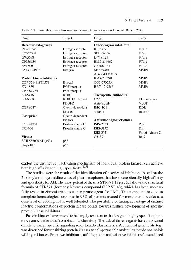
1195 Drug Discovery
exploit the distinctive inactivation mechanism of individual protein kinases can achieveboth high affinity and high specificity.[225]
The studies were the result of the identification of a series of inhibitors, based on the2-phenylaminopyrimidine class of pharmacophores that have exceptionally high affinityand specificity for Abl. The most potent of these is STI-571. Figure 5.1 shows the structuralformula of STI-571 (formerly Novartis compound CGP 57148), which has been success-fully tested in clinical trials as a therapeutic agent for CML. The compound has led tocomplete hematological response in 96% of patients treated for more than 4 weeks at adose level of 300 mg and is well tolerated. The possibility of taking advantage of distinctinactive conformations of protein kinase points towards further development of specificprotein kinase inhibitors.
Protein kinases have proved to be largely resistant to the design of highly specific inhibi-tors, even with the aid of combinatorial chemistry. The lack of these reagents has complicatedefforts to assign specific signaling roles to individual kinases. A chemical genetic strategywas described for sensitizing protein kinases to cell-permeable molecules that do not inhibitwild-type kinases. From two inhibitor scaffolds, potent and selective inhibitors for sensitized
Table 5.1. Examples of mechanism-based cancer therapies in development [Refs in 224].
Drug Target Drug Target
Receptor antagonists Raloxifene LY353381 GW5638 CP336156 EM-800 EMD-121974 Protein kinase inhibitors CGP 57148/STI 571 ZD-1839 CP-358,774 SU-5416 SU-6668 CGP 60474 Flavopiridol CGP 41251 UCN-01 Viruses SCH 58500 (AD-p53) Onyx-015
Estrogen receptor Estrogen receptor Estrogen receptor Estrogen receptor Estrogen receptor Integrin Bcr-abl EGF receptor EGF receptor KDR KDR; FGFR; and PDGFR Cyclin-dependent kinases Cyclin-dependent kinases Protein kinase C Protein kinase C p53 p53
Other enzyme inhibitors R115777 SCH 66336 L-778,123 BMS-214662 CP-609,754 Marimastat AG-3340 MMPs BMS-275291 CGS-27023A BAY 12-9566 Therapeutic antibodies C225 Anti-VEGF IMC-1C11 Vitaxin Antisense oligonucleotides ISIS-2503 ISIS-5132 ISIS-3521 G3139
FTase FTase FTase FTase FTase MMPs MMPs MMPs MMPs EGF receptor VEGF KDR Integrin Ras Raf Protein kinase C Bcl2
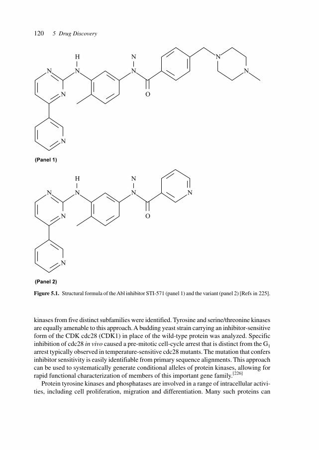
120 5 Drug Discovery
kinases from five distinct subfamilies were identified. Tyrosine and serine/threonine kinasesare equally amenable to this approach. A budding yeast strain carrying an inhibitor-sensitiveform of the CDK cdc28 (CDK1) in place of the wild-type protein was analyzed. Specificinhibition of cdc28 in vivo caused a pre-mitotic cell-cycle arrest that is distinct from the G1arrest typically observed in temperature-sensitive cdc28 mutants. The mutation that confersinhibitor sensitivity is easily identifiable from primary sequence alignments. This approachcan be used to systematically generate conditional alleles of protein kinases, allowing forrapid functional characterization of members of this important gene family.[226]
Protein tyrosine kinases and phosphatases are involved in a range of intracellular activi-ties, including cell proliferation, migration and differentiation. Many such proteins can
Figure 5.1. Structural formula of the Abl inhibitor STI-571 (panel 1) and the variant (panel 2) [Refs in 225].
N
N
N
H
N
N
N
O
(Panel 1)
N
N
N
N
N
H
N
N
N
O
(Panel 2)
N
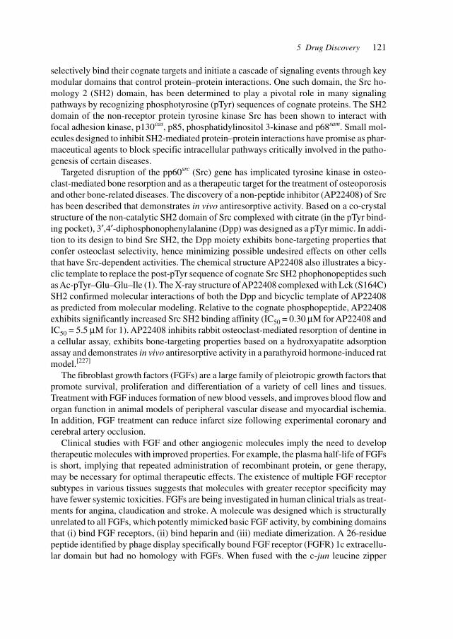
121
selectively bind their cognate targets and initiate a cascade of signaling events through keymodular domains that control protein–protein interactions. One such domain, the Src ho-mology 2 (SH2) domain, has been determined to play a pivotal role in many signalingpathways by recognizing phosphotyrosine (pTyr) sequences of cognate proteins. The SH2domain of the non-receptor protein tyrosine kinase Src has been shown to interact withfocal adhesion kinase, p130cas, p85, phosphatidylinositol 3-kinase and p68sam. Small mol-ecules designed to inhibit SH2-mediated protein–protein interactions have promise as phar-maceutical agents to block specific intracellular pathways critically involved in the patho-genesis of certain diseases.
Targeted disruption of the pp60src (Src) gene has implicated tyrosine kinase in osteo-clast-mediated bone resorption and as a therapeutic target for the treatment of osteoporosisand other bone-related diseases. The discovery of a non-peptide inhibitor (AP22408) of Srchas been described that demonstrates in vivo antiresorptive activity. Based on a co-crystalstructure of the non-catalytic SH2 domain of Src complexed with citrate (in the pTyr bind-ing pocket), 3′,4′-diphosphonophenylalanine (Dpp) was designed as a pTyr mimic. In addi-tion to its design to bind Src SH2, the Dpp moiety exhibits bone-targeting properties thatconfer osteoclast selectivity, hence minimizing possible undesired effects on other cellsthat have Src-dependent activities. The chemical structure AP22408 also illustrates a bicy-clic template to replace the post-pTyr sequence of cognate Src SH2 phophonopeptides suchas Ac-pTyr–Glu–Glu–Ile (1). The X-ray structure of AP22408 complexed with Lck (S164C)SH2 confirmed molecular interactions of both the Dpp and bicyclic template of AP22408as predicted from molecular modeling. Relative to the cognate phosphopeptide, AP22408exhibits significantly increased Src SH2 binding affinity (IC50 = 0.30 µM for AP22408 andIC50 = 5.5 µM for 1). AP22408 inhibits rabbit osteoclast-mediated resorption of dentine ina cellular assay, exhibits bone-targeting properties based on a hydroxyapatite adsorptionassay and demonstrates in vivo antiresorptive activity in a parathyroid hormone-induced ratmodel.[227]
The fibroblast growth factors (FGFs) are a large family of pleiotropic growth factors thatpromote survival, proliferation and differentiation of a variety of cell lines and tissues.Treatment with FGF induces formation of new blood vessels, and improves blood flow andorgan function in animal models of peripheral vascular disease and myocardial ischemia.In addition, FGF treatment can reduce infarct size following experimental coronary andcerebral artery occlusion.
Clinical studies with FGF and other angiogenic molecules imply the need to developtherapeutic molecules with improved properties. For example, the plasma half-life of FGFsis short, implying that repeated administration of recombinant protein, or gene therapy,may be necessary for optimal therapeutic effects. The existence of multiple FGF receptorsubtypes in various tissues suggests that molecules with greater receptor specificity mayhave fewer systemic toxicities. FGFs are being investigated in human clinical trials as treat-ments for angina, claudication and stroke. A molecule was designed which is structurallyunrelated to all FGFs, which potently mimicked basic FGF activity, by combining domainsthat (i) bind FGF receptors, (ii) bind heparin and (iii) mediate dimerization. A 26-residuepeptide identified by phage display specifically bound FGF receptor (FGFR) 1c extracellu-lar domain but had no homology with FGFs. When fused with the c-jun leucine zipper
5 Drug Discovery
122 5 Drug Discovery
domain, which binds heparin and forms heterodimers, the polypeptide specifically repro-duced the mitogenic and morphogenic activities of basic FGF with similar potency (EC50 =240 pM). The polypeptide required interaction with heparin for activity, demonstrating theimportance of heparin for FGFR activation, even with designed ligands structurally unre-lated to FGF. It is thus possible to engineer potent artificial ligands for the receptor tyrosinekinases, which has important implications for the design of non-peptidic ligands for FGFreceptors. Artificial FGFR agonists may be useful alternatives to FGF in the treatment ofischemic vascular disease.[228]
The heparin-binding FGF family consists of more than 20 members that play essentialroles in regulating cellular differentiation, growth, and development. Because FGF signaltransduction requires formation of three-member complexes made up of FGF, heparan sul-fate/heparin and FGFRs, heparin sulfate/heparin-bearing proteoglycans (PGs) located oncell surfaces or in the extracellular matrix are important regulators of FGF biological activ-ity. Several FGF isoforms, including FGF-1, are known to require heparin/heparin sulfatein order to manifest their full mitogenic activity in vitro, heparin facilitates formation ofbiologically active FGF-1 dimers and high-affinity binding of FGF-1 to FGFR and alsostabilizes the structure of FGF-1 and protects it from proteolytic inactivation.
In the absence of heparan sulfate on the surface of target cells or free heparin in thevicinity of their receptors, FGF family members cannot exert their biological activity andare easily damaged by proteolysis. This limits the utility of FGFs in a variety of applica-tions including treatment of surgical, burn and periodontal tissue wounds, gastric ulcers,segmental bony defects, and ligament and spinal cord injury. An FGF analog was engi-neered to overcome this limitation by fusing FGF-1 with heparin sulfate PG core protein.The fusion protein (PG–FGF-1), which was expressed in Chinese hamster ovary cells andcollected from the conditioned medium, possessed both heparin sulfate and chondroitinsulfate sugar chains. After fractionation, intact PG–FGF-1 proteins with little affinity toimmobilized heparin and high-level heparin sulfate modification, but not their heparitinaseor heparinase digests, exerted mitogenic activity independent of exogenous heparin towardheparin sulfate-free Ba/F3 transfectants expressing FGFR. Although PG–FGF-1 was resis-tant to tryptic digestion, its physiological degradation with a combination of heparitinaseand trypsin augmented its mitogenic activity toward human endothelial cells. The sametreatment abolished the activity of simple FGF-1 protein. By constructing a biologicallyactive PG–FGF-1 fusion protein, an approach was demonstrated that may be effective forengineering other FGF family members and other heparin-binding proteins as well.[229]
Protein–protein interactions are crucial to many biological processes. Such interactionscan be identified by phage display and the yeast two-hybrid system, as well as by bio-chemical methods. The identification of protein–protein interaction is only the first step inelucidating process and function. Modulation of interaction is necessary to produce trueinsight into the biological purpose.
The processes of protein–protein interaction include signal transduction, cell cycle regu-lation, gene regulation, and viral assembly and replication. Many proteins and enzymesmanifest their function as oligomers. An efficient means has been developed to sift throughlarge combinatorial libraries and identify molecules that block the interaction of nine-resi-due peptides from a combinatorial library that inhibit the intracellular dimerization of HIV-
123
1 protease. Fewer than 1 in 106 peptides do so. In vitro biochemical analyses of one suchpeptide demonstrate that it acts by dissociating HIV-1 protease into monomers, which areinactive catalysts. Inhibition is further enhanced by dimerizing the peptide. This approachenables the facile identification of new molecules that control cellular processes.[230]
Studies of protein–protein interactions and their specific interference are the basis ofdeveloping small molecules as therapeutic agents in the area of protein networks and theirregulation. Mutations introduced into human growth hormone (hGH) and the extracellulardomain of the hGH receptor created a cavity at the protein–protein interface that resulted inbinding affinity to be reduced by a factor of 106. A small library of indole analogs wasscreened for small molecules that bind the cavity created by the mutations and restorebinding affinity. The ligand 5-chloro-2-trichloromethylimidazole was found to increase theaffinity of the mutant hormone for its receptor more than 1000-fold. Cell proliferation andJAK2 phosphorylation assays showed that the mutant hGH activates growth hormone sig-naling in the presence of added ligand. This approach may allow other protein–protein andprotein-nucleic acid interactions to be switched on or off by addition or depletion of exog-enous small molecules.[231]
Eukaryotic transcriptional activators are minimally comprised of a DNA binding do-main and a separable activation domain; most activator proteins also bear a dimerizationmodule. These proteins modules were replaced with synthetic counterparts to create artifi-cial transcription factors. One of these, at 4.2 kDa, mediates high levels of DNA site-spe-cific transcriptional activation in vitro. This molecule contains a sequence-specific DNAbinding polyamide in place of the typical DNA binding region and a non-protein linker inplace of the usual dimerization peptide. The protein–DNA binding module was replacedwith a hairpin polyamide composed of N-methylpyrrole (Py) and N-methylimidazole (Im)amino acids that binds in the minor groove of DNA. Thus the activating region, a designedpeptide, functions outside of the archetypal protein context, as long as it is tethered toDNA. Because synthetic polyamides can, in principle, be designed to recognize any spe-cific sequence, this represents a key step toward the design of small molecules that can up-regulate any specified gene.[232]
Mutations in the p53 tumor suppressor gene are the most common specific genetic changesin human tumors. In over 90% of cervical cancers and cancer-derived cell lines, the p53tumor suppressor pathway is disrupted by human papillomavirus (HPV). The HPV E6protein promotes the degradation of p53, and thus inhibits the stabilization and activationof p53 that would normally occur in response to HPV E7 oncogene expression. Restorationof p53 function in these cells by blocking this pathway should promote a selective thera-peutic effect. Treatment with the small-molecule nuclear export inhibitor, leptomycin Band actinomycin D leads to the accumulation of transcriptionally active p53 in the nucleusof HeLa, CaSki and SiHa cells. Northern blot analysis showed that both leptomycin B andactinomycin D reduced the amount of HPV E6–E7 mRNA, whereas combined treatmentwith the drugs showed almost complete disappearance of the viral mRNA. The combinedtreatment activated p53-dependent transcription, and increases in both p21Waf1/Cip1 and Hdm2mRNA were observed. The combined treatment resulted in apoptotic death in the cells, asevidenced by nuclear fragmentation and PARP-cleavage indicative of caspase 3 activity.These effects were greatly reduced by expressing a dominant negative p53 protein. Small
5 Drug Discovery
124 5 Drug Discovery
molecules can re-activate p53 in cervical carcinoma cells and this reactivation is associatedwith an extensive biological response, including the induction of apoptotic death of thecells.[233]
Triple-helix-forming oligonucleotides (TFOs) bind in the major groove of double-strandedDNA at oligopyrimidine–oligopurine sequences and therefore are candidate molecules forartificial gene regulation, in vitro and in vivo. Oligonucleotide analogs containing N3′–P5′phosphoramidate (np) linkages were described that exhibited efficient inhibition of tran-scription elongation in vitro. Conclusive evidence was provided that np-modified TFOstargeted to the HIV-1 polypurine tract (PPT) sequence can inhibit transcriptional elonga-tion in cells, either in transient or stable expression systems. The same constructs were usedin transient expression assays (target sequence on transfected plasmid) and in the genera-tion of stable cell lines (target sequence integrated into cellular chromosomes). In bothcases the only distinguishable feature between the cellular systems is the presence of aninsert containing the wild-type PPT/HIV-1 sequence, a mutated version with two mismatches,or the absence of the insert altogether. The inhibitory action induced by np-TFOs wasrestricted to the cellular systems containing the complementary wild-type PPT/HIV-1 tar-get and consequently can be attributed only to a triple-helix-mediated mechanism. An im-aging technique was applied to quantitatively investigate the dynamics of TFO-mediatedspecific gene silencing in single cells.[234]
The drug discovery process in its non-rational form often begins with massive screeningof compound libraries to identify modest affinity leads. The screening process targets notspecific sites for drug design but only those sites where high-throughput assays are avail-able. Many traditional screening methods rely on inhibition assays that are often subject toartifacts caused by reactive chemical species or denaturants.
A strategy (called tethering) was developed to discover low molecular weight ligands(around 250 Da) that bind weakly to targeted sites on proteins through an intermediarydisulfide tether. A native or engineered cysteine in a protein is allowed to react reversiblywith a small library of disulfide-containing molecules (around 1200 compounds) at con-centrations typically used in drug screening (10–200 µM). The cysteine-captured ligands,which are readily identified by MS, are among the most stable complexes, even though inthe absence of the covalent tether the ligands may bind very weakly. This method wasapplied to generate a potent inhibitor for thymidylate synthase, an essential enzyme inpyrimidine metabolism with therapeutic applications in cancer and infectious diseases. Theaffinity of the untethered ligand (Ki ~ 1 mM) was improved 3000-fold by synthesis of asmall set of analogs with the aid of crystallographic structures of the tethered complex.Such site-directed ligand discovery allows one to nucleate drug design from a spatiallytargeted lead fragment.[235]
Influencing genetic regulatory networks may be achieved by small-molecule effects, byregulatory proteins and by gene therapeutic methods. The complexities to be consideredare illustrated by the phenomenon of RNA interference and co-suppression. Originallydiscovered in plants, the phenomenon of co-suppression by transgenic DNA has since beenobserved in many organisms from fungi to animals: introduction of transgenic copies of agene result in reduced expression of the transgene as well as the endogenous gene. Theeffect depends on sequence identity between transgene and endogenous gene. Some cases
125
of co-suppression resemble RNA interference (the experimental silencing of genes by theintroduction of double-stranded RNA), as RNA seems to be both an important initiator anda target in these processes. It was shown that co-suppression in C. elegans is also probablymediated by RNA molecules. Both RNA interference and co-suppression have been impli-cated in the silencing of transposons. Mutants of C. elegans that are defective in transposonsilencing and RNA interference (mut-2, mut-7, mut-8 and mut-9) are in addition resistant toco-suppression. Thus RNA interference and co-suppression in C. elegans may be mediatedat least in part by the same molecular machinery, possibly through RNA-guided degrada-tion of the mRNA molecules.[236]
RNA-mediated interference (RNAi) is a recently discovered method to determine genefunction in a number of organisms, including plants, nematodes, Drosophila, zebra fishand mice. Injection of double-stranded RNA (dsRNA) corresponding to a single gene intoan organism silences expression of the specific gene. Rapid degradation of mRNA in af-fected cells blocks gene expression. Despite the promise of RNAi as a tool for functionalgenomics, injection of dsRNA interferes with gene expression transiently and is not stablyinherited. Consequently, use of RNAi to study gene function in the late stages of develop-ment has been limited. It is particularly problematic for development of disease models thatrely on postnatal individuals. To circumvent this problem in Drosophila, a method wasdeveloped to express dsRNA as an extended hairpin-loop RNA. This method has recentlybeen successful in generating RNAi in the nematode C. elegans. The hairpin RNA is ex-pressed from a transgene exhibiting dyad symmetry in a controlled temporal and spatialpattern. It was reported that the stably inherited transgene confers specific interference ofgene expression in embryos, and tissues that give rise to adult structures such as the wings,eyes and brain. Thus, RNAi can be adapted to study late-acting gene function in Droso-phila. The success of this approach in Drosophila and C. elegans suggests that a similarapproach may be useful to study gene function in higher organisms for which transgenictechnology is available.[237]
RNAi of gene expression facilitates reverse genetic studies in C. elegans (reviews refer-ences 1–4 of Clemens[238]). RNAi of gene expression in generating knock-out phenotypesfor specific proteins in several Drosophila cell lines was demonstrated. The technique wasused for studying signaling cascades by dissecting the well-characterized insulin signaltransduction pathway. Inhibiting the expression of the DSOR1 [mitogen-activated proteinkinase kinase (MAPKK)] prevents the activation of the down-stream ERK-A (MAPK). Incontrast, blocking ERK-A expression results in increased activation of DSOR1. Droso-phila AKT (DAKT) activation depends on the IR substrate, CHICO (IRS1–4). Blockingthe expression of Drosophila PTEN results in the activation of DAKT. In all cases, theinterference of the biochemical cascade by RNAi is consistent with the known steps in thepathway. The technique was extended to study two proteins, DSH3PX1 and DrosophilaACK (DACK). DSH3PX1 is an SH3, phox homology domain-containing protein and DACKis homologous to the mammalian activated cdc42 tyrosine kinase, ACK. Using RNAi, DACKis demonstrated to be upstream of DSH3PX1 phosphorylation, making DSH3PX1 an iden-tified downstream target/substrate of ACK-like tyrosine kinases. RNAi is useful in dissect-ing complex biochemical signaling cascades and a highly efficient method for determiningfunction of the identified genes arising from Drosophila genome sequencing.[238]
5 Drug Discovery
126 5 Drug Discovery
In A. thaliana, reverse genetic techniques for isolating mutants corresponding to knownsequences, such as antisense suppression, co-suppression by overexpression of the targetgene, targeted gene disruption or the PCR approach of screening for T-DNA insertion li-braries, have been developed. The widespread identification of differentially expressed genes,homologous genes and interacting proteins has created a need for potent and efficient methodsfor obtaining their loss-of-function or reduction-of-function mutants. RNAi with expres-sion of specific genes has been observed in a number of organisms including C. elegans,plants, Drosophila, Trypanosoma brucei, and a planarian.
The potential of RNAi with gene activity in A. thaliana was investigated. To constructtransformation vectors that produce RNAs capable of duplex formation, gene-specific se-quences in the sense and antisense orientations were linked, and placed under the control ofa strong viral promoter. When introduced into the genome of A. thaliana by Agrobacterium-mediated transformation, dsRNA-expressing constructs corresponding to four genes, AGA-MOUS (AG), CLAVATA3, APETALA1 and PERIANTHIA, caused specific and heritablegenetic interference. The severity of phenotypes varied between transgenic lines. In situhybridization revealed a correlation between a declining AG mRNA accumulation and in-creasingly severe phenotypes in AG (RNAi) mutants, suggesting that endogenous mRNAis the target of RNAi. The ability to generate stably heritable RNAi and the resultant spe-cific phenotypes allows to selectively reduce gene function in A. thaliana.[239]
RNA, unlike DNA, folds into a myriad of tertiary structures that are responsible for itsdiverse function in cells. In addition to its role as a mediator of genetic information fromDNA to protein, RNA serves as genetic material (in some viruses), as a structural compo-nent of many ribonucleoprotein (RNP) particles and in some cases as a catalytic subunit ofRNPs. RNA folds into complex structures that can interact specifically with effector pro-teins. These interactions are essential for various biological functions. In order to discoversmall molecules that can affect important RNA–protein complexes, a thorough analysis ofthe thermodynamics and kinetics of RNA–protein binding is required. This can facilitatethe formulation of high-throughput screening strategies and the development of structure–activity relationships for compound leads. In addition to traditional methods, such as filterbinding, gel mobility shift assays and various fluorescence techniques, newer methods suchas SPR and MS are being used for the study of RNA–protein interactions.[240]
Intriguingly, RNA serves several functions and in addition it was shown that relatedprotein or RNA sequences with the same folded conformation can often perform very dif-ferent biochemical functions, indicating that new biochemical functions can arise frompre-existing folds. A single RNA sequence was described that can assume either of tworibozyme folds and catalyze the two respective reactions. The two ribozyme folds share noevolutionary history and are completely different, with no base pairs (and probably nohydrogen bonds) in common. Minor variants of this sequence are highly active for one orother of the reactions and can be accessed from prototype ribozymes through a series ofneutral mutations. Thus, in the course of evolution, new RNA folds could arise from pre-existing folds, without the need to carry inactive intermediate sequences. This raises thepossibility that biological RNAs having no structural or functional similarity might share acommon ancestry. Furthermore, functional and structural divergence might, in some cases,precede rather than follow gene duplication.[241]
127
Regulation at the transcriptional and translational level requires a molecular understand-ing of the processes, and the design of specific effectors to influence transcription andtranslation. A tRNA-like property has been speculated for release factors in reading stopcodons and experiments were designed to identify putative peptide anticodons equivalentto release factors. The two translational release factors of prokaryotes, RF1 and RF2, cata-lyze the termination of polypeptide synthesis at the UAG/UAA and UGA/UAA stop codons,respectively. How these polypeptide release factors read both non-identical and identicalstop codons is puzzling. The basis of this recognition has now been elucidated. Swaps ofeach of the conserved domains between RF1 and RF2 in an RF1–RF2 hybrid led to theidentification of a domain that could switch recognition specificity. Genetic selection amongclones encoding random variants of this domain showed that the tripeptides Pro–Ala–Thrand Ser–Pro–Phe determine release factor specificity in vivo in RF1 and RF2, respectively.An in vitro release study of tripeptide variants indicated that the first and third amino acidsindependently discriminate the second and third purine bases, respectively. Analysis withstop codons containing base analogs indicated that the C2 amino group of purine may bethe primary target of discrimination of G from A. These findings show that the discrimina-tor tripeptide of bacterial release factors is functionally equivalent to that of the anticodonof tRNA, irrespective of the differences between protein and RNA.[242]
Integrins are a superfamily of heterodimeric transmembrane glyoproteins that functionin cellular adhesion, migration, and signal transduction. These receptors consist of an αand β subunit, which associate non-covalently in defined combinations. The integrin αβ,also referred to as the vitronectin receptor, is expressed on a variety of cell types and isthought to play a key role in the initiation and/or progression of several human diseases,including osteoporosis, re-stenosis following percutaneous transluminal coronary angioplasty(PTCA), RA, cancer and ocular diseases. Antagonism of integrin αβ is therefore expectedto provide an approach for the treatment and/or prevention of these diseases. A variety ofpotent, small-molecule αβ antagonists have been identified, several of which are active indisease models, thereby demonstrating the therapeutic potential of αβ antagonism. Recentstudies have lead to the identification of small-molecule αβ antagonists and been validatedby proof-of-concept studies in vivo.[243]
Protein tyrosine kinases have emerged as crucial targets for therapeutic intervention incancer. Growth factor ligands and their respective receptor tyrosine kinases (RTKs) havebeen shown to be required for tumor cell growth. The latter aspect includes tumor angio-genesis where the growth of tumors leads to compensatory effects on host cells in the tumormicroenvironment leading to the growth of microvessels. Synthetic chemical approachesare used to block RTKs associated with tumor angiogenesis as a means to limit the growthand spread of human tumors.[244]
Numerous medically relevant physiological events rely on glycoconjugates for their vi-ability. The recognition of glycoprocessing enzymes as targets for therapeutic interventionhas spurred the development of numerous drug candidates, including the influenza drugsthat inhibit influenza virus neuraminidase. The discovery of selectins attracted research tothe study of carbohydrate–protein interactions in the inflammatory response. The observa-tion that cell-surface glycoforms are altered in certain cancers has served both as a diagnos-tic tool and as the foundation of glycoconjugate-based vaccine development. The recogni-
5 Drug Discovery

128 5 Drug Discovery
tion of complex carbohydrates and glycoconjugates as mediators of important biologicalprocesses has stimulated investigation into their therapeutic potential. New approaches forthe simplification of glycoconjugate synthesis are overcoming the limitations of existingmethods and are providing a diverse array of these biomolecules. As the accessibility ofglycoconjugates increases, carbohydrate-based constructs are becoming available for analysisas medical agents in a wide range of therapies.[245]
The biological activity of peptides is of enormous interest to the pharmaceutical indus-try, but endogenous peptides themselves typically have some limitations regardingbioavailability and oral activity. Peptide mimicry by design used to be touted as a solutionto these problems and was focused on impersonating secondary structural motifs, particu-larly β-turns, but this approach has yielded few pharmaceutical products. The process ofidentifying and optimizing peptide mimetics is driven mainly by screening to obtain hits,followed by optimization, which might include design based on arranging pharmacophoresappropriately in three dimensions. As a consequence, the identification of non-peptide ago-nists as peptide receptors is being solved involving research in opiates, enzyme active sites,agonists at GPCRs, tyrosine kinase receptors, growth factor and cytokine receptors.[246]
Histocompatibility proteins are cell-surface proteins that bind peptides within the celland present them for scrutiny by T cells as part of the immune system’s role of recognizingand responding to foreign antigens. Class II MHC DR proteins are a set of heterodimersconsisting of a monomorphic α and polymorphic β chains that bind peptide antigens in apeptide-binding groove for presentation to helper T cells. Susceptibility to autoimmunedisease is strongly associated with specific alleles encoding these proteins. More than 90%of RA patients carry at least one of the class II MHC DRB1 alleles, DRB*0101, 0401, 0404and 0405. The DRB1 locus affects several aspects of the disease, including time of onset,severity, speed of progression and pathological form.
A heptapeptide was identified with high affinity to RA-associated class II MHC mol-ecules. Using a model of its interaction with the class II binding site, a variety of mimeticsubstitutions were introduced into the peptide. Several unnatural amino acids and dipeptidemimetics were found to be appropriate substituents and could be combined into compoundswith binding affinities comparable to more than a 1000-fold more potent than the originalpeptide in inhibiting T cell responses to processed protein antigens presented by the targetMHC molecules. Peptidomimetic compounds of this type could find therapeutic use as MHC-selective antagonists of antigen presentation in the treatment of autoimmune diseases.[247]
Several ligand discovery techniques have been developed that mimic the process of naturalevolution. Phage display technology is the most established of these methods and has beenapplied to numerous technical problems including the discovery of novel drugs. Some newdisplay technologies have emerged which, unlike phage display, operate entirely in vitroand have concomitant advantages. These new methods have the potential to improve thedevelopment of new drugs by, for example, the ability to generate molecular probes to theproducts of genes having a disease association.[248]
Highly diverse molecular libraries have become a vital tool in the search for moleculeswith novel properties. The antibody molecule in particular has been the focus of extensivedevelopment of different combinatorial strategies in order to obtain antibodies with novelspecificities. These strategies include the re-assembly of naturally occurring genes encod-
129
ing the heavy- and light-chain domains from either immune or non-immune B cell sourcesor introduction of synthetic variability into the individual complementarity-determiningregions (CDRs), i.e. the hypervariable parts of the variable domains that interact with anantigen.
To introduce genetic diversity in antibody libraries, an approach consists of combiningdiverse natural CDRs into an antibody framework, using only CDR-encoding gene frag-ments originating from sequences formed and ‘proofread’ in vivo. Such proofread peptidesequences that have evolved and developed in vivo in a specific biological context mayhave superior functional qualities compared with those produced in vitro. A high degree offunctional variation is achieved by means of simultaneous and random combination of sixbiologically derived CDRs, which can extend the genetic diversity beyond what is natu-rally created in the immune system. The single master framework into which these CDRsare introduced was selected to be compatible with the bacterial expression system em-ployed. The modular design of the approach allows for the iterative shuffling to improvespecificities and affinities, and the contribution of individual CDRs can be analyzed bysystematic regional changes.
A single-chain Fv antibody library that permits human CDR gene fragments of anygermline to be incorporated combinatorially into the appropriate positions of the variableregion frameworks VH-DP47 and VL-DPL3. A library of 2 × 10⁹ independent transformantswas screened against haptens, peptides, carbohydrates and proteins, and the selected anti-body fragments exhibited dissociation constants in the subnanomolar range. The antibodygenes in this library were built on a single master framework into which diverse CDRswere allowed to recombine. These CDRs were sampled from in vivo-processed gene se-quences, thus potentially optimizing the levels of correctly folded and functional molecules,and resulting in a molecule exhibiting a lower computed immunogenicity compared tonaive immunoglobulins. Using the modularized assembly process to incorporate foreignsequences into an immunoglobulin scaffold, it is possible to vary as many as six CDRs atthe same time, creating genetic and functional variation in antibody molecules.[249]
Seven-membrane segment GPCRs represent 1–2% of the total proteins encoded by thehuman genome and are important targets for pharmaceutical intervention. Generally lowlevels of expression and the dependence of the native conformation of GPCRs on the hy-drophobic, intramembrane environment have complicated the study of these proteins. Analy-sis of ligand interactions with GPCRs and screening for inhibitors of such interaction arecommonly conducted using live cells or intact cell membranes. Interpretation of these stud-ies may be complicated by the presence of numerous cell surface proteins, many of whichare expressed at much higher levels than the GPCR of interest.
The entry of HIV-1 into host cells typically requires the sequential interaction of thegp120 exterior envelope glycoprotein with the CD4 glycoprotein and a GPCR of thechemokine receptor family on the cell membrane. CD4 binding induces conformationalchanges in gp120 that allow high-affinity binding to the chemokine receptor. The βchemokine receptor CCR5 is the principal HIV-1 co-receptor used during natural infectionand transmission. Individuals with homozygous defects in CCR5 are healthy but relativelyresistant to HIV-1 infection. Thus, inhibiting the gp120–CCR5 interaction may be a usefultherapeutic or prophylactic approach to HIV-1 infection.
5 Drug Discovery

130 5 Drug Discovery
Seven-transmembrane segment GPCRs play central roles in a wide range of biologicalprocesses, but their characterization has been hindered by the difficulty of obtaining homo-geneous preparations of native protein. Paramagnetic proteoliposomes were created con-taining pure and oriented CCR5, a seven-transmembrane segment protein that serves as theprincipal co-receptor for HIV-1. The CCR5 proteoliposomes bind the HIV-1 gp120 enve-lope glycoprotein and conformation-dependent antibodies against CCR5. The binding ofgp120 was enhanced by a soluble form of the other HIV-1 receptor, CD4, but did notrequire additional cellular proteins. Paramagnetic proteoliposomes are uniform in size, stablein a broad range of salt concentrations and pH, and can be used in FACS and competitionassays typically applied to cells. Integral membrane proteins can be inserted in either orien-tation into the liposomal membrane. The magnetic properties of these proteoliposomesfacilitate rapid buffer exchange useful in multiple applications. As an example, the CCR5proteoliposomes were used to select CCR5-specific antibodies from a recombinant phagedisplay library. Thus, paramagnetic proteoliposomes should be useful tools in the analysisof membrane protein interactions with extracellular and intracellular ligands, particularlyin establishing screens for inhibitors.[250]
The seven transmembrane receptors (7TMRs) belong to a large superfamily of severalthousand proteins and are involved in most major physiological processes. They share acommon structure, seven hydrophobic putative transmembrane domains and a commonfunction, the transduction of an extracellular signal to intracellular heterotrimeric G pro-teins. The constitutive activation of GPCRs is a major new approach to investigating their.physiopathology and pharmacology. A large number of spontaneous and site-directed mu-tations resulting in constitutive activity have been identified, but systematic mapping ofthe amino acids involved for a given receptor would be useful for complete elucidation ofthe molecular mechanisms underlying its activation. Such mapping was carried out forthe angiotensin II type 1A (AT1A) receptor by screening a randomly mutated cDNA li-brary after expressing the mutated clones in eukaryotic cells. To test the AT1A mutantsgenerated, an original, specific and highly sensitive assay was developed based on the prop-erties of CPG42112A. This classical AT2 agonist is a weak partial agonist of the wild-type AT1A receptor and becomes a full agonist for constitutively active AT1A mutants,as shown experimentally and in allostery-based theoretical models. Activation of the mu-tated receptors by CGP42112A was monitored by using the bioluminescent proteinaequorin, a very sensitive and specific sensor of intracellular calcium mobilization. Thescreening of 4800 clones, providing an exhaustive coverage of all of the mutations gener-ated, led to the identification of 16 mutations in sequences encoding the transmembranedomains that were responsible for high sensitivity to CPG42112A. The constitutive ac-tivity was confirmed by agonist-independent production of inositol phosphates (IP), whichshowed that at least half of the clones had significantly increased basal activity. This ap-proach is very efficient for the systematic identification of constitutively active mutantsof GPCRs.[251]
In vivo genetic analysis is a powerful approach to define the relative contribution ofsignaling interactions to physiological processes. Small genomes, ease of mutagenesis andrapid generation time have made D. melanogaster, C. elegans, and S. cerevisiae the majormodels for genetic dissection of eukaryotic signaling pathways.
131
Modifier screens have been powerful genetic tools to define signaling pathways in lowerorganisms. The identification of modifier loci in mice has begun to allow a similar dissec-tion of mammalian signaling pathways. Transgenic mice (Btklo) expressing 25% of endog-enous levels of Bruton’s tyrosine kinase (Btk) have B cell functional responses betweenthose of wild-type and Btk–/– mice. It was studied whether reduced dosage or completedeficiency of genes previously implicated as Btk regulators would modify the Btklo pheno-type. Two independent assays of Btk-dependent B cell function were used. Proliferativeresponse to B cell antigen receptor cross-linking in vitro was chosen as an example of arelatively simple, well-defined signaling system. In vivo response to type II T-independentantigens (TI-II) measures complex interactions among multiple cell types over time andmay identify additional Btk pathways. All modifiers identified differentially affected thesetwo assays, indicating that Btk mediates these processes via distinct mechanisms. Loss ofLyn, PTEN (phosphatase and tensin homolog) or SH2-containing inositol phosphatase sup-pressed the Btklo phenotype in vitro but not in vivo, whereas CD19 and the p85α form ofphosphoinositide 3-kinase behaved as Btklo enhancers in vivo but not in vitro. Effects ofLyn, PTEN or p85α haploinsufficiency were observed. Haploinsufficiency or completedeficiency of protein kinase C β, Fyn, CD22, Gαq, or Gα11 had no detectable effect on thefunction of Brklo B cells. A transgenic system creating a reduction in dosage of Btk cantherefore be used to identify modifier loci that affect B cell responses and quantitativelyrank their contribution to Btk-mediated processes.[252]
Table 5.2 gives a list of B cell phenotypes of candidate gene knock-outsA single-chain Tcell receptor (scTCR) scaffold with high stability and soluble expression efficiency bydirected evolution and yeast surface display was constructed. The scTCRs were evolved inparallel for either enhanced resistance to thermal denaturation at 46 °C or improved intra-cellular processing at 37 °C, with essentially equivalent results. This indicates that the effi-ciency of the consecutive kinetic processes of membrane translocation, protein folding,quality control, and vesicular transport can be well predicted by the single thermodynamicparameter of thermal stability. Selected mutations were recombined to create an scTCRscaffold that was stable for over an 1 h at 65 °C, had solubility of over 4 mg/ml and shake-flask expression levels of 7.5 mg/l, while retaining specific ligand binding to peptide–MHC(pMHCs) and bacterial superantigen. These properties are comparable to those for stablesingle-chain antibodies, but are markedly improved over existing scTCR constructs. Avail-ability of this scaffold allows engineering of high-affinity soluble scTCRs as antigen-spe-cific antagonists of cell-mediated immunity. Moreover, yeast displaying the scTCR formedspecific conjugates with APCs, which could allow development of novel cell-to-cell selec-tion strategies for evolving scTCRs with improved binding to various pMHC ligands insitu.[253]
NMR spectroscopy is a powerful tool for chemistry and structural biology, especiallywhen NMR is applied to study protein–ligand interactions. In the drug discovery process,NMR is used to study whether a compound binds to a protein up to the determination of thefull three-dimensional structure of the complex.[254] Rational drug discovery requires anearly appraisal of all factors impacting on the likely success of a drug candidate in thesubsequent preclinical, clinical and commercial phases of dug development. The study ofabsorption, distribution, metabolism and excretion/pharmacokinetics (ADME/PK) has de-
5 Drug Discovery
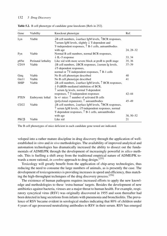
132 5 Drug Discovery
veloped into a rather mature discipline in drug discovery through the application of well-established in vitro and in vivo methodologies. The availability of improved analytical andautomation technologies has dramatically increased the ability to dissect out the funda-mentals of ADME/PK through the development of increasingly powerful in silico meth-ods. This is fuelling a shift away from the traditional empirical nature of ADME/PK to-wards a more rational, in cerebro approach to drug design.[255]
Toxicology will greatly benefit from the application of chip-array technologies, thusreducing the need to consume the large numbers of animals, as is presently the case. Thedevelopment of toxicogenomics is providing increases in speed and efficiency, thus match-ing the high-throughput techniques of the drug discovery process.[256]
The existence of human pathogens requires increased efforts to apply the new knowl-edge and methodologies to these ‘extra-human’ targets. Besides the development of newantibiotics against bacteria, viruses are a major threat to human health. For example, respi-ratory syncytical virus (RSV) was originally discovered in 1955 and soon thereafter hadbeen detected in lung secretions from infants with pneumonia and bronchiolitis. The preva-lence of RSV became evident in serological studies indicating that 80% of children under4 years of age possessed neutralizing antibodies to RSV in their serum. RSV has emerged
Table 5.2. B cell phenotype of candidate gene knockouts [Refs in 252].
Gene Viabillty Knockout phenotype Ref.
Lyn Viable ↓B cell numbers, ↓surface IgM levels, ↑BCR responses,↑serum IgM levels, slightly ↓ T-dependent andT-independent responses, ↑ B-1 cells, autoantibodieswith age 24, 28–32
Fyn Viable Normal B cell numbers, normal BCR responses,↓ IL–5 response 33, 34
p85α Perinatal lethality Like xid with more severe block at proB to preB stage 35, 36CD19 Viable ↓B cell numbers, ↓BCR responses, ↓serum Ig levels, 37–39
↓T-dependent responses,normal or ↑T-independent responses, ↑ B-1 cells
Gαq Viable No B cell phenotype described 40Gα11 Viable No B cell phenotype described 41SHIP Viable ↓B cell numbers, ↓surface IgM levels, ↑ BCR responses,
↓ FcãRIIb-mediated inhibition of BCR,↑ serum Ig levels, normal T-dependentresponses, ↑ T-independent responses 42–44
PTEN Embryonic lethal In +/- mice: ↑ number of activated B cells(polyclonal expansion), ↑ autoantibodies 45–49
CD22 Viable ↓B cell numbers, ↓surface IgM levels, ↑BCR responses,↑ serum IgM levels, ↓T-independent responses, normalT-dependent responses, ↑ B-1 cells, autoantibodieswith age 38, 50–52
PKCβ Viable Like xid 21
The B cell phenotypes of mice deficient in each candidate gene tested are indicated.
133
as the leading cause of lower respiratory tract infections in children and infants. The pres-ently only available option for treatment is the teratogen ribavirin, since earlier attempts todevelop a prophylactic RSV vaccine have been unsuccessful. Only a passive immunizationby means of a monoclonal antibody is available. Thus, there is strong need for the develop-ment of small-molecule therapeutics and progress is being made in that direction.[257]
A large number of drugs are currently used therapeutically without knowledge of theirtarget molecules or of the precise pharmacological mechanisms of action. This has resultednot only in complicated side effects and adverse reactions, but has also limited efficacy incertain patients and even in treatment failure. Identifying the target gene’s function canallow predictions about the drug’s efficacy and can indicate the likelihood of adverse reac-tions or individual sensitivity even before clinical trials have begun. Additionally, the de-velopment of a new drug depends on developing drug target molecules on the basis of theiraffinity for low-molecular-weight chemicals.
Developments in three crucial areas: chemical diversity, phenotype-based screening, andtarget identification, enable the identification of compounds inducing a specific cellular stateand leading to the identification of the proteins which regulate that state. This integratedchemical genetic approach is applicable to numerous biological or disease processes.
Chemical genetic screens have identified several small molecules targeting a TGF-β-responsive reporter gene, arresting mammalian cells in mitosis, inhibiting the function oftumor suppressor p53, and acting as subtype-selective somatostatin receptor agonist.
The combinatorial synthesis may be carried out on beads to generate a large number ofeasily separable compounds.
Phenotype-based screens are essential to assay small synthetic molecules and more com-plex substances for biological activity, such as the enhancement or suppression of a par-ticular phenotype. The different states may be morphologically identifiable, such as thespindly pathologic morphology of cells transformed with oncogenes v-ras and v-src. Thepathologic state is reverted by depudecin into the wild-type state.
High-throughput screening requires appropriate test systems to study large numbers ofcells simultaneously for the effects of different compounds. The automation of screeningassays is facilitateed by visual detection and the combination of optical methods with com-putational processing of images.
To facilitate drug receptor identification, latex beads were developed that can be adaptedto the small-scale purification of drug receptors.
Thus, a method using novel latex beads for rapid identification of drug receptors usingaffinity purification was developed. Composed of a glycidylmethacrylate (GMA) and astyrene copolmer core with a GMA polymer surface, the beads minimize non-specific pro-tein binding and maximize purification efficiency. Their performance was demonstrated byefficiently purifying FK506-binding protein using FK506-conjugated beads and it was foundthat the amount of material needed was significantly reduced compared with previous meth-ods. Using the latex beads, a redox-related factor was identified, Ref-1, as a target proteinof an anti-NF-κB drug, E3330, demonstrating that the existence of a new class of receptorsof anti-NF-κB drugs. The results suggest that the latex beads could provide a tool for theidentification and analysis of drug receptors and should therefore be useful in drug devel-opment.[258]
5 Drug Discovery
134 5 Drug Discovery
Genetic screens in S. cerevisiae were designed to identify mammalian non-receptormodulators of G protein signaling pathways. Strains lacking a pheromone-responsive GPCRsand expressing a mammalian–yeast Gα hybrid protein were made conditional for growthupon either pheromone pathway activation (activator screen) or pheromone pathway inac-tivation (inhibitor screen). Mammalian cDNAs that conferred plasmid-dependent growthunder restrictive conditions were identified. One of the cDNAs identified from the activatorscreen, a human Ras-related G protein that was termed AGS1 (for activator of G proteinsignaling), appears to function by facilitating guanosine triphosphate (GTP) exchange onthe heterotrimeric Gα. A cDNA product identified from the inhibitor screen encodes apreviously identified regulator of G protein signaling, human RGS5.[259]
5.1 Substances Derived from Bacteria, Plants, Insects,and Animals
Infectious diseases are making a comeback with menacing new features and increased re-sistance. Increased effort in anti-infectives research is necessary and on-going. Antibioticsare currently the third-largest selling class of drugs, with a worldwide market annuallybetween US$ 7 and 22 billion. Most current antibiotics are derived from approximately 15base compounds: β-lactams (e.g., penicillins, methicillin, cephalopsporin), aminoglycosides(e.g., strptomycin, getamycin, neomycin), quinolones (e.g., ciprofloxacin), macrolides (e.g.,erythromycin), lincosamides (e.g., clindamycin), sulfonamides (e.g., sulfadiazine),tetracyclins (e.g., glycycline) and glycopeptides (e.g., vancomycin), and are targeting only15 different bacterial targets.
Only two compounds presently in the clinic act on unconventional targets: oxazolidinones(which inhibit bacterial protein synthesis initiation by binding the 50S ribosomal subunit)and cationic peptides (which permeabilize bacterial membranes).[260]
Drug discovery has huge untapped reservoirs in nature as sources for novel active prin-ciples and lead structures. Molecular phylogeny has demonstrated that the majority ofmicrobial genomes are currently inaccessible. Key objectives of future efforts are the de-velopments of techniques for accessing ‘unculturable’ genomes, exploiting their bio-technologically valuable genes and products, and linking genome sequence data to mo-lecular structure and function.[261]
An intriguing way of actually stimulating nature to deliver new biologically active mol-ecules for drug development has been developed in the field of plants – more specificallyby milking plant roots stimulated by bacterial and fungal elicitors.[262]
Easy transformation and cultivation make plants suitable for production of many recom-binant proteins. Plants are capable of carrying out acetylation, phosphorylation andglycosylation as well as other post-translational protein modifications required for the bio-logical activity of many eukaryotic proteins. Numerous heterologous (recombinant) pro-teins have been produced in plant leaves, fruits, roots, tubers and seeds, and targeted todifferent subcellular compartments, such as the cytoplasm, endoplasmic reticulum or
135
apoplastic space. The extraction and purification of proteins from biochemically complexplant tissues is a laborious and expensive process, and is a major obstacle to large-scaleprotein manufacturing in plants. Secretion-based systems utilizing transgenic plant cell orplant organs aseptically cultivated in vitro have been investigated; however, though these invitro systems are expensive, slow growing, unstable and relatively low yielding.
The problems were addressed by engineering tobacco plants to continuously secreterecombinant proteins from their roots into a simple hydroponic medium. Three heterolo-gous proteins of diverse origins [GFP of jellyfish, human placental alkaline phosphatase(SEAP) and bacterial xylanase] were produced using the root secretion method (rhizo-secretion). Protein secretion was dependent on the presence of the endoplasmic reticulumsignal peptide fused to the recombinant protein sequence. All three secreted proteins re-tained their biological activity and, as shown for SEAP, accumulated in much higher amountsin the medium than in the root tissue.[263]
5.2 Sources of Active Principles
The huge reservoir of as-yet untapped molecular structures is evidenced in the microbialworld by the assessment of the microbial diversity.[264]
Natural products are an indispensable source for novel structures and chemical diversityfor drug discovery.[265] Aquatic sources and plant sources are an underutilized source ofactive principles or lead compounds.
Extremophiles are a source of evolution-hardened enzymes and principles, such as ther-mostable enzymes (Taq polymerase for PCR) and cold-adapted enzymes with applicationsin the detergent and food industries, fine chemicals production, bioremediation as well asbroader applicable mechanisms for their high catalytic efficiency based on structural X-raystudies.[266]
Natural products are the most consistently successful source of drug leads. Natural prod-ucts continue to provide greater structural diversity than standard combinatorial chemistryand offer major opportunities for finding novel low molecular weight lead structures thatare active against a wide range of assay targets. Since less than 10% of the world’s biodiversityhas been tested for biological activity – many more useful natural lead compounds areawaiting discovery. The challenge is how to access this natural chemical diversity. Of the520 new drugs approved between 1983 and 1994, 39% were natural products or derivedfrom natural products, and 60–80% of antibacterials and anti-cancer drugs were derivedfrom natural products.
Commercial evidence also supports the case for natural products. Of the 20 best-sellingnon-protein drugs in 1999, nine were either derived from or developed as the result of leadsgenerated by natural products (e.g., simvastatin, lovastatin, ciprofloxacin, clarithromycinand cyclosprin) with combined annual sales of greater than US$ 16 billion. Newer develop-ments based on natural products include the antimalarial drug artemisinin, and the antican-cer agents taxol, docetaxel and camptothecin. In addition to the historical success in drugdiscovery, natural products are likely to continue to be sources of new commercially viable
5.2 Sources of Active Principles
136 5 Drug Discovery
drug leads. The chemical novelty associated with natural products is higher than that of anyother source: 40% of the chemical scaffolds in a published database of natural products areabsent from synthetic chemistry. This is particularly important when searching for leadmolecules against newly discovered targets for which there is no known small-moleculelead. Natural products can be a more economical source of chemical diversity than thesynthesis of equivalent numbers of diverse chemicals.[267]
Toxins are a good source of active principles, since they have developed as part of long-term evolutionary strategies for survival, feeding and defense of organisms. Snake venomsprovide a rich source of active molecules that address ion channels and receptors with highaffinity and selectivity. Since high selectivity, the differentiation between pharmaco-logi-cally relevant targets and targets that have similar structure but different functions, is a keyto effective therapeutics, toxins may provide both novel structures for active principles aswell as lessons for achieving selectivity.[268]
Studies of physiological responses at the protein level lead to targets for therapies. Forexample, heat-shock and stress proteins, synthesized as a response to external or immuno-logic stress, provide targets for immunotherapeutic interventions.[269]
Amphibians could help in the search for new substances to treat antibiotic-resistant bac-teria such as the methicillin-resistant Staphylococcus aureus.
Peptides from the skin of the red-backed salamander (Plethodon cinereus) killed 90% ofa culture of staphyolococci within 2 h. Peptides may be leads to design and synthesizecompounds that act as novel antibiotics.[270]
The repertoire of active molecules has extended to include DNA itself. The therapeuticpotential of DNA enzymes was demonstrated with a stretch designed to bind to and cleavethe RNA made by a damage-sensing gene called Egr-1, which appears to keep balloonedarteries from closing up in rate models of heart disease.[271]
Prior to this, in an experiment of ‘directed evolution’, a DNA enzyme capable of effi-ciently cleaving RNA[426] had been generated, which has a higher cleavage efficiency thanany known ribozyme. To target a specific RNA molecule, the RNA-cleaving domain (RNA10–23) is fitted with stretches of DNA that bind to target sequences in the RNA. When the10–23 catalytic domain is within reach, it cleaves the RNA by speeding up the backgroundreaction making RNA inherently unstable.[272]
The rational design of active pharmaceutical compounds based on identification of genesinvolved in disease is demonstrated by the identification of compounds that stabilize theDNA binding domain of p53 in the active conformation. These small synthetic moleculesnot only promote the stability of wild-type p53 but also allowed mutant p53 to maintain anactive conformation. A prototype compound caused the accumulation of conformationallyactive p53 in cells with mutant p53, enabling it to activate transcription and to slow tumorgrowth in mice. This class of compounds may thus be developed into anticancer drugs ofbroad utility.[273]
New strategies are also delivering novel compounds in protein engineering. A recombi-nation strategy called iterative truncation for the creation of hybrid enzymes (ITCHY) isbased on the generation of N- or C-terminal fragment libraries of two genes by progressivetruncation of the coding sequences with exonuclease III followed by ligation of the prod-ucts to make a single-crossover hybrid library.[427] The studies involved a methodology,
137
ITCHY, that creates combinatorial fusion libraries between genes in a manner that is inde-pendent of DNA homology. The ability of ITCHY and DNA shuffling was compared increating interspecies fusion libraries between fragments of the E. coli and human glycin-amide ribonucleotide transformylase genes, which have only 50% identity on the DNAlevel. Sequencing of several randomly selected positives from each library illustrated thatITCHY identified a more diverse set of active fusion points including those regions of non-homology and those with crossover points that diverged from the sequence alignment.Some of the hybrids found by ITCHY that were fused at non-homologous locations hadactivities that were greater than or equal to the activity of the hybrids found by DNA shuf-fling.[274]
Alpha interferons (IFN-α) are members of the diverse helical-bundle superfamily ofcytokine genes. Although these proteins possess therapeutic value in the treatment of anumber of diseases, they have not been optimized for use as pharmaceuticals. For example,dose-limiting toxicity, receptor cross-reactivity and short serum half-lives significantly re-duce the clinical utility of many of the cytokines. DNA family shuffling is a method forpermutation of natural genetic diversity. It is a powerful tool for rapidly evolving genes,operons, and whole viruses for desired properties.
DNA shuffling of a family of over 20 human IFN-α (Hu-IFN-α) genes was used toderive variants with increased antiviral and antiproliferation activities in murine cells. Aclone with 135,000-fold improved specific activity over Hu-IFN-α2a was obtained in thefirst cycle of shuffling. After a second cycle of selective shuffling, the most active clonewas improved 285,000-fold relative to Hu-IFN-α2a and 185-fold relative to Hu-IFN-α1.Remarkably, the three most active clones were more active than the native murine IFN-α.These chimeras are derived from up to five parental genes but contained no random pointmutations. These results demonstrate that diverse cytokine gene families can be used asstarting material to rapidly evolve cytokines that are more active, or have superior selectiv-ity profiles, than native cytokine genes.[275]
Natural products are presently a major source of anti-fungal agents and lead structures.New inhibitors of cell wall biosynthesis are a major target for these efforts.[276]
For DNA as the target, a class of compounds called peptide nucleic acids (PNAs) hasbeen developed. PNAs are synthetic homologs of nucleic acids in which the phosphate–sugar polynucleotide backbone is replaced by a flexible polyamide, an uncharged mimicconsisting of repeating 2-aminoethyl-glycine units. The resulting decrease in electrostaticrepulsion allows the formation of a PNA–DNA hydrogen-bonded double helix, which ismore stable than the one formed by DNA–DNA interaction. Peptide nucleic acids are resis-tant to nucleases and proteases, and consequently are more stable in cells than oligonucle-otides. Though potentially capable of blocking gene expression in a selective and specificmanner, PNAs have never been shown to be effective anti-gene agents in intact live cells inculture because of their limited ability to reach cell nuclei. A study was carried out toexplore the condition s that would make anti-gene PNAs effective in intact cells in vitro.PNA was covalently linked to PKKKRKV, a basic NLS peptide. A 17mer anti-myc–PNA,complementary to a unique sequence located at the beginning of the second exon of theoncogene, was covalently linked at its N-terminus to the NLS peptide (PNA–mycwt–NLS).When BL cells were exposed to PNA–mycwt–NLS, the anti-gene construct was localized
5.2 Sources of Active Principles
138 5 Drug Discovery
predominantly in the cell nuclei and a rapid consequent down-regulation of c-myc expres-sion occurred. Both completion of a productive cell cycle and apoptosis were inhibited.[277]
The backbone of PNA is a polymer of N-(2-aminoethyl)glycine (AEG) which is occa-sionally referred to as ethylendiamine monoacetic acid.
Rational approaches are yielding active pharmaceutical ingredients. For example, nuclearhormone receptors are potential targets for therapeutic approaches to many clinical condi-tions, including cancer, diabetes and neurological diseases. The crystal structure of theligand-binding domain of agonist-bound nuclear hormone receptors enables the design ofcompounds with agonist activity. However, with the exception of the human estrogen re-ceptor-α, the lack of antagonist-bound ‘inactive’ receptor structures hinders the rationaldesign of receptor antagonists. A strategy was developed for designing such antagonists. Amodel of the inactive conformation of human retinoic acid receptor-α was constructed byusing information derived from antagonist-bound estrogen-receptor-α and a computer-basedvirtual screening algorithm was applied to identify retinoic acid receptors. The availablecrystal structures of nuclear hormone receptors may thus be used for the rational design ofantagonists, which could lead to the development of novel drugs for a variety of diseases.[278]
The increasing versatility of chemical and biotechnological syntheses are expanding therange of available molecular structures. Of special importance is access to complex carbo-hydrate and lipid structures since these are part of the cell surface and of membranes in-volved in transport, recognition and signal transduction. Biological membranes define theboundaries of the cellular compartments in higher eukaryotes and are active in many pro-cesses such as signal transduction and vesicular transport. Although post-translational lipidmodification of numerous proteins in signal transduction is crucial for biological function,analysis of protein–protein interaction has mainly focused on recombinant proteins in solu-tion under defined in vitro conditions. A new strategy was developed for the synthesis ofsuch lipid-modified proteins. It involves the bacterial expression of a C-terminally trun-cated non-lipidated protein, the chemical synthesis of differently lipidated peptides repre-senting the C terminus of the proteins, and their covalent coupling. The technique is dem-onstrated using Ras constructs, which exhibit properties very similar to fully processedRas, but can be produced in high yields and are open for selective modifications. Theseconstructs are operative in biophysical and cellular assay systems, showing specific recog-nition of effectors by Ras lipoproteins inserted into the membrane surface of biosensorsand transforming activity of oncogenic variants after microinjection into cultured cells.[279]
Another example of the power of specifically synthesized carbohydrate structures is inthe interplay with receptor-binding proteins such as toxins.
The diseases caused by Shiga and cholera toxin account for the loss of millions of liveseach year. Both belong to the clinically significant subset of bacterial AB5 toxins consist-ing of an enzymatically active A subunit that gains entry to susceptible mammalian cellsafter oligosaccharide recognition by the B5 homopentamer. Therapies might target the obliga-tory oligosaccharide–toxin recognition event, but the low intrinsic affinity of carbohydrate–protein interactions hampers the development of low-molecular-weight inhibitors. The toxinscircumvent low affinity by binding simultaneously to five or more cell-surface carbohy-drates. The use of the crystal structure of the B5 subunit of E. coli O157:H7 Shiga-liketoxin I (SLT-I) in complex with an analog of its carbohydrate receptor allowed to design an
139
oligovalent, water-soluble carbohydrate ligand (named STARFISH), with subnanomolarinhibitory activity. The in vitro inhibitory activity is 1–10 million-fold higher than that ofunivalent ligands and is by far the highest molar activity of any inhibitor yet reported forShiga-like toxins I and II. Crystallography of the STARFISH/Shiga-like toxin I complexexplains this activity. Two trisaccharide receptors at the tips of each of five spacer armssimultaneously engage all five B subunits of two toxin molecules.[280]
The fortuitous nature of drug discovery is illustrated by the discovery of nitroimidazopyranas a potential therapeutic agent against tuberculosis. Mycobacterium tuberculosis, whichcauses tuberculosis, is the greatest single infectious cause of mortality worldwide, killingroughly two million people annually. Estimates indicate that one-third of the world popula-tion is infected with latent M. tuberculosis and the surge of multidrug-resistant clinicalisolates of M. tuberculosis have reaffirmed tuberculosis as a primary public health threat.New antitubercular drugs with new mechanisms of action have not been developed in over30 years. A series of compounds was reported containing a nitroimidazopyran nucleus thatpossesses antitubercular activity. After activation by a mechanism dependent on M. tuber-culosis F420 cofactor, nitroimidazopyrans inhibited the synthesis of protein and cell walllipid. In contrast to current antitubercular drugs, nitroimidazopyrans exhibited bactericidalactivity against both replicating and static M. tuberculosis. Lead compound PA-824 showedpotent bactericidal activity against multidrug-resistant M. tuberculosis and promising oralactivity in animal infection models. Nitroimidazopyrans offer the practical qualities of asmall molecule with the potential for the treatment of tuberculosis.[281]
Structure-based drug design has emerged as a powerful tool to augment the empiricaland semi-empirical drug discovery processes. Successful application of this technique re-quires a disease target, a feasible therapeutic strategy including an assay system, a siteselection strategy and the computational chemistry skills for engineering appropriate leadcompounds.
The CD8–MHC class I crystal structure was used as a template for the de novo design oflow-molecular-weight surface mimetics. The analogs were designed from a local surfaceregion on the CD8 α chain directly adjacent to the bound MHC class I, to block the proteinassociations in the T cell activation cluster that occur upon stimulation of the CTLs. Onesmall conformationally restrained peptide showed dose-dependent inhibition of a primaryallogeneic CTL assay while having no effect on the CD4-dependent mixed lymphocytereaction. The analog’s activity could be modulated through subtle changes in its side chaincomposition. Administration of the analog prevented CD8-dependent clearance of a mu-rine retrovirus in BALB/c mice. In C57BL/6 mice challenged with the same retrovirus, theanalog selectively inhibited the antiviral CTL responses without affecting the ability of theCTLs to generate robust allogeneic responses.[282]
Aberrant apoptosis-mediated cell death is believed to result in a number of differenthuman diseases. For example, excessive apoptosis in the liver can result in fulminant andautoimmune forms of hepatitis. The possibility was studied that inhibition of Fas expres-sion in mice would reduce the severity of fulminant hepatitis. To do this, a chemicallymodified 2′-O-(2-methoxy)ethyl antisense oligonucleotide (ISIS 22023) inhibitor of mouseFas expression was developed. In tissue culture, this oligonucleotide induced a reduction inFas mRNA expression that was both concentration- and sequence-specific. In BALB/c
5.2 Sources of Active Principles
140 5 Drug Discovery
mice, dosing with ISIS 22023 reduced Fas mRNA and protein expressions in liver by 90%.The ID50 for this response was 8–10 mg/kg daily dosing, and the reduction was highlydependent on oligonucleotide sequence, oligonucleotide concentration in liver and treat-ment time. Pretreatment with ISIS 22023 completely protected mice from fulminant hepa-titis induced by agonistic Fas antibody, by a mechanism entirely consistent with an oligo-nucleotide antisense mechanism of action. In addition, oligonucleotide-mediated suppressionof Fas expression reduced the severity of acetaminophen-mediated fulminant hepatitis, butwas without effect on concanavalin A-mediated hepatitis. The results demonstrate that 2′-O-(2-methoxy)ethyl-containing antisense oligonucleotides targeting Fas can exert in vivopharmacological activity in liver and suggest that oligonucleotide inhibitors of Fas may beuseful in the treatment of human liver disease.[283]
5.3 Assay Systems and Models (e.g., Knock-out Mice)
Crucial to the processes of establishing the safety, efficacy, and mode of action of a drug isthe availability and choice of appropriate and validated models for the compound, proce-dure, test, device, theory, or therapy under investigation.
Knowledge of gene function is required to utilize sequence information to develop newdrugs to treat disease. In vitro techniques, such as antisense oligonucleotides (AS-ODNs),ribozymes and small molecules, can be used to inhibit gene expression and to provide cluesto gene function. Although functional genomics methods analogous to those used in vitroexist in organisms such as D. melanogaster, X. laevis and C. elegans, the only methods thatcan currently be used to analyze gene function in mammals are recombinant transforma-tion and the cre/lox conditional knock-out technology. Traditional knock-out technology islimited to the evaluation of a primary phenotype that reflects the first critical requirementof a gene during development. As many primary phenotypes are lethal, there may be littleuseful information at the end of the knock-out experiment. Secondary phenotypes can pro-vide insight into the role of the gene after its first requirement during development, provid-ing more data as to its function. A technique has been reported to define gene functionusing AS-ODN inhibition of gene expression in mice. A single intravenous injection of anAS-ODN targeting VEGF into pregnant mice resulted in a lack of primary angiogenesis.The critical window required to inhibit VEGF expression was defined and recapitulated theprimary loss of function phenotype observed in VEGF–/– embryos. This phenotype wassequence specific, and time and dose dependent. Injection of an AS-ODN targeting anothergene, E-adherin, into pregnant mice at E10 confirmed a hypothesized secondary pheno-type. AS-ODN inhibition of gene expression in utero provides a strategy for target valida-tion in functional genomics.[284]
Efficient approaches to uncover, confirm and evaluate the physiological functions ofgenes need to be developed continuously. At the level of molecular genetics, there are twomain routes to determine the function of genes with known sequences: one is through lossof function, the other is through gain of function. Gene function must often be studied inthe context of the entire organism or in a particular cell type at a specific stage of develop-

141
ment or disease. An ideal strategy for functional studies should encompass both effective-ness and versatility. Gene therapy research has provided effective tools to achieve geneticmanipulation through somatic gene transfer. In particular, the recombinant adenovirus is awidely used delivery vector for both gene therapy and functional studies.[285]
To accelerate the biological annotation of novel genes discovered in sequenced regionsof mammalian genomes, large deletions in the mouse genome are created targeted to in-clude clusters of such genes. The targeted deletion of a 450-kb region on mouse chromo-some 11 is described, which, based on computational analysis of the deleted murine se-quences and human 5q orthologous sequences, codes for nine putative genes. Micehomozygous for the deletion had a variety of abnormalities, including severe hypertri-glyceridemia, hepatic and cardiac enlargement, growth retardation, and premature mortal-ity. Analysis of triglyceride metabolism in these animals demonstrated a several-fold in-crease in hepatic very-low-density lipoprotein triglyceride secretion, the most prevalentmechanism responsible for hyper-triglyceridemia in humans. A series of mouse BAC andhuman YAC transgenes covering different intervals of the 450-kb deleted region were as-sessed for their ability to complement the deletion-induced abnormalities. These studiesrevealed that OCTN2, a gene recently shown to play a role in carnitine transport, was ableto correct the triglyceride abnormalities. The discovery of this previously unappreciatedrelationship between OCTN2, carnitine and hepatic triglyceride production is of particularimportance because of the clinical consequence of hypertriglyceridemia and the paucity ofgenes known to modulate triglyceride secretion.[286]
Another technique of interfering with and probing of genetic and biochemical networksis based on RNAi. This provides an elegant method of probing gene expression and gener-ating desired genotypes (respectively, phenotypes) for studying genetic mechanisms andfor the identification as well as the validation of drug targets. Gene-silencing mechanismsoperate in regulating gene expression and cellular differentiation, and contribute to chro-mosomal dosage compensation, genetic imprinting, virus resistance, transposon silencingand, thus, to normal and pathological genetic and phenotypical phenomena. RNA signalshave long been implied in these processes. dsRNA induces sequence-specific interferencein several organisms; this process is called RNA interference (RNAi).
In C. elegans, the introduction of double-stranded RNA triggers sequence-specific ge-netic interference (RNAi) that is transmitted to offspring. The inheritance properties asso-ciated with this phenomenon were examined. Transmission of the interference effect oc-curred through a dominant extragenic agent. The wild-type activities of the RNAi pathwaygenes rde-1 and rde-4 genes were required for the formation of this interfering agent butwere not needed for interference thereafter. In contrast, the rde-2 and mut-7 genes wererequired downstream for interference. These finding provide evidence for germline trans-mission of an extragenic sequence-specific silencing factor and implicate rde-1 and rde-4in the formation of the inherited agent.[287]
Pathogenic effects may be caused by mutant mRNA. In the case of myotonic dystrophy(MD, prevalence 1 in 7400 live births), is the most common form of muscular dystrophy inadult humans and results from the expansion of a CTG repeat in the 3′ untranslated regionof the DMPK gene. The mutant DMPK mRNA contains an expanded CUG repeat and isretained in the nucleus. An untranslated CUG repeat was expressed in an unrelated mRNA
5.3 Assay Systems and Models (e.g., Knock-out Mice)
142 5 Drug Discovery
in transgenic mice. Mice that expressed expanded CUG repeats developed myotonia andmyopathy, whereas mice expressing a non-expanded repeat did not. Transcripts with ex-panded CUG repeats are thus sufficient to generate a DM phenotype. This result argues fora role for RNA gain of function in disease pathogenesis.[288]
Epigenetic silencing by dsRNA is a widespread phenomenon for regulating gene ex-pression. This process, RNAi, is thought to involve targeted degradation of homologousmRNAs. In C. elegans, seven genes have been shown to be important for RNAi: the RNA-directed RNA polymerase homolog ego-1, mut-7, rde-2, rde-3, rde-4, mut-2 and rde-1, whichencodes a member of the eIF2c/zwille family.
To gain insight into the mechanism of degradation, RNAi was examined with respect tothe influence of mutations in the smg genes, which are required for nonsense-mediateddecay. For three of six smg genes tested, mutations resulted in animals that were initiallysilenced by dsRNA but then recovered; wild-type animals remained silenced. The levels oftarget mRNAs were restored during recovery, and RNA editing and degradation of thedsRNA were identical to those of the wild-type. Thus, persistence of RNAi relies on asubset of smg genes.[289]
The B lymphocytes of the immune system use multiple genetic mechanisms – gene re-arrangement, somatic hypermutation and gene conversion – to drive the generation of anti-body diversity. RNA editing provides another contribution to antibody gene diversifica-tion. RNA editing appears to be crucial for the production of the secondary antibody repertoirein mature B cells. A deficiency in a single gene product, activation-induced deaminase(AID), is sufficient to obliterate generation of the secondary antibody repertoire in bothhuman and mouse B cells.
Since both hypermutation and switching are abolished by AID deficiency, AID-directedmRNA editing could be required for these processes. Alternatively, AID-directed editingmight occur while the immunoglobulin transcripts are still attached to their genomic tem-plate.
AID may therefore turn out to be an attractive drug target in the therapeutic modulationof antibody-dependent autoimmune diseases.[290]
The identification of functional regions in complex proteins is key to understandingtheir mechanisms of action and is commonly directed by conserved structural motifs. Theeffect of expressing distinct domains of death-associated protein kinase (DAPK) was ana-lyzed and it was found that overexpression of the entire death domain could protect cellsfrom apoptosis induced by the complete DAPK and that the linker region could protectcells as well, whereas overexpressing the kinase domain had no effect. A random unbiasedapproach to identify minimal regions in DAPK that are critical for its ability to participatein apoptotic processes was utilized. Functional domains of DAPK were identified by ex-pression and selection of dominant-negative peptides that inhibited the activity of the pro-tein and thus prevented cells from undergoing apoptosis. DAPK is a Ca2+/calmodulin-regu-lated serine/threonine kinase with a multidomain structure that participates in apoptosisinduced by a variety of signals. To identify regions in this protein that are critical for itsproapoptotic activity, a genetic screen was performed on the basis of functional selection ofshort DAPK-derived fragments that could protect cells from apoptosis by acting in a domi-nant-negative manner. A library of randomly fragmented DAPK cDNA was expressed in
143
HeLa cells and these cells were treated with IFN-γ to induce apoptosis. Functional cDNAfragments were recovered from cells that survived the selection and those in the senseorientation were examined further in a secondary screen for their ability to protect cellsfrom DAPK-dependent TNF-α-induced apoptosis. Four biologically active peptides wereisolated that mapped to the ankyrin repeats, the linker region, the death domain and the C-terminal tail of DAPK. Molecular modeling of the complete death domain provided a struc-tural basis for the function of the death-domain-derived fragment by suggesting that theprotective fragment constitutes a distinct substructure. The last fragment, spanning the C-terminal serine-rich tail, defined a new regulatory region. Ectopic expression of the tailpeptide (17 amino acids) inhibited the function of DAPK, whereas removal of this regionfrom the complete protein caused enhancement of the killing activity, indicating that the C-terminal tail normally plays a negative regulatory role. This unbiased screen highlightsfunctionally important regions in the protein and reveals an additional level of regulation ofDAPK apoptotic function that does not affect the catalytic activity.[291]
Another example of utilizing the mouse system for elucidation of cellular and organismalphenomena is demonstrated in studies of bone formation, resorption and remodeling pro-cesses.
RANK (receptor activator of NF-κB) nullizygous mice were generated to determine themolecular genetic interactions between osteoprotegerin, osteoprotegerin ligand and RANKduring bone resorption and remodeling processes. RANK–/– mice lack osteoclasts, andhave a profound defect in bone resorption and remodeling and in the development of thecartilaginous growth plates of endochondral bone. The osteopetrosis observed in these micecan be reversed by transplantation of bone marrow from rag1–/– (recombinase activatinggene 1) mice, indicating that RANK–/– mice have an intrinsic defect in osteoclast function.Calciotropic hormones and proresorptive cytokines that are known to induce bone resorp-tion in mice and human were administered to RANK–/– mice without inducing hypercalce-mia, although TNF-α treatment leads to the rare appearance of osteoclast-like cells near thesite of injection. Osteoclastogenesis can be initiated in RANK–/– mice by transfer of theRANK cDNA back into hematopoietic precursors, suggesting a means to critically evalu-ate RANK structural features required for bone resorption. Together these data indicatethat RANK is the intrinsic cell surface determinant that mediates osteoprotegerin ligandeffects on bone resorption and remodeling as well as the physiological and pathologicaleffects of calciotropoic hormones and proresorptive cytokines.[292]
Validation of drugs or therapies requires definite relationships between active agentsand targets, be they genes, proteins, enzymes or receptors, preferably on the organismallevel.
A new approach was developed for obtaining high resolution in vivo imaging of geneexpression in opaque animals, previously not possible by existing techniques. Such imageswere obtained by MRI using an MRI contrast agent that can indicate reporter gene expres-sion in living animals. MRI contrast agents were prepared in which the access of water tothe first coordination sphere of a chelated paramagnetic ion is blocked with a substrate thatcan be removed by enzymatic cleavage. Following cleavage, the paramagnetic ion can in-teract directly with water protons to increase the MRI signal. An agent was synthesizedwhere galactopyranose is the blocking group. This group renders the MRI contrast agent
5.3 Assay Systems and Models (e.g., Knock-out Mice)

144 5 Drug Discovery
sensitive to expression of the commonly used marker gene, β-galactosidase. To cellularresolution, regions of higher intensity in the MRI correlate with regions expressing markerenzyme. These studies offer the promise of in vivo mapping of gene expression in transgenicanimals and validate a general approach for constructing a family of MRI contrast agentsthat respond to biological activity.[293]
Signaling pathways are the target of a combination of Drosophila genetics to identifythe genetic basis of diabetes type II and tumors, followed by validation of identified targetsin a mouse model.[660]
145
6 Molecular Pharma-Biotechnology
The increasing availability of DNA sequences, structural data and analyses of interactionsis opening a plethora of new targets for therapeutic intervention.
DNA may be the target of sequence changes (gene therapy), or interact with small mol-ecules (RNA, PNA, pharmaceuticals, peptides) or large proteins; RNA may be target forsmall molecules, and proteins for small molecules or interacting large ones.
The advances in the determination of RNA structure and function have a significantimpact on drug discovery and the pharmaceutical industry. RNA serves as a messengerbetween DNA and proteins and has a surprising complexity in its structure. Both RNA andproteins are potential drug-binding sites, thus the number of potential targets is more thandoubled and drugs interacting with RNA may produce effects that cannot be achieved byinteraction with proteins.[294]
The logical and necessary successors to the Human Genome Project, which deliversprimary sequence data, are comparable efforts in bioinformatics (to understand the mean-ing of the sequences), in structural studies (to understand the structures and interactions ofmolecules derived from gene sequences) and the molecular design of interacting (small)molecules as well as (finally?) the overall biological systems for validation, verificationand application (e.g., mouse, zebra fish, Drosophila and human).[295]
Structural genomics requires methods to determine the three-dimensional structures ofproteins and complexes.
Sample damage by X-rays and other types radiation limits the resolution of structuralstudies on individual biomolecules or cells. Cooling can slow sample deterioration, butcannot eliminate damage-induced sample movement during the time needed for conven-tional measurements. Analyses of the dynamics of damage formation suggest that the con-ventional damage barrier (about 200 X-ray photons/Å with X-rays of 12 keV energy or 1 Åwavelength) may be extended at very high dose rates and very short exposure times. Com-puter simulations were used to investigate the structural information that can be recoveredfrom the scattering of intense femtosecond X-ray pulses by single protein molecules andsmall assemblies. Estimations of radiation damage as a function of photon energy, pulselength, integrated pulse intensity, and sample size show that experiments using very highX-ray dose rates and ultrashort exposures may provide useful structural information beforeradiation damage destroys the sample. Such ultra short, high-intensity X-ray pulses fromfree-electron lasers that are currently under development, in combination with container-free sample handling methods based on spraying techniques, obviating the need for macro-scopic crystals, will provide a new approach to structural determinations with X-rays.[296]
The application of genomic technologies to the study of infectious agents is yieldingresults for anti-infective therapies. The combination of sequencing, bacterial genomics,
Industrial Pharmaceutical Biotechnology. Heinrich KlefenzCopyright © 2002 Wiley-VCH Verlag GmbH
ISBNs: 3-527-29995-5 (Hardcover); 3-527-60012-4 (Electronic)

146 6 Molecular Pharma-Biotechnology
bioinformatics and proteomics provides targets for drug development and also demon-strates the efficiency of genotyping (susceptibility to infections correlated with specificgene sequences) for drug development and definition of patient subpopulations.[297]
Integrated pathway–genome databases enable and facilitate drug discovery. Such a de-scription of the genes and the genome of an organism as well as its predicted pathways,reactions, enzymes and metabolites provide, in conjunction with visualization and analysissoftware, the framework for improved understanding of microbial physiology and for anti-microbial drug discovery. Pathway-based analyses of the genomes of a number of medi-cally relevant microorganisms and a novel software tool that visualizes gene expressiondata on a diagram showing the whole metabolic network of the microorganism have beendescribed.[298]
Signal transduction pathways provide a rich source of drug active principles. The rapididentification of proteins that interact with a novel gene product is an important element offunctional genomics. A phage-display-based technique for interactive screening of com-plex cDNA libraries using proteins or synthetic peptides as bait has been developed. Start-ing with the epidermal growth factor receptor (EGFR) cytoplasmic tail, known proteininteractions were identified that link EGFR to the Ras/MAPK signal transduction cascadeand several novel interactions. This approach can be used as a rapid and efficient tool forelucidating protein networks and mapping intracellular signal transduction pathways.[299]
Dynamic protein–protein interactions are a key component of biological regulatory net-works. Dimerization events – physical interactions between related proteins – represent animportant subset of protein–protein interactions and are frequently employed in transduc-ing signals from the cell surface to the nucleus. Dimerization between different members ofa protein family can generate considerable functional diversity when different protein com-binations have distinct regulatory properties. A survey of processes known to be controlledby dimerization illustrates the diverse physical and biological outcomes achieved throughthis regulatory mechanism. These include facilitated proximity and orientation, differentialregulation by heterodimerization, generation of temporal and spatial boundaries, enhance-ment of specificity, and regulated monomer-to-dimer transitions. Elucidation of these mecha-nisms has led to the design of new approaches to study and to manipulate signal transduc-tion pathways.[300]
6.1 Bioinformatics
Bioinformatics is an interdisciplinary science that includes conceptual and practical meth-ods for the understanding, generation, processing, propagation and integration of biologi-cal information. The bioinformatics realm encompasses the input of DNA sequences, RNAsequences, protein sequences and structures plus knowledge of biochemical pathways, cel-lular and developmental processes, tissue and organismal physiology, normal and patho-logical processes. It yields knowledge about predicting gene function and structure, proteinsequences, three-dimensional structures of proteins and RNAs, and simulations of geneexpression networks, and of metabolic and signal transduction networks.[301]
1476.1 Bioinformatics
Computational methods are essential in processing the vast data streams from sequenc-ing efforts, structural information, protein analysis and differential gene expression. Com-putational approaches serve to integrate the research, development and discovery phases.An example of such efforts is the study of drug-likeness with respect to ADME parameters,in particular BBB penetration.[302]
The enormous task of determining the function and cooperation of proteins to create andmaintain biological systems draws on various methods, e.g., patterns of co-occurrence iden-tified from fused genes.[303]
Efficiency is achieved by a combination of experiments and computation, i.e. mergingthe results of expression profiling with DNA micro arrays with computation (co-occur-rence, phylogenetic profiles and fused genes).
The large-scale effort to measure, detect, and analyze protein–protein interactions experi-mentally includes biochemistry (e.g., co-immunoprecipitation or cross-linking), molecularbiology (e.g., the two-hybrid or phage display system) and genetics (e.g., unlinked non-complementing mutant detection). Using the two-hybrid system, a concerted effort has beenput into the analysis of the whole yeast genome. These approaches tend to be tedious, laborintensive, and inaccurate. From a computational perspective, the question is how to predictthat two proteins interact from structure or sequence alone. A method was developed thatidentifies gene-fusion events in complete genomes, based solely on sequence comparison.
Because there must be selective pressure for certain genes to be fused over the course ofevolution, functional associations of proteins can be predicted. In total, 215 genes or pro-teins in the complete genomes of E. coli, Haemophilus influenza and Methanococcusjannaschii are involved in 64 unique fusion events. The approach is of general applicabilityand can be applied to genes of unknown function.[304]
Figure 6.1 illustrates the multitude of expertise and interaction in biology.The sequencing of microbial genomes first focused on pathogenic organisms of medical
importance such as Haemophilus influenza and Mycoplasma genitalium in order to allowresearchers to develop drugs and vaccines for these pathogens. The completion elucidationof several non-pathogenic genomes such as Thermotoga maritima, whose genome revealsextensive potential lateral transfer with archaea, Deinococcus radiodurans, the most radia-tion-resistant microorganism known, and Aeropyrum pernix, the first Crearchaeota to becompletely sequenced, enables progress in studies on evolution, environment, industrialsynthesis, food technology and medicine.[471]
Table 6.1 lists selected genome sequencing projects of primary interest or application.Table 6.2 gives selecting databases for extracting physiological and functional in-forma-
tion from gene sequences, and Table 6.3 describes the contribution of genomics to non-medical applications of biotechnology.
The availability of over 20 fully sequenced genomes has forced the development of newmethods to find protein functions and interactions. Proteins were grouped by correlatedevolution, correlated mRNA expression patterns and patterns of domain fusion to deter-mine functional relationships among the 6217 proteins of the yeast S. cerevisiae. Usingthese methods, over 93,000 pair-wise links between functionally related yeast proteins werediscovered. Links between characterized and uncharacterized proteins allow a general func-tion to be assigned to more than half of the 2557 previously uncharacterized yeast proteins.
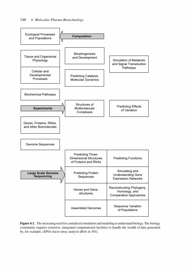
148 6 Molecular Pharma-Biotechnology
Figure 6.1. The increasing need for centralized simulation and modeling to understand biology. The biologycommunity requires extensive, integrated computational facilities to handle the wealth of data generatedby, for example, cDNA micro array analysis [Refs in 301].
Ecological Processes
and Populations
Tissue and Organismal
Physiology
Cellular and
Developmental
Processes
Biochemical Pathways
Morphogenesis
and Development
Predicting Catalysis,
Molecular Dynamics
Simulation of Metabolic
and Signal Transduction
Pathways
Genes, Proteins, RNAs,
and other Biomolecules
Genome Sequences
Structures of
Multimolecular
Complexes
Predicting Effects
of Variation
Predicting Three-
Dimensional Structures
of Proteins and RNAs
Predicting Functions
Predicting Protein
Sequences
Genes and Gene-
structures
Assembled Genomes
Simulating and
Understanding Gene
Expression Networks
Reconstructing Phylogeny,
Homology, and
Comparative Approaches
Sequence Variation
of Populations
Computation
Experiments
Large Scale GenomeSequencing
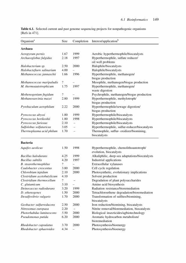
149
Table 6.1. Selected current and past genome sequencing projects for nonpathogenic organisms[Refs in 471].
Organisma Size Completion Interest/applicationb
Archaea
Aeropyrum pernix 1.67 1999 Aerobic hyperthermophile/biocatalystsArchaeoglobus fulgidus 2.18 1997 Hyperthermophile, sulfate reducer/
oil well problemsHalobacterium sp. 2.50 2000 Halophile/biocatalystsHalobactefiurn salinarium 4.00 – Halophile/biocatalystsMethanococcus jannaschii 1.66 1996 Hyperthermophile, methanogen/
biogas productionMethanococcus maripaludis ? – Mesophile, methanogen/biogas productionM. thermoautotrophicum 1.75 1997 Hyperthermophile, methanogen/
waste digestionMethenogenium fngidum ? – Psychrophile, methanogen/biogas productionMethanosarcinia mazei 2.80 1999 Hyperthermophile, methylotroph/
biogas productionPyrobaculum aerophilum 2.22 2000 Hyperthermophile/sewage digestion/
biogas productionPyrococcus abyssi 1.80 1999 Hyperthermophile/biocatalystsPyrococcus horikoshii 1.80 1998 Hyperthermophile/blocatalystsPyrococcus furiosus 2.10 – Hyperthermophile/biocatalystsSulfolobus solfataricus 3.05 – Hyperthermophile, sulfur-reducer/biocatalystsTherrnoplasma acid philum 1.70 – Thermophile, suffur- oxidizer/biomining,
biocatalysts
Bacteria
Aquifex aeolicus 1.50 1998 Hyperthermophile, chemolithoautotroph/evolution, biocatalysts
Bacillus halodurans 4.25 1999 Alkaliphilic, deep-sea adaptations/biocatalystsBacillus subtilis 4.20 1997 Industrial applicationsB. stearothermophilus ? – Extracellular xylanasesCaulobacter crescentus 3.80 2000 Cell cycle regulationChlorobium tepidum 2.10 2000 Photosynthetic, evolutionary implicationsClostridium acetobutylicum 4.10 Solvent productionClostridium thermocellum ? – Degradation of plant polysaccharidesC. glutamicum 3.10 – Amino acid biosynthesisDeinocaccus radiodurans 3.20 1999 Radiation resistance/bioremediationD. ethenogenes 1.50 2000 Tetrachloroethene degradation/bioremediationDesulfovibrio vulgaris 1.70 2000 Transformation of sulfites/biomining,
biocatalystsGeobacter sulfurreducens 2.50 2000 Iron reduction/biomining, biocatalystsNitrosomas europaea 2.20 – Nitrite removal/bloremediation, biocatalystsPhotorhabdus luminescons 5.50 2000 Biological insecticides/agbiotechnologyPseudomonas putida 6.20 2000 Aromatic hydrocarbon metabolism/
bioremediationRhodobacter capsulatus 3.70 2000 Photosynthesis/bioenergyRhodobacter sphaeroides 4.34 – Photosynthesis/bioenergy
6.1 Bioinformatics
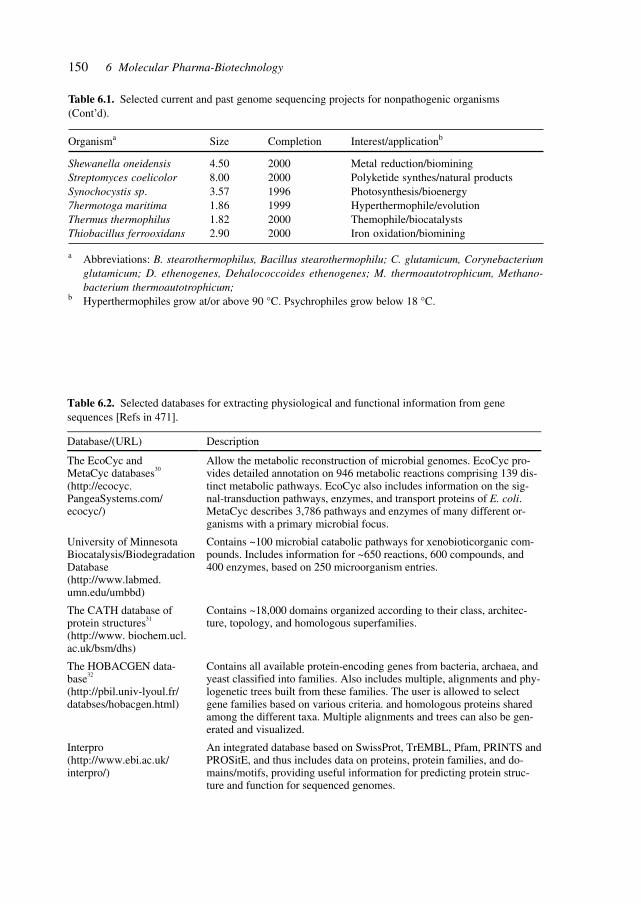
150 6 Molecular Pharma-Biotechnology
Table 6.1. Selected current and past genome sequencing projects for nonpathogenic organisms(Cont’d).
Organisma Size Completion Interest/applicationb
Shewanella oneidensis 4.50 2000 Metal reduction/biominingStreptomyces coelicolor 8.00 2000 Polyketide synthes/natural productsSynochocystis sp. 3.57 1996 Photosynthesis/bioenergy7hermotoga maritima 1.86 1999 Hyperthermophile/evolutionThermus thermophilus 1.82 2000 Themophile/biocatalystsThiobacillus ferrooxidans 2.90 2000 Iron oxidation/biomining
a Abbreviations: B. stearothermophilus, Bacillus stearothermophilu; C. glutamicum, Corynebacteriumglutamicum; D. ethenogenes, Dehalococcoides ethenogenes; M. thermoautotrophicum, Methano-bacterium thermoautotrophicum;
b Hyperthermophiles grow at/or above 90 °C. Psychrophiles grow below 18 °C.
Table 6.2. Selected databases for extracting physiological and functional information from gene sequences [Refs in 471].
Database/(URL) Description
The EcoCyc and MetaCyc databases30
(http://ecocyc. PangeaSystems.com/ ecocyc/)
Allow the metabolic reconstruction of microbial genomes. EcoCyc pro-vides detailed annotation on 946 metabolic reactions comprising 139 dis-tinct metabolic pathways. EcoCyc also includes information on the sig-nal-transduction pathways, enzymes, and transport proteins of E. coli. MetaCyc describes 3,786 pathways and enzymes of many different or-ganisms with a primary microbial focus.
University of Minnesota Biocatalysis/Biodegradation Database (http://www.labmed. umn.edu/umbbd)
Contains ~100 microbial catabolic pathways for xenobioticorganic com-pounds. Includes information for ~650 reactions, 600 compounds, and 400 enzymes, based on 250 microorganism entries.
The CATH database of protein structures31 (http://www. biochem.ucl. ac.uk/bsm/dhs)
Contains ~18,000 domains organized according to their class, architec-ture, topology, and homologous superfamilies.
The HOBACGEN data-base32
(http://pbil.univ-lyoul.fr/ databses/hobacgen.html)
Contains all available protein-encoding genes from bacteria, archaea, and yeast classified into families. Also includes multiple, alignments and phy-logenetic trees built from these families. The user is allowed to select gene families based on various criteria. and homologous proteins shared among the different taxa. Multiple alignments and trees can also be gen-erated and visualized.
Interpro (http://www.ebi.ac.uk/ interpro/)
An integrated database based on SwissProt, TrEMBL, Pfam, PRINTS and PROSitE, and thus includes data on proteins, protein families, and do-mains/motifs, providing useful information for predicting protein struc-ture and function for sequenced genomes.
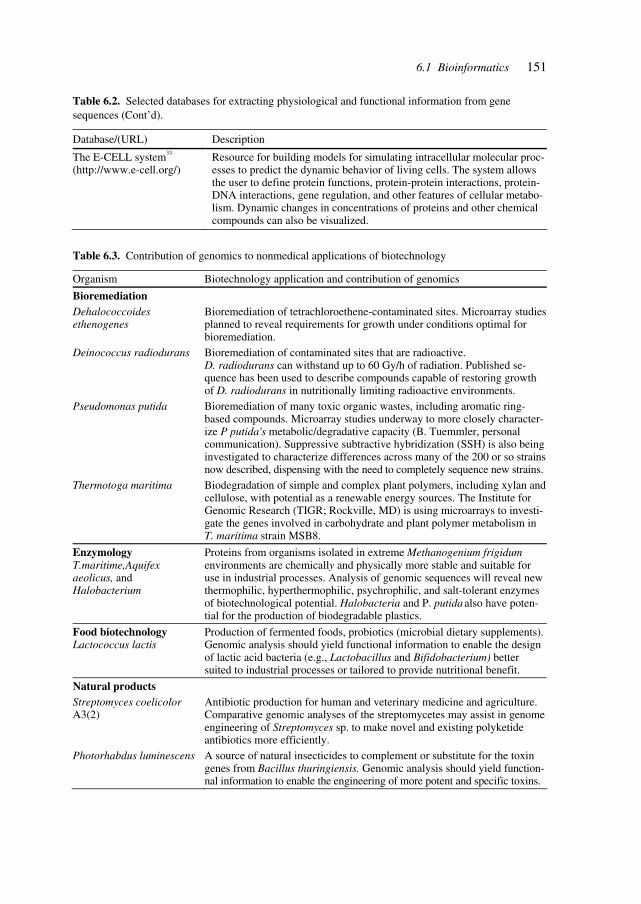
1516.1 Bioinformatics
Table 6.2. Selected databases for extracting physiological and functional information from gene sequences (Cont’d).
Database/(URL) Description
The E-CELL system33
(http://www.e-cell.org/) Resource for building models for simulating intracellular molecular proc-esses to predict the dynamic behavior of living cells. The system allows the user to define protein functions, protein-protein interactions, protein-DNA interactions, gene regulation, and other features of cellular metabo-lism. Dynamic changes in concentrations of proteins and other chemical compounds can also be visualized.
Table 6.3. Contribution of genomics to nonmedical applications of biotechnology
Organism Biotechnology application and contribution of genomics
Bioremediation Dehalococcoides ethenogenes
Bioremediation of tetrachloroethene-contaminated sites. Microarray studies planned to reveal requirements for growth under conditions optimal for bioremediation.
Deinococcus radiodurans Bioremediation of contaminated sites that are radioactive. D. radiodurans can withstand up to 60 Gy/h of radiation. Published se-quence has been used to describe compounds capable of restoring growth of D. radiodurans in nutritionally limiting radioactive environments.
Pseudomonas putida Bioremediation of many toxic organic wastes, including aromatic ring-based compounds. Microarray studies underway to more closely character-ize P putida's metabolic/degradative capacity (B. Tuemmler, personal communication). Suppressive subtractive hybridization (SSH) is also being investigated to characterize differences across many of the 200 or so strains now described, dispensing with the need to completely sequence new strains.
Thermotoga maritima Biodegradation of simple and complex plant polymers, including xylan and cellulose, with potential as a renewable energy sources. The Institute for Genomic Research (TIGR; Rockville, MD) is using microarrays to investi-gate the genes involved in carbohydrate and plant polymer metabolism in T. maritima strain MSB8.
Enzymology T.maritime,Aquifex aeolicus, and Halobacterium
Proteins from organisms isolated in extreme Methanogenium frigidum environments are chemically and physically more stable and suitable for use in industrial processes. Analysis of genomic sequences will reveal new thermophilic, hyperthermophilic, psychrophilic, and salt-tolerant enzymes of biotechnological potential. Halobacteria and P. putida also have poten-tial for the production of biodegradable plastics.
Food biotechnology Lactococcus lactis
Production of fermented foods, probiotics (microbial dietary supplements). Genomic analysis should yield functional information to enable the design of lactic acid bacteria (e.g., Lactobacillus and Bifidobacterium) better suited to industrial processes or tailored to provide nutritional benefit.
Natural products Streptomyces coelicolor A3(2)
Antibiotic production for human and veterinary medicine and agriculture. Comparative genomic analyses of the streptomycetes may assist in genome engineering of Streptomyces sp. to make novel and existing polyketide antibiotics more efficiently.
Photorhabdus luminescens A source of natural insecticides to complement or substitute for the toxin genes from Bacillus thuringiensis. Genomic analysis should yield function-nal information to enable the engineering of more potent and specific toxins.
152 6 Molecular Pharma-Biotechnology
Examples of functional links are given for a protein family of previously unknown func-tion, a protein whose human homologs is implicated in colon cancer and the yeast prionSup35.[305]
An increasing amount of DNA sequence is becoming available with the continuing ef-forts in deciphering the genomes of complex organisms. Only a small fraction (about 3% ofhuman chromosome 22) encodes proteins. Knowledge of the genetic code makes it feasibleto recognize the amino acid-coding parts of genes and to perform sophisticated compari-sons. The problem of recognizing gene regulatory sequences was tackled using data col-lected from microarray studies. The algorithm developed, MobyDick, reveals common motifsassociated with genes whose expression changes during either sporulatin or general repres-sion in yeast.
The availability of complete genome sequences has facilitated whole-genome expres-sion analysis and led to rapid accumulation of gene expression data from high-densityDNA microarray experiments. Correlating the information coded in the genome sequencesto these expression data is crucial to understanding how transcription is regulated at thegenomic scale. A great challenge is to decipher the information coded in the regulatoryregions of genes that control transcription. The development of computational tools foridentifying regulatory elements has lagged behind those for sequence comparison and genediscovery. One approach has been to delineate, as sharply as possible, a group of 10–100co-regulated genes and then find a pattern common to most of the upstream regions. Theanalysis tools used range from general multiple-alignment algorithms yielding a weightmatrix to comparison of the frequency counts of substrings with some reference set.
Although multiple copies of a single sequence motif may confer expression of a reportergene with a minimal core promoter, real genomes are not designed as disjoint groups ofgenes each controlled by a single factor. To tailor a gene’s expression to many differentconditions, multiple control elements are required and signals are integrated. Genome-wide control is considered to be achieved by combinatorial use of multiple sequence ele-ments. Microarray experiments for yeast have shown quantitatively that most occurrencesof proven binding motifs are in non-responding genes and that many responding genes donot have the motif.
The availability of complete genome sequences and mRNA expression data for all genescreates new opportunities and challenges for identifying DNA sequence motifs that controlgene expression. The algorithm MobyDick decomposes a set of DNA sequences into themost probable dictionary of motifs or words. This method is applicable to any set of DNAsequences, e.g., all upstream regions in a genome or all genes expressed under certainconditions. Identification of words is based on a probabilistic segmentation model in whichthe significance of longer words is deduced from the frequency of shorter ones of variouslengths, eliminating the need for a separate set of reference data to define probabilities. Adictionary was built with 1200 words for the 6000 upstream regulatory regions in the yeastgenome; the 500 most significant words (some with as few as 10 copies in all of the up-stream regions) match 114 of 443 experimentally determined sites (a significance level of18 standard deviations). When analyzing all of the genes up-regulated during sporulationas a group, many motifs are found in addition to the few previously identified by analyzingthe subclusters individually to the expression subclusters. By applying MobyDick to the
153
genes de-repressed when the general repressor Tup1 is deleted, both known as well asputative binding sites for its regulatory partners were found.[306]
DNA microarray technology and genome sequencing have advanced to the point that itis now possible to monitor gene expression levels on a genomic scale. These data promiseto enhance the fundamental understanding of life on the molecular level, from regulation ofgene expression and gene function to cellular mechanisms, and may prove useful in medi-cal diagnosis, treatment, and drug design. Analysis of these data requires mathematicaltools that are adaptable to the large quantities of data, while reducing the complexity of thedata to make them comprehensible. Analysis has so far been limited to the identification ofgenes and arrays with similar expression patterns by using clustering methods.
The use of a singular value decomposition in transforming genome-wide expressiondata from genes × arrays space to reduced diagonalized eigenarrays × eigenarrays spacehas been described, where the eigengenes (or eigenarrays) are unique orthonormal super-positions of the genes (or arrays). Normalizing the data by filtering out the eigengenes (oreigenarrays) that are inferred to represent noise or experimental artifacts enables meaning-ful comparison of the expression of different genes across different arrays in different ex-periments. Sorting the data according to the eigengenes and eigenarrays gives a globalpicture of the dynamics of gene expression, in which individual genes and arrays appear tobe classified into groups of similar regulation and function or similar cellular state andbiological phenotype, respectively. After normalization and sorting, the significanteigengenes and eigenarrays can be associated with observed genome-wide effects of regu-lators or with measured samples in which these regulators are overactive or underactive,respectively.[307]
The pharmaceutical industry has embraced genomics as a source of drug targets and hasrecognized bioinformatics as crucial for exploiting the data produced by genome-widesequencing and analysis. Bioinformatics is a cross-disciplinary endeavor including severalaspects of molecular biology, biochemistry, cell biology, computer science, software engi-neering, and techno-mathematics. Bioinformatics is a key aspect of drug discovery in thegenomic revolution contributing both to target discovery and target validation. In order tomake sense of the mass of data from genome-wide analyses, proteomics and protein func-tion as well as cell biology (cytomics) including expressed sequence tags, model organismsequences, microbial genome sequences, polymorphisms and gene expression data, it isessential that the data be handled in an integrated system-encompassing manner.[308]
Genome-wide transcript profiling has been used to monitor signal transduction during ayeast pheromone response and genetic manipulation allowed analysis of changes in geneexpression underlying pheromone signaling, cell cycle control and polarized morphogen-esis. This global transcript analysis reflects biological responses associated with the activa-tion and perturbation of signal transduction pathways, and thus demonstrates the power ofgenome-wide DNA microarrays to give broad correlation of gene activity with alterationsin physiological and developmental states.[309]
The assignment of function to novel genes uncovered by systematic genome-sequenc-ing programmes is a major challenge. Part of the necessary repertoire is the analysis ofpatterns of gene expression via the transcriptome, proteome and metabolome. Functionalgenomics is partly an exercise in pattern classification and, since many genes have known
6.1 Bioinformatics
154 6 Molecular Pharma-Biotechnology
functional classes, the prediction of their functional class is a learning task. The develop-ment of better-structured functional classes will facilitate the prediction of biochemicallytestable functions.[310]
Knowledge of gene sequences for the prediction of function does not yield descriptionsof multiple functional sites. Structural descriptors for protein functional sites are essentialfor deciphering sequence meaning.[311]
Computational methods for recognizing coding sequences in genomic DNA are capableof detecting most genes. The identification of regulatory elements in the 95% of the ge-nome composed of non-coding sequences is a substantial challenge.
Long-range regulatory elements are difficult to discover experimentally. They tend to beconserved among mammals, suggesting that cross-species comparisons should identify them.To search for regulatory sequences, about 1 Mb of orthologous human and mouse sequenceswas examined for conserved non-coding elements with greater than or equal to 70% iden-tity over at least 100 bp. Ninety non-coding sequences that met these criteria were discov-ered and the analysis of 15 of these elements found that about 70% were conserved acrossmammals. Characterization of the largest element in yeast artificial chromosome transgenicmice revealed it to be a coordinate regulator of three genes, IL-4, IL-13 and IL-5, spreadover 120 kb.[312]
The availability of whole-genome sequence information allows the identification of pro-tein motifs by sequence alignments, pattern recognition algorithms, protein-folding algo-rithms, and enables the identification of biologically relevant interacting molecules such asDNA, RNA, protein and small-molecule ligands. Computational methods can be used toidentify protein-interaction motifs in open reading frames (ORFs). The coiled-coil interac-tion motif allows the identification of candidate ligands for other interacting moleculesbeyond the computational or algorithmic capabilities. The coiled-coil interaction motifconsists of two or more α-helices that wrap around each other and the ligands for coiled-coil sequences are generally other coiled-coil sequences which facilitates the motif/ligandrecognition determination. A two-step approach was used to identify protein–protein inter-actions mediated by two-stranded coiled coils that occur in S. cerevisiae. Coiled coils fromthe yeast genome were first predicted computationally by the multicoil program and asso-ciations were then determined experimentally by using the yeast two-hybrid assay. Be-tween 162 putative coiled-coil sequences, 213 unique interactions were reported and theresulting interactions studied by focusing on associations between components of the spindlepole body (the yeast centromere).[470]
The increasing number of targets for therapeutic interventions requires concepts to dealwith the interdependencies of numerous protein–protein, protein–DNA and protein–DNAinteractions that are affected by targeting certain receptors, enzymes and control regions.The genetic and biochemical networks, which underlie such processes as homeostasis inmetabolism and the developmental programs of living cells must withstand considerablevariations and random perturbations of biochemical parameters. These occur as transientchanges in, for example, transcription, translation, and RNA and protein degradation. Theintensity and duration of these perturbations differ between cells in a population. The uniquestate of cells, and thus the diversity in a population is due to the different environmentalstimuli the individual cells experience and the inherent stochastic nature of biochemical
155
processes. Autoregulatory, negative feedback loops in gene circuits may provide stability,thereby limiting the range over which the concentrations of network components fluctuate.Simple gene circuits were designed and constructed consisting of a regulator and transcrip-tional repressor modules in E. coli, and a gain of stability was shown to be produced bynegative feedback.[313]
The study of complex phenotypes is gaining increasing importance with the elucidationof molecular mechanisms in genomics and proteomics. Attempts to influence mesoscopicrealms between the genotype and phenotype require an understanding of the networks andinteractions of numerous proteins, complexes, effectors, agonists and antagonists in a sys-tematic approach.
Chemotactic behavior and the control of metabolism are examples of complex pheno-types with complicated networks and pathways of signaling and other proteins. In the caseof metabolism, the rules describing complex, context-dependent processes depend directlyon thermodynamics and kinetics. Analyses of metabolic flux show the principle of distrib-uted control governing the phenotype in mammalian and bacterial systems interacting withthe environment, and explain the robust nature of these networks.
The study of network architecture allows the pinpointing of crucial interactions that areamenable to influence by genetic or pharmacological or other means to achieve desiredchanges in disease phenotypes. The study of complexity in embryology, development, car-cinogenesis, metabolism, genetic, and protein regulatory networks is a crucial area foraddressing complex traits or multifactorial disease phenomena.[314]
The complexity of intermediary metabolism and the containment of a combinatorialexplosion are addressed by looking at the origins of intermediary metabolism as a networkof catalytic reactions and molecules by thermodynamic and energetic considerations.
The core of intermediary metabolism in autotrophs is the citric acid cycle. In certaingroups of autotrophs, the reductive citric acid cycle is an engine of synthesis, taking incarbon dioxide and synthesizing the molecules of the cycle. The chemistry of a modelsystem of carbon, hydrogen and oxygen that starts with carbon dioxide and reductants anduses redox couples as an energy source was studied. To investigate the reaction networksthat might emerge, use was made of the largest available database of organic molecules, i.e.Beilstein online, and pruned by a set of physical and chemical constraints applicable to themodel system. From the 3.5 million entries in Beilstein, one comes up with 153 moleculesthat contain all 11 members of the reductive citric acid cycle. A small number of selectionrules generates a very constrained subset, suggesting that this is the type of reaction modelthat will prove useful in the study of biogenesis. The model indicates that the metabolismshown in the universal chart of pathways may be central to the origin of life, is emergentfrom organic chemistry and may be unique.[315]
The identification of genetic networks is a crucial basis for bioinformatics and an inter-esting experimental technique has been developed.
Homeogenes code for homeoproteins, a large family of transcription factors character-ized by their highly conserved 60-amino-acid DNA-binding homeodomain. They are highlyexpressed in the developing and adult nervous system, where they have been linked tomany normal and pathological processes. Phenotypic analyses of gene-targeted mouseembryos have been invaluable for understanding homeogene functions during develop-
6.1 Bioinformatics
156 6 Molecular Pharma-Biotechnology
ment; however, defining their precise mode of action requires that their target genes beidentified.
The identification of the homeoprotein target genes is an important issue in develop-mental biology. A strategy was developed based on the internalization and nuclear address-ing of exogenous homeodomains, using an engrailed homeodomain (EnHD) to screen anES cell gene trap library. Eight integrated gene trap loci responded to EnHD. One is withinthe bullous pemphipoid antigen 1 (BPAG1) locus, in a region that interrupts two neuralisoforms. By combining in vivo electroporation with organotypic cultures, it was shownthat an already identified BPAG1 enhancer/promoter is differentially regulated by homeo-proteins Hoxc-8 and Engrailed in the embryonic spinal cord and mesencephalon. This strat-egy can therefore be used for identifying and mutating homeoprotein targets. Becausehomeodomain third helices can internalize proteins, peptides, phosphopeptides and antisenseoligonucleotides, this strategy should be applicable to other intracellular targets for charac-terizing genetic networks involved in a large number of pathophysiological states.[316]
The study of the genetic network that regulates the development of the Drosophila vi-sual system has resulted in the identification of several transcription factors and other nuclearproteins that are required for the specification of early eye morphogenesis. These factorsseem to act in a hierarchy in which sine oculis (so) is regulated directly by Pax-6, themaster control function. In turn, so requires eyes absent (eya), encoding a nuclear protein,to induce ectopic eyes. This genetic pathway has been established in Drosophila, but ho-mologous proteins also regulate eye development in vertebrates, suggesting that this regu-latory network is old, is conserved in evolution and has been adapted to the control ofdevelopment of different visual systems found in both clades. The identification and func-tional characterization of homologous genes in more primitive organisms, such as the Platy-helminthes, helps to clarify the age and extent of conservation of this genetic cascade.
A sine oculis gene in the planarian Girardia tigrina (Platyhelminthes; Turbrellaria;Tricladida) was identified. The planarian sine oculis gene (Gtso) encodes a protein with asine oculis (Six) domain and a homeodomain that shares significant sequence similaritywith So proteins assigned to the Six-2 gene family. Gtso is expressed as a single transcriptin both regenerating and fully developed eyes. Whole-mount in situ hybridization studiesshowed exclusive expression in photoreceptor cells. Loss of function of Gtso by RNAinterference during planarian development inhibits eye regeneration completely. Gtso isalso essential for maintenance of the differentiated state of photoreceptor cells. The resultsand the expression of Pax-6 in planarian eyes suggest that the same basic gene regulatorycircuit required for eye development in Drosophila and mouse is used in the prototypic eyespots of platyhelminthes and is truly conserved during evolution.[317]
Several recently developed computational approaches in comparative genomics go be-yond sequence comparison. By analyzing phylogenetic profiles of protein families, do-main fusions, gene adjacency in genomes and expression patterns, these methods predictmany functional interactions between proteins and help deduce specific functions for nu-merous proteins. Although some of the resultant predictions may not be highly specific,these developments herald a new era in genomics in which the benefits of comparativeanalysis of the rapidly growing collection of complete genomes will become increasinglyobvious.[318]
157
The classification of organisms was originally based on morphological and anatomicalcriteria. The data yielded by molecular analysis include DNA sequences, RNA sequences,protein sequences and structures. The comparison of genes and their products with respectto function in functional genomics and with respect to structure in structural genomics is acapability required for biological and pharmaceutical research and development. The ap-plication of large-scale gene expression measurement techniques yielded multi-dimensionaland multi-relational data thus necessitating tools for analyzing, clustering and visualizingsuch data. Available packages include the Expression Profiler (www.ebi.ac.uk) and SpaceExplorer (www.soi.city.ac.uk).
Visualization is an efficient way to utilize the human ability to process large amounts ofdata. Traditional visualization methods are based on clustering and tree representation, andare complemented by projecting objects onto a Euclidean space to reflect their structural orfunctional differences. The data are visualized without preclustering and can be exploreddynamically and interactively, e.g., in protein topology and gene expression.[463]
Developing models as a basis for simulating interactions and effects is a crucial basis fordesigning therapeutic intervention. Of particular importance is the modeling of key func-tional genes and proteins.
Programmed cell death or apoptosis plays a critical role in nature. It is a process by whichthe cell commits suicide when malfunctions arise from cell stress, cell damage or conflict-ing division signals. Cell suicide is also required, for instance, during normal embryonicdevelopment as tissues are formed. Maintaining the balance between cell death and survivalis key. Many cancers are difficult to eradicate because they fail to respond to apoptotic sig-nals. Conversely, neurodegenerative disorders such as Parkinson’s, Alzheimer’s and Hunting-don’s disease are characterized by excessive apoptotic activity in certain classes of neurons.Autoimmune disorders and central immune system phenomena such as the elimination ofvirus-infected T cells and elimination of active immune cells after successful immune re-sponse are also strongly linked with caspase activity. Central to apoptotic cell death is a familyof proteases termed caspases (cysteine-containing aspartate-specific proteases).
Under normal circumstances caspases are present as inactive proteins termed zymogensor pro-caspases, which must be activated. Once activated they seek out and dismantle keyprotein targets by making selective cuts after aspartate residues. The activation of thesepowerful enzymes (whose inactive forms are constitutively expressed) is regulated at anumber of points. A cascade of events must occur before a cell is irreversibly committed toapoptotic death. In response to stress, damage or a signal to undergo programmed celldeath, a family of caspases termed initiator caspases is activated. Active initiator caspasesthen activate a second group of caspases termed executioner or effector caspases. Onceactivated, executioner caspases seek out and cleave their respective protein targets, therebydismantling the cell.
Due to the central role of caspases in cancer, and in neurodegenerative and autoimmunedisorders, they are subject to intense studies. A mechanistic mathematical model was for-mulated on the basis of newly emerging information, describing key elements of receptor-mediated and stress-induced caspase activation. Mass-conservation principles were used inconjunction with kinetic rate laws to formulate ordinary differential equations that describethe temporal evolution of caspase activation. Qualitative strategies for the prevention of
6.1 Bioinformatics
158 6 Molecular Pharma-Biotechnology
caspase activation are simulated and compared with experimental data. Model predictionsare consistent with available information. The model could aid in better understandingcaspase activation and identifying therapeutic approaches promoting or retarding apoptoticcell death.[319]
The E. coli MG1655 genome has been completely sequenced. The annotated sequence,biochemical information and other information were used to reconstruct the E. coli meta-bolic map. The stoichiometric coefficients for each metabolic enzyme in the E. coli meta-bolic map were assembled to construct a genome-specific stoichiometric matrix. The E. colistoichiometric matrix was used to define the system’s characteristics and the capabilities ofE. coli metabolism. The effects of gene deletions in the central metabolic pathways on theability of the in silico metabolic networks to support growth were assessed and the in silicopredictions were compared with experimental observations. It was shown that, based onstoichiometric and capacity constraints, the in silico analysis was able to qualitatively pre-dict the growth potential of mutant strains in 86% of the cases examined. The synthesis ofin silico metabolic genotypes based on genomic, biochemical and strain-specific informa-tion is possible, and systems analysis methods are available to analyze and interpret themetabolic phenotype.[320]
The reconstruction of complete metabolic networks is possible from the annotated genesequence, biochemical and physiological information. The hypothesis that Escherichia coliuses its metabolism to grow at a maximal rate was tested by quantitative relationship be-tween a primary carbon source uptake rate, oxygen uptake rate, and maximal cellular growthwere found consistent with the hypothesis that the E. coli metabolic network is optimizedfor maximum growth, using the E. coli MG1655 metabolic reconstruction. Experimentsdescribing the quantitative relationship between a primary carbon source uptake rate, oxy-gen uptake rate, and maximal cellular growth were found consistent with the hypothesisthat the E. coli metabolic network is optimized for maximum growth. The combination ofin silico and experimental biology delivers quantitative genotype-phenotype relationshipinformation for metabolism in bacterial cells and may be useful for studies of metabolicengineering.[596]
Digital organisms are computer programs that self-replicate, mutate and adapt by natu-ral selection. They offer an opportunity to test generalizations about living systems thatmay extend beyond the organic life that is the subject of the study of biology. Two classesof digital organisms were generated: (i) simple organisms selected solely for rapid replica-tion and (ii) complex programs selected to perform mathematical operations that acceleratereplication through a set of defined metabolic rewards. To examine the differences in theirgenetic architecture, millions of single and multiple mutations were introduced into eachorganism and measured for their effects on the organism’s fitness. Complex organisms aremore robust than the simple ones with respect to the average effects of single mutations.Interactions among mutations are common and usually yield higher fitness than predictedfrom the component mutations assuming multiplicative effects. Such interactions are espe-cially important in the complex organisms. Frequent interactions among mutations havealso been seen in bacteria, fungi and fruit flies. The findings in digital organisms supportthe notion that interactions are a general feature of genetic systems and may offer a tool forstudying such interactive networks.[321]
159
Analysis of published sets of DNA microarray gene expression data by singular valuedecomposition has uncovered underlying patterns or characteristic modes in their temporalprofiles. These patterns contribute unequally to the structure of the expression profiles. Theessential features of a given set of expression profiles are captured using just a small num-ber of characteristic modes. This leads to the striking conclusion that the transcriptionalresponse of a genome is orchestrated in a few fundamental patterns of gene expressionchange. These patterns are both simple and robust, dominating the alterations in expressionof genes throughout the genome. The characteristic modes of gene expression that changein response to environmental perturbations are similar in such distant organisms as yeastand human cells. This analysis reveals simple regularities in the seemingly complex tran-scriptional transitions of diverse cells to new states and these provide insights into theoperation of the underlying genetic networks.[322]
The evolution of complexity needs to be considered when studying the various networksof proteins, genes and cellular interactions. The evolution of genomic complexity was in-vestigated in populations of digital organisms and monitored in detail for the evolutionarytransitions that increase complexity. In order to study the evolution of biological complex-ity in biological evolution, complexity needs to be both rigorously defined and measurable.An information-theoretic definition identifies genomic complexity with the amount of in-formation a sequence stores about its environment. Since natural selection forces genomesto behave as a natural Maxwell Demon, within a fixed environment, genomic complexity isforced to increase.[323]
Studies involving large volumes of high-dimensional data, such as gene sequence data,human or plant gene distributions, global climate patterns and stellar spectra, are confrontedwith the need for dimensionality reduction, i.e. the identification of meaningful low-dimen-sional structures hidden in their high-dimensional observations. The human brain perma-nently confronts this problem in its perception of the environment and extracts a manage-ably small number of perceptually pertinent features from high-dimensional sensory inputsfrom 30,000 auditory nerve fibres or 106 optic nerve fibers. An approach towards solvingdimensionality reduction problems utilizing easily measured local metric information tolearn the underlying global geometry of a data set was developed. Unlike classical tech-niques such as principal component analysis and multidimensional scaling, this approachis capable of discovering the non-linear degrees of freedom that underlie complex naturalobservations, such as irregularly shaped objects, three-dimensional structures of molecules,human handwriting or images of a face under different viewing conditions. In contrast toprevious algorithms for non-linear dimensionality reduction, this algorithm efficiently com-putes a globally optimal solution and, for an important class of data manifolds, ascertainsan asymptotical convergence to the true structure. This approach has applications in thecomparison of three-dimensional biological and chemical structures, in high-volume dataanalysis and real time operations, and optimization of micro- and nano-devices.[464]
Another powerful algorithm was developed for the computation of low-dimensionalembedding from high-dimensional inputs. The requirements for analyzing large amountsof multivariate data raises the fundamental problem of dimensionality reduction, the dis-covery of compact representations of high-dimensional data in exploratory data analysisand visualization. Local linear embedding (LLE) is an unsupervised learning algorithm
6.1 Bioinformatics
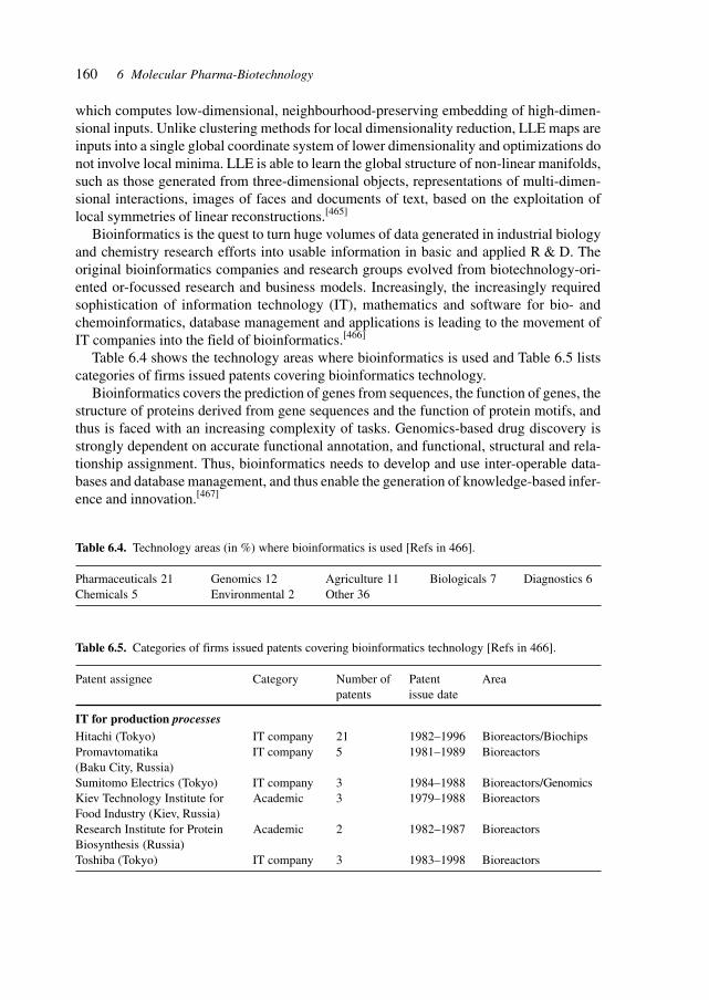
160 6 Molecular Pharma-Biotechnology
which computes low-dimensional, neighbourhood-preserving embedding of high-dimen-sional inputs. Unlike clustering methods for local dimensionality reduction, LLE maps areinputs into a single global coordinate system of lower dimensionality and optimizations donot involve local minima. LLE is able to learn the global structure of non-linear manifolds,such as those generated from three-dimensional objects, representations of multi-dimen-sional interactions, images of faces and documents of text, based on the exploitation oflocal symmetries of linear reconstructions.[465]
Bioinformatics is the quest to turn huge volumes of data generated in industrial biologyand chemistry research efforts into usable information in basic and applied R & D. Theoriginal bioinformatics companies and research groups evolved from biotechnology-ori-ented or-focussed research and business models. Increasingly, the increasingly requiredsophistication of information technology (IT), mathematics and software for bio- andchemoinformatics, database management and applications is leading to the movement ofIT companies into the field of bioinformatics.[466]
Table 6.4 shows the technology areas where bioinformatics is used and Table 6.5 listscategories of firms issued patents covering bioinformatics technology.
Bioinformatics covers the prediction of genes from sequences, the function of genes, thestructure of proteins derived from gene sequences and the function of protein motifs, andthus is faced with an increasing complexity of tasks. Genomics-based drug discovery isstrongly dependent on accurate functional annotation, and functional, structural and rela-tionship assignment. Thus, bioinformatics needs to develop and use inter-operable data-bases and database management, and thus enable the generation of knowledge-based infer-ence and innovation.[467]
Table 6.4. Technology areas (in %) where bioinformatics is used [Refs in 466].
Pharmaceuticals 21 Genomics 12 Agriculture 11 Biologicals 7 Diagnostics 6Chemicals 5 Environmental 2 Other 36
Table 6.5. Categories of firms issued patents covering bioinformatics technology [Refs in 466].
Patent assignee Category Number of Patent Areapatents issue date
IT for production processesHitachi (Tokyo) IT company 21 1982–1996 Bioreactors/BiochipsPromavtomatika IT company 5 1981–1989 Bioreactors(Baku City, Russia)Sumitomo Electrics (Tokyo) IT company 3 1984–1988 Bioreactors/GenomicsKiev Technology Institute for Academic 3 1979–1988 BioreactorsFood Industry (Kiev, Russia)Research Institute for Protein Academic 2 1982–1987 BioreactorsBiosynthesis (Russia)Toshiba (Tokyo) IT company 3 1983–1998 Bioreactors
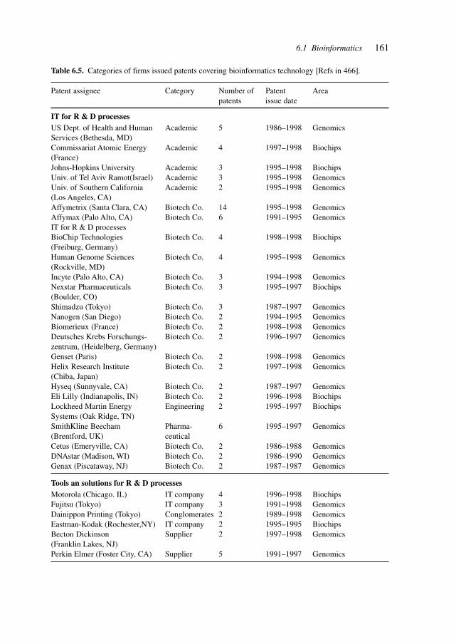
161
Table 6.5. Categories of firms issued patents covering bioinformatics technology [Refs in 466].
Patent assignee Category Number of Patent Areapatents issue date
IT for R & D processesUS Dept. of Health and Human Academic 5 1986–1998 GenomicsServices (Bethesda, MD)Commissariat Atomic Energy Academic 4 1997–1998 Biochips(France)Johns-Hopkins University Academic 3 1995–1998 BiochipsUniv. of Tel Aviv Ramot(Israel) Academic 3 1995–1998 GenomicsUniv. of Southern California Academic 2 1995–1998 Genomics(Los Angeles, CA)Affymetrix (Santa Clara, CA) Biotech Co. 14 1995–1998 GenomicsAffymax (Palo Alto, CA) Biotech Co. 6 1991–1995 GenomicsIT for R & D processesBioChip Technologies Biotech Co. 4 1998–1998 Biochips(Freiburg, Germany)Human Genome Sciences Biotech Co. 4 1995–1998 Genomics(Rockville, MD)Incyte (Palo Alto, CA) Biotech Co. 3 1994–1998 GenomicsNexstar Pharmaceuticals Biotech Co. 3 1995–1997 Biochips(Boulder, CO)Shimadzu (Tokyo) Biotech Co. 3 1987–1997 GenomicsNanogen (San Diego) Biotech Co. 2 1994–1995 GenomicsBiomerieux (France) Biotech Co. 2 1998–1998 GenomicsDeutsches Krebs Forschungs- Biotech Co. 2 1996–1997 Genomicszentrum, (Heidelberg, Germany)Genset (Paris) Biotech Co. 2 1998–1998 GenomicsHelix Research Institute Biotech Co. 2 1997–1998 Genomics(Chiba, Japan)Hyseq (Sunnyvale, CA) Biotech Co. 2 1987–1997 GenomicsEli Lilly (Indianapolis, IN) Biotech Co. 2 1996–1998 BiochipsLockheed Martin Energy Engineering 2 1995–1997 BiochipsSystems (Oak Ridge, TN)SmithKline Beecham Pharma- 6 1995–1997 Genomics(Brentford, UK) ceuticalCetus (Emeryville, CA) Biotech Co. 2 1986–1988 GenomicsDNAstar (Madison, WI) Biotech Co. 2 1986–1990 GenomicsGenax (Piscataway, NJ) Biotech Co. 2 1987–1987 Genomics
Tools an solutions for R & D processesMotorola (Chicago. IL) IT company 4 1996–1998 BiochipsFujitsu (Tokyo) IT company 3 1991–1998 GenomicsDainippon Printing (Tokyo) Conglomerates 2 1989–1998 GenomicsEastman-Kodak (Rochester,NY) IT company 2 1995–1995 BiochipsBecton Dickinson Supplier 2 1997–1998 Genomics(Franklin Lakes, NJ)Perkin Elmer (Foster City, CA) Supplier 5 1991–1997 Genomics
6.1 Bioinformatics
162 6 Molecular Pharma-Biotechnology
To support drug discovery efficiently, the biological information derived in bioinfor-matics needs to be combined with chemical data in order to facilitate the identification oflead structures and the development of small molecules as active pharmaceuticals. To thisend, different databases need to be brought together for the integration of disparate re-sults.[468]
Chemoinformatics needs to be brought to the fore and combined with bioinformatics inorder to accelerate the discovery of relationships between structure and biological activity,i.e. cross-database data mining. These activities need, in view of the huge amounts of data,to include advanced visualization techniques for efficient knowledge management.[469]
Iteration between models upon introduction of experimental results aids in verificationof hypotheses and the improvement of models.
The studies of interactions in the biological systems comprising genes, proteins, cellsa,tissues and respective networks leads to an integrative approach of biological and biomedi-cal research.
6.2 Biological Systems and Models
Biological systems are the key to validating the results of sequencing, structure analysisand functional predictions.
The models employed range from in vitro assays to cell cultures to whole organisms.Increasingly, the use of specific differentiated cells provides new models for understandingdifferentiation, function, drug discovery and testing plus transplantation and therapies.[324]
Complex biological systems are defined and studied which may provide the tools forstudying previously intractable problems. In the search for drugs to fight tumorigenesis,the study of tumor suppressor genes in humans and yeast with its homologs provides sucha biological model. The rare familial cancer phenotype called Li–Fraumeni syndrome, whichis highly penetrant, is usually associated with inherited mutations in the TP53 gene andheterozygous germline mutations in the hCHK2 gene are show to occur. The hCHK2 geneencodes the human homolog of yeast Cds1 and Rad 53 G2 checkpoint kinases, whoseactivation in response to DNA damage prevents cellular entry into mitosis. Thus, the hCHK2is a tumor suppressor gene conferring predisposition to sarcoma, breast cancer and braintumors, and links the central role of p53 inactivation in human cancer and the G2 check-point in yeast. The inactivation of this G2 checkpoint may have therapeutic implications inrendering cancer cells sensitive to DNA-damaging agents and thus triggering a mitoticcatastrophe during the attempt to segregate damaged chromosomes.[325]
Mouse models provide an important system to address studies of disease development,and for the development and testing of therapeutic compounds and strategies.
For cancer studies, several models are available, e.g., to study neurofibromatosis whichaffects about 1 in 3500 individuals worldwide. Neurofibromatosis type 1 (NF1) is a preva-lent familial cancer syndrome resulting from germline mutations in the NF1 tumor sup-pressor gene. Hallmark features of the disease are the development of benign peripheralnerve sheath tumors (neurofibromas), which can progress to malignancy. Unlike humans,
163
mice that are heterozygous for a mutation in Nf1 do not develop neurofibromas. Chimericmice, composed in part of Nf1–/– cells, do, which demonstrates that loss of the wild-typeNf1 allele is rate limiting in tumor formation.
In addition, mice carrying linked germline mutations in Nf1 and p53 develop malignantperipheral nerve sheath tumors (MPNSTs), which supports a cooperative and causal rolefor p53 mutations in MPNST development.[326]
Another resourceful biological system for studying genetic traits and elucidating func-tional and regulatory pathways is the zebra fish.
Defects in iron absorption and utilization lead to iron deficiency and overload disorders.Adult mammals absorb iron through the duodenum, whereas embryos obtain iron throughplacental transport. Iron uptake from the intestinal lumen through the apical surface ofpolarized duodenal enterocytes is mediated by the divalent metal transporter, DMT1. Asecond transporter has been postulated to export iron across the basolateral surface to thecirculation. Positional cloning was used to identify the gene responsible for the hypochro-mic anemia of the zebra fish mutant weissherbst. The gene, ferroportin1, encodes a mul-tiple transmembrane domain protein, expressed in the yolk sac, that is a candidate for theelusive iron exporter. Zebra fish ferroportin1 is required for the transport of iron frommaternally derived yolk stores to the circulation and functions as a iron exporter whenexpressed in Xenopus oocytes. Human Ferroportin1 is found at the basal surface of placen-tal syncytiotrophoblasts, suggesting that it also transports iron from mother to embryo.Mammalian Ferroportin1 is expressed at the basolateral of duodenal enterocytes and couldexport cellular iron into the circulation. Ferroportin1 function may be perturbed in mam-malian disorders of iron deficiency or overload.[327]
The Drosophila retina is patterned by a morphogenetic wave driven by the Hedgehogsignaling protein. Hedgehog, secreted by the first neurons, induces neuronal differentiationand Hedgehog expression in nearby uncommitted cells, thereby propagating the wave.Evidence was reported that the zebra fish Hedgehog homolog, Sonic Hedgehog, is alsoexpressed in the first retinal neurons, and that Sonic Hedgehog drives a wave of neurogenesisacross the retina, similar to the wave in Drosophila. The conservation of this patterningmechanism supports, in view of the highly divergent structures of vertebrate and inverte-brate eyes, a common evolutionary origin of the animal visual system.[328]
Genetic ablation (the elimination of genes in whole organisms by, for example, selectiveexpression of a toxin gene or by genetic deletion) is a powerful technique to study theeffects of genes in genetic and biochemical networks and pathways. This technique hasyielded valuable insights in neural development and in hormone production.
During neural development in vertebrates, a spatially ordered array of neurons is gener-ated in response to inductive signals derived from localized organizing centers. One orga-nizing center that has been proposed to have a role in the control of neural patterning is theroof plate. To define the contribution of signals derived from the roof plate to the specifica-tion of neuronal cell types in the dorsal neural tube, a strategy was devised to ablate the roofplate selectively in mouse embryos. Embryos without a roof plate lack all the interneuronsubtypes that are normally generated in the dorsal third of the neural tube. Using a geneti-cally based lineage analysis and in vitro assays, it was shown that the loss of these neuronsresults from the elimination of non-autonomous signals provided by the roof plate. These
6.2 Biological Systems and Models
164 6 Molecular Pharma-Biotechnology
results reveal that the roof plate is essential for specifying multiple classes of neurons in themammalian CNS.[329]
The study of receptors and their function may be aided by genetic methods of introduc-ing mutations or deletions in mice or other organisms and observing the phenotypical con-sequences. Reversing genetic defects by the introduction of transgenes validates the func-tional importance of the introduced gene and its product.
Targeted deletion of the metabotropic glutamate receptor subtype 1 (mGluR1) gene cancause defects in development and function in the cerebellum. The mGluR1 transgene wasintroduced into mGluR1-null mutant (mGluR1–/–) mice with a Purkinje cell-specific pro-moter. mGluR1-rescue mice showed normal cerebellar long-term depression and regres-sion of multiple climbing fiber innervation – events significantly impaired in mGluR1–/–
mice. The impaired motor coordination was rescued by this transgene, in a dose-dependentmanner. MGluR1 in Purkinje cells is a key molecule for normal synapse formation, synap-tic plasticity and motor control in the cerebellum.[330]
Olfactory receptors are G protein-coupled seven-transmembrane proteins encoded by adivergent multigene family in vertebrates and in Drosophila. Taste receptors are also thoughtto belong to the superfamily of GPCRs and several candidate genes have been reported,although their function as taste receptor has not been proved. In Drosophila, taste sensillaare present on the labelum, tarsi and wing margins. In a typical chemosensillum on thelabelum, there are four taste sensory cells, each of which responds to either water, salt orsugar. There are at least three separate receptor sites for sugars in the sugar receptor cell ofDrosophila. The Tre gene was identified through studies on natural variants.
Employing a differential screening strategy, a taste receptor gene, Tre1, was identifiedthat controls the taste sensitivity to trehalose in D melanogaster. The Tre1 gene encodes anovel protein with similarity to G protein-coupled seven-transmembrane receptors. Dis-ruption of the Tre1 gene lowered the taste sensitivity to trehalose, whereas sensitivities toother sugars were unaltered. Overexpression of the Tre1 gene restored the taste sensitivityto trehalose in the Tre1 deletion mutant. The Tre1 gene is expressed in taste sensory cells.The results provide direct evidence that Tre1 encodes a putative taste receptor for trehalosein Drosophila.[331]
The parathyroid glands are the only known source of circulating parathyroid hormone(PTH), which initiates an endocrine cascade that regulates serum calcium concentration.Glial cells missing2 (Gcm2), a mouse homolog of Drosophila Gcm, is the only transcrip-tion factor whose expression is restricted to the parathyroid glands. It could be shown thatGcm2-deficient mice lack parathyroid glands and exhibit a biological hypoparathyroidism,identifying Gcm2 as a master regulatory gene of parathyroid gland development. UnlikePTH-receptor-deficient mice, however, Gcm2-deficient mice are viable and fertile and haveonly a mildly abnormal bone phenotype. Despite their lack of parathyroid glands, Gcm2-deficient mice have PTH serum levels identical to those of wild-type mice, as do parathy-roidectomized wild-type animals. Expression and ablation studies identified the thymus,where Gcm1, another Gcm homolog, is expressed, as the additional, down-regulatable sourceof PTH. Thus, Gcm2 deletion uncovers an auxiliary mechanism for the regulation of cal-cium homeostasis in the absence of parathyroid glands. This backup mechanism may be ageneral feature of endocrine regulation.[332]
165
Based on the similarity of biochemical, phenotypic and genetic regulatory pathways,models may be derived from different spheres of biology and can contribute to research inother fields.
Parkinson’s disease is a common neurodegenerative syndrome characterized by loss ofdopaminergic neurons in the substantia nigra, formation of filamentous intraneuronal in-clusions (Lewy bodies) and an extrapyramidal movement disorder. Mutations in the α-synuclein gene are linked to familial Parkinson’s disease, and α-synuclein accumulates inLewy bodies and Lewy neurites. Normal and mutant forms of α-synuclein were expressedin Drosophila and produced adult-onset loss of dopaminergic neurons, filamentous intra-neuronal inclusions containing α-synuclein and locomotor dysfunction. The Drosophilamodel thus recapitulates the essential features of the human disorder and makes possible apowerful genetic approach to Parkinson’s disease.[333]
Another example is the development of a Drosophila model for studying the malariaparasite. Malaria is a devastating public health menace, killing over a million people everyyear and infecting about half a billion. It was shown that the protozoan Plasmodiumgallinaceum, a close relative of the human malaria parasite P. falciparum, can develop inthe fruit fly D. melanogaster. P. gallinaceum ookinetes injected into the fly developed intosporozoites infectious to the vertebrate host with similar kinetics seen in the mosquito hostAedes aegypti. In the fly, a component of the insect’s innate immune system, the macroph-age, can destroy Plasmodia. The experiments suggest that Drosophila can be used as asurrogate mosquito for defining the genetic pathways involved in both vector competenceand part of the parasite sexual cycle.[334]
Biochemical and genetic networks need to be studied experimentally. The understand-ing of their functioning relies mainly on data collected from populations rather than fromsingle cells. For the observation of phenotypic variability, single-cell measurements be-come indispensable. Understanding biology at the single-cell level requires simultaneousmeasurements of biochemical and genetic parameters as well as behavioral characteristicsin individual cells.
The power of micro- and nanotechnologies is enhanced by an understanding of the func-tioning of single cells and subcellular machinery.
The output of individual flagellar motors in E. coli was measured as a function of theintracellular concentration of the chemotactic signaling protein. The concentration of thismolecule, fused to GFP, was monitored with fluorescence correlation spectroscopy. Motorsfrom different bacteria exhibited an identical steep input–output relation, suggesting thatthey actively contribute to signal amplification in chemotaxis. These experimental studiescan be extended to quantitative in vivo studies of other biochemical networks.[335]
The studies have also been extended to single mammalian cells. A method has beendemonstrated for the simultaneous measurement of the activation of key regulatory en-zymes within single cells. To illustrate the capabilities of the technique, the activationof protein kinase C (PKC), protein kinase A (PKA), calcium-calmodulin-activated ki-nase II (CamKII) and cdc2 protein kinase (cdc2k) was measured in response to bothpharmacologic or physiological stimuli. This assay strategy should be applicable to abroad range of intracellular enzymes, including phosphatases, proteases, nucleases andother kinases.
6.2 Biological Systems and Models
166 6 Molecular Pharma-Biotechnology
Rather than microscopic imaging, the method relies on the strengths of chemical separa-tion technologies to identify and quantify fluorescent substrates from a cell. The determi-nation of enzyme activation requires only that the substrate undergo a change in its electro-phoretic mobility after being modified by the enzyme. The strengths of the technique includethe sensitivity to use physiological to subphysiological concentrations of substrate(nanomolar to micromolar), a temporal resolution (subsecond) appropriate for the chemi-cal reactions involved in cellular signal transduction, applicability to many different typesof enzymes and the ability to perform simultaneous measurements of several enzymes withinthe same cell.[336]
Valuable insights into biochemical and molecular mechanisms for the understandingand identification of drug targets are provided by studies of molecular interactions at thesingle-molecule level. Using an optical-trap/flow-control video microscopy technique, tran-scription by single molecules of E. coli RNA polymerase was followed in real time overlong template distances. These studies reveal that RNA polymerase molecules possess dif-ferent intrinsic transcription rates, and different propensities to pause and stop. The experi-ment also shows that reversible pausing is a kinetic intermediate between normal elonga-tion and the arrested state. The conformational metastability of RNA polymerase revealedby the single-molecule study of transcription has direct implications for the mechanisms ofgene regulation in both bacteria and eukaryotes.[337]
Single-molecule studies enhance the fundamental understanding of molecular interac-tions and further the development of rational drug molecules. T7 DNA polymerase cata-lyzes DNA replication in vitro at rates of more than 100 bases/s and has a 3′–5′ exonuclease(nucleotide removing) activity at a separate site. This enzyme possesses a right-hand shapethat is common to most polymerases with fingers, palm and thumb domains. The rate-limiting step for replication is thought to involve a conformational change between anopen-fingers state in which the active site samples nucleotides and a closed state in whichnucleotide incorporation occurs. DNA polymerase must function as a molecular motorconverting chemical energy into mechanical focus as it moves over the template. It wasshown, using a single-molecule assay based on the differential elasticity of single- anddouble-stranded DNA, that mechanical force is generated during the rate-limiting step andthat the motor can work against a maximum template tension of about 34 pN. Estimates ofthe mechanical and entropic work done by the enzyme show that T7 DNA polymeraseorganizes two template bases in the polymerization site during each catalytic cycle. A force-induced 100-fold increase in exonucleolysis above 40 pN was observed.[338]
The suitability of Caenorhabditis elegans as a model organism is enhanced by the ge-nome-wide profiling of the nematode and the analysis of gene function, gene expression,and interactions.[689] The usefulness of the zebrafish for studying metabolism is evidencedby studies of the lipid metabolism to identify genes instrumental in vertebrate digestivephysiology[670].
167
6.3 Assay Systems
Assay systems for the identification of biologically active substances in screening pro-grams, for the optimization of lead structures in drug discovery, for tests of efficacy, toxic-ity and dosage need to correspond to the biological target molecule and mechanism. Vali-dation of the assay system with respect to the biological system for which a pharmacologicalintervention and therapy is sought is a crucial criterion. A suitable assay system needs toyield reproducible results and should be amenable to automation.
Signal transduction pathways, receptors and ion channels are important targets of assaysystems due to their amenability to modulation by therapeutically active compounds, theirwidespread occurrence and the wide-ranging biological effects they produce.
Figure 6.2 shows schematic representations and flow diagrams illustrating the princi-pals of flux assays suitable for evaluating calcium- and swelling-activated Cl– channel ac-tivity plus CFTR.
Transmission of extracellular signals to the interior of the cell is dependent on receptor-mediated assembly of proteins into signaling complexes at the inner side of the plasmamembrane. Protein–protein and protein–lipid interactions are essential in these processes,whereby molecular interaction and proximity provide the spatial and temporal conditionsfor signaling. The hydrophobic phospholipid bilayer is part of the dynamic processes ofenrichment and modulation of lipid components. Utilization of fluorescent proteins inter-acting with lipid components allows the analysis of molecular proximity in intact cells andsheds light on the membrane dynamics of signaling processes.[456]
Research aimed at the identification of novel antiarrythmic agents is targeting Cl– chan-nels. Cl– channels are involved in cardiac arrythmia, asthma, CF and diarrhea, thus repre-senting targets for therapeutic approaches.
Voltage-gated sodium channels (VGSCs) play a central role in the generation and propa-gation of action potentials in neurons and other cells. Therapeutic substances modulatingVGSCs are applied as local anesthetics, antiarrythmics, analgesics and antiepileptics. Theidentification of a multigene family of VGSCs is enabling research on selective therapeuticinterventions. VGSCs are complex membrane proteins expressed inneural, neuroendocrine,skeletal muscle and cardiac cells. They respond to membrane depolarization and enable theinflux of sodium ions during the rising phase of the action potential. They are indispensablefor the function of the cells as evidenced by the lethal effects of their inhibition by neuro-toxins. Diseases of ion channel genes are called channelopathies and are usually domi-nantly inherited.[458]
Table 6.6 lists voltage-gated sodium channelopathies (disease, mutations and pheno-type), Table 6.7 describes the VGSC gene family and Table 6.8 describes ion-channel assaytechnologies, such as electrophysiology, membrane-potential measurement, flux assays,and binding assays.
6.3 Assay Systems
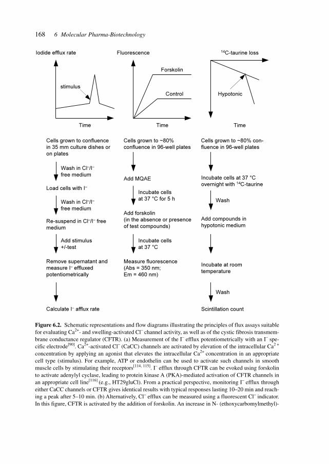
168 6 Molecular Pharma-Biotechnology
Time
Iodide efflux rate
Time
Fluorescence
Time
14C-taurine loss
stimulus
Forskolin
Control Hypotonic
Cells grown to confluence
in 35 mm culture dishes or
on plates
Cells grown to ~80%
confluence in 96-well plates
Cells grown to ~80% con-
fluence in 96-well plates
Load cells with I–
Re-suspend in Cl–/I– free
medium
Remove supernatant and
measure I– effluxed
potentiometrically
Calculate I– afflux rate
Incubate cells
at 37 °C for 5 h
Add forskolin
(in the absence or presence
of test compounds)
Measure fluorescence
(Abs = 350 nm;
Em = 460 nm)
Incubate cells at 37 °C
overnight with 14C-taurine
Wash
Add compounds in
hypotonic medium
Incubate at room
temperature
Wash
Scintillation count
Wash in Cl–/I–
free medium
Wash in Cl–/I–
free medium
Add stimulus
+/-test
Add MQAE
Incubate cells
at 37 °C
Figure 6.2. Schematic representations and flow diagrams illustrating the principles of flux assays suitablefor evaluating Ca2+- and swelling-activated Cl– channel activity, as well as of the cystic fibrosis transmem-brane conductance regulator (CFTR). (a) Measurement of the I– efflux potentiometrically with an I– spe-cific electrode[90]. Ca2+-activated Cl– (CaCC) channels are activated by elevation of the intracellular Ca2 +
concentration by applying an agonist that elevates the intracellular Ca2+ concentration in an appropriatecell type (stimulus). For example, ATP or endothelin can be used to activate such channels in smoothmuscle cells by stimulating their receptors[114, 115]. I– efflux through CFTR can be evoked using forskolinto activate adenylyl cyclase, leading to protein kinase A (PKA)-mediated activation of CFTR channels inan appropriate cell line[116] (e.g., HT29gluCl). From a practical perspective, monitoring I– efflux througheither CaCC channels or CFTR gives identical results with typical responses lasting 10–20 min and reach-ing a peak after 5–10 min. (b) Alternatively, Cl– efflux can be measured using a fluorescent Cl– indicator.In this figure, CFTR is activated by the addition of forskolin. An increase in N- (ethoxycarbomylmethyl)-
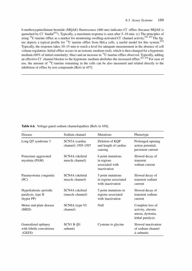
169
6-methoxyquinolinium bromide (MQAE) fluorescence (460 nm) indicates Cl– efflux (because MQAE isquenched by Cl– bindin[81]). Typically, a maximum response is seen after 5–10 min. (c) The principles ofusing 14C-taurine efflux as a marker for monitoring swelling-activated Cl– channel activity.[83, 85] The fig-ure depicts a typical profile for 14C taurine efflux from HeLa cells, a useful model for this system.[83]
Typically, the response takes 10–15 min to reach a level for adequate measurement in the absence of cellvolume regulation. Initial efflux occurs in an isotonic medium (red), which is then changed for a hypotonicmedium (60% of initial osmolarity: blue) and an increase in 14C-taurine efflux observed. Typically, addingan effective Cl– channel blocker to the hypotonic medium abolishes the increased efflux.[83, 84] For ease ofuse, the amount of 14C-taurine remaining in the cells can be also measured and related directly to theinhibition of efflux by test compounds [Refs in 457].
Table 6.6. Voltage-gated sodium channelopathies [Refs in 458].
Disease Sodium channel Mutations Phenotype
Long QT syndrome 3 SCN5A (cardiac Deletion of KQP Prolonged openingchannel) 1505-1507 and length of cardiac action potential,
causing persistent current
Potassium aggravated SCN4A (skeletal 6 point mutations Slowed decay ofmyotinia (PAM) muscle channel) in regions transient
associated with sodium currentinactivation
Paramyotonia congentia SCN4A (skeletal 5 point mutations Slowed decay of(PC) muscle channel) in regions associated transient sodium
with inactivation current
Hyperkalemic periodic SCN4A (skeletal 5 point mutations in Slowed decay ofparalysis, type II (muscle channel) regions associated transient sodium(hyper PP) with inactivation current
Motor end-plate disease SCN8A (type VI Null Complete loss of(MED) channel) activity, chronic
ataxia, dystonia,lethal paralysis
Generalized epilepsy SCN1 B (β1 Cysteine to glycine Slowed inactivationwith febrile convulsions subunit) of sodium channel (GEFS) á-subunits
6.3 Assay Systems
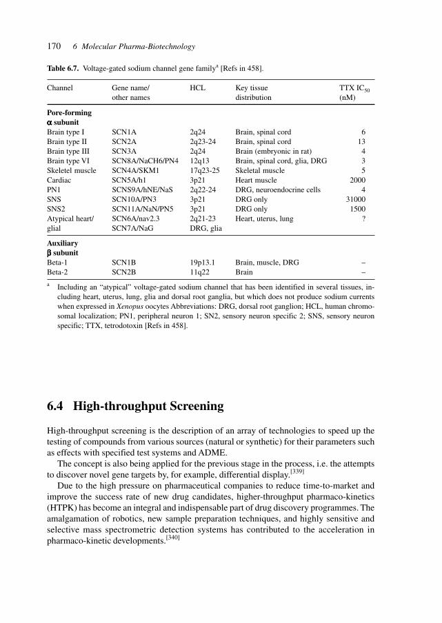
170 6 Molecular Pharma-Biotechnology
Table 6.7. Voltage-gated sodium channel gene familya [Refs in 458].
Channel Gene name/ HCL Key tissue TTX IC50other names distribution (nM)
Pore-formingααααα subunitBrain type I SCN1A 2q24 Brain, spinal cord 6Brain type II SCN2A 2q23-24 Brain, spinal cord 13Brain type III SCN3A 2q24 Brain (embryonic in rat) 4Brain type VI SCN8A/NaCH6/PN4 12q13 Brain, spinal cord, glia, DRG 3Skeletel muscle SCN4A/SKM1 17q23-25 Skeletal muscle 5Cardiac SCN5A/h1 3p21 Heart muscle 2000PN1 SCNS9A/hNE/NaS 2q22-24 DRG, neuroendocrine cells 4SNS SCN10A/PN3 3p21 DRG only 31000SNS2 SCN11A/NaN/PN5 3p21 DRG only 1500Atypical heart/ SCN6A/nav2.3 2q21-23 Heart, uterus, lung ?glial SCN7A/NaG DRG, glia
Auxiliaryβββββ subunitBeta-1 SCN1B 19p13.1 Brain, muscle, DRG –Beta-2 SCN2B 11q22 Brain –
a Including an “atypical” voltage-gated sodium channel that has been identified in several tissues, in-cluding heart, uterus, lung, glia and dorsal root ganglia, but which does not produce sodium currentswhen expressed in Xenopus oocytes Abbreviations: DRG, dorsal root ganglion; HCL, human chromo-somal localization; PN1, peripheral neuron 1; SN2, sensory neuron specific 2; SNS, sensory neuronspecific; TTX, tetrodotoxin [Refs in 458].
6.4 High-throughput Screening
High-throughput screening is the description of an array of technologies to speed up thetesting of compounds from various sources (natural or synthetic) for their parameters suchas effects with specified test systems and ADME.
The concept is also being applied for the previous stage in the process, i.e. the attemptsto discover novel gene targets by, for example, differential display.[339]
Due to the high pressure on pharmaceutical companies to reduce time-to-market andimprove the success rate of new drug candidates, higher-throughput pharmaco-kinetics(HTPK) has become an integral and indispensable part of drug discovery programmes. Theamalgamation of robotics, new sample preparation techniques, and highly sensitive andselective mass spectrometric detection systems has contributed to the acceleration inpharmaco-kinetic developments.[340]
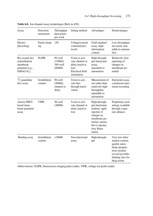
1716.4 High-throughput Screening
Table 6.8. Ion-channel assay technologies [Refs in 458].
Assay Detection instrument
Throughput data points per week
Gating method Advantages Disadvantages
Electro-physiology
Patch-clamp rig
150 Voltage/current command pro-tocols
Gold standard assay, high information content
Low-throughput not easily ame-nable to automa-tion
Bis-oxonal dye redistribution membrane potential [e.g., DiBAC(4)3]
FLIPR 96-well (15000)/. 384-well (60000)
Toxin to acti-vate channel or delay inactiva-tion. Electrical field stimulation
High-through-put functional assay, amenable to automation
Relatively slow reporting of changes in membrane po-tential
14C-guanidine flux assay
Scintillation counter
96-well (20000), channel or delay
Toxin to acti-vate flux through inacti-vation
Measurement of ion rather than relatively high-throughput, amenable to automation
End-point assay, continuous func-tional recording
Aurora-FRET-based mem-brane-potential assay
VIPR 96-well (20000)
Toxin to acti-vate channel or delay inactiva-tion
High-through-put functional readout, rapid reporter of changes in membrane po-tential, amena-ble to automa-tion, Ratio-metric
Proprietary tech-nology available through corpo-rate alliance
Binding assay Scintillation counter
>30000 Non-functional assay
High-through-put
Very low infor-mation content, quality mem-brane prepara-tions needed, several possible binding sites for drug action
Abbreviations: FLIPR, fluorescence imaging plate reader; VIPR, voltage ion probe reader.
172 6 Molecular Pharma-Biotechnology
6.5 Automation
Automation is required at all stages of the pharmaceutical, chemical and biotechnologicaldiscovery, development and manufacturing process in order to achieve increased speed andthroughput. Large central synthesis facilities exist for combinatorial chemistry programs todeliver thousands up to hundreds of thousands of compounds for mass screening. At thelaboratory level, the preparation of samples, the dosing of liquids, and the feeding ofsequencers, mass spectrometers, assay instruments needs to be carried out with minutevolumes, and with high speed and precision around the clock. A crucial component is theefficient data management of the huge amounts of information produced, and the capabil-ity to track compounds and results. An automated laboratory, pilot plant, combinatorialsynthesis facility, medical chemistry laboratory needs to be designed for automation, inte-gration into the network, documentation, efficient control and data management.[450]
Throughput, speed and data management are combined with a keen interest in contain-ing costs of the development and testing activities. High-throughput screening is now of-fering solutions to those requirements. Nanovolumes and optical tests enable the testing ofthe target number of 100,000 compounds per day at affordable costs.[451]
Robotics and automation are applied to synthesis, liquid handling and detection. Theinterface with humans provides for the flexibility and adaptation to differing tasks androutines. The delicate nature of many assays requires the inclusion of liquid handling, manualtransfers, pipetting, heating and cooling, filtration, evaporation, extraction, and chromatog-raphy in integrated systems. Laboratory instruments with a low level of automation andintelligence may be attached to a core of flexible central highly automated intelligent robotstations.[452]
The development of miniaturized analytical systems with integration of sample han-dling, fluidics, measurement and calculations is leading to ‘laboratory-on-a-chip’ devices.These devices are applicable in laboratory analyses for development and also in thephysician’s practice. The design and manufacture involves incorporating developments fromthe microsystems, micro-electronics and opto-electronics industry and technologies, suchas lithography, etching, placement of sensors and actuators, and the integration of the vari-ous functions. At the micro scales of these devices, there are special techniques applicablefor mixing, transportation, and separation, heating and cooling, such as electrophoresis,piezoelectric actuators, diffusion and conductive heating. Microfluidics is a key technol-ogy for the design and operation of such integrated devices.[453]
The application of microsystems (‘biochips’) with highly integrated functions coversthe analysis of DNA, protein, DNA–RNA and DNA–protein interactions, biochemical as-says, and structure and expression studies.[454]
The development of laboratory-on-a-chip systems will allow routine diagnostics to becarried out at the genetic, biochemical, pharmacological and cellular level, thus enablingthe researcher and the physician to study SNPs, pharmaco-genetic variation, metabolizationof compounds, genetic predisposition, prenatal diagnostics and tumor cell profiling on in-dividual patients.[455]
173
6.6 Combinatorial Synthesis:Chemistry, Biology, and Biotechnology
The primary use of combinatorial chemistry is for drug discovery and lead optimization.The technique has also significant potential in the fields of material science, catalyst devel-opment and studies of molecular recognition.
The basic study of intermolecular interactions is facilitated by one-bead–one-structurelibraries which can be powerful tools for the discovery of ligands to synthetic receptors andvice versa. Encoded combinatorial libraries have been useful for disclosing ligands forwell-designed macrocyclic host molecules and to elucidate their specificities for peptidesequences. These studies led via receptors with more flexibility to simple host moleculeswithout elaborate design that are accessible to combinatorial synthesis. One application isthe development of chemical sensors for analytes that are otherwise difficult to detect oronly non-specifically detected. Such libraries have been used to find new catalysts andenzyme mimics.
Understanding the nature of non-covalent intermolecular interactions and using thisknowledge for the rational design of synthetic receptor–ligand pairs is one of the mostintriguing tasks in supramolecular chemistry. Combinatorial libraries offer a powerful toolto gather empirical information about host–guest interactions. This may be studied by screen-ing of a ligand library against an individual receptor or a set of receptors, or by the screen-ing of a library of synthetic receptors for binding studies with a set of chosen ligands.[341]
Combinatorial syntheses aim at producing large numbers of candidate substances forfurther tests and screening. The methods employed are chemical, biological and biotechno-logical. An example of biological and biotechnological methods is the generation of mo-lecular diversity.[342]
Progress in combinatorial chemistry is aimed at the synthesis and testing of more com-plex molecules containing intricate ring systems, stereochemical features and elaboratepresentations of functional groups – molecules that resemble real drugs and natural prod-ucts. This involves the recruitment of methods and principles of complex molecule synthe-sis into combinatorial chemistry, thereby creating hybrid operating systems such as conver-gent automated parallel synthesis.
Synthetic organic chemistry is a vast, complex and expanding web of interrelated re-agents, reactions, procedures, protecting groups, catalysts, purification methods and char-acterization methods. Chemists seek to achieve productivity gains by leveraging the exist-ing body of knowledge and, to a relatively smaller degree, by adding to this body ofknowledge. There are two strategies, i.e. the conceptual and the mechanical, to further suchefforts. The conceptual levers are computer-assisted planning, retro-synthetic analysis, con-vergent pathways, and electronic search tools for reactions, methods and analogies. Themechanical levers and the main drivers of combinatorial chemistry are parallel processing,laboratory automation, solid-phase organic synthesis and split/combine synthesis proto-cols. The combination of both approaches offers the most potential for fulfilling the re-quirements for ever more complex molecules.[343]
6.6 Combinatorial Synthesis: Chemistry, Biology, and Biotechnology
174 6 Molecular Pharma-Biotechnology
These approaches and concepts of combinatorial chemistry, permeating the drug dis-covery and development processes, represent a paradigm shift in drug discovery and basicresearch. Combinatorial chemistry approaches decrease the time taken for discovery andincrease the throughput of chemical screening by as much as 1000-fold. The use of mix-ture-based synthetic combinatorial libraries is gaining ground. Numerous mixture-basedlibraries of peptides, peptidomimetics and heterocycles have been synthesized anddeconvoluted using the positional scanning approach. Mixture-based library approachesfor drug discovery have also shown promising results in vaccine development.[344]
Combinatorial synthesis has been applied to the development of new chiral stationaryphases (CSPs) for large-scale chromatographic separations, which are becoming increas-ingly important due to the complexity of both synthesized as well as natural products.
The technique of chromatographic enantioseparation has become the method of choicefor analytical determinations of enantiopurity and the use of preparative chromatographicseparation for the manufacture of enantiomerically pure forms of chiral drugs is gainingimportance. The technique of simulated moving-bed chromatography is a useful tool forlarge-scale chromatographic separations using CSPs. Greater enantioselectivity is desir-able in order to improve the economics of such separation processes.[345]
Understanding the cellular function of a protein requires either biochemical studies and/or a means to alter the function. This may be done by mutating the gene encoding theprotein – the genetic approach (see Chapter 5.3 ‘Assay Systems and Models’). It may alsobe done by binding the protein directly with a small-molecule ligand – the chemical geneticapproach. In fact both approaches are complementary, and may be necessary to elucidatefunction and to develop active pharmaceutical principles.
Such small-molecule ligands, primarily natural products or synthetic variants of them orrationally designed molecules or randomly synthesized molecules, can either inactivate oractivate protein function. Efficient methods of ligand synthesis and discovery are requiredto provide a small-molecule partner for every gene product or target.
In addition to synthesizing and screening large numbers of small molecules, syntheticstrategies are required which enable the synthesis of vast numbers of compounds withstructures reminiscent of natural products and compatible with miniaturized assays. Thiswill be necessary in order to discover non-natural compounds having the binding affinitiesand specificities characteristic of natural products.[346]
The ability of an enzyme to discriminate among many potential substrates is importantfor maintaining the fidelity of most biological functions. A method was developed for thepreparation and use of fluorogenic substrates that allows for the configuration of generalsubstrate libraries to rapidly identify the primary and extended specificity of proteases. Thesubstrates contain the fluorogenic 7-amino-4-carboxymethylcoumarin (ACC) leaving group.Substrates incorporating the ACC leaving group show kinetic profiles comparable to thosewith the traditionally used 7-amino-4-methylcoumarin (AMC) leaving group. The bifunc-tional nature of ACC allows for the efficient production of single substrates and substratelibraries by using 9-fluorenylmethoxycarbonyl (Fmoc)-based solid-phase synthesis tech-niques. The approximately 3-fold increased quantum yield of ACC over AMC permits areduction in enzyme and substrate concentrations. As a consequence, a greater number ofsubstrates can be tolerated in a single assay, thus enabling an increase in the diversity space
175
of the library. Soluble positional protease substrate libraries of 137, 180 and 6859 mem-bers, possessing amino acid diversity at the P4–P3–P2–P1 and P4–P3–P2 positions, re-spectively, were constructed. Employing this screening method, the substrate specificitiesof a diverse array of proteases was profiled, including the serine proteases thrombin, plas-min, Factor Xa, urokinase-type plasminogen activator, tissue plasminogen activator,granzyme B, trypsin, chymotrypsin, human neutrophil elastase, and the cysteine proteasespapain and cruzain. The resulting profiles create a pharmacophoric portrayal of the pro-teases to aid in the design of selective substrates and potent inhibitors.[347]
Soluble polymer supports provide homogeneous reaction conditions, and facilitate productpurification through selective precipitation and filtration of the polymer-bound product.The use of linear, non-cross-linked polymers in methods of liquid-phase synthesis avoidsthe difficulties of solution- and solid-phase methods while preserving the positive aspectsof both. The solubility properties of PEG, polyacrylamide, non-cross-linked polystyreneand other soluble supports have allowed for the development of numerous molecules, in-cluding peptides, oligonucleotides, oligosaccharides and complex organic compounds. Al-though the choice of polymer used in liquid-phase methods may be tailored to specificapplications, PEG is used most frequently due to its high solubilizing power. Its usage hasresulted in the development of liquid-phase combinatorial synthesis, wherein the integra-tion of liquid-phase synthesis with combinatorial chemistry has produced small-moleculelibraries of peptides, sulfonamides and neomycin B mimics.[348]
Chemical libraries can be screened for small molecules that affect a variety of cellularprocesses including signal transduction, the cell cycle and cellular metabolism. In particu-lar, it may be possible to identify molecules that induce cells to differentiate into specificcell types or, conversely, molecules that reverse differentiation or induce regenerative pro-cesses. The latter might result from either blockage of pathways that maintain that differen-tiated phenotype, or the induction of biochemical pathways active during development orwound healing. Molecules that affect the differentiated phenotype may ultimately find usein cellular replacement therapy or tissue engineering and regeneration.
A new microtubule-binding molecule, myoseverin, was identified from a library of 2,6,9-trisubstituted purines in a morphological differentiation screen. Myoseverin induces thereversible fission of multinucleated myotubes into mononucleated fragments. Myotube fis-sion promotes DNA synthesis and cell proliferation after the removal of the compound andtransfer of the cells to fresh growth medium. Transcriptional profiling and biochemicalanalysis indicate that myoseverin alone does not reverse the biochemical differentiationprocess. Instead, myoseverin affects the expression of a variety of growth factor, immuno-modulatory, extracellular matrix-remodeling and stress response genes, consistent with theactivation of pathways involved in wound healing and tissue regeneration.[349]
The sequencing of the human genome, and of numerous pathogen and microbial ge-nomes, has resulted in an explosion of potential drug targets. These targets represent bothan unprecedented opportunity and a technological challenge for the pharmaceutical indus-try. A novel strategy is required to implement small-molecule drug discovery with sets ofincompletely characterized, disease-associated proteins. Combinatorial chemistry and othertechnologies serve to discover bioactive small-molecule ligands that act on candidate drugtargets. Therapeutically active ligands serve to concurrently validate a target and provide
6.6 Combinatorial Synthesis: Chemistry, Biology, and Biotechnology
176 6 Molecular Pharma-Biotechnology
lead structures for further drug development in an accelerated drug discovery process. Thefuture of drug discovery thus holds great promise, largely because of the introduction of anarray of new technologies.[350]
The next step in efficiency increase is achieved by integrating combinatorial synthesisand bioassays. Highly parallel automation in drug development requires combinatorial syn-thesis, parallel analysis and screening. The bottleneck has shifted from the generation oflead structures (or at least compounds) to their transformation into orally active drugs withthe desired physiological properties and performance results in clinical trials. The synthe-sis of large and diverse libraries will have to be integrated with analysis of subsequent stepssuch as bioavailability, ADME and toxicity.[351]
Identification of new biological targets combined with combinatorial medical chemistryhas enormous potential to facilitate the discovery of new therapeutics. These new targetsrequire the use of different or more complex screening libraries. This complexity might beachieved through the use of new templates or the identification of new chemistries. Oneapproach is the use of chiral libraries that have been designed using three-dimensionalpharmacophores and shape descriptors to provide maximal structure–activity informationafter screening.[352]
Combinatorial chemistry is applying a multitude of approaches such as diversity librar-ies, drug-like libraries and combinatorial subset selection.[353]
Combinatorial chemistry combined with self-assembly processes studied in supramo-lecular chemistry, yields dynamic combinatorial chemistry (DCC). DCC is useful for thesynthesis of compounds binding to specific biological and nonbiological targets by selec-tively binding to target molecules and being removed from a pool of interconverting com-pounds.[633]
The solution-phase synthesis of organic compounds offers efficiency advantages whichare counter-balanced by the difficulty in separating, identifying, and purifying the resultingcomponents. A strategy for mixture synthesis which resolves the separation problem in-volves the tagging of a series of organic substrates with fluorous tags of increasing fluorinecontent. The compounds were mixed, multistep reactions were carried out, and the result-ing tagged products were demixed by fluorous chromatography and elution in order ofincreasing fluorine content to provide the individual pure compoonents which are detaggedto obtain the final products.[569]
The increasing importance discovered for carbohydrate structures and their structure-function relationships is necessitating improved techniques in synthesis and analysis ofcarbohydrates, glycoproteins, and glycopeptides. Progress toward the automated synthesishas been reported and provides compunds for biological and pharmaceutical studies.[640], [570]
The analytical methods range from fingerprinting by atomic force microscopy[571] to massspectrometry determination.[641]
177
6.7 Genotyping:Genetic Pre-Disposition, and Heterogeneity
SNPs allow us to pinpoint the genes involved in traits and diseases such as asthma, hyper-tension, obesity, cancers, and diabetes. They will enable both the localization of genes andgene families as well as the development of personalized medicine.
A method for the sensitive detection of single-base mutation in DNA has been describedwhich involves the application of a primer thiolated oligonucleotide, complementary to thetarget DNA as far as one base before the mutation site, on an electrode or a gold-quartzpiezoelectric crystal. After hybridizing the target DNA, normal or mutant, with the sensingoligonucleotide, the resulting assembly is reacted with the biotinylated nucleotide, comple-mentary to the mutation site, in the presence of polymerase.The labeled nucleotide is coupledonly to the double-stranded assembly with the mutant site. Faradaic impedance spectros-copy and microgravimetric quartz-crystal microbalance analyses were used for the ellectronicdetection of single-base mutants. The lower sensitivity limit for detection of mutant DNAis 1 × 10–14 mol/ml. The method was applied to the analysis of polymorphic blood sampleswith the Tay-Sachs disorder and allows the quantitaive analysis of the mutant with no PCR-pre-amplification.[560]
The International SNP Map Working Group has prepared a map which identifies andlocalizes 1.42 million single-nucleotide polymorphisms (SNPs) throughout te genome, mostof which are in noncoding regions. This list future and expanded versions provide thefoundation for the biomedical reseach focus on the systematic characterization of indi-vidual gene products and the effects of sequence variants on function.[561]
In 1999 several companies joined to form the SNP Consortium to create a database ofseveral 100,000 SNP markers and make the available to researchers. The companies andorganizations of the SNP Consortium presently are:
AstraZeneca Glaxo WellcomeAventis MotorolaAmersham Pharmacia Biotech NovartisBayer PfizerBristol-Myers Squibb Searle (Monsanto)Hoffmann-La Roche SmithKline BeechamIBM Wellcome Trust
The International SNP Map Working Group has prepared a map which identifies and local-izes 1.42 million single-nucleotide polymorphisms (SNPs) throughout te genome, most ofwhich are in noncoding regions. This list future and expanded versions provide the founda-tion for the biomedical reseach focus on the systematic characterization of individual geneproducts and the effects of sequence variants on function.[561]
SNPs and small insertions or deletions (grouped together as SNPs) are the largest set ofsequence variants in most organisms. They enable the identification (respectively, localiza-tion) of human traits, QTLs and disease traits.[354] SNPs spaced at distances of 30 kb al-
6.7 Genotyping: Genetic Pre-Disposition, and Heterogeneity
178 6 Molecular Pharma-Biotechnology
lowed the localization of several diseases, i.e. psoriasis, migraine, Alzheimer’s and diabe-tes.
The revolution in genetics has led to the determination of the precise genetic basis ofcommon and uncommon genetic hereditary diseases. The first fruits of this revolution arediagnostic – the ability to determine who is and who is not at risk for disease before theonset of symptoms. Such information is becoming indispensable for proper the manage-ment of patients and their families. In individuals who inherit mutant genes, simple preven-tive measures often can reduce morbidity and mortality, and allow more thoughtful plan-ning for the future. The benefits of genetic testing are equally important for those familymembers who are found not to carry the relevant mutation; these individuals are sparedunnecessary medical procedures and tremendous anxiety.
The problems of genetic testing are psychological or technical in nature. Issues relatedto insurance, employment discrimination and privacy have received much attention. Addi-tional ethical concerns arise when no effective intervention is available and when prenataltesting is considered for diseases with late-onset or minimal effects.
There are significant technical challenges for genetic testing as well. In many diseases,not all of the genes capable of causing or contributing to pathogenesis are known. Evenwhen the mutated gene is known, routine genetic testing may fail to identify mutations inmany cases (25–75% or more).
There are nearly 1000 different hereditary diseases for which the causative genes areknown and there are numerous ways in which genes can be mutated. The ease of detectionof these mutations covers a whole spectrum from compliant mutations to refractory muta-tions. Compliant mutations are identifiable by using DNA sequencing or PCR. Refractorymutations involve deletions of one or more exons, insertions, translocations or alterationswhich affect the expression of the gene at the RNA or protein level.
Direct detection of a genetic variant is done by either DNA sequencing or micro arrayanalysis. Even using DNA sequencing, mutations affecting only one allele may be difficultto distinguish from normal variations of sequencing baselines. Therefore, in thorough ge-netic testing, both strands of DNA are being sequenced.
A number of indirect methods have been developed for detecting mutations, such assingle-strand conformation polymorphism (SSCP) and denaturing gradient gel electrophore-sis (DGGE), which exploit the differential electrophoretic migration of nucleic acids thatvary by as little as a single base. DHPLC is a related technique that detects a variation instructure between mutant and wild-type molecules but uses HPLC instead of gel electro-phoresis for separation. Another group of indirect methods involves chemical or enzymaticcleavage of the DNA, which exploits the bulges or bends in the DNA duplex that are cre-ated by mismatched mutations. Alterations in protein structure resulting from certain ge-netic alterations may be used for analysis. Nonsense mutations, frameshifts and skippedexons are revealed by Western blot analysis or through analysis of proteins synthesized invitro from PCR products.
Refractory mutations are difficult to detect due to the nature of the molecular defects. Ifthe genomic region is deleted from the mutant allele, the PCR product from genomic DNAwill lead to a false conclusion that this gene region is wild-type. Other mutations affect theexpression or processing of mRNA from the affected allele, through mutations of promoter
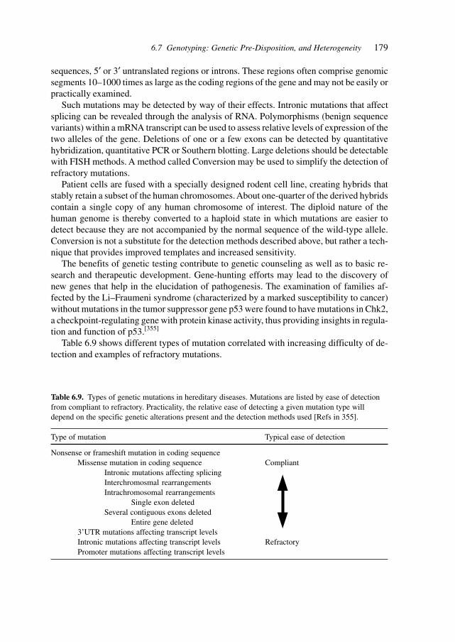
179
sequences, 5′ or 3′ untranslated regions or introns. These regions often comprise genomicsegments 10–1000 times as large as the coding regions of the gene and may not be easily orpractically examined.
Such mutations may be detected by way of their effects. Intronic mutations that affectsplicing can be revealed through the analysis of RNA. Polymorphisms (benign sequencevariants) within a mRNA transcript can be used to assess relative levels of expression of thetwo alleles of the gene. Deletions of one or a few exons can be detected by quantitativehybridization, quantitative PCR or Southern blotting. Large deletions should be detectablewith FISH methods. A method called Conversion may be used to simplify the detection ofrefractory mutations.
Patient cells are fused with a specially designed rodent cell line, creating hybrids thatstably retain a subset of the human chromosomes. About one-quarter of the derived hybridscontain a single copy of any human chromosome of interest. The diploid nature of thehuman genome is thereby converted to a haploid state in which mutations are easier todetect because they are not accompanied by the normal sequence of the wild-type allele.Conversion is not a substitute for the detection methods described above, but rather a tech-nique that provides improved templates and increased sensitivity.
The benefits of genetic testing contribute to genetic counseling as well as to basic re-search and therapeutic development. Gene-hunting efforts may lead to the discovery ofnew genes that help in the elucidation of pathogenesis. The examination of families af-fected by the Li–Fraumeni syndrome (characterized by a marked susceptibility to cancer)without mutations in the tumor suppressor gene p53 were found to have mutations in Chk2,a checkpoint-regulating gene with protein kinase activity, thus providing insights in regula-tion and function of p53.[355]
Table 6.9 shows different types of mutation correlated with increasing difficulty of de-tection and examples of refractory mutations.
Table 6.9. Types of genetic mutations in hereditary diseases. Mutations are listed by ease of detectionfrom compliant to refractory. Practicality, the relative ease of detecting a given mutation type willdepend on the specific genetic alterations present and the detection methods used [Refs in 355].
Type of mutation Typical ease of detection
Nonsense or frameshift mutation in coding sequenceMissense mutation in coding sequence Compliant
Intronic mutations affecting splicingInterchromosmal rearrangementsIntrachromosomal rearrangements
Single exon deletedSeveral contiguous exons deleted
Entire gene deleted3’UTR mutations affecting transcript levelsIntronic mutations affecting transcript levels RefractoryPromoter mutations affecting transcript levels
6.7 Genotyping: Genetic Pre-Disposition, and Heterogeneity
180 6 Molecular Pharma-Biotechnology
Mutation discovery and genetic profiling at the protein/peptide level has been demon-strated by MS analysis of N-terminally tagged test peptides generated in a coupled in vitrotranscription/translation reaction from a PCR product of any continuous region of codingsequence. Truncations and amino acid substitutions in peptides coded for by the breastcancer susceptibility gene BRCA1 were readily identified with this method.[356]
The analysis of mutations that cause genetic diseases and their diagnosis requires high-throughput methods for the identification of many different targets in an efficient, multi-plexed manner. High-throughput sequence analysis is a powerful tool for both population-based genetic assessment and rapid cost-effective diagnostic tests. DNA chips (microarrays)readily adapt to the parallel format necessary to screen many samples for many mutationssimultaneously.
These DNA chips are arrays of oligonucleotide probes which are spatially synthesizedusing either masking techniques or liquid dispersing methods. Major goals are the furtherreduction of overall substrate and individual feature sizes, increasing the simplicity andreducing the cost of fabrication, and elimination of target labeling. A randomly orientedfiber optic gene array for rapid, parallel detection of unlabeled DNA targets with surfaceimmobilized molecular beacons that undergo a conformational change accompanied by afluorescence change in the presence of a complementary DNA target has demonstrated theselective detection of genomic CF-related targets with an optical encoding scheme and animaging fluorescence microscope system.[357]
The pharmaceutical world is realizing the fact that genetic variation is the key to identi-fying the biological basis behind both susceptibility to disease and response to drugs. Ge-netic variation within drug targets is common and genetic factors influence almost everyhuman disease. The study of SNPs is crucial for characterizing molecular targets and alsoto validate the role of these targets in disease. Genetic variation as exemplified by SNPswill thus affect target, characterization and validation, and pharmaco-genetics. Examplesare genetic variation affecting the response to pravastatin, SKF38393, neuroleptics, tacrine,sensitivity to chemotherapy in breast cancer and asthma treatment by interfering with 5-lipoxygenase.[358]
The study of SNPs offers a way to specifically correlate genotype with (clinical) pheno-type at the individual level, thus leading to a very personalized treatment or medicine. Inorder to achieve that goal, gene-specific sequencing and the cataloguing of SNPs plus cor-relation to the phenotype is necessary.[359]
Systematic characterization of RNA splicing alterations using differential analysis oftranscripts with alternative splicing (DATAS) aids in quantitative gene profiling.[360]
High-throughput SNP scoring from unamplified genomic DNA (an alternative to PCR-based essays) utilizes a structure-spesific 5’-nuclease (or flap nuclease) to clieve a 5’-flapsequence when two synthetic oligonucleotide probes hybridize in tandem to the target se-quence[361]. Flap endonucleases (FENs) isolated from archaea are shown to recognize andcleave a structure formed when two overlapping oligonucleotides hybridize to a target DNAstrand. The downstream oligonucleotide probe is cleaved and the precise site of cleavage isdependent on the amount of overlap with the upstream oligonucleotide. The use of thermo-stable archeal FENs was demonstrated to allow the reaction to be performed at tempera-tures that promote probe turnover without the need for temperature cycling. The resulting

181
amplification of the cleavage signal enables the detection of specific DNA targets atsubattomole levels within complex mixtures. Evidence was provided that this cleavage issufficiently specific to enable discrimination of single-base differences and can differenti-ate homozygotes from heterozygotes in single-copy genes in genomic DNA.[362]
An improved sensitivity version of the invasive signal amplification assay was describedwith a limit of detection (LOD) of zeptomole (10–21 mol) levels of a target DNA in homo-geneous format.
The invasive signal amplification reaction has been developed for quantitative detectionof nucleic acids and discrimination of SNPs. The improved method couples two invasivereactions into a serial isothermal homogeneous assay using fluorescence resonance energytransfer detection. The serial version of the assay generates more than 107 reporter mol-ecules for each molecule of target DNA in a 4-h reaction; this sensitivity, coupled with theexquisite specificity of the reaction, is sufficient for direct detection of less than 1000 targetmolecules with no prior target amplification. A kinetic analysis of the parameters affectingsignal and background generation in the serial invasive signal amplification reaction and asimple kinetic model of the assay were described. The assay was able to detect as few as600 copies of the methylene tetrahydrofolate reductase gene in samples of human genomicDNA. The assay is capable of discriminating single base differences in this gene by using20 ng of human genomic DNA.[363]
Interleukin-1 polymorphisms have been associated with increased risk of gastric andmay influence the varying susceptibility to develop gastric cancers[696] and chromosome18q loss is connected to vascular invasion.[701]
The repertoire of technologies is expanding and includes SBE-TAGS, an array-basedmethod for SNP genotyping,[686] detection of single-base mismatches and DNA base le-sions by the electrocatalysis at DNA-modified electrodes,[687] the scanning of guanine-guanine mismatches in DNA by synthetic ligands with surface plasmon resonance,[699] andinterference-based detection of nucleic acid targets on optically coated silicon.[700]
6.8 Sequencing
Gene sequencing has reached an advanced technological stage where automation allowsthe rapid sequencing of several 10 000 base pairs per machine per day. The future of genesequencing may be in superfast sequencing.[364]
Large-scale sequencing efforts require efficient methods of ordering the sequences. Physi-cal mapping has been reintroduced as an important component of large-scale sequencingprojects. Restriction maps provide landmark sequences at defined intervals and high-reso-lution restriction maps can be assembled from ensembles of single molecules by opticalmeans. Such optical maps can be constructed from large-insert clones and genomic DNA,and are used as a scaffold for accurately aligning sequence contigs generated by shotgunsequencing.[365]
The acceleration of sequencing is being addressed by several approaches to measuringsingle molecules. A variety of different DNA polymers were electrophoretically driven
6.8 Sequencing
182 6 Molecular Pharma-Biotechnology
through the nanopore of an α-hemolysin channel in a lipid bilayer. Single-channel record-ing of the translocation duration and current flow during traversal of individual polynucle-otides yielded a unique pattern of events for each of the several polymers tested. Statisticaldata derived from this pattern of events demonstrate that in several cases a nanopore candistinguish between polynucleotides of similar length and composition that differ only insequence. Studies of temperature effects on the translocation process show that transloca-tion duration scales as ~T–2. A strong correlation exists between the temperature dependenceof the event characteristics and the tendency of some polymers to form secondary structure.Because nanopores can rapidly discriminate and characterize unlabeled DNA molecules atlow copy number, refinements of the experimental approach demonstrated could eventu-ally provide a low-cost high-throughput method of analyzing DNA polynucleotides.[366]
DNA and RNA molecules can be detected as they are driven through a nanopore by anapplied electric field at rates ranging from several hundred microseconds to a few millisec-onds per molecule. The nanopore can rapidly discriminate between pyrimidine and purinesegments along a single-stranded nucleic acid molecule. Nanopore detection and charac-terization of single molecules represents a new method for directly reading informationencoded in linear polymers. If single-nucleotide resolution can be achieved, it is possiblethat nucleic acid sequences can be determined at rates exceeding 1000 bases/s.[367]
A nanopore instrument, which can discriminate between individual DNA hairpins thatdiffer by one base pair or one nucleotide, has been demonstrated. RNA and DNA strandsproduce ionic current signatures when driven through an alpha-hemolysin channel by anapplied voltage. This nanopore detector was combined with a support vector machine (SVM)to analyze DNA hairpin molecules on the millisecond time scale. Measurable propertiesinclude duplex stem length, base pair mismatches, and loop length.[559]
Another development towards the creation of ultra-fast DNA sequencing is the progressin designing and producing micro fluidic arrays.
A nanofluidic channel device, consisting of many entropic traps, was designed and fab-ricated for the separation of long DNA molecules. The channel comprises narrow constric-tions and wider regions that cause size-dependent trapping of DNA at the onset of a con-striction. This process creates electrophoretic mobility differences, thus enabling efficientseparation without the use of a gel matrix or PEF. Samples of long DNA molecules (5000–160,000 bp) were efficiently separated into bands in 15-mm-long channels. Multiple-chan-nel devices operating in parallel were demonstrated. The efficiency, compactness and easeof fabrication of the device suggest the possibility of more practical integrated DNA analy-sis systems.[368]
A method for multiplexed detection of polymorphic sites and direct determination ofhaplotypes in 10-kb DNA fragments using single-walled carbon nanotubes (SWNT) atomicforce microscopy (AFM) probes was developed. Labeled oligonucleotides are hybridizedspecifically to complementary target sequences in template DNA and the positions of thetagged sequences are detected by direct SWNT tip imaging. This concept was demon-strated by detecting streptavidin and IRD800 labels at two different sequences in M13mp18.The approach also permits haplotype determination from simple visual inspection of AFMimages of individual DNA molecules, which was done on UGT1A7, a gene under study asa cancer risk factor. The haplotypes of individuals heterozygous at two critical loci, which
183
together influence cancer risk, can be easily and directly distinguished from AFM images.The application of this technique to haplotyping in population-based genetic disease stud-ies and in genomic screening studies will facilitate the correlation of genotype and pheno-type.[369]
A novel sequencing approach was described that combines non-gel-based signature se-quencing with in vitro cloning of millions of templates on separate 5-µm diametermicrobeads. After constructing a microbead library of DNA templates by in vitro cloning,a planar array of a million template-containing microbeads in a flow cell at a density greaterthan 3 × 10⁶ microbeads/cm2 was constructed. Sequences of the free ends of the clonedtemplates on each microbead were then simultaneously analyzed using a fluorescence-based signature sequencing method that does not require DNA fragment separation. Signa-ture sequences of 16–20 bases were obtained by repeated cycles of enzymatic cleavagewith a type IIs restriction endonuclease, adaptor ligation and sequence interrogation byencoded hybridization probes. The approach was validated by sequencing over 269,000signatures from two cDNA libraries constructed from a fully sequenced strain of S. cerevisiaeand by measuring gene expression levels in the human cell line THP-1. The approach pro-vides an unprecedented depth of analysis permitting application of powerful statistical tech-niques for discovery of functional relationships among genes, whether known or unknownbeforehand, or whether expressed at high or very low levels.[370]
Automated DNA sequencing in 16-channel microchips was reported. A microchipprefilled with sieving matrix is aligned on a heating plate affixed to a movable platform.Samples are loaded into sample reservoirs by using an eight-tip pipetting device and thechip is docked with an array of electrodes in the focal plane of a four-color scanning detec-tion system. Under computer control, high voltage is applied to the appropriate reservoirsin a programmed sequence that injects and separates DNA samples. An integrated four-color confocal fluorescent detector automatically scans all 16 channels. The system rou-tinely yields more than 450 bases in 15 min in all 16 channels. In the best case using anautomated base-calling program, 543 bases have been called at with an accuracy of greaterthan 99%. Separations, including automated chip loading and sample injection, normallyare completed in less than 18 min. The advantages of DNA sequencing on capillary electro-phoresis chips include uniform signal intensity and tolerance of high DNA template con-centration. To understand the fundamentals of these unique features, a theoretical treatmentof cross-channel chip injection called the differential concentration effect was developed.Experimental evidence demonstrating consistence with the predictions of the theory hasbeen presented.[371]
A special sequencing approach to quickly permit DNA signatures consistently associ-ated with phenotypes of interest has been developed and applied to dinoflagellates for rapididentification of Pfiesteria piscicida and related dinoflagellates from complex cultures.
The newly described heterotrophic estuarine dinoflagellate P. piscicida has been linkedwith fish kills in field and laboratory settings, and with a novel clinical syndrome of im-paired cognition and memory disturbance among humans after presumptive toxin expo-sure. It is necessary to better characterize the organisms and these associations. Advancesin Pfiesteria research were hampered by the absence of genomic sequence data. A sequenc-ing strategy was employed directed by heteroduplex mobility assay to detect P. piscicida
6.8 Sequencing
184 6 Molecular Pharma-Biotechnology
18S rDNA ‘signature’ sequences in complex pools of DNA and those data were used as thebasis for determination of the complete P. piscicida 18S rDNA sequence. Specific PCRassays for P. piscicida and other estuarine dinoflagellates were developed, permitting theirdetection in algal cultures and in estuarine water samples collected during fish kill and fishlesion events. These tools will enhance efforts to characterize these organisms and theirecological relationships. Heteroduplex mobility assay-directed sequence discovery is broadlyapplicable and may be adapted for the detection of genomic sequence data of other novel ornon-culturable organisms in complex assemblages.[372]
A method involving ligation-mediated PCR (polymerase chain reaction) for quantitativein vivo footprinting was developed which allows high-resolution analysis of transcription-factor binding and chromatin structure as well as sequencing.[690] The repertoire of meth-ods to sequence and identify genes was expanded by ORESTES (open reading frame ESTs)which will contribute to the construction of contigs covering full-length cDNAs.[693]
6.9 Pharmaco-Genomics
The availability of sequence data is the foundation for turning the data from modern genomicsinto therapies for diseases.[373] Pharmaco-genomics aims to refine the present overwhelm-ingly followed principle of ‘one-medicine-fits-all’ with the development of active princi-ples and therapies targeting patient groups and, in the final analysis, individual patientswith specific means of treatment. An ideal prior requirement is the definition of a disease atthe molecular level, and a relation to the genetic make-up of the patients, defined in termsof haplotypes, gene defects, gene alleles, SNPs, genetic linkage groups and imprintingpatterns. The next level is the definition with respect to protein variations, isozymes, ge-netic variants of receptors, and varying interactions in protein–protein, DNA–protein andRNA–protein networks, up to the cellular level of metabolic states and phenotypes.
Pharmaco-genomics may thus serve to customize pharmaceuticals and therapies for spe-cific subgroups of patients. The direct clinical evidence of pharmaco-genomic informationis demonstrated by the different susceptibility of patients to hydralazine, an arterial vasodi-lator, in correlation with different expression of α- and β-myosin heavy chains. Thepharmaco-genetic approach contributes to understanding the causes of phamaco-kineticsand pharmaco-dynamics based on isozymes of cytochrome P450, dihydropyrimidine de-hydrogenase, thiopurine methyltransferases, cholesteryl ester transfer protein, angiotensin-converting enzyme, and the serotonin transporter. The different responses may be due todifferent interactions with the active principle on any (or several) level(s) of the biochemi-cal and cellular machinery.
Pharmaco-genomics may also contribute to disease correction by providing disease-associated markers which are not themselves causally related to the variation in response.
Table 6.10 lists a selection of companies and their focus area, thus illustrating the rangeof applications of pharmaco-genomics.[474]
Differential gene expression profiling has been added to the repertoire of pharmaco-genomics for drug discovery and development. Differential gene expression technologies
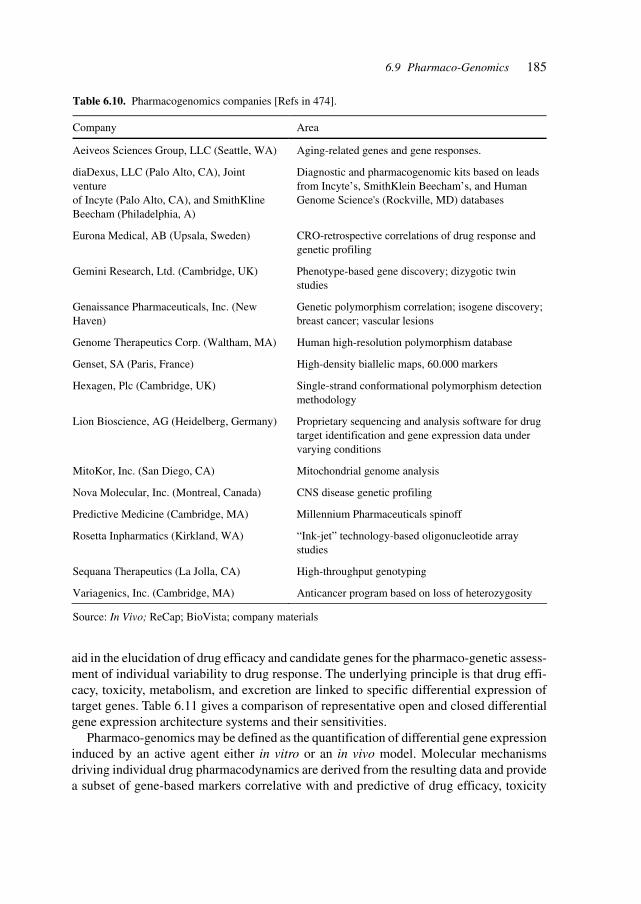
185
aid in the elucidation of drug efficacy and candidate genes for the pharmaco-genetic assess-ment of individual variability to drug response. The underlying principle is that drug effi-cacy, toxicity, metabolism, and excretion are linked to specific differential expression oftarget genes. Table 6.11 gives a comparison of representative open and closed differentialgene expression architecture systems and their sensitivities.
Pharmaco-genomics may be defined as the quantification of differential gene expressioninduced by an active agent either in vitro or an in vivo model. Molecular mechanismsdriving individual drug pharmacodynamics are derived from the resulting data and providea subset of gene-based markers correlative with and predictive of drug efficacy, toxicity
Table 6.10. Pharmacogenomics companies [Refs in 474].
Company Area
Aeiveos Sciences Group, LLC (Seattle, WA) Aging-related genes and gene responses.
diaDexus, LLC (Palo Alto, CA), Joint venture of Incyte (Palo Alto, CA), and SmithKline Beecham (Philadelphia, A)
Diagnostic and pharmacogenomic kits based on leads from Incyte’s, SmithKlein Beecham’s, and Human Genome Science's (Rockville, MD) databases
Eurona Medical, AB (Upsala, Sweden) CRO-retrospective correlations of drug response and genetic profiling
Gemini Research, Ltd. (Cambridge, UK) Phenotype-based gene discovery; dizygotic twin studies
Genaissance Pharmaceuticals, Inc. (New Haven)
Genetic polymorphism correlation; isogene discovery; breast cancer; vascular lesions
Genome Therapeutics Corp. (Waltham, MA) Human high-resolution polymorphism database
Genset, SA (Paris, France) High-density biallelic maps, 60.000 markers
Hexagen, Plc (Cambridge, UK) Single-strand conformational polymorphism detection methodology
Lion Bioscience, AG (Heidelberg, Germany) Proprietary sequencing and analysis software for drug target identification and gene expression data under varying conditions
MitoKor, Inc. (San Diego, CA) Mitochondrial genome analysis
Nova Molecular, Inc. (Montreal, Canada) CNS disease genetic profiling
Predictive Medicine (Cambridge, MA) Millennium Pharmaceuticals spinoff
Rosetta Inpharmatics (Kirkland, WA) “Ink-jet” technology-based oligonucleotide array studies
Sequana Therapeutics (La Jolla, CA) High-throughput genotyping
Variagenics, Inc. (Cambridge, MA) Anticancer program based on loss of heterozygosity
Source: In Vivo; ReCap; BioVista; company materials
6.9 Pharmaco-Genomics
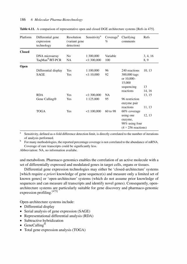
186 6 Molecular Pharma-Biotechnology
and metabolism. Pharmaco-genomics enables the correlation of an active molecule with aset of differentially expressed and modulated genes in target cells, organs or tissues.
Differential gene expression technologies may either be ‘closed-architecture’ systems[which require a priori knowledge of gene sequence(s) and measure only a limited set ofknown genes] or ‘open-architecture’ systems (which do not assume prior knowledge ofsequences and can measure all transcripts and identify novel genes). Consequently, open-architecture systems are particularly suitable for gene discovery and pharmaco-genomicexpression profiling.[475]
Open-architecture systems include:• Differential display• Serial analysis of gene expression (SAGE)• Representational differential analysis (RDA)• Subtractive hybridization• GeneCalling®
• Total gene expression analysis (TOGA)
Table 6.11. A comparison of representative open and closed DGE architecture systems [Refs in 475].
Platform Differential gene Resolution Sensitivitya Coverageb Clarifying Refsexpression (variant gene % commentstechnology detection)
ClosedDNA microarray No 1:300,000 Variable 3, 4, 16TaqMan®/RT-PCR NA <1:300,000 100 8, 9
OpenDifferential display Yes 1:100,000 96 240 reactions 10, 13SAGE Yes <1:10,000 92 300,000 tags
or 10,000–15,000sequencing 13reactions 14, 16
RDA Yes <1:300,000 NA 13, 15Gene Calling® Yes 1:125,000 95 96 restriction
enzyme pairreactions 11, 13
TOGA Yes <1:100,000 60 to 98 60% coverageusing one 12, 13enzyme,98% using four(4 × 256 reactions)
a Sensitivity, defined as n-fold difference detection limit, is directly correlated to the number of iterationsof analysis performed.
b For many methodologies, the reported percentage coverage is not correlated to the abundance of mRNA.Coverage of rare transcripts could be significantly less.
Abbreviation: NA, no information availabe.
187
A strategy for disease gene identification through nonsense-mediated mRNA decay inhibi-tion has been developed. Premature termination codons (PTCs) initiate degradation of mutanttranscripts via the nonsense-mediated messenger RNA decay (NMD) pathway which isinvolved in an estimated one-third of mutations underlying human disorders. Targeting thismechanism allows the identification of disease genes and was validated by testing coloncancer and Sandhoff disease cell lines.[664]
The study of functional relationships between RNA expression and chemotherapeuticsusceptibility allows the identification of relevance networks thus enabling identificationof biochemical associations.[695]
The importance of RNA in regulatory networks is evidenced by the phenomenon ofgene silencing by RNA interference (RNAi), which is the process of sequence-specific,post-transcriptional gene silencing in animals and plants by double-stranded RNA (dsRNA)homo-logous to the silenced gene. Understanding this regulatory mechanisms is essentialfor the control and modification of the concomitant genetic and biochemical as well ascellular processes in fundamental studies and for pharmaceutical intervention. Duplexes of21-nucleotide RNAs were demonstrated to mediate RNA interference in cultured mamma-lian cells, thus representing a tool for studying gene function and offering use as gene-specific therapeutics.[675]
An RNA-directed nuclease was found to mediate post-transcriptional gene silencingin cultured Drosophila cells by analysis of loss-of-function phenotypes obtained upontrans-fection with double-stranded RNAs.[681] Post-transcriptional gene-silencing was ana-lyzed in Arabidopsis mutants and several related proteins were identified which supportsthe hypo-thesis of a common ancestral mechanism of gene expression control in plants,fungi, and animals.[682] In the unicellular green alga Chlamydomonas reinhardtii a geneMut6 was cloned that is required for the silencing of a transgene and two transposon fami-lies and codes for a protein homologous to RNA helicases of the DEAH-box family, againsupporting the notion of an ancestral mechanism[678]. Similar proteins are used in thenematode Caenorhabditis elegans and the fungus Neurospora crassa to effect gene si-lencing.[683]
Functional genomic studies in Caenorhabditis elegans by systematic RNAi (RNA inter-ference) help to elucidate the interaction between genes, gene function, and the environ-ment. They also help to understand the functions of genes in other organisms with homo-logous genes resp. proteins[676] [677] and thus will advance the development of therapeutics.
Regulatory RNAs are also prominent in bacteria controlling at the posttranscriptionallevel and represent targets for regulation of metabolism and of infectivity.[679]
Epigenetic mechanism acting at the level of genes without affecting gene sequence formanother layer of gene regulation, exemplified by genomic imprinting which acts by severalmechanism such as methylation, promoters, enhancers, antisense RNA transcripts, silenc-ers, and chromatin boundaries.[684] Epigenetic modifications change transcription patternsin multicellular organism to effect tissue-specific gene expression. DNA methylation isinvolved in animals and plants for the heritability and flexibility of epigenetic states. InArabidopsis a gene (MOM) was identified the disruption of which releases transcriptionalsilencing of methylated genes while not affecting methylation patterns[680]. X chromosomeinactivation is an example of genetic regulation in which thousands of genes on one homo-
6.9 Pharmaco-Genomics
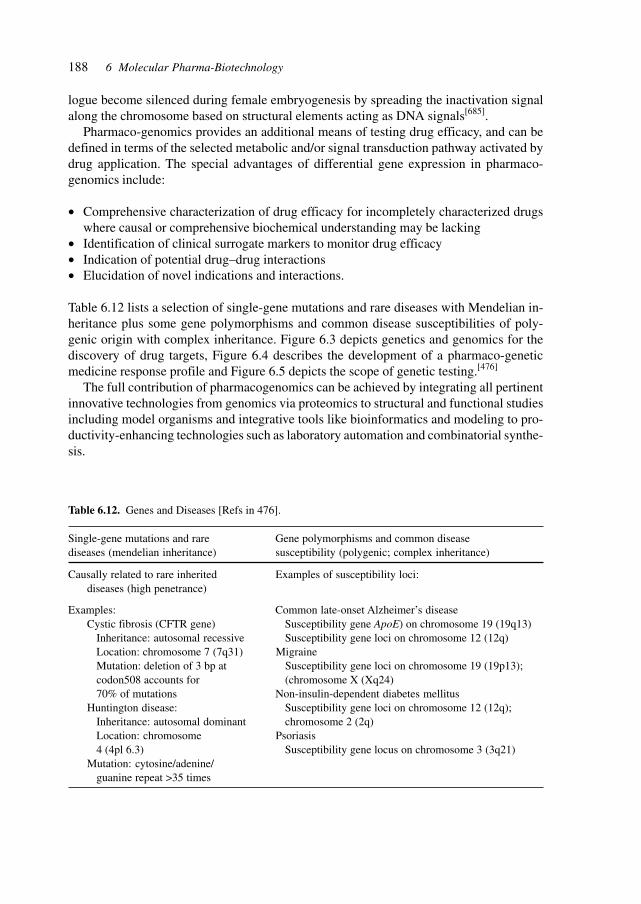
188 6 Molecular Pharma-Biotechnology
logue become silenced during female embryogenesis by spreading the inactivation signalalong the chromosome based on structural elements acting as DNA signals[685].
Pharmaco-genomics provides an additional means of testing drug efficacy, and can bedefined in terms of the selected metabolic and/or signal transduction pathway activated bydrug application. The special advantages of differential gene expression in pharmaco-genomics include:
• Comprehensive characterization of drug efficacy for incompletely characterized drugswhere causal or comprehensive biochemical understanding may be lacking
• Identification of clinical surrogate markers to monitor drug efficacy• Indication of potential drug–drug interactions• Elucidation of novel indications and interactions.
Table 6.12 lists a selection of single-gene mutations and rare diseases with Mendelian in-heritance plus some gene polymorphisms and common disease susceptibilities of poly-genic origin with complex inheritance. Figure 6.3 depicts genetics and genomics for thediscovery of drug targets, Figure 6.4 describes the development of a pharmaco-geneticmedicine response profile and Figure 6.5 depicts the scope of genetic testing.[476]
The full contribution of pharmacogenomics can be achieved by integrating all pertinentinnovative technologies from genomics via proteomics to structural and functional studiesincluding model organisms and integrative tools like bioinformatics and modeling to pro-ductivity-enhancing technologies such as laboratory automation and combinatorial synthe-sis.
Table 6.12. Genes and Diseases [Refs in 476].
Single-gene mutations and rare Gene polymorphisms and common diseasediseases (mendelian inheritance) susceptibility (polygenic; complex inheritance)
Causally related to rare inherited Examples of susceptibility loci:diseases (high penetrance)
Examples: Common late-onset Alzheimer’s diseaseCystic fibrosis (CFTR gene) Susceptibility gene ApoE) on chromosome 19 (19q13)
Inheritance: autosomal recessive Susceptibility gene loci on chromosome 12 (12q)Location: chromosome 7 (7q31) MigraineMutation: deletion of 3 bp at Susceptibility gene loci on chromosome 19 (19p13);codon508 accounts for (chromosome X (Xq24)70% of mutations Non-insulin-dependent diabetes mellitus
Huntington disease: Susceptibility gene loci on chromosome 12 (12q);Inheritance: autosomal dominant chromosome 2 (2q)Location: chromosome Psoriasis4 (4pl 6.3) Susceptibility gene locus on chromosome 3 (3q21)
Mutation: cytosine/adenine/guanine repeat >35 times
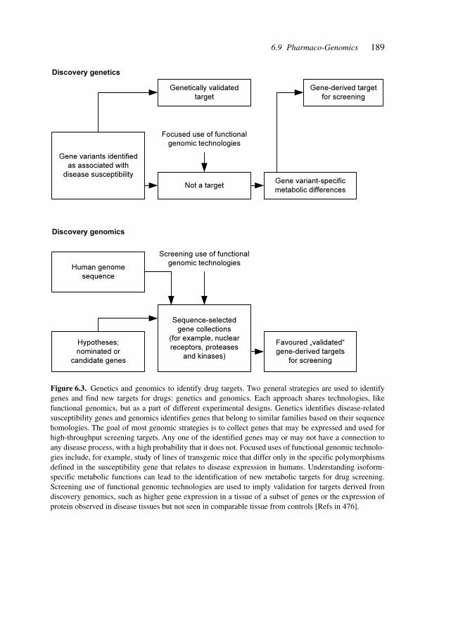
189
Figure 6.3. Genetics and genomics to identify drug targets. Two general strategies are used to identifygenes and find new targets for drugs: genetics and genomics. Each approach shares technologies, likefunctional genomics, but as a part of different experimental designs. Genetics identifies disease-relatedsusceptibility genes and genomics identifies genes that belong to similar families based on their sequencehomologies. The goal of most genomic strategies is to collect genes that may be expressed and used forhigh-throughput screening targets. Any one of the identified genes may or may not have a connection toany disease process, with a high probability that it does not. Focused uses of functional genomic technolo-gies include, for example, study of lines of transgenic mice that differ only in the specific polymorphismsdefined in the susceptibility gene that relates to disease expression in humans. Understanding isoform-specific metabolic functions can lead to the identification of new metabolic targets for drug screening.Screening use of functional genomic technologies are used to imply validation for targets derived fromdiscovery genomics, such as higher gene expression in a tissue of a subset of genes or the expression ofprotein observed in disease tissues but not seen in comparable tissue from controls [Refs in 476].
Gene variants identified
as associated with
disease susceptibility
Discovery genetics
Discovery genomics
Genetically validated
target
Not a targetGene variant-specific
metabolic differences
Gene-derived target
for screening
Human genome
sequence
Hypotheses;
nominated or
candidate genes
Sequence-selected
gene collections
(for example, nuclear
receptors, proteases
and kinases)
Favoured „validated“
gene-derived targets
for screening
Screening use of functional
genomic technologies
Focused use of functional
genomic technologies
6.9 Pharmaco-Genomics
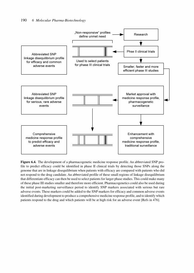
190 6 Molecular Pharma-Biotechnology
Figure 6.4. The development of a pharmacogenetic medicine response profile. An abbreviated SNP pro-file to predict efficacy could be identified in phase II clinical trials by detecting those SNPs along thegenome that are in linkage disequilibrium when patients with efficacy are compared with patients who didnot respond to the drug candidate. An abbreviated profile of these small regions of linkage disequilibriumthat differentiate efficacy can then be used to select patients for larger phase studies. This could make manyof these phase III studies smaller and therefore more efficient. Pharmacogenetics could also be used duringthe initial post-marketing surveillance period to identify SNP markers associated with serious but rareadverse events. These markers could be added to the SNP markers for efficacy and common adverse eventsidentified during development to produce a comprehensive medicine response profile, and to identify whichpatients respond to the drug and which patients will be at high risk for an adverse event [Refs in 476].
Abbreviated SNP
linkage disequilibrium profile
for efficacy and common
adverse events
Abbreviated SNP
linkage disequilibrium profile
for serious, rare adverse
events
Comprehensive
medicine response profile
to predict efficacy and
adverse events
Enhancement with
comprehensive
medicine response profile,
traditional surveillance
Market approval with
medicine response profile,
pharmacogenetic
surveillance
Smaller, faster and more
efficient phase III studies
Phse II clinical trials
Research„Non-responsive“ profiles
define unmet need
Used to select patients
for phase III clinical trials
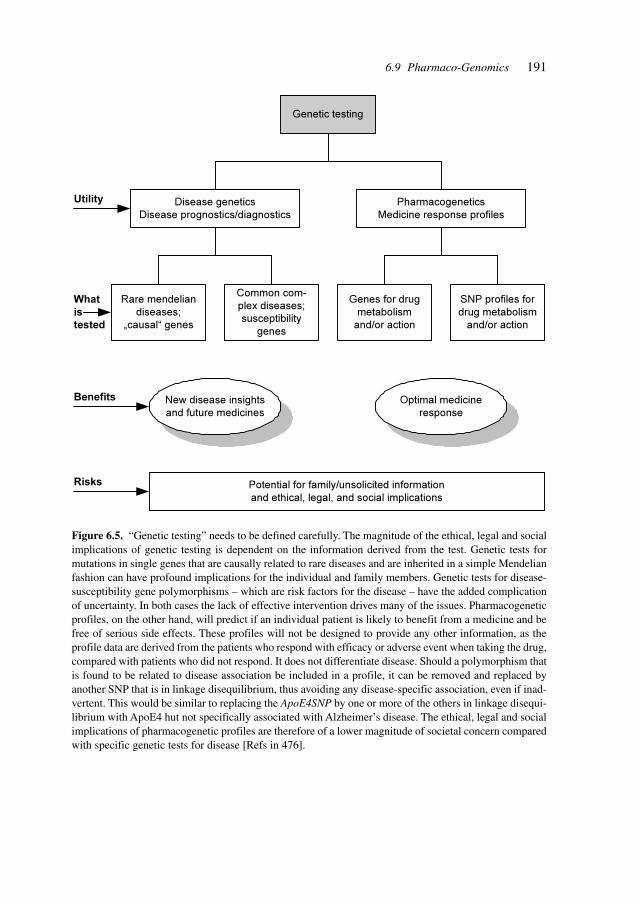
191
Figure 6.5. “Genetic testing” needs to be defined carefully. The magnitude of the ethical, legal and socialimplications of genetic testing is dependent on the information derived from the test. Genetic tests formutations in single genes that are causally related to rare diseases and are inherited in a simple Mendelianfashion can have profound implications for the individual and family members. Genetic tests for disease-susceptibility gene polymorphisms – which are risk factors for the disease – have the added complicationof uncertainty. In both cases the lack of effective intervention drives many of the issues. Pharmacogeneticprofiles, on the other hand, will predict if an individual patient is likely to benefit from a medicine and befree of serious side effects. These profiles will not be designed to provide any other information, as theprofile data are derived from the patients who respond with efficacy or adverse event when taking the drug,compared with patients who did not respond. It does not differentiate disease. Should a polymorphism thatis found to be related to disease association be included in a profile, it can be removed and replaced byanother SNP that is in linkage disequilibrium, thus avoiding any disease-specific association, even if inad-vertent. This would be similar to replacing the ApoE4SNP by one or more of the others in linkage disequi-librium with ApoE4 hut not specifically associated with Alzheimer’s disease. The ethical, legal and socialimplications of pharmacogenetic profiles are therefore of a lower magnitude of societal concern comparedwith specific genetic tests for disease [Refs in 476].
Genetic testing
Disease genetics
Disease prognostics/diagnostics
Pharmacogenetics
Medicine response profiles
Rare mendelian
diseases;
„causal“ genes
Common com-
plex diseases;
susceptibility
genes
Genes for drug
metabolism
and/or action
SNP profiles for
drug metabolism
and/or action
New disease insights
and future medicines
Optimal medicine
response
Potential for family/unsolicited information
and ethical, legal, and social implications
Risks
Benefits
Utility
What
is
tested
6.9 Pharmaco-Genomics
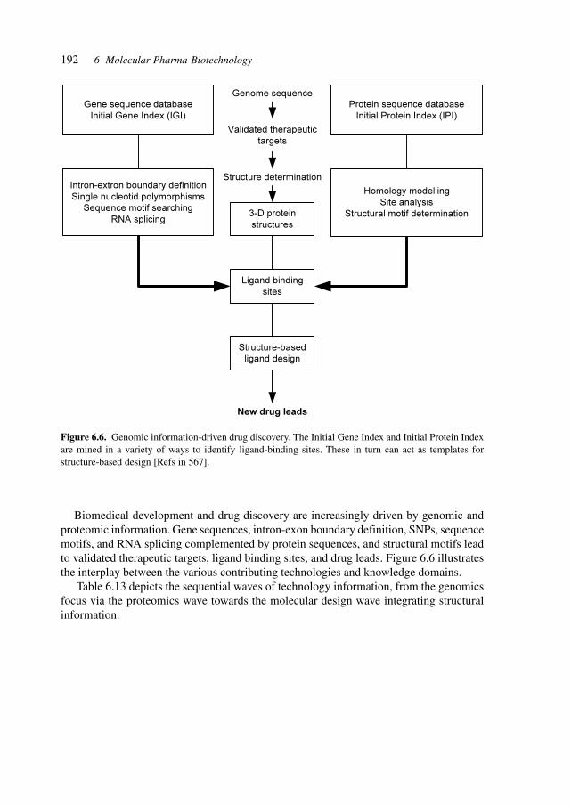
192 6 Molecular Pharma-Biotechnology
Biomedical development and drug discovery are increasingly driven by genomic andproteomic information. Gene sequences, intron-exon boundary definition, SNPs, sequencemotifs, and RNA splicing complemented by protein sequences, and structural motifs leadto validated therapeutic targets, ligand binding sites, and drug leads. Figure 6.6 illustratesthe interplay between the various contributing technologies and knowledge domains.
Table 6.13 depicts the sequential waves of technology information, from the genomicsfocus via the proteomics wave towards the molecular design wave integrating structuralinformation.
Figure 6.6. Genomic information-driven drug discovery. The Initial Gene Index and Initial Protein Indexare mined in a variety of ways to identify ligand-binding sites. These in turn can act as templates forstructure-based design [Refs in 567].
Gene sequence database
Initial Gene Index (IGI)
Protein sequence database
Initial Protein Index (IPI)
3-D protein
structures
Ligand binding
sites
Structure-based
ligand design
New drug leads
Structure determination
Validated therapeutic
targets
Genome sequence
Homology modelling
Site analysis
Structural motif determination
Intron-extron boundary definition
Single nucleotid polymorphisms
Sequence motif searching
RNA splicing
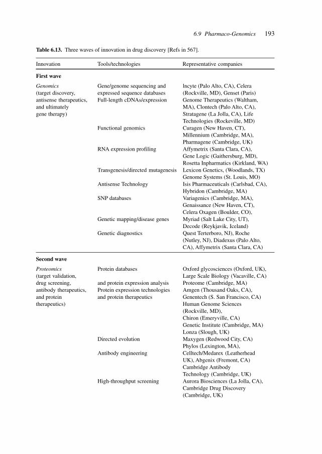
193
Table 6.13. Three waves of innovation in drug discovery [Refs in 567].
Innovation Tools/technologies Representative companies
First wave
Genomics Gene/genome sequencing and lncyte (Palo Alto, CA), Celera(target discovery, expressed sequence databases (Rockville, MD), Genset (Paris)antisense therapeutics, Full-length cDNAs/expression Genome Therapeutics (Waltham,and ultimately MA), Clontech (Palo Alto, CA),gene therapy) Stratagene (La Jolla, CA), Life
Technologies (Rockeville, MD)Functional genomics Curagen (New Haven, CT),
Millennium (Cambridge, MA),Pharmagene (Cambridge, UK)
RNA expression profiling Affymetrix (Santa Clara, CA),Gene Logic (Gaithersburg, MD),Rosetta Inpharmatics (Kirkland, WA)
Transgenesis/directed mutagenesis Lexicon Genetics, (Woodlands, TX)Genome Systems (St. Louis, MO)
Antisense Technology Isis Pharmaceuticals (Carlsbad, CA),Hybridon (Cambridge, MA)
SNP databases Variagenics (Cambridge, MA),Genaissance (New Haven, CT),Celera Oxagen (Boulder, CO),
Genetic mapping/disease genes Myriad (Salt Lake City, UT),Decode (Reykjavik, Iceland)
Genetic diagnostics Quest Terterboro, NJ), Roche(Nutley, NJ), Diadexus (Palo Alto,CA), Affymetrix (Santa Clara, CA)
Second wave
Proteomics Protein databases Oxford glycosciences (Oxford, UK),(target validation, Large Scale Biology (Vacaville, CA)drug screening, and protein expression analysis Proteome (Cambridge, MA)antibody therapeutics, Protein expression technologies Amgen (Thousand Oaks, CA),and protein and protein therapeutics Genentech (S. San Francisco, CA)therapeutics) Human Genome Sciences
(Rockville, MD),Chiron (Emeryville, CA)Genetic Institute (Cambridge, MA)Lonza (Slough, UK)
Directed evolution Maxygen (Redwood City, CA)Phylos (Lexington, MA),
Antibody engineering Celltech/Medarex (LeatherheadUK), Abgenix (Fremont, CA)Cambridge AntibodyTechnology (Cambridge, UK)
High-throughput screening Aurora Biosciences (La Jolla, CA),Cambridge Drug Discovery(Cambridge, UK)
6.9 Pharmaco-Genomics
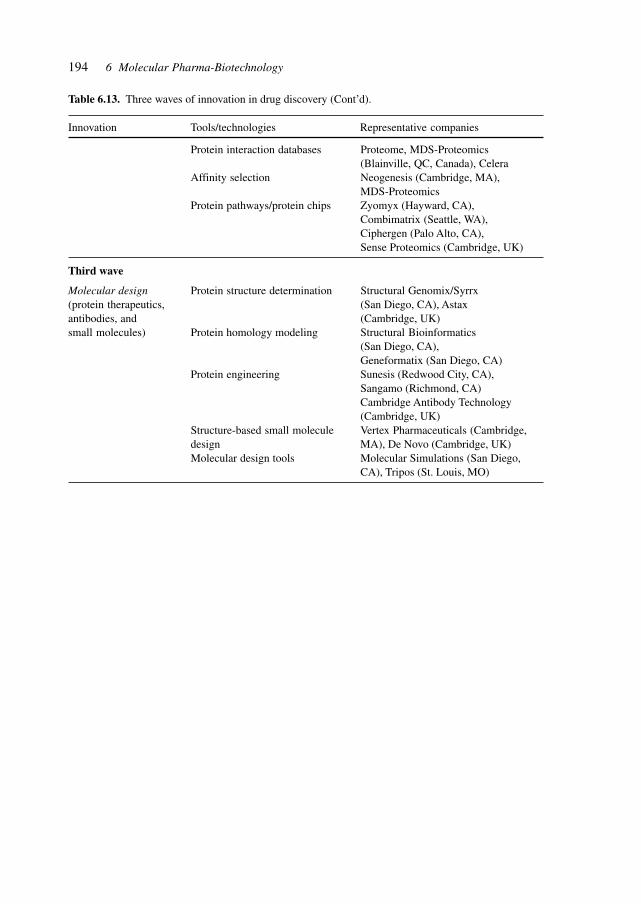
194 6 Molecular Pharma-Biotechnology
Table 6.13. Three waves of innovation in drug discovery (Cont’d).
Innovation Tools/technologies Representative companies
Protein interaction databases Proteome, MDS-Proteomics(Blainville, QC, Canada), Celera
Affinity selection Neogenesis (Cambridge, MA),MDS-Proteomics
Protein pathways/protein chips Zyomyx (Hayward, CA),Combimatrix (Seattle, WA),Ciphergen (Palo Alto, CA),Sense Proteomics (Cambridge, UK)
Third wave
Molecular design Protein structure determination Structural Genomix/Syrrx(protein therapeutics, (San Diego, CA), Astaxantibodies, and (Cambridge, UK)small molecules) Protein homology modeling Structural Bioinformatics
(San Diego, CA),Geneformatix (San Diego, CA)
Protein engineering Sunesis (Redwood City, CA),Sangamo (Richmond, CA)Cambridge Antibody Technology(Cambridge, UK)
Structure-based small molecule Vertex Pharmaceuticals (Cambridge,design MA), De Novo (Cambridge, UK)Molecular design tools Molecular Simulations (San Diego,
CA), Tripos (St. Louis, MO)

195
7 Research and Development
The R & D process is benefitting from the expanding knowledge base, and from the in-creasing contributions from robotics and automation, informatics and bioinformatics, equip-ment development, and the outsourcing of key processes and consolidation of the regula-tory environment.
7.1 Biology, Medicine, and Genetics
The recognition of similarities and differences between species and the use of this knowl-edge for the elucidation of biochemical mechanisms, genetic lesions and relationships,disease mechanisms, identification of molecular targets is facilitating the drug discoveryprocess.
Integration of experts from various fields of medicine, biology and genetics will bringresults from fundamental research quickly into the realm of therapy and medicine.
7.2 Pre-clinical and Clinical Development
The pre-clinical development stage is accelerated by additional models of disease and test-ing as well as high-throughput technologies in the laboratory.
Clinical development benefits from genetic and biochemical knowledge, and the spe-cific targeting of therapies to genetic (sub-)groups with concomitant stratification of pa-tients.
7.3 Processes
The classical methods of chemical and fermentative synthesis and purification are aug-mented by the powerful genetically modified cellular systems of production, by transgenicanimals and plants, as well as by sophisticated techniques of synthesis, analysis, purifica-tion, such as chiral catalysts, micro-reactors, and process intensification.
Industrial Pharmaceutical Biotechnology. Heinrich KlefenzCopyright © 2002 Wiley-VCH Verlag GmbH
ISBNs: 3-527-29995-5 (Hardcover); 3-527-60012-4 (Electronic)
196 7 Research and Development
7.4 Pilot Plants
The inceasing efficiency and potency of APIs as well as the fragmentation of patient groupsgenerally decreases the amounts of substances required for the pre-clinical, clinical andproduction phases. Versatile pilot plants are capable of providing such amounts, whereaslarge dedicated factories were often required for lower potency compounds.
7.5 Engineering
The engineering capabilities which need to be deployed are extending from classical chemicalor fermentation formulation engineering towards the inclusion of the additional technolo-gies in cell culture, metabolic engineering, micro- and nano-technology engineering, DNAor protein chip design, in silico biology, simulation, and bioinformatics.
7.6 Fermentation Process Development
These topics in fermentation process development include metabolic flux, metabolic engi-neering, process development, and downstream processing. Recent advances in metabolicengineering have led to the development of new methods for the synthesis of novel mol-ecules, improved production of existing compounds and improved degradation of recalci-trant environmental contaminates. Increasing the flux through an existing pathway andintroducing a new pathway into a host organism demand coordinated expression of thegenes that encode the enzymes, tight control over gene expression and consistent expres-sion in all cells. Several tools have been developed for the overproduction of specific pro-teins. Pathway redirection (metabolic engineering) requires certain specific characteristicsof gene-expression tools such as cloning vectors, plasmids, and promoters for the expres-sion of multiple genes.[374]
Metabolic engineering has been intensively studied in the eukaryotic organism Saccha-romyces cerevisiae, which is a model for the biology of eukaryotic organisms. S. cerevisiaeis susceptible to recombinant DNA technology and metabolic engineering is facilitated bythe availability of the complete genome sequence.
The importance of bacteria and eukaryotic microorganisms is illustrated in Table 7.1,which lists metabolite production by various microorganisms, such as amino acid biosyn-thesis, ethanol production, lactic acid, and xylitol production, 7-ACA and 7-ADCA-pro-duction.
In Saccharomyces cerevisiae, substrate extension has been achieved to increase the ver-satility and improve the economics of S. cerevisiae fermentations. Examples illustratingthe extension of substrate range to include Lactose, Malate, Starch and Dextrins, and Xy-lose are shown in Table 7.2.
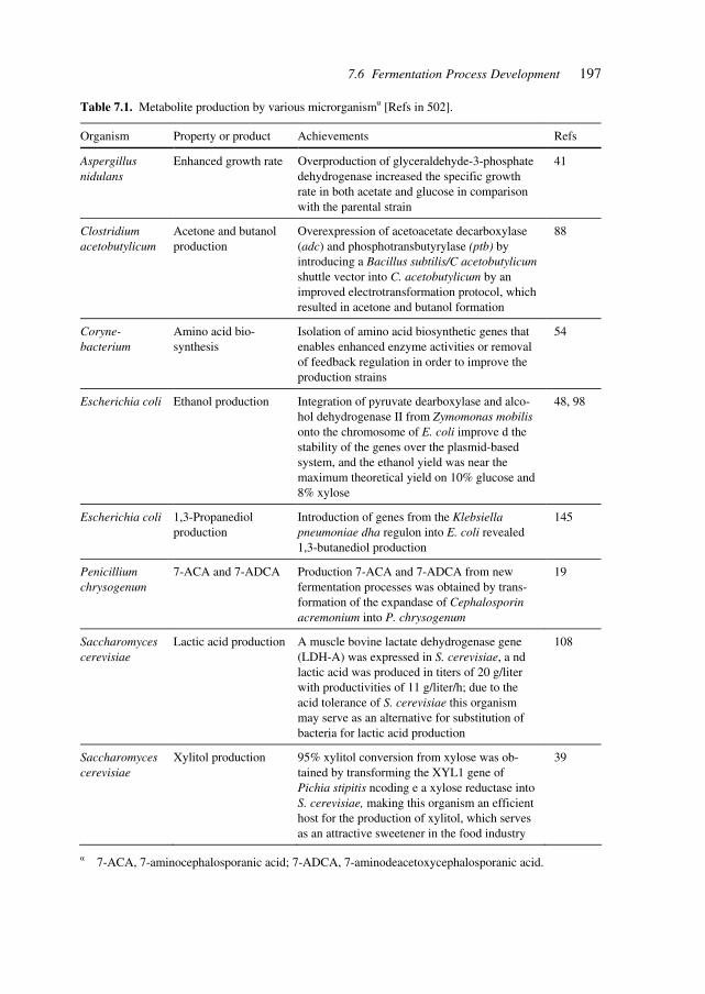
1977.6 Fermentation Process Development
Table 7.1. Metabolite production by various microrganismα [Refs in 502].
Organism Property or product Achievements Refs
Aspergillus nidulans
Enhanced growth rate Overproduction of glyceraldehyde-3-phosphate dehydrogenase increased the specific growth rate in both acetate and glucose in comparison with the parental strain
41
Clostridium acetobutylicum
Acetone and butanol production
Overexpression of acetoacetate decarboxylase (adc) and phosphotransbutyrylase (ptb) by introducing a Bacillus subtilis/C acetobutylicum shuttle vector into C. acetobutylicum by an improved electrotransformation protocol, which resulted in acetone and butanol formation
88
Coryne-bacterium
Amino acid bio-synthesis
Isolation of amino acid biosynthetic genes that enables enhanced enzyme activities or removal of feedback regulation in order to improve the production strains
54
Escherichia coli Ethanol production Integration of pyruvate dearboxylase and alco-hol dehydrogenase II from Zymomonas mobilis onto the chromosome of E. coli improve d the stability of the genes over the plasmid-based system, and the ethanol yield was near the maximum theoretical yield on 10% glucose and 8% xylose
48, 98
Escherichia coli 1,3-Propanediol production
Introduction of genes from the Klebsiella pneumoniae dha regulon into E. coli revealed 1,3-butanediol production
145
Penicillium chrysogenum
7-ACA and 7-ADCA Production 7-ACA and 7-ADCA from new fermentation processes was obtained by trans-formation of the expandase of Cephalosporin acremonium into P. chrysogenum
19
Saccharomyces cerevisiae
Lactic acid production A muscle bovine lactate dehydrogenase gene (LDH-A) was expressed in S. cerevisiae, a nd lactic acid was produced in titers of 20 g/liter with productivities of 11 g/liter/h; due to the acid tolerance of S. cerevisiae this organism may serve as an alternative for substitution of bacteria for lactic acid production
108
Saccharomyces cerevisiae
Xylitol production 95% xylitol conversion from xylose was ob-tained by transforming the XYL1 gene of Pichia stipitis ncoding e a xylose reductase into S. cerevisiae, making this organism an efficient host for the production of xylitol, which serves as an attractive sweetener in the food industry
39
α 7-ACA, 7-aminocephalosporanic acid; 7-ADCA, 7-aminodeacetoxycephalosporanic acid.
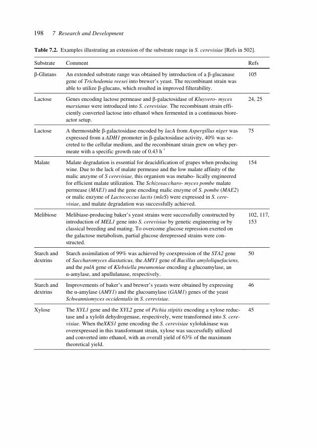
198 7 Research and Development
Table 7.2. Examples illustrating an extension of the substrate range in S. cerevisiae [Refs in 502].
Substrate Comment Refs
β-Glutans An extended substrate range was obtained by introduction of a β-glucanase gene of Trichodemia reesei into brewer’s yeast. The recombinant strain was able to utilize β-glucans, which resulted in improved filterability.
105
Lactose Genes encoding lactose permease and β-galactosidase of Kluyvero- myces marxianus were introduced into S. cerevisiae. The recombinant strain effi-ciently converted lactose into ethanol when fermented in a continuous biore-actor setup.
24, 25
Lactose A thermostable β-galactosidase encoded by lacA from Aspergillus niger was expressed from a ADH1 promoter in β-galactosidase activity, 40% was se-creted to the cellular medium, and the recombinant strain grew on whey per-meate with a specific growth rate of 0.43 h–1
75
Malate Malate degradation is essential for deacidification of grapes when producing wine. Due to the lack of malate permease and the low malate affinity of the malic anzyme of S cerevisiae, this organism was metabo- lically engineered for efficient malate utilization. The Schizosaccharo- myces pombe malate permease (MAE1) and the gene encoding malic enzyme of S. pombe (MAE2) or malic enzyme of Lactococcus lactis (mleS) were expressed in S. cere-visiae, and malate degradation was successfully achieved.
154
Melibiose Melibiase-producing baker’s yeast strains were successfully constructed by introduction of MEL1 gene into S. cerevisiae by genetic engineering or by classical breeding and mating. To overcome glucose repression exerted on the galactose metabolism, partial glucose derepressed strains were con-structed.
102, 117, 153
Starch and dextrins
Starch assimilation of 99% was achieved by coexpression of the STA2 gene of Saccharomyces diastaticus, the AMY1 gene of Bacillus amyloliquefaciens, and the pulA gene of Klebsiella pneumoniae encoding a glucoamylase, an α-amylase, and apullulanase, respectively.
50
Starch and dextrins
Improvements of baker’s and brewer’s yeasts were obtained by expressing the α-amylase (AMY1) and the glucoamylase (GAM1) genes of the yeast Schwanniomyces occidentalis in S. cerevisiae.
46
Xylose The XYL1 gene and the XYL2 gene of Pichia stipitis encoding a xylose reduc-tase and a xylolit dehydrogenase, respectively, were transformed into S. cere-visiae. When theXKS1 gene encoding the S. cerevisiae xylolukinase was overexpressed in this transformant strain, xylose was successfully utilized and converted into ethanol, with an overall yield of 63% of the maximum theoretical yield.
45

199
Metabolic engineering requires an integrated approach of analysis and synthesis. Stud-ies involve expression analysis, metabolite levels, pathway analysis, fermentation experi-ments, and protein characterization. Synthesis incorporates recombinant DNA techniquessuch as cloning, transformation, construction of vectors and artificial chromosomes.
The substrate extension to include the utilization of xylose by S. cerevisiae involves theintroduction of XYL1 and XYL2 from yeasts capable of utilizing xylose.
In order to maximize ethanol production in relation to glycerol and pyruvate, the meta-bolic pathways need to be manipulated. Figure 7.1 shows the biochemical routes to glyc-erol, pyruvate, and ethanol, illustrating targets for redirecting carbon flux to ethanol.
To overcome lag phases during fermentation of sugar mixtures containing glucose, su-crose, amd maltose, the pathways for sugar and saccharide utilization need to be alleviatedfrom glucose control. Improving maltose consumption was achieved by targeting regula-tory genes such as MIG1 and MIG2 as well as by constitutive expression of the structuralMAL genes.
The ultimate goal of the various genome-sequencing projects is to gain sufficient under-standing of the physiology of the organisms under study to enable exploitation of the ge-netic information to advance human health, agricultural production and industrial fermen-tation. A list of genes alone, however, is unlikely to provide sufficient information tomanipulate the metabolism or pathophysiology of an organism in a predictable way. Meta-bolic flux analysis is designed to convert a list of putative enzymes into a set of metabolicpathways. This approach involves a rigorous definition of the constitutive activities of agiven pathway as the sum of elementary flux modes, each of which represents a minimalsequence of metabolic steps that can cooperate independently of each other. Applicationsinclude more efficient identification of important drug targets and assigning and/or cor-roborating specific activities to orphan gene sequences.[375]
Applications include maximizing product yield in amino acid and antibiotic synthesis,reconstruction and consistency checks of metabolism from genome data, analysis of en-zyme deficiencies, and drug target identification in metabolic networks.
7.6 Fermentation Process Development
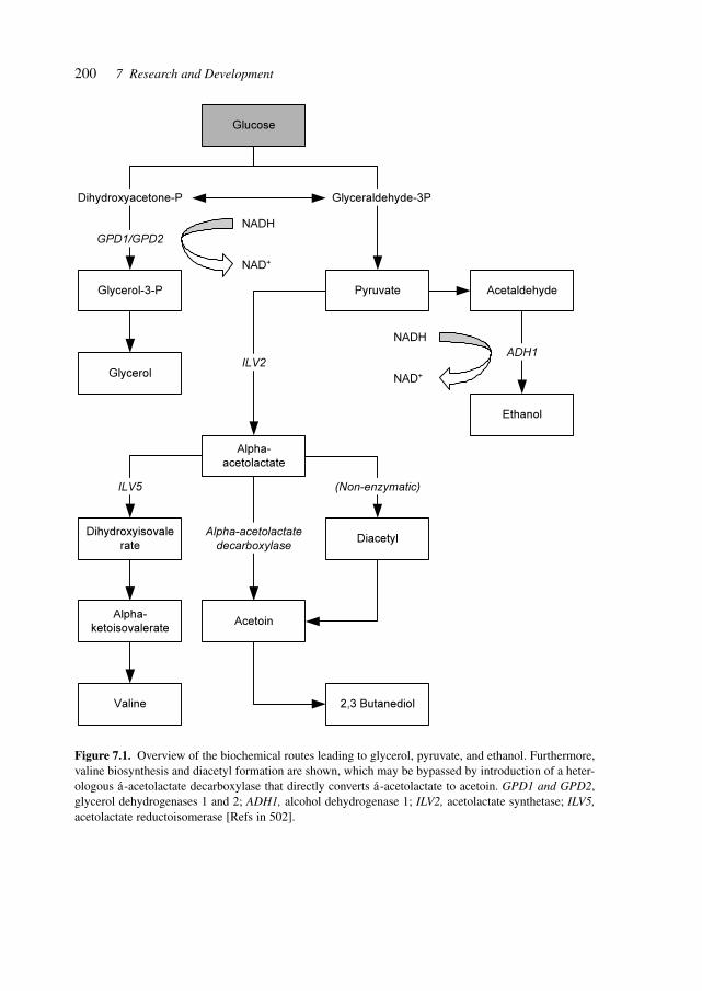
200 7 Research and Development
Figure 7.1. Overview of the biochemical routes leading to glycerol, pyruvate, and ethanol. Furthermore,valine biosynthesis and diacetyl formation are shown, which may be bypassed by introduction of a heter-ologous á-acetolactate decarboxylase that directly converts á-acetolactate to acetoin. GPD1 and GPD2,glycerol dehydrogenases 1 and 2; ADH1, alcohol dehydrogenase 1; ILV2, acetolactate synthetase; ILV5,acetolactate reductoisomerase [Refs in 502].
Glucose
Glycerol-3-P
Glycerol
Pyruvate Acetaldehyde
Ethanol
ADH1
Alpha-
acetolactate
ILV2
Dihydroxyisovale
rate
Alpha-
ketoisovalerate
Valine
Acetoin
Diacetyl
2,3 Butanediol
ILV5 (Non-enzymatic)
Alpha-acetolactate
decarboxylase
Glyceraldehyde-3PDihydroxyacetone-P
GPD1/GPD2
NADH
NAD+
NADH
NAD+
201
8 Pharmaceutical Production
Nature was the first source of active principles, albeit in crude, dilute, mixed and some-times toxic forms, an early example being quinine (present in Cinchona bark).
Microbial biotechnology, first exemplified by penicillin, yielded industrial amounts ofbiosynthetic compounds, followed (after the application of molecular biology) by signifi-cant numbers of microbial metabolites, engineered compounds and recombinant proteins.[442]
The ‘scale-up’ of chemical and biotechnological synthesis (from automated synthesis,laboratory synthesis to pilot production and full-scale commercial production) is gainingincreasing importance due to the required speed of development, the increasing complexityof the molecules (APIs) and the expanding technological repertoire being applied.
The industrial operation requires efficient design of production and optimized utiliza-tion of multi-product facilities.[376]
The increasing demand of the chemical and pharmaceutical industries for enantiomericallypure compounds has spurred the development of a range of so-called ‘chiral technologies’,which aim to exert the ultimate control over a chemical reaction by directing its enantio-selectivity. Heterogeneous enantioselective catalysis is particularly attractive because it al-lows the production and ready separation of large quantities of chiral product while usingonly small quantities of catalyst. Heterogeneous enantioselectivity is usually induced byadsorbing chiral molecules onto catalytically active surfaces. A mimic of one such catalystis formed by adsorbing (R,R)-tartaric acid molecules on Cu(110) surfaces: this generates avariety of surface phases, of which only one is potentially catalytically active and leaves thequestion of how adsorbed chiral molecules give rise to enantioselectivity. It was shown thatthe active phase consists of extended supramolecular assemblies of adsorbed (R,R)-tartaricacid, which destroy existing symmetry elements of the underlying metal and directly be-stow chirality to the modified surfaces. The adsorbed assemblies create chiral ‘channels’exposing bare metal atoms and it is these chiral spaces that are believed to be responsiblefor imparting enantioselectivity, by forcing the orientation of reactant molecules dockingonto catalytically active metal sites. The findings demonstrate that it is possible to sustain asingle chiral domain across an extended surface – provided that reflection domains of op-posite handedness are removed by a rigid and chiral local adsorption geometry, and thatinequivalent rotation domains are removed by successful matching of the rotational sym-metry of the adsorbed molecule with that of the underlying metal surface.[377]
Using metabolic engineering, the carotenoid biosynthesis pathway in tobacco (Nicoti-ana tabacum) was modified to produce astaxanthin, a red pigment of considerable eco-nomic value. To alter the carotenoid pathway in chromoplasts of higher plants, the cDNAof the gene CrtO from the alga Haematococcus pluvialis, encoding β-carotene ketolase,was transferred to tobacco under the regulation of the tomato Pds (phytoene desaturase)
Industrial Pharmaceutical Biotechnology. Heinrich KlefenzCopyright © 2002 Wiley-VCH Verlag GmbH
ISBNs: 3-527-29995-5 (Hardcover); 3-527-60012-4 (Electronic)

202 8 Pharmaceutical Production
promoter. The transit peptide of Pds from tomato was used to target the CRTO polypeptideto the plastids. Chromoplasts in the nectary tissue of transgenic plants accumulated (3S,3′S)astaxanthin and other ketocarotenoids, changing the color of the nectary from yellow tored. This accomplishment demonstrates that plants can be used as a source of novel caro-tenoid pigments such as astaxanthin. The procedures described can serve as a platformtechnology for future genetic manipulations of pigmentation of fruits and flowers of horti-cultural and floricultural importance.[378]
Combinatorial biosynthesis was used to synthesize novel lipophilic carotenoids that arepowerful cellular antioxidants. By co-expressing three different carotenoid desaturases incombination with a carotenoid hydratase, a cyclase and a hydroxylase on compatible plas-mids in E. coli, four novel carotenoids were synthesized that were not previously detectedin biological material or chemically synthesized. Their identification was based on theirrelative retention times on HPLC, spectroscopic properties, molecular weights, number ofhydroxy groups and 1H-NMR spectra. The carotenoids were designated as 1-HO-3′,4′-didehydrolycopene, 3,1′-(H)2-γ-carotene, 1,1′-(HO)2-3,4,3′,4′-tetradehydrolycopene and1,1′-(HO)2-3,4-didehydrolycopene These novel acyclic derivatives differ from structurallyrelated compounds by extension of the conjugated polyene chain as well as additionalhydroxy groups at position C-1′. Their antioxidative activity was determined in a lipo-some-membrane model system, which showed that their ability to protect against photo-oxidation and radical-mediated peroxidation reactions was linked to the length of the con-jugated double-bond system and the presence of a single hydroxy group. The protection ofmembrane degradation was superior to the related 1-HO and 1,1′-(HO)2 lycopene deriva-tives, making them interesting pharmaceutical candidates.[379]
The burgeoning demand for complex, biologically active molecules for medicine, mate-rials science, consumer products and agrochemicals is driving efforts to engineer new bio-synthetic pathways into microorganisms and plants. Principles of breeding, including mix-ing genes and modifying catalytic functions by in vitro evolution, were applied to createnew metabolic pathways for biosynthesis of natural products in E. coli. Shuffled phytoenedesaturases were expressed in the context of a carotenoid biosynthetic pathway assembledfrom different bacterial species and the resulting library screened for novel carotenoids.One desaturase chimera efficiently introduced six rather than four double bonds intophytoene, to favor production of the fully conjugated carotenoid, 3,4,3′,4′-tetradehydro-lycopene. This new pathway was extended with a second library of shuffled lyopene cycla-ses to produce a variety of colored products. This combined approach of rational pathwayassembly and molecular breeding may allow the discovery and production, in simple labo-ratory organisms, of new compounds that are essentially inaccessible from natural sourcesor by synthetic chemistry.[380]
Metabolic engineering in S. cerevisiae was carried out to increase the flux through cen-tral carbon metabolism. Regulatory network manipulation achieved a balanced increase inthe activity of all enzymes in the pathway and augmented the metabolic flux through thatpathway. Manipulation of the tightly regulated GAL gene regulatory network of S. cerevisiaeproduced prototroph mutant strains with increased flux through the galactose utilizationpathway by eliminating three negative regulators of the GAL system, Gal6, Gal80 andMig1, with a resulting 41% increase in flux through the galactose utilization pathway of
2038.1 GenePharming (Animals and Plants)
the mutant as compared with the wild-type strain. The improved consumption of galactose,which is a constituent of many industrial fermentation media, in the gal mutants resultednot in augmented biomass formation but in excessive respiro-fermentative metabolism,whereby the ethanol production rate increased linearly with glycolytic flux.[443]
Genetic and metabolic profiling aids in the identification of suitable plant species andvariants, in selecting desired mutants, and in identifying networks and the effects of genemutations or the influence of small molecules. Metabolic profiling in particular helps inelucidating links and relationships at the metabolic regulation level.
Multiparallel analyses of mRNA and proteins are complemented by metabolic profilingas a tool for comparative display of gene function. It provides insight into complex regula-tory processes and gives a characteristic description of different phenotypes. Using GC/MS, 326 distinct compounds from A. thaliana leaf extracts were automatically quantified.A chemical structure was assigned to approximately half of these compounds. Comparisonof four Arabidopsis genotypes (two homozygous ecotypes and a mutant of each ecotype)demonstrated a distinct metabolic profile for each genotype. Data mining tools such asprincipal component analysis enabled the assignment of metabolic phenotypes. The meta-bolic phenotypes of the two ecotypes were more divergent than were the metabolic pheno-types of the single-loci mutant and their parental ecotypes.[448]
8.1 GenePharming (Animals and Plants)
The application of gene-transfer technology to domestic animals offers a way for the intro-duction of genes encoding (human) proteins, thus creating biochemical pathways that arenot originally present or non-functional in these animals.
This provides mechanisms for producing therapeutic proteins and for the modificationof production characteristics. Efforts to introduce specific characteristics which involve theintegration of additional with existing biochemical pathways and homeostasis require higherlevels of transcriptional and translational control.[381]
Farm animal genomics as a method of identifying genes controlling commercially im-portant traits is developing by constructing maps of informative markers, scanning thosemaps to locate quantitative trait loci, identifying the trait genes themselves, and finallybridging the gap between the gene(s) and the ultimate trait.[382]
Transgenic plants have become attractive systems for production of human therapeuticproteins because of the reduced risk of mammalian viral contaminants in animal cell cul-tures and animals, the ability to do large scale-up at low cost, and the low maintenancerequirements. A feasibility study was reported for the production of a human therapeuticprotein through transplastomic transformation technology, which has the additional advan-tage of increased biological containment by apparent elimination of the transmission oftransgenes through pollen. It is shown that chloroplasts can express a secretory protein,human somatotropin, in a soluble, biologically active, disulfide-bonded form. High con-centrations of recombinant protein accumulation are observed (greater than 7% total solubleprotein), more than 300-fold higher than a similar gene expressed using a nuclear transgenic
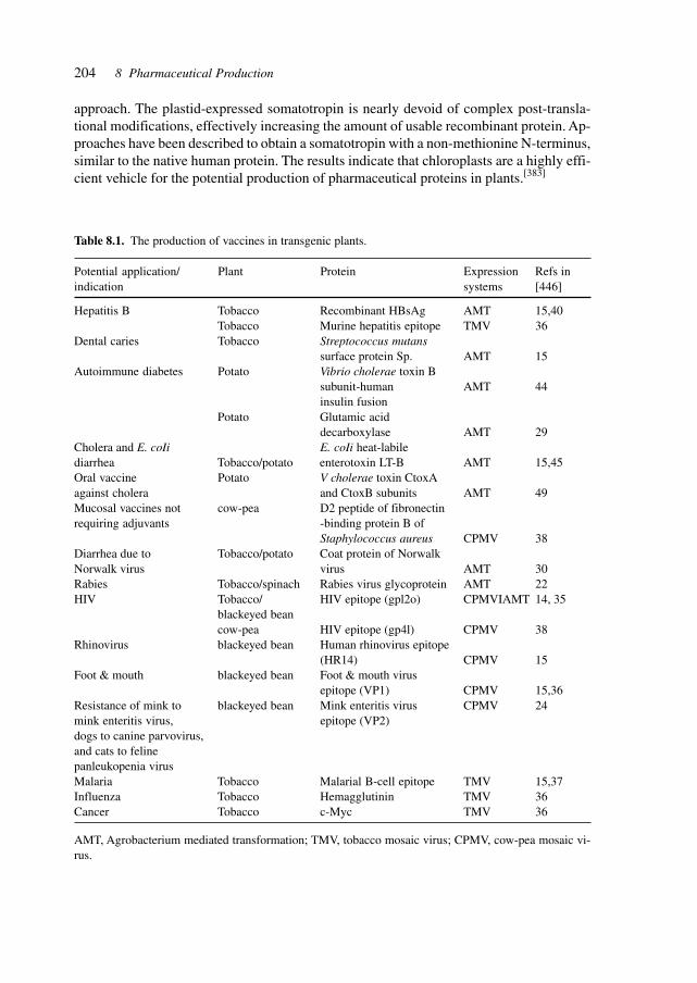
204 8 Pharmaceutical Production
approach. The plastid-expressed somatotropin is nearly devoid of complex post-transla-tional modifications, effectively increasing the amount of usable recombinant protein. Ap-proaches have been described to obtain a somatotropin with a non-methionine N-terminus,similar to the native human protein. The results indicate that chloroplasts are a highly effi-cient vehicle for the potential production of pharmaceutical proteins in plants.[383]
Table 8.1. The production of vaccines in transgenic plants.
Potential application/ Plant Protein Expression Refs inindication systems [446]
Hepatitis B Tobacco Recombinant HBsAg AMT 15,40Tobacco Murine hepatitis epitope TMV 36
Dental caries Tobacco Streptococcus mutanssurface protein Sp. AMT 15
Autoimmune diabetes Potato Vibrio cholerae toxin Bsubunit-human AMT 44insulin fusion
Potato Glutamic aciddecarboxylase AMT 29
Cholera and E. coIi E. coIi heat-labilediarrhea Tobacco/potato enterotoxin LT-B AMT 15,45Oral vaccine Potato V cholerae toxin CtoxAagainst cholera and CtoxB subunits AMT 49Mucosal vaccines not cow-pea D2 peptide of fibronectinrequiring adjuvants -binding protein B of
Staphylococcus aureus CPMV 38Diarrhea due to Tobacco/potato Coat protein of NorwalkNorwalk virus virus AMT 30Rabies Tobacco/spinach Rabies virus glycoprotein AMT 22HIV Tobacco/ HIV epitope (gpl2o) CPMVIAMT 14, 35
blackeyed beancow-pea HIV epitope (gp4l) CPMV 38
Rhinovirus blackeyed bean Human rhinovirus epitope(HR14) CPMV 15
Foot & mouth blackeyed bean Foot & mouth virusepitope (VP1) CPMV 15,36
Resistance of mink to blackeyed bean Mink enteritis virus CPMV 24mink enteritis virus, epitope (VP2)dogs to canine parvovirus,and cats to felinepanleukopenia virusMalaria Tobacco Malarial B-cell epitope TMV 15,37Influenza Tobacco Hemagglutinin TMV 36Cancer Tobacco c-Myc TMV 36
AMT, Agrobacterium mediated transformation; TMV, tobacco mosaic virus; CPMV, cow-pea mosaic vi-rus.
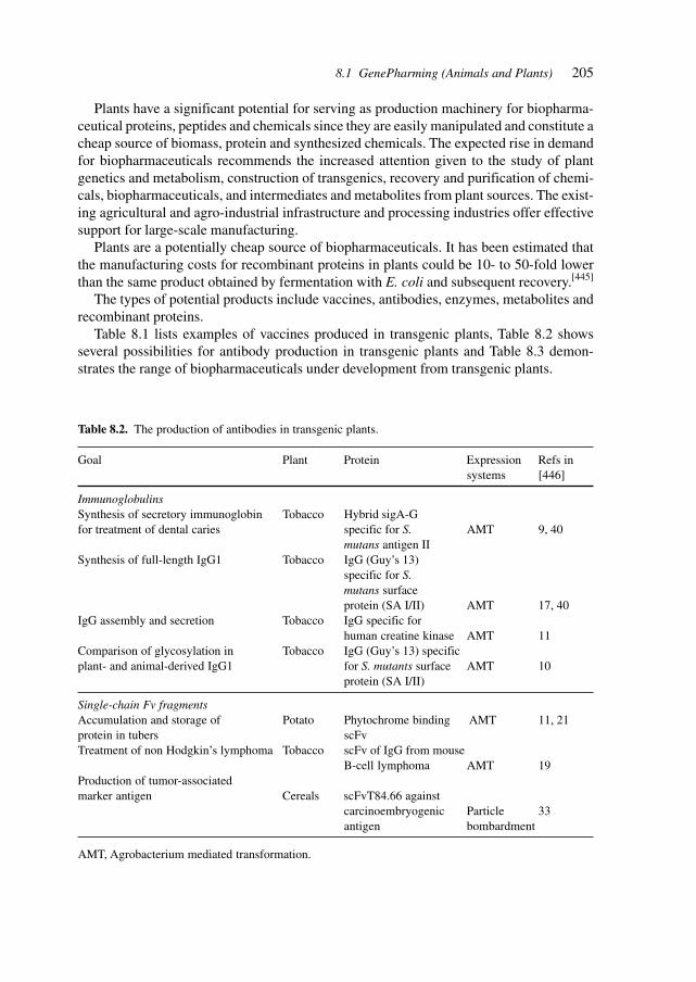
205
Plants have a significant potential for serving as production machinery for biopharma-ceutical proteins, peptides and chemicals since they are easily manipulated and constitute acheap source of biomass, protein and synthesized chemicals. The expected rise in demandfor biopharmaceuticals recommends the increased attention given to the study of plantgenetics and metabolism, construction of transgenics, recovery and purification of chemi-cals, biopharmaceuticals, and intermediates and metabolites from plant sources. The exist-ing agricultural and agro-industrial infrastructure and processing industries offer effectivesupport for large-scale manufacturing.
Plants are a potentially cheap source of biopharmaceuticals. It has been estimated thatthe manufacturing costs for recombinant proteins in plants could be 10- to 50-fold lowerthan the same product obtained by fermentation with E. coli and subsequent recovery.[445]
The types of potential products include vaccines, antibodies, enzymes, metabolites andrecombinant proteins.
Table 8.1 lists examples of vaccines produced in transgenic plants, Table 8.2 showsseveral possibilities for antibody production in transgenic plants and Table 8.3 demon-strates the range of biopharmaceuticals under development from transgenic plants.
Table 8.2. The production of antibodies in transgenic plants.
Goal Plant Protein Expression Refs insystems [446]
ImmunoglobulinsSynthesis of secretory immunoglobin Tobacco Hybrid sigA-Gfor treatment of dental caries specific for S. AMT 9, 40
mutans antigen IISynthesis of full-length IgG1 Tobacco IgG (Guy’s 13)
specific for S.mutans surfaceprotein (SA I/II) AMT 17, 40
IgG assembly and secretion Tobacco IgG specific forhuman creatine kinase AMT 11
Comparison of glycosylation in Tobacco IgG (Guy’s 13) specificplant- and animal-derived IgG1 for S. mutants surface AMT 10
protein (SA I/II)
Single-chain Fv fragmentsAccumulation and storage of Potato Phytochrome binding AMT 11, 21protein in tubers scFvTreatment of non Hodgkin’s lymphoma Tobacco scFv of IgG from mouse
B-cell lymphoma AMT 19Production of tumor-associatedmarker antigen Cereals scFvT84.66 against
carcinoembryogenic Particle 33antigen bombardment
AMT, Agrobacterium mediated transformation.
8.1 GenePharming (Animals and Plants)
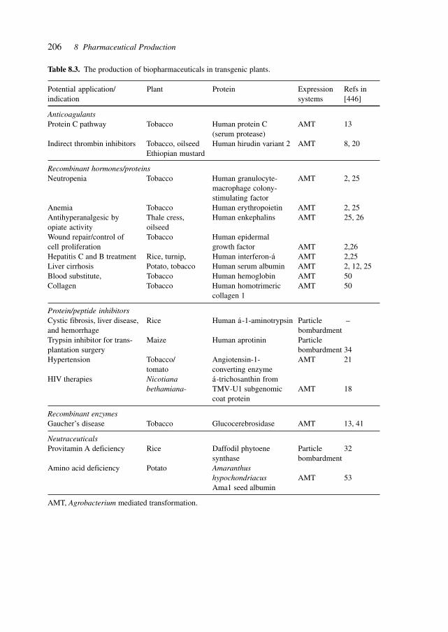
206 8 Pharmaceutical Production
Table 8.3. The production of biopharmaceuticals in transgenic plants.
Potential application/ Plant Protein Expression Refs inindication systems [446]
AnticoagulantsProtein C pathway Tobacco Human protein C AMT 13
(serum protease)Indirect thrombin inhibitors Tobacco, oilseed Human hirudin variant 2 AMT 8, 20
Ethiopian mustard
Recombinant hormones/proteinsNeutropenia Tobacco Human granulocyte- AMT 2, 25
macrophage colony-stimulating factor
Anemia Tobacco Human erythropoietin AMT 2, 25Antihyperanalgesic by Thale cress, Human enkephalins AMT 25, 26opiate activity oilseedWound repair/control of Tobacco Human epidermalcell proliferation growth factor AMT 2,26Hepatitis C and B treatment Rice, turnip, Human interferon-á AMT 2,25Liver cirrhosis Potato, tobacco Human serum albumin AMT 2, 12, 25Blood substitute, Tobacco Human hemoglobin AMT 50Collagen Tobacco Human homotrimeric AMT 50
collagen 1
Protein/peptide inhibitorsCystic fibrosis, liver disease, Rice Human á-1-aminotrypsin Particle –and hemorrhage bombardmentTrypsin inhibitor for trans- Maize Human aprotinin Particleplantation surgery bombardment 34Hypertension Tobacco/ Angiotensin-1- AMT 21
tomato converting enzymeHIV therapies Nicotiana á-trichosanthin from
bethamiana- TMV-U1 subgenomic AMT 18coat protein
Recombinant enzymesGaucher’s disease Tobacco Glucocerebrosidase AMT 13, 41
NeutraceuticalsProvitamin A deficiency Rice Daffodil phytoene Particle 32
synthase bombardmentAmino acid deficiency Potato Amaranthus
hypochondriacus AMT 53Ama1 seed albumin
AMT, Agrobacterium mediated transformation.
207
Specific products from transgenic plants include enkephalins, IFN-α, human serum al-bumin, glucocerebrosidase, granulocyte macrophage colony-stimulating factor, α1-antit-rypsin inhibitor (for the treatment of CF, liver diseases, hemorrhages) and hirudin (an anti-coagulant for the treatment of thrombosis) from the leech Hirudo medicinalis.[446]
Hepatitis B oral immunization with transgenic potato was demonstrated in preclinicalanimal trials with recombinant hepatitis B surface antigen (HbsAg). Mice fed transgenicHbsAg potato tubers showed an immune response, i.e. increased HbsAg-specific serumantibody, which was boosted by intraperitoneal application of a single sub-immunogenicdose of commercially available HbsAg vaccine. In order to be effective and applicable asan oral vaccine, higher resulting titers will have to be achieved by delivering larger doses ofantigen. Studies of the factors influencing the accumulation of HbsAg in transgenic potatoinclude 5′ and 3′ flanking sequences and protein targeting within the plant cells. The mostsignificant improvements resulted from alternative polyadenylation signals and fusion pro-teins containing targeting signals designed to enhance the integration or retention of HbsAgin the endoplasmic reticulum of plant cells.[447]
The grain of self-pollinating diploid barley species offers two modes of producingrecombinant enzymes or other proteins. One uses the promoters of genes with aleurone-specific expression duringgermination and the signal peptide code for export of the proteininto the endosperm. The other uses promoters of the structural genes for storage proteinsdeposited in the developing endosperm. Production of a protein-engineered thermotolerant(1,3–1,4)-β-glucanase with the D hordein gene (Hor3-1) promoter during endosperm de-velopment was analyzed in transgenic plants with four different constructs. High expres-sion of the enzyme and it activity in the endosperm of the mature grain required codonoptimization to a C + G content of 63% and synthesis as a precursor with a signal peptidefor transport through the endoplasmic reticulum and targeting into the storage vacuoles.Synthesis of the recombinant enzyme in the aleurone of germinating transgenic grain withan α-amylase promoter and the code for the export signal peptide yielded about 1 µg/mgsoluble protein, whereas 54 µg/mg soluble protein was produced on average in the matur-ing grain of 10 transgenic lines with the vector containing the gene for the (1,3–1,4)-β-glucanase under the control of the Hor3-1 promoter.[384]
Important chemicals and intermediates are produced by plants, in particular in glandulartissues. Glandular tissues are an anatomical plant feature widely observable in plants. Manyproducts produced in these tissues play biologically important roles by, for example, at-tracting pollinators, protecting against herbivores, and defining flavor and aroma. Glandu-lar tissues comprise trichomes which accumulate essential oils and resins, secretory cavi-ties which accumulate aromatic oils in the epidermis of citrus fruits, ducts and cavitiesaccumulating oleoresinsin conifers, and osmopheres in floral tissues secreting volatile com-pounds attracting pollinators. The compounds synthesized include monoterpenes and ses-quiterpenes, diterpene resin acids, sucrose, and glucose esters of fatty acids, indole, nico-tine and phenolic substances, fatty acids, nectar, proteins, and polysaccharides.[444]
Bioengineering may aim at increasing the production or concentration of such indig-enous compounds or introduce the production of novel substances.
The metabolism of secretory tissues is divided into non-photosynthetic and photosyn-thetic glands. Oil-producing glandular tissues are often non-photosynthetic whereas crops
8.1 GenePharming (Animals and Plants)
208 8 Pharmaceutical Production
like tomato, potato, sunflower and geranium have photosynthetic glandular trichomes. Thisdistinction is pertinent for the manipulation of the metabolism since photosynthetic tri-chomes assimilate carbon dioxide and convert tissue into secretory products, whereas non-photosynthetic trichomes need to import carbon from the leaf cells for their metabolism.
The elucidation of the gene sequence of A. thaliana will facilitate the manipulation ofregulatory and metabolic pathways since knowledge about the developmental genetics oftrichome formation originates primarily from research on the non-glandular trichomes ofArabidopsis and numerous mutants of Arabidopsis have been described.
Bioengineering the development and metabolism of glandular tissues will have to takeinto account the commercial applications, the price of the chemicals to be produced, theagro-industrial production techniques and the genetic, regulatory and metabolic suscepti-bility for manipulation, as well as the potential of plant metabolism to achieve the desiredincreased energy and synthetic load.
Transgenic mice expressing bacterial phytase in the digestive tract (salivary glands) weredeveloped which showed reduced fecal phosphorous. This demonstrates a biological ap-proach to reducing phosphorous pollution from animal agriculture[674].
The expression of lysostaphin protein in mammary glands of transgenic mice confersprotection against staphylococcal infection and shows the potential of genetic engineeringto combat the disease in dairy cattle[702].
8.2 Vitamins
Vitamins can be produced by microbial and fungal fermentation (e.g., vitamin B2, riboseand vitamin B12).
Advanced genetic engineering has introduced a desired nutritional trait into agricultur-ally important rice.[385]
8.3 Amino Acids
Using directed evolution, the hydantoinase process for the production of L-methionine (L-Met) in E. coli was improved. This was accomplished by inverting the enantioselectivityand increasing the total activity of a key enzyme in a whole-cell catalyst. The selectivity ofall known hydantoinases for D-5-(2-methylthioethyl) hydantoin (D-MTEH) over the L-enantiomer leads to the accumulation of intermediates and reduced productivity for the L-amino acid. By using random mutagenesis, saturation mutagenesis and screening it, waspossible to convert the D-selective hydantoinase from Arthrobacter sp. DSM 9771 into anL-selective enzyme and increase its total activity 5-fold. Whole E. coli cells expressing theevolved L-hydantoinase, an L-N-carbamoylase and a hydantoin racemase, produced 91 mML-Met from 100 mM L,D-MTEH in less than 2 h. The improved hydantoinase increasedproductivity 5-fold for >90% conversion of the substrate. The accumulation of the un-
209
wanted intermediate D-carbamoyl-methionine was reduced 4-fold compared to cells withthe wild-type pathway. Highly D-selective hydantoinase mutants were also discovered.Enantioselective enzymes rapidly optimized by directed evolution and introduced intomultienzyme pathways may lead to improved whole-cell catalysts for efficient productionof chiral compounds.[386]
8.4 Proteins
In addition to the classical production of naturally occurring proteins, such as insulin, IFNs,growth hormone, growth factors and blood-clotting proteins, the techniques of molecularbiotechnology enable the synthesis of superior proteins by synthesis of artificial genes orby directed evolution.
In biological systems, enzymes catalyze the efficient synthesis of complex moleculesunder benign conditions, but widespread industrial use of these biocatalysts depends cru-cially on the development of new enzymes with useful catalytic functions. The evolution ofenzymes in biological systems often involves the acquisition of new catalytic or bindingproperties by an existing protein scaffold. This strategy was experimentally mimicked us-ing the most common fold in enzymes, the α/β barrel, as the scaffold. By combining anexisting binding site for structural elements of phosphoribosylanthranilate with catalytictemplate required for isomerase activity, phosphoribosylanthranilate isomerase activityevolved from the scaffold of indole-3-glycerolphosphate synthetase. Targeting the catalytictemplate for in vitro mutagenesis and recombination, followed by in vivo selection, resultsin a new phosphoribosylanthranilate isomerase that has catalytic properties similar to thoseof the natural enzyme, with an even higher specificity constant. The demonstration of di-vergent evolution and the widespread occurrence of the α/β barrel suggests that this scaf-fold may be a fold of choice for the directed evolution of new biocatalysts.[387]
Enzymes offer an environmentally benign alternative to chemical catalysts in many com-mercial and industrial applications. The expansion of their use depends on the developmentof new protein catalysts. This will imply the construction or identification of enzymes thathave been genetically altered to improve their performance under defined, application-specific conditions. Directed evolution offers a fast and effective way of creating improvedenzymes from relatively ineffective catalysts to commercially viable products by a varietyof directed evolution techniques.[388]
The Coprinus cinereus (CiP) heme peroxidase was subjected to multiple rounds of di-rected evolution in an effort to produce a mutant suitable for use as a dye-transfer inhibitorin laundry detergent. The wild-type peroxidase is rapidly inactivated under laundry condi-tions due to the high pH (10.5), high temperature (50 °C) and high peroxidase concentra-tion (5–10 µm). Peroxidase mutants were initially generated using two parallel approaches:site-directed mutagenesis based on structure–function considerations and error-prone PCRto create random mutations. Mutants were expressed in S. cerevisiae and screened for im-proved stability by measuring residual activity after incubation under conditions mimick-ing those in a washing machine. Manually combining mutations from the site-directed and
8.4 Proteins
210 8 Pharmaceutical Production
random approaches led to a mutant with 110 times the thermal stability and 2.8 times theoxidative stability of wild-type CiP. In the final two rounds, mutants were randomly recom-bined by using the efficient yeast homologous recombination system to shuffle point muta-tions among a large number of parents. This in vivo shuffling led to the most dramaticimprovements in oxidative stability, yielding a mutant with 174 times the thermal stabilityand 100 times the oxidative stability of wild-type CiP.[389]
The extraordinary success of breeding as a technology for modifying eukaryotic organ-isms is reflected in the rapid generation of diversity in domestic animals and in the combi-nation of production-enhancing traits in crop plants. Permutation of existing variations bybreeding also avoids the accumulation of deleterious mutations that accompanies an asexualevolutionary strategy. Individual proteins have traditionally been modified by choosing asingle ‘parent’ protein and then either engineering changes based on structure and functionconsiderations or by making random mutations and screening for improvement.
This approach has the difficulty of making mutations that improve one property withoutcompromising others. A solution to this problem is the ‘breeding’ of proteins with theappropriate individual properties and then screening for ‘progeny’ with the desired combi-nation. Simulating meiotic recombination at the level of single proteins is possible usingthe techniques of molecular breeding, such as DNA family shuffling. Thereby, multiplerelated genes are used as parental sequences which are combined by random fragmentationand reassembly to generate libraries of chimeric genes.
DNA family shuffling of 26 protease genes was used to generate a library of chimericproteases that was screened for four distinct enzymatic properties. Multiple clones wereidentified that were significantly improved over any of the parental enzymes for each indi-vidual property. Family shuffling, also known as molecular breeding, efficiently created allof the combinations of parental properties, producing a great diversity of property combi-nations in the progeny enzymes. Molecular breeding, like classical breeding, is a powerfultool for recombining existing diversity to tailor biological systems for multiple functionalparameters.[390]
Enzymes generated by directed evolution may play a significant role in therapies. Genetherapy for correction of genetic diseases such as sickle cell anemia involves substituting amutant gene with its normal counterpart. This approach in gene therapy is unlikely to beeffective in cancer therapy because human cancer cells contain mutations at multiple lociand have been postulated to exhibit a mutator phenotype that accelerates accumulation offurther mutations. Rather than address the many mutant genes and variable combinationsof mutant genes in cancer cells, a logical approach is to introduce genes encoding prodrugmetabolizing enzymes that have the potential to kill cancer cells. Another approach in-volves protection of normal host tissues (e.g., bone marrow) against the toxicity of manychemotherapeutic agents. Chemoprotective genes can be transfected into bone marrow exvivo and only a small percentage of the cells need to be transfected. Cells expressing thedrug-resistance gene should have a selective proliferative advantage in the presence ofchemotherapeutic drugs. The success of gene therapy might be increased by using mutatedversions of enzymes for both tumor ablation and bone marrow protection.
New techniques of directed evolution make it feasible to tailor enzymes for cancer genetherapy. Novel enzymes with desired properties can be created and selected from vast li-
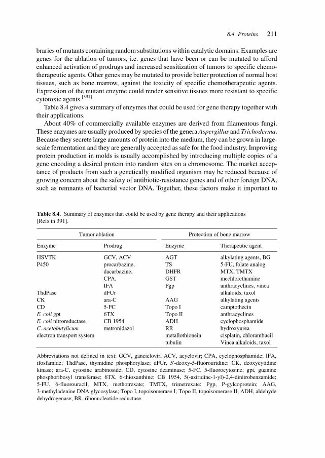
211
braries of mutants containing random substitutions within catalytic domains. Examples aregenes for the ablation of tumors, i.e. genes that have been or can be mutated to affordenhanced activation of prodrugs and increased sensitization of tumors to specific chemo-therapeutic agents. Other genes may be mutated to provide better protection of normal hosttissues, such as bone marrow, against the toxicity of specific chemotherapeutic agents.Expression of the mutant enzyme could render sensitive tissues more resistant to specificcytotoxic agents.[391]
Table 8.4 gives a summary of enzymes that could be used for gene therapy together withtheir applications.
About 40% of commercially available enzymes are derived from filamentous fungi.These enzymes are usually produced by species of the genera Aspergillus and Trichoderma.Because they secrete large amounts of protein into the medium, they can be grown in large-scale fermentation and they are generally accepted as safe for the food industry. Improvingprotein production in molds is usually accomplished by introducing multiple copies of agene encoding a desired protein into random sites on a chromosome. The market accep-tance of products from such a genetically modified organism may be reduced because ofgrowing concern about the safety of antibiotic-resistance genes and of other foreign DNA,such as remnants of bacterial vector DNA. Together, these factors make it important to
Table 8.4. Summary of enzymes that could be used by gene therapy and their applications [Refs in 391].
Tumor ablation Protection of bone marrow
Enzyme Prodrug Enzyme Therapeutic agent
HSVTK P450 ThdPase CK CD E. coli gpt E. coli nitroreductase C. acetobutylicum electron transport system
GCV, ACV procarbazine, dacarbazine, CPA, IFA dFUr ara-C 5-FC 6TX CB 1954 metronidazol
AGT TS DHFR GST Pgp AAG Topo I Topo II ADH RR metallothionein tubulin
alkylating agents, BG
5-FU, folate analog
MTX, TMTX
mechlorethamine anthracyclines, vinca alkaloids, taxol
alkylating agents
camptothecin anthracyclines cyclophosphamide hydroxyurea cisplatin, chlorambucil
Vinca alkaloids, taxol
Abbreviations not defined in text: GCV, ganciclovir, ACV, acyclovir; CPA, cyclophosphamide; IFA, ifosfamide; ThdPase, thymidine phosphorylase; dFUr, 5'-deoxy-5-fluorouridine; CK, deoxycytidine kinase; ara-C, cytosine arabinoside; CD, cytosine deaminase; 5-FC, 5-fluorocytosine; gpt, guanine phosphoribosyl transferase; 6TX, 6-thioxanthine; CB 1954, 5(-aziridine-1-yl)-2,4-dinitrobenzamide; 5-FU, 6-fluorouracil; MTX, methotrexate; TMTX, trimetrexate; Pgp, P-gylcoprotein; AAG, 3-methyladenine DNA glycosylase; Topo I, topoisomerase I; Topo II, topoisomerase II; ADH, aldehyde dehydrogenase; BR, ribonucleotide reductase.
8.4 Proteins
212 8 Pharmaceutical Production
develop methods of producing food-grade mold strains that contain multiple gene copies ofa gene integrated at a predetermined locus in the genome. Ideally, the recombinant micro-organism should contain only the heterologous gene encoding the desired protein and shouldbe free of unwanted foreign DNA. Although targeted integration of a single gene copy hasbeen described, it is difficult and sometimes impossible to obtain mold strains that containmultiple gene copies integrated at a predetermined site in the genome.
Whereas transformation techniques for bacteria and yeasts are well established, they areless so for fungi. An alternative transformation procedure for molds based on Agrobacteriumtumefaciens tumor-inducing DNA (T-DNA) transfer and genome integration by illegiti-mate recombination has been achieved, that is 100- to 1000-fold more efficient than previ-ous methods. T-DNA can also integrate via homologous recombination into Aspergillusawamori. Combining these two findings allows the efficient generation of food-grade re-combinant strains by targeted integration of multiple gene copies.
A. tumefaciens is known to transfer part of its tumor-inducing (Ti) plasmid to the fila-mentous fungus A. awamori by illegitimate recombination with the fungal genome. Whenthis Ti DNA shares homology with the A. awamori genome, integration can also occur byhomologous recombination. An efficient method was thus developed for constructing re-combinant mold strains free from bacterial DNA by A. tumefaciens-mediated transforma-tion. Multiple copies of a gene can be integrated rapidly at a predetermined locus in thegenome, yielding transformants free of bacterial antibiotic resistance genes or other for-eign DNA. Recombinant A. awamori strains were constructed containing up to nine copiesof a Fusarium solani pisi cutinase expression cassette integrated in tandem at the pyrGlocus. This allowed the study of how mRNA and protein levels are affected by gene copynumber, without the influence of chromosomal environmental effects. Cutinase mRNAand protein were maximal with four gene copies, indicating a limitation at the transcrip-tional level. This transformation system will potentially stimulate market acceptance ofderived products by avoiding introduction of bacterial and other foreign DNA into thefungi.[392]
8.5 Antibiotics
Antibiotics are increasingly being sought (respectively, developed) by the application ofgenomics and combinatorial approaches. This has been made possible by developing tech-niques to manipulate genes and gene clusters, and thus the metabolism of the importantproviders of antibiotics.
Bacteria belonging to the order Actinomycetales produce most microbial metabolitesthus far described, several of which have found applications in medicine and agriculture.Most strains were discovered by their ability to produce a given molecule and are thereforepoorly characterized physiologically and genetically. Methodologies for genetic manipula-tion of actinomycetes are not available and efficient tools have been developed for just afew strains. This constitutes a serious limitation to applying molecular genetics approachesto strain development and structural manipulation of microbial metabolites. To overcome
213
this hurdle, artificial bacterial chromosomes (BAC) were developed that can be shuttledamong E. coli, where they replicate autonomously, and a suitable Streptomyces host, wherethey integrate site-specifically into the chromosome. The existence of gene clusters and ofgenetically amenable host strains, such as S. coelicolor or S. lividans, makes this a sensibleapproach. Segments of 100 kb of actinomycete DNA can be cloned into these vectors andintroduced into genetically accessible S. lividans, where they are stably maintained in inte-grated form in its chromosomes.[393]
Whereas the science of genomics has largely been driven by the desire to understand theorganization and function of the human genome, the determination and characterization ofsmaller, less complex genomes, notably bacteria and yeast, has preceded that of the humangenome, providing a testing ground for high-throughput methods in synthesis, screening,etc.
In previous decades, the search for antibiotics has been largely restricted to well-knowncompound classes active against a standard set of drug tests. Although many effective com-pounds have been discovered, insufficient chemical variability (and lack of novel targetsand target mechanisms) has been generated to prevent a serious escalation in clinical resis-tance. Recent advances in genomics have provided an opportunity to expand the range ofpotential drug targets and have facilitated a fundamental shift from direct antimicrobialscreening programs toward rational target-based strategies. The application of genome-based technologies such as expression profiling and proteomics will lead to further changesin the drug discovery paradigm by combining the strengths and advantages of bothscreening strategies in a single program.[394]
Medically useful semisynthetic cephalosporins are made from 7-aminodeacetoxy-cephalosporanic acid (7-ADCA) or 7-aminocephalosporanic acid (7-ACA). A new indus-trially amenable bioprocess for the production of the important intermediate 7-ADCA thatcan replace the expensive and environmentally unfriendly chemical method classically usedhas been developed. The method is based on the disruption and one-step replacement of thecefEF gene, encoding the bifunctional expandase/hydroxylase activity, of an actual indus-trial cephalosporin C production strain of Acremonium chrysogenum. Subsequent cloningand expression of the cefEF gene from Streptomyces clavuligerus in A. chrysogenum yieldrecombinant strains producing high titers of deacetoxycephalosporin C (DAOC). The pro-duction level of DAOC is nearly equivalent (75–80%) to the total β-lactams biosynthesizedby the parental overproducing strain. DAOC deacylation is carried out by two final enzy-matic bioconversions catalyzed by D-amino acid oxidase (DAO) and glutaryl acylase (GLA)yielding 7-ADCA. In contrast to the data reported for recombinant strains of Penicilliumchrysogenum expressing ring expansion activity, no detectable contamination with othercephalosporin intermediates occurred.[395] Figure 8.1 illustrates the four different strategiesfor 7-ADCA production.
Identification of genes that encode essential products provides a promising approach tovalidation of new antibacterial drug targets. A mariner-based transposon was developed,TnAraOut, that allows efficient identification and characterization of essential genes bytranscriptionally fusing them to an outward-facing, arabinose-inducible promoter, locatedat one end of the transposon. In the absence of arabinose, such TnAraOut fusion strainsdisplay pronounced growth defects. Of a total of 16 arabinose-dependent TnAraOut mu-
8.5 Antibiotics
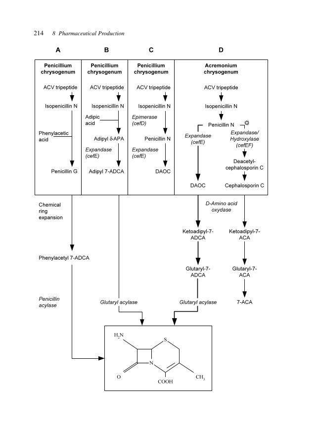
214 8 Pharmaceutical Production
A B C D
Penicillium
chrysogenum
Penicillium
chrysogenum
Penicillium
chrysogenum
Acremonium
chrysogenum
ACV tripeptide
Isopenicillin N
Phenylacetic
acid
Penicillin G
ACV tripeptide
Isopenicillin N
Adipic
acid
Adipyl δ-APA
Adipyl 7-ADCA
Expandase
(cefE)
ACV tripeptide
Isopenicillin N
Penicillin N
DAOC
Expandase
(cefE)
Epimerase
(cefD)
Chemical
ring
expansion
Phenylacetyl 7-ADCA
Penicillin
acylase
ACV tripeptide
Isopenicillin N
Penicillin N
DAOC Cephalosporin C
Deacetyl-
cephalosporin C
Expandase
(cefE)
Expandase/
Hydroxylase
(cefEF)
Ketoadipyl-7-
ACA
Glutaryl-7-
ACA
7-ACA
Ketoadipyl-7-
ADCA
Glutaryl-7-
ADCA
Glutaryl acylaseGlutaryl acylase
D-Amino acid
oxydase
S
COOH
CH3
O
H2N
N
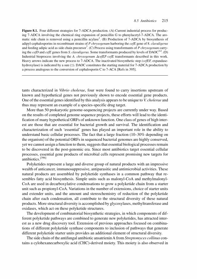
215
tants characterized in Vibrio cholerae, four were found to carry insertions upstream ofknown and hypothetical genes not previously shown to encode essential gene products.One of the essential genes identified by this analysis appears to be unique to V. cholerae andthus may represent an example of a species-specific drug target.
More than 50 prokaryotic genome-sequencing projects are currently under way. Basedon the results of completed genome sequence projects, these efforts will lead to the identi-fication of many hypothetical ORFs of unknown function. One class of genes of high inter-est are those that are essential for bacterial growth and survival. The identification andcharacterization of such ‘essential’ genes has played an important role in the ability tounderstand basic cellular processes. The fact that a large fraction (10–30% depending onthe organism) of the potential ORFs in sequenced bacterial genomes are highly conserved,yet we cannot assign a function to them, suggests that essential biological processes remainto be discovered in the post-genomic era. Since most antibiotics target essential cellularprocesses, essential gene products of microbial cells represent promising new targets forantibiotics.[396]
Polyketides represent a large and diverse group of natural products with an impressivewealth of anticancer, immunosuppressive, antiparasitic and antimicrobial activities. Thesenatural products are assembled by polyketide synthases in a common pathway that re-sembles fatty acid biosynthesis. Simple units such as malonyl-CoA and methylmalonyl-CoA are used in decarboxylative condensations to grow a polyketide chain from a starterunit such as propionyl-CoA. Variations in the number of extensions, choice of starter unitsand extender units, and the amount and stereochemistry of reduction of the polyketidechain after each condensation, all contribute to the structural diversity of these naturalproducts. More structural diversity is accomplished by glycosylases, methyltransferase andoxidases, which act on these polyketide structures.
The development of combinatorial biosynthetic strategies, in which components of dif-ferent polyketide pathways are combined to generate new polyketides, has attracted inter-est as a new drug discovery tool. Extension of previous approaches focused on combina-tions of different polyketide synthase components to inclusion of pathways that generatedifferent polyketide starter units provides an additional element of structural diversity.
The side chain of the antifungal antibiotic ansatrienin A from Streptomyces collinus con-tains a cylohexanecarboxylic acid (CHC)-derived moiety. This moiety is also observed in
Figure 8.1. Four different strategies for 7-ADCA production. (A) Current industrial process for produc-ing 7-ADCA involving the chemical ring expansion of penicillin G to phenylacetyl-7-ADCA. The aro-matic side chain is removed using a penicillin acylase1. (B) Production of 7-ADCA by biosynthesis ofadipyl-cephalosporins in recombinant strains of P chrysogenum harboring the cefE gene of 8. clavuligerusand feeding adipic acid as side chain precursor7. (C) Process using transformants of P chrysogenum carry-ing the cefD and cefE genes from S. clavuligerus. Some transformants produced by levels of DAOC3'4. (D)Industrial bioprocess involving the A. chrysogenum ∆cefEF-cefE transformants described in this work.Heavy arrows indicate the new process to 7-ADCA. The inactivated biosynthetic step (cefEF, expandase-hydroxylase) is indicated by a sun (I). DAOC constitutes the starting material for 7-ADCA production bya process analogous to the conversion of cephalosporin C to 7-ACA [Refs in 395].
8.5 Antibiotics
216 8 Pharmaceutical Production
trace amounts of ω-cyclohexyl fatty acids (typically less than 1% of total fatty acids) pro-duced by S. collinus. Coenzyme A-activated CHC (CHC-CoA) is derived from shikimicacid through a reductive pathway involving a minimum of nine catalytic steps. Five puta-tive CHC-CoA biosynthetic genes in the ansatrienin biosynthetic gene cluster of S. collinushave been identified. Plasmid-based heterologous expression of these five genes in S.avermitilis or S. lividans allows for production of significant amounts of ω-cyclohexyl fattyacids (as high as 49% of total fatty acids). In the absence of the plasmid these organisms aredependent on exogenously supplied CHC for ω-cyclohexyl fatty acid production. Doramectinis a commercial antiparasitic avermectin analog produced by fermenting a bkd mutant of S.avermitilis in the presence of CHC. Introduction of the S. collinus CHC-CoA biosyntheticgene cassette into this organism resulted in an engineered strain able to produce doramectinwithout CHC supplementation. The CHC-CoA biosynthetic gene cluster represents an im-portant genetic tool for precursor-directed biosynthesis of doramectin and has potential fordirected biosynthesis in other important polyketide-producing organisms.[397]
An enormous range of medically important polyketide and peptide natural productsassembled by modular polyketide synthases (PKSs), non-ribosomal peptide synthases(NRPSs) and mixed PKS/NRPS systems have macrocyclic structures, including the antibi-otics erythromycin (PKS) and daptomycin (NRPS), the immunosuppressants cyclosporin(NRPS) and rapamycin (PKS/NRPS), and the antitumor agent epothilone (PKS/NRPS).PKSs and NRPSs are large, multifunctional proteins that are organized into sets of func-tional domains termed modules. The order of modules corresponds directly to the sequenceof monomers in the product. Synthetic intermediates are covalently tethered by thioesterlinkages to a carrier protein domain in each module. The thiol tether on each carrier domainis phosphopantetheine, which is attached to a conserved serine residue in the carrier proteinin a post-translational priming reaction catalyzed by a phosphopantetheinyltransferase.
In the biosynthesis of many macrocyclic natural products by multidomain megasynthases,a C-terminal thioesterase (TE) domain is involved in cyclization and product release. It hasnot been determined whether TE domains can catalyze macrocyclization (and elongationin the case of symmetric cyclic peptides) independently of upstream domains. The inabilityto decouple the TE cyclization step from earlier chain assembly steps has precluded deter-mination of TE substrate specificity, which is important for the engineered biosynthesis ofnew compounds. The excised TE domain from tyrocidine synthetase efficiently catalyzescyclization of a decapeptide-thioester to form the antibiotic tyrocidine A and can catalyzepentapeptide-thioester dimerization followed by cyclization to form the antibiotic gramici-din S. By systematically varying the decapeptide-thioester substrate and comparing cy-clization rates, it was shown that only two residues (one near each end of the decapeptide)are critical for cyclization. This specificity profile indicates that the tyrocidine synthetaseTE and, by analogy many other TE domains, are able to cyclize and release a broad rangeof new substrates and products produced by engineered enzymatic assembly lines.[398]
Peptide antibiotics such as penicillins and cephalosporins, vancomycins, bacitracin, ac-tinomycin D, the antitumor peptide bleomycin, and the immunosuppressant cyclosporin Aare all assembled on NRPSs. The identity and sequence of amino acid residues in thesenon-ribosomal peptides is dictated by the organization of sets of iterated modules in themegasynthetases.
217
In non-ribosomal biosynthesis of peptide antibiotics by multimodular synthetases, aminoacid monomers are activated by the adenylation domains of the synthetase and loaded ontothe adjacent carrier protein domains as thioesters, then the formation of peptide bonds andtranslocation of the growing chain are effected by the synthetase’s condensation domains.Whether the condensation domains have any editing function has been unknown. Synthesisof aminoacyl-CoA molecules and direct enzymatic transfer of aminoacyl-phospho-pantetheine to the carrier domains allow the adenylation domain editing function to be by-passed. This method was used to demonstrate that the first condensation domain of tyroci-dine synthetase shows low selectivity at the donor residue (D-phenylalanine) and higherselectivity at the acceptor residue (L-proline) in the formation of the chain-initiating D-Phe–L-Pro dipeptidyl-enzyme intermediate.[399]
Modular polyketide synthases catalyze the biosynthesis of medically important naturalproducts through an assembly-line mechanism. Although these megasynthases display veryprecise overall selectivity, their constituent modules are remarkably tolerant toward diverseincoming acyl chains. By appropriate engineering of linkers, which exist within and be-tween polypeptides, it is possible to exploit this tolerance to facilitate the transfer of bio-synthetic intermediates between unnaturally linked modules. This protein engineering strat-egy provides a new strategy for combinatorial biosynthesis, in which modules rather thanindividual enzymatic domains, are the building blocks for genetic manipulation, and fur-thermore gives insights into the evolution of modular polyketide synthases.[400]
Lovastatin is an inhibitor of the enzyme (3S)-hydrooxymethylglutaryl-CoA (HMG-CoA)reductase that catalyzes the reduction of HMG-CoA to mevalonate, a key step in choles-terol biosynthesis. This activity confers on lovastatin its medically important anti-hyperchoilesterolemic activity and other potentially important uses. It is a secondary me-tabolite from the filamentous fungus Aspergillus terreus and has been shown to be derivedfrom acetate via a polyketide pathway.
Polyketides, the ubiquitous products of secondary metabolism in microorganisms, aremade by a process resembling fatty acid biosynthesis that allows the suppression of reduc-tion or dehydration reactions at specific biosynthetic steps, giving rise to a wide range ofoften medically useful products. The lovastatin biosynthesis cluster contains two type Ipolyketide synthase genes. Synthesis of the main nonaketide-derived skeleton was found torequire the previously known iterative lovastatin nonaketide synthase (LNKS), plus at leastone additional protein (LovC) that interacts with LNKS and is necessary for the correctprocessing of the growing polyketide chain and the production of dihydromonacolin L.The non-iterative lovastatin diketide synthase (LDKS) enzyme specifies formation of 2-methylbutyrate and interacts closely with an additional transesterase (LovD) responsiblefor assembling lovastatin from this polyketide and monacolin J.[401]
Additional enzymes may add to the versatility of gene clusters and novel compoundssynthesized by combining previously unrelated genes from different organisms.
Chalcone synthases, which biosynthesize chalcones (the starting materials for many fla-vonoids), were believed to be specific to plants. The rppA gene from the Gram-positive,soil-living filamentous bacterium Streptomyces griseus encodes a 372-amino-acid proteinthat shows significant similarity to chalcone synthases. Several rrpA-like genes are known,but their functions and catalytic properties have not been described. A homodimer of RppA
8.5 Antibiotics
218 8 Pharmaceutical Production
catalyzes polyketide synthesis: it selects malonyl-CoA as the starter, carries out four suc-cessive extensions and releases the resulting pentaketide to cyclize to 1,3,6,8-tetra-hydroxynaphthalene (THN). Site-directed mutagenesis showed that, as in other chalconesynthases, a cysteine residue is essential for enzyme activity. Disruption of the chromo-somal rppA gene in S. griseus abolished melanin production in hyphae, resulting in ‘al-bino’ mycelium. THN was readily oxidized to form 2,5,7-trihydroxy-1,4-naphtoquinone(flaviolin), which then randomly polymerized to form various colored compounds. THNformed by RppA appears to be an intermediate in the biosynthetic pathways for not onlymelanins but also various secondary metabolites containing a naphthoquinone ring. RppAis a chalcone-synthase-related synthase that synthesizes polyketides and is found in theStreptomyces and other bacteria.[402]
The elucidation of molecular structures of enzymes in biosynthetic pathways aids in thedesign of proteins with modified specificities.
Isopenicillin N synthase (IPNS), a non-heme iron-dependent oxidase, catalyzes the bio-synthesis of isopenicillin N (IPN), the precursor of all penicillins and cephalosporins. Thekey steps in this reaction are the two iron-dioxygen-mediated ring closures of the tripeptideδ-(L-α-aminoadipoyl)–L-cysteinyl–D-valine (ACV). The four-membered β-lactam ringforms initially, associated with a highly oxidized iron(IV)-oxo-(ferryl) moiety, which sub-sequently mediates closure of the five-membered thiazolidine ring. The observation of theIPNS reaction in crystals by X-ray crystallography has been described. IPNS-Fe2+ sub-strate crystals were grown anaerobically, exposed to high pressures of oxygen to promotereaction and frozen. Their structures were subsequently elucidated by X-ray crystallogra-phy. Using the natural substrate ACV, this resulted in the IPNS–Fe2+–IPN product com-plex. With the substrate analog, δ-(L-α-aminoadipoyl)–L-cysteinyl–L-S-methyl-cysteine(AcmC) in the crystal, the reaction cycle was interrupted at the monocyclic stage. Thesemono- and bicyclic structures support the hypothesis of a two-stage reaction sequence leadingto penicillin. The formation of a monocyclic sulfoxide product from AcmC is best ex-plained by the interception of a high-valency iron-oxo species.[403]
Escherichia coli was metabolically engineered to produce a crucial intermediate forcomplex polyketides synthesized by the bacterium Saccharopolyspora through the actionof a multifunctional polyketide synthase (PKS). Genetically engineered E.coli convertesexogenous propionate into the macrocyclic core of the antibiotic erythromycon, 6-deoxyerythronolide B (6dEB) with a productivity comparable to an industrially used high-producing mutant of S. erythraea.[503]
The sequential coordinated action of the modular PKSs is illustrated in Figure 8.10,where (A) shows the 6-deoxyerythronolide B synthase with catalytic domains, (B) depictsthe production of triketide lactone, and (C) illustrates the rifamycin synthetase, a polyketidesynthase naturally primed by a nonribosomal PKS loading module which may be engi-neered to utiliize exogenous acids for the sythesis of substituted macrocycles.
219
8.6 Biocatalysis
The chemical processes and transformations underpinning all biological phenomena areexecuted by enzymes that transform small molecules, macromolecular substrates and transferenergy. The numerous chemical reactions are enabled by hundreds to thousands of pro-teins, enzymes, and by RNAs and ribozymes, which have catalytic activity for the conver-sion of specific substrates to products. The location and lifetime of proteins is controlled bythe family of related proteases that hydrolyze peptide bonds in a controlled way. The de-gree of specificity ranges from highly specific actions on particular substrate bonds to ageneral non-specific hydrolysis action that attacks a broad set of substrates. The highspecificity is usually found in signaling pathways, whereas the broad reactions apply toprotein degradation. The signaling pathways are initiated by catalytic action of cascades ofprotein kinases that are related proteins and catalyze phosphoryl transfer from ATP to theside-chain hydroxyl of serine, threonine or tyrosine residues. The selectivity is achieved byspecific protein–protein interactions between kinase and protein substrate acting in cas-cades and ultimately producing changes in activity and location of proteins as well as selec-tive gene activation.
Enzymes may also catalyze transformations of a highly selective and unique nature suchas in transformations of small molecules. Examples are the fragmentation of 1-amino-cyclopropane-1-carboxylate to the fruit ripening hormone ethylene, the cycloreversion ofthymine dimers in DNA repair, the synthesis of isopenicillin and the reduction of molecu-lar nitrogen (N2) to ammonia (NH3).
[488]
Enzymes demonstrate both high specificities and significant reaction rate accelerations.The relative values of enzymic over non-enzymic reactions may be from 1010 to 1023
(orotidine decarboxylase) and the turnover numbers range from one catalytic event perminute to 105 per second (hydration of CO2 to HCO3
– by carbonic anhydrase). The molecu-lar entities of enzymes cover proteins, ribozymes and catalytic antibodies.
The sequential reactions in elongating acyl transfers in the synthesis of polyketide natu-ral products and non-ribosomal peptide antibiotics such as erythromycin, rapamycin,epothilone, lovastatin, penicillins, cyclosporin and vancomycin resemble molecular solid-state assembly lines. Such multimodular enzymes may be utilized in combinatorial biosyn-thesis by way of reprogramming for the manufacture of unnatural analogs of natural prod-ucts.
IPNS belongs to a family of iron-containing enzymes which use Fe2+ to activate O2 andsimultaneously the specific co-substrate for redox reactions whereby both atoms of O2 arereduced to water and the tripeptide ACV undergoes oxidation with C–S and C–C bondformation to generate β-lactam molecules.
A related enzyme, the expandase enzyme, is deployed by cephalosporin-producing or-ganisms to expand the five-membered penicillin ring to the six-membered cephalosporinring.
The increasingly stringent environmental requirements demand new catalytic syntheticprocesses as does the necessity to produce the novel types of organic compounds generatedin biomolecular research as active molecules or targets. Enzyme-catalyzed chemical trans-
8.6 Biocatalysis
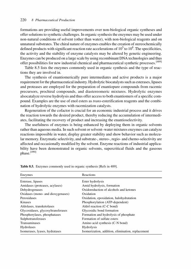
220 8 Pharmaceutical Production
formations are providing useful improvements over non-biological organic syntheses andoffer solutions to synthetic challenges. In organic synthesis the enzymes may be used undernon-natural conditions of solvent (other than water), with non-biological reagents and onunnatural substrates. The chiral nature of enzymes enables the creation of stereochemicallydefined products with significant reaction rate accelerations of 105 to 108. The specificities,the activity and the stability of enzyme catalysts may be altered by genetic engineering.Enzymes can be produced on a large scale by using recombinant DNA technologies and thusoffer possibilities for new industrial chemical and pharmaceutical synthetic processes.[489]
Table 8.5 lists the enzymes commonly used in organic synthesis and the type of reac-tions they are involved in.
The synthesis of enantiomerically pure intermediates and active products is a majorrequirement for the pharmaceutical industry. Hydrolytic biocatalysts such as esterases, lipasesand proteases are employed for the preparation of enantiopure compounds from racemicprecursors, prochiral compounds, and diastereomeric mixtures. Hydrolytic enzymesalsocatalyze reverse hydrolysis and thus offer access to both enantiomers of a specific com-pound. Examples are the use of enol esters as trans-esterification reagents and the combi-nation of hydrolytic enzymes with racemization catalysts.
Regeneration of the cofactor is crucial for an economic industrial process and it drivesthe reaction towards the desired product, thereby reducing the accumulation of intermedi-ates, facilitating the recovery of product and increasing the enantioselectivity.
The usefulness of enzymes is being enhanced by deploying them in organic solventsrather than aqueous media. In such solvent or solvent–water mixtures enzymes can catalyzereactions impossible in water, display greater stability and show behavior such as molecu-lar memory. Enzymatic selectivity, and substrate-, stereo-, regio- and chemo-selectivity areaffected and occasionally modified by the solvent. Enzyme reactions of industrial applica-bility have been demonstrated in organic solvents, supercritical fluids and the gaseousphase.[490]
Table 8.5. Enzymes commonly used in organic synthesis [Refs in 489].
Enzymes Reactions
Esterase, lipases Ester hydrolysisAmidases (proteases, acylases) Amid hydrolysis, formationDehydrogenases Oxidoreduction of alcohols and ketonesOxidases (mono- and dioxygenases) OxidationPeroxidases Oxidation, epoxidation, halohydratationKinases Phosphorylation (ATP-dependent)Aldolases, transketolases Aldol reaction (C-C bond)Glycosidases, glycosyltransferases Glycosidic bond formationPhosphorylases, phosphatases Formation and hydrolysis of phosphateSulphotransferases Formation of sulfate estersTransaminases Amino acid synthesis (C-N bond)Hydrolases HydrolysisIsomerases, lyases, hydratases Isomerization, addition, elimination, replacement
221
Modular enzymes fall into three categories of modular catalysts:• Enzymes where catalysis and substrate specificity reside in separate domains• Multisubstrate enzymes wherein the binding sites for individual substrates are modular• Multienzyme systems which catalyze programmable metabolic pathways
The ribosomal protein biosynthetic machinery encompasses all three types of modularity.The ribosome catalyst can be separated from the element determining the substrate specificity,i.e. the mRNA template. The two acylated tRNA substrates for the peptide-bond formationbind to different ribosomal sites, the A and P sites, and the multistep pathway catalyzed bythe modular system may be reprogrammed by codon choice.
The individual modules of polyketide synthases (PKSs) and of NRPSs represent anotherexample of modularity. An electrophile and a nucleophile are covalently attached to twodifferent subunits of a multidomain enzyme. The selectivity for the electrophile resides inthe domain catalyzing bond formation between two substrates and the selectivity for thenucleophile is controlled by a transfer domain which attaches the nucleophile onto a carrierdomain.[491]
Multienzyme modular assemblies such as PKSs and NRPSs have flexible swinging teth-ers which channel covalently bound intermediates between successive active sites. Swing-ing arms plus specific protein–protein interactions offer mechanisms for the transfer ofsubstrates between modules and offer concepts for the development of one-pot multi-reac-tion biocatalytic processes.
The quest for purpose-designed biocatalysts relies on the rational redesign of naturalbiocatalysts and on evolutionary procedures to obtain novel catalysts. To rationally de-sign biocatalysts requires knowledge of the connection between structure and function,for proteins as well as for ribozymes. The capabilities for manipulations of novel struc-tures, organisms, bio-electronic hybrid devices will be targeted at process biocatalysts,novel therapeutic enzymes, specifically designed cellular machinery and metabolic net-works.[492]
Rational design, directed evolutionary methods and combinatorial synthesis are the avail-able techniques for the development of novel biocatalysts. Evolutionary protein designmethods utilize random mutagenesis, gene recombination and high-throughput screening,and resemble breeding procedures. Molecular breeding can be extended to involve com-plex synthetic tasks involving several interacting enzymes and even the creation of newpathways in microorganisms, plants and animals. Genes encoding the enzymes for synthe-sizing in a series of reactions may be combined in suitable organisms. An example of engi-neering a biosynthetic pathway for carotenoid synthesis is the transfer of genes from Erwiniasp. into E. coli together with a gene library of shuffled versions of a Erwinia-saturase led tonovel carotenoids as well as novel synthetic pathways for known carotenoids.[380]
De novo catalyst design by rational principles is emerging as a tool for the design ofcatalysts. Iron and oxygen binding sites were introduced into thioredoxin by computationaldesign. The rudimentary active sites demonstrate enzyme activities selecting between dif-ferent oxygen chemistries and different control mechanisms are observed. The reactivity ofthe active sites is determined by the microenvironment and the interactions of the proteinmatrix with the metal center.[493]
8.6 Biocatalysis
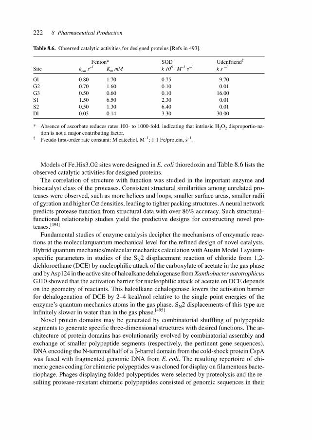
222 8 Pharmaceutical Production
Models of Fe.His3.O2 sites were designed in E. coli thioredoxin and Table 8.6 lists theobserved catalytic activities for designed proteins.
The correlation of structure with function was studied in the important enzyme andbiocatalyst class of the proteases. Consistent structural similarities among unrelated pro-teases were observed, such as more helices and loops, smaller surface areas, smaller radiiof gyration and higher Cα densities, leading to tighter packing structures. A neural networkpredicts protease function from structural data with over 86% accuracy. Such structural–functional relationship studies yield the predictive designs for constructing novel pro-teases.[494]
Fundamental studies of enzyme catalysis decipher the mechanisms of enzymatic reac-tions at the molecularquantum mechanical level for the refined design of novel catalysts.Hybrid quantum mechanics/molecular mechanics calculation with Austin Model 1 system-specific parameters in studies of the SN2 displacement reaction of chloride from 1,2-dichloroethane (DCE) by nucleophilic attack of the carboxylate of acetate in the gas phaseand by Asp124 in the active site of haloalkane dehalogenase from Xanthobacter autotrophicusGJ10 showed that the activation barrier for nucleophilic attack of acetate on DCE dependson the geometry of reactants. This haloalkane dehalogenase lowers the activation barrierfor dehalogenation of DCE by 2–4 kcal/mol relative to the single point energies of theenzyme’s quantum mechanics atoms in the gas phase. SN2 displacements of this type areinfinitely slower in water than in the gas phase.[495]
Novel protein domains may be generated by combinatorial shuffling of polypeptidesegments to generate specific three-dimensional structures with desired functions. The ar-chitecture of protein domains has evolutionarily evolved by combinatorial assembly andexchange of smaller polypeptide segments (respectively, the pertinent gene sequences).DNA encoding the N-terminal half of a β-barrel domain from the cold-shock protein CspAwas fused with fragmented genomic DNA from E. coli. The resulting repertoire of chi-meric genes coding for chimeric polypeptides was cloned for display on filamentous bacte-riophage. Phages displaying folded polypeptides were selected by proteolysis and the re-sulting protease-resistant chimeric polypeptides consisted of genomic sequences in their
Table 8.6. Observed catalytic activities for designed proteins [Refs in 493].
Fenton* SOD Udenfriend‡
Site kcat s–1 Km mM k 106 · M–1 s–1 k s –1
Gl 0.80 1.70 0.75 9.70G2 0.70 1.60 0.10 0.01G3 0.50 0.60 0.10 16.00S1 1.50 6.50 2.30 0.01S2 0.50 1.30 6.40 0.01Dl 0.03 0.14 3.30 30.00
* Absence of ascorbate reduces rates 100- to 1000-fold, indicating that intrinsic H2O2 disproportio-na-tion is not a major contributing factor.
‡ Pseudo first-order rate constant: M catechol, M–1; 1:1 Fe/protein, s–1.
223
original reading frames. One of the obtained proteins had a fold similar to CspA withoutshowing sequence homology. Several chimeric proteins were soluble polypeptides formingmonomers and exhibiting cooperative unfolding. One chimeric protein actually containedseveral very slowly exchanging amides and was more stable than CspA itself. Combinato-rial assembly of segments from non-homologous proteins and diverse sources can be usedto generate novel proteins, domains and architectures.[496]
Evolutionary methodology can be used to generate catalytic antibodies for chemicalprocess steps. The reactive immunization procedure employed uses reactive chemicals asimmunogens. The immune system selects chemical steps such as the formation of a cova-lent bond between the antibody and the antigen rather than a simple recognition and bind-ing process. Catalytic aldolase antibodies were generated by immunization with two differ-ent but structurally related β-diketone haptens and the resulting antibodies were sequencedto allow study of similarities, differences, and structural determinants of properties of inde-pendently evolved catalysts. Kinetic and sequence analysis, mutagenesis, and structuraland modeling studies demonstrate a somatic mutation placing a lysine residue into a deephydrophobic pocket as the crucial event of the catalyst evolution. In addition to the non-covalent interactions which have been the focus of immunochemistry, covalent chemistriescan be procured from the immune repertoire.[497]
Table 8.7 lists the kinetic parameters for antibody-catalyzed aldol and retro-aldol reactions.Biocatalysis on an industrial scale covers a range of reactions, solvents and products,
thus extending from the manufacture of large-volume chemicals such as in acrylamideproduction to numerous syntheses of optically active alcohols, amines, nitriles, peptidesand semisynthetic antibiotics, to numerous intermediates.[498]
Several recently developed biocatalytic systems at several chemical companies are listedin Table 8.8, illustrating the range of products (amides, alcohols, acids, amino acids, peni-cillins, N-heterocyclic compounds, and non-proteinogenic amino acids).
The factors determining the industrial and economic feasibility of biocatalytic processesare depicted in Figure 8.2, showing the key aspects of the synthetic process (and its eco-nomics), biocatalyst selection and characterization, biocatalyst engineering, its applicationin industrial use, and product recovery.
For reasons of stability, handling, separation and recovery, the industrial biocatalyst ispreferably applied as an entrained molecule.
Lipases are an important class of enzymes for biocatalysts. They serve to synthesizeenantiomerically pure alcohols, chiral amines, and nitrilases for amino- and hydroxy-car-boxylic acids.
The bioconversion of steroids, the products of which are used in steroidal hormonepharmaceuticals, is carried out at a scale of more than 1000 tons/year starting from naturalphytosterols from soya, conifers and from rape seed (β-sitosterol, campesterol, stigmas-terol, brassicasterol, and β-sitostanol).
Ribozymes cleave RNA molecules at specific sites. In order to modify and extend thenumber of cleavable target sequences on the mRNA, an allosteric version of a ribozyme, amaxizyme, in vivo activity and specificity was developed several custom-designedmaxizymes showed sensor functions, thus indicating the applicability of the technology forthe design of maxizymes with use in analytics and therapy.[499]
8.6 Biocatalysis
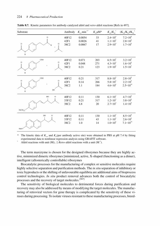
224 8 Pharmaceutical Production
Table 8.7. Kinetic parameters for antibody-catalyzed aldol and retro-aldol reactions [Refs in 497].
Substrate Antibody Kcat min–1* Km µM* Kcat/Kun
† (Kcat/Km)/Kun
‡
N
H
O
O
H
+
O
40F12 42F1 38C2
0.0054 0.0026 0.0067
33 41 17
2.4×10⁴ 1.1×10⁴
2.9×10⁴
7.2×10⁸ 2.8×10⁸
1.7×10⁹
N
H
O
CHO
+
O
40F12 42F1 38C2
0.071 0.048 0.21
203 271 123
6.5×10⁵ 4.3×10⁵ 1.9×10⁶
3.2×10⁹ 1.6×10⁹ 1.5×10⁷
N
H
O
CHO
+
O
HO
40F12 42F1 38C2
0.21 0.14 1.1
317 266 184
8.8×10⁵
5.8×10⁵
4.6×10⁶
2.8×10⁹
2.2×10⁹
2.5×10¹⁰
N(CH3)2
HO O
40F12 33F12 38C2
0.11 0.21 4.8
130 317 20
6.1×10²
1.2×10³ 2.7×10⁴
4.7×10⁶
3.8×10⁶ 1.4×10⁹
HO O
MeO
40F12 33F12 38C2
0.11 0.11 1.0
130 43 14
1.1×10⁵
1.1×10⁵
1.0×10⁶
8.5×10⁸
2.6×10⁹
7.1×10¹⁰
* The kinetic data of Kcat and Km(per antibody active site) were obtained in PBS at pH 7.4 by fitting experimental data to nonlinear regression analysis using GRAFIT software.
† Aldol reactions with unit (M);. ‡ Retro-aldol reactions with a unit (M–1).
The term maxizyme is chosen for the designed ribozymes because they are highly ac-tive, minimized dimeric ribozymes [minimized, active, X-shaped (functioning as a dimer),intelligent (allosterically controllable) ribozymes
Biocatalytic processes for the manufacturing of complex or sensitive molecules requirehighly selective separation and purification methods. The in situ separation of inhibitory ortoxic byproducts or the shifting of unfavourable equilibria are additional aims of bioprocesscontrol technologies. In situ product removal advances both the control of biocatalyticprocesses and the recovery of target molecules.[587]
The sensitivity of biological molecules to detrimental forces during purification andrecovery may also be addressed by means of modifying the target molecules. The manufac-turing of retroviral vectors for gene therapy is complicated by the sensitivity of these vi-ruses during processing. To isolate viruses resistant to these manufacturing processes, breed-
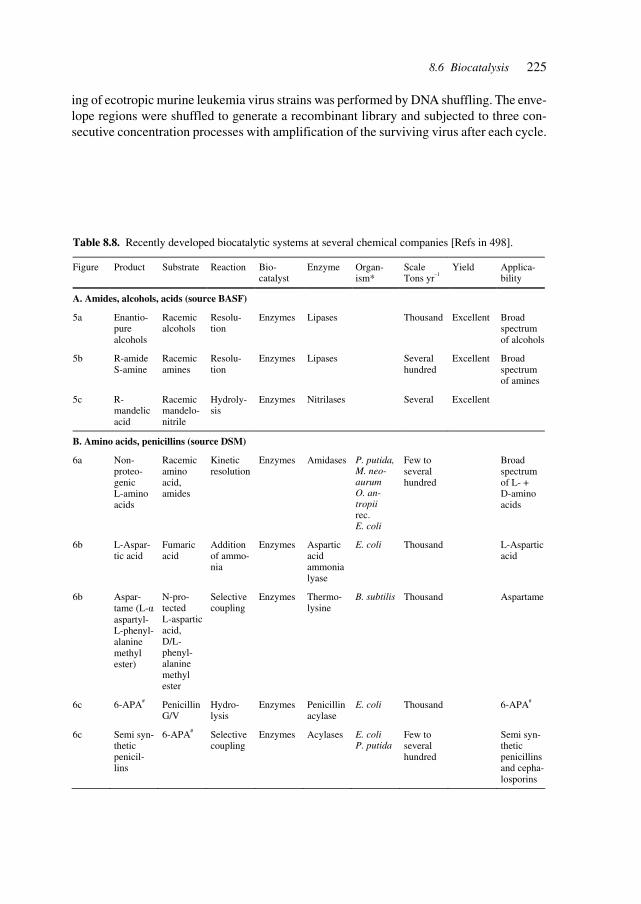
225
ing of ecotropic murine leukemia virus strains was performed by DNA shuffling. The enve-lope regions were shuffled to generate a recombinant library and subjected to three con-secutive concentration processes with amplification of the surviving virus after each cycle.
8.6 Biocatalysis
Table 8.8. Recently developed biocatalytic systems at several chemical companies [Refs in 498].
Figure Product Substrate Reaction Bio-catalyst
Enzyme Organ-ism*
Scale Tons yr–1
Yield Applica-bility
A. Amides, alcohols, acids (source BASF)
5a Enantio-pure alcohols
Racemic alcohols
Resolu-tion
Enzymes Lipases Thousand Excellent Broad spectrum of alcohols
5b R-amide S-amine
Racemic amines
Resolu-tion
Enzymes Lipases Several hundred
Excellent Broad spectrum of amines
5c R- mandelic acid
Racemic mandelo- nitrile
Hydroly-sis
Enzymes Nitrilases Several Excellent
B. Amino acids, penicillins (source DSM)
6a Non- proteo-genic L-amino acids
Racemic amino acid, amides
Kinetic resolution
Enzymes Amidases P. putida, M. neo-aurum O. an-tropii rec. E. coli
Few to several hundred
Broad spectrum of L- + D-amino acids
6b L-Aspar-tic acid
Fumaric acid
Addition of ammo-nia
Enzymes Aspartic acid ammonia lyase
E. coli Thousand L-Aspartic acid
6b Aspar-tame (L-α aspartyl-L-phenyl- alanine methyl ester)
N-pro-tected L-aspartic acid, D/L- phenyl- alanine methyl ester
Selective coupling
Enzymes Thermo-lysine
B. subtilis Thousand Aspartame
6c 6-APA# Penicillin G/V
Hydro-lysis
Enzymes Penicillin acylase
E. coli Thousand 6-APA#
6c Semi syn-thetic penicil-lins
6-APA# Selective coupling
Enzymes Acylases E. coli P. putida
Few to several hundred
Semi syn-thetic penicillins and cepha-losporins
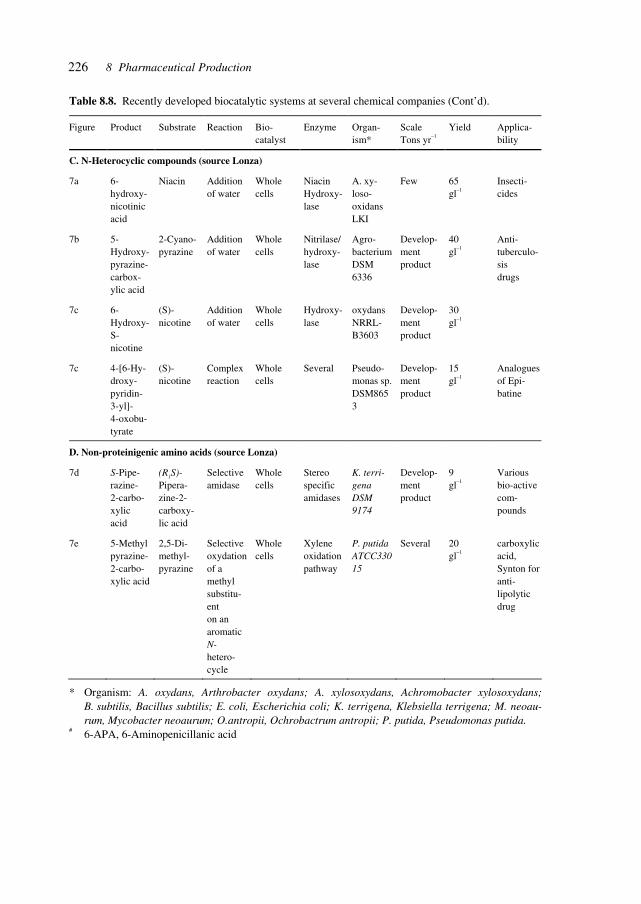
226 8 Pharmaceutical Production
Table 8.8. Recently developed biocatalytic systems at several chemical companies (Cont’d).
Figure Product Substrate Reaction Bio-catalyst
Enzyme Organ-ism*
Scale Tons yr–1
Yield Applica-bility
C. N-Heterocyclic compounds (source Lonza)
7a 6-hydroxy-nicotinic acid
Niacin Addition of water
Whole cells
Niacin Hydroxy-lase
A. xy-loso- oxidans LKI
Few 65 gl–1
Insecti-cides
7b 5-Hydroxy-pyrazine- carbox-ylic acid
2-Cyano-pyrazine
Addition of water
Whole cells
Nitrilase/ hydroxy-lase
Agro-bacterium DSM 6336
Develop-ment product
40 gl–1
Anti-tuberculo-sis drugs
7c 6-Hydroxy- S-nicotine
(S)-nicotine
Addition of water
Whole cells
Hydroxy-lase
oxydans NRRL- B3603
Develop-ment product
30 gl–1
7c 4-[6-Hy- droxy-pyridin-3-yl]- 4-oxobu-tyrate
(S)- nicotine
Complex reaction
Whole cells
Several Pseudo-monas sp. DSM8653
Develop-ment product
15 gl–1
Analogues of Epi-batine
D. Non-proteinigenic amino acids (source Lonza)
7d S-Pipe-razine- 2-carbo-xylic acid
(R1S)- Pipera-zine-2- carboxy-lic acid
Selective amidase
Whole cells
Stereo specific amidases
K. terri-gena DSM 9174
Develop-ment product
9 gl–1
Various bio-active com-pounds
7e 5-Methyl pyrazine- 2-carbo-xylic acid
2,5-Di- methyl- pyrazine
Selective oxydation of a methyl substitu-ent on an aromatic N- hetero-cycle
Whole cells
Xylene oxidation pathway
P. putida ATCC33015
Several 20 gl–1
carboxylic acid, Synton for anti-lipolytic drug
* Organism: A. oxydans, Arthrobacter oxydans; A. xylosoxydans, Achromobacter xylosoxydans; B. subtilis, Bacillus subtilis; E. coli, Escherichia coli; K. terrigena, Klebsiella terrigena; M. neoau-rum, Mycobacter neoaurum; O.antropii, Ochrobactrum antropii; P. putida, Pseudomonas putida.
# 6-APA, 6-Aminopenicillanic acid
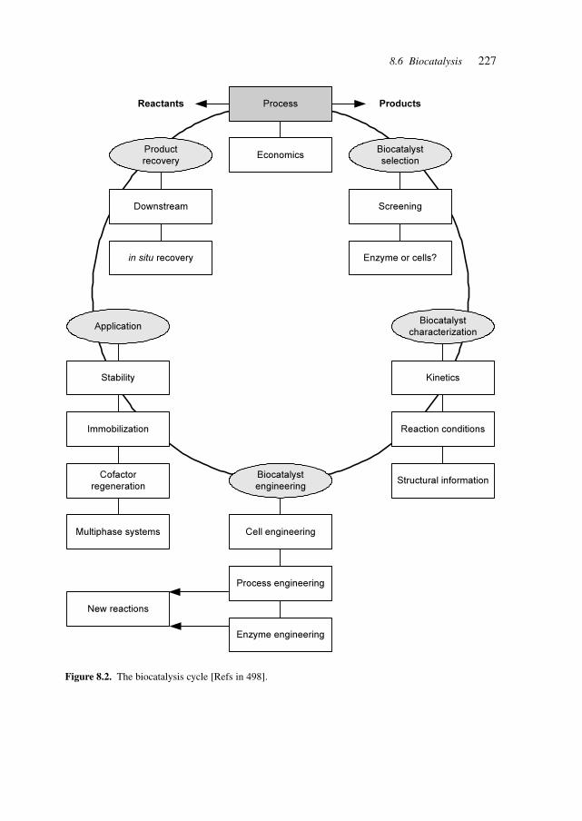
227
Figure 8.2. The biocatalysis cycle [Refs in 498].
Process
Economics
Downstream
in situ recovery
Screening
Enzyme or cells?
Kinetics
Reaction conditions
Structural information
Stability
Immobilization
Cofactor
regeneration
Multiphase systems
Enzyme engineering
Process engineering
Cell engineering
New reactions
ProductsReactants
Product
recovery
Biocatalyst
selection
Biocatalyst
characterizationApplication
Biocatalyst
engineering
8.6 Biocatalysis
228 8 Pharmaceutical Production
Several viral clones with greatly improved stability were isolated. The envelopes of theresistant viruses differed in DNA and protein sequence and were complex chimeras frommultiple parents. DNA shuffling is useful in breeding viral strains with improved charac-teristics for gene therapy and other manufacturing or biomedical applications.[597]
A high-throughput quantitative method was developed for the isolation of enzymes withnovel substrate specificities from large libraries of protein variants. Protein variants aredisplayed on the surface of microorganisms and incubated with a synthetic substrate com-prising a fluorescent dye, a positively charged moiety, the target fissile bond, and a fluores-cence resonance energy transfer (FRET) quenching moiety. Enzymatic cleavage of thescissile bond effects the release of the FRET quenching partner while the fluorescent prod-uct remains on the cell surface, allowing isolation of catalytically active clones by fluores-cence-activated cell sorting (FACS). Escherichia coli expressing the serine protease OmpTwas enriched over 5,000 fold in a single round from cells expressing an inactive OmpTvariant. Variant proteases with catalytic activities enhanced 60-fold were isolated. The methodallows the high-throughput screening of large catalytic libraries on the principle of cata-lytic turnover.[630]
The efforts to construct an allosteric enzyme which functions in vivo have thus led to amaxizyme, a construct, which includes a sensor function and thus allows to specificallycleave abnormal chimeric mRNA targets without affecting the wild-type mRNA, a taskthat cannot be achieved with conventional hammerhead ribozymes. Maxizymes can bedesigned to attack and cleave specific sequences of interest, such as in the expression of anabnormal chimeric gene lacking a NUX cleavage site, a target generated from reciprocalchromosomal trans-locations observed in several leukemia diseases such as acute lym-phoblastic leukemia (ALL), acute promyelocytic leukemia (APL), and chronic myelog-enous leukemia (CML). Specific cleavage of abnormal mRNA without affecting normaltranscripts was also observed in studies of Alzheimer’s disease.
The utilization of short ribozymes functioning as dimers enables the design of a maxi-zyme with one substrate-binding region for the unique junction sequence thus acting as thesensor and the second binding region for an efficient cleavage site thus operating as thescissors.
Ribozymes are also potential targets for pharmaceutical intervention with bio-activemolecules, small molecules, acting as modulators, agonists or antagonists. A screeningprocedure has been developed for identifying hammerhead-ribozyme inhibitors and wasused to detect inhibitory activities in extracts from actinomycete strains.[697]
Biotransformations, bioconversions impact on the whole range of biological, biotech-nological and biomedical applications, such as in the analysis of genes and transcripts, thetherapeutic treatment of abnormal molecules in diseases, the construction of specific en-zymes and proteins for therapy, analysis, production, the assembly and construction ofsynthetic bio-synthetic pathways in organisms for the synthesis of intermediates, antibiot-ics, vitamins, enzymes, flavors.
229
8.7 Natural Compounds
The supply of natural products can be increased from rare original sources by identificationof the relevant gene (gene cluster) and its transfer to a more suitable host(s).[404] This alsoallows the facile generation of novel compounds by reshuffling the genes, or introducing ordeleting genes or functions.
Organic compounds such as carotenoids can be produced by aqua-farming of microalgae.Microalgae are a potential source of single-cell protein, as a means of sequestering carbondioxide from effluent gases, and can be used in effluent purification and for producingbiofuels. High production costs preclude the use of microalgae as commercial sources ofcommodity products that can be produced inexpensively on a vast scale by plants.
Astaxanthin is a high-value carotenoid produced from microalgae that is produced com-mercially. Astaxanthin is ubiquitous in nature, especially in the marine environment, and isresponsible for eliciting the pinkish-red hue to the flesh of salmonids, shrimp, lobster andcrayfish. Cultivation methods have been developed to produce Haematococcus containing1.5–3.0% astaxanthin by dry weight, with potential applications as a pigment source inaquaculture, poultry feeds and in the nutraceutical area.[405]
8.8 Recovery/(Bio-) Processing
The delicate nature of biomolecules such as (glycosylated) proteins, polysaccharides andmetabolites necessitates appropriate methods for purification and separation, such as chro-matography, lyophilization and crystallization.
The chiral nature of biomolecules and, increasingly, of APIs requires the production ofenantiomerically pure compounds. The difference in biological activity of the two enanti-omers of a chiral drug has raised the demand for enantiomerically pure products, especiallyin the pharmaceutical and veterinary industry. Simulated moving-bed chromatography isdeveloping into an efficient tool for the separation of the two isomers of a chiral molecule,at all production scales, from laboratory to pilot plant to production plant.[406]
The cell possesses a complex protein machinery to help some proteins to fold afterbiosynthesis or heat shock. It consists of molecular chaperones, thought to prevent misfoldingand aggregation by binding to polypeptide chains that are not fully folded, and some en-zymes able to catalyze specific interactions in polypeptide chains.
Proteins that contain disulfide bonds often fold slowly in vitro because the oxidation andcorrect pairing of the cysteine residues becomes the rate-limiting step and the bonds formedare not always the correct ones. Many proteins, especially those that are secreted by eu-karyotes, are stabilized by disulfide bonds. Examples of such proteins include those usedfor medical or biotechnological purposes, such as interleukins, IFNs, antibodies and theirfragments, insulin, TGF, and many toxins and proteases. Expression of recombinant pro-teins as inclusion bodies in bacteria can be a very efficient way to produce cloned proteins,as long as the inclusion body protein can be successfully refolded.
8.8 Recovery/(Bio-) Processing
230 8 Pharmaceutical Production
Aggregation is the leading cause of decreased refolding yield. Refolding of non-co-valently immobilized denatured protein on gel beads has been used to avoid aggregation.Another approach is the in vitro method to cope with the aggregation by use of immobi-lized minichaperones. This technique was extended to include reagents for the oxidativecatalysis of disulfide bond formation and for the isomerization of cis–trans peptidyl–prolyllinkages.
An immobilized and reusable molecular chaperone system for oxidative refoldingchromatography was constructed. Its three components – GroEl minichaperone (191–345)(which can prevent protein aggregation), DsbA (which catalyzes the shuffling and oxida-tive formation of disulfide bonds) and peptidyl–prolyl isomerase – were immobilized on anagarose gel. The gel was applied to the refolding of denatured and reduced scorpion toxinCn5. The 66-residue toxin, which has four disulfide bridges and a cis peptidyl–prolinebond, had not previously been refolded in reasonable yield. The protein was recovered in87% yield with 100% biological activity.[407]
Inteins are self-splicing proteins that occur as in-frame insertions in specific host pro-teins. In a self-splicing reaction, inteins excise themselves from a precursor protein, whilethe flanking regions, the exteins, become joined to restore host gene function. The ability toconstruct intein fusions to proteins of interest has broad potential applications. One of theseis affinity fusion-based protein purification, where an intein is used in conjunction with anaffinity group to purify a desired protein. Self-cleavage, rather than splicing of the intein,releases the desired protein, thereby eliminating the need for protease addition and simpli-fying overall processing. For this application, the intein must be altered to yield control-lable cleavage rather than splicing, and the intein should be relatively small for scale-upconsiderations.
A self-cleaving element for use in bio separations has been derived from a naturallyoccurring, 43-kDa protein splicing element (intein) through a combination of protein engi-neering and random mutagenesis. A mini-intein (18 kDa) previously engineered for re-duced size had compromised activity and was therefore subjected to random mutagenesisand genetic selection. In one selection a mini-intein was isolated with restored splicingactivity, while in another, a mutant was isolated with enhanced, pH-sensitive C-terminalcleavage activity. The enhanced-cleavage mutant has utility in affinity fusion-based proteinpurification. These mutants also provide new insights into the structural and functionalroles of some conserved residues in protein splicing.[408]
Chirality is a determinative property of most biological molecules. A particular stereoi-somer is generally preferred in physiologic processes such as metabolism, intra- and inter-cellular communication, and as building blocks for the formation of macromolecules. Liv-ing organisms discriminate between the enantiomers of exogenous compounds (e.g., drugs)at virtually all levels of interaction and respond differently to them. Even minor enantio-meric impurities may cause severe pharmacologic and toxicologic side effects. The devel-opment of refined analytical techniques for the precise determination of biorelevant chiralmolecules is therefore of great interest. Various sensor technologies have been developedfor chiral compounds. Chiral selectors for enantiomer discrimination have largely builtupon empiric chromatographic phases such as metal complexes, cycodextrin derivativesand Chirasil-Val derivatives.
231
Based on the stereoselectivity of immunoglobulins, a new chiral sensor for the detectionof low-molecular-weight analytes was developed. Using surface plasmon resonance detec-tion, enantiomers of free, underivatized α-amino acids can be monitored in a competitiveassay by their interaction with antibodies specific for the chiral center of this class of sub-stances. The sensitivity to the minor enantiomer in non-racemic mixtures exceeds currentlyavailable methods; therefore, such immunosensors can readily detect traces of enantio-meric impurities and are attractive for a range of applications in science and industry.[409]
8.9 Chemical–Biotechnological Syntheses
Membranes and biotechnological tools can be used for improving traditional productionsystems to maintain the sustainable growth of society and industry. Typical examples in-clude new and improved foodstuffs, in which the desired nutrients are maintained duringthermal treatment, novel pharmaceutical products with defined enantiomeric compositionsand the treatment of wastewater, wherein pollution by traditional processes is problematic.
Table 8.9, 8.10 and 8.11 show the industrial application of enzymes, the application ofbiocatalytic membrane reactors in the agro-food industry, and the application of biocatalyticmembrane reactors in pharmaceutical and biomedical treatments, respectively.
Biocatalytic membrane reactors combine selective mass transport with chemical reac-tions and the selective removal of products from the reaction site increases the conversionof product-inhibited or thermodynamically unfavorable reactions. Membrane reactors us-ing biological catalysts can be used in production, processing and treatment operations.Recent advances towards environmentally friendly technologies make these membrane re-actors particularly attractive because they do not require additives, are able to function atmoderate temperatures and pressure, and reduce the formation of by-products. The cata-lytic action of enzymes is extremely efficient and selective compared with chemical cata-lysts. These enzymes demonstrate higher reaction rates, milder reaction conditions andgreater stereospecificity.
The potential advantages of membrane reactor technology over more conventional ap-proaches include its higher efficiency and reduced costs due to the integration of bio-conversion and product purification, thus reducing equipment costs and the number ofprocessing steps. Enzymatic membranes will also contribute to the growth of new researchareas, such as non-aqueous enzymology, the use of non-biological catalysts (such ascyclodextrins) and the development of novel biosensors for diagnostic purposes.[410]
Solid-state fermentation (SSF) using inert supports impregnated with chemically de-fined liquid media has several potential applications in both scientific studies and the in-dustrial production of high-value products, such as metabolites, biological control agentsand enzymes. As a consequence of its more defined system, SSF on inert supports offersnumerous advantages, such as improved process control and monitoring, and enhancedprocess consistency, compared with cultivation on natural solid substrates. SSF fermenta-tion processes involve the growth of microorganisms (typically fungi) on moist solid sub-strates in the absence of free-flowing water and have considerable economical potential in
8.9 Chemical–Biotechnological Syntheses
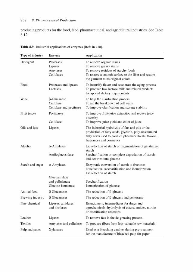
232 8 Pharmaceutical Production
Table 8.9. Industrial applications of enzymes [Refs in 410].
Type of industry Enzyme Application
Detergent Proteases To remove organic stainsLipases To remove greasy stainsAmylases To remove residues of starchy foodsCellulases To restore a smooth surface to the fiber and restore
the garment to its original colors
Food Proteases and lipases To intensify flavor and accelerate the aging processLactases To produce low-lactose milk and related products
for special dietary requirements
Wine β-Glucanase To help the clarification processCellulase To aid the breakdown of cell wallsCellulase and pectinase To improve clarification and storage stability
Fruit juices Pectinases To improve fruit-juice extraction and reduce juiceviscosity
Cellulase To improve juice yield and color of juice
Oils and fats Lipases The industrial hydrolysis of fats and oils or theproduction of fatty acids, glycerin, poly-unsaturatedfatty acids used to produce pharmaceuticals, flavors,fragrances and cosmetics
Alcohol α-Amylases Liquefaction of starch or fragmentation of gelatinizedstarch
Amiloglucosidase Saccharification or complete degradation of starchand dextrins into glucose
Starch and sugar α-Amylases Enzymatic conversion of starch to fructose:liquefaction, saccharification and isomerizationLiquefaction of starch
Glucoamylaseand pullulanase SaccharificationGlucose isomerase Isomerization of glucose
Animal feed β-Glucanases The reduction of β-glucans
Brewing industry β-Glucanases The reduction of β-glucans and pentosans
Fine chemical Lipases, amidases Enantiomeric intermediates for drugs andand nitrilases agrochemicals; hydrolysis of esters, amides, nitriles
or esterification reactions
Leather Lipases To remove fats in the de-greasing process
Textiles Amylases and cellulases To produce fibers from less-valuable raw materials
Pulp and paper Xylanases Used as a bleaching catalyst during pre-treatmentfor the manufacture of bleached pulp for paper
producing products for the food, feed, pharmaceutical, and agricultural industries. See Table8.12.
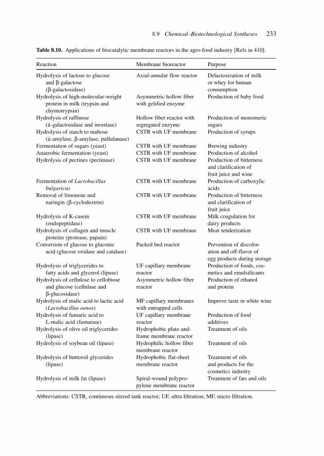
233
Table 8.10. Applications of biocatalytic membrane reactors in the agro-food industry [Refs in 410].
Reaction Membrane bioreactor Purpose
Hydrolysis of lactose to glucose Axial-annular flow reactor Delactosization of milkand β-galactose or whey for human(β-galactosidase) consumption
Hydrolysis of high-molecular-weight Asymmetric hollow fiber Production of baby foodprotein in milk (trypsin and with gelified enzymechymotrypsin)
Hydrolysis of raffinose Hollow fiber reactor with Production of monomeric(á-galactosidase and invertase) segregated enzyme sugars
Hydrolysis of starch to maltose CSTR with UF membrane Production of syrups(á-amylase, β-amylase, pullulanase)
Fermentation of sugars (yeast) CSTR with UF membrane Brewing industryAnaerobic fermentation (yeast) CSTR with UF membrane Production of alcoholHydrolysis of pectines (pectinase) CSTR with UF membrane Production of bitterness
and clarification offruit juice and wine
Fermentation of Lactobacillus CSTR with UF membrane Production of carboxylicbulgaricus acids
Removal of limonene and CSTR with UF membrane Production of bitternessnaringin (β-cyclodextrin) and clarification of
fruit juiceHydrolysis of K-casein CSTR with UF membrane Milk coagulation for
(endopeptidase) dairy productsHydrolysis of collagen and muscle CSTR with UF membrane Meat tenderization
proteins (protease, papain)Conversion of glucose to gluconic Packed bed reactor Prevention of discolor-
acid (glucose oxidase and catalase) ation and off-flavor ofegg products during storage
Hydrolysis of triglycerides to UF capillary membrane Production of foods, cos-fatty acids and glycerol (lipase) reactor metics and emulsificants
Hydrolysis of cellulose to cellobiose Asymmetric hollow fiber Production of ethanoland glucose (cellulase and reactor and proteinβ-glucosidase)
Hydrolysis of malic acid to lactic acid MF capillary membranes Improve taste in white wine(Lactobacillus oenos) with entrapped cells
Hydrolysis of fumaric acid to UF capillary membrane Production of foodL-malic acid (fumarase) reactor additives
Hydrolysis of olive oil triglycerides Hydrophobic plate-and- Treatment of oils(lipase) frame membrane reactor
Hydrolysis of soybean oil (lipase) Hydrophilic hollow fiber Treatment of oilsmembrane reactor
Hydrolysis of butteroil glycerides Hydrophobic flat-sheet Treatment of oils(lipase) membrane reactor and products for the
cosmetics industryHydrolysis of milk fat (lipase) Spiral-wound polypro- Treatment of fats and oils
pylene membrane reactor
Abbreviations: CSTR, continuous stirred tank reactor; UF, ultra filtration; MF, micro filtration.
8.9 Chemical–Biotechnological Syntheses
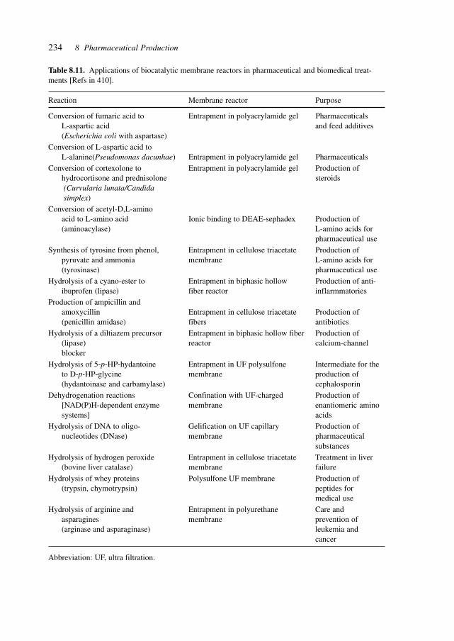
234 8 Pharmaceutical Production
Table 8.11. Applications of biocatalytic membrane reactors in pharmaceutical and biomedical treat-ments [Refs in 410].
Reaction Membrane reactor Purpose
Conversion of fumaric acid to Entrapment in polyacrylamide gel PharmaceuticalsL-aspartic acid and feed additives(Escherichia coli with aspartase)
Conversion of L-aspartic acid toL-alanine(Pseudomonas dacunhae) Entrapment in polyacrylamide gel Pharmaceuticals
Conversion of cortexolone to Entrapment in polyacrylamide gel Production ofhydrocortisone and prednisolone steroids (Curvularia lunata/Candida simplex)
Conversion of acetyl-D,L-aminoacid to L-amino acid Ionic binding to DEAE-sephadex Production of(aminoacylase) L-amino acids for
pharmaceutical use
Synthesis of tyrosine from phenol, Entrapment in cellulose triacetate Production ofpyruvate and ammonia membrane L-amino acids for(tyrosinase) pharmaceutical use
Hydrolysis of a cyano-ester to Entrapment in biphasic hollow Production of anti-ibuprofen (lipase) fiber reactor inflarmmatories
Production of ampicillin andamoxycillin Entrapment in cellulose triacetate Production of(penicillin amidase) fibers antibiotics
Hydrolysis of a diltiazem precursor Entrapment in biphasic hollow fiber Production of(lipase) reactor calcium-channelblocker
Hydrolysis of 5-p-HP-hydantoine Entrapment in UF polysulfone Intermediate for theto D-p-HP-glycine membrane production of(hydantoinase and carbamylase) cephalosporin
Dehydrogenation reactions Confination with UF-charged Production of[NAD(P)H-dependent enzyme membrane enantiomeric aminosystems] acids
Hydrolysis of DNA to oligo- Gelification on UF capillary Production ofnucleotides (DNase) membrane pharmaceutical
substances
Hydrolysis of hydrogen peroxide Entrapment in cellulose triacetate Treatment in liver(bovine liver catalase) membrane failure
Hydrolysis of whey proteins Polysulfone UF membrane Production of(trypsin, chymotrypsin) peptides for
medical use
Hydrolysis of arginine and Entrapment in polyurethane Care andasparagines membrane prevention of(arginase and asparaginase) leukemia and
cancer
Abbreviation: UF, ultra filtration.
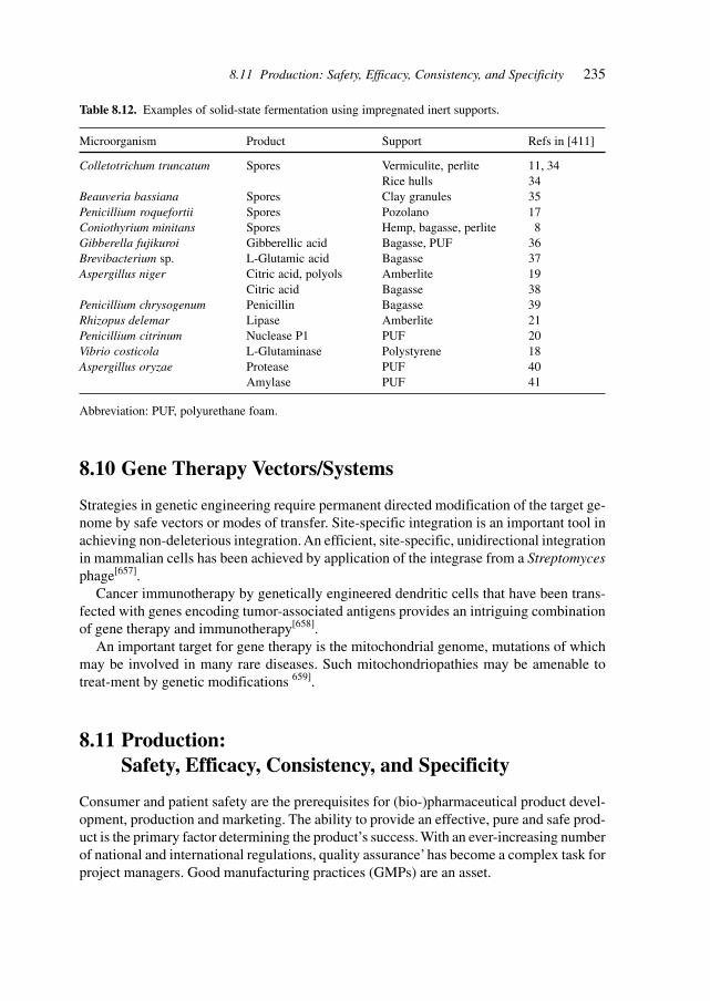
235
8.10 Gene Therapy Vectors/Systems
Strategies in genetic engineering require permanent directed modification of the target ge-nome by safe vectors or modes of transfer. Site-specific integration is an important tool inachieving non-deleterious integration. An efficient, site-specific, unidirectional integrationin mammalian cells has been achieved by application of the integrase from a Streptomycesphage[657].
Cancer immunotherapy by genetically engineered dendritic cells that have been trans-fected with genes encoding tumor-associated antigens provides an intriguing combinationof gene therapy and immunotherapy[658].
An important target for gene therapy is the mitochondrial genome, mutations of whichmay be involved in many rare diseases. Such mitochondriopathies may be amenable totreat-ment by genetic modifications 659].
8.11 Production:Safety, Efficacy, Consistency, and Specificity
Consumer and patient safety are the prerequisites for (bio-)pharmaceutical product devel-opment, production and marketing. The ability to provide an effective, pure and safe prod-uct is the primary factor determining the product’s success. With an ever-increasing numberof national and international regulations, quality assurance’ has become a complex task forproject managers. Good manufacturing practices (GMPs) are an asset.
Table 8.12. Examples of solid-state fermentation using impregnated inert supports.
Microorganism Product Support Refs in [411]
Colletotrichum truncatum Spores Vermiculite, perlite 11, 34Rice hulls 34
Beauveria bassiana Spores Clay granules 35Penicillium roquefortii Spores Pozolano 17Coniothyrium minitans Spores Hemp, bagasse, perlite 8Gibberella fujikuroi Gibberellic acid Bagasse, PUF 36Brevibacterium sp. L-Glutamic acid Bagasse 37Aspergillus niger Citric acid, polyols Amberlite 19
Citric acid Bagasse 38Penicillium chrysogenum Penicillin Bagasse 39Rhizopus delemar Lipase Amberlite 21Penicillium citrinum Nuclease P1 PUF 20Vibrio costicola L-Glutaminase Polystyrene 18Aspergillus oryzae Protease PUF 40
Amylase PUF 41
Abbreviation: PUF, polyurethane foam.
8.11 Production: Safety, Efficacy, Consistency, and Specificity
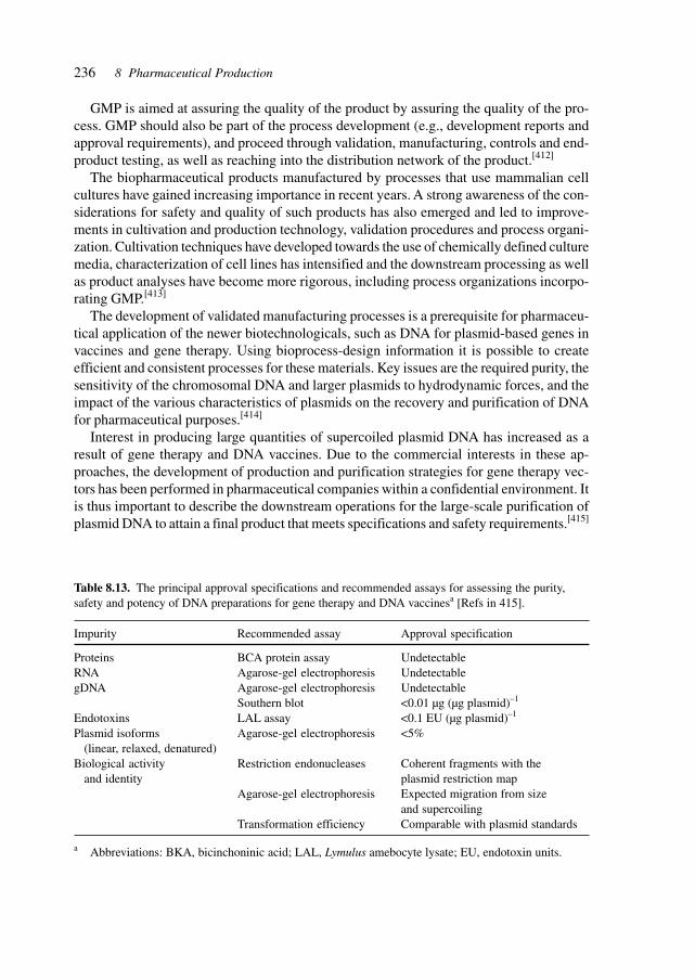
236 8 Pharmaceutical Production
GMP is aimed at assuring the quality of the product by assuring the quality of the pro-cess. GMP should also be part of the process development (e.g., development reports andapproval requirements), and proceed through validation, manufacturing, controls and end-product testing, as well as reaching into the distribution network of the product.[412]
The biopharmaceutical products manufactured by processes that use mammalian cellcultures have gained increasing importance in recent years. A strong awareness of the con-siderations for safety and quality of such products has also emerged and led to improve-ments in cultivation and production technology, validation procedures and process organi-zation. Cultivation techniques have developed towards the use of chemically defined culturemedia, characterization of cell lines has intensified and the downstream processing as wellas product analyses have become more rigorous, including process organizations incorpo-rating GMP.[413]
The development of validated manufacturing processes is a prerequisite for pharmaceu-tical application of the newer biotechnologicals, such as DNA for plasmid-based genes invaccines and gene therapy. Using bioprocess-design information it is possible to createefficient and consistent processes for these materials. Key issues are the required purity, thesensitivity of the chromosomal DNA and larger plasmids to hydrodynamic forces, and theimpact of the various characteristics of plasmids on the recovery and purification of DNAfor pharmaceutical purposes.[414]
Interest in producing large quantities of supercoiled plasmid DNA has increased as aresult of gene therapy and DNA vaccines. Due to the commercial interests in these ap-proaches, the development of production and purification strategies for gene therapy vec-tors has been performed in pharmaceutical companies within a confidential environment. Itis thus important to describe the downstream operations for the large-scale purification ofplasmid DNA to attain a final product that meets specifications and safety requirements.[415]
Table 8.13. The principal approval specifications and recommended assays for assessing the purity,safety and potency of DNA preparations for gene therapy and DNA vaccinesa [Refs in 415].
Impurity Recommended assay Approval specification
Proteins BCA protein assay UndetectableRNA Agarose-gel electrophoresis UndetectablegDNA Agarose-gel electrophoresis Undetectable
Southern blot <0.01 µg (µg plasmid)–1
Endotoxins LAL assay <0.1 EU (µg plasmid)–1
Plasmid isoforms Agarose-gel electrophoresis <5%(linear, relaxed, denatured)
Biological activity Restriction endonucleases Coherent fragments with theand identity plasmid restriction map
Agarose-gel electrophoresis Expected migration from sizeand supercoiling
Transformation efficiency Comparable with plasmid standards
a Abbreviations: BKA, bicinchoninic acid; LAL, Lymulus amebocyte lysate; EU, endotoxin units.
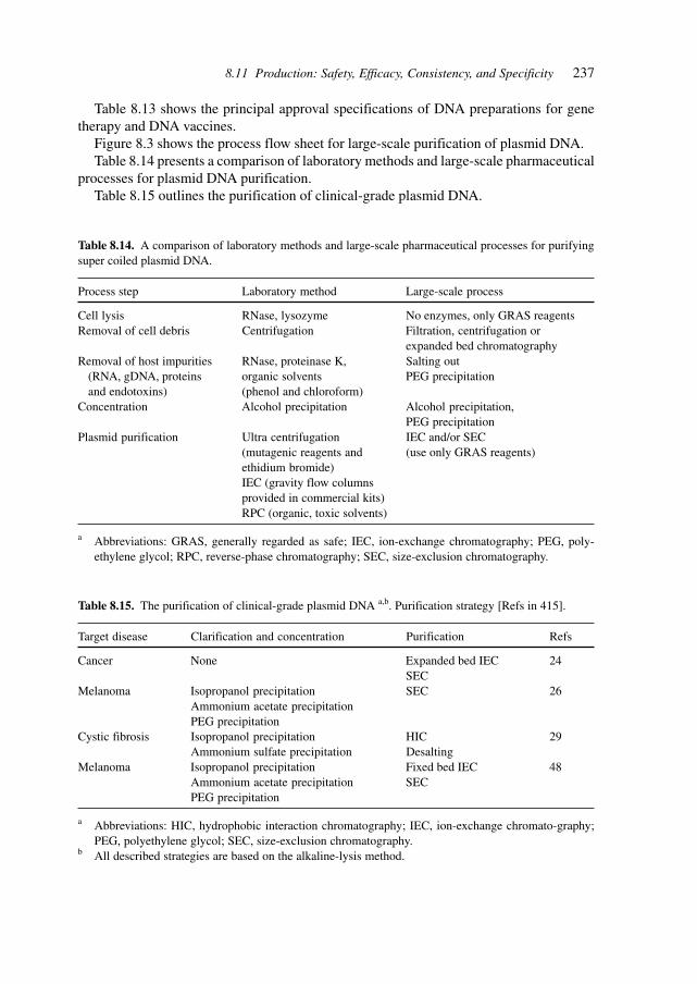
237
Table 8.14. A comparison of laboratory methods and large-scale pharmaceutical processes for purifyingsuper coiled plasmid DNA.
Process step Laboratory method Large-scale process
Cell lysis RNase, lysozyme No enzymes, only GRAS reagentsRemoval of cell debris Centrifugation Filtration, centrifugation or
expanded bed chromatographyRemoval of host impurities RNase, proteinase K, Salting out
(RNA, gDNA, proteins organic solvents PEG precipitationand endotoxins) (phenol and chloroform)
Concentration Alcohol precipitation Alcohol precipitation,PEG precipitation
Plasmid purification Ultra centrifugation IEC and/or SEC(mutagenic reagents and (use only GRAS reagents)ethidium bromide)IEC (gravity flow columnsprovided in commercial kits)RPC (organic, toxic solvents)
a Abbreviations: GRAS, generally regarded as safe; IEC, ion-exchange chromatography; PEG, poly-ethylene glycol; RPC, reverse-phase chromatography; SEC, size-exclusion chromatography.
Table 8.13 shows the principal approval specifications of DNA preparations for genetherapy and DNA vaccines.
Figure 8.3 shows the process flow sheet for large-scale purification of plasmid DNA.Table 8.14 presents a comparison of laboratory methods and large-scale pharmaceutical
processes for plasmid DNA purification.Table 8.15 outlines the purification of clinical-grade plasmid DNA.
Table 8.15. The purification of clinical-grade plasmid DNA a,b. Purification strategy [Refs in 415].
Target disease Clarification and concentration Purification Refs
Cancer None Expanded bed IEC 24SEC
Melanoma Isopropanol precipitation SEC 26Ammonium acetate precipitationPEG precipitation
Cystic fibrosis Isopropanol precipitation HIC 29Ammonium sulfate precipitation Desalting
Melanoma Isopropanol precipitation Fixed bed IEC 48Ammonium acetate precipitation SECPEG precipitation
a Abbreviations: HIC, hydrophobic interaction chromatography; IEC, ion-exchange chromato-graphy;PEG, polyethylene glycol; SEC, size-exclusion chromatography.
b All described strategies are based on the alkaline-lysis method.
8.11 Production: Safety, Efficacy, Consistency, and Specificity
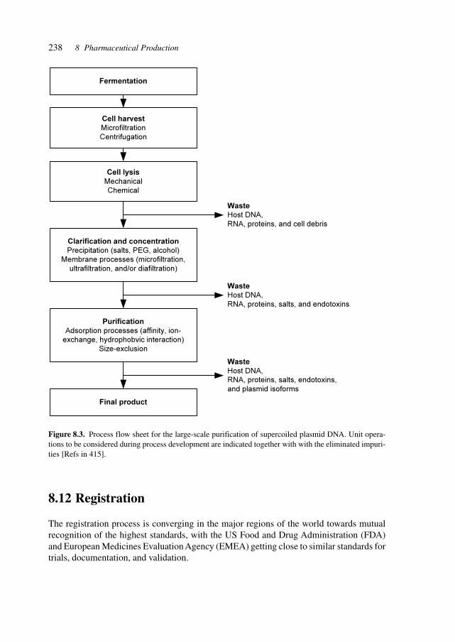
238 8 Pharmaceutical Production
Figure 8.3. Process flow sheet for the large-scale purification of supercoiled plasmid DNA. Unit opera-tions to be considered during process development are indicated together with with the eliminated impuri-ties [Refs in 415].
8.12 Registration
The registration process is converging in the major regions of the world towards mutualrecognition of the highest standards, with the US Food and Drug Administration (FDA)and European Medicines Evaluation Agency (EMEA) getting close to similar standards fortrials, documentation, and validation.
Fermentation
Cell harvest
Microfiltration
Centrifugation
Cell lysis
Mechanical
Chemical
Clarification and concentration
Precipitation (salts, PEG, alcohol)
Membrane processes (microfiltration,
ultrafiltration, and/or diafiltration)
Purification
Adsorption processes (affinity, ion-
exchange, hydrophobvic interaction)
Size-exclusion
Final product
Waste
Host DNA,
RNA, proteins, and cell debris
Waste
Host DNA,
RNA, proteins, salts, and endotoxins
Waste
Host DNA,
RNA, proteins, salts, endotoxins,
and plasmid isoforms
239
9 Safety
Safety is the uppermost concern for patients, physicians, producers, the population at large,the environment, the biosphere and (due to genetic modifications through, for example,gene therapies) also for future generations.
9.1 Medical Safety
Medical safety essentially involves ensuring the safety of the patients from unwanted andundesirable side effects, whereby side effects are assessed in the context of severity ofdisease and acceptable side effects versus therapeutic effect.
The use of transplanted organs from tissue engineering or from donor species providesnew and additional aspects of safety, e.g., dealing with unwanted gene transfer. Somaticgene therapy needs to consider undesirable integration in germ cells, and subsequent bio-logical effects and perpetuation.
9.2 Biological Safety
Biological safety concerns the effect of APIs, therapies, gene therapy vectors as well asbiological manufacturing systems (e.g., bacteria, cells and transgenics) on the humans in-volved and the biological systems potentially affected. Examples are considerations of an-tibiotic-resistance genes, virulence determinants and specific genetic traits.
9.3 Chemical Safety
Chemical safety is the acute toxicity of compounds which needs to be considered thor-oughly in view of the increasing potency and the potentially different effects on geneticallydifferent subtypes of humans or other entities of the biosphere.
Industrial Pharmaceutical Biotechnology. Heinrich KlefenzCopyright © 2002 Wiley-VCH Verlag GmbH
ISBNs: 3-527-29995-5 (Hardcover); 3-527-60012-4 (Electronic)

240 9 Safety
9.4 Equipment Safety
The safety of equipment considers primarily the function and effects of malfunction ofproduction equipment, diagnostic equipment, and the increasing numbers and types ofmedical devices, ranging from implanted pumps to prosthetics to the conceived nano-ro-bots carrying out essential supervisory or remedial functions in patients’ bodies.
241
10 Environment
Biotechnology is playing an increasing role in the environmental realm, in environmentalcontrol and in the remediation field by providing genetically modified plants, e.g., for car-bon dioxide fixation, by accumulating metal ions in plants, as a source of renewable rawmaterials and as an alternative for fossil fuels[502] More specifically, antibodies offer alter-natives to chemical and physical technologies in the multitude of possible topologies andthe resulting sensitive detection and specific interactions for removal of organic pollut-ants.[416]
A revealing study of the environmental impact of biotechnological production of PHA(polyhydroxy acid) in agricultural plants has been published with a comparative view onthe sustainability of biological processes. This study involves a thorough ‘cradle-to-grave’analysis and reveals that the total amount of fossil fuel required to produce 1 kg of PHAsexceeds that required to produce an equal amount of polystyrene. The replacement of con-ventional polymers with fermentation-derived PHAs does not yield benefits with respect tofossil fuel consumption. Future assessments of biological processes must incorporate theuse of raw materials (which are renewable) and also address the indirect consumption ofnon-renewable energy sources required for the process.[417]
Genetically engineered microorganisms or plants may be used for the remediation ofsoils contaminated with metals[707] or degradable organic compounds.
10.1 Pharmaceuticals and the Environment
Novel APIs, medical devices and gene therapy vectors/systems are increasingly being dis-covered and designed based on their interaction with genetic and biological networks. Thesimilarity of the genomes of many different species and the conservation of protein struc-ture and function necessitates consideration of the effects on the environment and the bio-sphere.
10.2 Biological Containment
Biological containment involves the use of cells, vectors, genes, and APIs which, uponrelease from the targeted or optimal environment, will not interact with the biological sys-tems they encounter. This means that high-performance strains are auxotrophic for rare
Industrial Pharmaceutical Biotechnology. Heinrich KlefenzCopyright © 2002 Wiley-VCH Verlag GmbH
ISBNs: 3-527-29995-5 (Hardcover); 3-527-60012-4 (Electronic)
242 10 Environment
compounds unlikely to be found in nature or are unable to function at the temperatures/oxygen levels found in the environment.
10.3 Physical/Chemical Containment
Physical/chemical containment takes into account the toxicity, virulence, and potential forchange and evolution of biological agents, and uses barriers, closed systems, and physicaldestruction by irradiation, chemicals and temperature to prevent the escape of viable agents.
10.4 Process-Integrated Environmental Protection
The manufacturing processes need to take a holistic view of the total life cycle of productsincluding their manufacture and disposal (from factory and from patient). The choice ofchiral syntheses, metabolically engineered cells, microreactors for optimized processes,and medical devices designed for function and recycling (and manufacturability) are keyconsiderations to be applied early in the development process.
10.5 Waste/Effluent Treatment and Recycling
Molecular/genetic techniques involve the direct modification of genetic material and re-quire special consideration of the waste streams produced. The adequacy of existing treat-ment, disposal and recycling processes of waste streams from biotechnological laborato-ries and industrial processes, especially those using genetically modified organisms, isfocused on the DNA content of these wastes, the properties of extracellular (or ‘naked’)DNA and the ability to transfer genetic information between bacteria (e.g., antibiotic resist-ance) or into higher organisms.[418]
Particle-based biofilm reactors provide the potential to develop compact and high-rateprocesses. In these reactors, a large biomass content can be maintained (up to 30 g/l) andthe large specific surface area (up to 3000 m2/m3) ensures that the conversions are notlimited by the biofilm liquid mass-transfer rate. Engineered design and control of particle-based biofilm reactors have been established and reliable correlations exist for the estima-tion of the design parameters. Thus, a new generation of high-load, efficient biofilm reac-tors are operating throughout the world, with several full-scale applications for industrialand municipal waste-water treatment.
Biofilm reactors are used in situations where the reactor capacity obtained by freelysuspended organisms is limited by the biomass concentration and hydraulic residence time.
The main reactor types are upflow sludge blankets, biofilm fluidized beds, expandedgranular sludge blankets, and biofilm airlift suspension and internal circulation reactors.
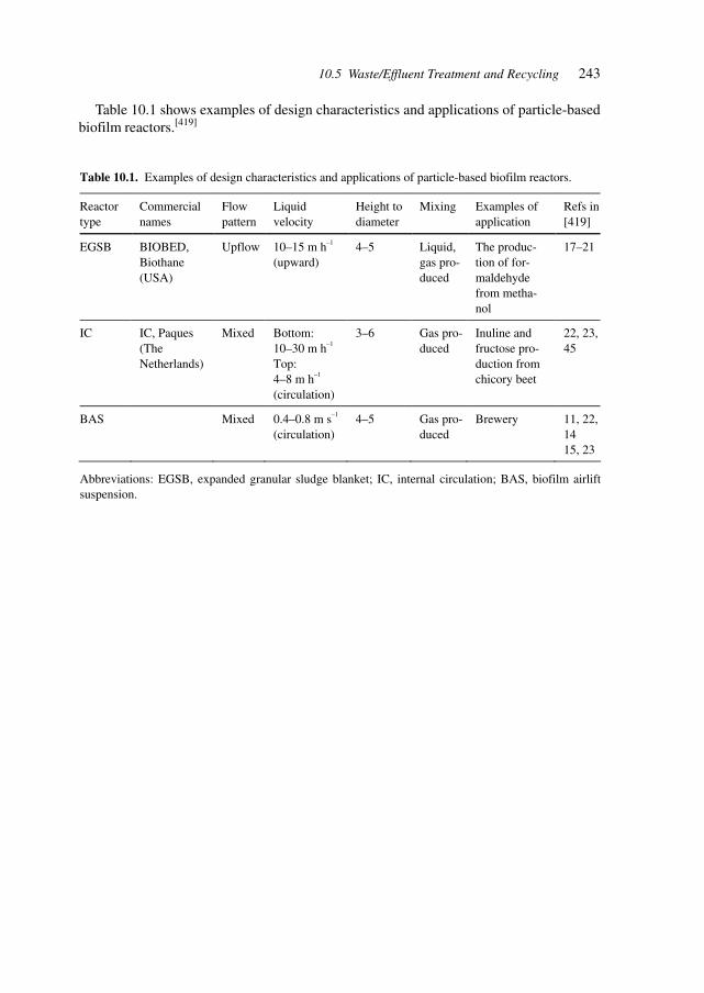
243
Table 10.1 shows examples of design characteristics and applications of particle-basedbiofilm reactors.[419]
10.5 Waste/Effluent Treatment and Recycling
Table 10.1. Examples of design characteristics and applications of particle-based biofilm reactors.
Reactor type
Commercial names
Flow pattern
Liquid velocity
Height to diameter
Mixing Examples of application
Refs in [419]
EGSB
BIOBED, Biothane (USA)
Upflow 10–15 m h–1
(upward) 4–5 Liquid,
gas pro-duced
The produc-tion of for-maldehyde from metha-nol
17–21
IC
IC, Paques (The Netherlands)
Mixed Bottom: 10–30 m h–1
Top: 4–8 m h–1 (circulation)
3–6 Gas pro-duced
Inuline and fructose pro-duction from chicory beet
22, 23, 45
BAS
Mixed 0.4–0.8 m s–1
(circulation) 4–5 Gas pro-
duced Brewery 11, 22,
14 15, 23
Abbreviations: EGSB, expanded granular sludge blanket; IC, internal circulation; BAS, biofilm airlift suspension.
245
11 Ethics
Gene therapy and ES cell research are a major focus of ethical considerations in terms ofresearch developments and applications.
The emphasis of underlying issues differs in Europe and the USA. Whereas Europeconfronts the debate from its historic plurality and with consideration of vast excesses theUSA debate brings the argument of public financing stronger into the debate. The patients’views and interests, which in the case of pressure groups during the AIDS epidemic havealready changed procedures of the FDA and public perceptions by bringing about fast-track transfer of pharmaceuticals into therapy, are significantly influencing the debate inUSA.[420]
The creation of novel organisms as exemplified by discussions and research on creatinga free-living organism with a minimal genome is also promoting ethical debate. To create anovel organism, scientists must determine which genes form the minimum set necessaryfor basic metabolism and replication, construct this minimum gene set, and provide orcreate the necessary non-genetic components for successful gene expression. There is stilla large technological gap between what has been achieved to date (defining a portion of aminimum set of genes necessary for an organism to survive under permissive laboratoryconditions) and actually ‘creating life’.[421]
Ethical considerations across the whole spectrum of biotechnology are being discussedin Europe, dealing with environmental applications, food production, human health, ani-mal welfare, research, industry, the ‘north/south’ divide, biodiversity, public perceptionsand media, and stress the consideration of the interest of future generations.[422]
Research dealing with generating organisms with a minimum set of genes has so farfocussed on the smallest known cellular genome, that of Mycoplasma genitalium (580 kb).M. genitalium (with 517 genes) has the smallest gene complement of any independentlyreplicating cell identified so far. Global transposon mutagenesis was used to identify non-essential genes in an effort to identify whether the naturally occurring gene complement isa true minimum genome under laboratory growth conditions. The positions of 2209transposon insertions in the completely sequenced genomes of M. genitalium and its closerelative M. pneumoniae were determined by sequencing across the junction of the transposonand the genomic DNA. These junctions define 1354 distinct sites of insertion that are notlethal. The analysis suggests that 265–350 of the 480 protein-coding regions of M. genitaliumare essential under laboratory growth conditions, including about 100 genes of unknownfunction.[423]
The ethical and practical aspects of applying biomedicine and life-extending therapiesto generate an ‘immortal’ phenotype are questions that society needs to consider as devel-opments unfold. Society, even in the advanced ‘rich’ parts of the world, will divide intothose that can afford expensive therapies and those that cannot.
Industrial Pharmaceutical Biotechnology. Heinrich KlefenzCopyright © 2002 Wiley-VCH Verlag GmbH
ISBNs: 3-527-29995-5 (Hardcover); 3-527-60012-4 (Electronic)

246 11 Ethics
On a global scale, there will exist societies that can hardly sustain the minimum require-ments for food, water, clothing, shelter, and medicine, while affluent individuals and soci-etal groups will co-exist. Unprecedented mechanisms need to be developed to cope withthese developments and their consequences, intra-societally as well as inter-societally (i.e.between nations or groups of nations).[424]
24712 Companies, Institutes, Networks, and Organizations
12 Companies, Institutes, Networks,and Organizations
Actinova Ltd 185 Cambridge Science Park, Cambridge CB4 0GA, UK
Advanced Cell Technology One Innovation Drive, Worcester, MA 01605, USA
Aeterna Laboratories (Arthritis) Quebec, PQ, Canada
Affymetrix Inc. 3380 Central Expressway, Santa Clara, CA 95051, USA
Agouron Pharmaceuticals Inc. (AIDS) 3565 General Atomics CT., La Jolla, CA 92037, USA
Agouron Pharmaceuticals Inc. (Arthritis) 10350 N Torrey Pines Rd., La Jolla, CA 92037-1018, USA
AGOWA Gesellschaft fuer molekular-biologische Technologie mbH,Glienicker Weg 185, 12489 Berlin, Germany
Agracetus Campus Monsanto Company, 8520 University Green, Middleton,WI 53562, USA
Akkadix Corp. 11099 North Torrey Pines Road, Suite 200, La Jolla, CA92037, USA
Alexion Pharmaceuticals 25 Science Park #360, New Haven, CT 06511, USA
Alliance for Ageing Research 2021 K Street, NW, Suite 305, Washington, DC 20006, USA
Alliance for Ageing Research, 800 W 9th Ave., Amarillo, TX 79101-3206, USAAmarillo Biosciences (AIDS)
Amgen Inc. One Amgen Center Drive, Thousand Oaks, CA 91320, USA
Amgen Staffing P. O. Box 2569, Thousand Oaks, CA 91319-2569, USA
Amrad (Malaria) 576 Swan St., Richmond VIC3121, Australia
AnalyticCon AG Hermannswerder Haus 17, 14473 Potsdam, Germany
AnorMed (Arthritis) (AIDS) 20353 64 Ave., Langley, BC V2Y 1N5, Canada
Antibioticos SA Laboratorios de Biotechnologia and Bioquimica, Avenida deAntibioticos 59–61, 24009 Leon, Spain
Antigen Express One Innovation Drive, Worcester, MA 01605, USA
Applied Biosystems Division PE Biosystems, 850 Lincoln Center Drive, Foster City, CA894404-1128, USAwww.appliedbiosystems.com
Applied Logic Associates Inc. 5615 Kirby Drive, Houston, TX 77005, USA
Applied Molecular Evolution Inc. 3520 Dunhill Street, San Diego, CA 92121, USA
Industrial Pharmaceutical Biotechnology. Heinrich KlefenzCopyright © 2002 Wiley-VCH Verlag GmbH
ISBNs: 3-527-29995-5 (Hardcover); 3-527-60012-4 (Electronic)

248 12 Companies, Institutes, Networks, and Organizations
Aquila Biopharmaceuticals Inc. 175 Crossing Blvd. #200, Framingham, MA 01702-4473,(Malaria) USA
Arena Pharmaceuticals 6166 Nancy Ridge Drive, San Diego, CA 92121, USA
Arena Pharmaceuticals 6166 Nancy Ridge Drive, San Diego, CA 92121, USA
Arevia GmbH Fennstrasse 49, 13353 Berlin, Germany
ARIAD Pharmaceuticals Inc. 26 Landsdowne Street, Cambridge, MA 02139, USA
Aronex Pharmaceuticals Inc. (Cancer) 8707 Technology Forest Pl., Spring, TX 77381-1191, USA(Antibacterial and Antifungal) (AIDS)
ArQule, Inc. 200 Boston Ave. Medford, MA 02155, USATel (++1) 781 395 4100Fax: (++1) 781 395 1225
Artemis Pharmaceuticals GmbH Neurather Ring 1, 51063 Koeln, Germany
AstraZeneca Alderley Park, Macclesfield, Cheshire SK10 4TG, UK
Atrix Laboratoies Inc. 701 Central Ave., Fort Collins, CO 80526-1843, USA(Antibacterial and Antifungal)
Atugen AG Robert-Roessle-Strasse 10, 13125 Berlin, Germany
Aurora Biosciences Corp. 11010 Torreyana Road, San Diego, CA 92121, USATel. (++1) 858 404 6600Fax: (++1) 858 404 8485
AutoImmun (Arthritis) Lexington, MA 781-860-0710, USA
Avanir Pharmaceuticals (AIDS) 9393 Towne Center Dr. #200, San Diego, CA 92121-3070,USA
Aventis Pharma London Road, Homes Chapel, Crewe, Cheshire CW4 8BE, UK
Aventis Research & Technologies Gebäude H 777, Industriepark Hoechst, GmbH & Co. KG,65926 Frankfurt/M, Germany
Avigen Inc. (Cancer) 1201 Harbor Bay Pkwy. #1000, Alameda, CA 94502-6586,USA
Axys Pharmaceuticals 180 Kimball Way, South San Francisco, CA 94080-6218, USA
BASF-LYNX Bioscience Im Neuenheimer Feld 515, 69120 Heidelberg, Germany
BASF Bio-research 100 Research Dr., Worcester, MA 01605-4312, USA
Bayer Corp., Pharmaceutical Division PO Box 3238, Scranton, PA 18505-0238, USA
Bayer Inc. 400 Morgan Ln., West Haven, CT 06516-4175, USA(Antibacterial and Antifungal)
Bayer AG (Arthritis) Kaiser-Wilhelm-Allee 1, 51373 Leverkusen, Germany
B. Braun Biotech International GmbH Schwarzenberger Weg 73–79, 34212 Melsungen, Germany
BD Biosciences 1 Becton Drive, Franklin Lakes, NJ 07417 USAwww.bdbiosciences.com
24912 Companies, Institutes, Networks, and Organizations
Bell Laboratories Lucent Technologies, 600 Mountain Avenue, Murray Hill,NJ 07974, USA
Biocarta, Inc 3830 Valley Center Drive 705-161, San Diego, CA 92130,USAwww.biocarta.com
Biochem Technology 1190 Saratoga Ave. # 140, San Jose, CA 95129-3433, USA
Biochemie GmbH A-6250 Kundl, AustriaFax and Tel. (++43) 5338200442
BioCryst Pharmaceuticals (AIDS) 2190 Parkway Lake Dr., Birmingham, AL 35244-1879, USA
Biogen Inc. 14 Cambridge Ctr., Cambridge, MA 02142-1481, USA
BioGenes Koepenicker Strasse 325, 12555 Berlin, GermanyGesellschaft für Biopolymere mbH
Bioimmune Systems (AIDS) 1177 S 1680 W, Orem, UT 84058-4930, USA
BioInvent Therapeutic AB Soelveg 41, Lund 22370, Sweden
Bioject Medical Technologies (Malaria) 7620 SW Bridgeport Rd., Portland, OR 97224-7700, USA
Biomatrix Inc. (Arthritis) 65 Railroad Ave. #2, Ridgefield, NJ 07657-2130, USA
BioSource Deutschland GmbH Tel. 0800 100 85 74, 12555 Berlin, Germany
BioSyn (AIDS) 3401 Market St. #300, Philadelphia, PA 19104-3319, USA(Antibacterial and Antifungal)
Biotechnology, Boston Life Sciences 137 Newbury Street, Boston, MA 02116, USA(Arthritis)
Biotecon GmbH Gesellschaft fuer Biotechnologische Entwicklung undConsulting mbH, Tegeler Weg 33, 10589 Berlin, Germany
Biozym GmbH Steinbrinksweg 27, POB 180, 31833 Hessisch-Oldendorf,Germany
Bitop Gesellschaft für Biotechnische Alfred-Herrhausen-Strasse 44, 58455 Witten, GermanyOptimierung mbH
Boehringer Ingelheim Anml Hlth 2621 N Belt Hwy., St Joseph, MO 64506-2002, USA
Boehringer Ingelheim GmbH Binger Strasse 173, 55216 Ingelheim am Rhein, Germany
Biotechnology Research and Darmstaedter Strasse 34, 64673 Zwingenberg, GermanyInformation Network
Brain GmbH Darmstaedter Strasse 34
Biotechnology Resesarch and 64673 Zwingenberg, GermanyInformation Network
Bristol-Myers-Squibb Pharmaceutical 5 Research Parkway, Wallingford, Research Institute,CT 06492, USA
Britsh Biotech Watlington Rd., Oxford, Oxfordshire, OX4 6LY,United Kingdom
250 12 Companies, Institutes, Networks, and Organizations
Brucker Daltonics Inc. 15 Fortune Dr. Bldg. #2, Billerica, MA 01821-3958, USA
Bruker Daltonik GmbH Fahrenheitstrasse 4, 28359 Bremen, Germany
BTX Inc., A Division of Genetronics 11199 Sormento Valoley Road, San Diego, CA 92121, USAwww.btxonline.com
Cadus Pharmaceutical Corp. 777 Old Saw Mill River Rd., Tarrytown, NY 10591, USA
Cambridge Antibody Technology The Science Park Melbourn, Royston,(Arthritis) Hertfordshire SG8 6JJ, UK
Carl Zeiss Inc. 1 Zeiss Dr., Thornwood, NY 1054-1996, Microscopy &Imaging Systems, USA
CELERA Genomics 45 West Gude Drive, Rockville, MD 20850, USAwww.celerajobs.com
Celgene (Arthritis) 7 Powderhorn Dr. #1,Warren, NJ 07059-5190, USA
Cell Genesis Inc. (Arthritis) 342 Lakeside Dr., Foster City, CA 94404-1146, USA
CellGenix Technology Transfer GmbH Elsässer Strasse 2N, 79110 Freiburg, Germany
Cellomics Inc. 635 William Pitt Way, Pittsburgh, PA 15238, USA
Cell Signaling Technology 166B Cummings Center, Beverly, MA 01915, USA
CellTec GmbH Biotechnologie Frohmestrasse 110, 22459 Hamburg, Germany
Cell-Tech Inc. 8982 Mansfield Rd., Shreveport, LA-71118-2128, USA
Celltech Ltd. 216 Bath Rd., Slough Berkshire SL1 4EN, UK
Cell Therapeutics (Cancer) 201 Ellrot Ave W#400, Seattle, WA 98119-4237, USA
Cellular Products GmbH Delitzscher Strasse 141, 04129 Leipzig, Germany
Centocor Inc. 200 Great Valley Pkwy, Malvern, PA 19355-1307, USA
Charles River Laboratories, Inc 251 Ballardvale Street, Wilmington, MA 01887-1000, USAwww.CRIVER.COM
Charles River Therion Rensselaer Technology Park, 185 Jordan Road, Troy, NY12180-7617, USA
Chemical Synthesis,Process Develop- 421 Wakara Way; Suite 201, Salt Lake City, UT 84108, USAment, SNP Consortium, Cognetics Tel.: (++1) 801 581 0400 ext. 222
Fax: (++1) 801 581 9555
Chiron Corp. 4560 Horton Street, Room 4.4144, Emeryville, CA 94608,USA
Clontech 1020 East Meadow Circle, Palo Alto, CA 94303, USA
Clontech Laboratories GmbH Tullastrasse 4, 69126 Heidelberg, Germany
Cobra Therapeutics The Science Park, University of Keele, Keele, StaffordshireST5 5SP, UK
Cold Spring Harbor Laboratory 1 Bungtown Road, Cold Spring Harbor, NY 11724, USA
25112 Companies, Institutes, Networks, and Organizations
Collateral Therapeutics Research 11622 El Camino Real # 300, San Diego, CA 92130-2051,(Cardiovascular Disease) USA
Columbia Laboratories Inc. (AIDS) 2875 NE 191st St., Miami, FL 33180-2831, USA
Combinature Biopharm AG, Robert-Roessle-Strasse 10, 13125 Berlin, Germanyc/o GenProfile AG
Comparative Genetics SmithKline Beecham Pharmaceuticals, Third Avenue, Harlow,Essex CM19 5AW, UK
Computational Chemistry GlaxoWellcome R & D, Medicines Research Centre,and Informatics Unit, Gunnels Wood Road, Stevenage, Hertfordshire SG1 2NY, UK
Connetics Corp. 3400 W Bay Shore Rd., Palo Alto, CA 94303-4227, USA
Connex Gesellschaft zur Optimierung Am Kopferspitz 19, 82152 Martinsried, Planegg, Germanyvon Forschung und Entwicklung
Cortech Tec. (Arthritis) 422 N Douty St. # 422, Hanford, CA 93230-3977, USA
Corvas Intl. Inc. 3030 Science Park Rd. # 301,San Diego, CA 92121-1183,(Cardiovascular Disease) (Malaria) USA
Cosmix Molecular Bilogicals GmbH, Mascheroder Weg 1b,38124 Braunschweig, Germany
CRO Laboratories 89 Adams Rd, North Grafton, MA 0153-2001, USA
CSORO Commonwealth Scientific Normanby Rd., Clayton, VIC 3168, Australiaand Industrial Research Org.
Cubist Pharmaceuticals 24 Emily Street, Cambridge, MA 02139, USA(antibacterial and antifungal)
CuraGen Corp. 555 Long Wharf Drive, 11th Floor, New Haven, CT 06511,USA
CV Therapeutics 3172 Porter Drive, Palo Alto, CA 94304, USA
Cyanotech Corp. 73-4460 Queen Kaahumanu Highway, Suite 102, Kailua-Kona,HI 96740, USA
Cyclacel Ltd. Dundee Technopole James Lindsay Place, Dundee DD1 5JJ,UK
Cypress Bioscience Inc. (Arthritis) 4350 Executive Dr. #325, San Diego, CA 92121-2118, USA
Cypress Pharmaceuticals 4350 Executive Dr. #325, San Diego, CA 92121-2118, USA(Cardiovascular Disease)
Cytomatrix 50 Cummings Park, Woburn, MA 01801-2123, USA
Cytran Inc. (AIDS) 10230 NE Points Dr., Kirkland, WA 98033-7869, USA
Cytrx (Cancer) (Malaria) 154 Technology Pkwy, Norcross, GA 30092-2911, USA
Daylight Inc. 1561 Laurel St., San Carlos, CA 94070-5114, USA
Department of Genomics, Pfizer Central Research, Eastern Point Road, Groton,Targets and Cancer Research CT 06340, USA
252 12 Companies, Institutes, Networks, and Organizations
Departments of Cardiovascular 1 DNA Way, South San Francisco, CA 94080, USAResearch and Pathology
Departments of Obstetrics & 5 Lower Kent Ridge Rd, Singapore119074GynaecologyNational University of Singapore
DeveloGen AG Rudolf-Wissell-Strasse 28, 37079 Goettingen, Germany
Diatide Inc. (Cardiovascular Disease) 9 Delta Dr. #7, Londonderry, NH 03053-2372, USA
Digital Gene Technologies 11149 North Torrey Pines Road, La Jolla, CA 92037, USA
Discovery Partners International 11149 North Torrey Pines Road, La Jolla, CA 92037, USA
DNAVEC Research Inc. Tsukuba-City, Ibaraki, Japan
DNX Transgenic Sciences 5, Cedar Brook Drive, Cranbury, NJ 08512, USAwww.dnxsciences.comTel.: (++1) 609 860 0806Fax: (++1) 609 860 8515
Double Twist.com Dr Kuebler GmbH, Siebertstrasse 6, 81675 Muenchen,Germany
Drug Discovery Ltd Royal College Building, 204 George Street, Glasgow G1 1XW,UK
DuPont 4570 Executive Drive, Suite 400, San Diego, CA 92121,Pharmaceuticals Research Laboratories USA
DuPont Pharmaceuticals Co. (AIDS) 974 Center Rd., Wilmington, DE-19805-1269, USA
Dura Pharmaceuticals Inc. 7475 Lusk Blvd., San Diego, CA 92121-5796, USA
Elan Pharmaceuticals (AIDS) South San Francisco, CA, USA
Eli Lilly Co. Bldg. 3 # 31, Indianapolis, IN 46285-0001, USA
Enzyme Design Novo Nordisk, Building 2C, 2880 Bagsvaerd, Denmark
Eppendorf Scientific Inc. One Cantiague Road, Westbury, NY 11590-0207, USA
Eppendorf-Netherler-Hinz GmbH Barkhausenweg 1, 22339 Hamburg, Germany
Epimmune Inc. (Malaria) 655 Nancy Ridge Dr., San Diego, CA 92121-3221, USA
Evotec Biosystems AG Schnackenburgallee 114, 22525 Hamburg, Germany
Exelixis Inc. P.O.Box 511, So. San Francisco, CA 94083-511, USAwww.exelixis.com
Frauenhofer, Max-Planck-Society/ Deerfield, IL 60015-4850, USAFijusawa (AIDS)
Fraunhofer Research Center 2901 Hubbard St., Ann Arbor, MI 48105-2435, USA
GAIFAR (German–American Institute Herrmannswerder 15–16, 14473 Potsdam, Germanyfor Applied Biomedical Research GmbH)
GATC GmbH Fritz-Arnold-Strasse 23, 78467 Konstanz, Germany
G D Searle Co (Arthritis) 5200 Old Orchard Rd., Skokie, IL 60077-1034, USA
25312 Companies, Institutes, Networks, and Organizations
GelTex Pharmaceuticals Inc. (AIDS) 153 Second Avenue, Waltham, MA 02451-1122, USA
Gene Alliance Max-Volmer-Strasse 4, 70724 Hilden, Germany
Gene Logic 708 Quince Orchard Road, Gaithersburg, MD 20878, USATel.: (++1) 301 987 1700Fax: (++1) 301 987 1701
Genelabs Technologies Inc. (Asthma) 505 Penobscot Dr., Redwood City, CA 94063-4738, USA
Genelabs Technologies Inc. (Cancer) 505 Penobscot Dr., Redwood City, CA 94063-4738, USA
Gene Therapy Systems 10190 Telesis Court, San Diego, CA 92121, USA
Geneformatics Inc. 5830 Oberlin Drive, Suite 200, San Diego, CA 92121-3754,USA
GeneMachines 935 Washington Street, San Carlos, CA 94070, USAwww.genemachines.com
Genencor 925 Page Mill Road, Palo Alto, CA 94304, USA
Genetics Institute GmbH Lochamer Strasse 11 82152 Planegg Germany
Genetic Technologies SmithKline Beecham Pharmaceuticals, New Frontiers SciencePark, Harlow, Essex CM19 5AW, UK
Genetic Therapy Inc. (Cancer) 9 W Watkins Mill Rd., Gaithersburg, MD 20878-4021, USA
Genetix Pharmaceuticals (Cancer) 840 Memorial Drive, Cambridge, MA 02139, USA
Genom Analytik GmbH Loebener Strasse, UFT, 28359 Bremen, Germanyc/o Zentrum fuer Umweltforschungund Technologie Germany
Genomatix Software GmbH Bioinformatics & Sequence Analysis, Karlstrasse 55, 80333Muenchen, Germany
Genome Pharmaceuticals Corporation Lochamer Strasse 29, 82152 Martinsried, Germany(GPC AG)
Genome Pharmaceuticals Corporation Fraunhoferstrasse 20, 82152 Martinsried/Munich, GermanyTel.: (++49) 8985652650Fax: (++49) 8985652610
Genomica Corp. 1745 38th Street, Boulder, CO 80301, USA
Genta (Arthritis) San Diego, CA 92121, USA
Genta (AIDS) 99 Hayden Avenue, Lexington, MA 02173 USA
Genzyme Transgenics TSI 2 Taft CT. #202, Rockville, MD 20850-5355, USA
Genzyme Corp. 1 Kendall Sq., Cambridge, MA 02139-1562, USA
Genzyme Molecular Oncology PO Box 9322, Framingham, MA 01701-9322, USAand Genzyme Corp.
Geron Corp. 230 Constitution Drive, Menlo Park, CA 94025, USA
Gilead Sciences Inc. (AIDS) 333 Lakeside Dr., Foster City, CA 94404-1147, USA
Glaxo Wellcome (AIDS) Research Triangle Park, NC 27709-5428 USA
254 12 Companies, Institutes, Networks, and Organizations
Glaxo Wellcome Research Bioanalysis and Drug Metabolism, Park Road, Ware,and Development SG12 ODP, UK
GuraGen Corp. 555 Long Wharf Drive, New Haven, CT 06511, USA
Hemispherx Biopharma (AIDS) 1617 JFK Blvd., Philadelphia, PA 19103-1821, USA
HepaVec AG Robert-Roessle-Strasse 10, 13122 Berlin, Germany
Hoechst Marion Roussel (now Aventis) RT 202.206 Bridgewater, NJ 08807, USA(Asthma) (Antibacterial and Antifungal)
Hoffmann-LaRoche Inc. 340 Kingsland St., Nutley, NJ 07110-1199, USAPreclinical Research
Hoffmann-La Roche Ltd Basel, Switzerland(Antibacterial and Antifungal)
Hollis-Eden Pharmaceuticals (Malaria) 9333 Genesee Ave.# 200, San Diego, CA 92121-2113, USA(AIDS)
Hybridon (AIDS) 620 Memorial Drive, Cambridge, MA 02139, USA
Hyseq Inc. 670 Almanor Avenue, Sunnyvale, CA 94085, USAwww.hysec.comFax: (++1) 408 524 8129
Ibis Therapeutics, 2292 Faraday Avenue, Carlsbad, CA 92008, USAIsis Pharmaceuticals Inc. Tel.: (++1) 760 603 2347
Fax: (++1) 760 431 2768
ICOS Inc. (Asthma) 22021 20 th Ave., Bothell, WA 98021-4406, USA(Cardiovascular Disease)
IDEA (Innovative Dermale Frankfurter Ring 193a, 80807, Muenchen, GermanyApplikationen GmbH)
IDEC Pharmaceuticals Corp. 11011 Torreyana Rd., San Diego, CA 92121-1104, USA
Idun Pharmaceuticals 11085 North Torrey Pines Road, Suite 300, La Jolla,CA 92307, USA
IGB Fraunhofer Institute for Inter- Nobelstrasse 12, 70569 Stuttgart, Germanyfacial Engineering and Biotechnology
Immune Response Corp. 5935 Darwin CT., Carlsbad, CA 92008-7399, USA
Immunex Corp. (Arthritis) (Cancer) 51 University St., Seattle, WA 98101-2936, USA(AIDS)
Incyte Genomics, Inc 4678 World Parkway Circle, St. Louis, MO 63134-3156, USAwww.incite.com
Incyte Microarray Systems & Genomics 6519 Dumbarton Dr., Fremont, CA 94555-3619, USA
Inflazyme Pharmaceuticals Ltd. 5600 Parkwood Way, Richmond, BCV6V 2M2, Canada(Arthritis)
Inflazyme (Asthma) Richmond, BC V6X 2GO, Canada
25512 Companies, Institutes, Networks, and Organizations
InforMax 610 Executive Boulevard,10 th Floor, North Bethesda,MD 20852, USAwww.informaxinc.com
Ingenex (Cancer) Menlo Park, CA 94025, USA
Integrated Genomics Inc. 2201 West Campbell Park Drive, Chicago, IL 60612, USA
Integrated Protein Technologies 8520 University Green, Middleton, WI 53562, USAAgracetus Campus, Monsanto Company
Interferon Sciences Inc. (AIDS) 783 Jersey Ave., New Brunswick, NJ 08901-3660, USA
Intra Biotics Pharmaceuticals) 1255 Terra Bella Ave., Mountain View, CA 94043-1833, USA(Antibacterial and Antifungal)
Invitrogen Corp. 1600 Faraday, Carlsbad, CA 92008, USA
Isenselt Fahrenheitstrasse 9, 28359 Bremen, Germany(Intelligente Sensorsoftware undBioinformatik AG iG)
IVAX Corp. (AIDS) 4400 Biscayne Blvd., Miami, FL 33137-3212, USA
Ixsis (Cancer) 1903 1/2 Capitol Ave., Sacramento, CA 95814-4212, USA
Jacobus Pharmaceuticals (AIDS) 37 Cleveland Ln., Princeton, NJ 08540-3790, USA
Janssen Pharmaceiticals 1125 Trenton Harbourton Rd.,Titusville, NJ 08560-1504, USA(antibacterial and antifungal)
Juergen Drews, International 767 Third Avenue, New York, NY 10158, USABiomedicine Management Partners, Tel.: (++1) 212 739 6400Orbimed Advisors Fax: (++1) 212 739 6444
Jerini Bio Tools GmbH Rudower Chaussee 5, 12489 Berlin, Germany
Kissei Pharmaceuticals Co. Ltd. Meijiseimei Sakaimachi Bldg., 1-9-6 Sakaimachi,(Arthritis) Kokurakita-Ku, Kitakyushi City, Fakuoka Pref., Japan
KOSAN Biosciences 3832 Bay Center Place, Hayward, CA 94545, USA
KTB Tumorforschungs GmbH Breisacher Strasse 117, 79106 Freiburg, Germany
LaVision BioTec GmbH Hoefeweg 74, 33619 Bielefeld, Germany
LETTI (CEA-Technologies Avancées) DEIN/SPE/GCO, CE Saclay, 91191 Gif-sur-Yvette Cedex,France
LeukoSite Inc. 75 Sydney St., Cambridge, MA 02139-4134, USA
Lexicon Genetics 4000 Research Forest Drive, The Woodlands, TX 77381, USA
Ligand Pharmaceuticals Inc. (AIDS) 10275 Science Center Dr., San Diego, CA 92121-1117, USA
Lion Bioscience AG Im Neuenheimer Feld 515, 69120 Heidelberg, Germany
Lorus Therapeutics Inc. (AIDS) 7100 Woodbine Ave., Markham, ON L3R 5JR, Canada
Lynx Therapeutics Inc. 25861 Industrial Boulevard, Hayward, CA 94545, USA
256 12 Companies, Institutes, Networks, and Organizations
Max-Planck-Gesellschaft Hofgartenstr. 8, 80539 Muenchen, GermanyMIPS Datenbank für Proteinsequenzenund Molekulare Strukturbiologie
Magainin Pharmaceuticals Inc. 5110 Campus Dr., Plymouth Meeting, PA 19462-1111, USA(Antibacterial and Antifungal) (Asthma)
Maxygen Inc. 3410 Central Expressway, Santa Clara, CA 95051, USA
Medarex Inc. 707 State Rd.206, Princeton, NJ 08540-1437, USA
MEDICS (The European Center of c/o FhG IBMT, Industriestrasse 5, 66280 Sulzbach, GermanyCompetence for Biomedical Microdevices)
MediGene AG Lochhamer Strasse 11, 82152 Martinsried, Germany
MediGenomix GmbH Lochamer Strasse 11, 82152 Martinsried Planegg, Germany
Memorec Stoffel GmbH Medical Molecular Research Cologne, Stockheimer Weg 1,50829 Koeln, Germany
Merck & Co Inc. (AIDS) 1 Merck Dr., White House Station, NJ 08889-3497, USA
Merck KGaA Frankfurter Strasse 250, 64293 Darmstadt, Germany
Merck Research Laboratories P. O. Box 2000, Mail Drop RY80Y-215, Rahway, NJ 07065,USA
Merck Research Laboratories 770 Sumneytown Pike, West Point, PA 19486, USA
Merck Sharp and Dohme Terlings Park, Eastwick Road, Harlow, Essex CM20 2QR, UKResearch laboratoriesNeuroscience Research Centre
Message Pharmaceuticals 30 Spring Mill Drive, Malvern, PA 19355, USA
Metanomics GmbH & CoKG Tegeler Weg 33, 10589 Berlin, Germany
MIKROGEN GmbH Fraunhofer Strasse 20, 82152 Martinsried, Germany
Millennium Pharmaceuticals P. O. Box 798, Burlington, MA 01803, USA
Millennium Pharmaceuticals and 640 Memorial Drive, Cambridge, MA 02139, USAMillennium Predictive Medicine
Millenium Pharmaceuticals (Asthma) 640 Memorial Drive, Cambridge, MA 02139, USA
Miltenyi Biotec GmbH Friedrich-Ebert-Strasse 68, 51429 Bergisch Gladbach,Germany
MIT Artificial Intelligence Lab 545 Technology Square, Cambridge, MA 02139, USA
MitoKor 11494 Sorrento Valley Road, San Diego, CA 92121, USA
Mixture Sciences Torrey Pines Institute for Molecular Studies,3550 General Atomics Court, San Diego, CA 92121, USA
Molecular Devices Corporation 1311 Orleans Drive, Sunnyvale, CA 94089, USAwww.moleculardevices.comTel.: (++1) 800 635 5577
25712 Companies, Institutes, Networks, and Organizations
Molecular Dynamics 928 E Arques Ave., Sunnyvale, CA 94085-4520, USAAmersham Pharmacia Biotech
Monsanto Company 700 Chesterfield Village Parkway North, St Louis, MO 63198,USA
MorphoSys AG Am Klopferspitz 19, 82152 Martinsried, Germany
MPB Cologne GmbH Eupener Strasse 161, 50923 Koeln, GermanyMolecular Plant & Protein Biotechnology
MWG Biotech AG Anzinger Strasse 7a, 85560 Ebersberg, Germany
Myriad Genetics Inc. 320 Wakara Way, Salt Lake City, UT 84108, USA
NABI Biomedical 1000 Chestnut St., Evansville, IN 47713-1952, USA(Antibacterial and Antifungal)
Nanogate GmbH Gewerbepark, Eschberger Weg, 66121 Saarbruecken, Germany
Nanogen Inc. 10398 Pacific Center Center, San Diego, CA 92121, USA
National Institute of Health 9000 Rockville Pike, Bethesda, MD 20892, USA
National Institute on Aging Gerontology Research Center, 5600 Nathan Shock Drive,Baltimore, MD 21224-6825, USA
NeoGenesis 840 Memorial Drive, Cambridge, MA 02139, USA
Neo Rx Corp. (cardiovascular disease) 410 W Harrison St., Seattle, WA 98119-4000, USA
Neose Technologies Inc. 102 Witmer Road, Horsham, PA 19044, USA
Neurocrine Biosciences Inc. 10555 Science Center Dr., San Diego, CA 92121-1100, USA(autoimmune disease)
New World Science and 8401 Colesville Road, Suite 610, Silver Spring, MD 20910,Technology Inc. USA
Nexell Therapeutics Inc. (AIDS) 9 Parker, Irvine, CA 92618-1605, USA
Novartis Agricultural Discovery 3115 Merryfield Row, Suite 100, San Diego, CA 92121, USAInstitute Inc.
Novartis Pharmaceuticals Corp. 556 Morris Avenue, Building SEF 1029, Summit,NJ 07901-1398, USA
Novartis (Antibacterial and Antifungal) Basel, Switzerland
Novartis Corp. 59 State Route 10, East Hanover, NJ 07936-1005, USA(Antibacterial and Antifungal)
Novo Nordisk A/S Enzyme Design, 2CS.01, 1 Novo Alle, 2880 Bagsvaerd,Denmark
Novo Nordisk Biotech Inc. 1445 Drew Avenue, Davis, CA 95616, USA
NOXXON Pharma AG Gustav-Meyer-Allee 25, 13355 Berlin, Germany
Oncotest GmbH Am Flughafen 8–10, 79110 Freiburg, Germany
Ono Pharmaceuticals Co. Ltd (Arthritis) 2-1-5 Doshomachi, Chuo-Ku, Osaka-City, Osaka Pref., Japan

258 12 Companies, Institutes, Networks, and Organizations
Ontogeny Inc. 45 Moulton Street, Cambridge, MA 02138 USA
Orchid BioSciences 303 College Road East, Princeton, NJ 08540, USA
Organon Inc. Department of Immunology, PO Box 20, 5340 BH Oss,The Netherlands
Origene Technologies Inc. 6 Taft Court, Suite 300, Rockville, MD 20850, USA
Orpegen Pharma Gesellschaft fuer Czernyring 22, 69115 Heidelberg, GermanyBiotechnologische Forschung,Entwicklung und Produktion mbH
Pangea Systems 4040 Campbell Ave, Menlo Park, CA 94025, USA
PanVera Corp. 545 Science Drive, Madison WI 53711, USA
Paradigm Genetics 104 Alexander Drive, Building 2, PO Box 14528,Research Triangle Park, NC 27709-5428, USA
Pasteur Merieux Connaught (Malaria) Discovery Dr., Swiftwater, PA 18370, USA(AIDS)
PathoGenesis Corp. 201 Elliott Avenue West, Seattle, WA 98119, USA
PE Biosystems 500 Old Connecticut Path, Framingham, MA 01701, USA
Penederm Inc. 320 Lakeside Dr. #A, Foster City, CA 94404-1146, USA(Antimicrobial and Antifungal)
Pfizer Inc. (Antibacterial and 235E 42 nd St., New York, NY 10017-5755, USAAntifungal) (AIDS)
Pharmabiodyn GmbH Ferdinand-Porsche-Strasse 51, 79211 Denzlingen, Germany
Pharmacia & Upjohn 7000 Portage Road, Kalamazoo, MI 49001, USA
Pharmacia & Upjohn 103 Carnegie Ctr., Princeton, NJ 08540-6235, USA
Pharmacia Corp. 100 RT 206N, Peapack, NJ 07977, USA(Antibacterial and Antifungal)
Pharmacopoeia Inc. (Asthma) 3000 Eastpark Blvd., Cranbury, NJ 08512-3516, USA
Phytotech Inc. 1 Deer Park Drive, Suite 1, Monmouth Junction, NJ 08852,USA
Pico Rapid Technologie GmbH Leobener Strasse, UFT, 28359 Bremen, Germany
Polydex Pharmaceuticals Inc. (AIDS) 2705 Lajoie, Trois Rivières, QC G8Z 3G4 Canada
PPL Therapeutics Inc. 1700 Kraft Drive, Blacksburg, VA 24060, USA
PPL Therapeutics Ltd Roslin, Edingburgh, EH25 9PP, UK
Preclinical Research 340 Kingsland St., Nutley, NJ 07110-1199, USAHoffmann-La Roche, Inc
Price Waterhouse Coopers Harman House, 1 George Street, Uxbridge,Middlesex UB8 1QQ, UK
ProBioGen GmbH Rudower Chaussee 5, 12489 Berlin, Germany
25912 Companies, Institutes, Networks, and Organizations
Progen Biotechnik GmbH Maasstrasse 30, 69123 Heidelberg, Germany
Progenitor (Asthma) 1730 Ackerman Dr. #4, Lodi, CA 95240-6385, USA
Proligo Biochemie GmbH Georg-Heyken-Strasse 14, 21147 Hamburg, Germany
Promega Corp. 2800 Woods Hollow Road, Madison, WI 53177-5399, USA
Promega GmbH Hi-Tech-Park, Schildkroetstrasse 15, 68199 Mannheim,Germany
Protana A/S Staermosegaardsvej 16, 5230 Odense M, Denmark
Protein Design Labs Inc. 34801 Campus Dr., Fremont, CA 94555-3606, USA
Qiagen, Inc. 28159 Avenue Stanford, Valencia, CA 91355, USAwww.qiagen.com
Qiagen GmbH Max-Volmer-Strasse 4, 40724 Hilden, Germany
R & D Division 1250 South Collegeville Road, P.O.Box 5089, Collegeville,Smith-Kline-Beecham Pharmaceuticals PA 19426-0989, USA
R W Johnson Pharmaceutical Research 920 RT 202, Raritan, NJ 08869-1420, USAInstitute (Antibacterial and Antifungal)
Regeneron Pharmaceuticals Inc. 777 Old Saw Mill River Road, Tarrytown, NY 10591, USA
Regis Technologies Inc. 8210 Austin Avenue, Morton Grove, IL 60053-3205, USA
Rentschler Biotechnolgie GmbH Erwin-Rentschler-Str. 21, 88471 Laupheim, Germany
Research Genetics 2130 Memorial Parkway, SW Huntsville, AL 35801, USA
Rhein Biotech GmbH Eichsfelder-Strasse 11, 40595 Duesseldorf, Germany
RheoGene 706 Forest Street, Charlottsville, VA 22903, USA
Rhone-Poulenc Rorer 13 Quai Jules Guesdo, Vitry-Sur-Seine 94400, France
Rhone-Poulenc Rorer Ltd. Dagenham Research Centre, Rainham Road South, Dagenham,Essex RM10 7XS, UK
Rigel Corp. (Asthma) 240 E Grand Ave., South San Francisco, CA 94080-4811, USA
Roche Bioscience 3401 Hillview Avenue, Palo Alto, CA 94304, USA
Rosetta Inpharmatics 12040 115th Ave., NE, Kirkland, WA 98034-6947, USA
Roslin Institute Midlothian, Edinburgh EH25 9PS, UK
Santa Fe Institute 1399 Hyde Park Rd., Santa Fe, NM 87501-8943, USA
(Sarawak) MediChem Research Inc. 12305 New Ave., Lemont, IL 60439-3687, USA(AIDS)
Schering-Plough Research Institute, 2015 Galloping Hill Road, Kenilworth,NJ 07033-1300, USA
Schering-Plough Lab 1 Giralda Farms, Madison, NJ 07940-1010, USA
Science Research Laboratory Inc. 15 Ward Street, Somerville, MA 02143-4241, USA
SCIOS Inc. (Cardiovascular Disease) 820 W Maude Ave., Sunnyvale, CA 94085-2910, USA
260 12 Companies, Institutes, Networks, and Organizations
SensLab GmbH Permoserstrasse 15, 04318 Leipzig, Germany
Sepracor Inc. (Asthma) 33 Locke Dr., Marlborough, MA 01752-1146, USA
Sequenom Inc. 11555 Sorrento Road, San Diego, CA 92121, USA
Serono Laboratories (AIDS) 100 Longwater Dr., Norwell, MA 02061-1616, USA
Serono Pharmaceutical Research 14, Chemin des Aulx, 1228 Plan-les-Ouates, Geneva,Institute Serono International SA Switzerland
Smith-Kline Beecham Pharmaceuticals 709 Swedeland Road, PO Box 1539, King of Prussia,PA 19106, USA
Smith-Kline-Beecham Pharmaceuticals R & D Division, 1250 South Collegeville Road, PO Box 5089,Collegeville, PA 19426-0989, USA
Smith-Klein-Beecham (Arthritis) 1 Franklin Plz #200, Philadelphia, PA 19102-1225, USA(AIDS) (Antibacterial and Antifungal)
Squibb Institute for medical Research RT 206, Trenton, NJ 08648, USA(Cardiovascular Disease)
Stern Cell Sciences Pty Ltd Level 1, 28 Riddell Parade, Elsternwick Victoria 3185,Australia
Strathmann Biotech GmbH Sellhopsweg 1, 22459 Hamburg, Germany
SUGEN Inc. 230 E Grand Avenue, South San Francisco, CA 94080-4811,USA
SUGEN Inc. 1048 El Camino Real # 8, Redwood City, CA 94063-1687,(AIDS Community Research Cnsrt) USA
Sunesis Pharmaceuticals Inc. 3696 Haven Avenue, Suite C, Redwood City, CA 94063, USA
Super Gen Inc. (Arthritis) (Cancer) 2 Annabel Ln. #220, San Ramon, CA 94583-1398, USA
Synsorb Biotech Inc. (AIDS) 1204 Kensington Rd NW, Calgary, AB T2N 3P5, Canada
Tanox Biosystems Inc. 10301 Stella Link Rd. #110, Houston, TX 77025-5497, USA
Target Genomics c/o Pfizer Ltd, Ramsgate Rd., Sandwich, Kent CT13 9NJ, UK
Targeted Genetics Corp. (AIDS) 1100 Olive Way # 100, Seattle, WA 98101-1823, USA
Texas Biotechnology Corp. (Asthma) 7000 Fannin St.#1920, Houston, TX 77030-5500, USA
The Automation Partnership Melbourn Science Park, Melbourn, Royston SG8 6HB, UK
The Genetics Company Winterthurerstrasse 190, 8057 Zuerich, Switzerlandwww.the-genetics.comTel. (++41) 1 635 66 20eistetter@the-genetics. com
Therapeutic Antibodies (Malaria) Blasenwaun Ffostrasol, Llandysul Dyfed SA 44 5JT, UK(now Protherics)
Third Wave Technologies Inc. 502 South Rosa Road, Madison, WI 53719, USA
TissUse GmbH Delitzscher Strasse 141, 04129 Leipzig, Germany
26112 Companies, Institutes, Networks, and Organizations
Titan Pharmaceuticals Inc. (Cancer) 400 Oyster Point Blvd. # 505, South San Francisco,CA 94080-1920, USA
TOPLAB (angewandte Biologie) Fraunhoferstrasse 18a, 82152 Martinsried/Muenich, Germany
TRACE Biotech AG Mascheroder Weg 1b, 38124 Braunschweig, Germany
TranXenoGen Fuller Building, P. O. Box 707, 222 Maple Avenue,Shrewsbury, MA 01545, USA
Trianle Pharmaceuticals Inc. (AIDS) 4611 University Dr., Durham, NC 27707-3458, USA
Tularik Inc. 2 Corporate Dr., South San Francisco, CA 94080-7047, USA
United Biomedical Lab. (AIDS) 25 David’s Dr., Hauppauge, NY 11788-2037, USA
Univlever Research Colworth House Laboratory, Colworth House, Sharnbrook,Bedford, MK44 1LQ, UK
US Bioscience (AIDS) 100 Front St. # 400, W. Conshohocken, PA 19428-2874, USA
Vanguard Medica (Asthma) Chancellor Court Surrey Research Park, Guilford Surrey,GU2 7YG, UK
Vax Gen Inc. (AIDS) 1000 Marina Blvd. #200, Brisbane, CA 94005-1841, USA
Verex Labs. Inc. (AIDS) 14 Inverness Dr. E #D100, Englewood, CO 80112-5604, USA
Vical Inc. (Malaria) 4510 Executive Dr. #215, San Diego, CA 92121-3023, USA
Warner Lambert 182 Tabor Rd, Morris Plains, NJ 07950-2597, USA(Antibacterial and Antifungal)
Wyeth-Ayerst Labs. 31 Morehall Road, Malvern, PA 193551759, USA
Wyeth-Ayerst Research 37 Commonwealth Ave., Boston, MA 02116-2354, USA(CNS Disorders)
Xenova Ltd. (Cancer) 240 Bath Rd., Slough Berkshire SL1 4EF, UK
Xerox PARC 3333 Cayote Hill Road, Palo Alto, CA 94304, USA
Xoma Corp. 2910 7 th St. #100, Berkeley, CA 94710-2743, USA(antibacterial and antifungal)
Zymark Corp. 68 Elm Street, Hopkinton, MA 01748-1668, USA
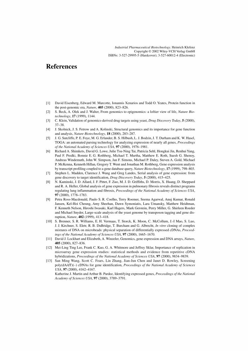
263References
References
[1] David Eisenberg, Edward M. Marcotte, Ionannis Xenarios and Todd O. Yeates, Protein function inthe post-genomic era, Nature, 405 (2000), 823–826.
[2] S. Beck, A. Olek and J. Walter, From genomics to epigenomics: a loftier view of life, Nature Bio-technology, 17 (1999), 1144.
[3] C. Klein, Validation of genomics-derived drug targets using yeast, Drug Discovery Today, 5 (2000),37–38.
[4] J. Skolnick, J. S. Fetrow and A. Kolinski, Structural genomics and its importance for gene functionand analysis, Nature Biotechnology, 18 (2000), 283–287.
[5] J. G. Sutcliffe, P. E. Foye, M. G. Erlander, B. S. Hilbush, L. J. Bodzin, J. T. Durham and K. W. Hasel,TOGA: an automated parsing technology for analyzing expression of nearly all genes, Proceedingsof the National Academy of Sciences USA, 97 (2000), 1976–1981.
[6] Richard A. Shimkets, David G. Lowe, Julie Tsu-Ning Tai, Patricia Sehl, Hongkui Jin, Renhui Yang,Paul F. Predki, Bonnie E. G. Rothberg, Michael T. Murtha, Matthew E. Roth, Suesh G. Shenoy,Andreas Windemuth, John W. Simpson, Jan F. Simons, Michael P. Daley, Steven A. Gold, MichaelP. McKenna, Kenneth Hillan, Gregory T. Went and Jonathan M. Rothberg, Gene expression analysisby transcript profiling coupled to a gene database query, Nature Biotechnology, 17 (1999), 798–803.
[7] Stephen L. Madden, Clarence J. Wang and Greg Landes, Serial analysis of gene expression: fromgene discovery to target identification, Drug Discovery Today, 5 (2000), 415–425.
[8] N. Kaminski, J. D. Allard, J. F. Pittet, F. Zuo, M. J. D. Griffiths, D. Morris, X. Huang, D. Sheppardand R. A. Heller, Global analysis of gene expression in pulmonary fibrosis reveals distinct programsregulating lung inflammation and fibrosis, Proceedings of the National Academy of Sciences USA,97 (2000), 1778–1783.
[9] Petra Ross-Macdonald, Paulo S. R. Coelho, Terry Roemer, Seema Agarwal, Anuj Kumar, RonaldJansen, Kel-Hoi Cheung, Amy Sheehan, Dawn Symoniatis, Lara Umansky, Matthew Heidtman,F. Kenneth Nelson, Hiroshi Iwasaki, Karl Hagers, Mark Gerstein, Perry Miller, G. Shirleen Roederand Michael Snyder, Large-scale analysis of the yeast genome by transposon tagging and gene dis-ruption, Nature, 402 (1999), 413–418.
[10] S. Brenner, S. R. Williams, E. H. Vermaas, T. Storck, K. Moon, C. McCollum, J.-I Mao, S. Luo,J. J. Kirchner, S. Eletr, R. B. DuBridge, T. Burcham and G. Albrecht, In vitro cloning of complexmixtures of DNA on microbeads: physical separation of differentially expressed cDNAs, Proceed-ings of the National Academy of Sciences USA, 97 (2000), 1665–1670.
[11] David J. Lockhart and Elizabeth, A. Winzeler, Genomics, gene expression and DNA arrays, Nature,405 (2000), 827–836.
[12] Mei-Ling Ting Lee, Frank C. Kuo, G. A. Whitmore and Jeffrey Sklar, Importance of replication inmicroarray gene expression studies: statistical methods and evidence from repetitive cDNAhybridizations, Proceedings of the National Academy of Sciences USA, 97 (2000), 9834–9839.
[13] San Ming Wang, Scott C. Fears, Lin Zhang, Jian-Jun Chen and Janet D. Rowley, Screeningpoly(dA/dT)(–) cDNAs for gene identification, Proceedings of the National Academy of SciencesUSA, 97 (2000), 4162–4167.Katherine J. Martin and Arthur B. Pardee, Identifying expressed genes, Proceedings of the NationalAcademy of Sciences USA, 97 (2000), 3789–3791.
Industrial Pharmaceutical Biotechnology. Heinrich KlefenzCopyright © 2002 Wiley-VCH Verlag GmbH
ISBNs: 3-527-29995-5 (Hardcover); 3-527-60012-4 (Electronic)
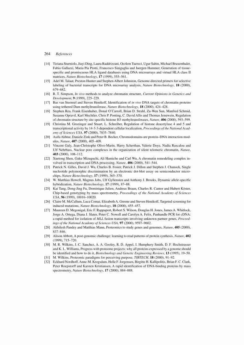
264 References
[14] Tiziana Sturniolo, Jiayi Ding, Laura Raddrizzani, Oezlem Tuereci, Ugur Sahin, Michael Braxenthaler,Fabio Gallazzi, Maria Pia Protti, Francesco Sinigaglia and Juergen Hammer, Generation of tissue-specific and promiscuous HLA ligand databases using DNA microarrays and virtual HLA class IImatrices, Nature Biotechnology, 17 (1999), 555–561.
[15] Adel M. Talaat, Preston Hunter and Stephen Albert Johnston, Genome-directed primers for selectivelabeling of bacterial transcripts for DNA microarray analysis, Nature Biotechnology, 18 (2000),679–682.
[16] R. T. Simpson, In vivo methods to analyze chromatin structure, Current Opinions in Genetics andDevelopment, 9 (1999), 225–229.
[17] Bas van Steensel and Steven Henikoff, Identification of in vivo DNA targets of chromatin proteinsusing tethered Dam methyltransferase, Nature Biotechnology, 18 (2000), 424–428.
[18] Stephen Rea, Frank Eisenhaber, Donal O’Carroll, Brian D. Strahl, Zu-Wen Sun, Manfred Schmid,Susanne Opravil, Karl Mechtler, Chris P. Ponting, C. David Allis and Thomas Jenuwein, Regulationof chromatin structure by site-specific histone H3 methyltransferases, Nature, 406 (2000), 593–599.
[19] Christina M. Grozinger and Stuart, L. Schreiber, Regulation of histone deacetylase 4 and 5 andtranscriptional activity by 14-3-3-dependent cellular localization, Proceedings of the National Acad-emy of Sciences USA, 97 (2000), 7835–7840.
[20] Asifa Akhtar, Daniele Zink and Peter B. Becker, Chromodomains are protein–DNA interaction mod-ules, Nature, 407 (2000), 405–409.
[21] Vincent Galy, Jean-Christophe Olivo-Marin, Harry Scherthan, Valerie Doye, Nadia Rascalou andUlf Nehrbass, Nuclear pore complexes in the organization of silent telomeric chromatin, Nature,403 (2000), 108–112.
[22] Xuetong Shen, Gaku Mizuguchi, Ali Hamiche and Carl Wu, A chromatin remodeling complex in-volved in transcription and DNA processing, Nature, 406 (2000), 541–544.
[23] Patrick N. Gilles, David J. Wu, Charles B. Foster, Patrick J. Dillon and Stephen J. Chanock, Singlenucleotide polymorphic discrimination by an electronic dot-blot assay on semiconductor micro-chips, Nature Biotechnology, 17 (1999), 365–370.
[24] W. Matthias Howell, Magnus Jobs, Ulf Gyllensten and Anthony J. Brooks, Dynamic allele-specifichybridization, Nature Biotechnology, 17 (1999), 87–88.
[25] Kai Tang, Dong-Jing Fu, Dominique Julien, Andreas Braun, Charles R. Cantor and Hubert Köster,Chip-based genotyping by mass spectrometry, Proceedings of the National Academy of SciencesUSA, 96 (1999), 10016–10020.
[26] Claire M. McCallum, Luca Comai, Elizabeth A. Greene and Steven Henikoff, Targeted screening forinduced mutations, Nature Biotechnology, 18 (2000), 455–457.
[27] Maureen D. Megonigal, Eric F. Rappaport, Robert S. Wilson, Douglas H. Jones, James A. Whitlock,Jorge A. Ortega, Diana J. Slater, Peter C. Nowell and Carolyn A. Felix, Panhandle PCR for cDNA:a rapid method for isolation of MLL fusion transcripts involving unknown partner genes, Proceed-ings of the National Academy of Sciences USA, 97 (2000), 9597–9602.
[28] Akhilesh Pandey and Matthias Mann, Proteomics to study genes and genomes, Nature, 405 (2000),837–846.
[29] Alison Abbott, A post-genomic challenge: learning to read patterns of protein synthesis, Nature, 402(1999), 715–720.
[30] M. R. Wilkins, J.-C. Sanchez, A. A. Gooley, R. D. Appel, I. Humphery-Smith, D. F. Hochstrasserand K. L. Williams, Progress with proteome projects: why all proteins expressed by a genome shouldbe identified and how to do it, Biotechnology and Genetic Engineering Reviews, 13 (1995), 19–50.
[31] M. Wilkins, Proteomic paradigms for perceiving purpose, TIBTECH, 18 (2000), 91–92.[32] Eckhard Nordhoff, Anne-M. Krogsdam, Helle F. Jorgensen, Birgitte H. Kallipolitis, Brian F. C. Clark,
Peter Roepstorff and Karsten Kristiansen, A rapid identification of DNA-binding proteins by massspectrometry, Nature Biotechnology, 17 (2000), 884–888.
265References
[33] G. Marius Clore, Accurate and rapid docking of protein–protein complexes on the basis of inter-molecular nuclear Overhauser enhancement data and dipolar couplings by rigid body minimization,Proceedings of the National Academy of Sciences USA, 97 (2000), 9021–9025.
[34] Steven P. Gygi, Garry L. Corthals, Yanni Zhang, Yvan Rochon and Ruedi Aebersold, Evaluation oftwo-dimensional gel electrophoresis-based proteome analysis technology, Proceedings of the Na-tional Academy of Sciences USA, 97 (2000), 9390–9395.
[35] M. J. Grossel, H. Wang, B. Gadea, W. Yeung and P. H. Hinds, A yeast two-hybrid system for discern-ing differential interactions using multiple baits, Nature Bio-technology, 17 (1999), 1232–1233.
[36] Stephen McCraith, Ted Holtzman, Bernard Moss and Stanley Fields, Genome-wide analysis of vac-cinia virus protein–protein interactions, Proceedings of the National Academy of Sciences USA, 97(2000), 4879–4884.
[37] Fang Liu, Qi Wan, Zdenek B. Pristupa, Xian-Min Yu, Yu Tian Wang and Hyman B. Niznik, Directprotein–protein coupling enables cross-talk between dopamine D5 and γ-aminobutyric acid Areceptors, Nature, 403 (2000), 274–280.
[38] Steven P. Gygi, Beate Rist, Scott A. Gerber, Frantisek Turecek, Michael H. Gelb and Ruedi Aebersold,A quantitative analysis of complex protein mixtures using isotope-coded affinity tags, Nature Bio-technology, 17 (1999), 994–999.
[39] Ruud M. T. de Wildt, Chris R. Mundy, Barbara D. Gorick and Ian M. Tomlinson, Antibody arrays forhigh-throughput screening of antibody–antigen interactions, Nature Biotechnology, 18 (2000),989−994.
[40] Alia Qureshi Emili and Gerard Cagney, Large-scale functional analysis using peptide or proteinarrays, Nature Biotechnology, 18 (2000), 393–397.
[41] Gavin MacBeath and Stuart L. Schreiber, Printing proteins as microarrays for high-throughput func-tion determination, Science, 289 (2000), 1760–1763.
[42] T. A. Craig, L. M. Breson, A. J. Tomlinson, T. D. Veenstra, S. Naylor and R. Kumar, Analysis oftranscription complexes and effects of ligands by microelectrospray ionization mass spectrometry,Nature Biotechnology, 17 (1999), 1214–1218.
[43] Alfred Maelicke, Proteomics, Nachrichten aus Chemie, Technik und Laboratorium, 47 (1999),1433−1435.
[44] S. Müllner, Eine Bewertung des Technologie- und Marktpotentials für die Life-Science-Industrie,transkript, 8 (5) (1999), 40–44.Georgius Amexis, Paul Oeth, Kenneth Abel, Anna Ivashina, François Pelloquin, Charles R. Cantor,Andreas Brau, Konstantin Chumakow, Quantitative Mutant Analysis of Viral Quasispecies by Chip-Based Matrix-Assisted Laser Desorption/Ionization Time-of-Flight Mass Spectrometry, Proccedingsof the National Academy of Sciences USA, 98 (2001), 12097–12102.
[45] T. Ito, K. Tashiro, S. Muta, R. Ozawa, T. Chiba, M. Ishizawa, K. Yamamoto, S. Kuhara and Y. Sakaki,Toward a protein–protein interaction map of the budding yeast: a comprehensive system to examinetwo-hybrid interactions in all possible combinations between the yeast proteins, Proceedings of theNational Academy of Sciences USA, 97 (2000), 1143–1147.
[46] Peter Uetz, Loic Giot, Gerard Cagney, Traci A. Mansfield, Richard S. Judson, James R. Knight,Daniel Lockshon, Vaibhav Narayan, Maithreyn Srinivasan, Pascale Pochart, Alia Qureshi-Emili,Ying Li, Brian Godwin, Diana Conover, Theodore Kalbfleisch, Govindan Vijayadamodar, MeijiaYang, Mark Johnston, Stanley Fields, Jonathan M. Rothberg, A comprehensive analysis of protein–protein interactions in Saccharomyces cerevisiae, Nature, 403 (2000), 623–627.Benno Schwikowski, Peter Uetz, Stanley Fields, A network of protein-protein interactions in yeast,Nature Biotechnology, 18 (2000), 1257–1261.
[47] J. Keith Joung, Elizabeth I. Ramm and Carl O. Pabo, A bacterial two-hybrid selection system forstudying protein–DNA and protein–protein interactions, Proceedings of the National Academy ofSciences USA, 97 (2000), 7382–7387.
266 References
[48] Carl T. Rollins, Victor M. Rivera, Derek N. Woolfson, Terence Keenan, Marcos Hatada, SusanE. Adams, Lawrence J. Andrade, David Yaeger, Marie Rose van Schravendijk, Dennis A. Holt, MichaelGilman and Tim Clackson, A ligand-reversible dimerization system for controlling protein–proteininteractions, Proceedings of the National Academy of Sciences USA, 97 (2000), 7096–7101.
[49] L. Li, R. W. Garde and J. V. Sweedler, Single-cell MALDI: a new tool for direct peptide profiling,TIBTECH, 18 (2000), 151–160.
[50] F. Oesterhelt, D. Oesterhelt, M. Pfeiffer, A. Engel, H. E. Traub and D. J. Müller, Unfolding pathwaysof individual bacteriorhodopsins, Science, 288 (2000), 143–146.J. G. Forbes and G. H. Lorimer, Unraveling a membrane protein, Science, 288 (2000), 63–64.
[51] D. Baker, A surprising simplicity to protein folding, Nature, 405 (2000), 39–42.[52] G. Milligan, Receptors as kissing cousins, Science, 288 (2000), 65–67.[53] M. Rocheville, D. C. Lange, U. Kumar, S. C. Patel, R. C. Patel and Y. C. Patel, Receptors for dopamine
and somatostatin: formation of hetero-oligomers with enhanced functional activity, Science, 288(2000), 154–157.
[54] P.-A. Binz, M. Müller, D. Walther, W. V. Bienvenut, R. Gras, C. Hoogland, G. Bouchet, E. Gasteiger,R. Fabretti, S. Gay, P. Palagi, M. R. Wilkins, V. Rouge, L. Tonella, S. Paesano, G. Rossellat, A.Karmime, A. Bairoch, J.-C. Sanchez, R. D. Appel and D. F. Hochstrasser, A molecular scanner toautomate proteomic research and to display proteome images, Analytical Chemistry, 71 (1999),4981–4988.
[55] J. Rappsilber, S. Siniossoglou, E. C. Hurt and M. Mann, A generic strategy to analyze the spatialorganization of multi-protein complexes by cross-linking and mass spectro-metry, Analytical Chem-istry, 72 (2000), 267–275.
[56] E. Saxon and C. R. Bertozzi, Cell surface engineering by a modified Staudinger reaction, Science,287 (2000), 2007–2010.
[57] B. D. Stahl and C. D. Allis, The language of covalent histone modifications, Nature, 403 (2000),41−45.
[58] V. Galy, J.-C. Olivo-Marin, H. Scherthan, V. Doye, N. Rascalou and U. Nehrbass, Nuclear porecomplexes in the organization of silent telomeric chromatin, Nature, 403 (2000), 108–112.
[59] Ulrich Schubert, Luis C. Anton, James Gibbs, Christopher C. Norbury, Jonathan W. Yewdell andJack R. Bennink, Rapid degradation of a large fraction of newly synthesized proteins by proteasomes,Nature, 404 (2000), 770–774.
[60] Sangeet Singh-Gasson, Roland D. Green, Yongjian Yue, Clark Nelson, Fred Blattner, MichaelR. Sussman and Franco Cerrina, Maskless fabrication of light-directed oligo-nucleotide microarraysusing a digital micromirror array, Nature Biotechnology, 17 (1999), 974–978.
[61] R. C. Merckle, Biotechnology as a route to nanotechnology, TIBTECH, 17 (1999), 271–274.[62] M. A. Unger, H.-P. Chou, T. Thorsen, A. Scherer and S. R. Quake, Monolithic microfabricated valves
and pumps by multilayer soft lithography, Science, 288 (2000), 113–116.[63] Dawn E. Kataoka and Sandra M. Troian, Patterning liquid flow on the microscopic scale, Nature,
402 (1999), 794–797.[64] Douglas B. Chrisey, The power of direct writing, Science, 289 (2000), 797–881.[65] Seunghun Hong and Chad A. Mirkin, A nanoplotter with both parallel and serial writing capabilities,
Science, 288 (2000), 1808–1811.[66] Gongwen Peng, Feng Qiu, Valeriy V. Ginzburg, David Jasnow and Anna C. Balazs, Forming
supramolecular networks from nanoscale rods in binary, phase-separating mixtures, Science, 288(2000), 1802–1804.
[67] David H. Gracias, Joe Tien, Tricia L. Breen, Carey Hsu and George M. Whitesides, Forming electri-cal networks in three dimensions by self-assembly, Science, 289 (2000), 1170–1172.
[68] Mei Li, Heimo Schnablegger and Stephen Mann, Coupled synthesis and self-assembly of nanoparticlesto give structures with controlled organization, Nature, 402 (1999), 393–395.
267References
[69] Tadashi Okamoto, Tomohiro Suzuki and Nobuko Yamamoto, Microarray fabrication with covalentattachment of DNA using bubble jet technology, Nature Biotechnology, 18 (2000), 438–441.
[70] Edwin W. H. Jager, Olle Inganäds and Ingemar Lundström, Microrobots for micro-meter-size ob-jects in aqueous media: potential tools for single-cell manipulation, Science, 288 (2000), 2335−2338.
[71] Michael J. B. Krieger, Jean-Bernard Billeter and Laurent Keller, Ant-like task allocation and recruit-ment in cooperative robots, Nature, 406 (2000), 992–995.
[72] Hod Lipson and Jordan B. Pollack, Automatic design and manufacture of robotic lifeforms, Nature,406 (2000), 974–978.Rodnay Brooks, From robot dreams to reality, Nature, 406 (2000), 945–947.
[73] Michael Knoblauch, Julian M. Hibberd, John C. Gray and Aart J. E. van Bel, A galinstan expansionfemtosysringe for micro-injection of eukaryotic organelles and prokaryotes, Nature Biotechnology,17 (1999), 906–909.
[74] Brian M. Cullum and Tuan Vo-Dinh, The development of optical nanosensors for biological meas-urements, TIBTECH, 18 (2000), 388–393.
[75] Nily Dan, Synthesis of hierarchical materials, TIBTECH, 18 (2000), 370–374.[76] John Cumings and A. Zettl, Low-friction nanoscale linear bearing realized from multiwall carbon
nanotubes, Science, 289 (2000), 602–604.[77] Thomas Rueckes, Kyoungha Kim, Ernesto Joselevich, Greg Y. Tseng, Chin-Li Cheung and Charles
M. Lieber, Carbon nanotube-based nonvolatile random access memory for molecular computing,Science, 289 (2000), 94–97.
[78] Rolf Schuster, Viola Kirchner, Philippe Allongue and Gerhard Ertl, Electro-chemical micromachining,Science, 289 (2000), 98–101.
[79] Bernard Yurke, Andrew J. Turberfield, Allen P. Mills, Jr, Friedrich C. Simmel and Jennifer L. Neumann,A DNA-fuelled molecular machine made of DNA, Nature, 406 (2000), 605–608.
[80] R. C. Hayward, D. A. Saville and I. A. Aksay, Electrophoretic assembly of colloidal crystals withoptically tunable micro-patterns, Nature, 404 (2000), 56–59.
[81] Hongyou Fan, Yunfeng Lu, Aaron Stump, Scott T. Reed, Tom Baer, Randy Schunk, Victor Perez-Luna, Gabriel P. Lopez and C. Jeffrey Brinker, Rapid prototyping of patterned functional nano-structures, Nature, 405 (2000), 56–60.
[82] Sandra R. Whaley, D. S. English, Evelyn L. Hu, Paul F. Barbara and Angela M. Belcher, Selection ofpeptides with semiconductor binding specificity for directed nanocrystal assembly, Nature, 405 (2000),665–668.Chad A. Mirkin and T. Andrew Taton, Semiconductors meet biology, Nature, 405 (2000), 626–627.
[83] G. P. Lopinski, D. D. M. Wayner and R. A. Wolkow, Self-directed growth of molecular nanostructureson silicon, Nature, 406 (2000), 48–51.
[84] Nae-Lih Wu, Sze-Yen Wang and I. A. Rusakova, Inhibition of crystallite growth in the sol–gel syn-thesis of nanocrystalline metal oxides, Science, 285 (1999), 1375–1377.
[85] David J. Beebe, Jeffrey S. Moore, Joseph M. Bauer, Qing Yu, Robin H. Liu, Chelladurai Devadossand Byung-Ho Jo, Functional hydrogel structures for autonomous flow control inside micro-fluidicchannels, Nature, 404 (2000), 588–590.
[86] J. Fritz, M. K. Baller, H. P. Lang, H. Rothuizen, P. Vettiger, E. Meyer, H.-J. Güntherodt, Ch. Gerberand J. K. Gimzewski, Translating biomolecular recognition into nanomechanics, Science, 288 (2000),316–318.
[87] Veronica Bermudez, Nathalie Capron, Torsten Gase, Francesco G. Gatti, Francois Kaijzar, David A.Leigh, Francesco Zerbetto and Songwei Zhang, Influencing intramolecular motion with an alternat-ing electric field, Nature, 406 (2000), 608–611.
[88] James C. Weaver, Timothy E. Vaughan and R. Dean Astumian, Biological sensing of small fielddifferences by magnetically sensitive chemical reactions, Nature, 405 (2000), 707–709.
[89] N. C. Seeman, DNA engineering and its application to nanotechnology, TIBTECH, 17 (1999),437−443.
268 References
[90] D. Faulhammer, A. R. Cukras, R. J. Lipton and L. F. Landweber, Molecular computation: RNA so-lutions to chess problems, Proceedings of the National Academy of Sciences USA, 97 (2000),1385−1389.
[91] J. Chen and D. H. Wood, Computation with biomolecules, Proceedings of the National Academy ofSciences USA, 97 (2000), 1328–1330.
[92] K. Sakamoto, H. Gouzu, K. Komiya, D. Kiga, S. Yokoyama, T. Yokomori and M. Hagiya, Molecularcomputation by DNA hairpin formation, Science, 288 (2000), 1223–1226.
[93] Qinghua Liu, Liman Wang, Anthony G. Frutos, Anne E. Condon, Robert M. Corn and Lloyd M. Smith,DNA computing on surfaces, Nature, 403 (2000), 175–179.
[94] Mitsunori Ogihara and Animesh Ray, DNA computing on a chip, Nature, 403 (2000), 143–144.[95] Robert P. Lanza, Jose B. Cibelli and Michael D. West, Human therapeutic cloning, Nature Medicine,
5 (1999), 975–977.A. W. S. Chan, T. Dominko, C. M. Luetjens, E. Neuber, C. Martinovich, L. Hewitson, C. R. Simerly,G. P. Schatten, Clonal Propagation of Primate Offspring by Embryo Splitting, Science, 287 (2000),317–321.
[96] L. R. Abeydeera et al., Development and Viability of Pig Oocytes Matured in A Protein-Free Me-dium Containing Epidermal Growth Factor, Theriogenology, 54 (5) (2000), 787–797.Randall S. Prather, Pigs is pigs, Science, 289 (2000), 1886–1887.
[97] Mindy Tsai, Jochen Wedemeyer, Soula Ganiatsas, See-Ying Tam, Leonard I. Zon and Stephen J. Galli,In vivo immunological function of mast cells derived from embryonic stem cells: an approach forthe rapid analysis of even embryonic lethal mutations in adult mice in vivo, Proceedings of theNational Academy of Sciences USA, 97 (2000), 9186–9190.
[98] Oliver Brüstle, Kimberly N. Jones, Randall D. Learish, Khaled Karram, Khalid Choudhary, OtmarD. Wiestler, Ian D. Duncan, Ronald D. G. McKay, Embryonic Stem Cell-Derived Glial Precursors:A Source of Myelinating Transplants, Science, 285 (1999), 754–756.
[99] Ingrid Wickelgren, Rat Spinal Cord Function Partially Restored, Science, 286 (1999), 1826–1827.[100] Henry M. Eppich, Russell Foxall, Kate Gaynor, David Dombkowski, Nobuyuki Miura, Tao Cheng,
Sandra Silva-Arrieta, Richard H. Evans, Joseph A. Mangano, Frederic I. Preffer and David T. Scadden,Pulsed electric fields for selection of hematopoietic cells and depletion of tumor cell contaminants,Nature Biotechnology, 18 (2000), 882–887.
[101] D. Perry, Patients’ voices: the powerful sound in the stem cell debate, Science, 287 (2000), 1423.J. M. W. Slack, Stem cells in epithelial tissues, Science, 287 (2000), 1431–1433.F. H. Gage, Mammalian neural stem cells, Science, 287 (2000), 1433–1438.D. van der Kooy and S. Weiss, Why stem cells, Science, 287 (2000), 1439–1441.P. J. Hines, B. A. Purnell and J. Marx, Stem cells branch out, Science, 287 (2000), 1417.
[102] Benjamin E. Reubinoff, Martin F. Pera, Chui-Yee Fong, Alan Trounson and Ariff Bongso, Embry-onic stem cell lines from human blastocysts: somatic differentiation in vitro, Nature Biotechnology,18 (2000), 399–404. Nature Genetics, 24 (2000), 372ff.
[103] Motonari Kondo, David C. Scherer, Toshihiro Miyamoto, Angela C. King, Koichi Akashi, KazuoSugamura and Irving L. Weissman, Cell-fate conversion of lymphoid-committed progenitors byinstructive actions of cytokines, Nature, 407 (2000), 383–386.
[104] Maite Lewin, Nadia Carlesso, Ching-Hsuan Tung, Xiao-Wu Tang, David Cory, David T. Scaddenand Ralph Weissleder, Tat peptide-derivatized magnetic nanoparticles allow in vivo tracking andrecovery of progenitor cells, Nature Biotechnology, 18 (2000), 410–414.
[105] T. Cheng, N. Rodrigues, H. Shen, Y.-G. Yang, D. Dombkowski, M. Sykes and D. T. Scadden,Hematopoietic stem cell quiescence maintained by p21(cip1/waf1), Science, 287 (2000), 1804–1808.
[106] E. Marshall, New genetic tricks to rejuvenate ailing livers, Science, 287 (2000), 1185–1186.[107] K. L. Rudolph, S. Chang, M. Millard, N. Schreiber-Agus and R. A. DePinho, Inhibition of experi-
mental liver cirrhosis in mice by telomerase gene delivery, Science, 287 (2000), 1253–1258.
269References
[108] Naoya Kobayashi, Toshiyoshi Fujiwara, Karen A. Westerman, Yusuke Inoue, Masakiyo Sakaguchi,Hirofumi Noguchi, Masahiro Miyazaki, Jin Cai, Noriaki Tanaka, Ira J. Fox, Philippe Leboulch,Prevention of Acute Liver Failure in Rats with Reversibly Immortalized Human Hepatocytes, Sci-ence, 287 (2000), 1258–1262.
[109] K. J. McCreath, J. Howcroft, K. H. S. Campbell, A. Colman, A. E. Schnieke and A. J. Kind, Produc-tion of gene-targeted sheep by nuclear transfer from cultured somatic cells, Nature, 405 (2000),1066–1069.
[110] Akira Onishi, Masaki Iwamoto, Tomiji Akita, Satoshi Mikawa, Kumiko Takeda, Takashi Awata,Hirohumi Hanada and Anthony C. F. Perry, Pig cloning by microinjection of fetal fibroblast nuclei,Science, 289 (2000), 1188–1190.
[111] C. Kubota, H. Yamakuchi, J. Todoroki, K. Mizoshita, N. Tabara, M. Barber and X. Yang, Six clonedcalves produced from adult fibroblast cells after long-term culture, Proceedings of the NationalAcademy of Sciences USA, 97 (2000), 990–995.
[112] Yoko Kato, Tetsuya Tani, Yusuke Sotomaru, Kazuo Kurokawa, Jun-ya Kato, Hiroshi Doguchi, HiroshiYasue and Yukio Tsunoda, Eight calves cloned from somatic cells of a single adult, Science, 282(1998), 2095–2098.
[113] Irina A. Polejaeva, Shu-Hung Chen, Todd D. Vaught, Raymond L. Page, Mullins, Suyapa Ball, YifanDai, Jeremy Boone, Shawn Walker, David L. Ayares, Alan Colman and Keith H. S. Campbell, Clonedpigs produced by nuclear transfer from adult somatic cells, Nature, 407 (2000), 86–90.
[114] Andy Coghlan, Cloning without embryos, New Scientist, 165 (No. 2223) (2000), 4.[115] Michael Thomas, Lianqing Yang and Peter J. Hornsby, Formation of functional tissue from trans-
planted adrenocortical cells expressing telomerase reverse transcriptase, Nature Biotechnology, 18(2000), 39–42.J. W. Shay and W. E. Wright, The use of telomerized cells for tissue engineering, Nature Biotechnol-ogy, 18 (2000), 22–23.Floyd E. Bloom, Breakthroughs 1999, Science, 286 (1999), 2267.Gretchen Vogel, Capturing the Promise of Youth, Science, 286 (1999), 22438–2239.
[116] Robert P. Lanza, Jose B. Cibelli and Michael D. West, Prospects for the use of nuclear transfer inhuman transplantation, Nature Biotechnology, 17 (1999), 1171–1174.
[117] I. L. Weissman, Translating stem and progenitor cell biology to the clinic: barriers and opportuni-ties, Science, 287 (2000), 1442–1446.
[118] D. Ferber, Growing human corneas in the lab, Science, 286 (1999), 2051–2052.[119] May Griffith, Rosemarie Osborne, Rejean Munger, Xiaojuan Xiong, Charles J. Doillon, Noelani
L. C. Laycock, Malik Hakim, Ying Song, Mitchell A. Watsky, Functional Human Corneal Equiva-lents Constructed from Cell Lines, Science, 286 (1999), 2169–2172.
[120] L. E. Niklason, J. Gao, W. M. Abbott, K. K. Hirschi, S. Houser, R. Marini and R. Langer, Functionalarteries grown in vitro, Science, 284 (1999), 489–493.
[121] Jeffrey S. Schechner, Anjali K. Nath, Lian Zheng, Martin S. Kluger, Christopher C. W. Hughes, M.Rocio Sierra-Honigmann, Marc I. Lorber, George Tellides, Michael Kashgarian, Alfred L. M. Bothwelland Jordan S. Pober, In vivo formation of complex micro-vessels lined by human endothelial cells inan immunodeficient mouse, Proceedings of the National Academy of Sciences USA, 97 (2000),9191−9196.
[122] Ioannis V. Yannas, Synthesis of organs: in vitro or in vivo?, Proceedings of the National Academy ofSciences USA, 97 (2000), 9354–9356.
[123] V. Tropepe, B. L. K. Coles, B. J. Chiasson, D. J. Horsford, A. J. Elia, R. R. McInnes and D. van derKooy, Retinal stem cells in the adult mammalian eye, Science, 287 (2000), 2032–2036.
[124] Sang-Hun Lee, Nadya Lumelsky, Lorenz Studer, Jonathan M. Auerbach and Ron D. McKay, Effi-cient generation of midbrain and hindbrain neurons from mouse embryonic stem cells, Nature Bio-technology, 18 (2000), 675–679.
270 References
[125] Diana L. Clarke, Clas B. Johansson, Johannes Wilbertz, Biborka Veress, Erik Nilsson, HelenaKarlström, Urban Lendahl and Jonas Frisen, Generalized potential of adult neural stem cells, Sci-ence, 288 (2000), 1660–1663.
[126] Sujata Kale, Sybil Biermann, Claire Edwards, Catherine Tarnowski, Michael Morris and MichaelWilliam Long, Three-dimensional cellular development is essential for ex vivo formation of humanbone, Nature Biotechnology, 18 (2000), 954–958.
[127] Herve Petite, Veronique Viateau, Wassila Bensaid, Alain Meunier, Cindy de Pollak, MarianneBourguignon, Karim Oudina, Laurent Sedel and Genevieve Guillemin, Tissue-engineered bone re-generation, Nature Biotechnology, 18 (2000), 959–963.
[128] Frank Oberpenning, Jun Meng, James J. Yoo and Anthony Atala, De novo reconstitution of a func-tional mammalian urinary bladder by tissue engineering, Nature Biotechnology, 17 (1999), 149−155.
[129] Susan Bonner-Weir, Monica Taneja, Gordon C. Weir, Krystyna Tatarkiewicz, Ki-Ho Song, ArunSharma and John J. O’Neill, In vitro cultivation of human islets from expanded ductal tissue, Pro-ceedings of the National Academy of Sciences USA, 97 (2000), 7999–8004.
[130] Gene C. Webb, Murtaza S. Akbar, Choongjian Zhao and Donald F. Steiner, Expression profiling ofpancreatic β-cells: glucose regulation of secretory and metabolic pathways, Proceedings of the Na-tional Academy of Sciences USA, 97 (2000), 5773–5778.
[131] Toshio Imaizumi, Karen L. Lankford, Willis V. Burton, William L. Fodor and Jeffery D. Kocsis,Xeno-transplantation of transgenic pig olfactory ensheathing cells promotes axonal regeneration inrat spinal cord, Nature Biotechnology, 18 (2000), 949–953.
[132] Toru Kondo and Martin Raff, Oligodendrocyte precursor cells reprogrammed to become multipotentialCNS stem cells, Science, 289 (2000), 1754–1757.
[133] Luc J. W. van der Laan, Christopher Lockey, Bradley C. Griffeth, Francine S. Frasier, Carolyn A.Wilson, David E. Onions, Bernard J. Hering, Zhifeng Long, Edward Otto, Bruce E. Torbett andDaniel R. Salomon, Infection by porcine endogenous retrovirus after islet xenotransplantation inSCID mice, Nature, 207 (2000), 90–94.
[134] R. P. Lanza, J. B. Cibelli and M. D. West, Prospects for the use of nuclear transfer in human trans-plantation, Nature Biotechnology, 17 (1999), 1171–1174.
[135] Megan J. Munsie, Anna E. Michalska, Carmel M. O’Brien, Alan O. Trounson, Martin F. Perta andPeter S. Mountford, Isolation of pluripotent embryonic stem cells from reprogrammed adult mousesomatic cell nuclei, Current Biology, 10 (2000), 989–992.
[136] Michael Thomas, Lianqing Yang and Peter J. Hornsby, Formation of functional tissue from trans-planted adrenocortical cells expressing telomerase reverse transcriptase, Nature Biotechnology, 18(2000), 39–42.
[137] Jerry W. Shay and Woodring E. Wright, The use of telomerized cells for tissue engineering, NatureBiotechnology, 18 (2000), 22–23.
[138] W. Mark Saltzman, Delivering tissue regeneration, Nature Biotechnology, 17 (1999), 534–535.[139] Lonnie D. Shea, Elizabeth Smiley, Jeffrey Bonadio and David J. Mooney, DNA delivery from poly-
mer matrices for tissue engineering, Nature Biotechnology, 17 (1999), 551–554.[140] Mark C. Poznansky, Richard H. Evans, Russell B. Foxall, Ivona T. Olszak, Anita H. Piascik, Kelly
E. Hartman, Christian Brander, Thomas H. Meyer, Mark J. Pykett, Karissa T. Chabner, Spyros A.Kalams, Michael Rosenzweig and David T. Scadden, Efficient generation of human T cells from atissue-engineered thymic organoid, Nature Biotechnology, 18 (2000), 729–734.
[141] T. Lebestky, T. Chang, V. Hartenstein and U. Banerjee, Specification of Drosophila hematopoieticlineage by conserved transcription factors, Science, 288 (2000), 146–149.
[142] A. Colman and A. Kind, Therapeutic cloning: concepts and practicalities, TIBTECH, 18 (2000),192–196.
[143] I. Cerina, Biochips: from science to business, transkript, 3 (6) (2000), 30–32.[144] J. N. Cha, G. D. Stucky, D. E. Morse and T. J. Deming, Biomimetic synthesis of ordered silica struc-
tures mediated by block copolypeptides, Nature, 403 (2000), 289–292.
271References
[145] A. Katz and M. E. Davis, Molecular imprinting of bulk, micro-porous silica, Nature, 403 (2000),286–289.
[146] S. Schultz, D. R. Smith, J. J. Mock and D. A. Schultz, Single-target molecule detection withnonbleaching multicolor optical immunolabels, Proceedings of the National Academy of SciencesUSA, 97 (2000), 996–1001.
[147] Tuan Vo-Dinh, Jean-Pierre Alarie, Brian M. Cullum and Guy D. Griffin, Antibody-based nanoprobefor measurement of a fluorescent analyte in a single cell, Nature Biotechnology, 218 (2000), 764−767.
[148] T. Andrew Taton, Chad A. Mirkin and Robert L. Letsinger, Scanometric DNA array detection withnanoparticle probes, Science, 289 (2000), 1757–1760.
[149] J. Hodgson, Crystal gazing the new biotechnologies, Nature Biotechnology, 18 (2000), 29–31.[150] T. Peakman and Y. Bonduelle, Steering a course through the technology maze, Drug Discovery
Today, 5 (2000), 337–343.[151] J. Miller, Navigating the technology maze, Drug Discovery Today, 5 (2000), 265–266.[152] C. Gubser and P. Hiscocks, How much of a threat is virtual pharma? Scrip Magazine, (November
1999), 41–42.[153] T. G. Klopack, Strategic outsourcing: balancing the risks and the benefits, Drug Discovery Today, 5
(2000), 157–160.[154] C. Sander, Genomic medicine and the future of health care, Science, 287 (2000), 1977–1978.[155] M. Xu, G. Qiu, Z. Jiang, E. von Hofe and R. E. Humphreys, Genetic modulation of tumor antigen
presentation, TIBTECH, 18 (2000), 167–172.[156] Ramon Alemany, Cristina Balague and David T. Curiel, Replicative adenoviruses for cancer therapy,
Nature Biotechnology, 18 (2000), 723–727.[157] Jason C. Schense, Jecelyne Bloch, Patrick Aebischer and Jeffrey A. Hubbell, Enzymatic incorpora-
tion of bioactive peptides into fibrin matrices enhances neurite extension, Nature Biotechnology, 18(2000), 415–419.
[158] A. Mountain, Gene therapy: the first decade, TIBTECH, 18 (2000), 119–128.[159] G. Thurston, C. Suri, K. Smith, J. McClain, T. Sato, G. D. Yancopoulos and D. M. McDonald, Leak-
age-resistant blood vessels in mice transgenically overexpressing angiopoietin-1, Science, 286 (1999),2511–2514.
[160] R. Draghia-Akli, M. L. Fiorotto, L. A. Hill, P. B. Malone, D. R. Deaver and R. J. Schwartz, Myo-genic expression of an injectable protease-resistant growth hormone-releasing hormone augmentslong-term growth in pigs, Nature Biotechnology, 17 (1999), 1179–1183.
[161] K. M. L. Gaensler, G. Tu, S. Bruch, D. Liggitt, G. S. Lipshutz, A. Metkus, M. Harrison, T. D. Heathand R. J. Debs, Fetal gene transfer by transuterine injection of cationic liposome–DNA complexes,Nature Biotechnology, 17 (1999), 1188–1192.
[162] Ningya Shi and William M. Pardridge, Noninvasive gene targeting to the brain, Proceedings of theNational Academy of Sciences USA, 97 (2000), 7567–7572.
[163] Toshihiro Takenaka, Gary J. Murray, Gangjian Qin, Jane M. Quirk, Toshio Ohshima, Pankaj Qasba,Kelly Clark, Ashok B. Kulkarni, Roscoe O. Brady and Jeffry A. Medin, Long-term enzyme correc-tion and lipid reduction in multiple organs of primary and secondary transplanted Fabry mice receiv-ing transduced bone marrow, Proceedings of the National Academy of Sciences USA, 97 (2000),7515–7520.
[164] Paul Blezinger, Jijun Wang, Margaret Gondo, Abraham Quezada, Dorothy Mehrens, Martha French,Arun Singhal, Sean Sullivan, Alain Rolland, Rob Ralston and Wang Min, Systemic inhibition oftumor growth and tumor metastases by intramuscular administration of the endostatin gene, NatureBiotechnology, 17 (2000), 343–348.
[165] W. R. A. Brown, P. J. Mee and M. H. Shen, Artificial chromosomes: ideal vectors?, TIBTECH, 18(2000), 218–223.
272 References
[166] Chad, Stefao Rivella, John Callegari, Glenn Heller, Karen M. L. Gaensler, Lucio Luzzatto and MichelSadelain, Therapeutic haemoglobin synthesis in β-thalassemic mice expressing lentivirus-encodedhuman β-globin, Nature, 406 (2000), 82–86.
[167] Christian P. Kalberer, Robert Pawliuk, Suzan Imren, Thomas Bachelot, Ken J. Takekoshi, MaryFabry, Connie J. Eaves, Irving M. London, R. Keith Humphries and Philippe Leboulch, Preselectionof retrovirally transduced bone marrow avoids subsequent stem cell gene silencing and age-depend-ent extinction of expression of human β-globin in engrafted mice, Proceedings of the National Acad-emy of Sciences USA, 97 (2000), 5411–5415.
[168] David W. Emery, Evangelia Yannaki, Julie Tubb and George Stamatoyannopoulos, A chromatininsulator protects retrovirus vectors from chromosomal position effects, Proceedings of the NationalAcademy of Sciences USA, 97 (2000), 9150–9155.
[169] Tsuyoshi Tanabe, Tomoko Kuwabara, Masaki Warashina, Kenzaburo Tani, Kazunari Taira andShigetaka Asano, Oncogene inactivation in a mouse model, Nature, 406 (2000), 473.
[170] Aubrey D. N. J. de Grey, Mitochondrial gene therapy: an arena for the biomedical use of inteins,TIBTECH, 18 (2000), 394–399.
[171] Huatao Guo, Michael Karberg, Meredith Long, J. P. Jones III, Bruce Sullenger and Alan M. Lambo-witz, Group II introns designed to insert into therapeutically relevant DNA target sites in humancells, Science, 289 (2000), 452–457.
[172] Vitali Alexeev, Olga Igoucheva, Alla Domashenko, George Cotsarelis and Kyonggeun Yoon, Local-ized in vivo genotypic and phenotypic correction of the albino mutation in skin by RNA–DNAoligonucleotide, Nature Biotechnology, 18 (2000), 43.
[173] O. A. Kolesnikova, N. S. Entelis, H. Mireau, T. D. Fox, R. P. Martin and I. A. Tarassov, Suppressionof mutations in mitochondrial DNA by tRNAs imported from the cytoplasm, Science, 289 (2000),1931–1933.
[174] Jean-Baptiste Latouche and Michael Sadelain, Induction of human cytotoxic T lymphocytes by arti-ficial antigen-presenting cells, Nature Biotechnology, 18 (2000), 405–409.
[175] Michael A. Chattergoon, J. Joseph Kim, Joo-Sung Yang, Tara M. Robinson, Daniel J. Lee, TzveteDentchev, Darren M. Wilson, Velpandi Ayyavoo and David B. Weiner, Targeted antigen delivery toantigen-presenting cells including dendritic cells by engineered Fas-mediated apoptosis, Nature Bio-technology, 18 (2000), 974–979.
[176] Alevtina Domashenko, Sonya Gupta and George Cotsarelis, Efficient delivery of transgenes to hu-man hair follicle progenitor cells using topical lipoplex, Nature Biotechnology, 18 (2000), 420–423.
[177] Silvia M. Kreda, Raymond J. Pickles, Eduardo R. Lazarowski and Richard C. Boucher, G-protein-coupled receptors as targets for gene transfer vectors using natural small-molecule ligands, NatureBiotechnology, 18 (2000), 635–640.
[178] Richard J. Bartlett, Sabine Stockinger, Melvin M. Denis, William T. Bartlett, Luca Inverardi, T. T. Le,Nguyen thi Man, Glenn E. Morris, Daniel J. Bogan, Janet Metcalf-Bogan and Joe N. Kornegay, Invivo targeted repair of a point mutation in the canine dystrophin gene by a chimeric RNA/DNAoligonucleotide, Nature Biotechnology, 18 (2000), 615–622.
[179] Andrea W. Bledsoe, Cheryl A. Jackson, Sylvia McPherson and Casey D. Morrow, Cytokine produc-tion in motor neurons by poliovirus replicon vector gene delivery, Nature Biotechnology, 18 (2000),964–969.
[180] Yoshikazu Yonemitsu, Christopher Kitson, Stefano Ferrari, Raymond Farley, Uta Griesenbach, DianeJudd, Rachel Steel, Philippe Scheid, Jie Zhu, Peter K. Jeffery, Atsushi Kato, Mohammad K. Hasan,Yoshiyuki Nagai, Ichiro Masaki, Masayuki Fukumura, Mamoru Hasegawa, Duncan M. Geddes andEric W. F. W. Alton, Efficient gene transfer to airway epithelium using recombinant Sendai virus,Nature Biotechnology, 18 (2000), 970–973.
[181] Zahida Parveen, Anna Krupetsky, Martin Engel-Städter, Klaus Cichutek, Roger J. Pomerantz andRalph Dornburg, Spleen necrosis virus-derived C-type retroviral vectors for gene transfer to quies-cent cells, Nature Biotechnology, 18 (2000), 623–629.
273References
[182] Ziying Yan, Yulong Zhang, Dongsheng Duan and John F. Engelhardt, Trans-splicing vectors expandthe utility of adeno-associated virus for gene therapy, Proceedings of the National Academy of Sci-ences USA, 97 (2000), 6716–6721.
[183] Duncan Graham-Rowe, Mini motivators – Bionic nerves are battling muscle wasting after a stroke,New Scientist, 164 (No. 2216) (1999), 5.
[184] Ian Sample, Crystal fix – Biodegradable rods could help mend broken bones, New Scientist, 164(No. 2216) (1999), 17.
[185] T. T. Ton-that, D. Doron, B. S. Pollard, J. Bacher and H. B. Pollard, In vivo bypass of hemophilia Acoagulation defect by Factor XIIa implant, Nature Biotechnology, 18 (2000), 289–295.
[186] Robert F. Service, Scanners Get a Fix on Lab Animals, Science, 286 (1999), 2261–2263.[187] Cecilia H. Jiang, Joe Z. Tsien, Peter G. Schultz, Yinghe Hu, The effects of aging on gene expression
in the hypothalamus and cortex of mice, Proceedings of the National Academy of Sciences USA 98(2001), 1930–1934.
[188] Cheol-Koo Lee, Roger G. Klopp, Richard Weindruch and Thomas A. Prolla, Gene expression profileof aging and its retardation by caloric restriction, Science, 285 (1999), 1390–1393.
[189] A. S. Kamath-Loeb, E. Johansson, P. M. J. Burgers and L. A. Loeb, Functional interaction betweenthe Werner syndrome protein and DNA polymerase δ, Proceedings of the National Academy ofSciences USA, 97 (2000), 4603–4608.
[190] Joseph Landry, Ann Sutton, Stefan T. Tafrov, Ryan C. Heller, John Stebbins, Lorraine Pillus and RolfSternglanz, The silencing protein SIR2 and its homologs are NAD-dependent protein deacetylases,Proceedings of the National Academy of Sciences USA, 97 (2000), 5807–5811.
[191] Deborah J. Burks, Jalme Font de Mora, Markus Schubert, Dominic J. Withers, Martin G. Myers,Heather H. Towery, Shari L. Altamuro, Carrie L. Flint and Morris F. White, IRS-2 pathways inte-grate female reproduction and energy homeostasis, Nature, 407 (2000), 377–382.
[192] Su-Ju Lin, Pierre-Antoine Defossez and Leonard Guarente, Requirement of NAD and SIR2 for life-span extension in Saccharomyces cerevisiae, Science, 289 (2000), 2126–2128.
[193] Jens C. Brüning, Dinesh Gautam, Deborah J. Burks, Jennifer Gillette, Markus Schubert, Paul C.Orban, Rüdiger Klein, Wilhelm Krone, Dirk Müller-Wieland and C. Ronald Kahn, Role of braininsulin receptor in control of body weight and reproduction, Science, 289 (2000), 2122–2125.
[194] Derek J. Symula, Kelly A. Frazer, Yukihiko Ueda, Patrice Denefle, Mary E. Stevens, Zhi-En Wang,Richard Locksley, Edward M. Rubin, Functional screening of an asthma QTL in YAC transgenicmice, Nature Genetics, 23 (1999), 241–244.
[195] Anne Frary, T. Clint Nesbitt, Amy Frary, Silvana Grandillo, Esther van der Knaap, Bin Cong, JipingLiu, Jaroslaw Meller, Ron Elber, Kevin B. Alpert and Steven D. Tanksley, fw2.2: a quantitative traitlocus key to the evolution of tomato fruit size, Science, 289 (2000), 85–88.
[196] John C. Clapham, Jonathan R. S. Arch, Helen Chapman, Andrea Haynes, Carolyn Lister, GaryB. T. Moore, Valerie Plercy, Sabrina A. Carter, Ines Lehner, Stephen A. Smith, Lee J. Beeley, RobertJ. Godden, Nicole Herrity, Mark Skehel, K. Kumar Changani, Paul D. Hockings, David G. Reid,Sarah M. Squires, Jonathan Hatcher, Brenda Trail, Judy Latcham, Sohalla Rastan, Alexander J. Harper,Susana Cadenas, Julie A. Buckingham, Martin D. Brand and Alejandro Abuin, Mice overexpressinghuman uncoupling protein-3 in skeletal muscle are hyperphagic and lean, Nature, 406 (2000), 415−418.
[197] Jo Whelan, Reversing age-related and diabetic cardiovascular disease, Drug Discovery Today, 5(2000), 272–273.
[198] Donald Metcalf, Christopher J. Greenhalgh, Elizabeth Viney, Tracy A. Wilson, Robyn Starr, NicosA. Nicola, Douglas J. Hilton and Warren S. Alexander, Gigantism in mice lacking suppressor ofcytokine signaling-2, Nature, 405 (2000), 1069–1073.
[199] Thomas M. Loftus, Donna E. Jaworsky, Gojeb L. Frehywot, Craig A. Townsend, Gabriele V. Ronnett,M. Daniel Lane and Francis P. Kuhajda, Reduced food intake and body weight in mice treated withfatty acid synthase inhibitors, Science, 288 (2000), 2379–2381.
274 References
[200] K. P. White, S. A. Rifkin, P. Hurban and D. S. Hogness, Microarray analysis of Drosophila develop-ment during metamorphosis, Science, 286 (1999), 2179–2184.
[201] D. H. Ly, D. J. Lockhart, R. A. Lerner and P. G. Schultz, Mitotic misregulation and human aging,Science, 287 (2000), 2486–2492.
[202] Martijn E. T. Dolle, Wendy K. Snyder, Jan A. Gossen, Paul H. M. Lohman and Jan Vijg, Distinctspectra of somatic mutations accumulated with age in mouse heart and small intestine, Proceedingsof the National Academy of Sciences USA, 97 (2000), 8403–8408.
[203] Bey-Dih Chang, Keiko Watanabe, Eugenia V. Broude, Jing Fang, Jason C. Poole, Tatiana V. Kalini-chenko and Igor B. Roninson, Effects of p21(Waf1/Cip1/Sdi1) on cellular gene expression: implica-tions for carcinogenesis, senescence, and age-related diseases, Proceedings of the National Acad-emy of Sciences USA, 97 (2000), 4291–4296.
[204] Shin-Ichiro Imai, Christopher M. Armstrong, Matt Kaeberlein and Leonard Guarente, Transcrip-tional silencing and longevity protein Sir2 is an NAD-dependent histone deacetylase, Nature, 403(2000), 795–800.
[205] John J. Wyrick, Frank C. P. Holstege, Ezra G. Jennings, Helen C. Causton, David Shore, MichaelGrunstein, Eric S. Lander and Richard A. Young, Chromosomal landscape of nucleosome-depend-ent gene expression and silencing in yeast, Nature, 402 (1999), 418–421.
[206] Itamar Simon, Toyoaki Tenzen, Benjamin E. Reubinoff, Dahlia Hillman, John R. McCarrey andHoward Cedar, Asynchronous replication of imprinted genes is established in the gametes and main-tained during development, Nature 401 (1999), 929–932.
[207] Mark Pearson, Roberta Carbone, Carla Sebastiani, Mario Cioce, Marta Fagioli, Shin’ichi Saito,Yuichiro Higashimoto, Ettore Appella, Saverio Minucci, Pier Paolo Pandolfi and Pier Guiseppe Pelicci,PML regulates p53 acetylation and premature senescence induced by oncogenic Ras, Nature, 406(2000), 207–210.
[208] Jing Wang, Gregory J. Hannon and David H. Beach, Risky immortalization by telomerase, Nature,405 (2000), 755–756.
[209] David Kipling and Harry Rubin, Telomere-dependent senescence [Correspondence], Nature Bio-technology, 17 (1999), 313–314.
[210] David W. Walker, Gawain McGoll, Nicole L. Jenkins, Jennifer Harris and Gordon J. Lithgow, Evo-lution of lifespan in C. elegans, Nature, 405 (2000), 296–297.
[211] Shawn Ahmed and Jonathan Hodgkin, MRT-2 checkpoint protein is required for germline immortal-ity and telomere replication in C. elegans, Nature, 403 (2000), 159–164.
[212] Robert P. Lanza, Jose B. Cibelli, Catherine Blackwell, Vincent J. Cristofalo, Mary Kay Francis,Gabriela M. Baerlocher, Jennifer Mak, Michael Schertzer, Elizabeth A. Chavez, Nancy Sawyer,Peter Lansdorp and Michael West, Extension of cell life-span and telomere length in animals clonedfrom senescent somatic cells, Science, 288 (2000), 665–669.
[213] Teruhiko Wakayama, Yoichi Shinkai, Kellie L. K. Tamashiro, Hiroyuki Niida, D. Caroline Blanchard,Robert J. Blanchard, Atsuo Ogura, Kentaro Tanemura, Makoto Tachibana, Anthony C. F. Perry, DianaF. Colgan, Peter Mombaerts and Ryuzo Yanagimachi, Cloning of mice to six generations, Nature,407 (2000), 318–319.
[214] Anne E. Pitts and David R. Corey, Inhibition of human telomerase by 2′-O-methyl-RNA, Proceed-ings of the National Academy of Sciences USA, 95 (1998), 11549–11554.
[215] Steven E. Artandi, Sandy Chang, Shwu-Luan Lee, Scott Alson, Geoffrey J. Gottlieb, Lynda Chin andRonald A. DePinho, Telomere dysfunction promotes non-reciprocal translocations and epithelialcancers in mice, Nature, 406 (2000), 641–645.
[216] Simon Melov, Joanne Ravenscroft, Sarwatt Malik, Matt S. Gill, David W. Walker, Peter E. Clayton,Douglas C. Wallace, Bernard Malfroy, Susan R. Doctrow and Gordon J. Lithgow, Extension of life-span with superoxide dismutase/catalase mimetics, Science, 289 (2000), 1567–1569.
275References
Structures of EUK-8 and EUK-134: K. Baker, C. Bucay Marcus, K. Huffman, H. Kruk, B. Malfroy,S. R. Doctrow, Synthetic combined superoxide dismutase/catalase mimics are protective as a de-layed treatment in a rat stroke model: a key role for reactive oxygen species in ischemic brain injury,Journal of Pharmacology and Experimental Therapeutics, 284 (1998), 215–221.Effects in mouse ALS model: www.eukarion.com
[217] Ash A. Alizadeh, Michael B. Eisen, R. Eric Davis, Chi Ma, Izidore S. Lossos, Andreas Rosenwald,Jennifer C. Boldrick, Hajeer Sabet, Truc Tan, Xib Yu, John I. Powell, Liming Yang, Gerald E. Marti,Troy Moore, James Hudson, Jr, Lishend Lu, David B. Lewis, Robert Tibshirani, Gavin Sherlock,Wing C. Chan, Timothy C. Greiner, Dennis D. Weisenburger, James O. Armitage, Roger Warnke,Ronald Levy, Wyndham Wilson, Michael R. Grever, John C. Byrd, David Botstein, Patrick O. Brownand Louis M. Staudt, Distinct types of diffuse large B-cell lymphoma identified by gene expressionprofiling, Nature, 403 (2000), 503–511.
[218] M. Bittner, P. Meltzer, Y. Chen, Y. Jiang, E. Seftor, M. Hendrix, M. Radmacher, R. Simo, Z. Yakhini,A. Ben-Dor, N. Sampas, E. Dougherty, E. Wang, F. Marincola, C. Gooden, J. Lueders, A. Glatfelter,P. Pollck, J. Carpten, E. Gillanders, D. Leja, K. Dietrich, C. Beaudry, M. Berens, D. Alberts, V.Sondak, N. Hayward and J. Trent, Molecular classification of cutaneous malignant melanoma bygene expression profiling, Nature, 406 (2000), 536–540.
[219] Brad St Croix, Carlo Rago, Victor Velculescu, Giovanni Traverso, Katherine E. Romans, ElizabethMontgomery, Anita Lal, Gregory J. Riggins, Christoph Lengauer, Bert Vogelstein and Kenneth W.Winzler, Genes expressed in human tumor endothelium, Science, 289 (2000), 1197–1202.
[220] Charles M. Perou, Therese Sorlie, Michael B. Eisen, Matt van de Rijn, Stefanie S. Jeffrey, ChristianA. Rees, Jonathan R. Pollack, Douglas T. Ross, Hilde Johnsen, Lars A. Akslen, Öystein Fluge, Alex-ander Pergamenschikov, Cheryl Williams, Shirley X. Zhu, Per E. Lönning, Anne-Lise Börresen-Dale, Patrick O. Brown and David Botstein, Molecular portraits of human breast tumors, Nature,406 (2000), 747–752.
[221] J. Drews, Drug discovery today – and tomorrow, Drug Discovery Today, 5 (2000), 2–4.[222] S. L. Schreiber, Target-oriented and diversity-oriented organic synthesis in drug discovery, Science,
287 (2000), 1964–1969.[223] Martin J. Valler and Darren Green, Diversity screening versus focussed screening in drug discovery,
Drug Discovery Today, 5 (2000), 286–293.[224] J. B. Gibbs, Mechanism-based target identification and drug discovery in cancer research, Science,
287 (2000), 1969–1973.[225] Thomas Schindler, William Bornmann, Patricia Pellicena, W. Todd Miller, Bayard Clarkson and
John Kuriyan, Structural mechanism for STI-571 inhibition of Abelson tyrosine kinase, Science,289 (2000), 1938–1942.
[226] Anthony C. Bishop, Jeffrey A. Ubersax, Dejah T. Petsch, Dina P. Matheos, Nathanael S. Gray, JustinBlethrow, Elji Shimizu, Joe Z. Tsien, Peter G. Schultz, Mark D. Rose, John L. Wood, David O. Morganand Kevan M. Shokat, A chemical switch for inhibitor-sensitive alleles of any protein kinase, Na-ture, 407 (2000), 395–401.
[227] William Shakespeare, Michael Yang, Regine Bohacek, Franklin Cerasoli, Karin Stebbins, RajiSundaramoorthi, Mihai Azimioara, Chi Vu, Selvi Pradeepan, Chester Metcalf, III, Chad Haraldson,Taylor Merry, David Dalgarno, Surinder Narula, Marcos Hatada, Xiaode Lu, Marie Rose vanSchravendijk, Susan Adams, Shelia Violette, Jeremy Smith, Wie Guan, Catherine Bartlett, Jay Herson,John Iuliucci, Manfred Weigele and Tomi Sawyer, Structure-based design of an osteoclast-selective,nonpeptide Src homology 2 inhibitor with in vivo antiresorptive activity, Proceedings of the Na-tional Academy of Sciences USA, 97 (2000), 9373–9378.
[228] Marcus D. Ballinger, Venkatakrishna Shyamala, Louise D. Forrest, Maja Deuter-Reinhard, LauraV. Doyle, Jian-xin Wang, Lootsee Panganiban-Lustan, Jennifer R. Stratton, Gerald Apell, Jill A. Winter,Michael V. Doyle, Steven Rosenberg and W. Michael Kavanaugh, Semirational design of a potent,artificial agonist of fibroblast growth factor receptors, Nature Biotechnology, 17 (1999), 1199.
276 References
[229] Atsuko Yoneda, Masahiro Asada, Yuko Oda, Masashi Suzuki and Toru Imamura, Engineering of anFGF–proteoglycan fusion protein with heparin-independent, mitogenic activity, Nature Biotechnol-ogy, 18 (2000), 641–644.
[230] Sang-Hyun Park and Ronald T. Raines, Genetic selection for dissociative inhibitors of designatedprotein–protein interactions, Nature Biotechnology, 18 (2000), 847–851.
[231] Zhihong Guo, Demin Zhou and Peter G. Schultz, Designing small-molecule switches for protein–protein interactions, Science, 288 (2000), 2042–2045.
[232] Anna M. Kapp, Aseem Z. Ansari, Mark Ptashne and Peter B. Dervan, Activation of gene expressionby small molecule transcription factors, Proceedings of the National Academy of Sciences USA, 97(2000), 3930–3935.
[233] Sakari Hietanen, Sonia Lain, Eberhard Krausz, Christine Blattner and David P. Lane, Activation ofp53 in cervical carcinoma cells by small molecules, Proceedings of the National Academy of Sci-ences USA, 97 (2000), 8501–8506.
[234] M. Faria, C. D. Wood, L. Perrouault, J. S. Nelson, A. Winter, M. R. H. White, C. Helene andC. Giovannangeli, Targeted inhibition of transcription elongation in cells mediated by triplex-form-ing oligonucleotides, Proceedings of the National Academy of Sciences USA, 97 (2000), 3862–3867.
[235] Daniel A. Erlanson, Andrew C. Braisted, Darren R. Raphael, Mike Randal, Robert M. Stroud, EricM. Gordon and James A. Wells, Site-directed ligand discovery, Proceedings of the National Acad-emy of Sciences USA, 97 (2000), 9367–9372.
[236] Rene F. Ketting, Ronald H. A. Plasterk, A genetic link between co-suppression and RNA interfer-ence in C. elegans, Nature, 404 (2000), 296–298.
[237] Jason R. Kennerdell and Richard W. Carthew, Heritable gene silencing in Drosophila using double-stranded RNA, Nature Biotechnology, 18 (2000), 896–898.
[238] James C. Clemens, Carolyn A. Worby, Nancy Simonson-Leff, Marco Muda, Tomohiko Maehama,Brian A. Hemmings and Jack E. Dickson, Use of double-stranded RNA interference in Drosophilacell lines to dissect signal transduction pathways, Proceedings of the National Academy of SciencesUSA, 97 (2000), 6499–6503.
[239] Chiou-Fen Chuang and Elliott M. Meyerowitz, Specific and heritable genetic interference by dou-ble-stranded RNA in Arabidopsis thaliana, Proceedings of the National Academy of Sciences USA,97 (2000), 4985–4990.
[240] K. Asish Xavier, Paul S. Eder and Tony Giordano, RNA as a drug target: methods for biophysicalcharacterization and screening, TIBTECH, 18 (2000), 349–356.
[241] Erik A. Schultes and David P. Bartel, One sequence, two ribozymes: implications for the emergenceof new ribozyme folds, Science, 289 (2000), 448–452.
[242] Koichi Ito, Makiko Uno and Yoshikazu Nakamura, A tripeptide,anticodon’ deciphers stop codons inmessenger RNA, Nature, 403 (2000), 680–684.
[243] William H. Miller, Richard M. Keenan, Robert N. Willette, Michael W. Lark, Identification and invivo efficacy of small-molecule antagonists of integrin αβ (the vitronectin receptor), Drug Discov-ery Today, 5 (2000), 397–408.
[244] Li Sun and Gerald McMahon, Inhibition of tumor angiogenesis by synthetic receptor tyrosine kinaseinhibitors, Drug Discovery Today, 5 (2000), 344–353.
[245] Kathryn M. Koeller and Chi-Huey Wong, Emerging themes in medicinal glycoscience, Nature Bio-technology, 18 (2000), 835–841.
[246] Nigel R. A. Beeley, Can peptides be mimicked?, Drug Discovery Today, 5 (2000), 354–363.[247] Fiorenza Falcioni, Kouichi Ito, Damir Vidovic, Charles Belunis, Robert Campbell, Steven J. Berthel,
David R. Bolin, Paul B. Gillespie, Nicholas Huby, Gary L. Olson, Ramakanth Sarabu, JeanmarieGuenot, Vincent Madison, Jürgen Hammer, Francesco Sinigaglia, Michael Steinmetz and ZoltanA. Nagy, Peptidomimetic compounds that inhibit antigen presentation by autoimmune disease-asso-ciated class II major histocompatibility molecules, Nature Biotechnology, 17 (1999), 562–567.
277References
[248] Kevin FitzGerald, In vitro display technologies – new tools for drug discovery, Drug DiscoveryToday, 5 (2000), 253–258.
[249] Eskil Söderlind, Leif Strandberg, Pernilla Jirholt, Norihiro Kobayashi, Vessela Alexeiva, Anna-MariaAberg, Anna Nilson, Bo Jansson, Mats Ohlin, Christer Wingren, Lena Danielsson, Roland Carlssonand Carl A. K. Borrebaeck, Recombining germline-derived CDR sequences for creating diversesingle-framework antibody libraries, Nature Biotechnology, 18 (2000), 852–856.
[250] Tajib Mirzabekov, Harry Kontos, Michael Farzan, Wayne Marasco and Joseph Sodroski, Paramag-netic proteo-liposomes containing a pure, native and oriented seven-membrane segment protein,CCR5, Nature Biotechnology, 18 (2000), 649–654.
[251] Charles Parnot, Sabine Bardin, Stephanie Miserey-Lenkei, Denis Guedin, Pierre Corvol and EricClauser, Systematic identification of mutations that constitutively activate the angiotensin II type1A receptor by screening a randomly mutated cDNA library with an original pharmacological bioassay,Proceedings of the National Academy of Sciences USA, 97 (2000), 7615–7620.
[252] Anne B. Satterthwaite, Fiona Willis, Prim Kanchanastit, David Fruman, Lewis C. Cantley, CherylD. Helgason, R. Keith Humphries, Clifford A. Lowell, Melvin Simon, Michael Leitges, AlexanderTarakhovsky, Thomas F. Tedder, Ralf Lesche, Hong Wu and Owen N. Witte, A sensitized geneticsystem for the analysis of murine B lympho-cyte signal transduction pathways dependent on Bruton’styrosine kinase, Proceedings of the National Academy of Sciences USA, 97 (2000), 6687–6692.
[253] Eric V. Shusta, Philip D. Holler, Michele C. Kieke, David M. Kranz and K. Dane Wittrup, Directedevolution of a stable scaffold for T-cell receptor engineering, Nature Biotechnology, 18 (2000),754−759.
[254] Gordon C. K. Roberts, Applications of NMR in drug discovery, Drug Discovery Today, 5 (2000),230–240.
[255] Peter J. Eddershaw, Alan P. Beresford and Martin K. Bayliss, ADME/PK as part of a rational ap-proach to drug discovery, Drug Discovery Today, 5 (2000), 409–414.
[256] Richard A. Lovett, Toxicologists brace for genomics revolution, Science, 289 (2000), 536–537.[257] Nicholas A. Meanwell and Mark Krystal, Respiratory syncytical virus: recent progress towards the
discovery of effective prophylactic and therapeutic agents, Drug Discovery Today, 5 (2000), 241−252.[258] Noriaki Shimizu, Kotaro Sugimoto, Jianwei Tang, Takeyuki Nishi, Iwao Sato, Masaki Hiramoto,
Shin Aizawa, Mamoru Hatakeyama, Reiko Ohba, Hideaki Hatori, Tatsufumi Yoshikawa, FumihikoSuzuki, Akira Oomori, Hirotoshi Tanaka, Haruma Kawaguchi, Hajime Watanabe and Hiroshi Handa,High-performance affinity beads for identifying drug receptors, Nature Biotechnology, 18 (2000),877–881.
[259] Mary J. Cismowski, Aya Takesono, Chienling Ma, Jeffrey S. Lizano, Xiaobing Xie, Hans Fuernkranz,Stephen M. Lanier and Emir Duzic, Genetic screens in yeast to identify mammalian nonreceptormodulators of G-protein signaling, Nature Biotechnology, 17 (1999), 878–883.
[260] H. Breithaupt, The new antibiotics: can novel antibacterial treatments combat the rising tide of drug-resistant infections?, Nature Biotechnology, 17 (1999), 1165–1169.
[261] D. A. Cowan, Microbial genomes – the untapped resource, TIBTECH, 18 (2000), 14–16.[262] Doloressa Gleba, Nikolai V. Borisjuk, Ludmyla G. Borysjuk, Ralf Kneer, Alexander Poulev, Marina
Skarzhinskaya, Slavik Dushenkov, Sithes Logendra, Yuri Y. Gleba and Ilya Raskin, Use of plantroots for phytoremediation and molecular farming, Proceedings of the National Academy of Sci-ences USA, 96 (1999), 5973–5977.
[263] Nikolai V. Borisjuk, Ludmyla G. Borisjuk, Sithes Logendra, Frank Peterson, Yuri Gleba and IlyaRaskin, Production of recombinant proteins in plant root exudates, Nature Biotechnology, 17 (1999),466–469.
[264] M. R. Rondon, R. M. Goodman and J. Jadelsman, The Earth’s bounty: assessing and accessing soilmicrobial diversity, TIBTECH, 17 (1999), 403–409.
[265] W. R. Strohl, The role of natural products in a modern drug discovery program, Drug DiscoveryToday, 5 (2000), 39–41.
278 References
[266] Charles Gerday, Mohamed Aittaleb, Mostafa Bentahir, Jean-Pierre Chessa, Paule Claverie, TonyCollins, Salvino D’Amico, Joelle Dumont, Genevieve Garsoux, Daphne Georlette, Anne Hoyoux,Thierry Lonhienne, Marie-Alice Meuwis, Georges Feller, Cold-adapted enzymes: from fundamen-tals to biotechnology, TIBTECH, 18 (2000), 103–107.
[267] Alan Harvey, Strategies for discovering drugs from previously unexplored natural products, DrugDiscovery Today, 5 (2000), 294–300.
[268] G. S. Shen, R. T. Layer and R. T. McCabe, Conopeptides: from deadly venoms to novel therapeu-tics, Drug Discovery Today, 5 (2000), 98–106.
[269] W. van Eden, Stress proteins as targets for anti-inflammatory therapies, Drug Discovery Today, 5(2000), 115–120.
[270] P. Fredericks and J. Dankert, Journal of Experimental Zoology, 287 (2000), 340ff.[271] Fernando S. Santiago, Harry S. Lowe, Mary M. Kavurma, Colin N. Chesterman, Andrew Baker,
David G. Atkins, Levon M. Khachichian et al., New DNA enzyme targeting Egr-1 mRNA inhibitsvascular smooth muscle proliferation and regrowth after injury, Nature Medicine, 5 (1999), 1264−1269.Nature Medicine, 5 (1999), 1438. (Correction)
[272] E. Finkel, DNA Cuts its teeth – as an enzyme, Science, 286 (1999), 2441–2442.[273] A. Foster, H. A. Coffey, M. J. Morin and F. Rastinejad, Pharmacological rescue of mutant p53 con-
formation and function, Science, 286 (1999), 2507–2510.[274] M. Ostermeier, J. H. Shim and S. J. Benkovic, A combinatorial approach to hybrid enzymes inde-
pendent of DNA homology, Nature Biotechnology, 17 (1999), 1205–1209.[275] Chia-Chun J. Chang, Teddy T. Chen, Brett W. Cox, Glenn N. Dawes, Willem P. C. Stemmer, Juha
Punnonen and Phillip A. Patten, Evolution of a cytokine using DNA family shuffling, Nature Bio-technology, 17 (2000), 793–797.
[276] J. M. Fostel and P. A. Lartey, Emerging novel antifungal agents, Drug Discovery Today, 5 (2000),25–32.
[277] G. Cutrona, E. M. Carpaneto, M. Ulivi, S. Roncella, O. Landt, M. Ferrarini and L. C. Boffa, Effectsin live cells of a c-myc anti-gene PNA linked to a nuclear localization signal, Nature Biotechnology,18 (2000), 300–303.
[278] M. Schapira, B. M. Raaka, H. H. Samuels and R. Abagyan, Rational discovery of novel nuclearhormone receptor antagonists, Proceedings of the National Academy of Sciences USA, 97 (2000),1008–1013.
[279] Benjamin Bader, Karsten Kuhn, David J. Owen, Herbert Waldmann, Alfred Wittinghofer and JürgenKuhlmann, Bioorganic synthesis of lipid-modified proteins for the study of signal transduction,Nature, 403 (2000), 223–226.
[280] Pavel I. Kitov, Joanna M. Sadowska, George Mulvey, Glen D. Armstrong, Hong Ling, Navraj S. Pannu,Randy J. Read and David R. Bundle, Shiga-like toxins are neutralized by tailored multivalent carbo-hydrate ligands, Nature, 403 (2000), 669–672.
[281] C. Kendall Stover, Paul Warrener, Donald R. VanDevanter, David R. Sherman, Taraq M. Arain,Michael H. Langhorne, Scott W. Anderson, J. Andrew Towell, Ying Yuan, David N. McMurray,Barry N. Kreiswirth, Clifton E. Barry and William R. Baker, A small-molecule nitroimidazopyrandrug candidate for the treatment of tuberculosis, Nature, 405 (2000), 962–966.
[282] Anna P. Tretiakova, C. Scott Little, Kenneth J. Blank and Bradford A. Jameson, Rational design ofcytotoxic T-cell inhibitors, Nature Biotechnology, 18 (2000), 984–988.
[283] Hong Zhang, Jesse Cook, Jeffrey Nickel, Rosie Yu, Kimberley Stecker, Kathleen Myers and NicholasM. Dean, Reduction of liver Fas expression by an antisense oligonucleotide protects mice fromfulminant hepatitis, Nature Biotechnology, 18 (2000), 862–867.
[284] S. E. Driver, G. S. Robinson, J. Flanagan, W. Shen, L. E. H. Smith, D. W. Thomas and P. C. Roberts,Oligonucleotide-based inhibition of embryonic gene expression, Nature Biotechnology, 17 (1999),1184–1187.
279References
[285] Y. Wang and S. Huang, Adenovirus technology for gene manipulation and functional studies, DrugDiscovery Today, 5 (2000), 10–16.
[286] Y. Zhu, M. C. Jong, K. SA. Frazer, E. Gong, R. M. Krauss, J.-F. Cheng, D. Boffelli and E. M. Rubin,Genomic interval engineering of mice identifies a novel modulator of triglyceride production, Pro-ceedings of the National Academy of Sciences USA, 97 (2000), 1137–1142.
[287] A. Grishok, H. Tabara and C. C. Mello, Genetic requirements for inheritance of RNAi in C. elegans,Science, 287 (2000), 2494–2497.
[288] Ami Mankodi, Eric Logigian, Linda Callahan, Carolyn McClain, Robert White, Don Henderson,Matt Krym and Charles A. Thornton, Myotonic dystrophy in transgenic mice expressing an ex-panded CUG repeat, Science, 289 (2000), 1769–1772.
[289] Mary Ellen Domeier, Daniel P. Morse, Scott W. Knight, Michael Potereiko, Brenda L. Bass andSusan E. Mango, A link between RNA interference and nonsense-mediated decay in Caenorhabditiselegans, Science, 289 (2000), 1928–1930.
[290] Michael S. Neuberger and James Scott, RNA editing AIDS antibody diversification?, Science, 289(2000), 1705–1706.M. Muramatsu et al., Cell, 102 (2000), 553ff.P. Revy et al., Cell, 102 (2000), 565ff.
[291] T. Raveh, H. Berissi, M. Eisenstein, T. Spivak and A. Kimchi, A functional genetic screen identifiesregions at the C-terminal tail and death-domain of death-associated protein kinase that are criticalfor its proapoptotic activity, Proceedings of the National Academy of Sciences USA, 97 (2000),1572–1577.
[292] Ji Li, Ildiko Sarosi, Xiao-Qiang Yan, Sean Morony, Casey Capparelli, Hong-Lin Tan, Susan McCabe,Robin Elliott, Sheila Scully, Gwyneth Van, Stephen Kaufman, Shao-Chieh Juan, Yu Sun, John Tarpley,Laura Martin, Kathleen Christensen, James McCabe, Paul Kostenuik, Hailing Hsu, Frederick Fletcher,Colin R. Dunstan, David L. Lacey, William J. Boyle, RANK is the intrinsic hematopoietic cell sur-face receptor that controls osteoclastogenesis and regulation of bone mass and calcium metabolism,Proceedings of the National Academy of Sciences USA, 97 (2000), 1566–1571.
[293] A. Y. Louie, M. M. Hübner, E. T. Ahrens, U. Rothbächer, R. Moats, R. E. Jsacobs, S. E. Fraser andT. J. Meadde, In vivo visualization of gene expression using magnetic resonance imaging, NatureBiotechnology, 18 (2000), 321–325.
[294] D. J. Ecker and R. H. Griffey, RNA as a small-molecule drug target: doubling the value of genomics,Drug Discovery Today, 4 (1999), 420–429.
[295] Stephen K. Burley, Steven C. Almo, Jeffrey B. Bonanno, Malcolm Capel, Mark R. Chance, TerryGaasterland, Dawei Lin, Andrej Sali, F. William Studier, Subramanyam Swaminathan, Structuralgenomics: beyond the Human Genome Project, Nature Genetics, 23 (1999), 151–157.
[296] Richard Neutze, Remco Wouts, David van der Spoel, Edgar Weckert and Janos Hajdu, Potential forbiomolecular imaging with femtosecond X-ray pulses, Nature, 406 (2000), 752–757.
[297] H. Loferer, A. Jacobi, A. Posch, C. Gauss, S. Meier-Eweret and B. Seizinger, Integrated bacterialgenomics for the discovery of novel antimicrobials, Drug Discovery Today, 5 (2000), 107–114.
[298] P. D. Karp, M. Krummenacker, S. Paley and J. Wagg, Integrated pathway-genome databases andtheir role in drug discovery, TIBTECH, 17 (1999), 275–281.
[299] S. Zozulya, M. Lioubin, R. J. Hill, C. Abram and M. L. Gishizky, Mapping signal transduction path-ways by phage display, Nature Biotechnology, 17 (1999), 1193–1198.
[300] J. D. Klemm, S. L. Schreiber and G. R. Crabtree, Dimerization as a regulatory mechanism in signaltransduction, Annual Review of Immunology, 16 (1998), 569–59.
[301] S. J. Spengler, Bioinformatics in the information age, Science, 287 (2000), 1221–1222.[302] D. E. Clark and S. D. Pickett, Computational methods for the prediction of drug-likeness, Drug
Discovery Today, 5 (2000), 49–58.
280 References
[303] Matteo Pellegrini, Edward M. Marcotte, Michael J. Thompson, David Eisenberg, Todd O. Yeates,Assigning protein functions by comparative genome analysis: Protein phylogenetic profiles, Pro-ceedings of the National Academy of Sciences USA 96 (1999), 4285–4288.Sarah A. Teichmann and G. Mitchison, Computing protein function, Nature Biotechnology, 18 (2000),27.
[304] Anton J. Enright, Ioannis Iliopoulos, Nikos C. Kyrpides and Christos A. Ouzounis, Protein interac-tion maps for complete genomes based on gene fusion events, Nature, 402 (2000), 86–90.
[305] Edward M. Marcotte, Matteo Pellegrini, Michael J. Thompson, Todd O. Yeates and David Eisenberg,A combined algorithm for genome-wide prediction of protein function, Nature, 402 (2000), 83–86.
[306] Harmen J. Bussemaker, Hao Li and Eric D. Siggia, Building a dictionary for genomes: identificationof presumptive regulatory sites by statistical analysis, Proceedings of the National Academy of Sci-ences USA, 97 (2000), 10096–10100.
[307] Orly Alter, Patrick O. Brown and David Botstein, Singular value decomposition for genome-wideexpression data processing and modeling, Proceedings of the National Academy of Sciences USA,97 (2000), 10101–10106.
[308] D. B. Searls, Using bioinformatics in gene and drug discovery, Drug Discovery Today, 5 (2000),135–143.
[309] Christopher J. Roberts, Bryce Nelson, Matthew J. Marton, Roland Stoughton, Michael R. Meyer,Holly A. Bennett, Yudong D. He, Hongyue Dai, Wynn L. Walker, Timothy R. Hughes, Mike Tyers,Charles Boone, Stephen H. Friend, Signaling and Circuitry of Multiple MAPK pathways Revealedby a Matrix of Global Gene Expression Profiles, Science, 287 (2000), 873–880.
[310] D. B. Kell and R. D. King, On the optimization of classes for the assignment of un-identified readingframes in functional genomics programmes: the need for machine learning, TIBTECH 18 (2000),93–98.
[311] J. Skolnick and J. S. Fetrow, From genes to protein structure and function: novel applications ofcomputational approaches in the genomic era, TIBTECH, 18 (2000), 34–39.
[312] G. G. Loots, R. M. Locksley, C. M. Blankespoor, Z. E. Wang, W. Miller, E. M. Rubin and K. A.Frazer, Identification of a coordinate regulator of interleukins 4, 13, and 5 by cross-species compari-sons, Science, 288 (2000), 136–140.
[313] Attila Becskel and Luis Serrano, Engineering stability in gene networks by auto-regulation, Nature,405 (2000), 590–593.
[314] Richard C. Strohman, Organization becomes cause in the matter, Nature Bio-technology, 18 (2000),575–576.
[315] Harold J. Morowitz, Jennifer D. Kostelnik, Jeremy Yang and George D. Cody, The origin of interme-diary metabolism, Proceedings of the National Academy of Sciences USA, 97 (2000), 7704–7708.Peter Schuster, Taming combinatorial explosion, Proceedings of the National Academy of SciencesUSA, 97 (2000), 7678–7680.
[316] Gaell Mainguy, Maria Luz Montesinos, Brigitte Lesaffre, Branco Zevnik, Mika Karasawa, RashmiKothary, Wolfgang Wurst, Alain Prochiantz and Michael Volovitch, An induction gene trap for iden-tifying a homeoprotein-regulated locus, Nature Biotechnology, 18 (2000), 746–749.
[317] D. Pineda, J. Gonzalez, P. Callaets, K. Ikeo, W. J. Gehring and E. Salo, Searching for the prototypiceye genetic network: sine oculis is essential for eye regeneration in planarians, Proceedings of theNational Academy of Sciences USA, 97 (2000), 4525–4529.
[318] Michael Y. Galperin and Eugene V. Koonin, Who’s your neighbor? New computational approachesfor functional genomics, Nature Biotechnology, 18 (2000), 609–613.
[319] Martin Fussenegger, James E. Bailey and Jeffrey Varner, A mathematical model of caspase functionin apoptosis, Nature Biotechnology, 18 (2000), 768–774.
[320] J. S. Edwards and B. O. Palsson, The Escherichia coli MG1655 in silico metabolic genotype: Itsdefinition, characteristics, and capabilities, Proceedings of the National Academy of Sciences USA,97 (2000), 5528–5533.
281References
[321] Richard E. Lenski, Charles Ofria, Travis C. Collier and Christoph Adami, Genome complexity, ro-bustness and genetic interactions in digital organisms, Nature, 400 (1999).
[322] Neal S. Holter, Madhusmita Mitra, Amos Maritan, Marek Cieplak, Jayanth R. Banavar and NinaV. Federoff, Fundamental patterns of underlying gene expression profiles: simplicity from complex-ity, Proceedings of the National Academy of Sciences USA, 97 (2000), 8409–8414.
[323] Christoph Adami, Charles Ofria and Travis C. Collier, Evolution of biological complexity, Proceed-ings of the National Academy of Sciences USA, 97 (2000), 4463–4468.
[324] J. A. Thomson and J. S. Odorico, Human embryonic stem cell and embryonic germ cell lines,TIBTECH, 18 (2000), 53–57.
[325] Daphne W. Bell, Jennifer M. Varley, Tara E. Szydlo, Deborah H. Kang, Doke C. R. Wahrer, KristenE. Shannon, Marcie Lubratovich, Sigitas J. Verselis, Kurt J. Isselbacher, Joseph F. Fraumeni, JillianM. Birch, Frederick P. Li, Judy E. Garber, Daniel A. Haber, Heterozygous Germ Line hCHK2 Muta-tions in Li–Fraumeni Syndrome, Science, 286 (1999), 2528–2431.
[326] Karen Cichowski, T. Shane Shih, Earlene Schmitt, Sabrina Santiago, Karlyne Reilly, MargaretE. McLaughlin, Roderick T. Bronson, Tyler Jacks, Mouse models of tumor development in neu-rofibromatosis type 1, Science, 287 (1999), 2172–2176;Kristine S. Vogel, Laura J. Klesse, Susana Velasco-Miguel, Kimberley Meyers, Elizabeth J. Rush-ing, Luis F. Parada, Mouse Tumor Model for Neurofibromatosis Type 1, Science, 286 (1999),2176−2179.
[327] Adriana Donovan, Alison Brownelle, Yi Zhou, Jennifer Shepard, Stephen J. Pratt, John Moynihan,Barry H. Paw, Anna Drejer, Bruce Barut, Agustin Zapata, Terence C. Law, Carlo Brugnara, SamuelE. Lux, Geraldine S. Pinkus, Jack L. Pikus, Paul D. Kingsley, James Palis, Mark D. Fleming, NancyC. Andrews and Leonard I. Zon, Positional cloning of zebrafish ferroportin1 identifies a conservedvertebrate iron exporter, Nature, 403 (2000), 776–781.
[328] Carl J. Neumann and Christiane Nuesslein-Vollhard, Patterning of the zebrafish retina by a wave ofSonic Hedgehog activity, Science, 289 (2000), 2137–2139.
[329] Kevin J. Lee, Paula Dietrich and Thomas M. Jessell, Genetic ablation reveals that the roof plate isessential for dorsal interneuron specification, Nature, 403 (2000), 734–740.
[330] Taeko Ichise, Masanobu Kano, Kouichi Hashimoto, Dai Yanagihara, Kazuki Nakao, RyuichiShigemoto, Motoya Katsuki and Atsu Aiba, mGluR1 in cerebellar Purkinje cells essential for long-term depression, synapse elimination, and motor coordination, Science, 288 (2000), 1832–1835.
[331] Hiroshi Ishimoto, Akira Matsumoto and Teiichi Tanimura, Molecular identification of a taste receptorgene for trehalose in Drosophila, Science, 289 (2000), 116–119.
[332] Thomas Günther, Zhou-Feng Chen, Jaesang Kim, Matthias Priemel, Johannes M. Rueger, MichaelAmling, Jane M. Moseley, T. John Martin, David J. Anderson and Gerard Karsenty, Genetic ablationof parathyroid glands reveals another source of para-thyroid hormone, Nature, 406 (2000), 199–203.
[333] Mel B. Feany and Welcome W. Bender, A Drosophila model of Parkinson’s disease, Nature, 404(2000), 394–398.
[334] David Schneider and Mohammed Shahabuddin, Malaria parasite development in a Drosophila model,Science, 288 (2000), 2376–2379.
[335] P. Cluzel, M. Surette and S. Leibler, An ultrasensitive bacterial motor revealed by monitoring signalingproteins in single cells, Science, 287 (2000), 1652–1655.
[336] G. D. Meredith, C. E. Sims, J. S. Soughayer and N. L. Allbritton, Measurement of kinase activationin single mammalian cells, Nature Biotechnology, 18 (2000), 309–312.
[337] R. J. Davenport, G. J. L. Wuite, R. Landick and C. Bustamante, Single-molecule study of transcrip-tional pausing and arrest by E. coli RNA polymerase, Science, 287 (2000), 2497–2500.
[338] Gljs J. L. Wulte, Steven B. Smith, Mark Young, David Keller and Carlos Bustamante, Single-mol-ecule studies of the effect of template tension on T7 DNA polymerase activity, Nature, 404 (2000),103–106.
282 References
[339] G. G. Lennon, High-throughput gene expression analysis for drug discovery, Drug Discovery Today,5 (2000), 59–66.
[340] A. P. Watt, D. Morrison and D. C. Evans, Approaches to higher-throughput pharmacokinetics (HTPK)in drug discovery, Drug Discovery Today, 5 (2000), 17–24.
[341] H. P. Nestler and R. Liu, Combinatorial libraries: studies in molecular recognition, CombinatorialChemistry and High Throughput Screening, 1 (1998), 113–126.
[342] C. Karan and B. L. Miller, Dynamic diversity in drug discovery: putting small-molecule evolution towork, Drug Discovery Today, 5 (2000), 67–75.
[343] D. G. Powers and D. L. Coffen, Convergent automated parallel synthesis, Drug Discovery Today, 4(1999), 377–383.
[344] Richard A. Houghten, Darcy B. Wilson and Clemencia Pinilla, Drug discovery and vaccine develop-ment using mixture-based synthetic combinatorial libraries, Drug Discovery Today, 5 (2000), 276−285.
[345] C. J. Welch, M. N. Protopopova and G. A. Baht, Microscale synthesis and screening of combinato-rial libraries of new chromatographic stationary phases (Special Publication), Royal Society of Chem-istry, 235 (1999), 129–138.
[346] D. S. Tan, M. A. Foley, M. D. Shair and S. L. Schreiber, Stereoselective synthesis of over two mil-lion compounds having structural features both reminiscent of natural products and compatible withminiaturized cell-based assays, Journal of the American Chemical Society, 120 (1998), 8565–8566.S. L. Schreiber, Chemical genetics resulting from a passion for synthetic organic chemistry, Bioorganicand Medicinal Chemistry, 6 (1998), 1127–1152.
[347] Jennifer L. Harris, Bradley J. Backes, Francesco Leonetti, Sami Mahrus, Jonathan A. Ellman, CharlesS. Craik, Rapid and general profiling of protease specificity by using combinatorial fluorogenicsubstrate libraries, Proceedings of the National Academy of Sciences USA, 97 (2000), 7754–7759.
[348] C. W. Harwig, D. J. Gravert, K. D. Janda, Soluble polymers: new options in both traditional andcombinatorial synthesis, CHEMTRACTS Organic Chemistry, 12 (1999), 1–26.Nicholas W. Hird, Automated synthesis: new tools for the organic chemist, Drug Discovery Today, 4(1999), 265–274.Dominique Gorse, Anthony Rees, Michel Kaczorek, Roger Lahana, Molecular diversity and its analy-sis, Drug Discovery Today, 4 (1999), 257–264.
[349] G. R. Rosania, Y.-T. Chang, O. Perez, D. Sutherlin, H. Dong, D. J. Lockhart and P. G. Schultz,Myoseverin, a microtubule-binding molecule with novel cellular effects, Nature Biotechnology, 18(2000), 304–308.D. G. Hall, Combinatorial chemistry gives cell biology some muscle, Nature Bio-technology, 18(2000), 261–262.
[350] G. R. Lenz, H. M. Nash and S. Jindal, Chemical ligands, genomics and drug discovery, Drug Dis-covery Today, 5 (2000), 145–156.
[351] J. Rademann and G. Jung, Integrating combinatorial synthesis and bioassays, Science, 287 (2000),1947–1948.
[352] Paul Beroza and Mark J. Suto, Designing chiral libraries for drug discovery, Drug Discovery Today,5 (2000), 364–372.
[353] Andrew R. Leach and Michael M. Hann, The in silico world of virtual libraries, Drug DiscoveryToday, 5 (2000), 326–336.
[354] Raymond J. Cho, Michael Mindrinos, Daniel R. Richards, Ronald J. Sapolsky, Mary Anderson,Eliana Drenkard, Julia Dewdney, T. Lynne Reuber, Melanie Stammers, Nancy Federspiel, AthanasiosTheologis, Wie-Hsien Yang, Earl Hubbell, Melinda Au, Edward Y. Chung, Deval Lashkari, BertrandLemieux, Caroline Dean, Robert J. Lipshutz, Frederick M. Ausubel, Ronald W. Davis, Peter J. Oefner,Genome-wide mapping with biallelic markers in Arabidopsis thaliana, Nature Genetics, 23 (1999),203–207.
[355] Hai Yan, Kenneth W. Kinzler and Bert Vogelstein, Genetic testing – present and future, Science, 289(2000), 1890–1892.
283References
[356] A. M. Garvin, K. C. Parker and L. Haff, MALDI-TOF based mutation detection using tagged in vitrosynthesized peptides, Nature Biotechnology, 18 (2000), 95–97.
[357] F. J. Steemers, J. A. Ferguson and D. R. Walt, Screening unlabeled DNA targets with radomly or-dered fiber-optic gene arrays, Nature Biotechnology, 18 (2000), 91–94.
[358] David A. Campbell, AnaMaria Valdes and Nigel Spurr, Making drug discovery a SN(i)P, Drug Dis-covery Today, 5 (2000), 388–396.
[359] Dale R. Pfost, Michael T. Boyce-Jacino and Denis M. Grant, A SNPshot: pharmacogenetics and thefuture of drug therapy, TIBTECH, 18 (2000), 334–338.
[360] F. Schweighoffer, et al., Qualitative gene profiling: a novel tool in genomics and pharmacogenomicsthat deciphers messenger RNA isoforms diversity, Pharmacogenomics, 1 (2000), 187–197.
[361] Fors, L. et al., Large-scale SNP scoring from unamplified genomic DNA, Pharmacogenomics, 1(2000), 219–229.
[362] Victor Lyamichev, Andrea L. Mast, Jeff G. Hall, James R. Prudent, Michael W. Kaiser, TsetskaTakova, Robert W. Kwiatkowski, Tamara J. Sander, Monika de Arruda, David A. Arco, Bruce P. Neriand Mary Ann D. Brow, Polymorphism identification and quantitative detection of genomic DNA byinvasive cleavage of oligonucleotide probes, Nature Biotechnology, 17 (1999), 292–296.
[363] Jeff G. Hall, Peggy S. Eis, Scott M. Law, Luis P. Reynaldo, James R. Prudent, David J. Marshall,Hatim T. Allawi, Andrea L. Mast, James E. Dahlberg, Robert W. Kwiatkowski, Monika de Arruda,Bruce P. Neri and Victor I. Lyamichev, Sensitive detection of DNA polymorphisms by the serialinvasive signal amplification reaction, Proceedings of the National Academy of Sciences USA, 97(2000), 8272–8277.
[364] David Voss, Gene express, New Scientist, 164 (No. 2208) (1999), 40–43.[365] C. Aston, B. Mishra and D. C. Schwartz, Optical mapping and its potential for large-scale screening
projects, TIBTECH, 17 (1999), 297–302.[366] A. Meller, L. Nivon, E. Brandin, J. Golovchenko and D. Branton, Rapid nanopore discrimination
between single polynucleotide molecules, Proceedings of the National Academy of Sciences USA,97 (2000), 1079–1084.
[367] D. W. Deamer and M. Akeson, Nanopores and nucleic acids: prospects for ultrarapid sequencing,TIBTECH, 18 (2000), 147–151.
[368] J. Han and H. G. Craighead, Separation of long DNA molecules in a micro-fabricated entropic traparray, Science, 288 (2000), 1026–1029.
[369] Adam T. Woolley, Chantal Guillemette, Chin Li Cheung, David E. Housman and Charles M. Lieber,Direct haplotyping of kilobase-size DNA using carbon nanotube probes, Nature Biotechnology, 18(2000), 760–763.
[370] Sydney Brenner, Maria Johnson, John Bridgham, George Golda, David H. Lloyd, Davida Johnson,Shujun Luo, Sarah McCurdy, Michael Foy, Mark Ewan, Rithy Roth, Dave George, Sam Eletr, GlannAlbrecht, Eric Vermaas, Steven R. Williams, Keith Moon, Timothy Burcham, Michael Pallas, RobertB. DuBridge, Kames Kirchner, Karen Fearon, Jen-i Mao and Kevin Corcoran, Gene expressionanalysis by massively parallel signature sequencing (MPSS) on microbead arrays, Nature Biotech-nology, 18 (2000), 630–634.
[371] Shaorong Liu, Hongji Ren, Qiufeng Gao, David J. Roach, Robert T. Loder, Jr, Thomas M. Armstrong,Qinglu Mao, Iuliu Blaga, David L. Barker and Stevan B. Jovanovich, Automated parallel DNAsequencing on multiple channel microchips, Proceedings of the National Academy of Sciences USA,97 (2000), 5369–5374.
[372] David W. Oldach, Charles F. Delwiche, Kjetill S. Jakobsen, Torstein Tengs, Ernest G. Brown, JasonW. Kempton, Eric F. Schaefer, Holly A. Bowers, Howard B. Glasgow, Jr, JoAnn M. Burkholder,Karen A. Steidinger and Parke A. Rublee, Heteroduplex mobility assay-guided sequence discovery:elucidation of the small subunit (18S) rDNA sequences of Pfiesteria piscicida and relateddinoflagellates from complex algal culture and environmental sample DNA pools, Proceedings ofthe National Academy of Sciences USA, 97 (2000), 4303–4308.
284 References
Douglas W. Selinger, Kevin J. Cheung, Rui Mei, Erik M. Johannson, Craig S. Richmond, FrederickR. Blattner, David J. Lockhart, George M. Church, RNA expression analysis using a 30 base pairresolution Escherichia coli genome array, Nature Biotechnology, 18 (2000), 1262–1268.
[373] I. Wickelgren, Mining the genome for drugs, Science, 285 (1999), 998–1001.[374] J. D. Keasling, Gene-expression tools for the metabolic engineering of bacteria, TIBTECH, 17 (1999),
452–460.[375] S. Schuster, D. A. Fell and T. Dandekar, A general definition of metabolic pathways useful for the
systematic organization and analysis of complex metabolic networks, Nature Biotechnology, 18 (2000),326–332.
[376] Gregor Daum, Bernd Bartenbach and Mike Battrum, Praxisrelevante Simulations-werkzeuge fürmehrstufige Batch-Prozesse, Chemie Ingenieur Technik, 71 (1999), 1253–1261.
[377] M. Ortega Lorenzo, C. J. Baddeley, C. Muryn and R. Raval, Extended surface chirality fromsupramolecular assemblies of adsorbed chiral molecules, Nature, 404 (2000), 376–379.
[378] Varda Mann, Mark Harker, Iris Pecker and Joseph Hirschberg, Metabolic engineering of astaxanthinproduction in tobacco flowers, Nature Biotechnology, 18 (2000), 888–892.
[379] Manuela Albrecht, Shinichi Takaichi, Sabine Steiger, Zheng-Yu Wang and Gerhard Sandmann, Novelhydroxy-carotenoids with improved antioxidative properties produced by gene combination in Es-cherichia coli, Nature Biotechnology, 18 (2000), 843–846.Gerhard Sandmann, Manuela Albrecht, Georg Schnurr, Oliver Knörzer, Peter Böger, The biotechno-logical potential and design of novel carotenoids by gene combination in Escherichia coli, TIBTECH,17 (1999), 233–237.
[380] Claudia Schmidt-Dannert, Daisuke Umeno and Frances H. Arnold, Molecular breeding of caroten-oid biosynthetic pathways, Nature Biotechnology, 18 (2000), 750–753.
[381] K. A. Ward, Transgene-mediated modifications to animal biochemistry, TIBTECH, 18 (2000), 99–102.
[382] G. Bulfield, Farm animal biotechnology, TIBTECH, 18 (2000), 10–13.[383] J. M. Staub, B. Garcia, J. Graves, P. T. J. Hajdukiewicz, P. Hunter, N. Nehra, V. Paradkar, M. Schlittler,
J. A. Carroll, L. Spatola, D. Ward, G. Ye and D. A. Russell, High-yield production of a human thera-peutic protein in tobacco chloroplasts, Nature Biotechnology, 18 (2000), 333–338.
[384] H. Horvath, J. Huang, O. Wong, E. Kohl, T. Okita, C. G. Kannangara and D. von Wettstein, Theproduction of recombinant proteins in transgenic barley grains, Proceedings of the National Acad-emy of Sciences USA, 97 (2000), 1914–1919.
[385] Xudong Ye, Salim Al-Babili, Andreas Klöti, Jing Zhang, Paoloa Lucca, Peter Beyer, Ingo Potrykus,Engineering the Provitamin A (β-Carotene) Biosynthetic Pathway into (Carotenoid-Free) RiceEndosperm, Science, 287 (2000), 303–305.
[386] Oliver May, Peter T. Nguyen and Frances H. Arnold, Inverting enantioselectivity by directed evolu-tion of hydantoinase for improved production of L-methionine, Nature Biotechnology, 18 (2000),317–320.
[387] M. M. Altamirano, J. M. Blackburn, C. Aguayo and A. R. Fersht, Directed evolution of new catalyticactivity using the α/β-barrel scaffold, Nature, 403 (2000), 617–622.
[388] K. Kuchner and F. H. Arnold, Directed evolution of enzyme catalysts [Review], Trends in Biotech-nology, 15 (1997), 523–530.
[389] Joel R. Cherry, Michael H. Lamsa, Palle Schneider, Jesper Vind, Allan Svendsen, Aubrey Jones andAnders H. Petersen, Directed evolution of a fungal peroxidase, Nature Biotechnology, 17 (1999),379–384.
[390] Jon E. Ness, Mark Welch, Lori Giver, Manuel Bueno, Joel R. Cherry, Torben V. Borchert, WillemP. C. Stemmer and Jeremy Minshull, DNA shuffling of subgenomic sequences of subtilisin, NatureBiotechnology, 17 (1999), 893–896.
[391] Lance P. Encell, Daniel M. Landis and Lawrence A. Loeb, Improving enzymes for cancer therapy,Nature Biotechnology, 17 (2000), 143–147.
285References
[392] Robin J. Gouka, Casper Gerk, Paul J. J. Hooykaas, Paul Bundock, Wouter Musters, C. Theo Verripsand Marcel J. A. de Groot, Transformation of Aspergillus awamori by Agrobacterium tumefaciens-mediated homologous recombination, Nature Bio-technology, 17 (1999), 598–601.
[393] M. Sosio, F. Giusino, C. Cappellano, E. Bossi, A. M. Puglia and S. Donadio, Artificial chromosomesfor antibiotic-producing actinomycetes, Nature Bio-technology, 18 (2000), 343–345.
[394] J. Rosamund and A. Allsop, Harnessing the power of the genome in the search for new antibiotics,Science, 287 (2000), 1973–1976.
[395] Javier Velasco, Jose Luis Adrio, Miguel Angel Moreno, Bruno Diez, Gloria Soler and Jose LuisBarredo, Environmentally safe production of 7-aminodeacetoxy-cephalo-sporanic acid (7-ADCA)using recombinant strains of Acremonium chrysogenum, Nature Biotechnology, 18 (2000), 857–861.
[396] Nicholas Judson and John J. Mekalanos, TnAraOut, a transposon-based approach to identify andcharacterize essential bacterial genes, Nature Biotechnology, 18 (2000), 740–745.
[397] T. Ashton Cropp, Dennis J. Wilson and K. A. Reynolds, Identification of a cyclo-hexylcarbonyl CoAbiosynthetic gene cluster and application in the production of doramectin, Nature Biotechnology, 18(2000), 980–983.
[398] John W. Trauger, Rahul M. Kohll, Henning D. Mootz, Mohamed A. Marahiel and Christopher T. Walsh,Peptide cyclization catalysed by the thioester domain of tyrocidine synthetase, Nature, 407 (2000),215–218.
[399] Peter J. Belshaw, Christopher T. Walsh and Torsten Stachelhaus, Aminoacyl-CoAs as probes of con-densation domain selectivity in nonribosomal peptide synthesis, Science, 284 (1999), 486–489.
[400] Rajesh S. Gokhale, Stuart Y. Tsuji, David E. Cane and Chaitan Khosla, Dissecting and exploitingintermodular communication in polyketide synthases, Science, 284 (1999), 482–485.
[401] Jonathan Kennedy, Karine Auclair, Steven G. Kendrew, Cheonseok Park, John C. Vederas andC. Richard Hutchinson, Modulation of polyketide synthase activity by accessory proteins duringlovastatin biosynthesis, Science, 284 (1999), 1368–1372.
[402] Nobutaka Funa, Yasuo Ohnishi, Isao Fujii, Masaaki Shibuya, Yutaka Ebizuka and Suehara Horinouchi,A new pathway for polyketide synthesis in microorganisms, Nature, 400 (2000), 897–899.
[403] Nicolai I. Burzlaff, Peter J. Rutledge, Ian J. Clifton, Charles M. H. Hensgens, Michael Pickford,Robert M. Adlington, Peter L. Roach and Jack E. Baldwin, The reaction cycle of isopenicillin Nsynthase observed by X-ray diffraction, Nature, 401 (1999), 721–724.
[404] L. Tang, S. Shah, L. Chung, J. Carney, L. Katz, C. Khosla and B. Julien, Cloning and heterologousexpression of the epothilone gene cluster, Science, 287 (2000), 640–642.
[405] R. T. Lorenz and G. R. Cysewski, Commercial potential for Haematococcus microalgae as a naturalsource of astaxanthin, TIBTECH, 18 (2000), 160–167.
[406] M. Juza, M. Mazzotti and M. Morbidelli, Simulated moving-bed chromatography and its applica-tion to chirotechnology, TIBTECH, 18 (2000), 108–118.
[407] Myriam M. Altamirano, Consuelo Garcia, Lourival D. Possani and Alan R. Fersht, Oxidative refoldingchromatography: folding of the scorpion toxin Cn5, Nature Bio-technology, 17 (2000), 187–191.
[408] David W. Wood, Wie Wu, Georges Belfort, Victoria Derbyshire and Marlene Belfort, A geneticsystem yields self-cleaving inteins for bioseparations, Nature Bio-technology, 17 (1999), 889–892.
[409] Oliver Hofstetter, Heike Hofstetter, Meir Wilchek, Volker Schurig and Bernard S. Green, Chiraldiscrimination using an immunosensor, Nature Biotechnology, 17 (1999), 371–374.
[410] Lidietta Giorno and Enrico Drioli, Biocatalytic membrane reactors: applications and perspectives,TIBTECH, 18 (2000), 339–349.
[411] Lydia P. Ooijkaas, Frans J. Weber, Reinetta M. Buitelaar, Johannes Tramper and Arjen Rinzema,Defined media and inert supports: their potential as solid-state fermentation production systems,TIBTECH, 18 (2000), 356–360.
[412] O. Doblhoff-Dier and R. Bliern, Quality control and assurance from the development to the produc-tion of biopharmaceuticals, TIBTECH, 17 (1999), 266–270.
286 References
[413] F. Hesse and R. Wagner, Developments and improvements in the manufacturing of human therapeu-tics with mammalian cell cultures, TIBTECH, 18 (2000), 173–180.
[414] M. Susana Levy, Ronan D. O’Kennedy, Parviz Ayazi-Shamlou and Peter Dunhill, Biochemical engi-neering approaches to the challenges of producing pure plasmid DNA, TIBTECH, 18 (2000), 296−305.
[415] Guilherme N. M. Ferreira, Gabriel A. Monteiro, Duarte M. F. Prazeres and Joaquim M. S. Cabral,Down-stream processing of plasmid DNA for gene therapy and DNA vaccine applications, TIBTECH,18 (2000), 380–388.
[416] B. Harris, Exploiting antibody-based technologies to manage environmental pollution, TIBTECH,17 (1999), 290–296.John D. Coates, Robert T. Anderson, Emerging techniques for anaerobic bio-remediation of con-taminated environments, TIBTECH, 18 (2000), 408–412.
[417] Tillman U. Gerngross, Can biotechnology move us toward a sustainable society?, Nature Biotech-nology, 17 (1999), 541–544.Bernhard Zechendorf, Sustainable development: how can biotechnology contribute?, TIBTECH, 17(1999), 219–225.
[418] The Safety in Biotechnology Working Party of the European Federation of Biotechnology, SafeBiotechnology 10: DNA content of biotechnological process waste, TIBTECH, 18 (2000), 141–146.
[419] Cristiano Nicolella, Mark C. M. van Loosdrecht and Sef J. Heijnen, Particle-based biofilm reactortechnology, TIBTECH, 18 (2000), 312–320.
[420] D. Perry, Patients’ voices: the powerful sound in the stem cell debate, Science, 287 (2000), 1423.F. E. Young, A time for restraint, Science, 287 (2000), 1424.N. Lenoir, Europe confronts the embryonic stem cell research challenge, Science, 287 (2000),1425−1427.
[421] M. K. Cho, D. Magnus, A. L. Caplan, D. McGee and the Ethics of Genomics Group, Ethical consid-erations in synthesizing a minimal genome, Science, 286 (1999), 2087–2090.
[422] J. Kinderlerer, Is a European convention on the ethical use of modern biotechnology needed?,TIBTECH, 18 (2000), 87–90.
[423] C. A. Hutchison, III, S. N. Peterson, S. R. Gill, R. T. Cline, O. White, C. M. Fraser, H. O. Smith andJ. C. Venter, Global transposon mutagenesis and a minimal mycoplasma genome, Science, 286 (1999),2165–2169.J. C. Polkinghorne, Ethical issues in biotechnology, TIBTECH, 18 (2000), 8–10.
[424] J. Harris, Intimations of Immortality, Science, 288 (2000), 59.[425] S.-I. Imai, C. M. Armstrong, M. Kaeberlein and L. Guarente, Transcriptional silencing and longev-
ity protein Sir2 is an NAD-dependent histone deacetylase, Nature, 403 (2000), 795–800.[426] R. Breaker, G. Joyce and S. Santoro (1994), The Scripps Research Institute.[427] S. W. Michnik and F. H. Arnold, ‘Itching’ for new strategies in protein engineering, Nature Biotech-
nology, 17 (1999), 1159–1180.[428] Janet M. Thornton, Annabel E. Todd, Duncan Milburn, Neera Borkakoti and Christine A. Orengo,
From structure to function: approaches and limitations, Nature Structural Biology, Structural GenomicsSupplement, 7 (2000), 991–994.
[429] Steven E. Brenner, Target selection for structural genomics, Nature Structural Biology, StructuralGenomics Supplement, 7 (2000), 967–969.
[430] Aled M. Edwards, Cheryl H. Arrowsmith, Dinesh Christendat, Akil Dharamsi, James D. Friesen,Jack F. Greenblatt and Masoud Vedadi, Protein production: feeding the crystallographers and NMRspectroscopists, Nature Structural Biology, Structural Genomics Supplement, 7 (2000), 970–972.
[431] Enrique Abola, Peter Kuhn, Thomas Earnest and Raymond C. Stevens, Automation of X-raycrystallography, Nature Structural Biology, Structural Genomics Supplement, 7 (2000), 973–977.
[432] Victor S. Lamzin and Anastassis Perrakis, Current state of automated crystallographic data analysis,Nature Structural Biology, Structural Genomics Supplement, 7 (2000), 978–981.
287References
[433] Gaetano T. Montelione, Deyou Zheng, Yuanpeng J. Huang, Kristin C. Gunsalus and Thomas Szyperski,Protein NMR spectroscopy in structural genomics, Nature Structural Biology, Structural GenomicsSupplement, 7 (2000), 982–985.
[434] Roberto Sanchez, Ursula Pieper, Francisco Melo, Narayanan Eswar, Marc A. Marti-Renom,M. S. Madhusudhan, Nebosja Mirkovic and Andrej Sali, Protein structure modeling for structuralgenomics, Nature Structural Biology, Structural Genomics Supplement, 7 (2000), 986–990.
[435] Jennifer A. Doudna, Structural genomics of RNA, Nature Structural Biology, Structural GenomicsSupplement, 7 (2000), 954–956.
[436] Thomas C. Terwilliger, Structural genomics in North America, Nature Structural Biology, Struc-tural Genomics Supplement, 7 (2000), 935–939.
[437] Udo Heinemann, Structural genomics in Europe: slow start, strong finish?, Nature Structural Biol-ogy, Structural Genomics Supplement, 7 (2000), 940–942.
[438] Shigeyuki Yokoyama, Hiroshi Hirota, Takanori Kigawa, Takashi Yabuki, Mikako Shirouzu, TakahoTerada, Yutaka Ito, Yo Matsuo, Yutaka Kuroda, Yoshifumi Nishimura, Yoshimasa Kyogoku, KunioMiki, Ryoji Masui and Seiki Kuramitsu, Structural genomics projects in Japan, Nature StructuralBiology, Structural Genomics Supplement, 7 (2000), 943–945.
[439] Sarah Dry, Sean McCarthy and Tim Harris, Structural genomics in the biotechnology sector, NatureStructural Biology, Structural Genomics Supplement, 7 (2000), 946–949.
[440] Alison Abbott, Structures by numbers, Nature, 408 (2000), 130–132.[441] Vicki L. Nienaber, Paul L. Richardson, Vered Klighofer, Jennifer J. Bouska, Vincent L. Giranda and
Jonathan Greer, Discovering novel ligands for macromolecules using X-ray crystallographic screen-ing, Nature Biotechnology, 18 (2000), 1105–1108.
[442] Arnold L. Demain, Microbial biotechnology, TIBTECH, 18 (2000), 26–31.[443] Simon Ostergaard, Lisbeth Olsson, Mark Johnston and Jens Nielsen, Increasing galactose consump-
tion by Saccharomyces cerevisiae through metabolic engineering of the GAL gene regulatory net-work, Nature Biotechnology, 18 (2000), 1283–1286.
[444] David McCaskill and Rodney Croteau, Strategies for bioengineering the development and metabo-lism of glandular tissues in plants, Nature Biotechnology, 17 (1999), 31–36.
[445] A. Kusnadi, Z. L. Nikolov and J. A. Howard, Production of recombinant proteins in transgenic plants:practical considerations, Biotechnology and Bioengineering, 56 (1997), 473–484.
[446] Glynis Giddings, Gordon Allison, Douglas Brooks and Adrian Carter, Transgenic plants as factoriesfor biopharmaceuticals, Nature Biotechnology, 18 (2000), 1151–1155.
[447] Liz J. Richter, Yasmin Thanavala, Charles J. Arntzen and Hugh S. Mason, Production of hepatitis Bsurface antigen in transgenic plants for oral immunization, Nature Bio-technology, 18 (2000),1167−1171.
[448] Oliver Fiehn, Joachim Kopka, Peter Dörmann, Thomas Altmann, Richard N. Trethe-wey and LotharWillmitzer, Metabolite profiling for plant functional genomics, Nature Biotechnology, 18 (2000),1157–1161.
[449] David M. Horn, Roman A. Zubarev and Fred W. McLafferty, Automated de novo sequencing ofproteins by tandem high-resolution mass spectrometry, Proceedings of the National Academy ofSciences USA, 97 (2000), 10313–10317.
[450] William J. Coates, David J. Hunter and William S. MacLachlan, Successful implementation of auto-mation in medicinal chemistry, Drug Discovery Today, 5 (2000), 521–527.
[451] Henning Vollert, Birgit Jordan and Irvin Winkler, Wandel in der Wirkstoffsuche – Ultra-High-Through-put-Screening-Systeme in der Pharmaindustrie, LABORWELT, 1 (2000), 5–10.
[452] Martin Winter, Robotik und Automatisierungskonzepte in der Kombinatorischen Chemie – Synthese-und Pipettierroboter, LABORWELT, 1 (2000), 25–29.
[453] Odilo Müller, Lab-on-a-Chip-Technologie – Revolution im (bio-)analytischen Labor, LABORWELT,1 (2000), 36–38.
288 References
[454] Holger Eickhoff, Eckhard Nordhoff, Wilfried Nietfeld, Dolores Cahill, Martin Horn, Konrad Büssowand Hans Lehrach, Vom Gen zur Proteinstruktur – Hochparallele Ansätze zur Expressions-, Protein-und Strukturanalyse, LABORWELT, 1 (2000), 19–23.
[455] Egon Amann, Genom-Diagnose, SNPs und Pharmakogenetik – Einzug in das diagnostischeRoutinelabor?, LABORWELT, 1 (2000), 11–18.
[456] Tamas Balla, Identification of intracellular signaling domains, New Technologies for Life sciences:A Trends Guide, December (2000), 51–55.
[457] Andrew W. Mulvaney, C. Ian Spencer, Steven Culliford, John J. Borg, Stephen G. Davies and RolandZ. Koslowski, Cardiac chloride channels: physiology, pharma-cology and approaches for identify-ing novel modulators of activity, Drug Discovery Today, 5 (2000), 492–505.
[458] Jeffrey J. Clare, Simon N. Tate, Malcolm Nobbs and Mike A. Romanos, Voltage-gated sodium chan-nels as therapeutic targets, Drug Discovery Today, 5 (2000), 506–520.
[459] Ricky K. Soong, George D. Bachand, Hercules P. Neves, Anatoli G. Olkhovets, Harold G. Craigheadand Carlo D. Montemagno, Powering an inorganic nanodevice with a bio-molecular motor, Science,290 (2000), 1555–1558.
[460] David J. Beebe, Jeffrey S. Moore, Qing Yu, Robin H. Liu, Mary L. Kraft, Byung-Ho Jo and ChelladuraiDevadoss, Microfluidic tectonics: a comprehensive construction platform for microfluidic systems,Proceedings of the National Academy of Sciences USA, 97 (2000), 13488–13493.
[461] Jürgen Drews, Quo vadis, biotech? (Part 1), Drug Discovery Today, 5 (2000), 547–553.[462] Jürgen Drews, Quo vadis, biotech? (Part 2), Drug Discovery Today, 6 (2001), 21–26.[463] David R. Gilbert, Michael Schroeder and Jacques van Helden, Interactive visualization and explora-
tion of relationships between biological objects, TIBTECH, 18 (2000), 487–494.[464] Joshua B. Tenenbaum, Vin de Silva and John C. Langford, A global framework for nonlinear
dimensionality reduction, Science, 290 (2000), 2319–2323.[465] Sam T. Roweis and Lawrence K. Saul, Nonlinear dimensionality reduction by local linear embed-
ding, Science, 290 (2000), 2323–2326.[466] Paolo P. Saviotti, Marie-Angele deLooze, Sylvie Michelland and David Catherine, The changing
marketplace of bioinformatics, Nature Biotechnology, 18 (2000), 1247–1249.[467] Teresa K. Attwood, The Babel of bioinformatics, Science, 290 (2000), 471–473.[468] Christopher Ahlberg, Visual exploration of HTS data bases: bridging the gap between chemistry and
biology, Drug Discovery Today, 4 (1999), 370–376.[469] Nicholas J. Hrib and Norton P. Peet, Chemoinformatics: are we exploiting this new science?, Drug
Discovery Today, 5 (2000), 483–485.[470] John R. S. Newman, Ethan Wolf and Peter S. Kim, A computationally directed screen identifying
interacting coiled coils from Saccharomyces cerevisiae, Proceedings of the National Academy ofSciences USA, 97 (2000), 13203–13208.
[471] Karen E. Nelson, Ian T. Paulsen, John F. Heidelberg and Claire M. Fraser, Status of genome projectsfor nonpathogenic bacteria and archaea, Nature Biotechnology, 18 (2000), 1049–1054.
[472] Bernhard Palsson, The challenges of in silico biology, Nature Biotechnology, 18 (2000), 1147–1150.[473] Christophe H. Schilling, Stefan Schuster, Bernhard O. Palsson, Reinhart Heinrich, Metabolic Path-
way Analysis: Basic Concepts and Scientific Applications in the Post-genomic Era, BiotechnologyProgress, 15 (1999), 296–303.
[474] Aris Persidis, The business of pharmacogenomics, Nature Biotechnology, 16 (1998), 209–210 (re-printed in Nature Biotechnology, 18 (Suppl.) (2000), IT40–IT42.
[475] Joseph A. Rininger, Vincent A. DiPippo and Bonnie E. Gould-Rothberg, Differential gene expres-sion technologies for identifying surrogate markers of drug efficacy and toxicity, Drug DiscoveryToday, 5 (2000), 560–568.
[476] Allan D. Roses, Pharmacogenetics and the practice of medicine, Nature, 405 (2000), 857–865.[477] Paul L. Gourley, Mark F. Gourley, Biocavity lasers for biomedicine, TIBTECH, 18 (2000), 443–448.
289References
[478] M. Werner, S.-P. Heyn and Th. Köhler, Microsystems in health care markets: recent achievementsand future trends, mstnews, 4 (2000), 4–7.
[479] Patrick Griss, Helene Andersson and Göran Stemme, BioMEMS activities at S3, mstnews, 4 (2000),24–25.
[480] Gert Blankenstein, Microfluidic devices for biomedical applications, mstnews, 4 (2000), 14–15.[481] A. Offenhäuser and W. Knoll, Living cells as functional elements of a hybrid electronic/bio-
microsystem, mstnews, 4 (2000), 26–28.[482] Johan Wessberg, Christopher R. Stambaugh, Jerald D. Kralik, Pamela D. Beck, Marl Laubach, John
K. Chapin, Jung Kim, S. James Biggs, Mandayam A. Srinivasan, Miguel A. L. Nicolelis, Real-timeprediction of hand trajectory by ensembles of cortical neurons in primates, Nature, 408 (2000),361−365.Sandro Mussa-Ivaldi, Real brains for real robots, Nature 408 (2000), 305–306.
[483] Kathleen M. O’Craven, Paul E. Downing and Nancy Kanwisher, fMRI evidence for objects as theunits of attentional selection, Nature, 401 (1999), 584–587.
[484] Andro Zangaladze, Charles M. Epstein, Scott T. Grafton and K. Sathian, Involvement of visualcortex in tactile discrimination of orientation, Nature, 401 (1999), 587–591.
[485] Mark Hallett, Transcranial magnetic stimulation and the human brain, Nature, 406 (2000), 147–150.[486] Harley I. Kornblum, Dalia M. Araujo, Alexander J. Annala, Keith J. Tatsukawa, Michael E. Phelps
and Simon R. Cherry, In vivo imaging of neuronal activation and plasticity in the rat brain by highresolution positron emission tomography (microPET), Nature Biotechnology, 18 (2000), 655–660.
[487] Alexey Terskikh, Arkady Fradkov, Galina Ermakova, Andrey Zaraisky, Patrick Tan, Andrey V. Kajava,Xiaoning Zhao, Sergey Lukyanov, Mikhail Matz, Stuart Kim, Irving Weissman and Paul Siebert,‘Fluorescent Timer’: protein that changes color with time, Science, 290 (2000), 1585–1588.
[488] Christopher Walsh, Enabling the chemistry of life, Nature, 409 (2001), 226–231.[489] Kathryn M. Koeller and Chi-Huey Wong, Enzymes for chemical synthesis, Nature, 409 (2001),
232–240.[490] Alexander M. Klibanov, Improving enzymes by using them in organic solvents, Nature, 409 (2001),
241–246.[491] Chaitan Khosla and Pehr B. Harbury, Modular enzymes, Nature, 409 (2001), 247–252.[492] Frances H. Arnold, Combinatorial and computational challenges for biocatalyst design, Nature, 409
(2001), 253–257.[493] David E. Benson, Michael S. Wisz and Homme W. Hellinga, Rational design of nascent
metalloenzymes, Proceedings of the National Academy of Sciences USA, 97 (2000), 6292–6297.[494] Eric W. Stawinski, Albion E. Baucom, Scott C. Lohr and Lydia M. Gregoret, Predicting protein
function from structure: unique structural features of proteases, Proceedings of the National Acad-emy of Sciences USA, 97 (2000), 3954–3958.
[495] Edmond Y. Lau, Kalju Kahn, Paul A. Bash and Thomas C. Bruice, The importance of reactantpositioning in enzyme catalysis: a hybrid quantum mechanics/molecular mechanics study of ahaloalkane dehalogenase, Proceedings of the National Academy of Sciences USA, 97 (2000),9937−9942.
[496] Lutz Riechmann and Greg Winter, Novel folded protein domains generated by combinatorial shuf-fling of polypeptide segments, Proceedings of the National Academy of Sciences USA, 97 (2000),10068–10073.
[497] Amelie Karlstrom, Guofu Zhong, Christoph Rader, Nicholas A. Larsen, Andreas Heine, RobertsaFuller, Benjamin List, Fujie Tanaka, Ian A. Wilson, Carlos F. Barbas, III and Richard A. Lerner,Using antibody catalysis to study the outcome of multiple evolutionary trials of a chemical task,Proceedings of the National Academy of Sciences USA, 97 (2000), 3873–3883.
[498] A. Schmid, J. S. Dordick, B. Hauer, A. Kiener, M. Wubbolts and B. Witholt, Industrial bio-catalysistoday and tomorrow, Nature, 409 (2001), 258–268.
290 References
[499] Tomoko Kuwabara, Masaki Warashina and Kazunari Raira, Allosterically controllable maxizymescleave mRNA with high efficiency and specificity, TIBTECH, 18 (2000), 462–468.
[500] Hyotcherl Ihee, Vladimir A. Lobastov, Udo M. Gomez, Boyd M. Goodson, Ramesh Srinivasan,Chong-Yu Ruan and Ahmed H. Zewail, Direct imaging of transient molecular structures with ultrafastdiffraction, Science, 291 (2001), 458–462.
[501] Sergey A. Piletsky, Susan Alcock and Anthony P. F. Turner, Molecular imprinting: at the edge of thethird millennium, TIBTECH, 19 (2001), 9–12.
[502] Simon Ostergaard, Lisbeth Olsson, Jens Nielsen, Metabolic Engineering of Saccharomyces cerevisiae,Microbiology and Molecular Biology Reviews, 64 (2000), 34–50.
[503] Blaine A. Pfeifer, Suzanne J. Admiraal, Hugo Gramajo, David E. Cane, Chaitan Khosla, Biosynthe-sis of Complex Polyketides in a Metabolically Engineered Strain of E. coli, Science, 291 (2001),1790–1792.
[504] L. P. Wackett, Environmental biotechnology, TIBTECH, 18 (2000), 19–21.[505] Brent R. Stockwell, Frontiers in chemical genetics, TIBTECH, 18 (2000), 449–454.[506] Bernhard Straub, Elisabeth Meyer, Peter Fromherz, Nature Biotechnology, 19 (2001), 121–124.[507Jean-Christophe Rain, Luc Selig, Hilde De Reuse, Veronique Battaglia, Celine Reverdy, Stephane
Simon, Gerlinde Lenzen, Fabien Petel, Jerome Wojcik, Vincent Schächter, Y. Chemama, AgnesLabigne, Pierre Legrain, The protein-protein inter-action map of Helicobacter pylori, Nature, 490(2001), 211–215.
[508] Nicole F. Mathon, Denise S. Malcolm, Marie C. Harrisingh, Lili Cheng, Alison C. Lloyd, Lack ofReplicative Senescence in Normal Rodent Glia, Science, 291 (2001), 872–875.
[509] Dean G. Tang, Yasuhito M. Tokumoto, James A. Apperly, Alison C. Lloyd, Martin C. Raff, Lack ofReplicative Senescence in Cultured Rat Oligodendryte Precursor Cells, Science, 291 (2001), 868−871.
[510] Jerry W. Shay, Woodring E. Wright, When Do Telomers Matter?, Science, 291 (2001), 839–840.[511] Catherine A. Wolkow, Koutarou D. Kimura, Ming-Sum Lee, Gary Ruvkun, Regulation of C. elegans
Life-Span by Insulinlike Signaling in the Nervous system, Science, 290 (2000), 147–150.[512] Javier Apfeld, Cynthia Kenyon, Regulation of lifespan by sensory perception in Caenorhabditis
elegans, Nature, 402 (1999), 804–809.[513] Enrica Migliaccio, Marco Giorgio, Simonetta Mele, Giuliana Pelicci, Paolo Reboldi, Pier Paolo
Pandolfi, Luisa Lanfrancone, Pier Giuseppe Pelicci, The p66shc adaptor protein controls oxidativestress response and life span in mammals, Nature, 402 (1999), 309–313.
[514] Sige Zou, Sarah Meadows, Linda Sharp, Lily Y. Jan, Yuh Nung Jan, Genome-wide study of agingand oxidative stress response in Drosophila melanogaster, Proceedings of the National Academy ofSciences USA 97 (2000), 13726–13731.
[515] Blanka Rogina, Robert A. Reenan, Steven P. Nilsen, Stephen L. Helfand, Extended Life-Span Con-ferred by Cotransporter Gene Mutations in Drosophila, Science, 290 (2000), 2137–2140.
[516] Yoshiko Takahashi, Makoto Kuro-o, Fuyuki Ishikawa, Aging mechanisms, Proceedings of the Na-tional Academy of Sciences USA, 97 (2000), 12407–12408.
[517] Maarten H. K. Linskens, Junli Feng, William H. Andrews, Brett E. Enlow, Shahin M. Saati, LeathA. Tonkin, Walter D. Funk, Bryant Villeponteau, Cataloging altered gene expression in young andsenescent cells using enhanced differential display, Nucleic Acids Research, 23 (1995), 3244–3251.
[518] Eric Dufour, Joceline Boulay, Vincent Rincheval, Annie Sainsard-Chanet, A causal link betweenrespiration and senescence in Podospora anserina, Proceedings of the National Academy of Sci-ences USA, 97 (2000), 4138–4143.
[519] Thomas B. L. Kirkwood, Steven N. Austad, Why do we age?, Nature, 408 (2000), 233–238.[520] Toren Finkel, Nikki J. Holbrook, Oxidants, oxidative stress and the biology of ageing, Nature, 408
(2000), 239–247.[521] Ronald A. DePinho, The age of cancer, Nature, 408 (2000), 248–254.[522] Leonard Guarente, Cynthia Kenyon, Genetic pathways that regulate ageing in model organisms,
Nature, 408 (2000), 255–262.
291References
[523] George M. Martin, Junko Oshima, Lessons from human progeroid syndromes, Nature, 408 (2000),263–266.
[524] Leonard Hayflick, The future of ageing, Nature, 408 (2000), 267–269.[525] Elizabeth Blackburn, Telomere states and cell fates, Nature, 408 (2000), 53–56.[526] James R. Mitchell, Emily Wood, Kathleen Collins, A telomerase component is defective in the hu-
man disease dyskeratosis congenita, Nature, 402 (1999), 551–555.[527] Tanya Jonassen, Pamela L. Larsen, Catherine F. Clarke, A dietary source of coenzyme Q is essential
for growth of long-lived Caenorhabditis elegans clk-1 mutants, Proceedings of the National Acad-emy of Sciences USA, 98 (2001), 421–426.
[528] Markus Drescher, Michael Hentschel, Reinhard Kienberger, Gabriel Tempea, Christian Spielmann,Georg A. Reider, Paul B. Corkum, Ferenc Krausz, X-ray Pulses Approaching the Attosecond Fron-tier, Science, 291 (2001), 1923–1927.
[529] Albert M. Brouwer, Celine Frochot, Francesco G. Gatti, David A. Leigh, Loic Mottier, FrancescoPaolucci, Sergio Roffia, George W. H. Wurpel, Photoinduction of Fast, Reversible TranslationalMotion in a Hydrogen-Bonded Molecular Shuttle, Science, 291 (2001), 2124–2128.
[530] Jean-Pierre Suvage, A Light-Driven Linear Motor at the Molecular Level, Science, 291 (2001),2105−2106.
[531] Guanghua Wu, Haifeng Ji, Karolyn Hansen, Thomas Thundat, Ram Datar, Richard Cote, Michael F.Hagan, Arup K. Chakraborty, Arunava Majumdar, Origin of nano-mechanical cantilever motiongenerated from biomolecular interactions, Proceedings of the National Academy of Sciences USA,98 (2001), 1560–1564.
[532] H. B. Chan, V. A. Aksyuk, R. N. Kleiman, D. J. Bishop, Federico Capasso, Quantum MechanicalActuation of Microelectromechanical Systems by the Casimir Force, Science, 291 (2001), 1941−1944.
[533] Bin Zhao, Jeffrey S. Moore, David J. Beebe, Surface-Directed Liquid Flow Inside Microchannels,Science, 291 (2001), 1023–1026.
[534] A. Hatzor, P. S. Weiss, Molecular Rulers for Scaling Down Nanostructures, Science, 291 (2001),1019–1020.Philip G. Collins, Michael S. Arnold, Phaedon Avouris, Engineering Carbon Nanotubes and NanotubeCircuits Using Electrical Breakdown, Science, 292 (2001), 706–709.Min Ouyang, Jin-Lin Huang, Chin Li Cheung, Charles M. Lieber, Energy Gaps in ‘Metallic’ Single-Walled Carbon Nanotubes, Science, 292 (2001), 702–705.Mildred S. Dresselhaus, Burn and Interrogate, Science, 292 (2001), 650–651.
[535] Li-Qun Gu, Stephen Cheley, Hagan Bayley, Capture of a Single Molecule in a Nanocavity, Science,291 (2001), 636–640.
[536] Hideo Ohno, Toward Functional Spintronics, Science, 291 (2001), 840–841.[537] Yuji Matsumoto, Makoto Murakami, Tomoji Shono, Tetsuya Hasegawa, Tomoteru Fukumura, Masashi
Kawasaki, Parhat Ahmet, Toyohiro Chikyow, Shin-ya Koshihara, Hideomi Koinuma, Room-Tem-perature Ferromagnetism in Transparent Transition Metal-Doped Titanium Dioxide, Science, 291(2001), 854–856.
[538] Yu Huang, Xiangfeng Duan, Qingqiao Wie, Charles M. Lieber, Directed Assembly of One-Dimen-sional Nanostructures into Functional Networks, Science, 291 (2001), 630–633.
[539] Susan Daniel, Manoj K. Chaudhury, John C. Chen, Fast Drop Movements Resulting from a PhaseChange on a Gradient Surface, Science, 291 (2001), 633–636.
[540] Darsh T. Wasan, Alex D. Nikolov, Howard Brenner, Droplets Speeding on Surfaces, Science, 291(2001), 605–606.M. Heuberger, M. Zäch, N. D. Spencer, Density Fluctuations Under Confinement: When Is a FluidNot a Fluid?, Science, 292 (2001), 905–908.
[541] Yi Cui, Charles M. Lieber, Functional Nanoscale Electronic Devices Assembled Using SiliconNanowire Building Blocks, Science, 291 (2001), 851–853.
292 References
[542] M. W. J. Prins, W. J. J. Welters, J. W. Weekamp, Fluid Control in Multichannel Structures byElectrocapillary Pressure, Science, 291 (2001), 277–280.
[543] Jan Genzer, Kirill Efimenko, Creating Long-Lived Superhydrophobic Polymer Surfaces ThroughMechanically Assembled Monolayers, Science, 290 (2000), 2130–2133.
[544] A. Prasanna de Silva, David B. Fox, Thomas S. Moody, Sheenagh M. Weir, The development ofmolecular fluorescent switches, TIBTECH, 19 (2001), 29–34.
[545] S. Komiyama, O. Astafiev, V. Antonov, T. Kutsuwa, H. Hirai, A single-photon detector in the far-infrared range, Nature, 403 (2000), 405–407.
[546] Leo Kouwenhoven, One photon seen by one electron, Nature, 403 (2000), 374–375.[547] K. Schwab, E. A. Henriksen, J. M. Worlock, M. L. Roukes, Measurement of the quantum of thermal
conductance, Nature, 404 (2000), 974–977.[548] Leo P. Kouwenhoven, Lisbeth C. Venema, Heat flow through nanobridges, Nature, 404 (2000),
943−944.[549] C. Joachim, J. K. Gimzewski, A. Aviram, Electronics using hybrid-molecular and mono-molecular
devices, Nature, 408 (2000), 541–548.[550] David I. Gittins, Donald Bethell, David J. Schiffrin, Richard J. Nichols, A nanometre-scale elec-
tronic switch consisting of a metal cluster and redox-addressable groups, Nature, 408 (2000), 67–69.[551] Dan Feldheim, Flipping a molecular switch, Nature, 408 (2000), 45–46.[552] Nagatoshi Koumura, Robert W. J. Zijlstra, Richard A. van Delden, Nobuyuki Harada, Ben L. Feringa,
Light-driven monodirectional molecular motor, Nature, 401 (1999), 152–155.[553] T. Ross Kelly, Harshani De Silva, Richard A. Silva, Unidirectional rotary motion in a molecular
system, Nature, 401 (1999), 150–152.[554] Dhaval A. Doshi, Nicola K. Huesing, Mengcheng Lu, Hongyou Fan, Yunfeng Lu, Kelly Simmons-
Potter, B. G. Potter Jr., Alan J. Hurd, C. Jeffrey Brinker, Optically Defined Multifunctional Patterningof Photosensitive Thin-Film Silica Mesophases, Science, 290 (2000), 107–111.
[555] H. Sirringhaus, T. Kawase, R. H. Friend, T. Shimoda, M. Inbasekaran, W. Wu, E. P. Woo, High-Resolution Inkjet Printing of All-Polymer Transistor Circuits, Science, 290 (2000), 2123–2126.
[556] T. Thurn-Albrecht, J. Schotter, G. A. Kästle, N. Emley, T. Shibauchi, L. Krusin-Elbaum, K. Guarini,C. T. Black, M. T. Tuominen, T. P. Russell, Ultrahigh-Density Nanowire Arrays Grown in Self-As-sembled Diblock Copolymer Templates, Science, 290 (2000), 2126–2129.
[557] Peng Jiang, Jane F. Bertone, Vicki L. Colvin, A Lost-Wax Approach to Monodisperse Colloids andTheir Crystals, Science, 291 (2001), 453–457.
[558] Thomas E. Mallouk, Stretching the Mold, Science, 291 (2001), 443–444.[559] Wenonah Vercoutere, Stephen Winters-Hilt, Hugh Olsen, David Deamer, David Haussler, Mark
Akeson, Rapid discrimination among individual DNA hairpin molecules at single-nucleotide reso-lution using an ion channel, Nature Biotechnology, 19 (2001), 248–252.
[560] Fernando Patolsky, Amir Lichtenstein, Itamar Willner, Detection of single-based DNA mutations byenzyme-amplified electronic transduction, Nature Biotechnology, 19 (2001), 253–257.
[561] Bonnie E. Gould Rothberg, Mapping a role for SNPs in drug development, Nature Biotechnology, 19(2001), 209–211.
[562] Heiko O. Jacobs, George M. Whitesides, Submicrometer Patterning of Charge in Thin-Film Electrets,Science, 291 (2001), 1763–1766.
[563] Michael P. Washburn, Dirk Wolters, John R. Yates III, Large-scale analysis of the yeast proteome bymultidimensional protein identification technology, Nature Bio-technology, 19 (2001), 242–247.Trey Ideker, Vesteinn Thorsson, Jeffrey A. Ranish, Rowan Christmas, Jeremy Buhler, Jimmy K. Eng,Roger Bumgarner, David R. Goodlett, Ruedi Aebersold, Leroy Hood, Integrated Genomic andProteomic Analyses of a Systematically Perturbed Metabolic Network, Science, 292 (2001), 929−934.
[564] Tom Misteli, Protein Dynamics: Implications for Nuclear Architecture and Gene Expression, Sci-ence, 291 (2001), 843–847.
293References
[565] Jiwei Yang, Usha Nagavarapu, Kenneth Relloma, Michael D. Sjaastad, William C. Moss, AntoninoPassaniti, G. Scott Herron, Telomerized human microvasculature is functional in vivo, Nature Bio-technology, 19 (2001), 219–224.
[566] Sophie Petit-Zeman, Regenerative medicine, Nature Biotechnology, 19 (2001), 201–206.[567] David Bailey, Edward Zanders, Philip Dean, The end of the beginning for genomic medicine, Nature
Biotechnology, 19 (2001), 207–209.[568] Fumihiko Sato, Takahashi Hashimoto, Akira Hachiya, Ken-ichi Tamura, Kum-Boo Choi, Takashi
Morishige, Hideki Fujimoto, Yasuyuki Yamada, Metabolic engineering of plant alkaloid biosynthe-sis, Proceedings of the National Academy of Sciences USA, 98 (2001), 367–372.
[569] Zhiyong Luo, Qisheng Zhang, Yoji Oderaotoshi, Dennis P. Curran, Fluorous Mixture Synthesis:A fluorous-Tagging Strategy for the Synthesis and Separation of Mixtures of Organic Compounds,Science, 291 (2001), 1766–1769.
[570] Obadiah J. Plante, Emma R. Palmacci, Peter H. Seeburger, Automated Solid-Phase Synthesis ofOligosaccharides, Science, 291 (2001), 1523–1527.
[571] Piotr E. Marzalek, Hongbin Li, Julio M. Fernandez, Fingerprinting polysaccharides with single-molecule atomic force microscopy, Nature Biotechnology, 19 (2001), 258–262.
[572] Mary Sara McPeek, From mouse to human: Fine mapping of quantitative trait loci in a model organ-ism, Proceedings of the National Academy of Sciences USA, 97 (2000), 12389–12390.
[573] Richard Mott, Christopher J. Talbot, Maria G. Turri, Allan C. Collins, Jonathan Flint, A method forfine mapping quantitative trait loci in outbred animal stocks, Proceedings of the National Academyof Sciences USA, 97 (2000), 12649–12654.
[574] R. W. Doerge, Bruce A. Craig, Model selection for quantitative trait locus analysis in polypolids,Proceedings of the National Academy of Sciences USA, 97 (2000), 7951–7956.
[575] Eyal Fridman, Tzili Pleban, Dani Zamir, A recombination hotspot delimits a wild-species quantita-tive trait locus for tomato sugar content to 484 bp within an invertase gene, Proceedings of theNational Academy of Sciences USA, 97 (2000), 4718–4723.
[576] Dirk-Jan de Koning, Annemieke P. Rattink, Barbara Harlizius, Johan A. M. van Arendonk,E. W. Brascamp, Martien A. M. Groenen, Genome-wide scan for body composition in pigs revealsimportant role of imprinting, Proceedings of the National Academy of Sciences USA, 97 (2000),7947–7950.
[577] C. Lonjou, K. Barnes, H. Chen, W. O. C. M. Cookson, K. A. Deichmann, I. P. Hall, J. W. Holloway,T. Laitinen, L. J. Palmer, M. Wjst, N. E. Morton, A first trial of retrospective collaboration for positionalcloning in complex inheritance: Assay of the cytokine region on chromosome 5 by the Consortiumon Asthma Genetics (COAG), Proceedings of the National Academy of Sciences USA, 97 (2000),10942–10947.
[578] Linda M. Brzustowicz, Kathleen A. Hodgkinson, Eva W. C. Chow, William G. Honer, Anne S. Bassett,Location of a Major Susceptibility Locus for Familial Schizophrenia on Chromosome 1q21-q22,Science, 288 (2000), 678–682.
[579] Federico Sesti, Geoffrey W. Abbott, Jian Wie, Katherine T. Murray, Sanjeev Saksena, Peter J. Schwartz,Silvia G. Priori, Dan M. Roden, Alfred L. George, Jr., Steve A. N. Goldstein, A common polymor-phism associated with antibiotic-induced cardiac arrhythmia, Proceedings of the National Academyof Sciences USA, 97 (2000), 10613–10618.
[580] Pamela M. Greenwood, Trey Sunderland, Judy L. Friz, Raja Parasuraman, Genetics and visual atten-tion: Selective deficits in healthy adult carriers of the epsilon4 allele of the apolipoprotein E gene,Proceedings of the National Academy of Sciences USA, 97 (2000), 11661–11666.
[581] Richard Bellamy, Nulda Beyers, Keith P. W. J. McAdam, Cyril Ruwende, Robert Gie, Priscilla Samaai,Danite Bester, Mandy Meyer, Tumani Corrah, Matthew Collin, D. Ross Camidge, David Wilkinson,Eileen Hoal-van Helden, Hilton C. Whittle, William Amos, Paul van Helden, Adrian V. S. Hill, Ge-netic susceptibility to tuberculosis in Africans: A genome-wide scan, Proceedings of the NationalAcademy of Sciences USA, 97 (2000), 8005–8009.
294 References
[582] John E. J. Rasko, Jean-Luc Battini, Leonid Kruglyak, David R. Cox, A. Dusty Miller, Precise genelocalization by phenotypic assay of radiation hybrid cells, Proceedings of the National Academy ofSciences USA, 97 (2000), 7388–7392.
[583] Akihito Ozawa, Mark R. Band, Joshua H. Larson, Jena Donovan, Cheryl A. Green, James E. Womack,Harris A. Lewin, Comparative organization of cattle chromosome 5 revealed by comparative map-ping by annotation and sequence similarity and radiation hybrid mapping, Proceedings of the Na-tional Academy of Sciences USA, 97 (2000), 4150–4155.
[584] Adam C. Bell, Adam G. West, Gary Felsenfeld, Insulators and Boundaries: Versatile RegulatoryElements in the Eukaryotic Genome, Science, 291 (2001), 447–450.
[585] Ekaterina Muravyova, Anton Golovnin, Elena Gracheva, Aleksander Parshikov, Tatiana Belenkaya,Vincenzo Pirrotta, Pavel Georgiev, Loss of Insulator Activity by Paired Su(Hw) Chromatin Insula-tors, Science, 291 (2001), 495–498.
[586] Haini N. Cai, Ping Shen, Effects of cis Arrangement of Chromatin Insulators on Enhancer-BlockingActivity, Science, 291 (2001), 493–495.
[587] Gary J. Lye, John M. Woodley, Application of in situ product-removal techniques to biocatalyticprocesses, TIBTECH, 17 (1999), 395–402.
[588] Min Jiang, Jubin Ryu, Monika Kiraly, Kyle Duke, Valerie Reinke, Stuat K. Kim, Genome-wideanalysis of developmental and sex-regulated gene expression profiles in Caenorhabditis elegans,Proceedings of the National Academy of Sciences USA, 98 (2001), 218–223.
[589] Yao-Cheng Li, Tzu-Hao Cheng, Marc R. Gartenberg, Establishment of Transcriptional Silencing inthe Absence of DNA Replication, Science, 291 (2001), 650–653.
[590] Ann L. Kirchmaier, Jasper Rine, DNA Replication-Independent Silencing in S. cerevisiae, Science,291 (2001), 646–650.
[591] Jeffrey S. Smith, Jef D. Boeke, Is S Phase Important for transcriptional Silencing?, Science, 291(2001), 608–609.
[592] Marina N. Nikiforova, James R. Stringer, Ruthann Blough, Mario Medvedovic, James A. Fagin, YuriE. Nikiforov, Proximity of Chromosomal Loci That Participate in Radiation-Induced Rearrangementsin Human Cells, Science, 290 (2000), 138–141.
[593] Cesar Llave, Kristin D. Kasschau, James C. Carrington, Virus-encoded suppressor of post-transcriptional gene silencing targets a maintenance step in the silencing pathway, Proceedings ofthe National Academy of Sciences USA, 97 (2000), 13401–13406.
[594] Wolfgang Mayer, Alain Niveleau, Jörn Walter, Reinald Fundele, Thomas Haaf, Demethylation ofthe zygotic paternal genome, Nature 403 (2000), 501–502.
[595] Frank Lyko, Bernard H. Ramsahoye, Rudolf Jaenisch, DNA methylation in Droso-phila melanogaster,Nature, 408 (2000), 538–540.
[596] Jeremy S. Edwards, Rafael U. Ibarra, Bernhard O. Palsson, In silico predictions of Escherichia colimetabolic capabilities are consistent with experimental data, Nature Biotechnology, 19 (2001),125−130.
[597] Sharon K. Powell, Michele A. Kaloss, Anne Pinkstaff, Rebecca McKee, Irina Burimski, MichaelPensiero, Edward Otto, Willem P. C. Stemmer, Nay-Wie Soong, Breeding of retroviruses by DNAshuffling for improved stability and processing yields, Nature Biotechnology, 18 (2000), 1279–1282.
[598] Kevin Walker, Rodney Croteau, Taxol biosynthesis: Molecular cloning of a benzoyl-CaA:taxane2alpha-O-benzoyltransferase cDNA from Taxus and functional expression in Escherichia coli, Pro-ceedings of the National Academy of Sciences USA, 97 (2000), 13591–13596.
[599] W. Christian Wigley, Rheas D. Stidham, Nathan M. Smith, John F. Hunt, Philip J. Thomas, Proteinsolubility and folding monitored in vivo by structural complementation of a genetic marker protein,Nature Biotechnology, 19 (2001), 131–136.
[600] Eileen E. M. Furlong, David Profitt, Matthew P. Scott, Automated sorting of live transgenic em-bryos, Nature Biotechnology, 19 (2001), 153–156.
295References
[601] Kevin Eggan, Hidenori Akutsu, Konrad Hochedlinger, William Rideout III, Ryuzo Yanagimachi,Rudolf Jaenisch, X-Chromosome Inactivation in Cloned Mouse Embryos, Science, 290 (2000),1578−1581.
[602] S. Gronthos, M. Mankani, J. Brahim, P. Gehron Robey, S. Shi, Postnatal human dental pulp stemcells (DPSCs) in vitro and in vivo, Proceedings of the National Academy of Sciences USA, 97 (2000),13625–13630.
[603] Robert M. Lavker, Tung-Tien Sun, Epidermal stem cells: Properties, markers, and location, Pro-ceedings of the National Academy of Sciences USA, 97 (2000), 13473–13475.
[604] Martin J. Cohn, Giving limbs a hand, Nature, 406 (2000), 953–954.[605] Eva Mezey, Karen J. Chandross, Gyöngyi Harta, Richard A. Maki, Scott R. McKercher, Turning
Blood into Brain: Cells Bearing Neuronal Antigens Generated in Vivo from Bone Marrow, Science,290 (2000), 1779–1782.
[606] Jeff Betthauser, Erik Forsberg, Monica Augenstein, Lynette Childs, Kenneth Eilertsen, Joellyin Enos,Todd Forsythe, Paul Golueke, Gail Jurgella, Richard Koppang, Tiffany Lesmeister, Kelly Mallon,Greg Mell, Pavla Misica, Marvin Pace, Martha Pfister-Genskow, Nikolai Strelchenko, Gary Voelker,Steven Watt, Simon Thompson, Michael Bishop, Production of cloned pigs from in vitro systems,Nature Biotechnology, 18 (2000), 1055–1059.
[607] Alan Dove, Milking the genome for profit, Nature Biotechnology, 18 (2000), 1045–1048.[608] Mark Westhusin, Jorge Piedrahita, Three little pigs worth the huff and puff?, Nature Biotechnology,
18 (2000), 1144–1145.[609] Mark Noble, Can neural stem cells be used to track down and destroy migratory brain tumor cells
while also providing a means of repairing tumor-associated damage, Proceedings of the NationalAcademy of Sciences USA, 97 (2000), 12393–12395.
[610] Karen S. Aboody, Alice Brown, Nikolai G. Rainov, Kate A. Bower, Shaoxiong Liu, Wendy Yang,Juan E. Small, Ulrich Herrlinger, Vaclav Ourednik, Peter McL. Black, Xandra O. Brakefield, EvanY. Snyder, Neural stem cells display extensive tropism for pathology in adult brains: Evidence fromintracranial gliomas, Proceedings of the National Academy of Sciences USA, 97 (2000), 12846−12851.
[611] Maya Schuldiner, Ofra Yanuka, Joseph Itskovitz-Eldor, Douglas A. Melton, Nissim Benvenisty, Ef-fects of eight growth factors on the differentiation of cells derived from human embryonic stemcells, Proceedings of the National Academy of Sciences USA, 97 (2000), 11307–11312.
[612] Koichi Akashi, David Traver, Toshihiro Miyamoto, Irving L. Weissman, A clonogenic commonmyeloid progenitor that gives rise to all myeloid lineages, Nature, 404 (2000), 193–197.
[613] Malcolm R. Alison, Richard Poulsom, Rosemary Jefferey, Amar P. Dhillon, Alberto Quaglia, JoeJacob, Marco Novelli, Grant Prentice, Jill Williamson, Nicholas A. Wright, Hepatocytes from non-hepatic adult stem cells, Nature, 406 (2000), 257.
[614] Ron McKay, Stem cells – hype and hope, Nature 406 (2000), 361–364.[615] Diana L. Clarke, Clas B. Johansson, Johannes Wilbertz, Biborka Veress, Erik Nilsson, Helena
Karlström, Urban Lendahl, Jonas Frisen, Generalized Potential of Adult Neural Stem Cells, Science,288 (2000), 1660–1663.
[616] Robert L. Phillips, Robin E. Ernst, Brian Brunk, Natalia Ivanova, Mark A. Mahan, Julia K. Deanehan,Kateri A. Moore, G. Christian Overton, Ihor R. Lemischka, The Genetic Program of HematopoieticStem Cells, Science, 288 (2000), 1635–1640.
[617] Nobuaki Kikyo, Paul A. Wade, Dmitry Guschin, Hui Ge, Alan P. Wolffe, Active Remodeling ofSomatic Nuclei in Egg Cytoplasm by the Nucleosomal ATPase ISWI, Science, 289 (2000), 2360−2362.
[618] Qiang Tong, Gökhan Dalgin, Haiyan Xu, Chao-Nan Ting, Jeffrey M. Leiden, Gökhan S. Hotamisligil,Function of GATA Transcription Factors in Preadipocyte-Adipocyte Transition, Science, 290 (2000),134–138.
[619] Ting Xie, Allan C. Spradling, A Niche Maintaining Germ Line Stem Cells in the Drosophila Ovary,Science, 290 (2000), 328–330.
296 References
[620] Zhongtao Zhang, Rongguang Zhang, Andrzej Joachimiak, Joseph Schlessinger, Xiang-Peng Kong,Crystal structure of human stem cell factor: Implication for stem cell factor receptor dimerizationand activation, Proceedings of the National Academy of Sciences USA, 97 (2000), 7732–7737.
[621] Ognjenka Goga Vukmirovic, Shirley M. Tilghman, Exploring genome space, Nature, 405 (2000),820–822.
[622] Randall T. Peterson, Brian A. Link, John E. Dowling, Stuart L. Schreiber, Small molecule develop-mental screens reveal the logic and timing of vertebrate development, Proceedings of the NationalAcademy of Sciences USA, 97 (2000), 12965–12969.
[623] Gary P. Kobinger, Daniel J. Weiner, Qian-Chun Yu, James M. Wilson, Filovirus-pseudotyped lentiviralvector can efficiently and stably transduce airway epithelia in vivo, Nature Biotechnology, 19 (2001),225–230.
[624] Roberto P. Stock, Alejandro Olvera, Ricardo Sanchez, Andreas Saralegui, Sonia Scarfi, RosanaSanchez-Lopez, Marco A. Ramos, Lidia C. Boffa, Umberto Benatti, Alejandro Alagon, Inhibition ofgene expression in Entamoeba histolytica with antisense peptide nucleic acid oligomers, NatureBiotechnology, 19 (2001), 231–234.
[625] Marianne D. De Backer, Bart Nelissen, Marc Logghe, Jasmine Viaene, Inge Loonen, Sandy Vandonink,Ronald de Hoogt, Sylviane Dewaele, Fermin A. Simons, Peter Verhasselt, Greet Vanhoof, RolandContreras, Walter H. M. L. Luyten, An antisense-based functional genomics approach for identifica-tion of genes critical for growth of Candida albicans, Nature Biotechnology, 19 (2001), 235–241.
[626] Dominique Sanglard, Integrated antifungal drug discovery in Candida albicans, Nature Biotechnol-ogy, 19 (2001), 212–213.
[627] Paul A. Wender, Dennis J. Mitchell, Kanaka Pattabiraman, Erin T. Pelkey, Lawrence Steinman,Jonathan B. Rothbard, The design, synthesis, and evaluation of molecules that enable or enhancecellular uptake: Peptoid molecular transporters, Proceedings of the National Academy of SciencesUSA, 97 (2000), 13003–13008.
[628] Ting-Fung Chan, John Carvalho, Linda Riles, X. F. Steven Zheng, A chemical genomics approachtoward understanding the global functions of the target of rapamycin protein (TOR), Proceedings ofthe National Academy of Sciences USA, 97 (2000), 13227–13232.
[629] Michelle A. Blaskovich, Qing Lin, Frederic L. Delarue, Jiazhi Sun, Hyung Soon Park, DomenicoCoppola, Andrew D. Hamilton, Said M. Sebti, Design of GBF-111, a platelet-derived growth factorbinding molecule with antiangiogenic and anticancer activity against human tumors in mice, NatureBiotechnology, 18 (2000), 1065–1070.
[630] Mark J. Olsen, Daren Stephens, Devin Griffiths, Patrick Daugherty, George Georgiou, Brent L. Iverson,Function-based isolation of novel enzymes from a large library, Nature Biotechnology, 18 (2000),1071–1074.
[631] John V. Frangioni, Leah M. LaRiccia, Lewis C. Cantley, Marc R. Montminy, Minimal activators thatbind to the KIX domain of p300/CBP identified by phage display screening, Nature Biotechnology,18 (2000), 1080–1085.
[632] Bing Wang, Juan Li, Xiao Xiao, Adeno-associated virus vector carrying human minidystrophin geneseffectively ameliorates muscular dystrophy in mdx mouse model, Proceedings of the National Acad-emy of Sciences USA, 97 (2000), 13714–13719.
[633] Jean-Marie Lehn, Alexey V. Eliseev, Dynamic Combinatorial Chemistry, Science, 291 (2001),2331−2332.
[634] Brian F. Volkman, Doron Lipson, David E. Wemmer, Dorothee Kern, Two-State Allosteric Behav-iour in a Single-Domain Signaling Protein, Science, 291 (2001), 2429–2433.
[635] Matthias Buck, Michael K. Rosen, Flipping a Switch, Science, 291 (2001), 2329–2330.[636] Huib Caron, Barbera van Schaik, Merlijn van der Mee, Frank Baas, Gregory Riggins, Peter van
Sluis, Marie-Christine Hermus, Ronald van Asperen, Kathy Boon, P. A. Voute, Siem Heisterkamp,Antoine van Kampen, Rogier Versteeg, The Human Transcriptome Map: Clustering of Highly Ex-pressed Genes in Chromosomal Domains, Science, 291 (2001), 1289–1292.
297References
Junaid Ziauddin, David M. Sabatini, Microarrays of cells expressing defined cDNAs, Nature, 411(2001), 107–110.
[637] Richard T. Wood, Michael Mitchell, John Sgouros, Thomas Lindahl, Human DNA Repair Genes,Science, 291 (2001), 1284–1289.
[638] L. Aravind, Vishva M. Dixit, Eugene V. Koonin, Apoptotic Molecular Machinery: Vastly IncreasedComplexity in Vertebrates Revealed by Genome Comparisons, Science, 291 (2001), 1279–1284.
[639] Joseph Alper, Searching for Medicine’s Sweet Spot, Science, 291 (2001), 2338–2343.[640] Pamela Sears, Chi-Huey Wong, Toward Automated Synthesis of Oligosaccharides and Glycoproteins,
Science, 291 (2001), 2344–2350.[641] Anne Dell, Howard R. Morris, Glycoprotein Structure Determination by Mass Spectrometry, Sci-
ence, 291 (2001), 2351–2356.[642] Carolyn R. Bertozzi, Laura L. Kiessling, Chemical Glycobiology, Science, 291 (2001), 2357–2363.[643] Ari Helenius, Markus Aebi, Intracellular Functions of N-Linked Glycans, Science, 291 (2001),
2364−2369.[644] Pauline M. Rudd, Tim Elliott, Peter Cresswell, Ian A. Wilson, Raymond A. Dwek, Glycosylation
and the Immune System, Science, 291 (2001), 2370–2376.[645] Lance Wells, Keith Vosseller, Gerald W. Hart, Glycosylation of Nucleocytoplasmic Proteins: Signal
Transduction and O-GlcNAc, Science, 291 (2001), 2376–2378.[646] Neil J. Risch, Searching for genetic determinants in the new millenium, Nature, 405 (2000), 847−856.[647] David Eisenberg, Edward M. Marcotte, Ioannis Xenarios, Todd O. Yeates, Protein function in the
post-genomic era, Nature, 405 (2000), 823–826.[648] Hyun Chul Lee, Su-Jin Kim, Kyung-Sup Kim, Hang-Cheol Shin, Ji-Won Yoon, Remission in models
of type 1 diabetes by gene therapy using a single-chain insulin analogue, Nature, 408 (2000), 483−488.[649] Marina Cavazzano-Calvo, Salima Hacein-Bey, Genevieve de Saint Basile, Fabian Gross, Eric Yvon,
Patrick Nusbaum, Francoise Selz, Christophe Hue, Stephanie Certain, Jean-Laurent Casanova,Philippe Bousso, Francoise Le Deist, Alain Fischer, Gene Therapy of Human Severe CombinedImmunodeficiency (SCID)-X1 Disease, Science, 288 (2000), 669–672.
[650] Bruce D. Klugherz, Peter L. Jones, Xiumin Cui, Weiliam Chen, Nicolas F. Meneveau, SuzanneDeFelice, Jeanne Connolly, Robert L. Wilensky, Robert J. Levy, Gene delivery from a DNA control-led release stent in porcine coronary arteries, Nature Biotechnology, 18 (2000), 1181–1184.
[651] Richard Wade-Martins, Robert E. White, Hiroshi Kimura, Peter R. Cook, Michael R. James, Stablecorrection of a genetic deficiency in human cells by an episome carrying a 115 kb genomic transgene,Nature Biotechnology, 18 (2000), 1311–1314.
[652] Ryohei Yasuda, Hiroyuki Noji, Masasuke Yoshida, Kazuhiko Kinosita Jr., Hiroyasu Itoh, Resolutionof distinct rotational substeps by millisecond kinetic analysis of F1-ATPase, Nature, 410 (2001),898–904.
[653] Vincenzo Balzani, Alberto Credi, Francisco M. Raymo, J. Fraser Stoddart, Künstliche molekulareMaschinen, Angewandte Chemie, 112 (2000), 3484–3530.
[654] David J. Clancy, David Gems, Lawrence G. Harshman, Sean Oldham, Hugo Stocker, Ernst Hafen,Sally J. Leevers, Linda Partridge, Extension of Life-Span by Loss of CHICO, a Drosophila InsulinReceptor Substrate Protein, Science, 292 (2001), 104–106.Yi-Jyun Lin, Laurent Seroude, Seymour Benzer, Extended Life-Span and Stress Resistance in theDrosophila Mutant methuselah, Science, 282 (1998), 943–946.
[655] Tatar, A. Kopelman, D. Epstein, M.-P. Tu, C.-M. Yin, R. S. Garofalo, A Mutant Drosophila InsulinReceptor Homolog that Extends Life-Span and Impairs Neuro-endocrine Function, Science, 292(2001), 107–110.
[656] Evelyn Strauss, Growing Old Together, Science, 292 (2001), 41–43.[657] Adam Curtis, Chris Wilkinson, Nanotechniques and approaches in biotechnology, TIBTECH, 19
(2001), 97–101.
298 References
[658] Amy C. Groth, Eric C. Olivares, Bhaskar Thyagarajan, Michele P. Calos, A phage integrase directsefficient site-specific integration in human cells, Proceedings of the National Academy of SciencesUSA, 97 (2001), 5995–6000.
[659] Alistair S. Irvine, Peter K. E. Trinder, David L. Laughton, Helen Ketteringham, Ruth H. McDermott,Sophie C. H. Reid, Adrian M. R. Haines, Abdu Amir, Rhonda Husain, Rajeev Doshi, LawrenceS. Young, Andrew Mountain, Efficient nonviral transfection of dendritic cells and their use for invivo immunization, Nature Biotechnology, 18 (2000), 1273–1278.
[660] Aubrey D. N. J. de Grey, Mitochondrial gene therapy: an arena for the biomedical use of inteins,TIBTECH, 18 (2000), 394–399.
[661] Donald Orlic, Jan Kajstura, Stefano Chimenti, Igor Jakoniuk, Stacie M. Anderson, Baosheng Li,James Pickel, Ronald McKay, Bernardo Nadal-Ginard, David M. Bodine, Annarosa Leri, PieroAnversa, Bone marrow cells regenerate infarcted myo-cardium, Nature, 410 (2001), 701–705.Teruhiko Wakayama, Viviane Tabar, Ivan Rodriguez, Anthony C. F. Perry, Lorenz Studer, PeterMombaerts, Differentiation of Embryonic Stem Cell Lines Generated from Adult Somatic Cells byNuclear Transfer, Science 292 (2001), 740–743.
[662] H. Stocker, E. Hafen, Genetic control of cell size, Current Opinion in Genetics and Development, 10(2000), 529–535.F. Wittwer, A. van Straaten, K. Keleman, B. J. Dickson, E. Hafen, Lilliputian: anAF4/FMR2-relatedprotein that controls cell identitiy and cell growth, Development,128 (2001), 791–800.R. Bohni, J. Riesgo-Escovar, S. Oldham, W. Brogiolo, H. Stocker, B. F. Andruss, K. Beckingham,E. Hafen, Autonomous control of cell and organ size by CHICO, a Drosophila homolog of verte-brate IRS1-4, Cell, 97 (1999), 865–875.R. Burke, D. Nellen, M. Bellotto, E. Hafen, K. A. Senti, B. J. Dickson, K. Basler, Dispatched, anovel sterol-sensing domain protein dedicated to the release of cholesterol-modified hedgehog fromsignalling, Cell, 99 (1999), 803–815.
[663] Masaru Tomita, Whole-cell simulation: a grand challenge of the 21st century, TIBTECH, 19 (2001),205–210.
[664] Erick Noensie, Harry Dietz, A strategy for disease gene identification through nonsense-mediatedmRNA decay inhibition, Nature Biotechnology, 19 (2001), 434–439.
[665] John J. Harrington, Bruce Sherf, Stephen Rundlett, P. David Jackson, Rob Perry, Scott Cain, ChristinaLeventhal, Mark Thornton, Rakesh Ramachandran, Jessica Whittington, Laura Lerner, Dana Costanzo,Karen McElligott, Sherry Boozer, Robert Mays, Emery Smith, Neil Veloso, Alison Klika, JenniferHess, Kevin Cothren, Kalok Lo, Jason Offenbacher, Joel Danzig, Matt Ducar, Creation of genome-wide protein expression libraries using random activation of gene expression, Nature Bio-technol-ogy, 19 (2001), 440–445.
[666] Itamar Willner, Bilha Willner, Biomaterials integrated with electronic elements: en route tobioelectronics, TIBTECH, 19 (2001), 222–230.
[667] Kelvin H. Lee, Proteomics: a technology-driven and technology-limited discovery science, TIBTECH,19 (2001), 217–222.
[668] Yoshiharu Ishii, Akihiko Ishijima, Toshio Yanagida, Single molecule nano-manipulation ofbiomolecules, TIBTECH, 19 (2001), 211–216.
[669] Volker Sieber, Carlos A. Martinez, Frances H. Arnold, Libraries of hybrid proteins from distantlyrelated sequences, Nature Biotechnology, 19 (2001), 456–460.
[670] Steven A. Farber, Michael Pack, Shiu-Ying Ho, Iain D. Johnson, Daniel S. Wagner, Roland Dosch,Mary C. Mullins, H. Stewart Hendrickson, Elizabeth K. Hendrickson, Marnie E. Halpern, GeneticAnalysis of Digestive Physiology Using Fluorescent Phospholipid Markers, Science, 292 (2001),1385–1388.
[671] Maeve A. Caldwell, Xiaoling He, Neil Wilkie, Scott Pollack, George Marshall, Keith A. Wafford,Clive N. Svendsen, Growth factors regulate the survival and fate of cells derived from humanneurospheres, Nature Biotechnology, 19 (2001), 475–479.
299References
[672] Nadya Lumelsky, Oliver Blondel, Pascal Laeng, Ivan Velasco, Rea Ravin, Ron McKay, Differentia-tion of Embryonic Stem Cells to Insulin-Secreting Structures Similar to Pancreatic Islets, Science,292 (2001), 1389–1394.
[673] Joost A. Kolkman, Willem P. C. Stemmer, Directed evolution of proteins by exon shuffling, NatureBiotechnology, 19 (2001), 423–428.
[674] Serguei P. Golovan, M. Anthony Hayes, John P. Phillips, Cecil W. Forsberg, Transgenic mice ex-pressing bacterial phytase as a model for phosphorous pollution control, Nature Biotechnology, 19(2001), 429–433.
[675] Sayda M. Elbashir, Jens Harborth, Winfried Lendeckel, Abdullah Yalcin, Klaus Weber, Thomas Tuschl,Duplexes of 21-nucleotide RNAs mediate RNA interference in cultured mammalian cells, Nature,411 (2001), 494–498.
[676] Andrew G. Fraser, Ravi S. Kamath, Peder Zipperlen, Maruxa Martinez-Campos, Marc Sohrmann,Julie Ahringer, Functional genomic analysis of C. elegans chromo-some I by systematic RNA inter-ference, Nature, 408 (2000), 325–330.
[677] Pierre Gönczy, Christophe Echeverri, Karen Oegema, Alan Coulson, Steven J. M. Jones, RichardR. Copley, John Duperon, Jeff Oegema, Michael Brehm, Etienne Cassin, Eva Hannak, MatthewKirkham, Silke Pichler, Kathrin Flohrs, Anoesjka Goessen, Sebastian Leidel, Anne-Marie Alleaume,Cecille Martin, Nurhan Özlü, Peer Bork, Anthony A. Hyman, Functional genomic analysis of celldivision in C. elegans using RNAi of genes on chromosome III, Nature, 408 (2000), 331–336.
[678] Dancia Wu-Scharf, Byeong-Ryool Jeong, Chaomei Zhang, Heriberto Cerutti, Trans-gene andTransposon Silencing in Chlamydomonas reinhardtii by a DEAH-Box RNA Helicase, Science, 290(2000), 1159–1162.
[679] Shoshy Altuvia, E. Gerhart H. Wagner, Switching on and off with RNA, Proceedings of the NationalAcademy of Sciences USA, 97 (2000), 9824–9826.
[680] Paolo Amedeo, Yoshiko Habu, Karin Afsar, Ortrun Mittelsten Scheid, Jerzy Paszkowski, Disruptionof the plant gene MOM releases transcriptional silencing of methylated genes, Nature, 405 (2000),203–206.
[681] Scott M. Hammond, Emily Bernstein, David Beach, Gregory J. Hannon, An RNA-directed nucleasemediates post-transcriptional gene silencing in Drosophila cells, Nature, 406 (2000), 293–296.
[682] Mathilde Fagard, Stephanie Boutet, Jean-Benoit Morel, Catherine Bellini, Herve Vaucheret, AGO1,QDE-2, and RDE-1 are related proteins required for post-trans-criptional gene silencing in plants,quelling in fungi, and RNA interference in animals, Proceedings of the National Academy of Sci-ences USA, 97 (2000), 11650–11654.
[683] Caterina Catalanotto, Gianluca Azzalin, Giuseppe Macino, Carlo Cogoni, Gene silencing in wormsand fungi, Nature, 404 (2000), 245.
[684] Wolf Reik, Adele Murrell, Silence across the border, Nature, 405 (2000), 408–409.Adam C. Bell, Gary Felsenfeld, Methylation of a CTFC-dependent boundary controls imprintedexpression of the Igf2 gene, Nature, 405 (2000), 482–485.Amy T. Hark, Christopher J. Schoenherr, David J. Katz, Robert S. Ingram, John M. Levorse, ShirleyM. Tilghman, CTFC mediates methylation-sensitive enhancer-blocking activity at the H19/Igf2 lo-cus, Nature, 405 (2000), 486–489.
[685] Jeffrey A. Bailey, Laura Carrel, Aravinda Chakravarti, Evan Eichler, Molecular evidence for a rela-tionship between LINE-1 elements and X chromosome in-activation: The Lyon repeat hypothesis,Proceedings of the National Academy of Sciences USA, 97 (2000), 6634–6639.
[686] Joel N. Hirschhorn, Pamela Sklar, Kerstin Lindblad-Toh, Yin-Mei Lim, Melisa Ruiz-Gutierrez, StaceyBolk, Bradley Langhorst, Steven Schaffner, Ellen Winchester, Eric S. Lander, SBE-TAGS: An array-based method for efficient single-nucleotide polymorphism genotyping, Proceedings of the NationalAcademy of Sciences USA, 97 (2000), 12164–12169.
300 References
[687] Elizabeth M. Boon, Donato M. Ceres, Thomas G. Drummond, Michael G. Hill, Jacqueline K. Barton,Mutation detection by electrocatalysis at DNA-modified electrodes, Nature Biotechnology, 18 (2000),1096–1100.
[688] Hongping Jiang, Dong-chul Kang, Deborah Alexandre, Paul B. Fisher, RaSH, a rapid subtractionhybridization approach for identifying and cloning differentially expressed genes, Proceedings ofthe National Academy of Sciences USA, 97 (2000), 12684–12689.
[689] A. A. Hill, C. P. Hunter, B. T. Tsung, G. Tucker-Kellogg, E. L. Brown, Genomic Analysis of GeneExpression in C. elegans, Science, 290 (2000), 809–812.
[690] Shu-Mei Dai, Hsiu-Hua Chen, Cheng Chang, Arthur D. Riggs, Steven D. Flanagan, Ligation-medi-ated PCR for quantitative in vivo footprinting, Nature Biotechnology, 18 (2000), 1108–1111.
[691] Eaves, I. A. et al., The genetically isolated populations of Finland and Sardinia may not be a panaceafor linkage disequilibrium mapping of common disease genes, Nature Genetics, 25 (2000), 320−323.
[692] Taillon-Miller, P. et al., Juxtaposed regions of extensive and minimal linkage disequilibrium in hu-man Xq25 and Xq28, Nature Genetics, 25 (2000), 324–328.
[693] Neto E. D. et al., Shotgun sequencing of the human transcriptome with ORF expressed sequencetags, Proceedings of the National Academy of Sciences USA, 97 (2000), 3491–3496.
[694] Liu H. et al., Development of multichannel devices with an array of electrospray tips for high-throughput mass spectrometry, Anal. Chem., 72 (2000), 3303–3310.
[695] Atul J. Butte, Pablo Tamayo, Donna Slonim, Todd R. Golub, Isaac S. Kohane, Discovering func-tional relationships between RNA expression and chemotherapeutic susceptibility using relevancenetworks, Proceedings of the National Academy of Sciences USA, 97 (2000), 12182–12186.
[696] Emad M. El-Omar, Mary Carrington, Wong-Ho Chow, Kenneth E. L. McColl, Jay H. Bream, HowardA. Young, Jesus Herrera, Jolanta Lissowska, Chiu-Chin Yuan, Nathaniel Rothman, George Lanyon,Maureen Martin, Joseph F. Fraumeni Jr., Charles S. Rabkin, Interleukin-1 polymorphisms associ-ated with increased risk of gastric cancer, Nature, 404 (2000), 398–402.
[697] Andreas Jenne, Jörg S. Hartig, Nicolas Piganeau, Andreas Tauer, Dmitry A. Samarsky, MichaelR. Green, Julian Davies, Michael Famoluk, Rapid identification and characterization of hammer-head-ribozyme inhibitors using fluorescence-based technology, Nature Biotechnology, 19 (2001),56–61.
[698] Leonie M. Raamsdonk, Bas Teusink, David Broadhurst, Nianshu Zhang, Andrew Hayes, Michael C.Walsh, Jan A. Berden, Kevin M. Brindle, Douglas B. Kell, Jem J. Rowland, Hans V. Westerhoff,Karel van Dam, Stephen G. Oliver, A functional genomics strategy that uses metabolome data toreveal the phenotype of silent mutations, Nature Biotechnology, 19 (2001), 45–50.
[699] Kazuhiko Nakatani, Shinsuke Sando, Isao Saito, Scanning of guanine-guanine mis-matches in DNAby synthetic ligands using surface plasmon resonance, Nature Biotechnology, 19 (2001), 51–55.
[700] Robert Jenison, Shao Yang, Ayla Haeberli, Barry Polisky, Interference-based detection of nucleicacid targets on optically coated silicon, Nature Biotechnology, 19 (2001), 62–65.
[701] Wie Zhou, Gennaro Galizia, Steven N. Goodman, Katharine E. Romans, Kenneth W. Kinzler, BertVogelstein, Michael A. Choti, Elizabeth A. Montgomery, Counting alleles reveals a connection be-tween chromosome 18q loss and vascular invasion, Nature Biotechnology, 19 (2001), 78–81.
[702] David E. Kerr, Karen Plaut, A. John Bramley, Christine M. Williamson, Alistair J. Lax, Karen Moore,Kevin D. Wells, Robert J. Wall, Lysostaphin expression in mammary glands confers protection againststaphylococcal infection in transgenic mice, Nature Biotechnology, 19 (2001), 66–70.
[703] Tatsuhiro Joki, Marcelle Machluf, Anthony Atala, Jianhong Zhu, Nicholas T. Seyfried, Ian F. Dunn,Toshiaki Abe, Rona S. Carroll, Peter McL. Black, Continuous release of endostatin from micro-encapsulated engineered cells for tumor therapy, Nature Biotechnology, 19 (2001), 35–39.
[704] Tracy-Ann Read, Dag R. Sorensen, Rupavathana Mahesparan, Per O. Enger, Rupert Timpl, BjornR. Olsen, Mari H. B. Hjelstuen, Olav Haraldseth, Rolf Bjerkvig, Local endostatin treatment of glio-mas administered by microencapsulated producer cells, Nature Biotechnology, 19 (2001), 29–34.
301References
[705] Gabriele Bergers, Douglas Hanahan, Cell factories for fighting cancer, Nature Bio-technology, 19(2001), 20–21.Antonia E. Stephen, Peter T. Masiakos, Dorry L. Segev, Joseph P. Vacanti, Patricia K.Donahue,David T. MacLaughlin, Tissue-engineered cells producing complex recombinant proteins inhibitovarian cancer in vivo, Proceedings of the National Academy of Sciences USA, 98 (2001), 3214−3219.
[706] David T. Page, Sally Cudmore, Innovations in oral gene delivery: challenges and potentials, DrugDiscovery Today, 6 (2001), 92–101.
[707] Malin Mejare, Leif Bülow, Metal-binding proteins and peptides in bioremediation and phyto-remediation of heavy metals, TIBTECH, 19 (2001), 67–73.
[708] Andreas Offenhäusser, Wolfgang Knoll, Cell-transistor hybrid systems and their potential applica-tions, TIBTECH, 19 (2001), 62–66.
[709] John A. Rogers, Zhenan Bao, Kirk Baldwin, Ananth Dodabalapur, Brian Crone, V. R. Raju, ValerieKuck, Howard Katz, Karl Amundson, Jay Ewing, Paul Drzaic, Paper-like electronic displays: Large-area rubber-stamped plastic sheets of electronics and microencapsulated electrophoretic inks, Pro-ceedings of the National Academy of Sciences USA, 98 (2001), 4835–4840.
[710] Ralph G. Nuzzo, The future of electronics manufacturing is revealed in the fine print, Proceedings ofthe National Academy of Sciences USA, 98 (2001), 4827–4829.


![Prof. V. KALARANI - Asia | Australia | Medical | Pharma. V. KALARANI Department of Biotechnology ... Aeromonas hydrophila and ... Kalarani Varado .ppt [Compatibility Mode]](https://img.dokumen.tips/doc/110x75/5ada57817f8b9aee348ca8ef/prof-v-kalarani-asia-australia-medical-v-kalarani-department-of-biotechnology.jpg)