Embed Size (px)
Citation preview
Dr Sabin SahuM.S. Ophthalmology
SCEH, Lahan
Retinal diagramsMost retinal surgeons are trained to create
formal retinal drawings of the fundus. Retinal drawings are useful to document
pathology, although more and more people now prefer fundus photographs.
Can be used for serial follow up of patients to document changes in the pathology.
Fundus evaluationA. Optic Disc evaluationSize, shape, colour of the discVertical cup-to-disc ratio (CDR)Neuroretinal rim Disc margins: distinct/ blurredPeripapillary changes
B. Retinal vasculature Changes: attenuation tortuous dilated nicking
A/V ratio: ratio of artery size compared to vein size, should be checked after the 1st bifurcation.
(normal 2/3)

C. MaculaFlat/intact and uniformly pigmentedYellowish foveal reflexLook for any abnormal pigment/ blood or
fluid
D. Vitreous and retinal peripheryVitreous: clear/ cells posterior vitreous
detachment
Periphery complete 3600
look for retinal holes/ breaks/ blood
Technique of retinal drawingView in the condensing lens is real and in
front of the patient:
Image is inverted and reversed
You may invert the paper and draw anomaly as it appears inside the condensing lens; in same location as you are observing.

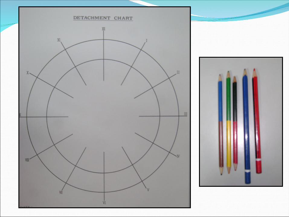
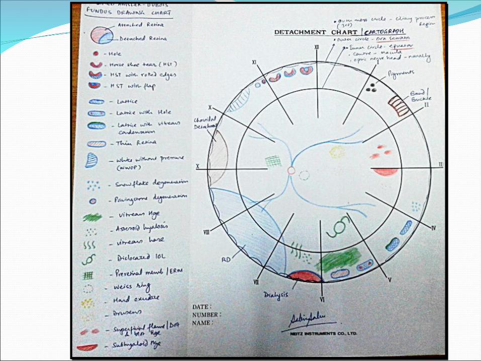
Retinal charts/ Cartographs2 concentric circles:Outer: ora serrataInner: equator
Macula is located centrallyOptic nerve head is located nasal to the
macula

BLUE Retinal vesselsSub retinal fluid Detached RetinaEdema
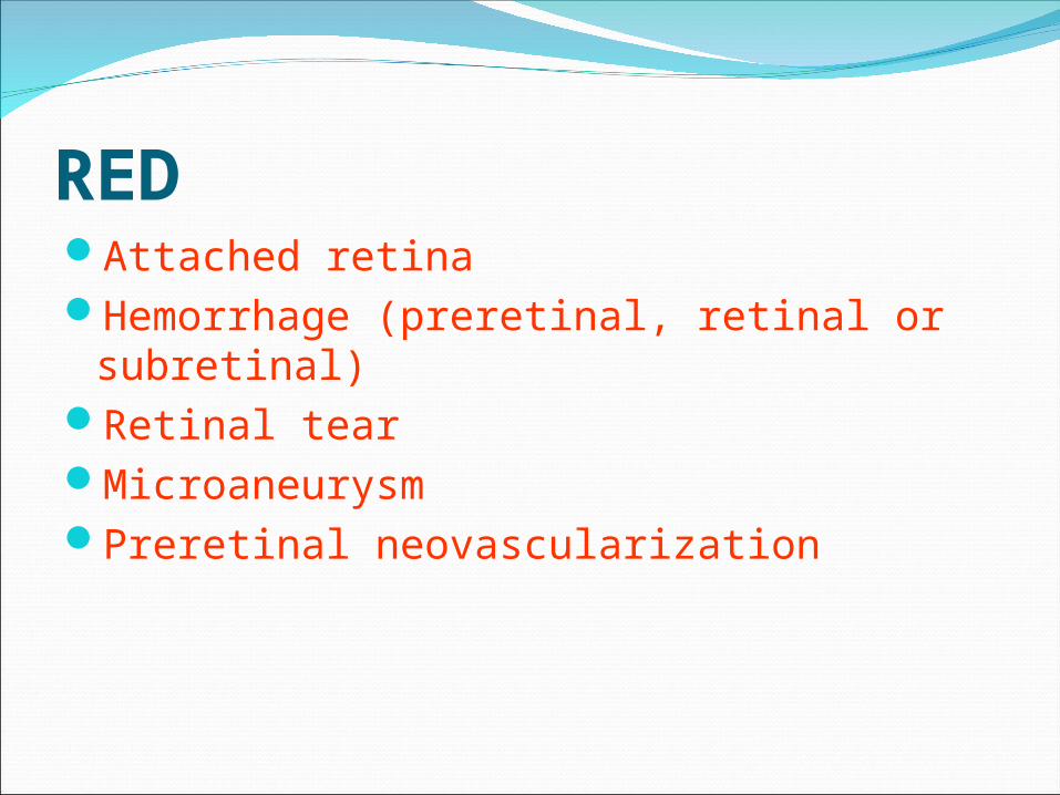
REDAttached retinaHemorrhage (preretinal, retinal or
subretinal)Retinal tearMicroaneurysmPreretinal neovascularization
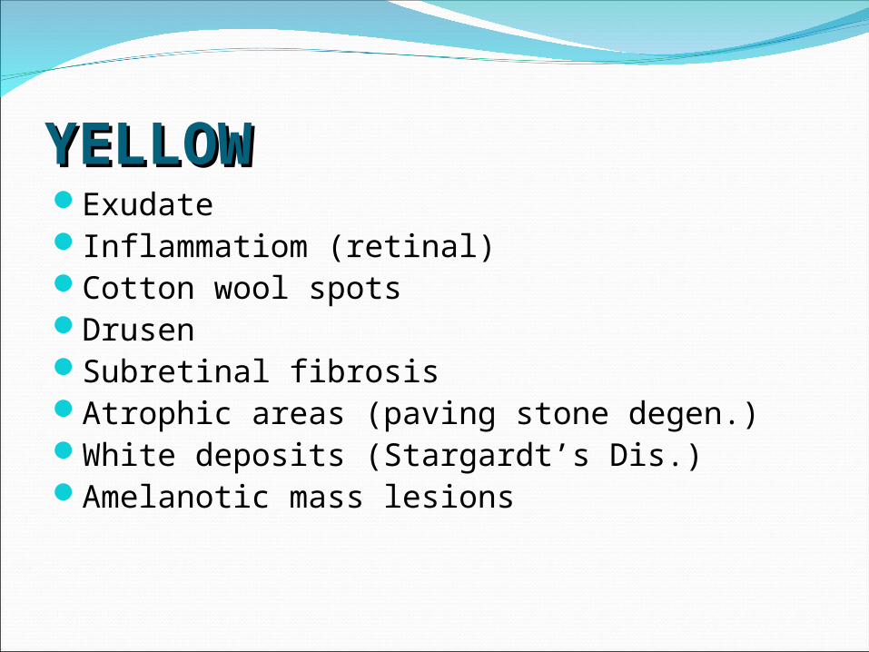
YELLOWYELLOWExudateInflammatiom (retinal)Cotton wool spotsDrusenSubretinal fibrosisAtrophic areas (paving stone degen.)White deposits (Stargardt’s Dis.)Amelanotic mass lesions
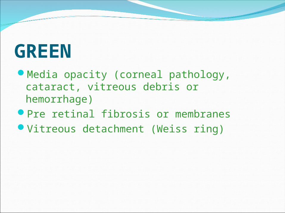
GREENMedia opacity (corneal pathology, cataract,
vitreous debris or hemorrhage)Pre retinal fibrosis or membranesVitreous detachment (Weiss ring)

BROWNMelanocytic lesionsUveal tissueMalignant choroidal melanomasEdge of buckle beneath detached retinaChoroidal detachment
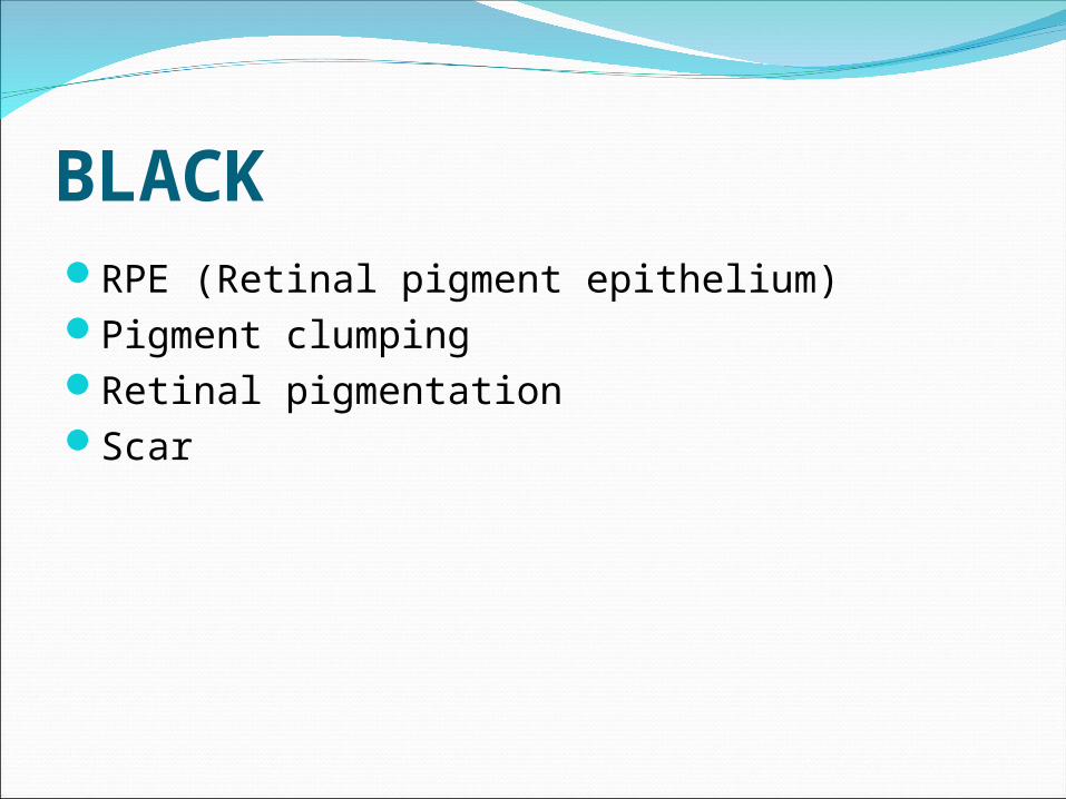
BLACKRPE (Retinal pigment epithelium)Pigment clumpingRetinal pigmentationScar
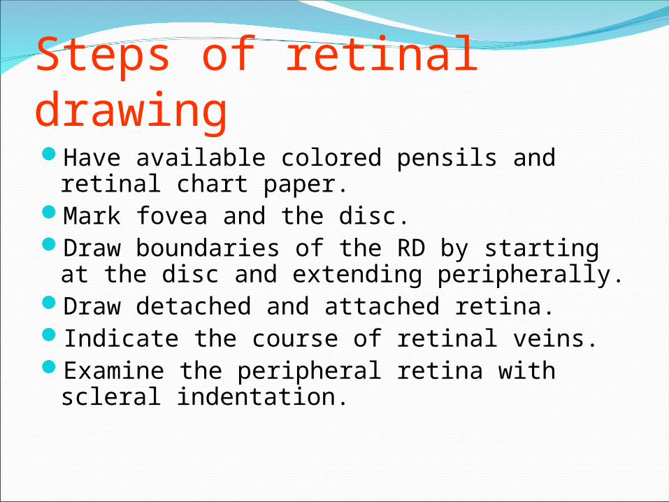
Steps of retinal drawingHave available colored pensils and retinal
chart paper.Mark fovea and the disc.Draw boundaries of the RD by starting at the
disc and extending peripherally.Draw detached and attached retina.Indicate the course of retinal veins.Examine the peripheral retina with scleral
indentation.

How big is the lesion?Size in disc diameters (DD) compare lesion to optic nerve head size.

Where is the lesion:Location:Clock-dialsuperior, supero-nasal, nasal, infero-nasal,
inferior, infero-temporal, temporal, supero-temporal.
Anterior/ posterior to the equator/ oral lesion.
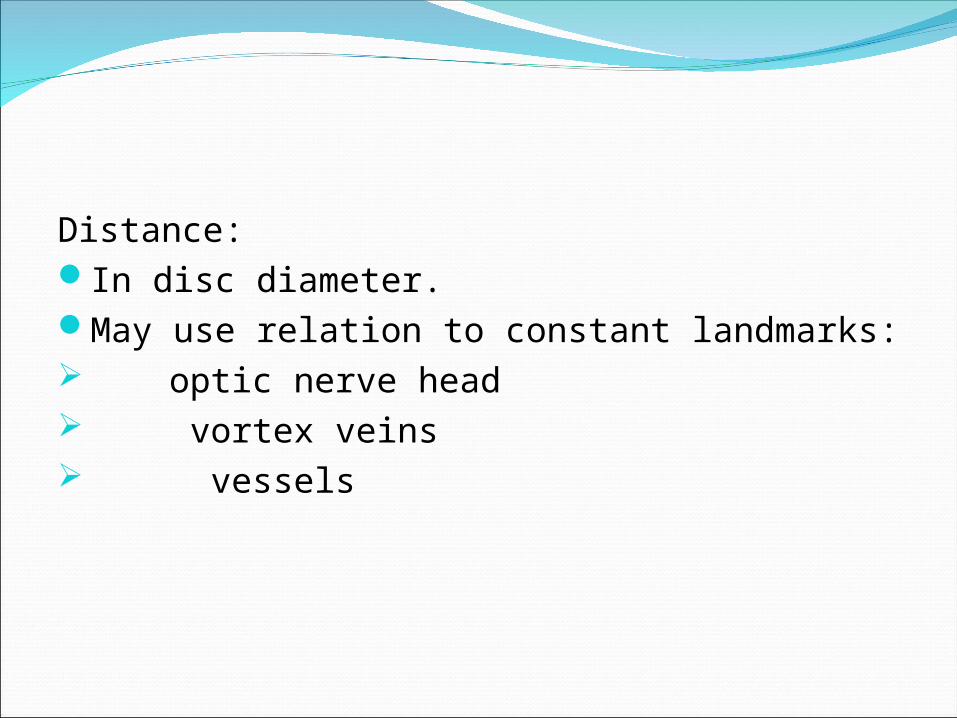
Distance:In disc diameter.May use relation to constant landmarks: optic nerve head vortex veins vessels