Embed Size (px)
DESCRIPTION
Walid M. Reda Ashour M.D Neurology, Lecturer of Neurology, Faculty of Medicine, Zagazig University, Egypt [email protected]
Citation preview


NEUROLOGYNEUROLOGY
DR.WALID REDA ASHOURDR.WALID REDA ASHOUR
LECTURER OF NEUROLOGYLECTURER OF NEUROLOGY

INTRODUCTIONINTRODUCTION

WHAT IS NEUROLOGY?
Neurology is the branch of medicine that
deals with diseases of the nervous system.

MAJOR DIVISIONSMAJOR DIVISIONS
NERVOUSNERVOUSSYSTEMSYSTEM
CENTRALCENTRAL NERVOUSNERVOUS SYSTEMSYSTEM
PERIPHERALPERIPHERALNERVOUSNERVOUSSYSTEMSYSTEM

CENTRAL NERVOUS SYSTEMCENTRAL NERVOUS SYSTEM

1. Cerebrum2. Brain stem3. Cerebellum
Intracranial part
Intracranial part
Spinal part
Spinal part
1- Spinal cord
2- Cauda equina

I- INTRACRANIAL PART
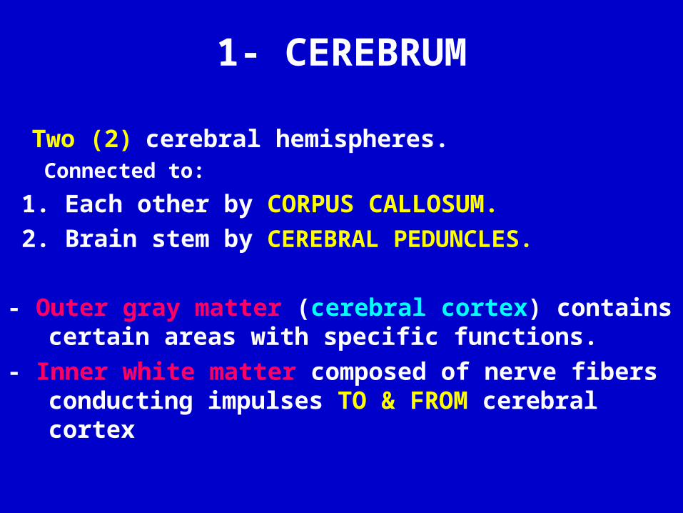
1- CEREBRUM
Two (2) cerebral hemispheres. Connected to:
1. Each other by CORPUS CALLOSUM.
2. Brain stem by CEREBRAL PEDUNCLES.
- Outer gray matter (cerebral cortex) contains certain areas with specific functions.
- Inner white matter composed of nerve fibers conducting impulses TO & FROM cerebral cortex

EACH cerebral hemisphere divided into :
FOUR ( 4 ) LOBES:
1. Frontal
2. Parietal
3. Temporal
4. Occipital

2- Brain stem
Formed of :
1- Midbrain
2- Pons
3- Medulla
Connected to:
1. Cerebral hemisphere
by cerebral peduncles
2. Cerbellum by cerebellar
peduncles

BRAIN STEM contains motor nuclei of cranial nerves:
• 3 & 4 in MIDBRAIN
• 5, 6 &7 in PONS
• 9, 10, 11 & 12 in MEDULLA
N.B. 1, 2 & 8
are sensory nerves have no motor nuclei

NAMES OF CRANIAL NERVES• 1 Olfactory
• 2 Optic
• 3 Oculomotor
• 4 Trochlear
• 5 Trigeminal
• 6 Abducent
• 7 Facial
• 8 Vestibulocochlear
• 9 Glossopharyngeal
• 10 Vagus
• 11 Accessory
• 12 Hypoglossal

CLASSIFICATION OF CRANIAL NERVES
• SENSORY : contain only sensory fibers1- Olfactory2- Optic
8- Vestibulocochlear • MOTOR : contain only motor fibers
3- Oculomotor 4- Trochlear 6- Abducent 11- Accessory
12- Hypoglossal • MIXED : contain both sensory and motor fibers
5- Trigeminal 7- Facial9- Glossopharyngeal 10- Vagus

Sensory cranial nerves
N. Name Function Assessment
1 Olfactory Smell Examination of smell
2 Optic Vision Acuity, field & fundus
8 Vestibular part
Equilibrium Caloric and rotational chair
Cochlear part
Hearing Watch test, Weber test

Motor cranial nervesN. Name Function Assessment
3 Oculomotor Motor to superior, inferior and medial recti; inferior oblique & levator palpebrae superioris
Ptosis, pupil, eye movements & squint
Parasympathetic to sphincter papillae and ciliary muscle
4 Trochlear Motor to superior oblique Look to opposite shoulder
6 Abducent Motor to lateral rectus Lateral eye movement
11 Accessory Motor to sternocleidomastoid and trapezius
Elevaton of shoulders and neck rotation
12 Hypoglossal Motor to muscles of tongue Tongue movements

Mixed cranial nervesN. Name Function Assessment
5 Trigeminal Muscles of mastication & sensations of face
Face sensations & power of muscles of mastication
7 Facial Muscles of facial expression & taste sensation in ant. 1/3 of tongue
Muscles of facial expression & taste sensation in ant. 1/3 of tongue
10 Vagus Sensation and movements of Larynx, pharynx, thoracic and abdominal organs
Palatal and pharyngeal reflexes & movements of uvula
9 Glossopharyngeal Taste in post 2/3 of tongue & gen. sensation
Taste & general sensation and pharyngeal reflex

3- CEREBELLUM
• Behind the brain stem
• Formed of : * Medline vermis
* Two cerebellar hemispheres
• Composed of :
Outer grey matter and inner
white matter

MBMB
PonsPons CC
MOMO
Superior peduncle connects cerebellum with MIDBRAIN. Middle peduncle connects cerebellum with PONS. Inferior peduncle connects cerebellum with MEDULLA
OBLONGATA
CEREBELLAR PEDUNCLES

LeftHemisphere
righthemisphere
CEREBELLAR HEMISPHERES AND VERMIS
Cerebellar Hemispheres are the two bilateral structures.Vermis is the unpaired midline structure between
the two hemispheres.
VERMIS

II- SPINAL PART

1- SPINAL CORD
• Inside spinal canal
• End at lower border of L1 vertebra.
• Its lowermost 3 segments S3,4,5 = Conus.
• The above 4 segments L4,5 & S1,2 = Epiconus.
• Inner grey matter (cells) surrounded by
white matter.

SPINAL CORD

SPINAL CORD TRACTS
• White matter contains ascending and descending nerve fibers (TRACTS):
1- ASCENDING• Lateral & Ventral spinothalamic.• Gracile & Cuneate.• Spinocerebellar.• Lissauer’s.
2- DESCENDING• Corticospinal (pyramidal).• Reticulospinal, vestibulospinal, tectospinal, rubrospinal &
olivospinal

2- CAUDA EQUINA
• Collection of LUMBOSACRAL ROOTS which fills the lower part of the spinal canal below L1 vertebra.

THE MOTOR SYSTEM

Motor pathway

UPPER MOTOR NEURON(pyramidal system)
* Originates in motor area 4 & premotor area 6
* Terminates at the anterior horn cells (AHCs)
of the different levels of the spinal cord.
* It supplies the opposite side of the body.

LOWER MOTOR NEURON
• Formed of AHCs & Peripheral motor nerves
(which transmit the motor impulses to the
voluntry muscles).

EXTRA PYRAMIDAL SYSTEM
• Originates from centers at various levels of CNS mainly the
BASAL GANGLIA.
• It controls the opposite side of the body.
CEREBELLAR SYSTEM
• Composed of Neo, Archi & Paleo- cerebellum.
• It co-ordinates the movements of the same side of the body.

UMNL LMNL
Tone Increased, with ‘clasp knife’ quality
Decreased
Clonus Present Absent Fasciculations Absent Present Muscle Wasting Absent, but disuse
atrophy eventually results
Present (within 2-3 weeks)
Tendon Reflexes Increased. Extensor plantar reflexes.
Decreased or absent. Flexor plantar reflexes.
Distribution Whole limbs, with more weakness in the upper limb extensors and lower limb flexors
Specific muscle groups affected (e.g. in the distribution of a spinal segment, or just the proximal muscles, etc.)


Thank you