Embed Size (px)
Citation preview

HumanPsychologyAssignmentShas Production
1: Structure and function of eye
2: Human ear
3: Sensation of taste and smell

P a g e | 1
Shas Production
Syed Harris
The Human Eye: - The eye is a very complex, delicate, and vital structurethat is responsible for an organism’s Interaction with the external world. It is themost important and influential sense organ.
· It receives information from the outside world inthe form of light, and sends loads of information to the brain all the time
· The human eye is a little less than one inch in diameter and almost spherical
· The eye has a very specific design or form, which captures and processes lightcoming from outside...light reflected by the stimuli
Eyes function like a camera, which has its aperture, and a lens through whichthe light enters and cells present in it process the received light just as do theintricate internal parts of the camera.
Anatomy/ Structure of the Eye
The anatomy of the eye is broadly divided into three parts along with itschambers.
I. The external structure of the eye
ii. The immediate structure of the eye
iii. The internal structure of the eye
All structures are important in terms of their processing and functioning
1: The External Structure:-
The entire external structure of the eye is a "light-tight" box

P a g e | 2
Shas Production
Syed Harris
Cornea:-
· A transparentexternal surface, a five layeredmembrane thatcovers both thepupil and theiris.
· It isthe first andmost powerfullens or layer ofthevisual apparatus that helps to form the sharp image on the retinal photoreceptor cells, along withthe crystalline lens.
Sclera:-
· Outer walls of the eye are formed by a hard, white substance called `sclera',hence sclerotic coat that covers 5/6th of the surface of the eye.
· The outside of the eye is light-tight and its mechanism is designed in such amanner that only small amount of light can enter into a small opening thatenables the production of a clearer vision, because a smaller openingalso acting as the `aperture', creates a sharper image
2: The Immediate Part:-
Pupil:-
· A dark, adjustable opening in the center of the eye through which the lightenters. It changes its size as the amount of light entering the eye varies.

P a g e | 3
Shas Production
Syed Harris
· It looks dark and black in appearance, because of the absorbing pigments in theretina
Iris:-
·Around the pupil of the eye, there is a ring of muscle tissue that controlsthe size of the pupil opening, through its contraction and expansion
·It contains the color pigments and thus gives color to the eye__ the color whichthe eyes possess such as brown, black, green, blue etc. are due to the iris muscles
Lens:-
·The transparent part of the eye that is located behind the pupil that changes itshape in order to focus images on the retina.
The lens changes its own thickness in order to focus image properly on retina__this ability of the lens is called “accommodation"
·The process of accommodation depends largely on the location of theobject with respect to the observer's body __ distant objects require arelatively flat lens and the muscles that are controlling it are relaxingas compared to when focusing the nearer objects, when muscles contract, takingtensions off the lens thus making the lens more round shaped
Fluids in the Chambers of the Eye:-
· Eye has three important layers or chambers:
I. Anterior layer that lies between the cornea and iris
Ii Posterior layer that lies between iris and lens
iii. Vitreous layer that lies between the lens and the retina
·Anterior and posterior chambers are filled with aqueous humor
·Whereas the vitreous chamber is filled with a more viscous fluid, thevitreous humor

P a g e | 4
Shas Production
Syed Harris
·The eye is filled with these two liquids that are important because they help maintain the shape of the eye and provide nourishment to the cells present in the eye
·The function of these fluids is the same as that of blood in other parts ofthe body; the difference being that these liquids are nearly transparent, so thatthey can nourish the cells of the eye without interfering with the light that entersin the eye.
Choroid:-
It is the middle layer of the eye, and the lining of the 3/5th of the eyeball
· The choroid serves two important functions; i.e. nourishment and absorption
· The choroid carries blood to the retina and the humors in order to providenourishment to the eye·
.The other important function is that choroid absorbs any light that strikes on it__this is extremely important, because light that passes through the rods and conesdoes not reflect back. But, in any case, if it (light) reflects back, the photo-sensitive cells would receive the light message twice, and would percept wrong,that there was twice as much light as there really was
3: Internal Structure of the Eye:-
· Includes the most important structure; "the retina".
· Retina is the light- sensitive inner surface or chamber of the eye that convertsthe electromagnetic energy of the light into useful information for the brain.
· It contains about 130 million nerve cells.
· It contains the receptors rods and cones plus the neurons; these cells are very important as they initiate the processing of visual information.
· The retina has an area of 5 square centimeters located at the back ofthe eye, which is a location where all light detection takes place.
P a g e | 5
Shas Production
Syed Harris
· The retina is a network of nerve cells connected with over 100 millionphotosensitive rod and cones. The signals generated by these rods and cones arethen sent, via the optic nerve, to the visual areas in the brain
Plexiform Layer:-
· It is the top layer of the retina, which does not interpret the light striking thispart.
· A web of optical nerves that carry signals of rods and cones to the optic nerves
· It is located between the photosensitive cells and the vitreous humor so that theweb's cells can be nourished.
· These cells are nearly transparent so only minimal interference takes place whenlight strikes on the photosensitive cells.
Rods and Cones:-
a. Rods:-
· Retinal receptors which are long, cylindrical, and light sensitive; that can only detect black, white and gray; they functions well in dim light, and are largelyinsensitive to color and small details__ functions when cones do not respond
· Rods are used for `peripheral vision', i.e. the objects that are outside the main center of focus, and for night vision
b. Cones:-
· Retinal receptors, cone shaped and light sensitive, concentrated near the centerof the retina that is concerned with sharp focusing, fine details and colorsensation; they work well in well- lit conditions i.e., bright or sufficient light.
· Rods and cones are distributed unevenly throughout the retina
· There are fewer cones i.e. about seven million than rods i.e., 125 million

P a g e | 6
Shas Production
Syed Harris
Important Regions of Retina; Fovea and Blind Spot
a. Fovea:-
· The very sensitive and important part of the retina that aids in focusing; it isthe area of best vision.
· The largest concentration of cones is present in fovea
· There are no rods present in fovea
b. Blind Spot:-
· The area/ point where the optic nerve leaves the eye; no receptor cells arelocated here, thus creating a “blind" spot__ area of no vision
OR
blind spot small portion of the visual field of each eye that corresponds to theposition of the optic disk (also known as the optic nerve head) within the retina.There are no photoreceptors (i.e., rods or cones) in the optic disk, and, therefore,there is no image detection in this area
Image of eye showing blind spot
P a g e | 7
Shas Production
Syed Harris
Color Blindness:-
Colour (color) blindness (colour vision deficiency, or CVD) affectsapproximately 1 in 12 men (8%) and 1 in 200 women in the world. In Britain thismeans that there are approximately 2.7 million colour blind people (about 4.5%of the entire population), most of whom are male.There are different causes of colour blindness. For the vast majority of peoplewith deficient colour vision the condition is genetic and has been inherited fromtheir mother, although some people become colour blind as a result of otherdiseases such as diabetes and multiple sclerosis or they acquire the condition overtime due to the aging process, medication etc. Most colour blind people are ableto see things as clearly as other people but they unable to fully ‘see’ red, green orblue light. There are different types of colour blindness and there are extremelyrare cases where people are unable to see any colour at all.
Total Color Blindness:-
Total color blindness is a severe vision impairment leaving a personcompletely unable to distinguish any color – seeing things only in grayscale(shades of black and white). There are two types of total color vision (ormonochromacy).
Monochromacy:-
Monochromacy, more commonly referred to as “total color blindness”, is causedby the total absence of either 2 or 3 of the pigmented retinal cones, reducingvision to one dimension. It comes in two forms:
Rod monochromacy is a rare, non-progressive inability to distinguish anycolor, resulting from non-functioning or absent retinal cones. Rodmonochromacy is typically associated with sensitivity to light (Photophobia)and poor vision.

P a g e | 8
Shas Production
Syed Harris
Cone monochromacy is also a rare type of total color blindness,however is accompanied by relatively normal vision.
Being totally color blind makes everyday life much more challenging. The ability totell the difference between colors of a similar level of brightness becomes nearlyimpossible, whereas to a normal person – these colors may be totally different.For example, confusion of colors such as green and blue, and yellow and white isto be expected.
Partial colour blindness:-
“Exhibiting partial loss of green color vision so that anincreased intensity of green is required in a mixture of red and green tomatch a given yellow”
Deutan color vision deficiencies are by far the most common forms of colorblindness. This subtype of red-green color blindness is found in about 6%of the male population, mostly in its mild form deuteranomaly.

P a g e | 9
Shas Production
Syed Harris
Normal and Deuteranopia Color Spectrum
When you have a look at the color spectrum of a deuteranopic person youcan see that a variety of colors look different than in a normal colorspectrum. Whereas red and green are the main problem colors, thereare also for example some gray, purple and a greenish blue-green whichcan’t be distinguished very well.
The well-known term red-green color blindness is actually split into twodifferent subtypes. On one side persons which either lack or haveanomalous long wavelength sensitive cones (protan color visiondeficiency), which are more responsible for the red part of vision. And onthe other side deutan color vision deficiencies, which again are split intotwo different types:
1. Dichromats: Deuteranopia (also called green-blind). In this case themedium wavelength sensitive cones (green) are missing at all. Adeuteranope can only distinguish 2 to 3 different hues, whereassomebody with normal vision sees 7 different hues.
2. Anomalous Trichromats: Deuteranomaly (green-weak). This can beeverything between almost normal color vision and deuteranopia. Thegreen sensitive cones are not missing in this case, but the peak ofsensitivity is moved towards the red sensitive cones.
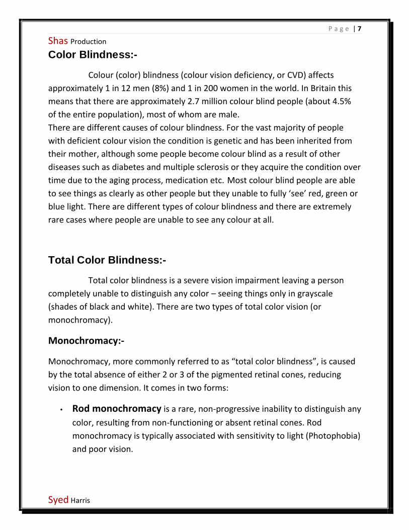
Below you can see a picture with normal colors on the left side and alteredcolors on the right side. The picture on the right side shows you how aperson affected by deuteranopia would see the scenery.
P a g e | 10
Shas Production
Syed Harris
Normal image Deuteranopia image

P a g e | 11
Shas Production
Syed Harris
Ear: - The ears are organs that provide two main functions —
hearing and balance — that depend on specialized receptors called haircells.
The ear is divided into three parts:
1:outer
2:Middle
3:inner

P a g e | 12
Shas Production
Syed Harris
Outer ear: The outer ear is lined with hairs and glands thatsecrete wax. This part of the ear provides protection andchannels sound.
Middle ear: Three tiny bones — the malleus, incus, andstapes — within the middle ear transfer sound vibrations fromthe eardrum to the inner ear. The middle ear is importantbecause it is filled with numerous air spaces that provide routes

P a g e | 13
Shas Production
Syed Harris
for infections to travel. It is also the location of the Eustachiantube, which provides equalization between the inner and outersurfaces of the tympanic membrane.
Inner ear: The inner ear, also called the labyrinth, operatesthe body’s sense of balances and contains the hearing organ. Abony casing houses a complex system of membranous cells.The inner ear is called the labyrinth because of its complexshape. There are two main sections within the inner ear: thebony labyrinth and the membranous labyrinth. The cochlea, thehearing organ, is located inside the inner ear.
The snail-like cochlea ismade up of three fluid-filled chambers thatspiral around a bony
core that contains a central channel called the cochlear duct.Inside the cochlear duct is the main hearing organ, the spiralshaped organ of Corti. Hair cells inside the organ of Corti detectsound and send the information through the cochlear nerve.
P a g e | 14
Shas Production
Syed Harris
Sound waves enter through the outer ear, move into themiddle ear, and finally reach the inner ear and its intricatenetwork of nerves, bones, canals, and cells.
Sensation:-
A sensation is something that produces excitement. A newsong or a new play might be termed a "sensation" or producing"sensation". A thrilling ride on a roller coaster might bedescribed as "sensational".
Sensation of taste: - (Gustatory sense)
Taste, or gustation, is the ability to detect sensorychanges in the tongue, through the use of taste buds, situateddeep into the papillae. Intriguingly, the sense called gustation isin fact comprised of varying ratios of multiple sensory systems,shifting in importance and attention as food is chewed, tastedand swallowed. These include the taste buds, the sense oftouch in the structures of the mouth and digestive system,chemical sensation of irritation in the trigeminal nerve system,and unique receptors for sensing the properties of waterlocated at the rear of the oral cavity.Sensation of smell: - (Olfactory sense)
Smell, or olfaction, is received by the olfactorybulb and connected to the brain by the olfactory nerve, the first

P a g e | 15
Shas Production
Syed Harris
cranial nerve of the brain, just after the nasal turbinate’s of thenose warm, strain and filter the air.
Shas Production