Embed Size (px)
Citation preview


13-3

! Arteries (and arterioles) take blood away from the heart.
! Artery walls contain smooth muscle that are able to constrict. " Regulates blood flow " Regulates blood pressure
! Arterioles can also constrict to regulate blood pressure.


X Capillaries have walls only one cell thick to allow exchange of gases and nutrients with tissue fluid.
X Capillary beds are present in all regions of the body but not all capillary beds are open at the same time.
X Contraction of a sphincter muscle closes off a bed and blood can flow through an arteriovenous shunt that bypasses the capillary bed.

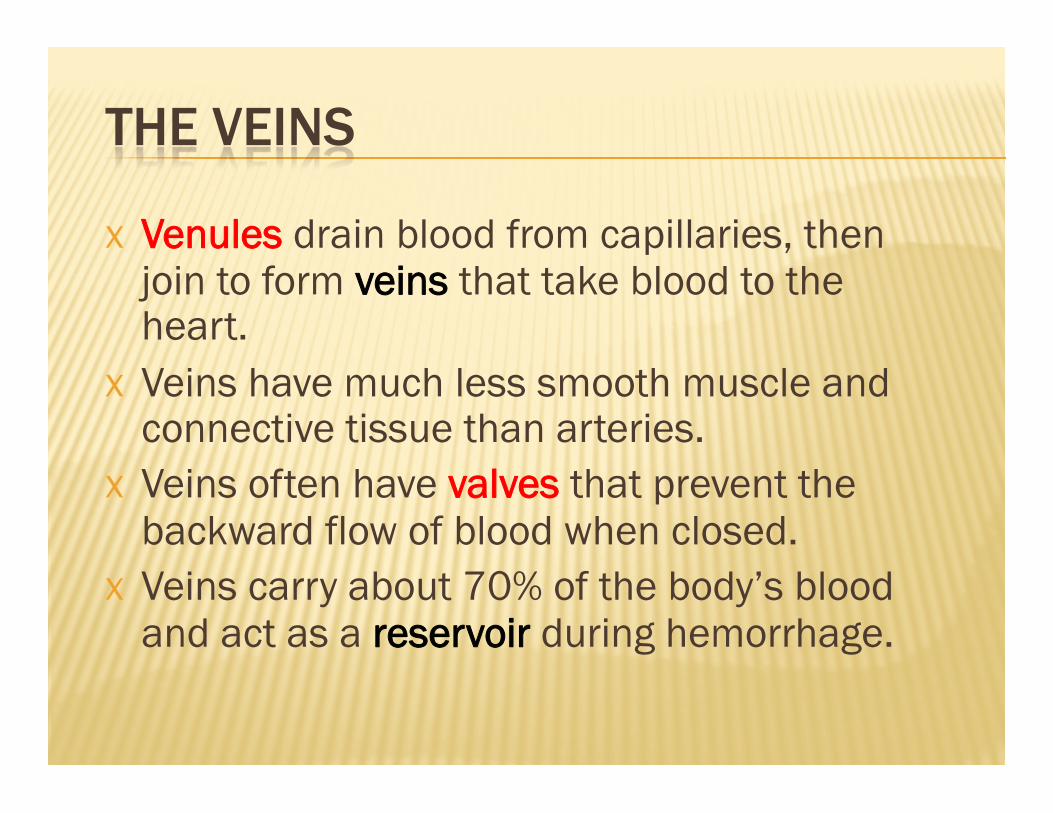
X Venules drain blood from capillaries, then join to form veins that take blood to the heart.
X Veins have much less smooth muscle and connective tissue than arteries.
X Veins often have valves that prevent the backward flow of blood when closed.
X Veins carry about 70% of the body’s blood and act as a reservoir during hemorrhage.

X The heart is a cone-shaped, muscular organ located between the lungs behind the sternum.
X The heart muscle forms the myocardium, with tightly interconnect cells of cardiac muscle tissue.
X The pericardium is the outer membranous sac with lubricating fluid.


X The heart has four chambers: two upper, thin-walled atria, and two lower, thick-walled ventricles.
X The septum is a wall dividing the right and left sides.
X Atrioventricular valves occur between the atria and ventricles – the tricuspid valve on the right and the bicuspid valve on the left; both valves are reinforced by chordae tendinae attached to muscular projections within the ventricles.


X Semilunar valves occur between the ventricles and the attached arteries
X The aortic semilunar valve lies between the left ventricle and the aorta
X The pulmonary semilunar valve lies between the right ventricle and the pulmonary trunk.


→ anterior and posterior vena cava → right atrium → tricuspid valve → right ventricle → → pulmonary semilunar valve → pulmonary trunk → → pulmonary veins → left atrium → → bicuspid valve → left ventricle → → aortic semilunar valve → aorta → to the body.


X The pumping of the heart sends out blood under pressure to the arteries.
X Blood pressure is greatest in the aorta; the wall of the left ventricle is thicker than that of the right ventricle and pumps blood to the entire body.
X Blood pressure then decreases as the cross-sectional area of arteries and then arterioles increases.

X Each heartbeat is called a cardiac cycle. X When the heart beats, the two atria contract
together, then the two ventricles contract; then the whole heart relaxes.
X Systole is the contraction of heart chambers; diastole is their relaxation.
X The heart sounds, lub-dup, are due to the closing of the atrioventricular valves, followed by the closing of the semilunar valves.

13-25

X The SA (sinoatrial) node, or pacemaker, initiates the heartbeat and causes the atria to contract on average every 0.85 seconds.
X The AV (atrioventricular) node conveys the stimulus and initiates contraction of the ventricles.
X The signal for the ventricles to contract travels from the AV node through the atrioventricular bundle to the smaller Purkinje fibers.


X A cardiac control center in the medulla oblongata speeds up or slows down the heart rate via the ANS. X PNS – constant stimulation. Increase
stimulation decreases HR. Decrease stimulation increases HR.
X SNS – Increased stimulation increases HR. X Hormones epinephrine and norepinephrine
from the adrenal medulla also stimulate faster heart rate.

X An electrocardiogram (ECG) is a recording of the electrical changes that occur in the myocardium during a cardiac cycle.
X Atrial depolarization creates the P wave, ventricle depolarization creates the QRS complex, and repolarization of the ventricles produces the T wave.

This diagram demonstrates the relationship between the cardiac cycle (ECG) and the pressure within the Aorta, L.V., and L.A.

The cardiovascular system includes two circuits: 1) Pulmonary circuit which circulates blood
through the lungs, and 2) Systemic circuit which circulates blood to the
rest of the body.


X The pulmonary circuit begins with the pulmonary trunk from the right ventricle which branches into two pulmonary arteries that take oxygen-poor blood to the lungs.
X In the lungs, oxygen diffuses into the blood, and carbon dioxide diffuses out of the blood to be expelled by the lungs. (more to come on this in the next unit)
X Four pulmonary veins return oxygen-rich blood to the left atrium.

X The systemic circuit starts with the aortic arch. The Aortic Arch begins from the left ventricle and branches into: X Brachiocephalic, left common carotid, left
subclavian X The aorta then branches to arteries going to
each specific organ. X Generally, an artery divides into arterioles
and capillaries which then lead to venules.

X Upper Body and Head – branch from Aortic Arch X Subclavian – blood to the arms X Carotid – blood to the head
X Lower Body – all branch from the Aorta X Hepatic – blood to the liver X Iliac – blood to the legs X Renal – blood to the kidneys X Mesenteric – blood to the GI tract

13-39

X Blood flow to the Heart X Coronary arteries serve the heart muscle. X Part of the systemic system. X Easily clogged
X The hepatic portal system X Blood flow between the GI tract and the liver X Rich in nutrients, but low in oxygen X Allows the liver to regulate levels of certain
substances in the blood before entering the rest of the body.

X Blood pressure due to the pumping of the heart accounts for the flow of blood in the arteries.
X Systolic pressure is high when the heart expels the blood.
X Diastolic pressure occurs when the heart ventricles are relaxing.
X Both pressures decrease with distance from the left ventricle because blood enters more and more arterioles and arteries.

! Mean Arterial Pressure (MAP) is the average blood pressure in an individual.
! Normal Range – 70-110 mmHg ! A minimum of 60 mmHg is required to sustain
organ function in the body


X Blood moves slowly in capillaries because of the large x-sectional area.
X This allows time for substances to be exchanged between the blood and tissues.

1) Skeletal muscle contraction: Compression of veins causes blood to move forward past a valve that then prevents it from returning backward.

2. Valves: bicuspid valves within valves stop backflow within veins.

3. Respiratory Movements: Inspiration increases venous return (contraction of diaphragm) and expiration decreases return.

X Varicose veins develop when the valves of veins become weak.
X Hemorrhoids (piles) are due to varicose veins in the rectum.
X Phlebitis is inflammation of a vein and can lead to a blood clot and possible death if the clot is dislodged and is carried to a pulmonary vessel.


X Blood separates into two main parts: plasma and formed elements.
X Plasma accounts for 55% and formed elements 45% of blood volume.
X Plasma contains mostly water (90–92%) and plasma proteins (7–8%), but it also contains nutrients and wastes.
X Albumin is a large group of plasma proteins that transport various biologically active molecules and helps regulate blood pressure.
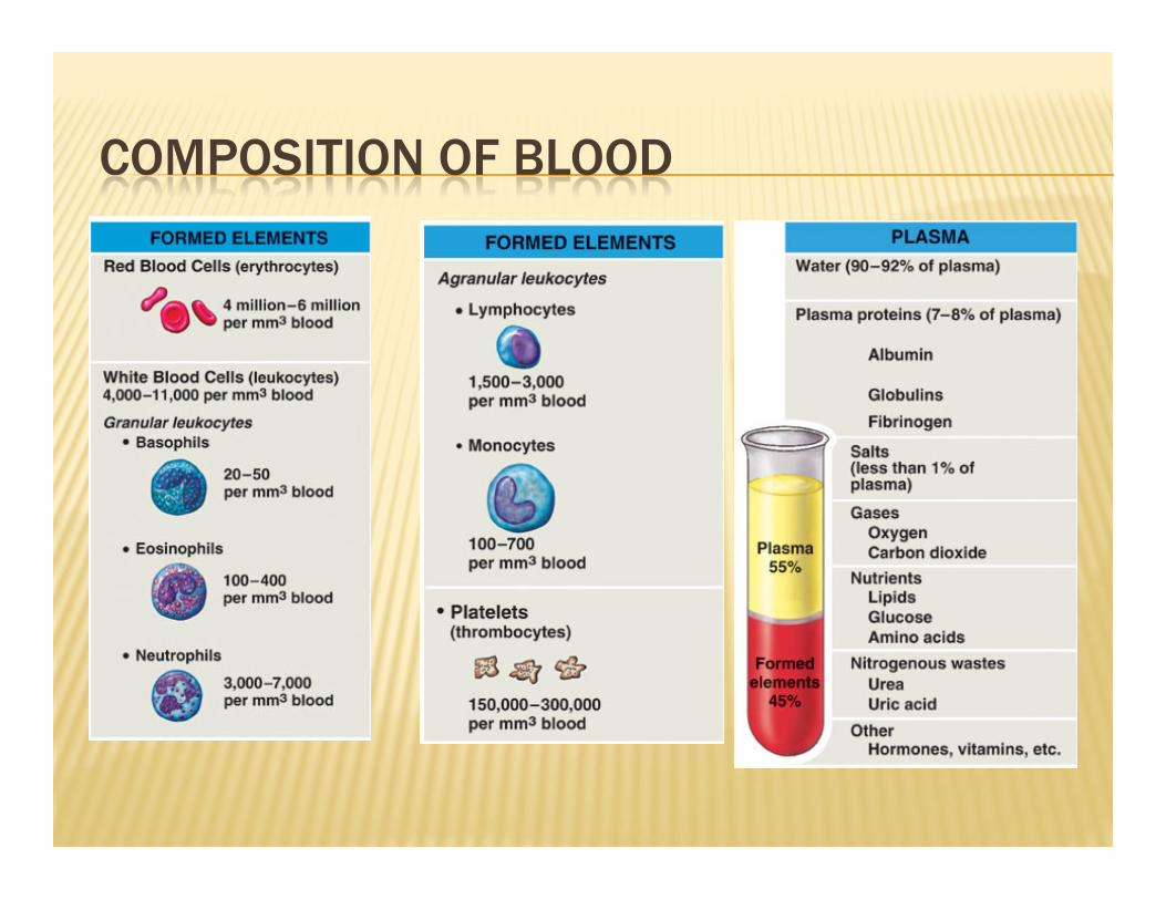

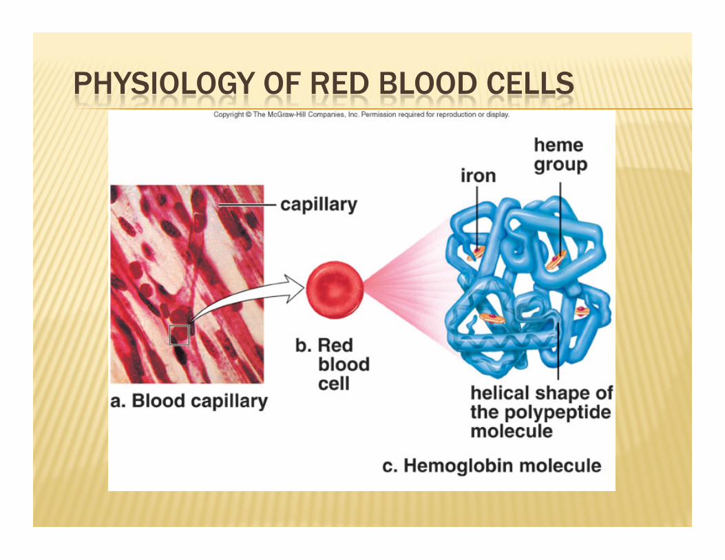
! Red blood cells (erythrocytes or RBC’s) are made in the red bone marrow of the skull, ribs, vertebrae, and the ends of long bones.
! Between 4-6 million RBC’s per mm3 of whole blood exist.
! Hemoglobin (pigment) contains heme, an complex iron-containing group that transports oxygen
! CO more readily bonds to hemoglobin and may cause suffocation
! RBC’s have a 120 day lifespan and are dismantled in the Liver and Spleen.

! RBC’s lack a nucleus. ! Iron from RBC dismantle is reused. ! Lack of enough hemoglobin results in anemia. ! The kidneys produce the hormone
erythropoietin (EPO) to increase blood cell production when oxygen levels are low.
! EPO is used as a performance enhancing drug in endurance events.

! White blood cells (leukocytes) have nuclei, are fewer in number than RBCs, with 5,000 – 10,000 cells per mm3, and defend against disease.
! Leukocytes are divided into granular and agranular based on appearance.
! Granular leukocytes (neutrophils, eosinophils, and basophils) contain enzymes and proteins that defend the body against microbes.

! The agranular leukocytes (monocytes and lymphocytes) have a spherical or kidney-shaped nucleus.
! Monocytes can differentiate into macrophages that phagocytize microbes and stimulate other cells to defend the body.
! Lymphocytes are involved in immunity. ! An excessive number of white blood cells may
indicate an infection or leukemia; HIV infection drastically reduces the number of lymphocytes.


! Red bone marrow produces large cells called megakaryocytes that fragment into platelets at a rate of 200 billion per day; blood contains 150,000–300,000 platelets per mm3.
! Twelve clotting factors in the blood help platelets form blood clots.
! The liver produces fibrinogen and prothrombin, two plasma proteins involved in the clotting process.

! Injured tissues release a clotting factor called prothrombin activator, which converts prothrombin into thrombin.
! Thrombin, in turn, acts as an enzyme and converts fibrinogen into insoluble threads of fibrin.
! These conversions require the presence of calcium ions (Ca2+).
! Trapped red blood cells make a clot appear red.


! Hemophilia is an inherited clotting disorder due to a deficiency in a clotting factor.
! Bumps and falls cause bleeding in the joins; cartilage degeneration and re-absorption of bone can follow.
! The most frequent cause of death is bleeding into the brain with accompanying neurological damage.

! A stem cell is capable of dividing into new cells that differentiate into particular cell types.
! Bone marrow is multipotent, able to continually give rise to particular types of blood cells.
! The skin and brain also have stem cells, and mesenchymal stem cells give rise to connective tissues including heart muscle.


! At the arteriole end of a capillary. Water moves out of the blood due to the force of blood pressure.
! At the venule end, water moves into the blood due to osmotic pressure of the blood.
! Substances that leave the blood contribute to tissue fluid, the fluid between the body’s cells.
! In the midsection of the capillary, nutrients diffuse out and wastes diffuse into the blood.

! Plasma proteins remain in the blood stream and create a concentration gradient for osmosis to occur.
! Excess tissue fluid is returned to the blood stream as lymph in lymphatic vessels.


! Cardiovascular disease (CVD) is the leading cause of death in Western countries.
! Modern research efforts have improved diagnosis, treatment, and prevention.
! Major cardiovascular disorders include atherosclerosis, stroke, heart attack, aneurysm, and hypertension.

! Atherosclerosis is due to a build-up of fatty material (plaque), mainly cholesterol, under the inner lining of arteries.
! The plaque can cause a thrombus (blood clot) to form.
! The thrombus can dislodge as an embolus and lead to thromboembolism.


Top sample shows the beginnings of lipid build-up.


! A cerebrovascular accident, or stroke, results when an embolus lodges in a cerebral blood vessel or a cerebral blood vessel bursts; a portion of the brain dies due to lack of oxygen.
! A myocardial infarction, or heart attack, occurs when a portion of heart muscle dies due to lack of oxygen.

! Partial blockage of a coronary artery causes angina pectoris, or chest pain.
! An aneurysm is a ballooning of a blood vessel, usually in the abdominal aorta or arteries leading to the brain.
! Death results if the aneurysm is in a large vessel and the vessel bursts.
! Atherosclerosis and hypertension weaken blood vessels over time, increasing the risk of aneurysm.
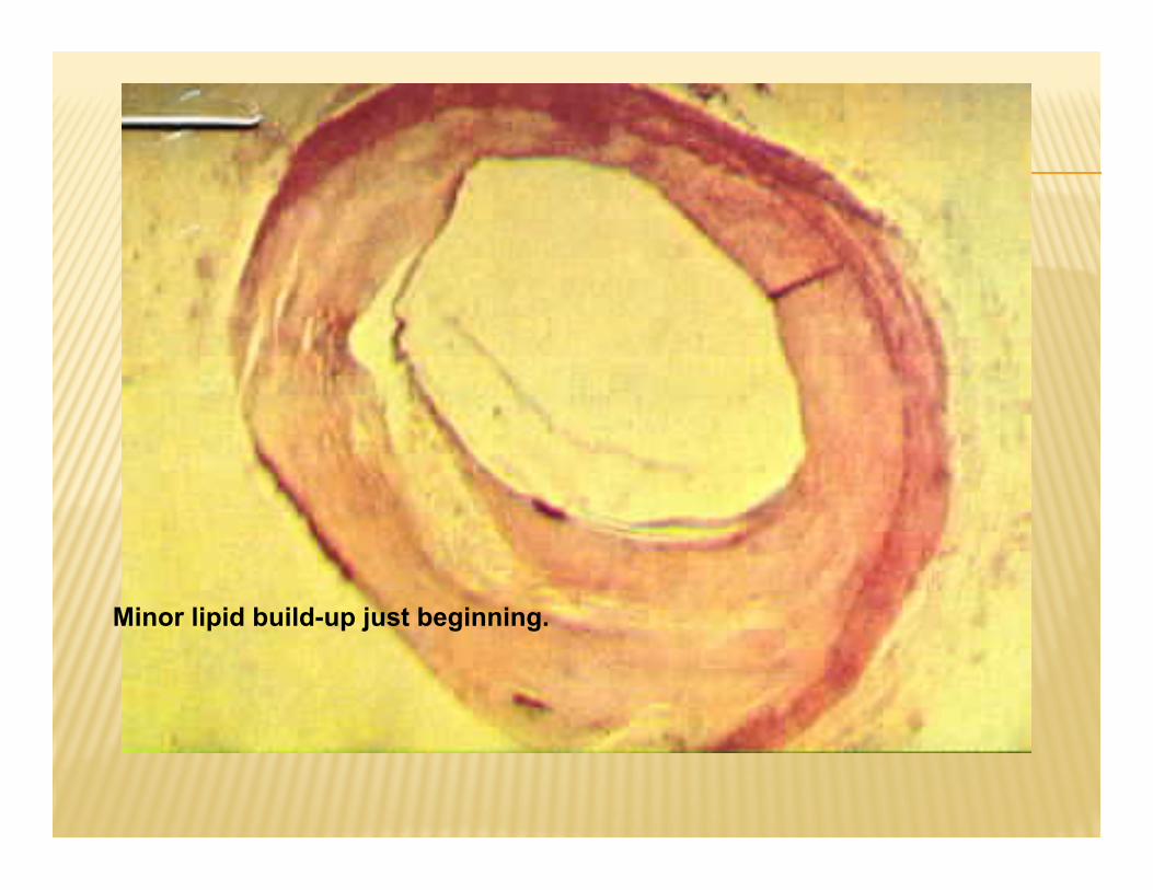
Minor lipid build-up just beginning.

X-sectional view shows the complexity of lipid build-up.


! A coronary bypass operation involves removing a segment of another blood vessel and replacing a clogged coronary artery.
! It may be possible to replace this surgery with gene therapy that stimulates new blood vessels to grow where the heart needs more blood flow.


! Angioplasty uses a long tube threaded through an arm or leg vessel to the point where the coronary artery is blocked; inflating the tube forces the vessel open.
! Small metal stents are expanded inside the artery to keep it open.
! New procedure uses this idea to increase blood flow to brain in MS patients. Controversial.


! Medical treatments for dissolving blood clots include use of t-PA (tissue plasminogen activator) that converts plasminogen into plasmin, an enzyme that dissolves blood clots, but can cause brain bleeding.
! Aspirin reduces the stickiness of platelets and reduces clot formation and lowers the risk of heart attack.
! High doses of Aspirin can increase the risk of internal bleeding in contact sport participants.

! Heart transplants are routinely performed but immunosuppressive drugs must be taken thereafter.
! There is a shortage of human organ donors. ! Work is currently underway to improve self-
contained artificial hearts, and muscle cell transplants may someday be useful.

! About 20% of Americans suffer from hypertension (high blood pressure).
! Hypertension is present when systolic pressure is 140 or greater or diastolic pressure is 90 or greater; diastolic pressure is emphasized when medical treatment is considered.
! A genetic predisposition for hypertension occurs in those who have a gene that codes for angiotensinogen, a powerful vasoconstrictor.

13-88
Specialized vessels deliver blood from heart to capillaries, where exchange of substances takes place; another series of vessels delivers blood from capillaries back to heart. The human heart is a double pump: the right side pumps blood to the lungs, and the left side pumps blood to the rest of body.

13-89
Pulmonary arteries transport blood low in oxygen to lungs; pulmonary veins return blood high in oxygen to the heart. Systemic circulation transports blood from the left ventricle of the heart to the body and then returns it to the right atrium of the heart. Blood is composed of cells and a fluid containing proteins and various other molecules and ions.

13-90
Blood clotting is a series of reactions; a clot forms when fibrin threads entrap red blood cells. Nutrients pass from blood and tissue fluid across capillary walls to cells; wastes move the opposite direction. The cardiovascular system is efficient but it is still subject to degenerative disorders.