Embed Size (px)
Citation preview
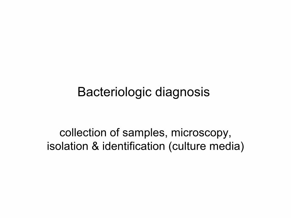
Bacteriologic diagnosis
collection of samples, microscopy, isolation & identification (culture media)
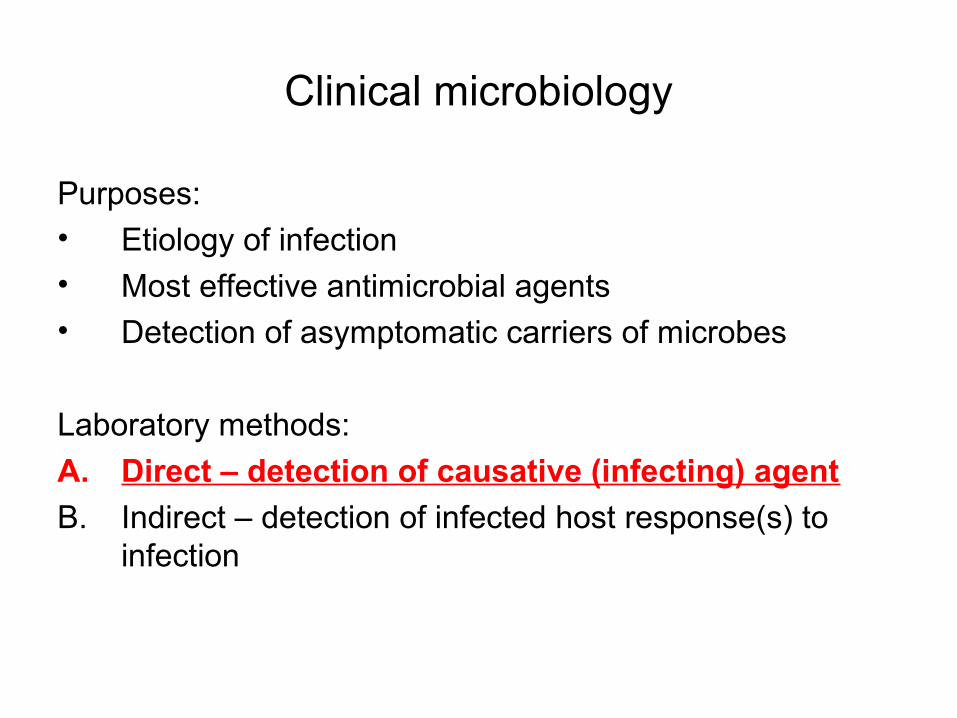
Clinical microbiology
Purposes:• Etiology of infection• Most effective antimicrobial agents• Detection of asymptomatic carriers of microbes
Laboratory methods:
A. Direct – detection of causative (infecting) agent
B. Indirect – detection of infected host response(s) to infection
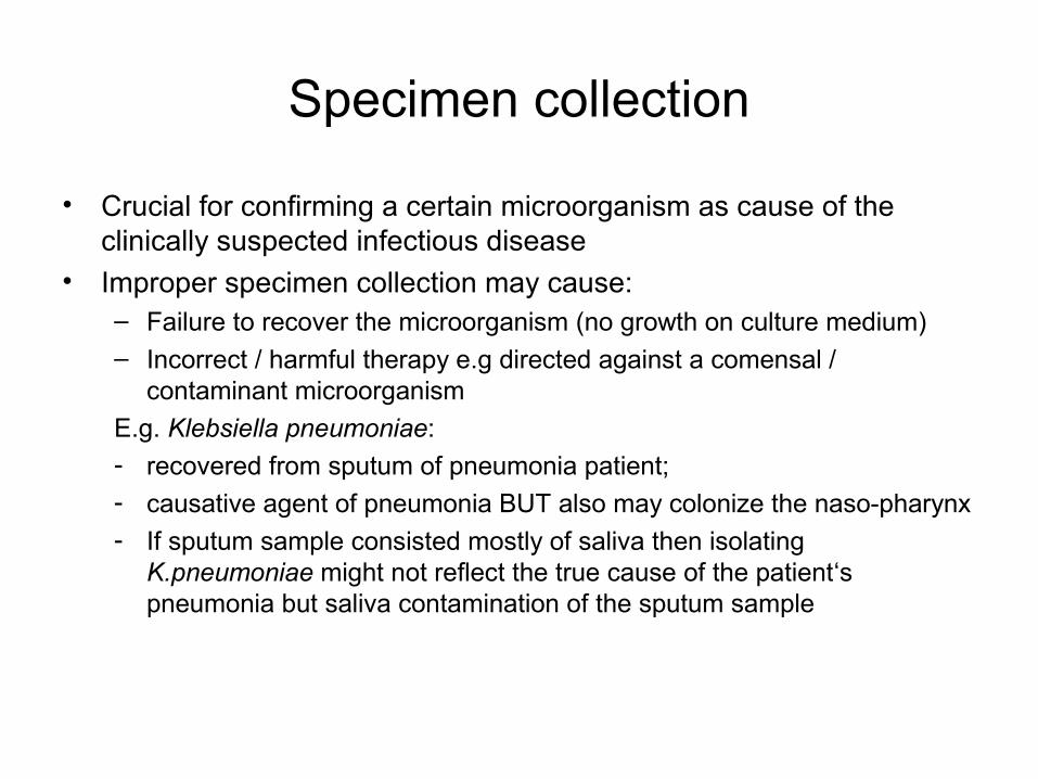
Specimen collection
• Crucial for confirming a certain microorganism as cause of the clinically suspected infectious disease
• Improper specimen collection may cause:– Failure to recover the microorganism (no growth on culture medium)– Incorrect / harmful therapy e.g directed against a comensal /
contaminant microorganism
E.g. Klebsiella pneumoniae:
- recovered from sputum of pneumonia patient; - causative agent of pneumonia BUT also may colonize the naso-pharynx- If sputum sample consisted mostly of saliva then isolating
K.pneumoniae might not reflect the true cause of the patient‘s pneumonia but saliva contamination of the sputum sample
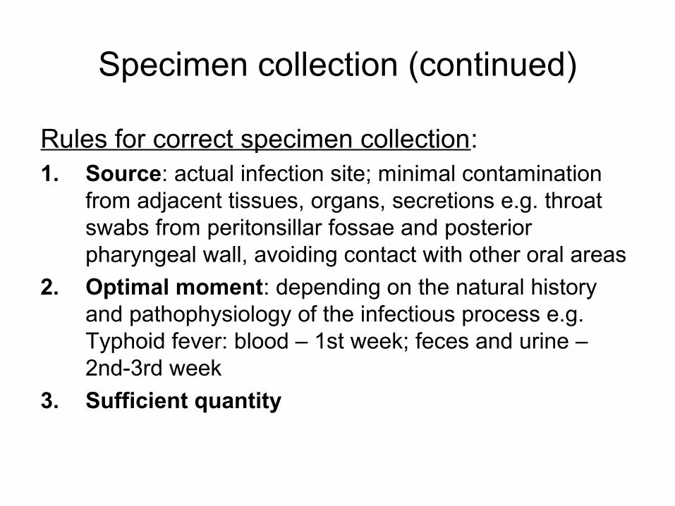
Specimen collection (continued)
Rules for correct specimen collection:1. Source: actual infection site; minimal contamination
from adjacent tissues, organs, secretions e.g. throat swabs from peritonsillar fossae and posterior pharyngeal wall, avoiding contact with other oral areas
2. Optimal moment: depending on the natural history and pathophysiology of the infectious process e.g. Typhoid fever: blood – 1st week; feces and urine – 2nd-3rd week
3. Sufficient quantity
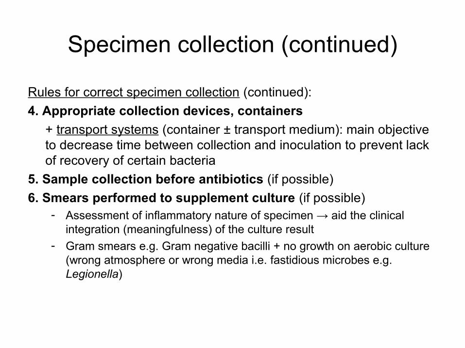
Specimen collection (continued)
Rules for correct specimen collection (continued):
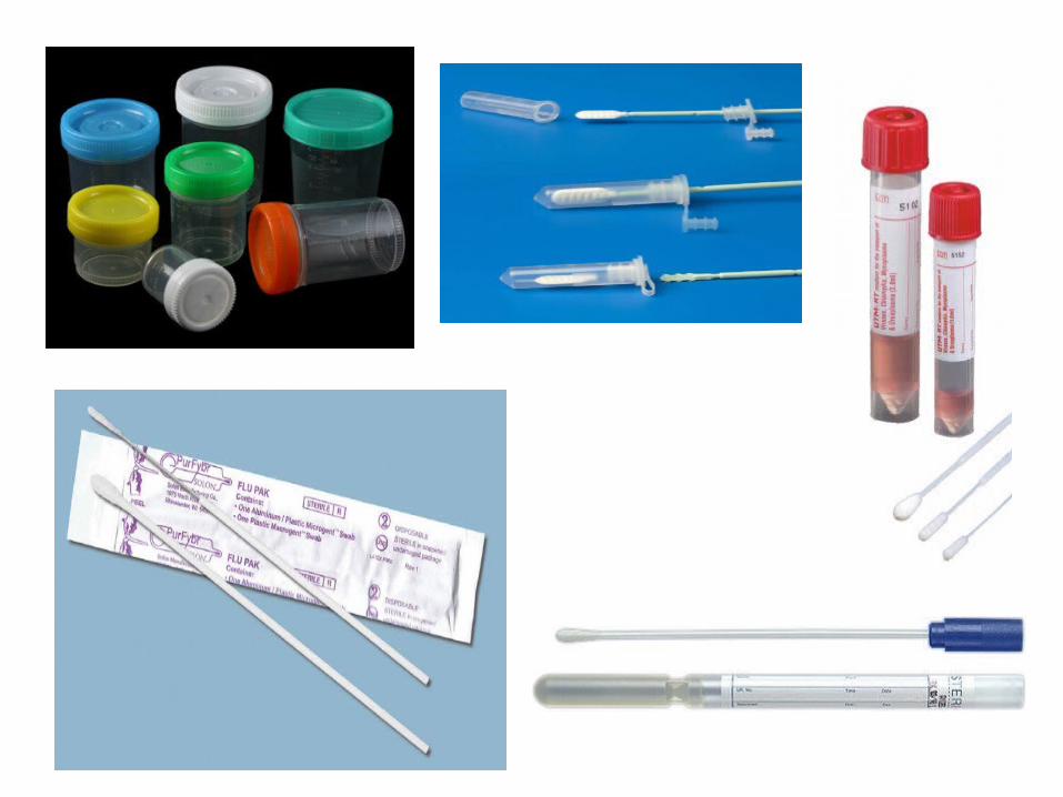
4. Appropriate collection devices, containers
+ transport systems (container ± transport medium): main objective to decrease time between collection and inoculation to prevent lack of recovery of certain bacteria
5. Sample collection before antibiotics (if possible)
6. Smears performed to supplement culture (if possible)- Assessment of inflammatory nature of specimen → aid the clinical
integration (meaningfulness) of the culture result - Gram smears e.g. Gram negative bacilli + no growth on aerobic culture
(wrong atmosphere or wrong media i.e. fastidious microbes e.g. Legionella)
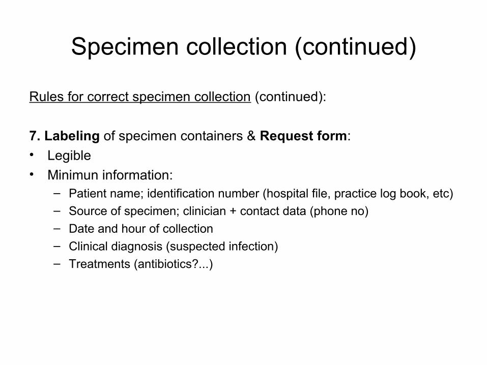
Specimen collection (continued)
Rules for correct specimen collection (continued):
7. Labeling of specimen containers & Request form:• Legible• Minimun information:
– Patient name; identification number (hospital file, practice log book, etc)– Source of specimen; clinician + contact data (phone no)
– Date and hour of collection– Clinical diagnosis (suspected infection)– Treatments (antibiotics?...)
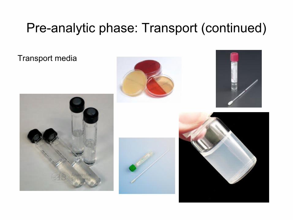
Pre-analytic phase: Transport (continued)
Transport media
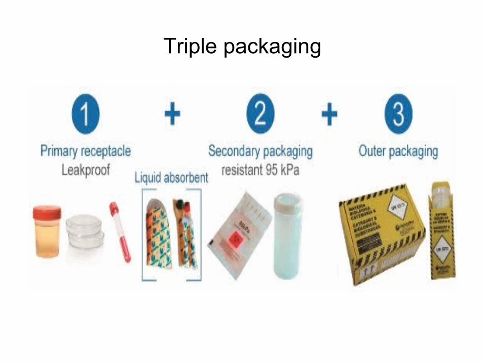
Triple packaging
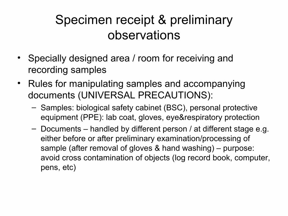
Specimen receipt & preliminary observations
• Specially designed area / room for receiving and recording samples
• Rules for manipulating samples and accompanying documents (UNIVERSAL PRECAUTIONS):– Samples: biological safety cabinet (BSC), personal protective
equipment (PPE): lab coat, gloves, eye&respiratory protection– Documents – handled by different person / at different stage e.g.
either before or after preliminary examination/processing of sample (after removal of gloves & hand washing) – purpose: avoid cross contamination of objects (log record book, computer, pens, etc)
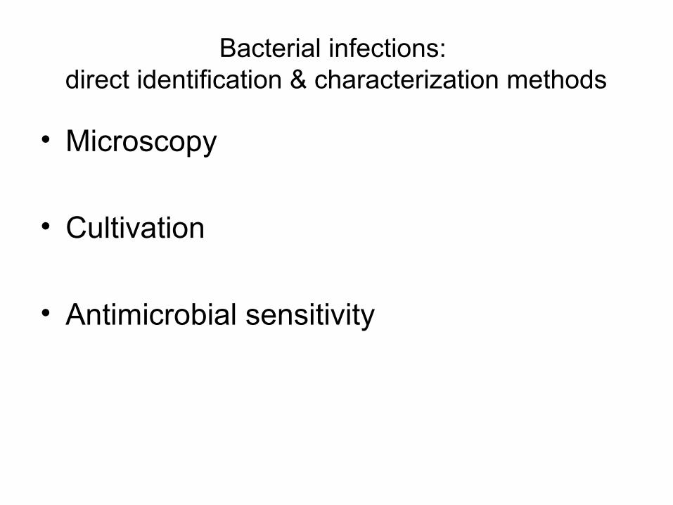
Bacterial infections: direct identification & characterization methods
• Microscopy
• Cultivation
• Antimicrobial sensitivity
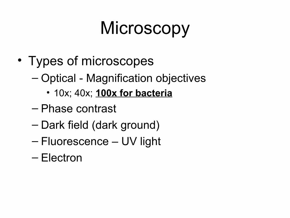
Microscopy
• Types of microscopes– Optical - Magnification objectives
• 10x; 40x; 100x for bacteria
– Phase contrast – Dark field (dark ground) – Fluorescence – UV light – Electron
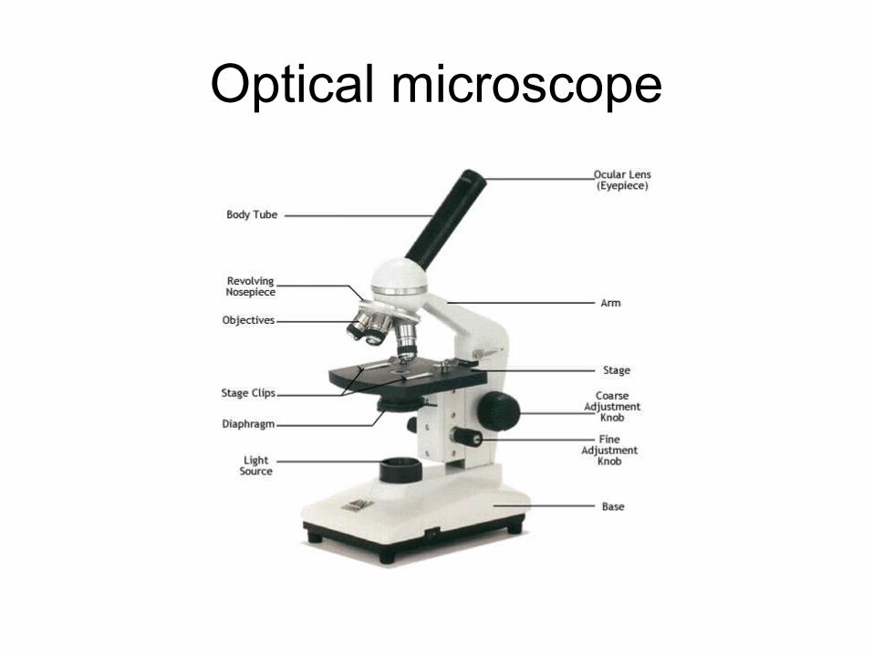
Optical microscope

Microscopic examination
• Wet mounts (unstained materials)– Direct light
– Observation of cells (PMN, macrophages), mobile germs in liquid samples (urine, CSF), shape and disposition of germs (cocci/bacilli/spirilli/vibrios)
• Stained smears
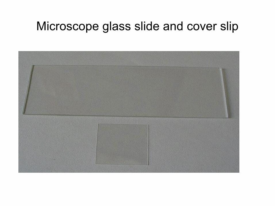
Microscope glass slide and cover slip
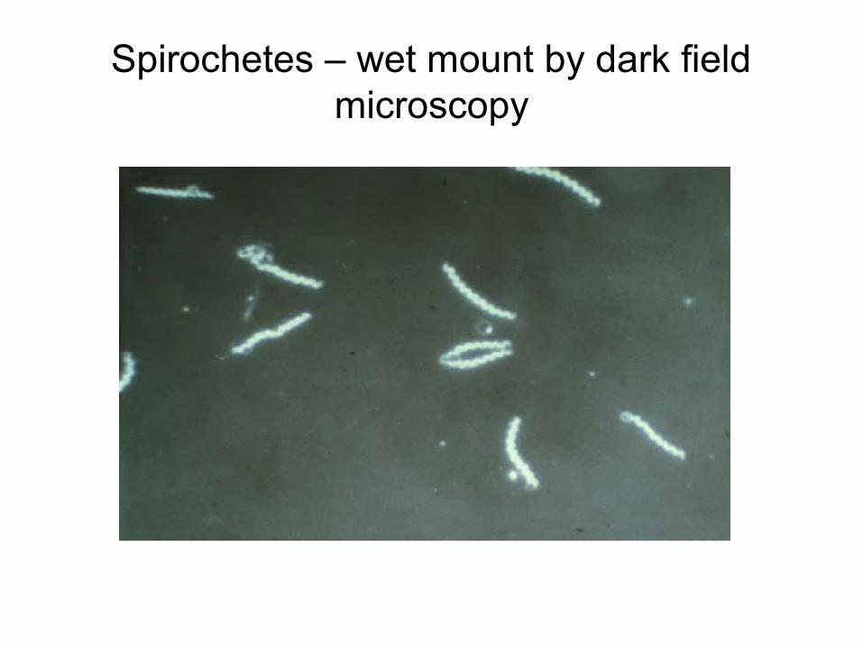
Spirochetes – wet mount by dark field microscopy
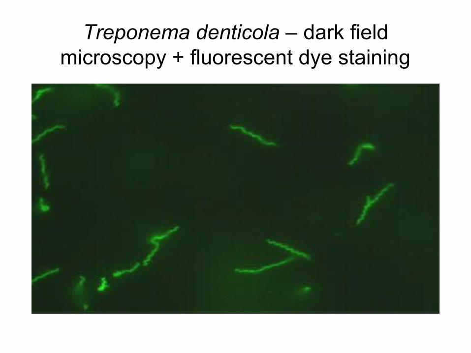
Treponema denticola – dark field microscopy + fluorescent dye staining
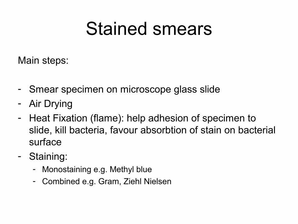
Stained smears
Main steps:
- Smear specimen on microscope glass slide - Air Drying
- Heat Fixation (flame): help adhesion of specimen to slide, kill bacteria, favour absorbtion of stain on bacterial surface
- Staining: - Monostaining e.g. Methyl blue- Combined e.g. Gram, Ziehl Nielsen
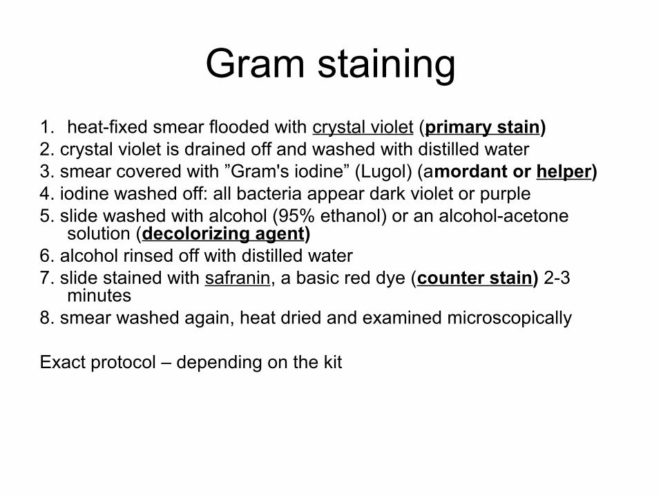
Gram staining1. heat-fixed smear flooded with crystal violet (primary stain)2. crystal violet is drained off and washed with distilled water 3. smear covered with ”Gram's iodine” (Lugol) (amordant or helper)4. iodine washed off: all bacteria appear dark violet or purple5. slide washed with alcohol (95% ethanol) or an alcohol-acetone
solution (decolorizing agent) 6. alcohol rinsed off with distilled water 7. slide stained with safranin, a basic red dye (counter stain) 2-3
minutes8. smear washed again, heat dried and examined microscopically
Exact protocol – depending on the kit
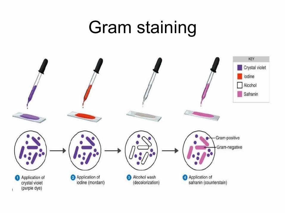
Gram staining
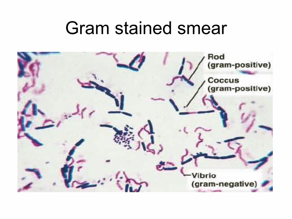
Gram stained smear
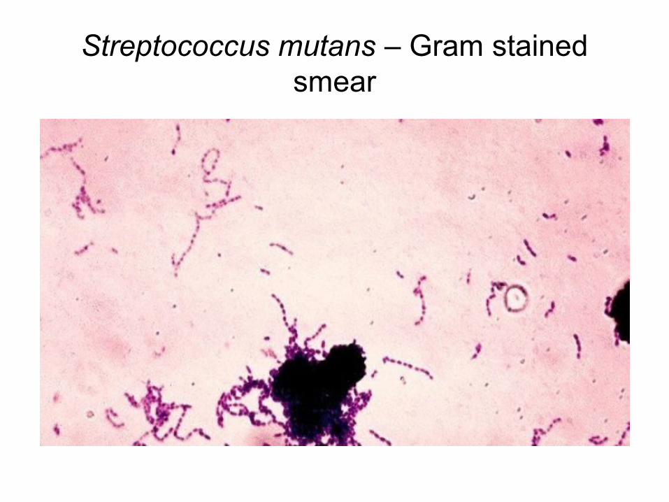
Streptococcus mutans – Gram stained smear
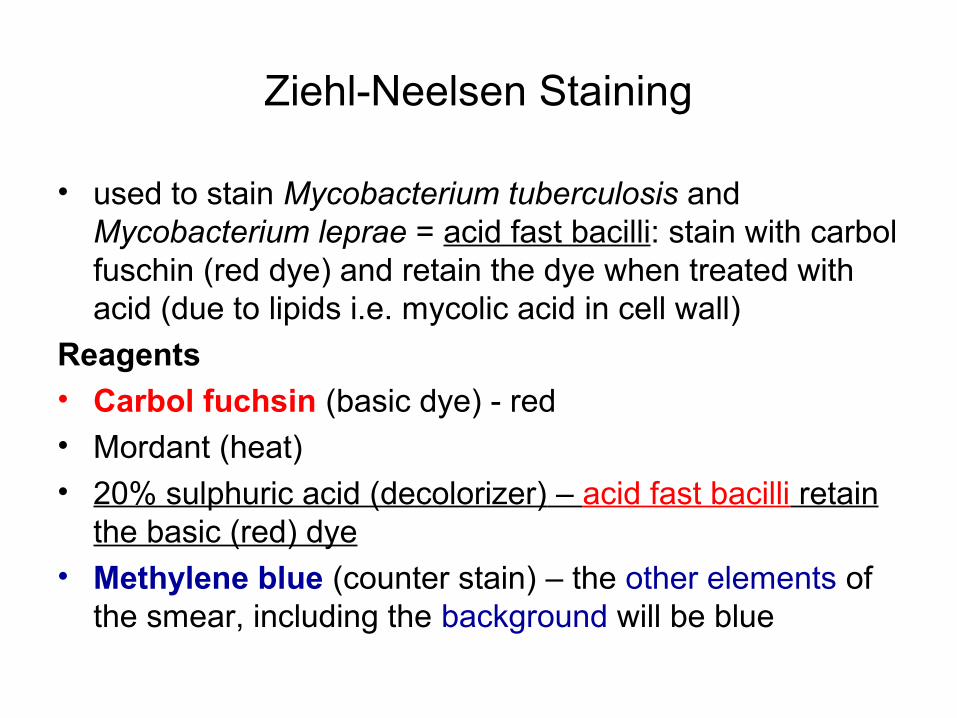
Ziehl-Neelsen Staining
• used to stain Mycobacterium tuberculosis and Mycobacterium leprae = acid fast bacilli: stain with carbol fuschin (red dye) and retain the dye when treated with acid (due to lipids i.e. mycolic acid in cell wall)
Reagents• Carbol fuchsin (basic dye) - red• Mordant (heat)
• 20% sulphuric acid (decolorizer) – acid fast bacilli retain the basic (red) dye
• Methylene blue (counter stain) – the other elements of the smear, including the background will be blue
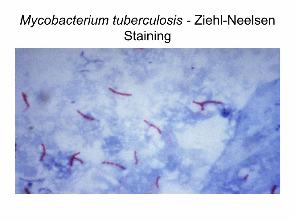
Mycobacterium tuberculosis - Ziehl-Neelsen Staining
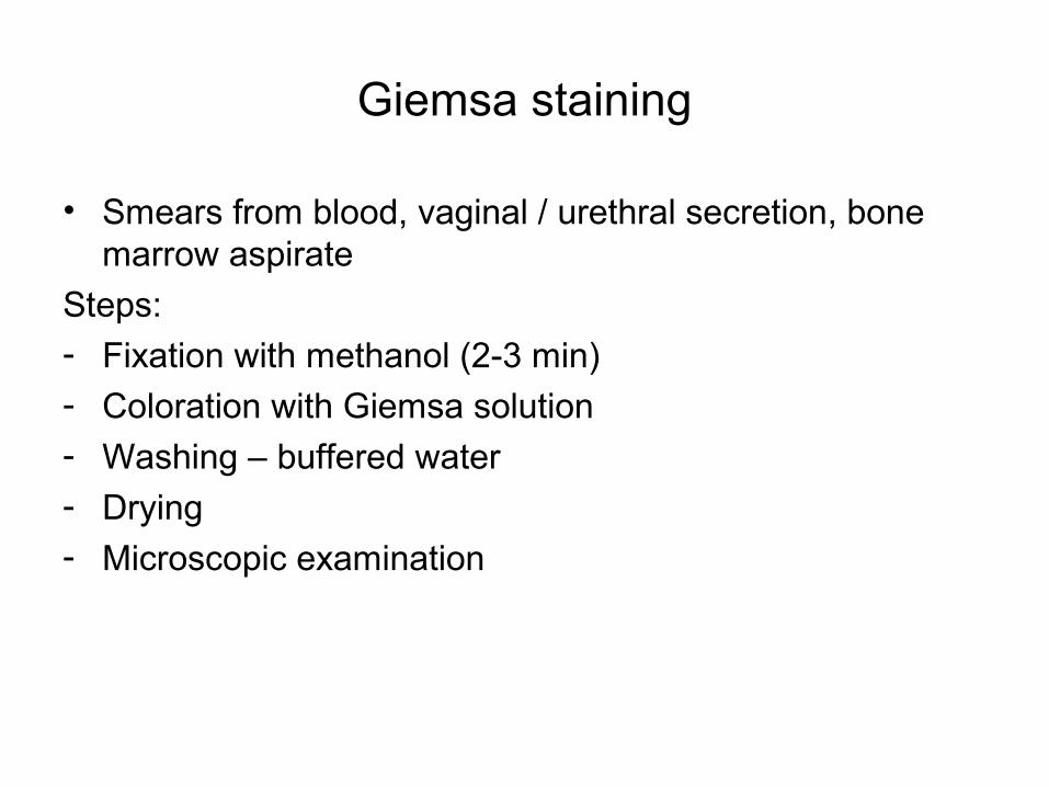
Giemsa staining
• Smears from blood, vaginal / urethral secretion, bone marrow aspirate
Steps:- Fixation with methanol (2-3 min)- Coloration with Giemsa solution
- Washing – buffered water
- Drying- Microscopic examination
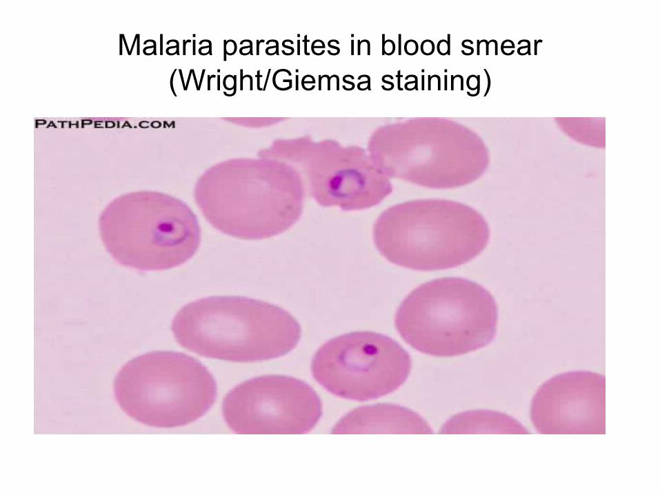
Malaria parasites in blood smear (Wright/Giemsa staining)
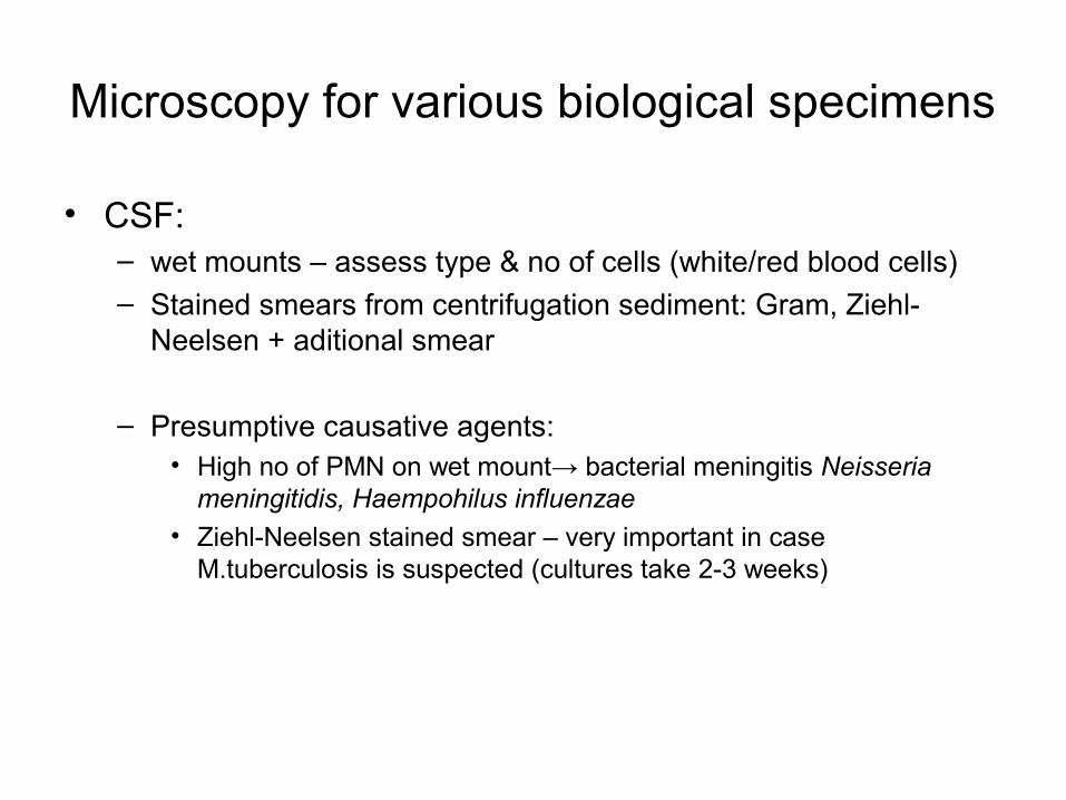
Microscopy for various biological specimens
• CSF: – wet mounts – assess type & no of cells (white/red blood cells)– Stained smears from centrifugation sediment: Gram, Ziehl-
Neelsen + aditional smear
– Presumptive causative agents:• High no of PMN on wet mount→ bacterial meningitis Neisseria
meningitidis, Haempohilus influenzae• Ziehl-Neelsen stained smear – very important in case
M.tuberculosis is suspected (cultures take 2-3 weeks)
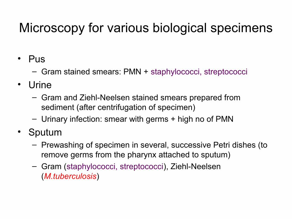
Microscopy for various biological specimens
• Pus– Gram stained smears: PMN + staphylococci, streptococci
• Urine– Gram and Ziehl-Neelsen stained smears prepared from
sediment (after centrifugation of specimen)– Urinary infection: smear with germs + high no of PMN
• Sputum– Prewashing of specimen in several, successive Petri dishes (to
remove germs from the pharynx attached to sputum)– Gram (staphylococci, streptococci), Ziehl-Neelsen
(M.tuberculosis)
Cultivation of microbes on culture media
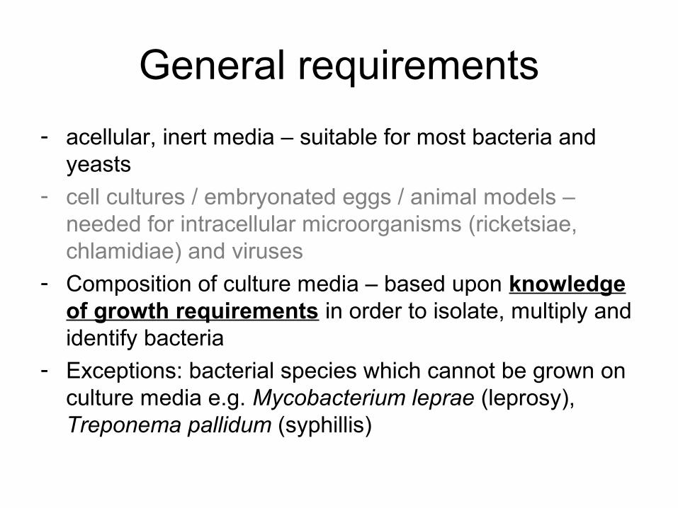
General requirements
- acellular, inert media – suitable for most bacteria and yeasts
- cell cultures / embryonated eggs / animal models – needed for intracellular microorganisms (ricketsiae, chlamidiae) and viruses
- Composition of culture media – based upon knowledge of growth requirements in order to isolate, multiply and identify bacteria
- Exceptions: bacterial species which cannot be grown on culture media e.g. Mycobacterium leprae (leprosy), Treponema pallidum (syphillis)
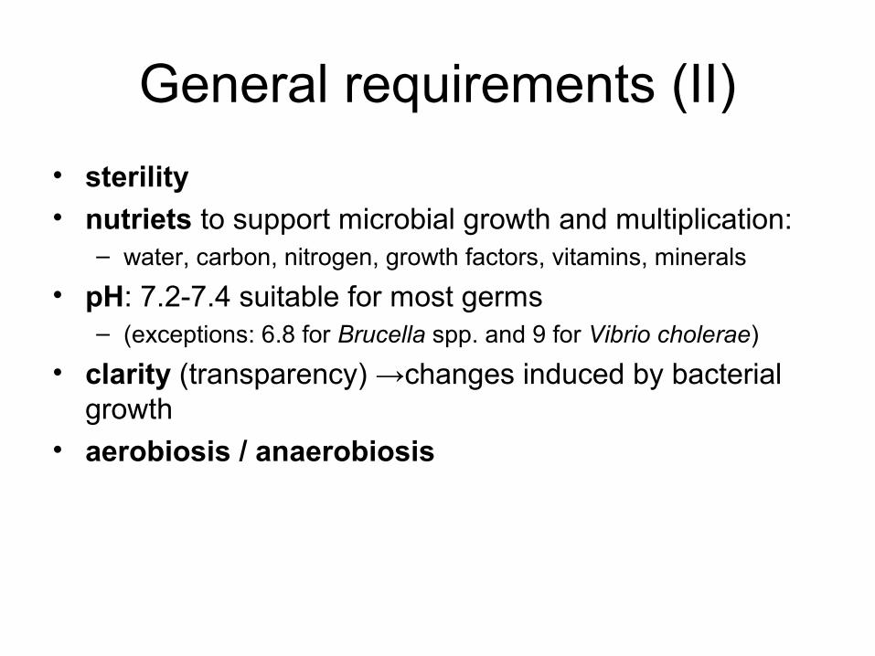
General requirements (II)
• sterility• nutriets to support microbial growth and multiplication:
– water, carbon, nitrogen, growth factors, vitamins, minerals
• pH: 7.2-7.4 suitable for most germs – (exceptions: 6.8 for Brucella spp. and 9 for Vibrio cholerae)
• clarity (transparency) →changes induced by bacterial growth
• aerobiosis / anaerobiosis
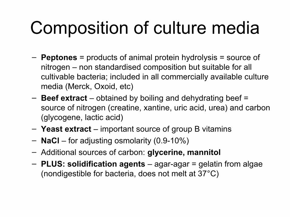
Composition of culture media
– Peptones = products of animal protein hydrolysis = source of nitrogen – non standardised composition but suitable for all cultivable bacteria; included in all commercially available culture media (Merck, Oxoid, etc)
– Beef extract – obtained by boiling and dehydrating beef = source of nitrogen (creatine, xantine, uric acid, urea) and carbon (glycogene, lactic acid)
– Yeast extract – important source of group B vitamins– NaCl – for adjusting osmolarity (0.9-10%)– Additional sources of carbon: glycerine, mannitol– PLUS: solidification agents – agar-agar = gelatin from algae
(nondigestible for bacteria, does not melt at 37°C)
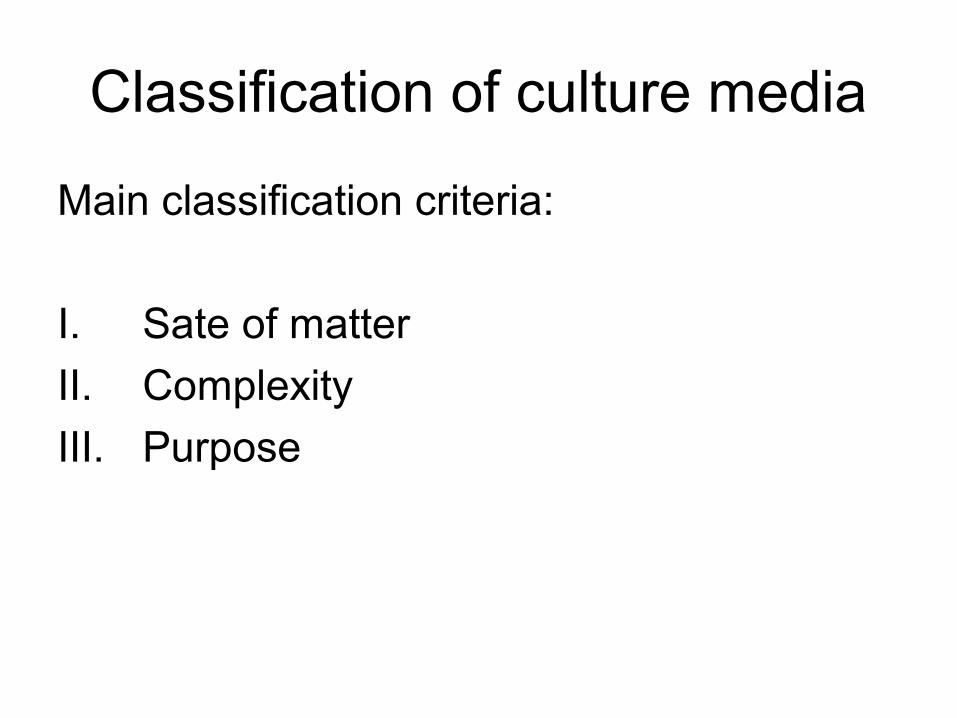
Classification of culture media
Main classification criteria:
I. Sate of matter
II. Complexity
III. Purpose
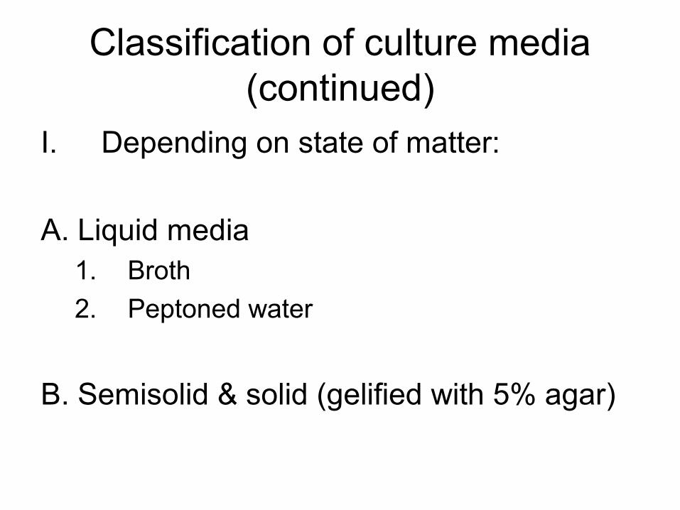
Classification of culture media (continued)
I. Depending on state of matter:
A. Liquid media 1. Broth
2. Peptoned water
B. Semisolid & solid (gelified with 5% agar)
Classification of culture media (continued)
A. Liquid media:
1. Nutrient broth = powdered beef extract (peptone content) dissolved in water – commercially available; used to be prepared by actually boiling beef/horse meat
- Widely used in microbiology laboratories:
- hemoculture – blood innoculated in liquid media
- identification of isolated bacterial strains by biochemical tests (e.g.fermentation of sugars)
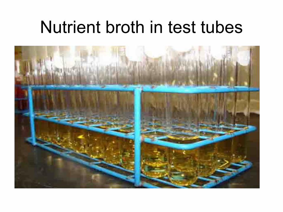
Nutrient broth in test tubes
Classification of culture media (continued)
B. Solid media - Obtained from liquid media by adding
agar-agar (gelification) - 1st reported use: Robert Koch 1882 –
cultivation of M. tuberculosis- Initially gelatin was used - disadvantages:
- Digested by some bacteria- Liquifies at 37°C – most frequently used
incubation temperature
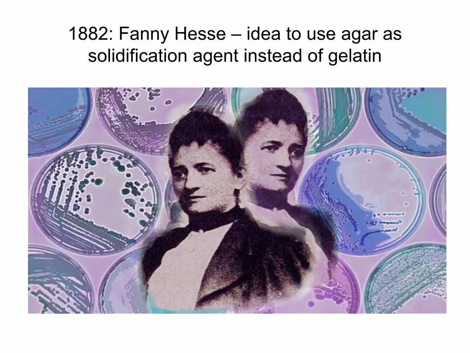
1882: Fanny Hesse – idea to use agar as solidification agent instead of gelatin
Classification of culture media (continued)
B. Solid media – Agar (continued)
1000 ml nutrient broth + 25-30 g agar-agar →melted by boiling + pH adjustment (7.2-7.4)
Features:
- odourless, colourless, nontoxic for microbes
- Nonsoluble in cold water, soluble in boiling water; upon cooling causes gelification
Classification of culture media (continued)
B. Solid media – Agar (continued)
Advantages:- Isolated colonies (resulting by multiplication of a single
microbe) → pure cultures can be obtained- Morphology of bacterial colonies: shape, size, changes
induced in the medium e.g. hemolysis, colour changes, etc.
- Counting microbes in a biological sample e.g. urinary infections
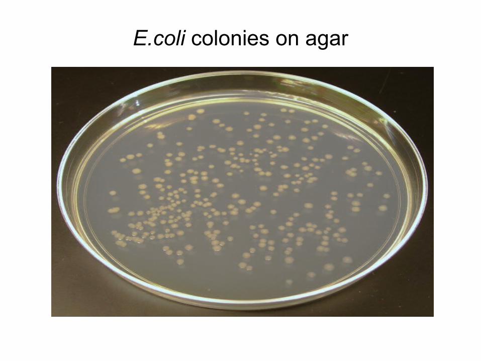
E.coli colonies on agar

Classification of culture media
Main classification criteria:
I. Sate of matter
II. Complexity
III. Purpose
Classification of culture media (continued)
II. Depending on complexity:
1. Simple media (previously described)
2. Enriched media: blood and other special nutrients may be added to simple media to encourage the growth of fastidious microbes e.g. blood agar, ”chocolate” agar
Classification of culture media (continued)
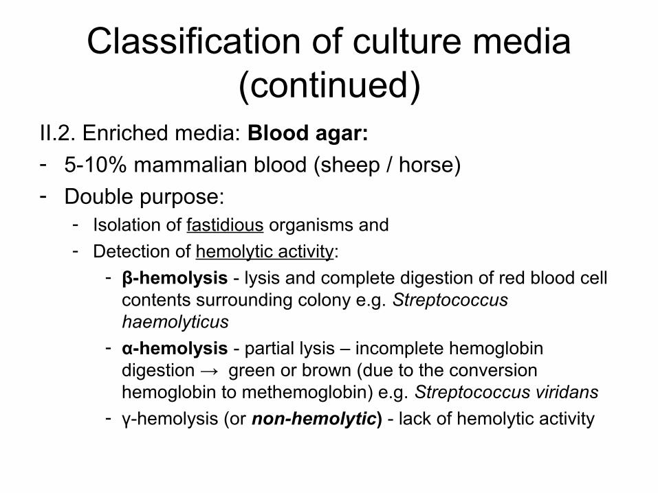
II.2. Enriched media: Blood agar: - 5-10% mammalian blood (sheep / horse)- Double purpose:
- Isolation of fastidious organisms and - Detection of hemolytic activity:
- β-hemolysis - lysis and complete digestion of red blood cell contents surrounding colony e.g. Streptococcus haemolyticus
- α-hemolysis - partial lysis – incomplete hemoglobin digestion → green or brown (due to the conversion hemoglobin to methemoglobin) e.g. Streptococcus viridans
- γ-hemolysis (or non-hemolytic) - lack of hemolytic activity
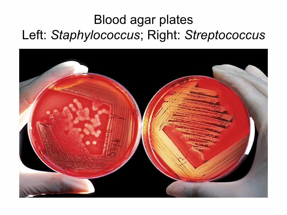
Blood agar platesLeft: Staphylococcus; Right: Streptococcus
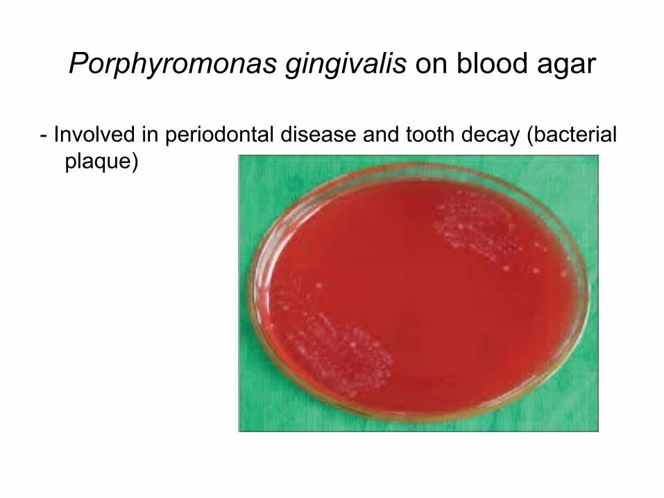
Porphyromonas gingivalis on blood agar
- Involved in periodontal disease and tooth decay (bacterial plaque)
Classification of culture media (continued)
II.2. Enriched media (continued): Chocolate agar- variant of blood agar in which red blood cells have been
lysed by slow, gradual heating to 80°C in order to provide additional growth factors contained in red blood cells
- !Does not contain chocolate!! The name is suggestive for the brownish colour resulted after red blood cell lysis
- used for growing fastidious respiratory bacteria e.g. Haemophilus influenzaze, Neisseria meningitidis
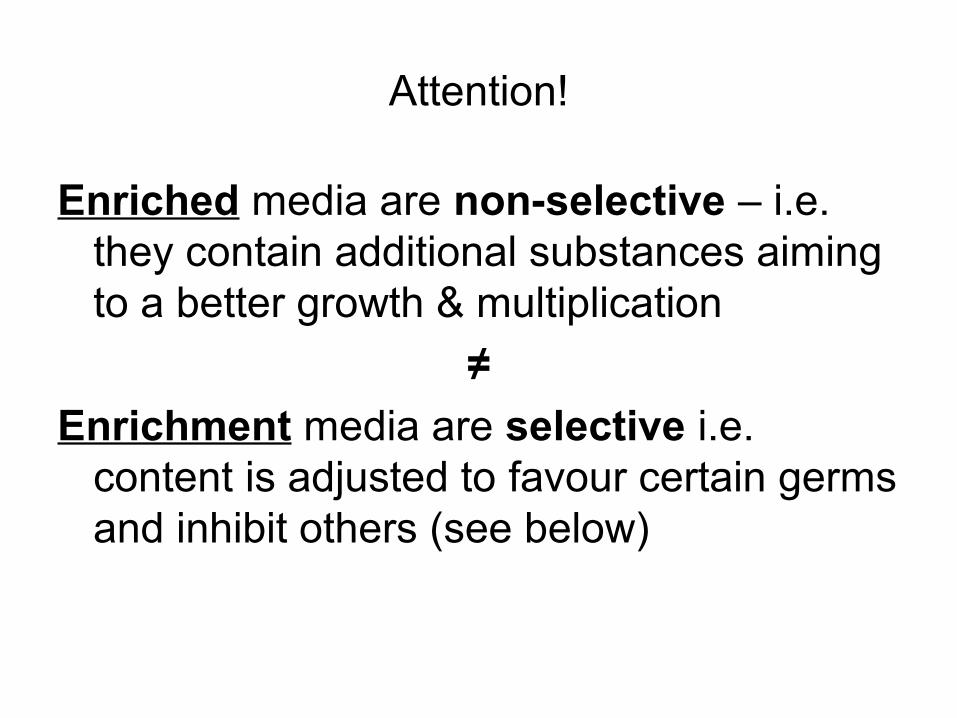
Attention!
Enriched media are non-selective – i.e. they contain additional substances aiming to a better growth & multiplication
≠
Enrichment media are selective i.e. content is adjusted to favour certain germs and inhibit others (see below)
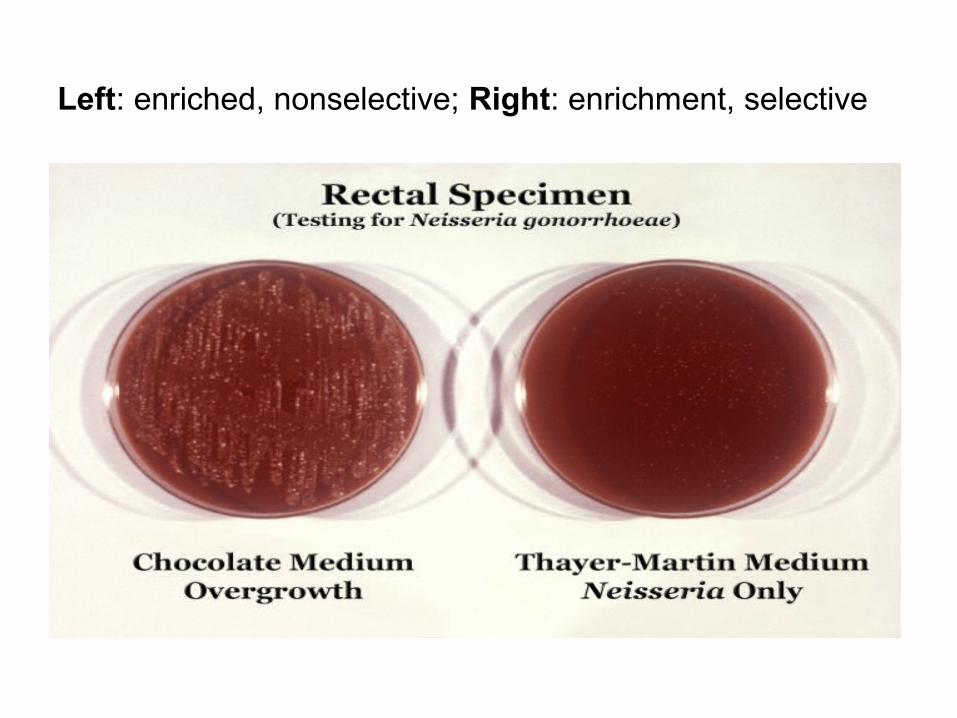
Left: enriched, nonselective; Right: enrichment, selective
Classification of culture media
Main classification criteria:
I. Sate of matter
II. Complexity
III. Purpose
Classification of culture media (continued)
III. Depending on purpose:
1. Selective & enrichment media
2. Diagnostic media
3. Special media
Classification of culture media (continued)
III.1. Selective & enrichment media- Favour the growth and multiplication of certain bacteria
while suppresing other species- Very useful for polymicrobial biological products when
attempting to isolate pure cultures- Used for inoculation of biological products (primary
isolation)
- Composition & cultivation conditions (temperature, aero/anaerobiosis, etc) adjusted according to the known growth characters & requirements of the suspected microbe
Classification of culture media (continued)
III.1. Selective & enrichment media (continued)
Liquid selective media and/or cultivation condition – examples:
- Nutrient broth + acid sodium selenite – Salmonella spp- Peptone water – Vibrio cholerae – the alkaline pH (9)
inhibits other species
- Temperature: +4°C – inhibits the growth of most bacteria EXCEPT Listeria spp
Classification of culture media (continued)
III.1. Selective & enrichment media (continued)
Solid selective media – same principles, same inhibition criteria
Chemical inhibitors: antibiotics (chosen depending on the known natural sensitivity of bacteria) e.g. Vancomycin added when trying to isolate gram negative anaerobic bacteria (gram positive anaerobic bacteria are vancomycin sensitive and their growth will be inhibited)
Classification of culture media (continued)
III. Depending on purpose:
1. Selective & enrichment media
2. Diagnostic media
3. Special media
Classification of culture media (continued)
III.2. Diagnostic media- Contain indicator systems demonstrating metabolic
characters of certain microbial species (fermentation of sugars, production of H2S, etc)
E.g. Fermentation of sugars:
nutrients + sugar + pH indicator – in case fermentation occurs the colour will change indicating the presence of a bacteria which ferments that particular sugar
- Identification relies on performing a number of tests and analyzing the ”profile” which is further matched to known metabolic & growth characters of bacteria

III.2. Diagnostic media (continued)
Mannitol Salt Agar (Chapman) - selective medium with a high salt concentration for the isolation, growth and enumeration of Staphylococcus species: organisms that use mannitol turn the medium colour to yellow
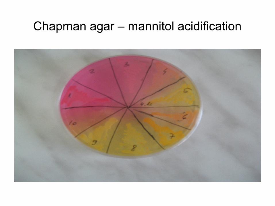
Chapman agar – mannitol acidification
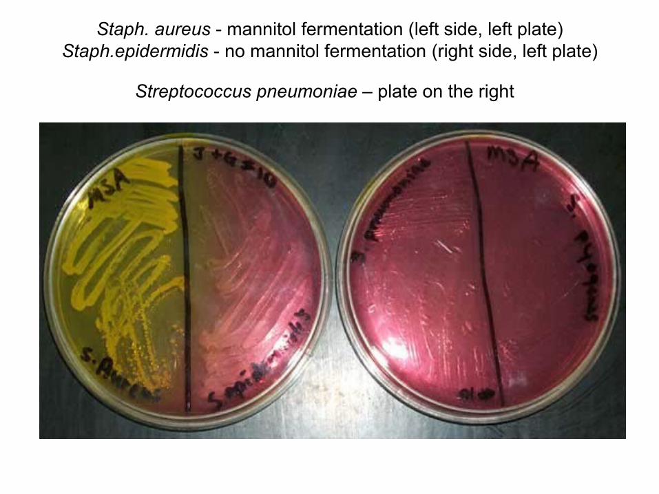
Staph. aureus - mannitol fermentation (left side, left plate) Staph.epidermidis - no mannitol fermentation (right side, left plate)
Streptococcus pneumoniae – plate on the right
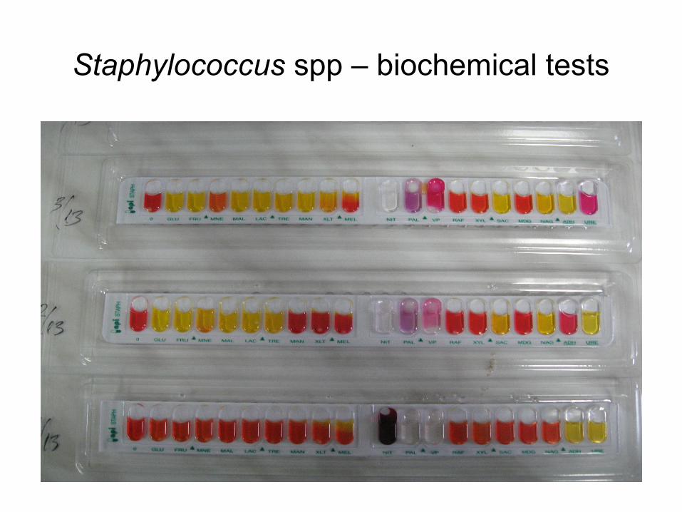
Staphylococcus spp – biochemical tests
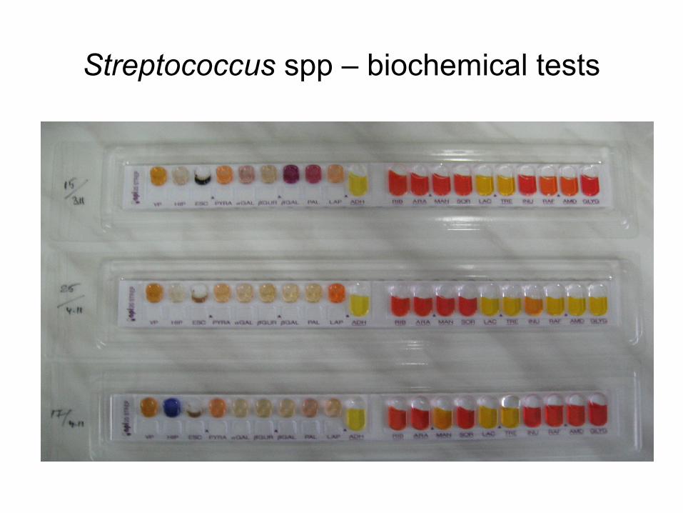
Streptococcus spp – biochemical tests
Classification of culture media (continued)
III. Depending on purpose:
1. Selective & enrichment media
2. Diagnostic media
3. Special media
Classification of culture media (continued)
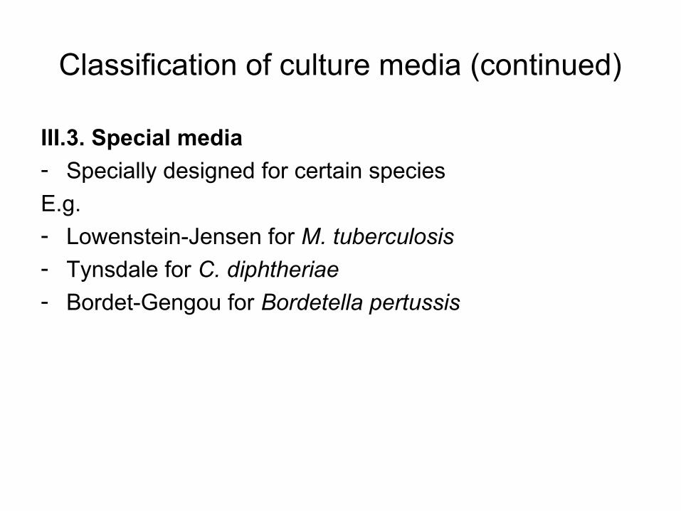
III.3. Special media- Specially designed for certain species
E.g. - Lowenstein-Jensen for M. tuberculosis
- Tynsdale for C. diphtheriae
- Bordet-Gengou for Bordetella pertussis
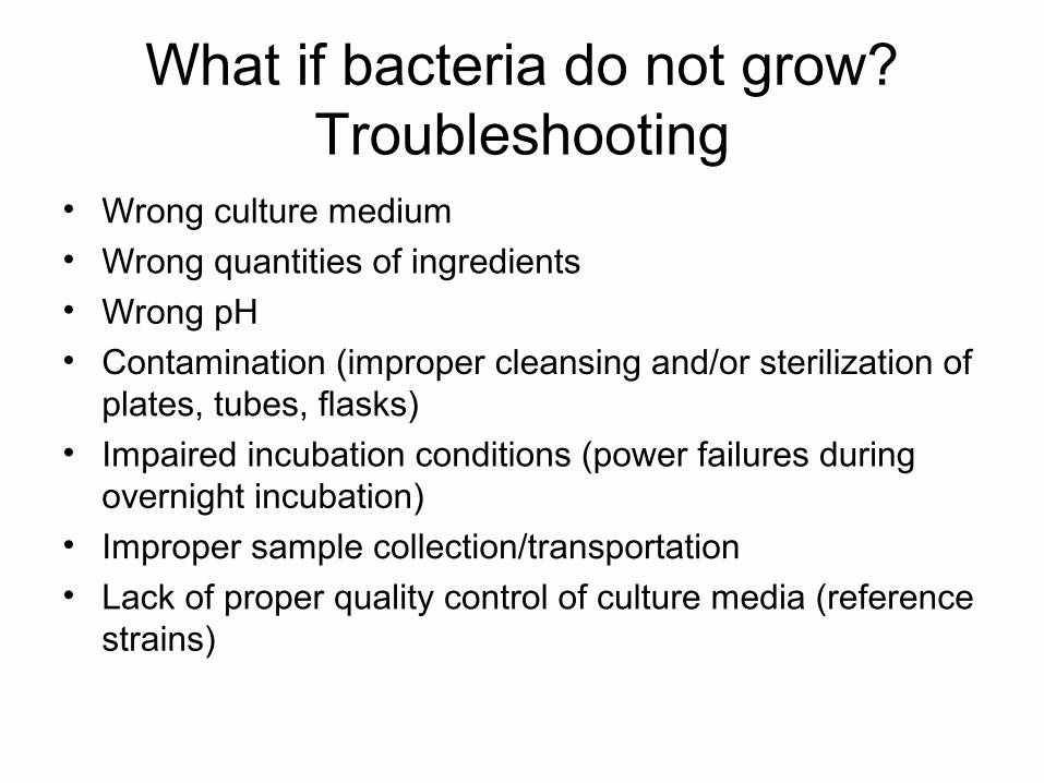
What if bacteria do not grow? Troubleshooting
• Wrong culture medium• Wrong quantities of ingredients• Wrong pH• Contamination (improper cleansing and/or sterilization of
plates, tubes, flasks)
• Impaired incubation conditions (power failures during overnight incubation)
• Improper sample collection/transportation• Lack of proper quality control of culture media (reference
strains)