Embed Size (px)
Citation preview
6 Supratentorial Masses on Magnetic Resonance Imaging

CLINICAL IMAGAGINGAN ATLAS OF DIFFERENTIAL DAIGNOSIS
EISENBERG
DR. Muhammad Bin Zulfiqar PGR-FCPS III SIMS/SHL

• Fig SK 6-1 Low-grade astrocytoma. T2-weighted image shows a high-signal-intensity lesion with well-defined margins, no surrounding edema, and little mass effect.7
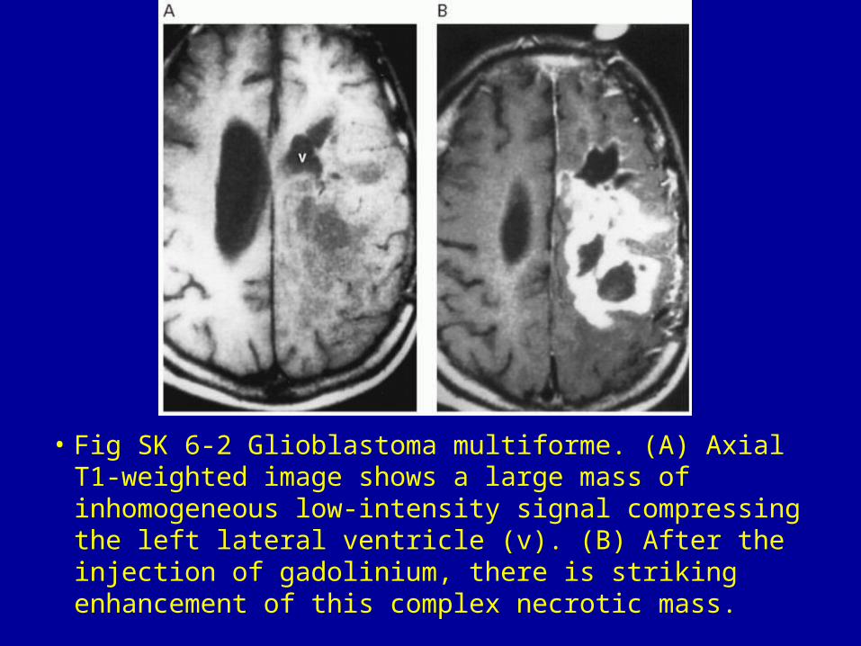
• Fig SK 6-2 Glioblastoma multiforme. (A) Axial T1-weighted image shows a large mass of inhomogeneous low-intensity signal compressing the left lateral ventricle (v). (B) After the injection of gadolinium, there is striking enhancement of this complex necrotic mass.
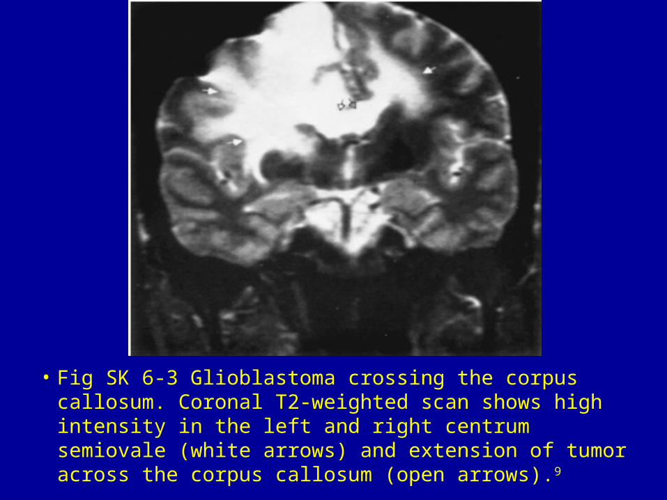
• Fig SK 6-3 Glioblastoma crossing the corpus callosum. Coronal T2-weighted scan shows high intensity in the left and right centrum semiovale (white arrows) and extension of tumor across the corpus callosum (open arrows).9

• Fig SK 6-4 Oligodendroglioma. (A) T1-weighted and (B) proton-density images show a well-differentiated left parietal lobe mass containing a central cystic component. The arrows (B) point to the thickened wall of the enhancing lesion.10

• Fig SK 6-5 Metastases. Axial T2-weighted scan demonstrates three large masses (arrows) surrounded by extensive high-signal edema.

Fig SK 6-6 Lymphoma. Homogeneous mass of increased signal intensity (arrows) extending to involve the uncus.

• Fig SK 6-7 Meningioma. Huge mass (black and white arrowheads) that appears hypointense on a T1-weighted coronal scan (A) and hyperintense on a T2-weighted image (B). Note the dramatic shift of the ventricle (v) caused by the mass effect of the tumor. The arrows point to areas of hemorrhage in the neoplasm.
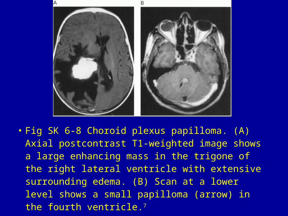
• Fig SK 6-8 Choroid plexus papilloma. (A) Axial postcontrast T1-weighted image shows a large enhancing mass in the trigone of the right lateral ventricle with extensive surrounding edema. (B) Scan at a lower level shows a small papilloma (arrow) in the fourth ventricle.7
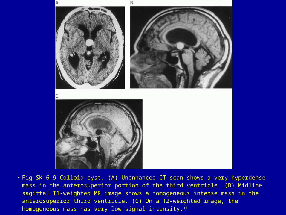
• Fig SK 6-9 Colloid cyst. (A) Unenhanced CT scan shows a very hyperdense mass in the anterosuperior portion of the third ventricle. (B) Midline sagittal T1-weighted MR image shows a homogeneous intense mass in the anterosuperior third ventricle. (C) On a T2-weighted image, the homogeneous mass has very low signal intensity.11
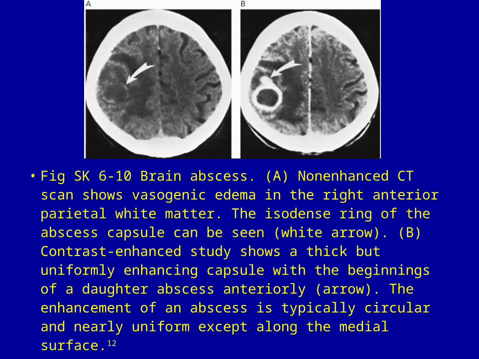
• Fig SK 6-10 Brain abscess. (A) Nonenhanced CT scan shows vasogenic edema in the right anterior parietal white matter. The isodense ring of the abscess capsule can be seen (white arrow). (B) Contrast-enhanced study shows a thick but uniformly enhancing capsule with the beginnings of a daughter abscess anteriorly (arrow). The enhancement of an abscess is typically circular and nearly uniform except along the medial surface.12

• Fig SK 6-11 Intraparenchymal hematoma. Coronal T2-weighted scan shows a large hematoma in the left thalamic region (arrow). The hematoma consists of two portions: a central area of increased signal intensity representing methemoglobin, and a surrounding area of low signal intensity representing hemosiderin.
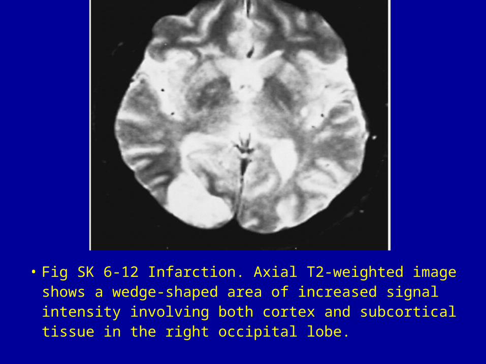
• Fig SK 6-12 Infarction. Axial T2-weighted image shows a wedge-shaped area of increased signal intensity involving both cortex and subcortical tissue in the right occipital lobe.
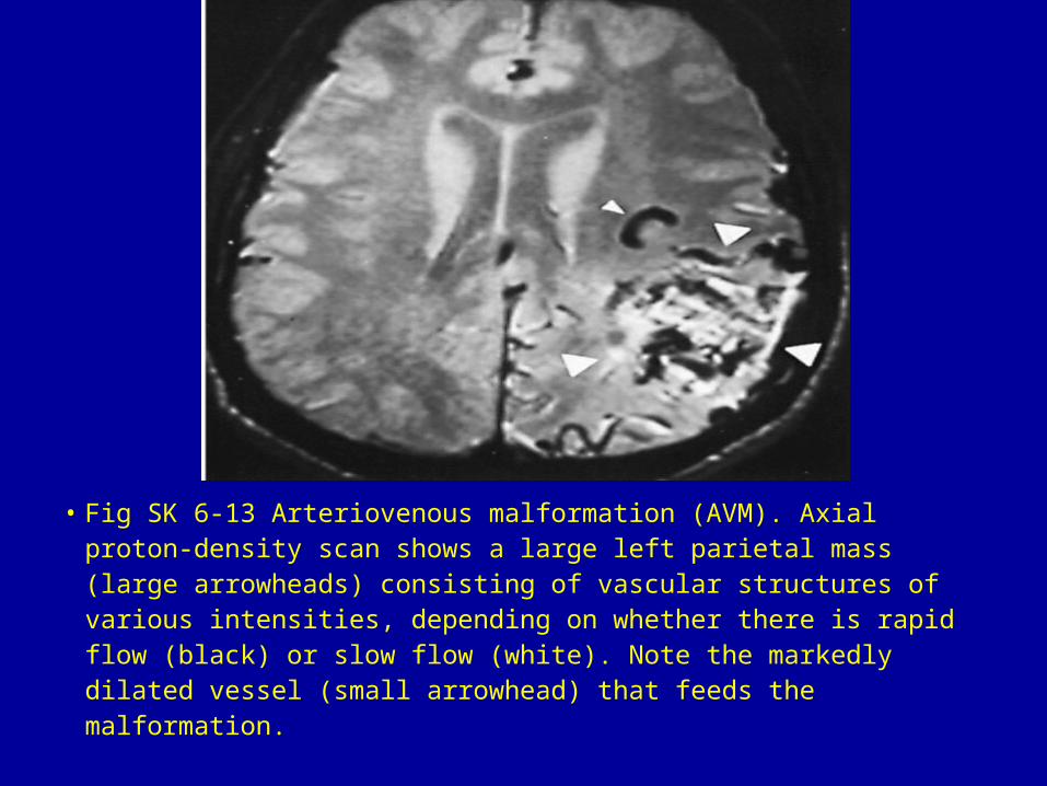
• Fig SK 6-13 Arteriovenous malformation (AVM). Axial proton-density scan shows a large left parietal mass (large arrowheads) consisting of vascular structures of various intensities, depending on whether there is rapid flow (black) or slow flow (white). Note the markedly dilated vessel (small arrowhead) that feeds the malformation.
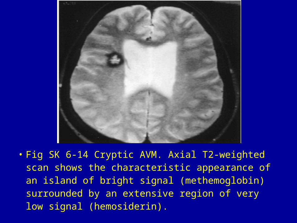
• Fig SK 6-14 Cryptic AVM. Axial T2-weighted scan shows the characteristic appearance of an island of bright signal (methemoglobin) surrounded by an extensive region of very low signal (hemosiderin).
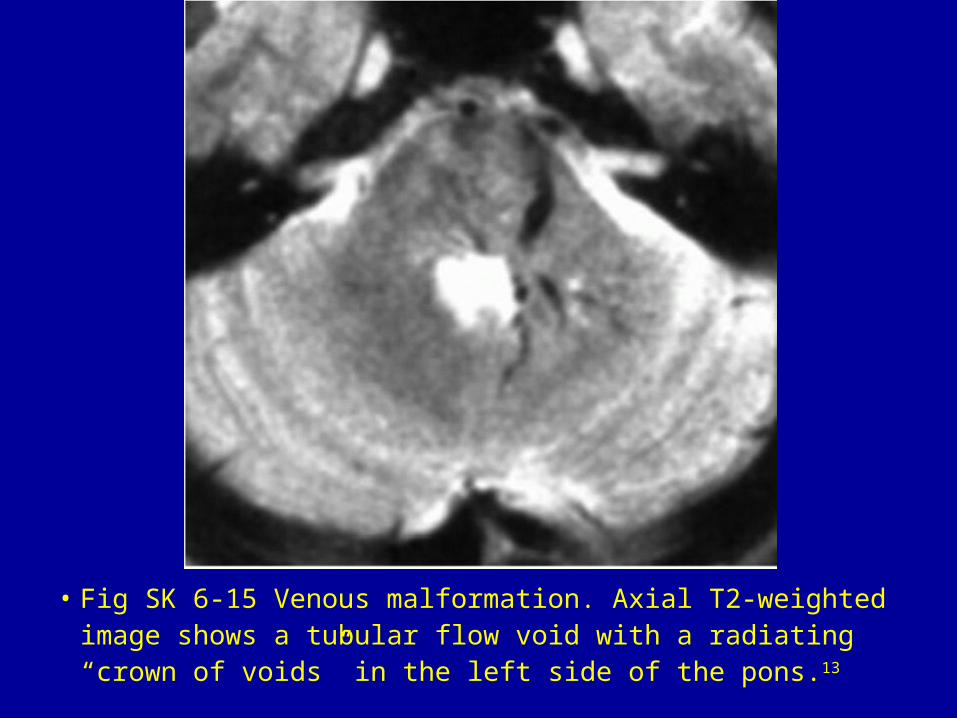
• Fig SK 6-15 Venous malformation. Axial T2-weighted image shows a tubular flow void with a radiating “crown of voids” in the left side of the pons.13
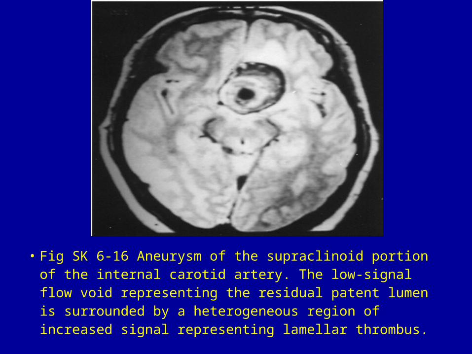
• Fig SK 6-16 Aneurysm of the supraclinoid portion of the internal carotid artery. The low-signal flow void representing the residual patent lumen is surrounded by a heterogeneous region of increased signal representing lamellar thrombus.