Embed Size (px)
Citation preview
Premedical BiologyMotor mechanism
Skeletal system
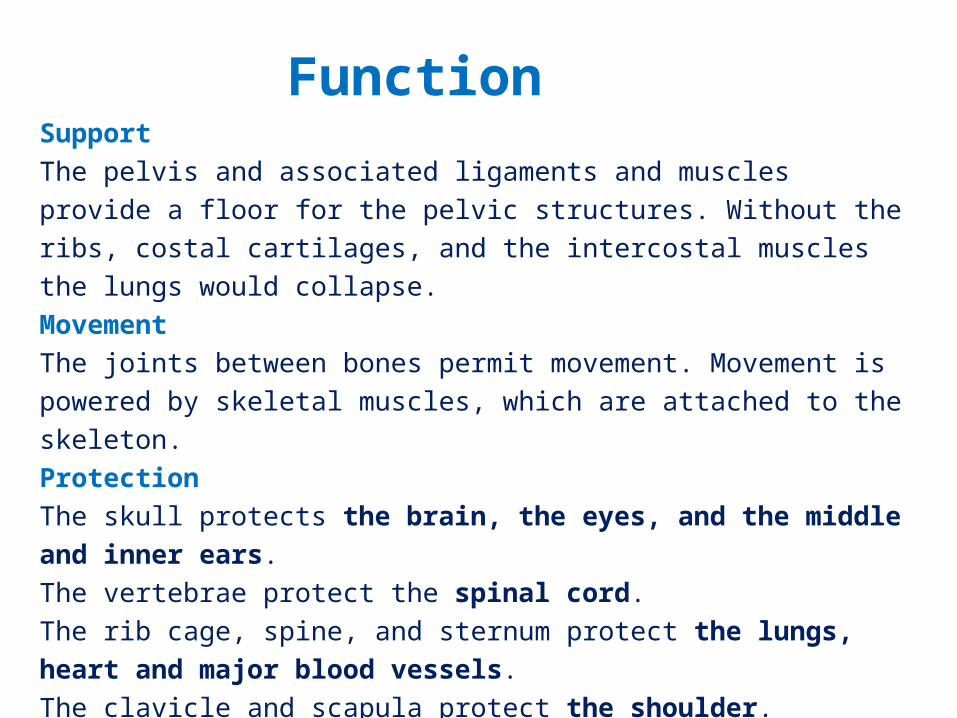
SupportThe pelvis and associated ligaments and muscles provide a floor for the pelvic structures. Without the ribs, costal cartilages, and the intercostal muscles the lungs would collapse.MovementThe joints between bones permit movement. Movement is powered by skeletal muscles, which are attached to the skeleton. ProtectionThe skull protects the brain, the eyes, and the middle and inner ears. The vertebrae protect the spinal cord. The rib cage, spine, and sternum protect the lungs, heart and major blood vessels. The clavicle and scapula protect the shoulder. The ilium and spine protect the digestive and urogenital systems and the hip.
Function
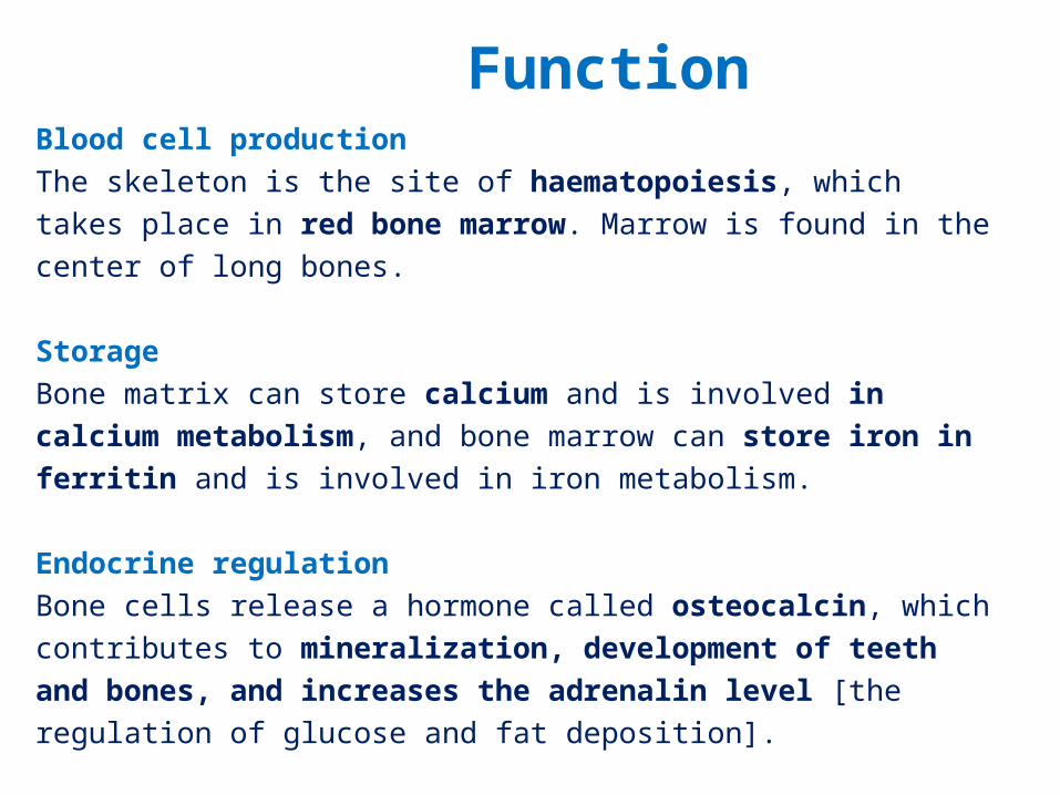
Blood cell productionThe skeleton is the site of haematopoiesis, which takes place in red bone marrow. Marrow is found in the center of long bones.
StorageBone matrix can store calcium and is involved in calcium metabolism, and bone marrow can store iron in ferritin and is involved in iron metabolism.
Endocrine regulationBone cells release a hormone called osteocalcin, which contributes to mineralization, development of teeth and bones, and increases the adrenalin level [the regulation of glucose and fat deposition].
Function
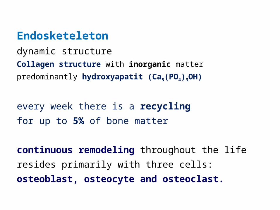
Endosketeletondynamic structureCollagen structure with inorganic matter predominantly
hydroxyapatit (Ca5(PO4)3OH)
every week there is a recycling for up to 5% of bone matter
continuous remodeling throughout the life resides primarily with three cells:osteoblast, osteocyte and osteoclast.
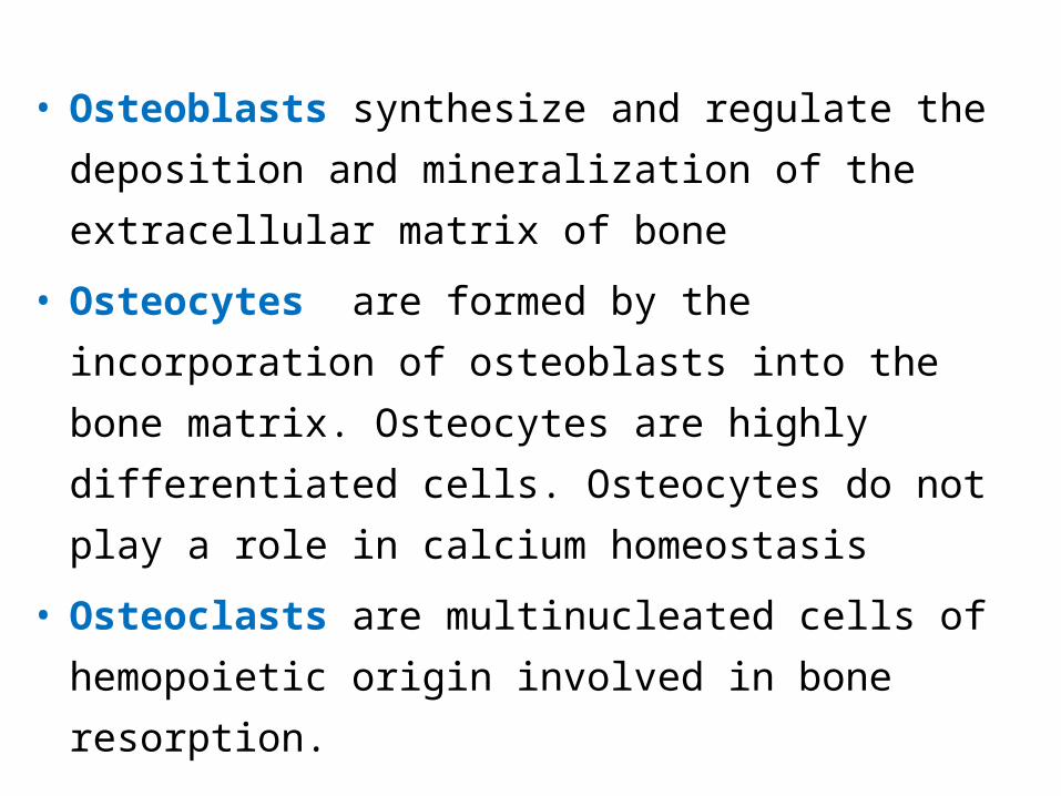
• Osteoblasts synthesize and regulate the deposition and mineralization of the extracellular matrix of bone
• Osteocytes are formed by the incorporation of osteoblasts into the bone matrix. Osteocytes are highly differentiated cells. Osteocytes do not play a role in calcium homeostasis
• Osteoclasts are multinucleated cells of hemopoietic origin involved in bone resorption.
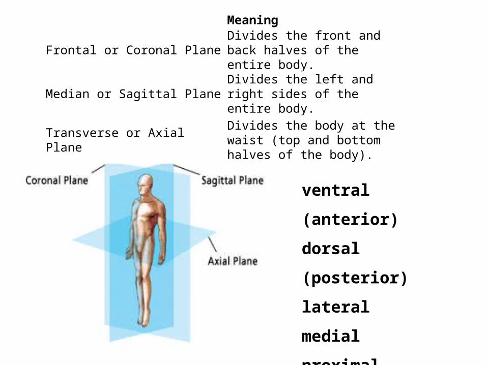
Meaning
Frontal or Coronal Plane Divides the front and back halves of the entire body.
Median or Sagittal Plane Divides the left and right sides of the entire body.
Transverse or Axial Plane Divides the body at the waist (top and bottom halves of the body).
ventral (anterior)
dorsal (posterior)
lateral
medial
proximal
distal
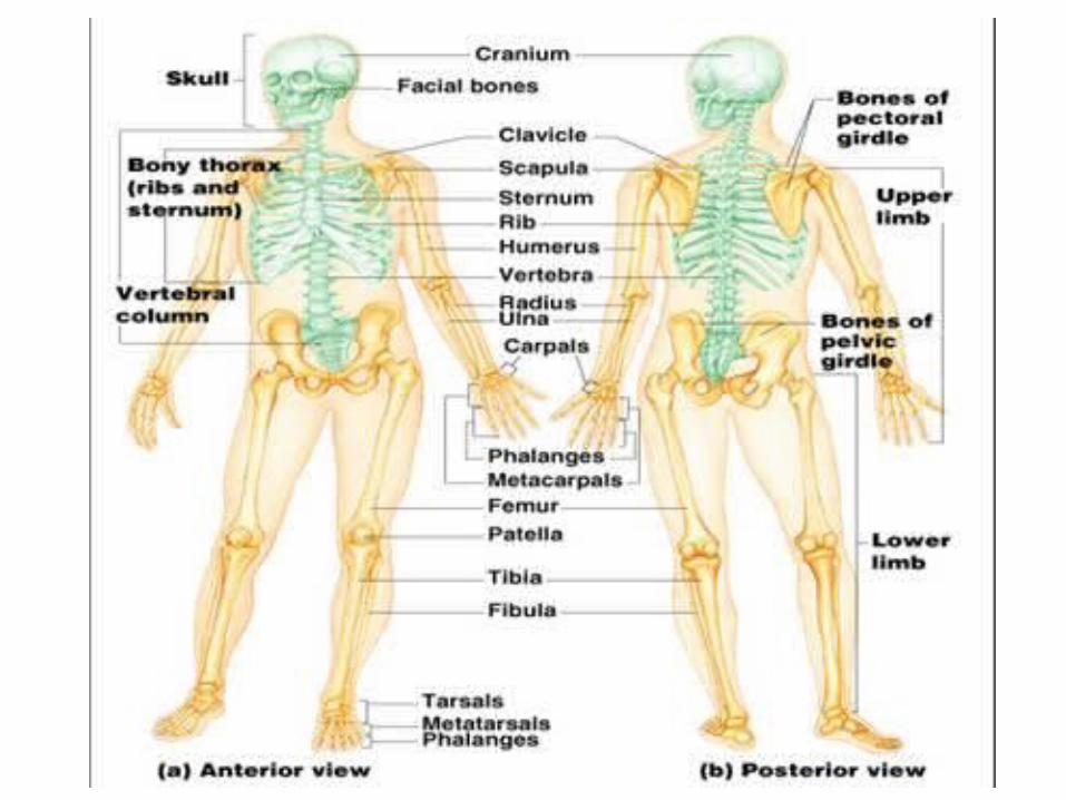
consists of both fused and individual bones supported and
supplemented by ligaments, tendons, anchoring muscles and cartilage.
Skeleton of adult human consists of 206 bonesNew-born children have about 300 bones [grow together].Fused bones include those of the pelvis and the cranium.
The development of whole skeleton is accomplished in the
age of 20 years.
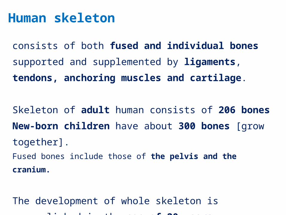
Human skeleton
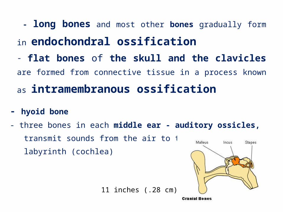
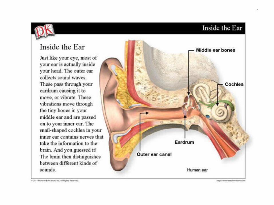
- hyoid bone - three bones in each middle ear - auditory ossicles, transmit sounds
from the air to the fluid-filled labyrinth (cochlea)
- long bones and most other bones gradually form in
endochondral ossification- flat bones of the skull and the clavicles are formed from
connective tissue in a process known as intramembranous ossification
11 inches (.28 cm)
Joints
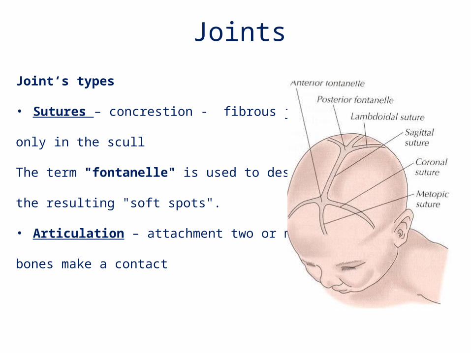
Joint‘s types
• Sutures – concrestion - fibrous joint
only in the scull
The term "fontanelle" is used to describe
the resulting "soft spots".
• Articulation – attachment two or more
bones make a contact
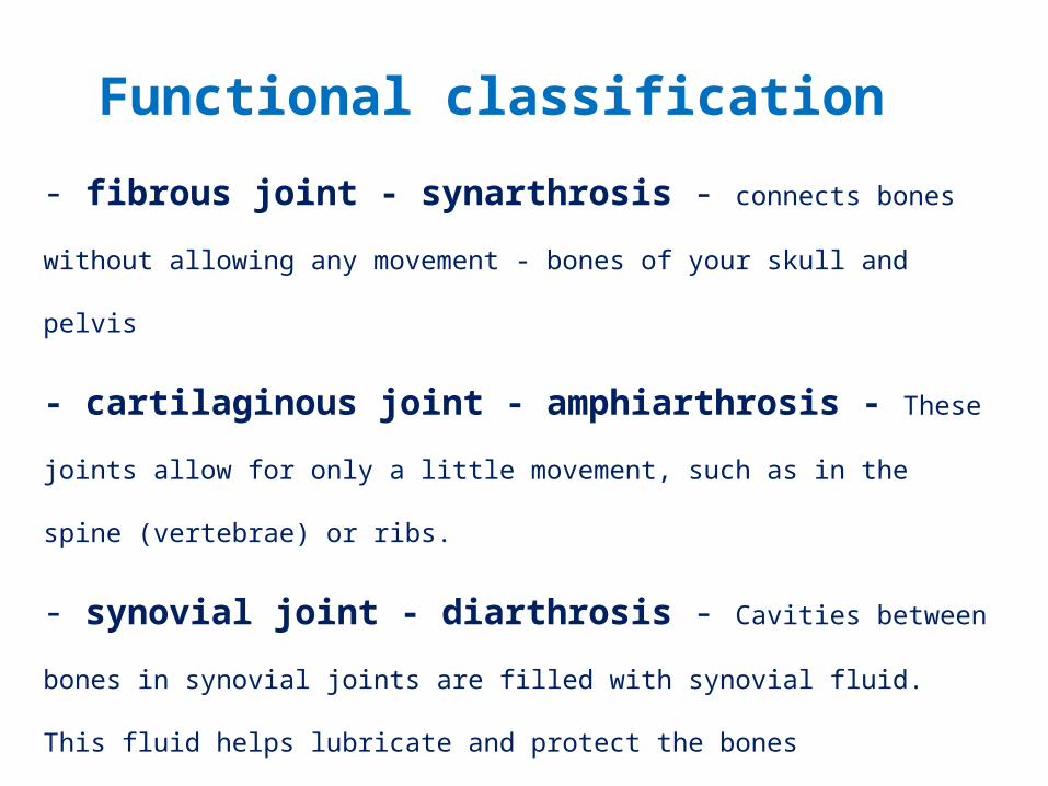
- fibrous joint - synarthrosis - connects bones without allowing any
movement - bones of your skull and pelvis
- cartilaginous joint - amphiarthrosis - These joints allow for only
a little movement, such as in the spine (vertebrae) or ribs.
- synovial joint - diarthrosis - Cavities between bones in synovial
joints are filled with synovial fluid. This fluid helps lubricate and protect the bones
permits a variety of movements (e.g., shoulder, hip, elbow, knee, etc.).
Functional classification
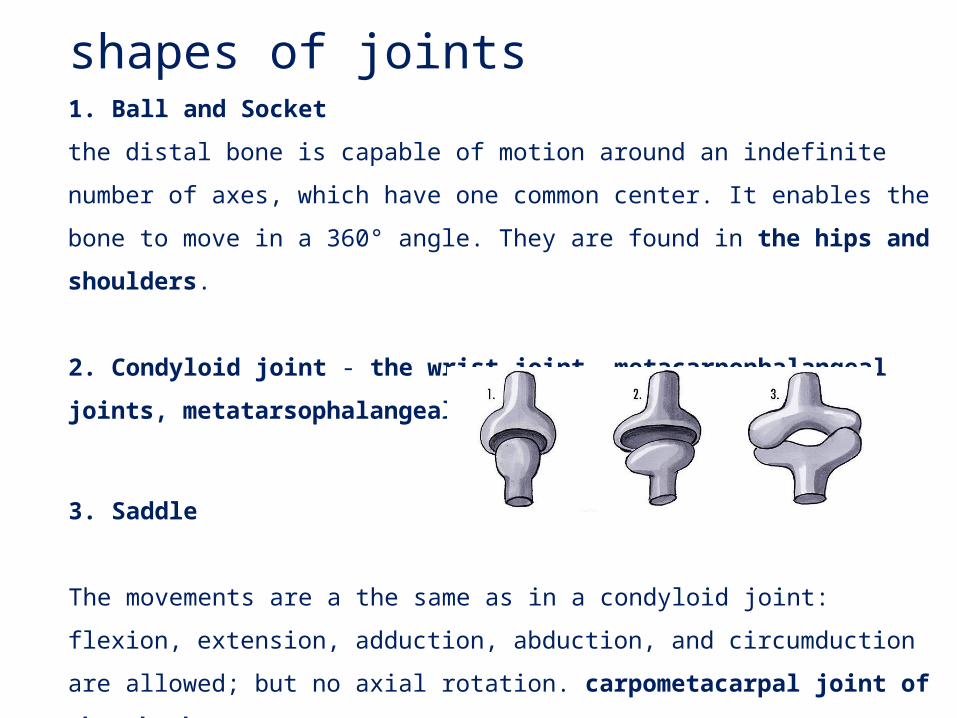
shapes of joints1. Ball and Socket
the distal bone is capable of motion around an indefinite number of axes, which
have one common center. It enables the bone to move in a 360° angle. They are
found in the hips and shoulders.
2. Condyloid joint - the wrist-joint, metacarpophalangeal joints,
metatarsophalangeal joints
3. Saddle
The movements are a the same as in a condyloid joint: flexion, extension,
adduction, abduction, and circumduction are allowed; but no axial rotation.
carpometacarpal joint of the thumb
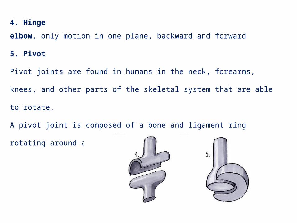
4. Hinge
elbow, only motion in one plane, backward and forward
5. Pivot
Pivot joints are found in humans in the neck, forearms, knees, and other parts of
the skeletal system that are able to rotate.
A pivot joint is composed of a bone and ligament ring rotating around another
bone.
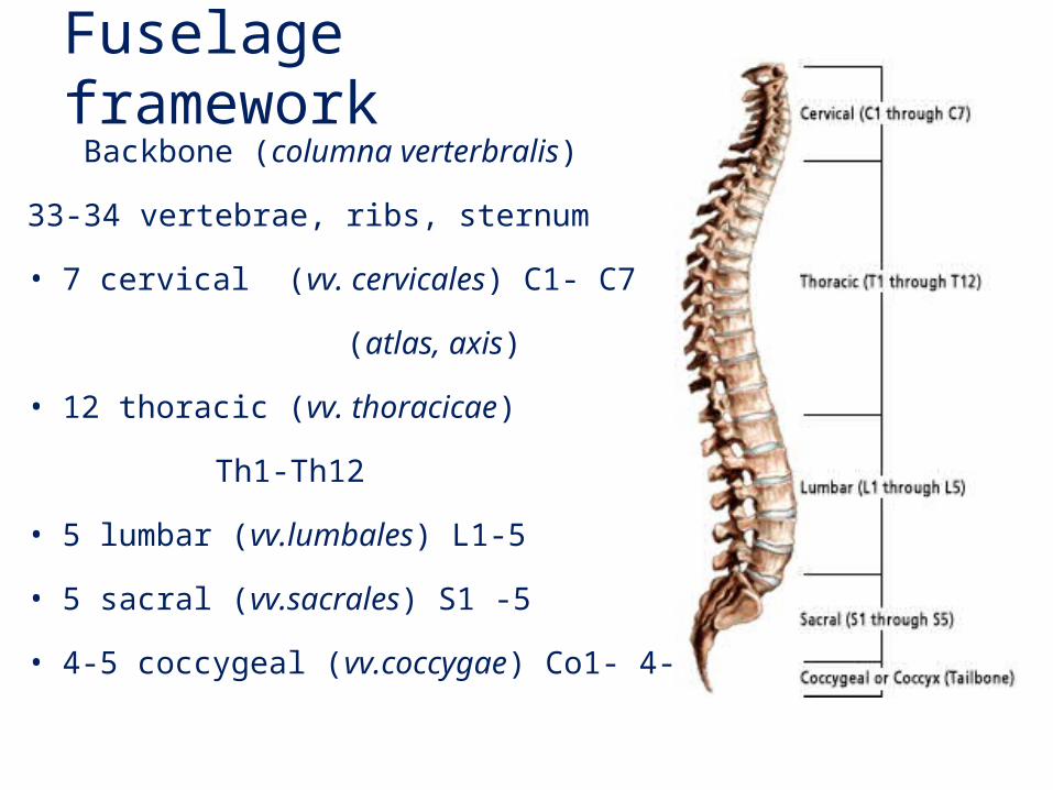
Fuselage framework Backbone (columna verterbralis)
33-34 vertebrae, ribs, sternum
• 7 cervical (vv. cervicales) C1- C7
(atlas, axis)
• 12 thoracic (vv. thoracicae)
Th1-Th12
• 5 lumbar (vv.lumbales) L1-5
• 5 sacral (vv.sacrales) S1 -5
• 4-5 coccygeal (vv.coccygae) Co1- 4-5
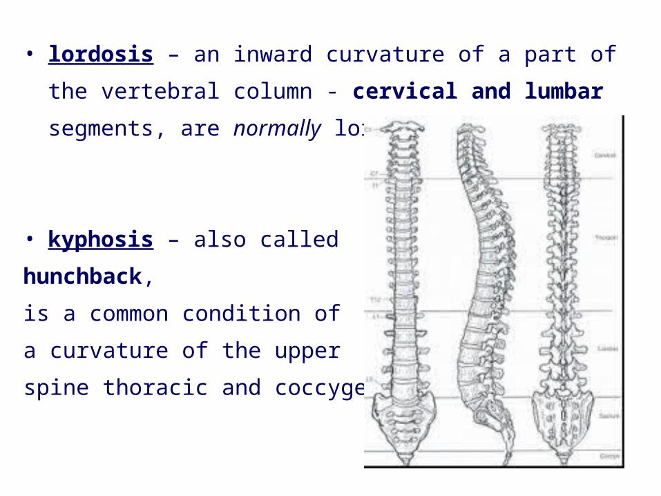
• lordosis – an inward curvature of a part of the vertebral
column - cervical and lumbar segments, are normally
lordotic
• kyphosis – also called
hunchback,
is a common condition of
a curvature of the upper
spine thoracic and coccygeal
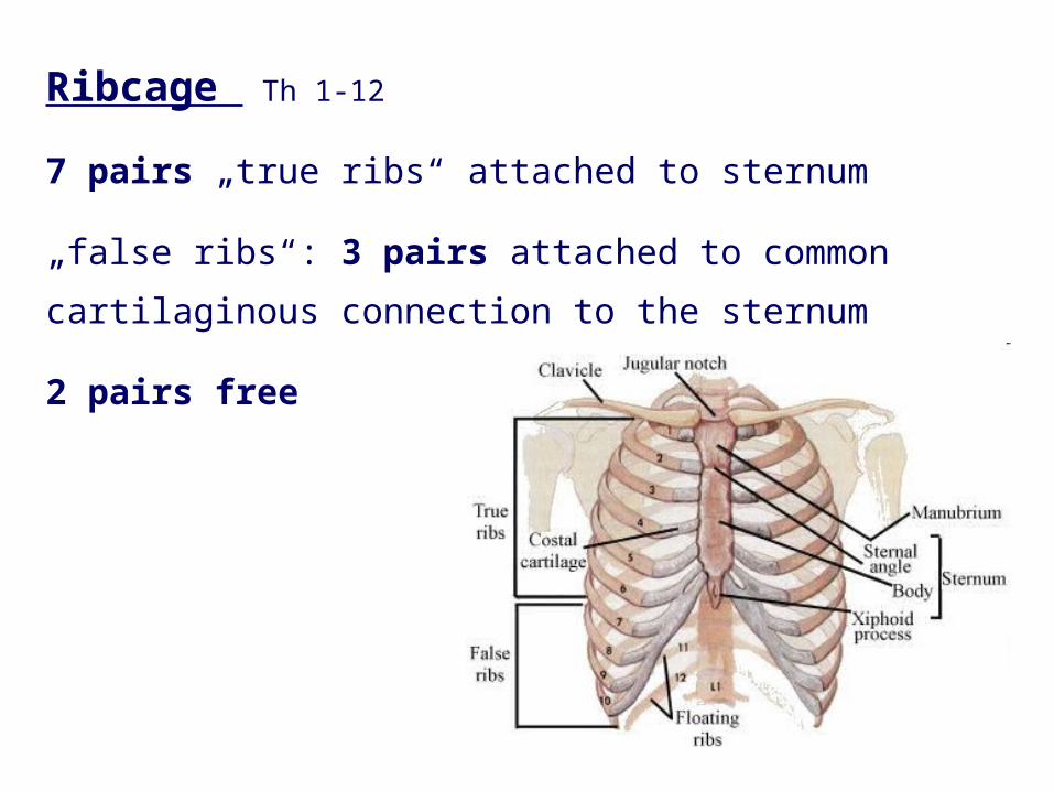
Ribcage Th 1-12
7 pairs „true ribs“ attached to sternum
„false ribs“: 3 pairs attached to common cartilaginous
connection to the sternum
2 pairs free
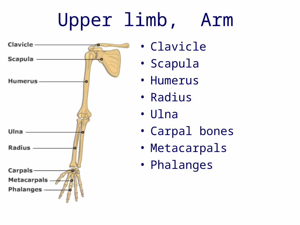
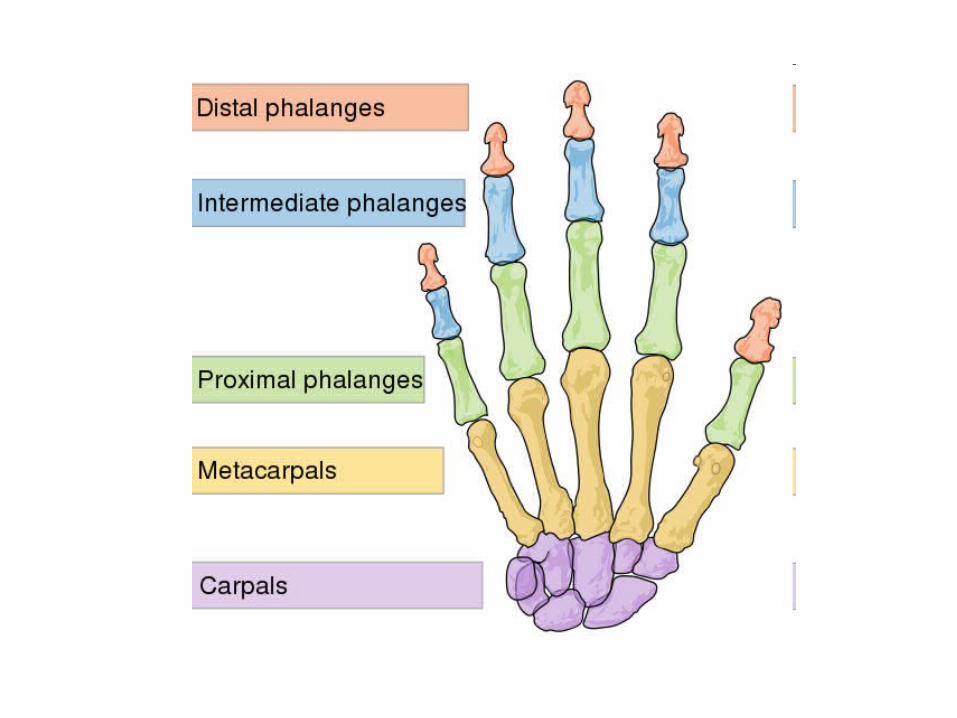
Upper limb, Arm• Clavicle • Scapula • Humerus • Radius • Ulna • Carpal bones • Metacarpals • Phalanges
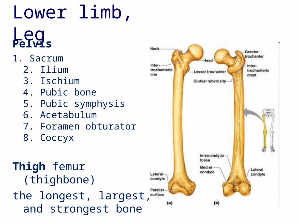
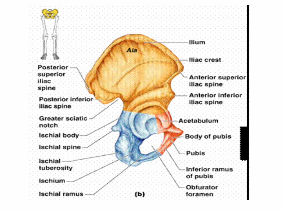
Lower limb, Leg Pelvis1. Sacrum
2. Ilium3. Ischium4. Pubic bone5. Pubic symphysis6. Acetabulum7. Foramen obturator8. Coccyx
Thigh femur (thighbone) the longest, largest, and
strongest bone
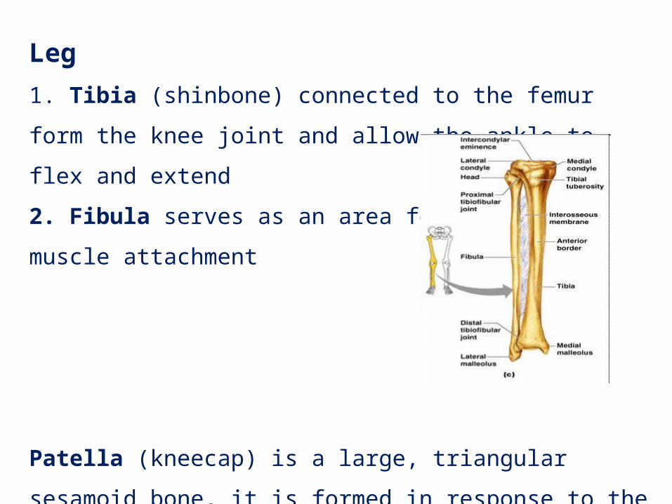
Leg1. Tibia (shinbone) connected to the femur form the knee joint
and allow the ankle to flex and extend
2. Fibula serves as an area for
muscle attachment
Patella (kneecap) is a large, triangular sesamoid bone, it is
formed in response to the strain in the tendon
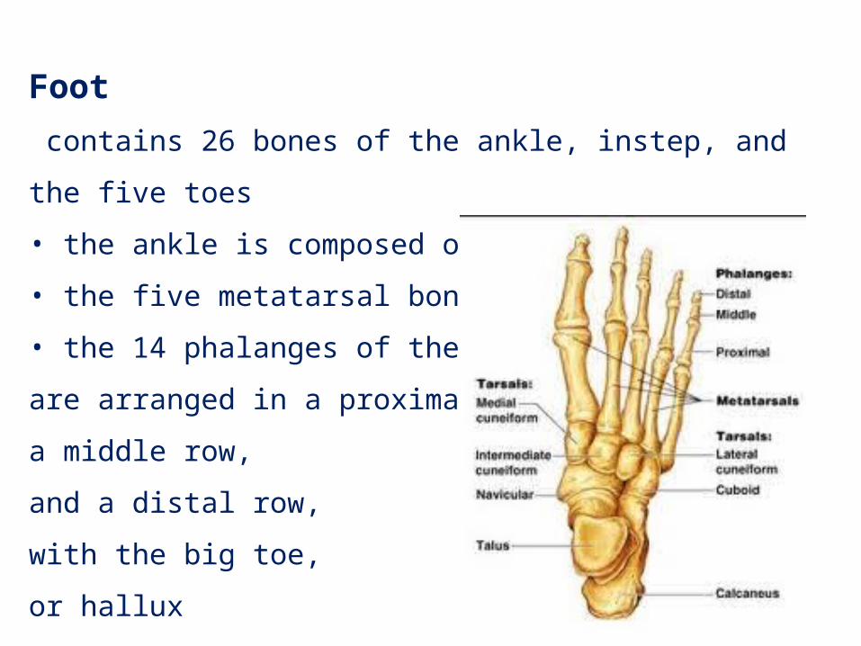
Foot contains 26 bones of the ankle, instep, and the five toes
• the ankle is composed of the 7 tarsal bones
• the five metatarsal bones
• the 14 phalanges of the foot,
are arranged in a proximal row,
a middle row,
and a distal row,
with the big toe,
or hallux
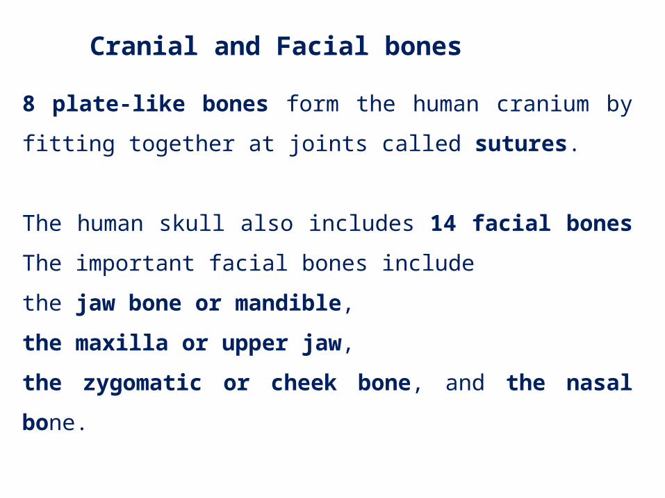
8 plate-like bones form the human cranium by fitting together
at joints called sutures.
The human skull also includes 14 facial bones
The important facial bones include
the jaw bone or mandible,
the maxilla or upper jaw,
the zygomatic or cheek bone, and the nasal bone.
Cranial and Facial bones
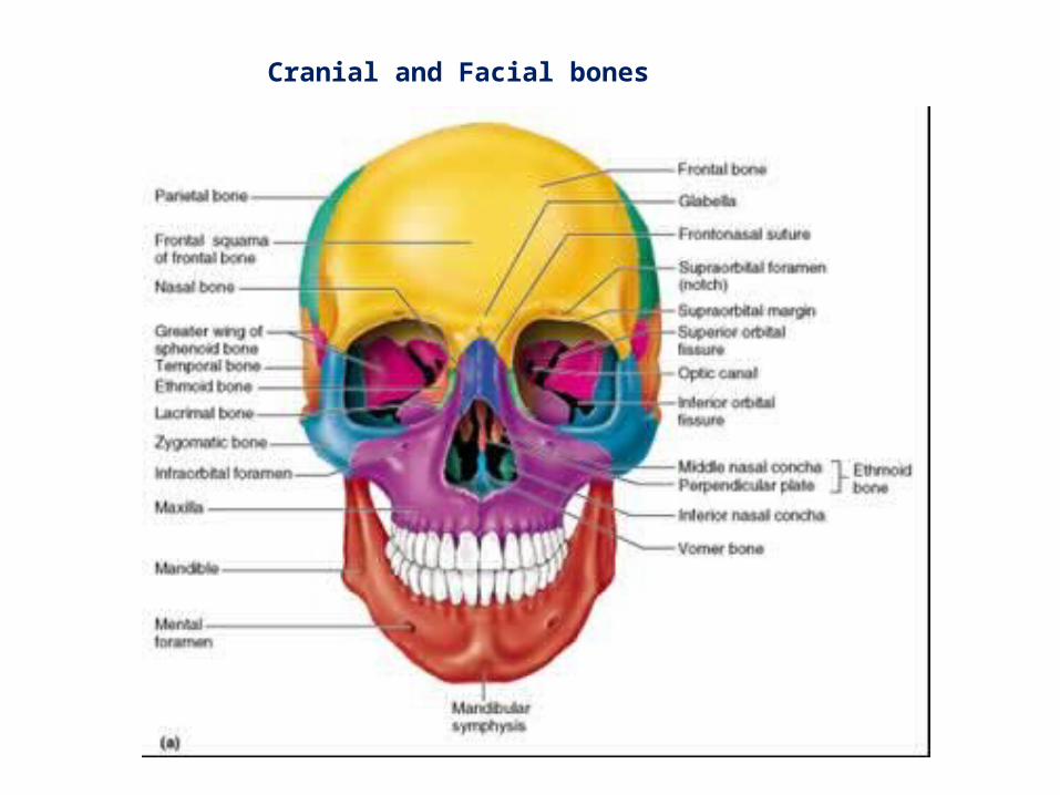
Cranial and Facial bones
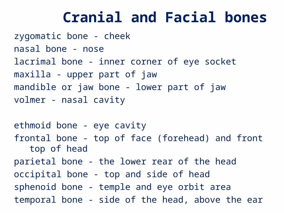
zygomatic bone - cheeknasal bone - noselacrimal bone - inner corner of eye socketmaxilla - upper part of jawmandible or jaw bone - lower part of jaw volmer - nasal cavity
ethmoid bone - eye cavityfrontal bone - top of face (forehead) and front top of headparietal bone - the lower rear of the headoccipital bone - top and side of headsphenoid bone - temple and eye orbit areatemporal bone - side of the head, above the ear
Cranial and Facial bones
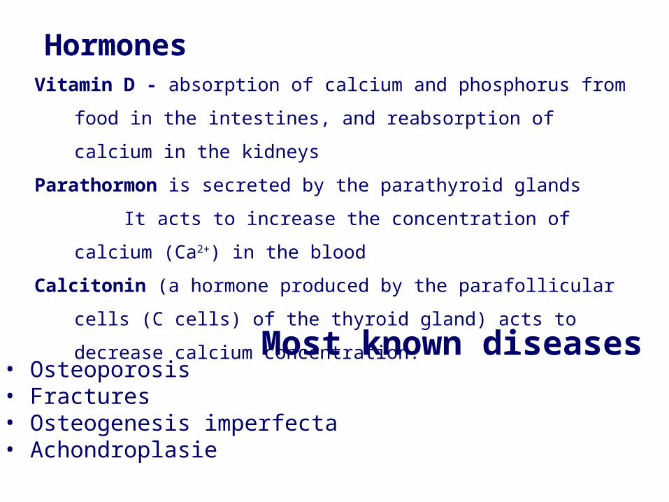
Most known diseases• Osteoporosis • Fractures• Osteogenesis imperfecta• Achondroplasie
HormonesVitamin D - absorption of calcium and phosphorus from food in the
intestines, and reabsorption of calcium in the kidneys
Parathormon is secreted by the parathyroid glands
It acts to increase the concentration of calcium (Ca2+) in the blood
Calcitonin (a hormone produced by the parafollicular cells (C cells) of
the thyroid gland) acts to decrease calcium concentration.
Muscular system
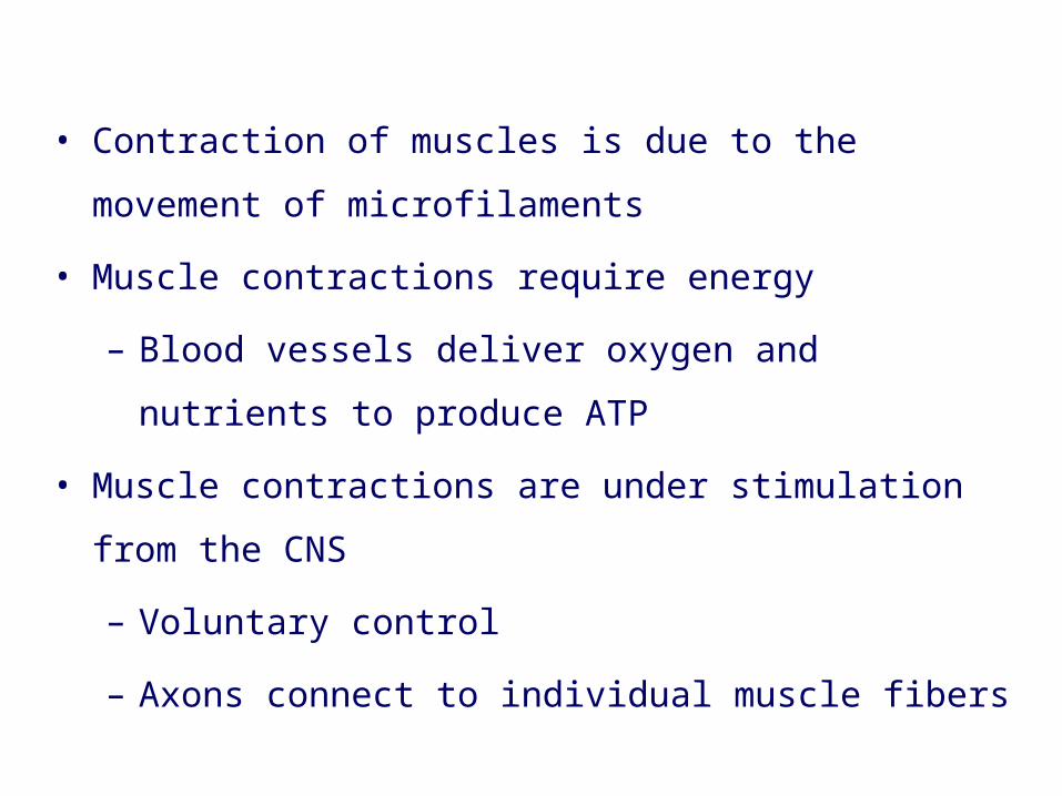
• Contraction of muscles is due to the movement of
microfilaments
• Muscle contractions require energy
– Blood vessels deliver oxygen and nutrients to produce
ATP
• Muscle contractions are under stimulation from the CNS
– Voluntary control
– Axons connect to individual muscle fibers
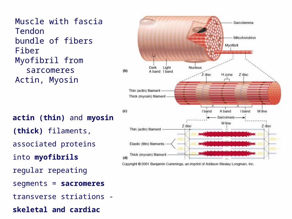
actin (thin) and myosin (thick)
filaments, associated proteins
into myofibrils
regular repeating segments =
sacromeres transverse
striations - skeletal and
cardiac
Muscle with fasciaTendon bundle of fibers FiberMyofibril from sarcomeres Actin, Myosin
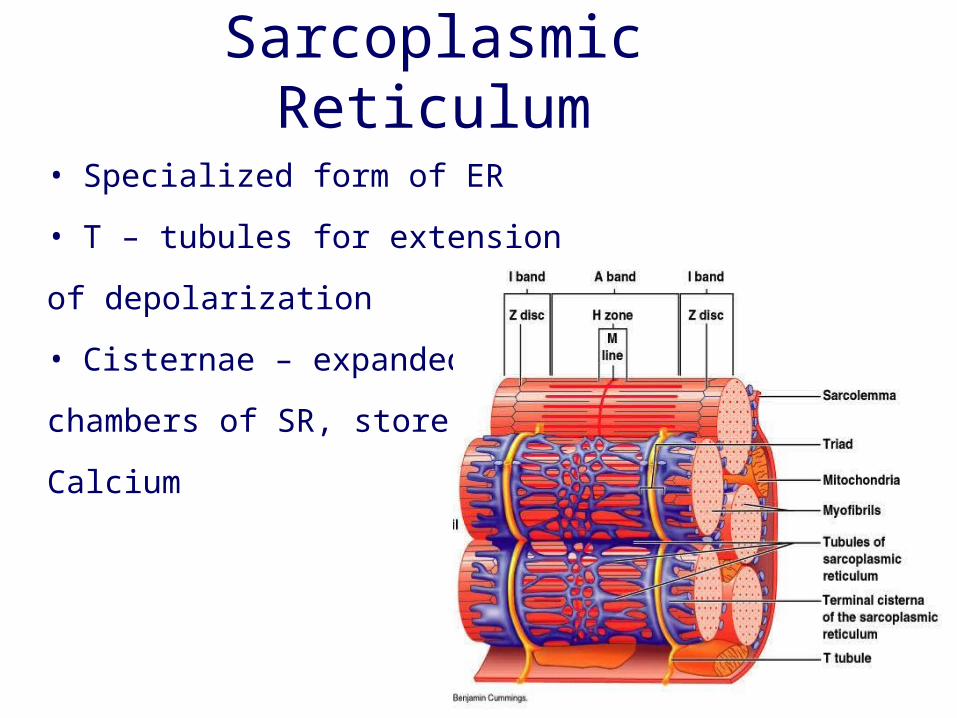
• Specialized form of ER
• T – tubules for extension
of depolarization
• Cisternae – expanded
chambers of SR, store
Calcium
Sarcoplasmic Reticulum
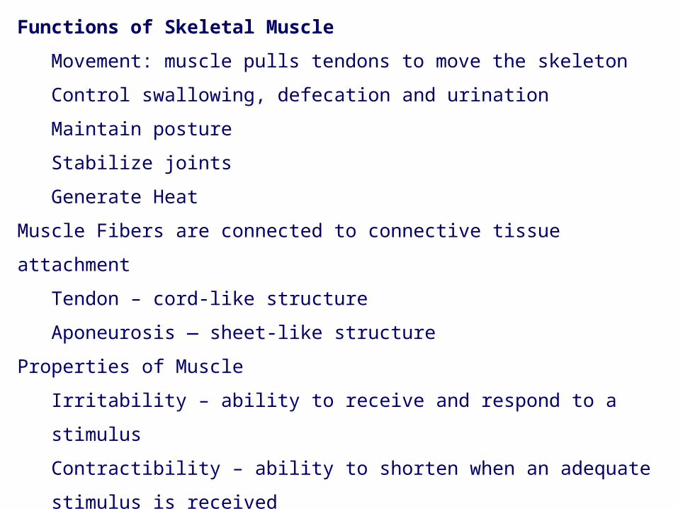
Functions of Skeletal Muscle
Movement: muscle pulls tendons to move the skeleton
Control swallowing, defecation and urination
Maintain posture
Stabilize joints
Generate Heat
Muscle Fibers are connected to connective tissue attachment
Tendon – cord-like structure
Aponeurosis — sheet-like structure
Properties of Muscle
Irritability – ability to receive and respond to a stimulus
Contractibility – ability to shorten when an adequate stimulus is received
Extensibility – ability to stretch when an adequate stimulus is received
Elasticity – ability to return to normal shape
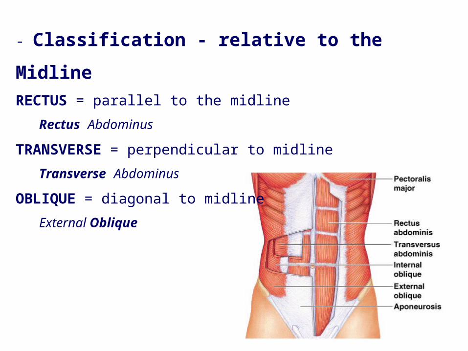
- Classification - relative to the MidlineRECTUS = parallel to the midline
Rectus Abdominus
TRANSVERSE = perpendicular to midline
Transverse Abdominus
OBLIQUE = diagonal to midline
External Oblique

Classification - relative to structure near which muscle is foundFRONTALIS = near FRONTAL bone
OCCIPITALIS = near OCCIPITAL bone
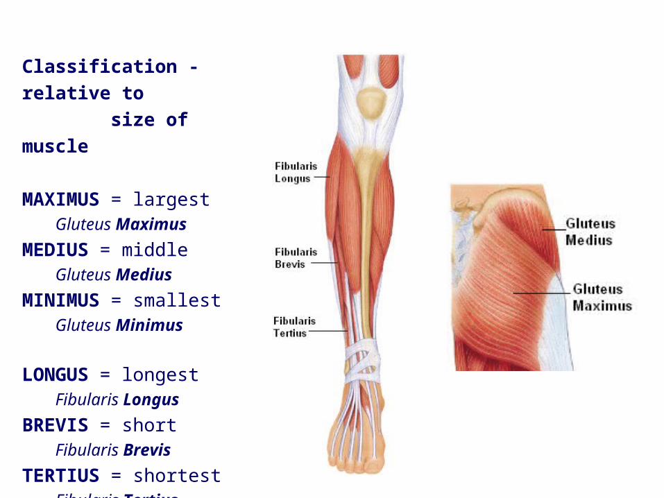
Classification - relative to size of muscle
MAXIMUS = largestGluteus Maximus
MEDIUS = middleGluteus Medius
MINIMUS = smallestGluteus Minimus
LONGUS = longestFibularis Longus
BREVIS = shortFibularis Brevis
TERTIUS = shortestFibularis Tertius
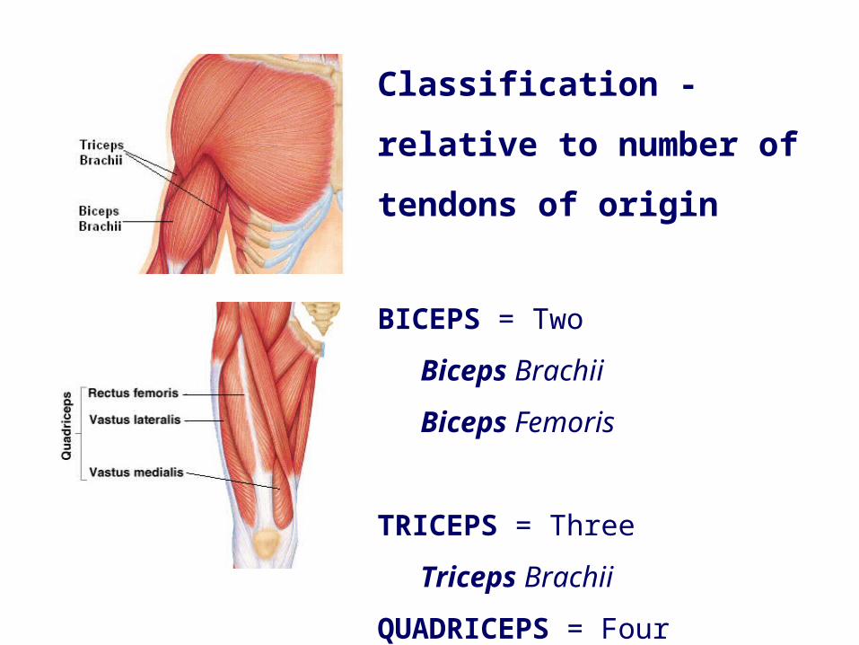
Classification - relative to
number of tendons of origin
BICEPS = Two
Biceps Brachii
Biceps Femoris
TRICEPS = Three
Triceps Brachii
QUADRICEPS = Four
Quadriceps Femoris
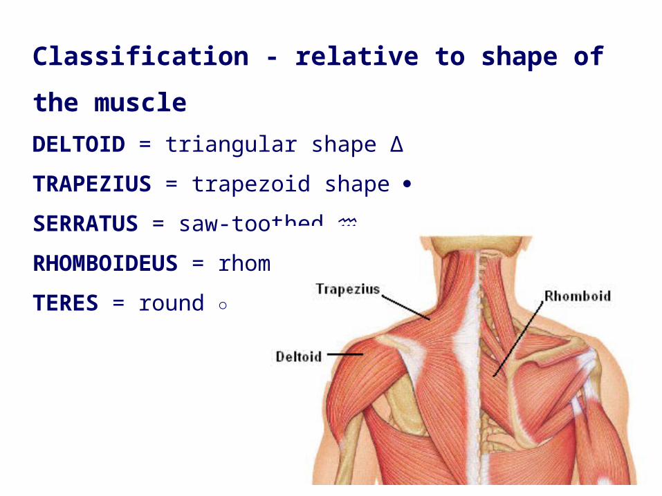
Classification - relative to shape of the muscleDELTOID = triangular shape Δ
TRAPEZIUS = trapezoid shape
SERRATUS = saw-toothed ♒
RHOMBOIDEUS = rhomboid shape TERES = round ○
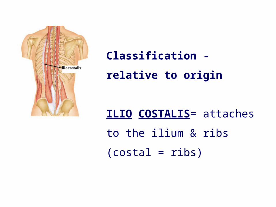
Classification - relative to
origin
ILIO COSTALIS= attaches to
the ilium & ribs (costal = ribs)
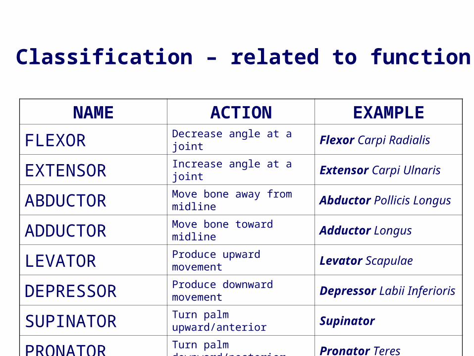
NAME ACTION EXAMPLEFLEXOR Decrease angle at a joint Flexor Carpi Radialis
EXTENSOR Increase angle at a joint Extensor Carpi Ulnaris
ABDUCTOR Move bone away from midline Abductor Pollicis Longus
ADDUCTOR Move bone toward midline Adductor Longus
LEVATOR Produce upward movement Levator Scapulae
DEPRESSOR Produce downward movement Depressor Labii Inferioris
SUPINATOR Turn palm upward/anterior Supinator
PRONATOR Turn palm downward/posterior Pronator Teres
Classification – related to function
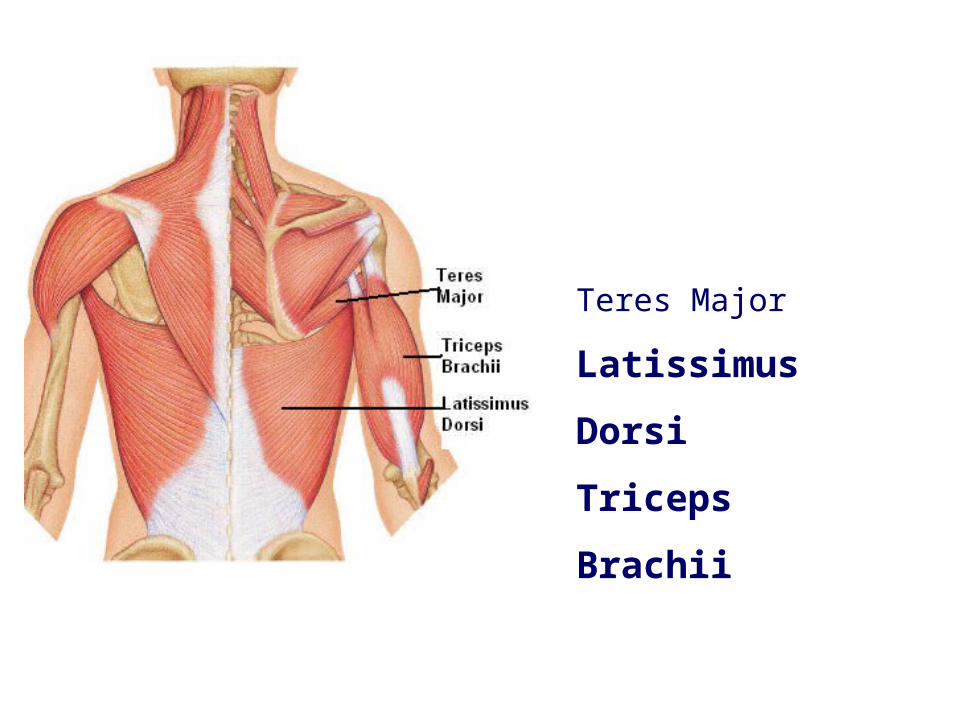
Teres Major
Latissimus Dorsi
Triceps Brachii
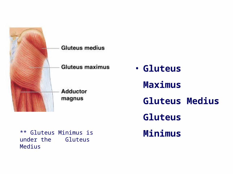
• Gluteus Maximus
Gluteus Medius
Gluteus Minimus
** Gluteus Minimus is under the Gluteus Medius
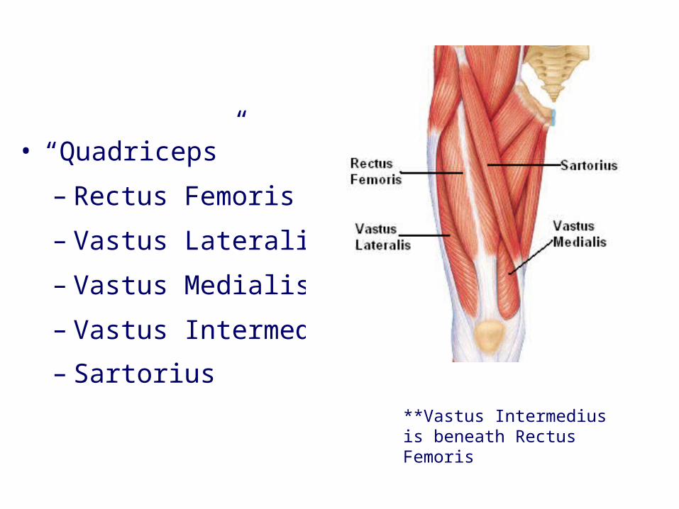
• “Quadriceps”
– Rectus Femoris
– Vastus Lateralis
– Vastus Medialis
– Vastus Intermedius – Sartorius
**Vastus Intermedius is beneath Rectus Femoris
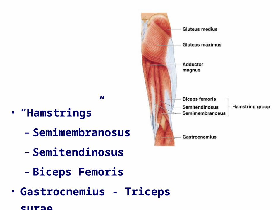
• “Hamstrings”– Semimembranosus– Semitendinosus– Biceps Femoris
• Gastrocnemius - Triceps surae
Campbell, Neil A., Reece, Jane B., Cain Michael L., Jackson, Robert B., Minorsky, Peter V., Biology, Benjamin-Cummings Publishing Company, 1996 –2010.
Thank you for your attention