IQ2000TM WSSV Detection and Prevention System
White Spot Syndrome Virus (WSSV)
In-vitro use only
No contagious materials included
2013/12
1
Table of Contents
I. Introduction
II. System Components
1. DNA extraction reagents
2. WSSV specific sequence amplification kit
III. Equipment and Reagents Required
IV. Detection Limit and Sensitivity
V. Sample Preparation and DNA Extraction
1. Sample preparation
2. DTAB-CTAB DNA extraction procedure
3. DNA extracted by Lysis Buffer
4. DNA dissolution
VI. Amplification Reaction Protocol
1. Reagents preparation
2. Reaction condition
3. Reaction procedure
VII. Electrophoresis
1. Agarose gel preparation
2. Electrophoresis
3. Gel staining and data assay
VIII. Diagnosis
IX. Troubleshooting
X. Reference
XI. Appendix
2
I. Introduction
White Spot Syndrome Virus (WSSV) is a major shrimp disease, which has
caused high mortality rate and economic losses to the major shrimp farming
countries in South East Asia, Central America and Southern USA. It is a
pathogen found in different penaeid shrimp species including P. monodon, P.
japonicus and L. vannamei as well as other crustaceans, such as crab and
crayfish.
Various molecular methods, such as microtomy, immuno-assay,
hybridization, and PCR (Polymerase Chain Reaction) for pathogen detection
have been developed for shrimp diseases. At present, nested PCR method is
recognised to be the most effective diagnostic tool for this pathogen. For the
development of effective diagnostic tools, a WSSV genomic library has been
constructed and analysed. In addition, a very conserved segment of WSSV
has also been selected as the target for PCR diagnosis.
Working in collaboration with Prof. Guang-Hsiung Kou and Prof. Chu-Fang
Lo from National Taiwan University, GeneReach has successfully developed
IQ2000TM
WSSV Detection and Prevention System which can differentiate
infected shrimps into 4 different levels of infection: very light, light, medium,
and severe. The diagnostic results produced by IQ2000TM
Systems are very
helpful for shrimp disease control program in the shrimp farming industry.
To provide a more convenient reaction condition and time saving procedure
for multi-viral diagnosis, IQ2000TM
WSSV Detection and Prevention System
can be performed not only by its own PCR reaction condition but also by the
Uni-IQ Program, a universal PCR profile which can be used for all IQ2000TM
Systems. Under this reaction profile, different kinds of shrimp viral diseases
can be screened at the same batch of PCR reaction.
The validation data for this kit have been certified by the OIE, based on
expert review, as fit for:

3
1) To certify freedom from infection (<10 virions/sample) in individual animals or
products for trade/movement purposes;
2) To confirm diagnosis of suspect or clinical cases (confirmation of a diagnosis by
histopathology or clinical signs);
3) To estimate prevalence of infection to facilitate risk analysis (surveys/herd health
schemes/disease control).
4
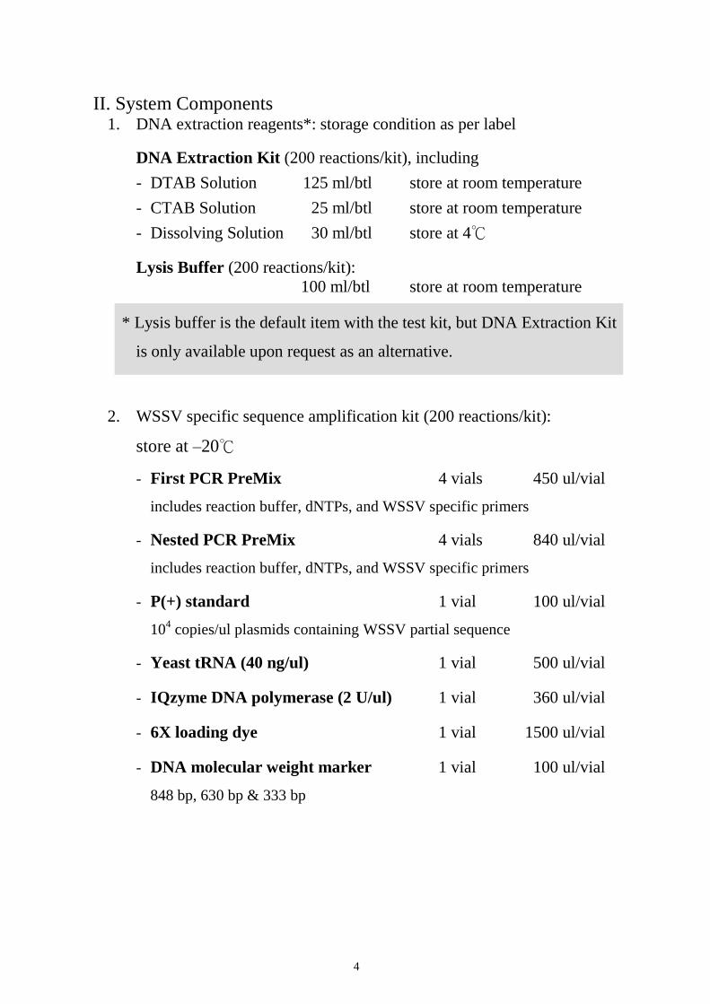
II. System Components 1. DNA extraction reagents*: storage condition as per label
DNA Extraction Kit (200 reactions/kit), including
- DTAB Solution 125 ml/btl store at room temperature
- CTAB Solution 25 ml/btl store at room temperature
- Dissolving Solution 30 ml/btl store at 4℃
Lysis Buffer (200 reactions/kit):
100 ml/btl store at room temperature
* Lysis buffer is the default item with the test kit, but DNA Extraction Kit
is only available upon request as an alternative.
2. WSSV specific sequence amplification kit (200 reactions/kit):
store at –20℃
- First PCR PreMix 4 vials 450 ul/vial
includes reaction buffer, dNTPs, and WSSV specific primers
- Nested PCR PreMix 4 vials 840 ul/vial
includes reaction buffer, dNTPs, and WSSV specific primers
- P(+) standard 1 vial 100 ul/vial
104 copies/ul plasmids containing WSSV partial sequence
- Yeast tRNA (40 ng/ul) 1 vial 500 ul/vial
- IQzyme DNA polymerase (2 U/ul) 1 vial 360 ul/vial
- 6X loading dye 1 vial 1500 ul/vial
- DNA molecular weight marker 1 vial 100 ul/vial
848 bp, 630 bp & 333 bp
5
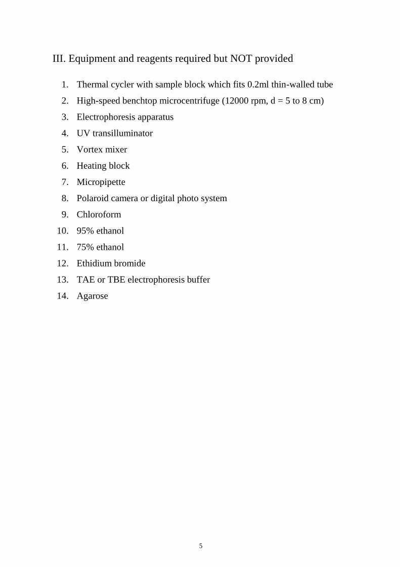
III. Equipment and reagents required but NOT provided
1. Thermal cycler with sample block which fits 0.2ml thin-walled tube
2. High-speed benchtop microcentrifuge (12000 rpm, d = 5 to 8 cm)
3. Electrophoresis apparatus
4. UV transilluminator
5. Vortex mixer
6. Heating block
7. Micropipette
8. Polaroid camera or digital photo system
9. Chloroform
10. 95% ethanol
11. 75% ethanol
12. Ethidium bromide
13. TAE or TBE electrophoresis buffer
14. Agarose
6
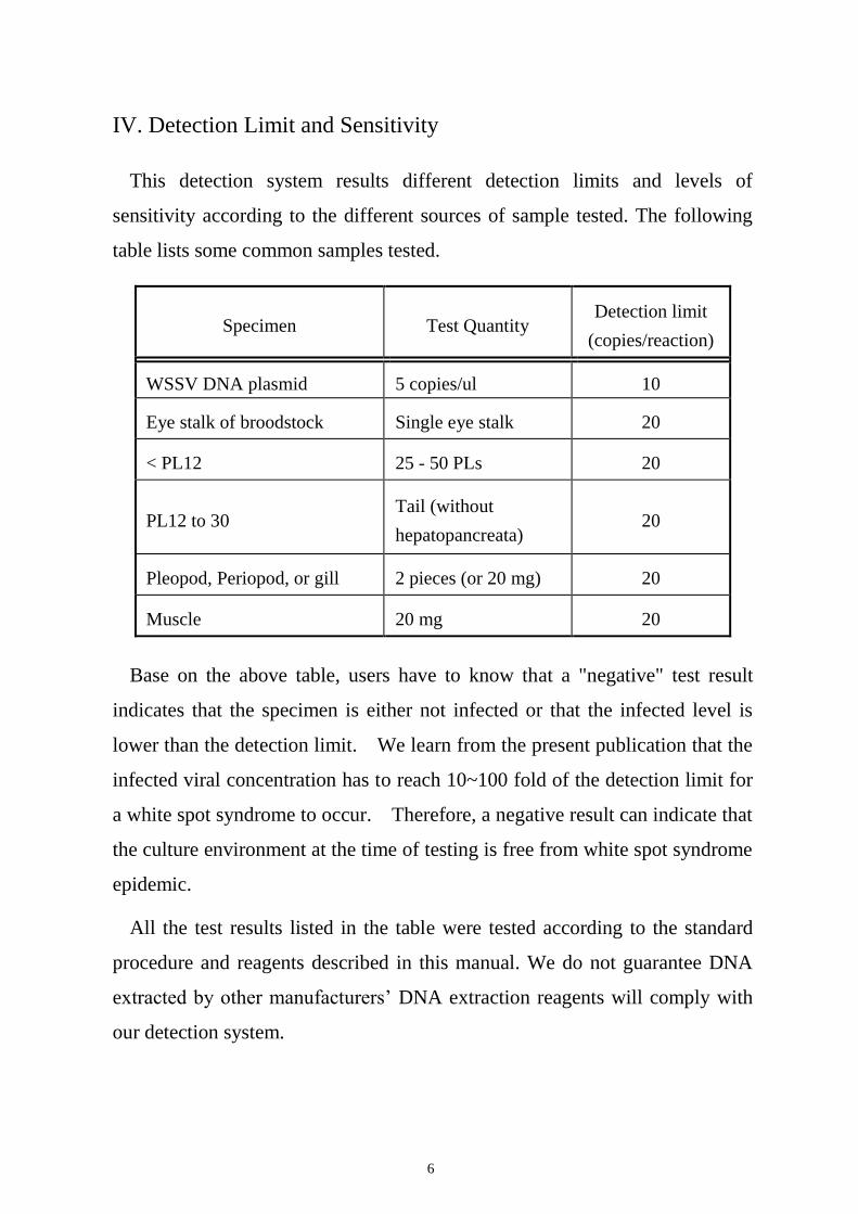
IV. Detection Limit and Sensitivity
This detection system results different detection limits and levels of
sensitivity according to the different sources of sample tested. The following
table lists some common samples tested.
Specimen Test Quantity Detection limit
(copies/reaction)
WSSV DNA plasmid 5 copies/ul 10
Eye stalk of broodstock Single eye stalk 20
< PL12 25 - 50 PLs 20
PL12 to 30 Tail (without
hepatopancreata) 20
Pleopod, Periopod, or gill 2 pieces (or 20 mg) 20
Muscle 20 mg 20
Base on the above table, users have to know that a "negative" test result
indicates that the specimen is either not infected or that the infected level is
lower than the detection limit. We learn from the present publication that the
infected viral concentration has to reach 10~100 fold of the detection limit for
a white spot syndrome to occur. Therefore, a negative result can indicate that
the culture environment at the time of testing is free from white spot syndrome
epidemic.
All the test results listed in the table were tested according to the standard
procedure and reagents described in this manual. We do not guarantee DNA
extracted by other manufacturers’ DNA extraction reagents will comply with
our detection system.
7
V. Sample preparation and DNA extraction
1. Sample preparation (For DTAB-CTAB method)
a. Broodstock eye stalk
(i) Rinse the cut eye stalk with clean water.
(ii) Put eye stalk into a 2ml tube that contains 0.6 ml DTAB solution.
(iii) Grind the eye stalk in the tube with a disposable grinder.
b. Larvae, PL or Juvenile
(i) Place about 20 mg specimen into a 2ml tube containing 0.6 ml
DTAB solution (The test requires at least 50 pieces for larvae; 30
pieces for <PL12; for PL12-30, only half tail should be used. Do not
sample the hepatopancrea or head. ).
(ii) Grind the sample in the tube with a disposable grinder.
c. Pleopod, pereopod, or gill of adult shrimp
(i) Place 2 pieces into a 2ml tube containing 0.6 ml DTAB solution.
(ii) Grind the sample in the tube with a disposable grinder.
d. Tail or muscle of adult shrimp
(i) Place a tail or about 20 mg muscle into a 2ml tube containing 0.6 ml
DTAB solution.
(ii) Grind the sample in the tube with a disposable grinder.
2. DTAB-CTAB DNA extraction procedure
a. Incubate the prepared sample at 75℃ for 5 minutes, then cool down to
room temperature.
b. Vortex briefly and spin down the mixture, then add 0.7 ml of
chloroform, vortex for another 20 seconds and centrifuge at 12000g
8
(12000 rpm, r = 5~7 cm) for 5 minutes.
c. Transfer 200 ul of the upper aqueous phase to a new 1.5ml tube. Add
100 ul of CTAB Solution and 900 ul ddH2O. Vortex briefly, then
incubate at 75℃ for 5 minutes.
d. Cool down to room temperature and centrifuge at 12000g for 10
minutes.
e. Carefully decant the supernatant, resuspend the pellet with 150 ul
Dissolving Solution, incubate at 75℃ for 5 minutes then cool down to
room temperature.
f. Spin at 12000g for 5 minutes. Transfer the clear solution to a fresh
1.5ml tube with 300 ul of 95% ethanol.
g. Vortex briefly, centrifuge at 12000g for 5 minutes, then wash the pellet
with 200 ul of 75% ethanol, spin down, dry the pellet and dissolve in
ddH2O or TE buffer. Refer to 4. DNA dissolution for volume of TE
buffer to be used.
3. DNA extracted by Lysis Buffer (for pleopod, gill, or < PL12 samples
only)
a. Add 500 ul Lysis Buffer in a 1.5ml tube.
b. Put shrimp sample into the tube and grind it with a disposable grinder.
c. Incubate the prepared sample at 95℃ for 10 minutes, then centrifuge at
12000g (12000 rpm r=5~7cm) for 10 minutes.
d. Transfer 200 ul of the upper clear solution to a fresh 1.5ml tube with
400 ul 95% ethanol.
e. Vortex briefly, centrifuge at 12000g for 5 minutes, then decant the
ethanol and dry the pellet.
f. Dissolve the pellet by ddH2O or TE buffer. Refer to 4. DNA dissolution
for volume of TE buffer to be used.

9
4. DNA dissolution
a. The concentration of DNA are different from different sample sources,
therefore the concentration of DNA needs to be adjusted by dissolving
the DNA pellet in different volume of ddH2O or TE buffer.
SAMPLE SOURCE VOLUME
Eye stalk of brood stock 100 ul
PL 200 ul
Pleopod or periopod 200 ul
Gill 50 ul
b. If sample needed to be preserved for longer period, TE buffer is
recommended. Sample can be stored in -20℃ for one year.
c. Please fine tune the volume of ddH2O or TE buffer according to the real
recovery efficiency.

10
VI. Amplification Protocol
The following amplification conditions apply to 0.2ml thin-wall tube or
96-well plate. Before executing the following PCR procedures, please confirm
hot start function of the machine has already been shut down.
1. Reagents preparation:
a. First PCR reaction reagent mixture: 8 ul/reaction
Mix the following:
First PCR PreMix 7.5 ul
IQzyme DNA Polymerase (2 U/ul) 0.5 ul
b. Nested PCR reaction reagent mixture: 15ul/reaction
Mix the following:
Nested PCR PreMix 14 ul
IQzyme DNA Polymerase (2 U/ul) 1 ul
2. Reaction condition:
a. First PCR reaction profile:
94℃ 30 seconds; 62℃ 30 seconds; 72℃ 30 seconds, repeat 5 cycles,
then
94℃ 15 seconds; 62℃ 15 seconds; 72℃ 20 seconds, repeat 15 cycles,
then
add 72℃ 30 seconds; 20℃ 30 seconds at the end of the final cycle.
b. Nested PCR reaction profile:
94℃ 20 seconds; 62℃ 20seconds; 72℃ 30 seconds, repeat 25 cycles,
add 72℃ 30 seconds; 20℃ 30 seconds at the end of the final cycle.
11
* IQ2000TM
WSSV Detection and Preservation System can also be
performed by the Uni-IQ Program. The reaction condition of Uni-IQ
Program is:
a. First PCR reaction profile:
42℃ 30min; 94℃ 2min; then
94℃ 20 sec; 62℃ 20sec; 72℃ 30sec, repeat 15 cycles, then add
72℃ 30 sec; 20℃ 30 sec at the end of the final cycle.
b. Nested PCR reaction profile:
94℃ 20 sec; 62℃ 20sec; 72℃ 30 sec, repeat 30 cycles, then add
72℃ 30 sec; 20℃ 30 sec at the end of the final cycle.
3. Reaction procedure:
a. Prepare first and nested PCR reaction reagent mixtures required
according to the sample number. For each reaction mixture
preparation, user also needs to take into account 3 positive standards
(103, 10
2 and 10
1) and 1 negative control (ddH2O or Yeast tRNA).
b. Pipette 8 ul of first PCR reaction reagent mixture into each 0.2ml
reaction tube with proper label.
c. Add 2 ul of the extracted sample DNA or standard into each reaction
mixture.
d. Cover each reaction mixture with 20 ul of mineral oil unless your
thermal cycler is equipped with oil-free design.
e. Perform first PCR reaction.
f. Add 15 ul of nested PCR reaction reagent mixture to each tube after first
PCR was completed. Be sure that the reagents go through oil overlay.
g. Perform nested PCR reaction.

12
h. After nested reaction is completed, add 5 ul of 6X loading dye to each
reaction tube and mix well.
i. After mixing, sample is ready for electrophoresis.
* 10 copies/ul standard is highly recommended for every batch of experiment
to monitor the sensitivity of reactions. We also recommend Yeast tRNA to
be used as diluents for diluting positive standards. Under this condition, the
standard can be kept at -20℃ for a week.
13
VII. Electrophoresis
1. Agarose gel preparation
a. First, decide a buffer system of electrophoresis between TAE and TBE.
Then, dilute the buffer to 1X operation concentration to process
electrophoresis and produce agarose gel. Note that the buffer for
processing electrophoresis and producing agarose gel must be the same
system.
b. A 2% agarose gel is recommended for electrophoresis. To prepare 2%
agarose gel, add 2 g agarose into a glass-made wide mouth bottle or
flask with 100ml electrophoresis buffer.
c. Heat the mixture until it becomes hyaline without any gel particles.
Heating can be done by using alcohol lamp, gas lamp or heat plate to
heat, microwave oven is able to heat as well. To avoid the boiled gel
from slopping over, a bigger glass-made container (twice the solution
volume) is recommended.
d. Cool down the clear agarose gel under room temperature until the
temperature is around 50℃ and slowly pour the gel into the gel box.
The volume of the gel varies from the size of the gel box. Generally
speaking, the height of agarose gel only has to go above the bottom of
the gel comb for about 0.3~0.5 cm, and thickness is suggested to be no
less than 0.8 cm.
e. Carefully remove the plastic comb and blockers at both sides of the gel
box when agarose gel is completely coagulated. This agarose gel, then,
is ready for electrophoresis. The finished agarose gel shouldn't be
exposed at room temperature for longer than 4 hours.
14
2. Electrophoresis
a. Lay the coagulated agarose gel inside the gel box. DNA molecules will
swift toward (+) because DNA molecular is negative charged.
b. Add 1X electrophoresis buffer into the gel box until the buffer lever is
just covering the gel.
c. Load 5 ul each of the “PCR product-loading dye mixture” into each well.
The mixture will sink to the bottom of the wells because its density is
heavier than buffer. This step should be carefully handled in order to
avoid contamination.
d. DNA marker is required for every electrophoresis. About 5 ul of DNA
marker is recommended. The DNA molecular weight marker is a
reference for PCR product size.
e. When all the samples are loaded, connect the gel box to the power
supply before switching on. Constant voltage between 100 V ~ 150 V is
recommended for electrophoresis.
f. The loading dye in the kit contains 2 colorants: Bromphenol Blue gives
deep blue color; Xylene Cyanol gives light blue color. When the dark
blue dye approaches 1/2 to 2/3 of the gel, stop the electrophoresis.
Then, remove the gel from the gel box to proceed with the EtBr staining
procedures.
g. To avoid contamination, DO NOT re-use the gel electrophoresis buffer
unless several gels will be used in the same day. When the
electrophoresis is finished, wash the gel box with plenty of water.
15
3. Gel staining and data assay
a. Ethidium Bromide (EtBr) is usually prepared for 10mg/ml stock
solution. This solution should be stored in an amber bottle because EtBr
is a light degradable chemical. Note that EtBr is a known carcinogen,
protective suit, gloves, and goggles are highly recommended.
b. Dilute the 10 mg/ml stock solution 20,000 times (i.e. add 5 ul of the
above stock solution into 100 ml distilled water to prepare the staining
solution.).
c. Pour the above staining solution into the plastic tray or zip-lock bag
with electrophoresis-finished gel. The solution must cover the whole
gel.
d. Shake lightly at room temperature for 10 minutes. Then, destain the gel
in another plastic tray with distilled water for another 10 minutes to
eliminate the background.
e. Lay the gel on a UV transilluminator to read the final result.
16
VIII. Diagnosis
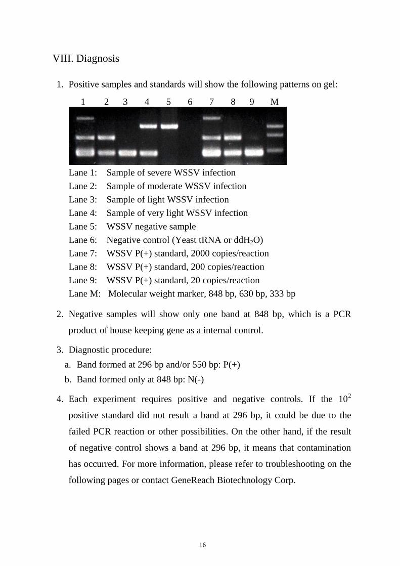
1. Positive samples and standards will show the following patterns on gel:
1 2 3 4 5 6 7 8 9 M
Lane 1: Sample of severe WSSV infection
Lane 2: Sample of moderate WSSV infection
Lane 3: Sample of light WSSV infection
Lane 4: Sample of very light WSSV infection
Lane 5: WSSV negative sample
Lane 6: Negative control (Yeast tRNA or ddH2O)
Lane 7: WSSV P(+) standard, 2000 copies/reaction
Lane 8: WSSV P(+) standard, 200 copies/reaction
Lane 9: WSSV P(+) standard, 20 copies/reaction
Lane M: Molecular weight marker, 848 bp, 630 bp, 333 bp
2. Negative samples will show only one band at 848 bp, which is a PCR
product of house keeping gene as a internal control.
3. Diagnostic procedure:
a. Band formed at 296 bp and/or 550 bp: P(+)
b. Band formed only at 848 bp: N(-)
4. Each experiment requires positive and negative controls. If the 102
positive standard did not result a band at 296 bp, it could be due to the
failed PCR reaction or other possibilities. On the other hand, if the result
of negative control shows a band at 296 bp, it means that contamination
has occurred. For more information, please refer to troubleshooting on the
following pages or contact GeneReach Biotechnology Corp.
17
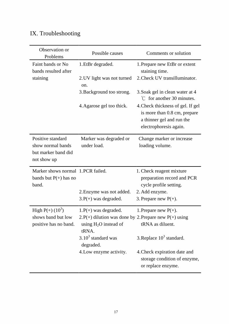
IX. Troubleshooting
Observation or
Problems Possible causes Comments or solution
Faint bands or No
bands resulted after
staining
1. EtBr degraded.
2. UV light was not turned
on.
3. Background too strong.
4. Agarose gel too thick.
1. Prepare new EtBr or extent
staining time.
2. Check UV transilluminator.
3. Soak gel in clean water at 4
℃ for another 30 minutes.
4. Check thickness of gel. If gel
is more than 0.8 cm, prepare
a thinner gel and run the
electrophoresis again.
Positive standard
show normal bands
but marker band did
not show up
Marker was degraded or
under load.
Change marker or increase
loading volume.
Marker shows normal
bands but P(+) has no
band.
1. PCR failed.
2. Enzyme was not added.
3. P(+) was degraded.
1. Check reagent mixture
preparation record and PCR
cycle profile setting.
2. Add enzyme.
3. Prepare new P(+).
High P(+) (103)
shows band but low
positive has no band.
1. P(+) was degraded.
2. P(+) dilution was done by
using H2O instead of
tRNA.
3. 103 standard was
degraded.
4. Low enzyme activity.
1. Prepare new P(+).
2. Prepare new P(+) using
tRNA as diluent.
3. Replace 103 standard.
4. Check expiration date and
storage condition of enzyme,
or replace enzyme.
18
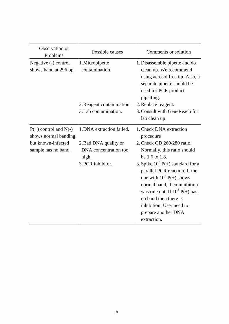
Observation or
Problems Possible causes Comments or solution
Negative (-) control
shows band at 296 bp.
1. Micropipette
contamination.
2. Reagent contamination.
3. Lab contamination.
1. Disassemble pipette and do
clean up. We recommend
using aerosol free tip. Also, a
separate pipette should be
used for PCR product
pipetting.
2. Replace reagent.
3. Consult with GeneReach for
lab clean up
P(+) control and N(-)
shows normal banding,
but known-infected
sample has no band.
1. DNA extraction failed.
2. Bad DNA quality or
DNA concentration too
high.
3. PCR inhibitor.
1. Check DNA extraction
procedure
2. Check OD 260/280 ratio.
Normally, this ratio should
be 1.6 to 1.8.
3. Spike 103 P(+) standard for a
parallel PCR reaction. If the
one with 103 P(+) shows
normal band, then inhibition
was rule out. If 103 P(+) has
no band then there is
inhibition. User need to
prepare another DNA
extraction.
19
X. Reference:
1. Chang PS, Lo CF, Wang YC, Kou GH (1996) Identification of white spot
syndrome associated baculovirus (WSSV) target organs in shrimp,
Penaeus monodon by in situ hybridization. Dis. Aquat. Org. 27:131–139.
2. Chou HY, Huang CY, Wang CH, Chiang HC, Lo CF (1995)
Pathogenicity of a baculovirus infection causing white spot syndrome in
cultured penaeid shrimp in Taiwan. Dis. Aquat. Org. 23: 165-173.
3. Lightner DV (ed.) (1996) A handbook of pathology and diagnostic
procedures for diseases of penaeid shrimp. World Aquaculture Soc.,
Baton Rouge. Section 3.11
4. Lo CF, Leu JH, Ho CH, Chen CH, Peng SE, Chen YT, Chou CM, Yeh
PY, Huang CJ, Chou HY, Wang CH, and Kou GH (1996 a) Detection of
baculovirus associated with white spot syndrome (WSSV) in penaeid
shrimps using polymerase chain reaction. Dis. Aquat. Org. 25: 133-141.
5. Lo CF, Ho CH, Peng SE, Chen CH, Hsu HC, Chiu YL, Chang CF, Liu
KF, Su MS, Wang CH, Kou GH (1996 b) White spot syndrome
baculovirus (WSSV) detected in cultured and captured shrimps, crabs
and other arthropods. Dis. Aquat. Org. 27:215–225.
6. Lo CF, Ho CH, Chen CH, Liu KF, Chiu YL, Yeh PY, Peng SE, Hsu HC,
Liu HC, Chang CF, Su MS, Wang CH, Kou GH (1997) Detection and
tissue tropism of white spot syndrome baculovirus (WSSV) in captured
brooders of Penaeus monodon with a special emphasis on reproductive
organs. Dis. Aquat. Org. 30:53–72.
7. Peng SN, Lo CF, Ho CH, Chang CF, Kou GH (1996) Detection of white
spot syndrome baculovirus (WSSV) in giant freshwater prawn,
Macrobrachium rosenbergii using polymerase chain reaction.
Aquaculture 164:253–262.
8. Wang CH, Lo CF, Leu JH, Chou CM, Yeh PY, Chou HY, Tung MC,
Chang CF, Su MS, Kou GH (1995) Purification and genomic analysis of
baculovirus associated with white spot syndrome (WSSV) of Penaeus
monodon. Dis. Aquat. Org. 23: 239-242.
9. Wongteerasupaya C, Vickers JE, Sriurairatana S, Nash GL, Akarajamorn

20
A, Boonsaeng V, Panyim S, Tassanakajon A, Withyachumnarnkul B,
Flegel TW (1995) A non-occluded, systemic baculovirus that occurs in
cells of ectodermal and mesodermal origin and causes high mortality in
the black tiger prawn Penaeus monodon. Dis Aquat Org 21: 69-77.
21
XI. Appendix
OIE Procedure for Registration of Diagnostic Kits
Abstract sheet
Name of the diagnostic kit: IQ 2000TM
WSSV Detection and Prevention System
Manufacturer: GeneReach Biotechnology Corporation
OIE Approval number: 20080304
Date of Registration: May 2008
Disease: White Spot Disease
Pathogen Agent: White spot syndrome virus (WSSV) found in different penaeid shrimp
species including P. monodon, P. japonicus and L. vannamei as well as other crustaceans,
such as crab and crayfish.
Type of Assay: The IQ 2000TM
WSSV Detection and Prevention System is a PCR test.
Purpose of Assay: Certified by the OIE in May 2008 as fit for the diagnosis of white spot
syndrome in crustaceans and for the following purposes:
1. To certify freedom from infection (<10 virions/sample) in individual animals or
products for trade/movement purposes;
2. To confirm diagnosis of suspect or clinical cases (confirmation of a diagnosis by
histopathology or clinical signs);
3. To estimate prevalence of infection to facilitate risk analysis (surveys/herd health
schemes/disease control).
Species and Specimen: Validated in shrimp but may be useful for detection of WSSV in
other crustaceans.
1. Information on the kit
Please refer to the kit insert available on the OIE Registry web page or contact manufacturer
at:
GeneReach Biotechnology Corporation TEL: 886-4-24639869 FAX: 886-4-24638255
Email: [email protected]
No. 19, Keyuan Second Road. Central Taiwan Science Park, Taichung, Taiwan, 407
22
2. Summary of validation studies
Analytical characteristics
Analytical sensitivity - The analytical sensitivity data were done by comparing the
sensitivity of IQ2000TM
WSSV Detection and Prevention System
and the PCR diagnostic method listed by OIE (Lo, et al., 1996).
DNA extracted from WSSV infected L. vannamei was 10 fold
serial diluted, and subsequently taken as DNA template for both
detection methods. The dilutants are ddH2O or DNA extract of
SPF L. vannamei. No apparent difference on sensitivity between
the 2 detection methods was observed.
Analytical specificity - SPF L. vannamei, Monodon baculovirus or Baculovirus Penai
infected P. monodon, as well as WSSV infected shrimp collected
from different geographic areas were acquired from OIE
reference laboratories. Results of all SPF samples and MBV or
BP infected samples have shown no detectable non-specific band;
while all WSSV infected samples can be detected by IQ2000TM
WSSV Detection and Prevention System.
Repeatability data - The repeatability study was carried out using pleopods of L.
vannamei with 3 production batches in triplicates. The data were
consistent.
Diagnostic Characteristics
Two evaluation studies were carried out for the estimation of the diagnostic sensitivity and
specificity:
- In one study, the OIE recommended nested PCR method (Lo, et al., 1996; Chapter 4.1.2.
of the Manual of Diagnostic Tests for Aquatic Animals, 2007, available on the OIE website)
was applied to classify certain populations of shrimp as either negative or positive. After
screening, 300 white spot syndrome negative samples and 300 white spot syndrome positive
samples were used for this study.
- In a second study, 100 un-defined samples collected from market were used to compare
IQ2000TM
WSSV Detection and Prevention System with the OIE recommended method.
Diagnostic sensitivity (DSn) and specificity (DSp) estimates with 95% confidence limits (Cl)
Tests performed with virus negative and positive animals:
WSS virus positive by
reference method
WSS virus negative by
reference method
Test Positive by IQ2000TM
289 0
Test Negative by IQ2000TM
11 300
Relative Diagnostic Sensitivity: 96.33%, CI [93.53 – 98.16%]
Relative Diagnostic Specificity: 100.00%, CI [98.78 – 100.00%]
23
Tests performed with undefined samples collected from market:
WSS virus positive by
reference method
WSS virus negative by
reference method
Test Positive by IQ2000TM
51 0
Test Negative by IQ2000TM
0 49
Relative Diagnostic Sensitivity: 100.00%, CI [92.89 – 100.00%]
Relative Diagnostic Specificity: 100.00%, CI [92.89 – 100.00%]
Agreement between tests
The results presented above reveal that the consistency between IQ2000TM
WSSV
Detection and Prevention System and the OIE recommended method is very good.
Reproducibility
Different lots of IQ2000TM
WSSV Detection and Prevention System were sent to three
different laboratories in Chinese Taipei (including one OIE reference laboratory) to be
tested. 35 shrimp samples (L. vannamei) were collected and sent to all 3 laboratories. Each
sample was analysed with 3 different lots of IQ2000TM
WSSV Detection and Prevention
System. Results of the 3 independent laboratories were collected and the data were analysed
statistically
The reproducibility data indicates that there is no significant difference in assay
performance between laboratories. Furthermore, good reproducibility was demonstrated
using three different lots of kits, suggesting that lot to lot variability is minimal.
Applications
The IQ2000TM
WSSV Detection and Prevention System kit is currently used by several
laboratories worldwide as an official test.
References
1. LO CF, LEU JH, HO CH, CHEN CH, PENG SE, CHEN YT, CHOU CM, YEH PY, HUANG CJ,
CHOU HY, WANG CH, AND KOU GH (1996 a) Detection of baculovirus associated with
white spot syndrome (WSSV) in penaeid shrimps using polymerase chain reaction. Dis
Aquat Org 25: 133-141.
2. LO CF, HO CH, PENG SE, CHEN CH, HSU HC, CHIU YL, CHANG CF, LIU KF, SU MS,
WANG CH, KOU GH (1996) White spot syndrome baculovirus (WSSV) detected in
cultured and captured shrimps, crabs and other arthropods. Dis. Aquat. Org.,
27,215-225.
3. NUNAN L.M., POULOS B.T. & LIGHTNER D.V. (1998) The detection of white spot
24
syndrome virus (WSSV) and yellow head virus (YHV) in imported commodity shrimp.
Aquaculture, 160, 19-30.
4. PENG SN, LO CF, HO CH, CHANG CF, KOU GH (1998) Detection of white spot
syndrome baculovirus (WSSV) in giant freshwater prawn, Macrobrachium rosenbergii
using polymerase chain reaction. Aquaculture 164:253-262.
5. THAKUR P. C., F. CORSIN, J. F. TURNBULL, K. M. SHANKAR, N. V. HAO4, P. A. PADIYAR,
M. MADHUSUDHAN, K. L. MORGAN, AND C. V. MOHAN (2002). Estimation of
prevalence of white spot syndrome virus (WSSV) by polymerase chain reaction in
Penaeus monodon postlarvae at time of stocking in shrimp farms of Karnataka, India: a
population-based study. Dis Aquat Org 49:235-243.
6. Chapter 2.2.6., White spot disease; Manual of Diagnostic Tests for Aquatic Animals,
2009, OIE, pp. 177-190.

25
Recommended