Use of Stem Cells in Dental Implants and Enamel Regenerative TherapiesUpadhyay RK*
Department of Zoology, Deen Dayal Upadhyay Gorakhpur University, India*Corresponding author: Upadhyay RK, Department of Zoology, DDU Gorakhpur University, Gorakhpur 273009, Uttar Pradesh, India, Tel:9838448495; E-mail: [email protected]
Received date: Oct 08, 2015; Accepted date: Jan 15, 2015; Published date: Jan 18, 2015
Abstract
Present review article emphasizes use of variousstem cell types, biological scaffold materials, genes andfactors essentially required in woundhealing, transplantation and regeneration of toothimplants. All recent developments in tooth engineeringused for successful regeneration of tooth, inductionof enamel and formation of dentin complexes arehighlighted. In addition, genes, factors and mineralsrequired in vascularization and maintenance ofmicroenvironment for responsiveness to cells are alsoelucidated as most attractive candidates for regenerationtherapy. Present review also elucidates use of stable anddurable biodegradable polymer scaffolds materials andcementum/periodontal-ligament complex formation.There is a need to widen the horizon of bio-rootengineering technology for successful repairing of toothinjuries and replacement of fractured or traumatizedtooth. For this purpose, use of implantation ofcultured stem cells, adhesion factors and biomaterials canprovide stable attachment of dental implants. This reviewalso sketch upon need of newer technologies and moreadhesive biocompatible biomaterials for successful dentalimplants and enamel regenerative therapies.
Keywords: Stem cells; Dental cell implants;Enamel matrix derivatives; Tooth regeneration; Enamel matrixderivatives; Adhesion factors and biomaterials
Abbreviations:PDL: Periodontal Ligament; PDLSCs: Periodontal Ligament
Stem Cells; EMD: Enamel Matrix Derivative; DPSC: Dental PulpStromal Cells; DMCs: Dermal Multipotent Cells; MSC:Mesenchymal Stem Cells; FBS: Fetal Bovine Serum; MDO:Mandibular Distraction in Osteogenesis; MSCT: MesenchymalStem Cell Transplantations; DBM: Demineralized Bone Matrix;DFDBA: Human Demineralized Freeze-Dried Bone Allograft;BMP: Bone Morphogenetic Proteins; BMMSCs: Bone MarrowMesenchymal Stem Cells
IntroductionPeriodontal diseases and tooth decay and enamel
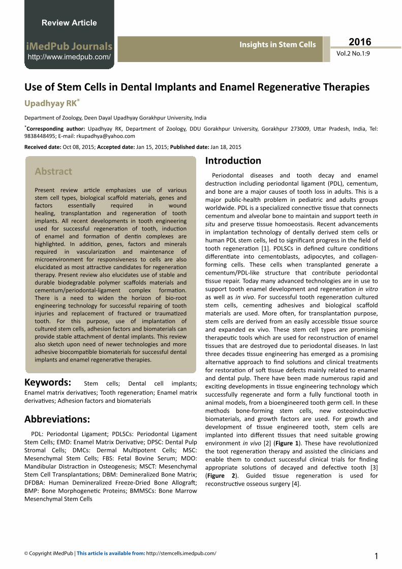
destruction including periodontal ligament (PDL), cementum,and bone are a major causes of tooth loss in adults. This is amajor public-health problem in pediatric and adults groupsworldwide. PDL is a specialized connective tissue that connectscementum and alveolar bone to maintain and support teeth insitu and preserve tissue homoeostasis. Recent advancementsin implantation technology of dentally derived stem cells orhuman PDL stem cells, led to significant progress in the field oftooth regeneration [1]. PDLSCs in defined culture conditionsdifferentiate into cementoblasts, adipocytes, and collagen-forming cells. These cells when transplanted generate acementum/PDL-like structure that contribute periodontaltissue repair. Today many advanced technologies are in use tosupport tooth enamel development and regeneration in vitroas well as in vivo. For successful tooth regeneration culturedstem cells, cementing adhesives and biological scaffoldmaterials are used. More often, for transplantation purpose,stem cells are derived from an easily accessible tissue sourceand expanded ex vivo. These stem cell types are promisingtherapeutic tools which are used for reconstruction of enameltissues that are destroyed due to periodontal diseases. In lastthree decades tissue engineering has emerged as a promisingalternative approach to find solutions and clinical treatmentsfor restoration of soft tissue defects mainly related to enameland dental pulp. There have been made numerous rapid andexciting developments in tissue engineering technology whichsuccessfully regenerate and form a fully functional tooth inanimal models, from a bioengineered tooth germ cell. In thesemethods bone-forming stem cells, new osteoinductivebiomaterials, and growth factors are used. For growth anddevelopment of tissue engineered tooth, stem cells areimplanted into different tissues that need suitable growingenvironment in vivo [2] (Figure 1). These have revolutionizedthe toot regeneration therapy and assisted the clinicians andenable them to conduct successful clinical trials for findingappropriate solutions of decayed and defective tooth [3](Figure 2). Guided tissue regeneration is used forreconstructive osseous surgery [4].
Review Article
iMedPub Journalshttp://www.imedpub.com/
Insights in Stem CellsVol.2 No.1:9
2016
© Copyright iMedPub | This article is available from: http://stemcells.imedpub.com/ 1
Figure 1 showing integration of stem cell therapy, tissueengineering and biomaterial technology in regenerativemedicine for clinical management and therapeutics basedon academic researches, industry and hospitals
Figure 2 Showing use of regeneration therapies for varioustooth defects
Table 1 Different types of tooth related problems and solutions based on available technology and methods
Problem Orthodontic reason/defect Solution Reference
Erupted tooth Expressed cytokeratin 14, dentin matrix protein-1, vascularendothelial growth factor, and osteopontin
DBC-fibrin glue-PRF composite wasautografted back into the original alveolarsockets
Yang, et al. [27]
Structural erosion orfragile tooth
Induced morphological changes occur due to increasedalkaline phosphatase (ALP) activity, runt-related transcriptionfactor 2 (RUNX2), osteocalcin (OCN), and bone sialoprotein(BSP) expression in PDLSCs
Correction of microRNAs (miRNAs) inhuman periodontal ligament stemcells(PDLSCs
Wei, et al. (2015)
Periodontal trauma Structural and functional integrity of the periodontiumfollowing periodontal trauma such as orthodontic toothmovement
High mobility group box protein-1 (HMGB1) Wolf, et al. (2015)
Traumatic injuries Loss of structural/compositional sensitivity of enamel tissue A mixed population of bone marrow-derived autologous stem andprogenitor cells are seeded onto β-tricalcium phosphate (β-TCP)
Rajan, et al. (50)
Osteoporosis Osteocalcium disorder, elemental loss and pulp and enameldestruction due to loss of calcium and phosphates
FasL pathways mediated differentiation ofERK and GSK-3β-catenin pathway
Ming, et al. (2014)
Osteoporosis intermittent mechanical strain (IMS) Promoted osteogenic differentiation ofOVX BMSCs by activating Runt-relatedtranscription factor 2
Zhang, et al. (2015)
Tumor progression andmetastasis/ Tumorblood vessels
Upregulation of vascular endothelial growth factor (VEGF)and VEGF receptor 2
Antitumor therapy, radioactive and laserbased destruction
Ohmura-Kakutani, etal. (2014)
Craniofacial problems Implantation of stem cells Use of cultured stem cells from sourcetissue and organ from which stem cells canbe derived
Mohanty, et al. (2015)
Fibrodysplasiaossificans progressiva
Low level of protein expression in receptor cells Bone morphogenetic protein (BMP)receptor ALK2, R206H are used
Fujimoto, et al. (2014)
Bone and tooth pain Limited capacity to induce bone formation Restoration by using rhBMP-2, DFDBAand EMD
Intini, et al. [33]
Demineralization embryonic tooth morphogenesis and promotes continuoustooth development
beta-catenin signaling Liu, et al. (2010)
Faulty and defectivetooth arrangment
Organization of predentin/dentin, enamel, and cementum Mineralization after implantation Lechguer, et al. (37)
Hard tissue formation Post-natal mesenchymal stromal cells (MSCs) includingbone marrow stromal cells (BMSCs) and periodontalligament fibroblasts (PDLFs).
Straumann Bone Ceramic coated withStraumann Emdogain
Mrozik, et al. (16)
Insights in Stem Cells
Vol.2 No.1:9
2016
2 This article is available from:http://stemcells.imedpub.com/

Loss of dental pulp andnon clinical damage
FGF molecules are able to maintain epithelial Tbx1expression during odontogenesis, Expression of Tbx1in dental epithelium of FGF receptor 2b(-/-)
Mesenchyme-derived signals Mitsiadis, et al. (34)
Odontogenesis Forced expression of Gli1, a major transcription factor in Shhsignaling
Amelogenin and ameloblastin. Takahashi, et al.(35,57)
Odontogenesis Sonic hedgehog (Shh) in enamel knot Use of Sonic hedgehog (Shh) Takahashi, et al.(35,57)
Low osteogenicdifferentiation
miR-26a potentially targeted on GSK3β and Smad1 toregulate Wnt and BMP signaling pathway
MicroRNAs (miRNAs) used as importantregulators of stem cell
Su, et al. (2015)
Orthodontic toothmovement (OTM)
induces local inflammation in periodontium OTM induced a significant elevation of type1 T helper cell (Th1) cytokines tumornecrosis factor-α (TNF-α) and interferon-γ(IFN-γ) around periodontal tissue in WT
Yan, et al. (2015)
Untimely enamel loss Obstruction in muscle formation and decreasing scar tissuecontraction
Scaffold-free cells and mesenchymal stemcells scaffold
Zhou, et al. (2015)
Loss of osteogeniccapability
Mitogen-activated protein kinase (MAPK) signaling pathways Use of BMMSCs and PBMSCs cells,regenerative medicine
Zheng, et al. (63)
Enamel cell and dentalpulp infection
von Willebrand factor (vWF) and CD31 immunofluorescentstaining, different energy densities of infrared LED on the cellviability
Vascular endothelial growth factor (VEGF)and basic fibroblast growth factor (bFGF)are used for pulp and enamel repairing
Feng, et al. (2015)
Fragile deciduous teeth Enamel cell destruction and low calcium level Human exfoliated deciduous teeth (SHED)and adipose stem cells (ASC)
Loo, et al. (2014)
Root cancer Inhibition of fucosylation may be used to block CSCs andmetastatic spread.
Oral squamous cell carcinoma (OSCC) Desiderio, et al.(2015)
Traumatic tooth and jawinjury
Skeletal regenerative medicine, mesenchymal stemcells (MSC)
Bone tissue engineering Asatrian, et al. (2015)
Large bone defects CACB/ADSCs compos Therapeutic potential of ERK signalingpathway
Wei, et al. (2015)
Defects in growingbones
Loss of majority of osteoblasts, Cxcl12 (chemokine (C-X-Cmotif) ligand 12)-abundant stromal cells and bone marrowstromal/mesenchymal progenitor cells in postnatal life
Implantation of periodontal humanmesenchyme cells
Ono, et al. (2014)
Bone breakage Osteoinductive signals are used as potent tool for boneregeneration, fabricated poly(L-lactic acid) (PLLA)electrospun nanofibers with random and aligned morphologyimmobilized with bone morphogenic protein-2 (BMP-2
Engineering bone tissue Perikamana, et al.(2015)
Skeletal deformities Mediates p53/miR-17/Smurf1 pathway Engineering bone tissue Liu, et al. (2015)
Delayed growth ofdeciduous tissues
Kruppel-like factors (KLFs) are evolutionarily conserved zincfinger-containing transcription factors
Differentiation, proliferation,embryogenesis and pluripotency.
Ding, et al. (2015)
Tooth decay anddamage
Organic matrix mediated mineralization paves a way forformation of synthetic enamel
Bone marrow-derivedmesenchymal stem andstromal cells (MSCs)
Jayasudha, et al.(2014)
Tooth decay Toll-like receptor 3, ankylosis-progressive homolog, decorin,osteocalcin, and runt-related transcription factor-2
Increasing cellular resistance and RCT Ramis, et al. (17)
Thermal hyper-sensitivity
Impairs the development of the tooth root in lactational ratsand alters the function of apical papilla-derived stem cell
Exposure to a continuous low dose oftetrachlorodibenzo-p-dioxin
Guo, et al. (2015)
For development of bioengineered tooth important growthfactors, stable and durable biodegradable polymer scaffoldsmaterials are highly needful [5]. For framing, growth andregeneration of enamel and pulp tissue, cementum/periodontal-ligament complex formation is highly essential [6].This is also cooperatively regulated by the epithelialameloblasts and mesenchymal odontoblasts [7]. For successfulregeneration of tooth, formation of dentin complexes, removalof demineralized matrix, induction of enamel, vascularizationof tooth need maintenance of microenvironment forresponsiveness to cells and factors [8,9] (Figure 2). Similarly,
for successful replacement of tooth and cellimplantation adhesion of cells to periodontal connective tissueamelogenin, bone sialoprotein and vimentin proteins is alsoimportant [10]. Similarly, enamel matrix derivative (EMD) alsoinfluence activities of cementoblasts and osteoblasts, whichmay able to regulate cell activities at a periodontalregenerative site. EMD and the type of cell populationspresent in the implant wound-healing environment may alterthe implant-connective tissue interface [11] (Table 1). Inaddition, subrenal capsule implantation is used as a newalternative method for tissue-engineering in vivo [2].
Insights in Stem Cells
Vol.2 No.1:9
2016
© Copyright iMedPub 3
Moreover, tooth loss due to periodontal disease, dental caries,trauma, or a variety of genetic disorders continues to affectmost adults adversely at some stage in their lives.
Hence, they essentially need a biological tooth substitutethat could replace lost teeth and may provide a vitalalternative to currently available clinical treatments. For thispurpose dissociated porcine third molar tooth buds are usedto make single-cell suspensions and seeded ontobiodegradable polymers. The recent technology upholds theslow but highly effective regenerative therapy for successfultreatment and replacement of tooth in pain. For toothregeneration and healing of traumatic injuries niche factors,growth factors, and implantation of stem cells, are mostessential requirements [12]. No doubt tooth bioengineeringpromises to be at the forefront of the next generation ofdental treatments [3] (Table 1 and Figure 2).
More specifically, mammalian tooth root development is along-term process during which root elongates along the apicaldirection and is accompanied with the formation ofperiodontium. In heterogeneous apical region of developingroot as a functional entity, a developing apical complex isformed the root [13]. The developing apical complex showssustainable development ability and functions as a growthcenter of tooth root. This is promising source of cells for toothroot and periodontal regeneration [13]. Similarly, periodontalligament stem cells are considered as one of the bestcandidates for periodontal regeneration therapy [14].Meanwhile, PDLSC cell pellet may be a promising alternative topromote periodontal defect repair for future clinicalapplications [15]. Still, there are limitations of conventionalregeneration modalities which underscore the necessity ofrecapitulating development for periodontal tissue engineering[15]. No doubt stem cell derived cell types show enormousregenerative capacity but how these cells act in ainflammatory or toxic microenvironment. There is anotherquestion how stem cell origin affects optimal differentiationand regeneration is a major question [8].
Periodontal tissue engineering requires suitablebiocompatible scaffold, growth factors, regenerative cells andinstructional molecules. For clinical purposes an enamel matrixprotein (EMP) is used to aid in hard tissue formation by post-natal mesenchymal stromal cells (MSCs) including bonemarrow stromal cells (BMSCs) and periodontal ligamentfibroblasts (PDLFs) [16]. Similarly, Straumann Bone Ceramiccoated with Straumann Emdogain impose significantstimulatory effect in the commitment of mesenchymal cells toosteogenic differentiation in vitro while Emdogain inhibited APactivity and appeared not to induce ectopic bone formation.EMPs truly possess the capacity to induce the regeneration ofbone or other components of the periodontium but it remainsto be established. Similarly, new methods for restoration ofcrown, root, pulp, enamel, dentin, odontoblast, cementum,blood vessels, and periodontal ligaments to improve theindiscriminate shape of tooth are also highly needful [16]. Inaddition, tissue response after implantation determines thesuccess of the healing process which is not only dependent onthe chemical properties of the implant surface but also
determines by the surface topography or roughness [17]. Forbetter regeneration inducers of molecular pathways that drivetooth morphogenesis and enamel secretion are also needed togenerate teeth from organ cultures fortherapeutic implantation [18]. However, p38α MAPK isrequired for tooth morphogenesis and enamel secretion [19].Similarly, CCN proteins and cell-associated molecules are alsoinvolved in several developmental processes are also needed[20]. Further, to induce complete tooth formation, in conditionof tooth loss due to trauma or diseases, implantation of stemcells, morphogens and growth factors are needed to stimulateentire tooth development and progression [6] (Table 1).Epithelial ameloblasts in combination to mesenchymalodontoblasts cooperatively regulate tooth development andsecrete enamel matrix, which is highly criticalfor enamel formation [7]. Formation of enamel knots isregulated by a cascade of gene activity where Fgf4, Shh, BMP4,Lef1 and p21 are the prime movers of the processes.Homeobox genes (Msx, Dlx) are the orchestrators of theframing and a series of proteins (adhesion molecules,extracellular matrix components) are the executors of toothframing [6]. Formation of cementum/periodontal-ligamentcomplex is an important field of tissue engineering (Table 1).
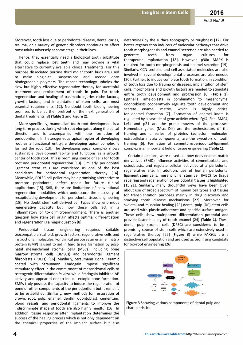
Certain questions, were raised i.e. how does enamel matrixderivatives (EMD) influence activities of cementoblasts andosteoblasts, and regulate cellular activities at a periodontalregenerative site. In addition, use of human periodontalligament stem cells, mesenchymal stem cell (MSC) for tissuerepairing and regeneration of periodontal tissues is highlighted[15,21]. Similarly, many thoughtful views have been givenabout use of broad spectrum of human cell types and tissuesfor transplantation purposes mainly in drug discovery andstudying tooth disease mechanisms [22]. Moreover, forskeletal and muscular healing [23] dental pulp (DP) stem cellsare used with plastic adherence and specific surface antigen.These cells show multipotent differentiation potential andprovide faster healing of tooth enamel [24] (Table 1). Thesedental pulp stromal cells (DPSC) are considered to be apromising source of stem cells which are extensively used inregenerative therapy [25] (Figure 3) while PAFSCs are adistinctive cell population and are used as promising candidatefor bio-root engineering [26].
Figure 3 Showing various components of dental pulp andcharacteristics
Insights in Stem Cells
Vol.2 No.1:9
2016
4 This article is available from:http://stemcells.imedpub.com/
Tooth Generation and FramingOdontogenesis is a complex process in which a series of
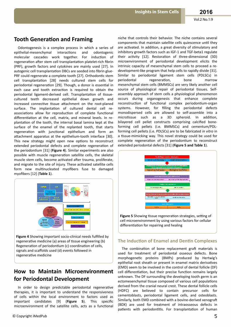
epithelial-mesenchymal interactions and odontogenicmolecular cascades work together. For induction ofregeneration after stem cell transplantation platelet-rich fibrin(PRF), growth factors and cytokines are mainly used [27]. Inautogenic cell transplantation DBCs are seeded into fibrin glue-PRF could regenerate a complete tooth [27]. Orthodontic stemcell transplantation [28] needs cultured stem cells forperiodontal regeneration [29]. Though, a donor is essential ineach case and tooth extraction is required to obtain theperiodontal ligament-derived cell. Transplantation of tissue-cultured teeth decreased epithelial down growth andincreased connective tissue attachment on the root-planedsurface. The implantation of cultured dental cell re-associations allow for reproduction of complete functionaldifferentiation at the cell, matrix, and mineral levels. In re-plantation of the tooth, the internal basal lamina kept at thesurface of the enamel of the replanted tooth, that startsregeneration with junctional epithelium and form anattachment apparatus at the epithelium-tooth interface [30].This new strategy might open new options to reconstructextended periodontal defects and complete regeneration ofthe periodontium [31] (Figure 4). Similar experiments are alsopossible with muscle regeneration satellite cells, the skeletalmuscle stem cells, become activated after trauma, proliferate,and migrate to the site of injury. These activated satellite cellsform new multinucleated myofibers fuse to damagedmyofibers [12] (Table 1).
Figure 4 Showing important socio-clinical needs fulfilled byregenerative medicine (a) areas of tissue engineering (b)Regeneration of periodontium (c) coordination of cells,signals and scaffolds used (d) events followed inregenerative medicine
How to Maintain Microenvironmentfor Periodontal Development
In order to design predictable periodontal regenerativetherapies, it is important to understand the responsivenessof cells within the local environment to factors used asimportant candidates [9] (Figure 5). This specificmicroenvironment of the satellite cells, acts as a functional
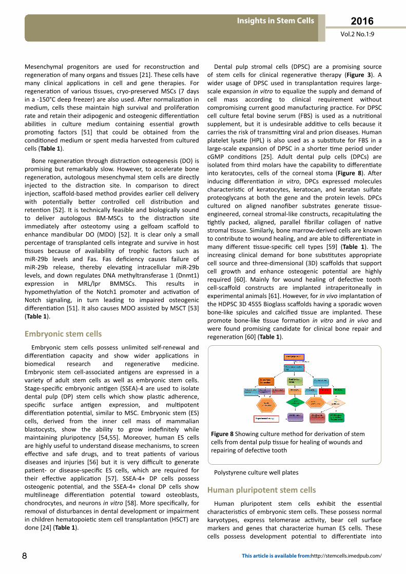
niche that controls their behavior. The niche contains severalcomponents that maintain satellite cells quiescence until theyare activated. In addition, a great diversity of stimulatory andinhibitory growth factors such as IGF-1 and TGF-beta1 regulatetheir activity [12]. Restoration of three-dimensional (3D)microenvironment of periodontal development elicits theintrinsic capacity of mesenchymal stem cells to proceed a re-development-like program that help cells to rapidly divide [31].Similar to periodontal ligament stem cells (PDLSCs) inperiodontal regeneration, bone marrowmesenchymal stem cells (BMMSCs) are very likely another cellsource of physiological repair of periodontal tissues. Self-assembly approach of stem cells a physiological phenomenonoccurs during organogenesis that enhance completereconstruction of functional complex periodontium-organsystems. However, for filling the periodontal defectsmonodispersed cells are allowed to self-assemble into amicrotissue such as a 3D spheroid. In addition,bilayered cell pellet constructs comprising calcified bone-forming cell pellets (i.e. BMMSCs) and cementum/PDL-forming cell pellets (i.e. PDLSCs) are to be fabricated in vitro ina tissue-mimicking way. This novel strategy could be used forcomplete regeneration of the periodontium to reconstructextended periodontal defects [31] (Figure 5 and Table 1).
Figure 5 Showing tissue regeneration strategies, setting ofcell microenvironment by using various factors for cellulardifferentiation for repairing and healing
The Induction of Enamel and Dentin ComplexesThe combination of bone replacement graft materials is
used for treatment of periodontal osseous defects. Bonemorphogenetic proteins (BMPs) produced by Hertwig’sepithelial root sheath or present in enamel matrix derivatives(EMD) seem to be involved in the control of dental follicle (DF)cell differentiation, but their precise function remains largelyunknown. The DF surrounding the developing tooth germ is anectomesenchymal tissue composed of various cell populationsderived from the cranial neural crest. These dental follicle cells(HDFC) are believed to contain precursor cells forcementoblasts, periodontal ligament cells, and osteoblasts.Similarly, both EMD combined with a bovine-derived xenograft(BDX) are used for treatment of intraosseous defects inpatients with periodontitis. For transplantation of human
Insights in Stem Cells
Vol.2 No.1:9
2016
© Copyright iMedPub 5

tooth germ components, heterologously recombinedmouse dental epithelia and xenogenic graft tissue are used tomake reconstructed transplants. Further, differentiation ofmouse dental epithelia is restrained by putative suppressivefactors derived from human dental papilla until they areseparated by mineralized dentin layers that serve as a diffusionbarrier. The mouse enamel organ nevertheless retains its ownphenotypic characteristics and intrinsic timingof cell differentiation and function [32].
ds and enamel matrix derivativeHuman demineralized freeze-dried bone allograft (DFDBA)
and enamel matrix derivative (EMD) are used for treatment ofbone and periodontal defects [33]. Tbx1 expression inepithelium requires mesenchyme-derived signalsbecause dental mesenchyme induces expression of Tbx1 inrecombined dental and non-dentalepithelia. Forced expressionof Tbx1 in dental explants activates amelogenin expression.Thus, Tbx1 expression in developing teeth takes place underdirect control of FGF signaling that correlates withdetermination of the ameloblast lineage [34]. Sonic hedgehog(Shh), one of the essential molecules for embryogenesis andorganogenesis, is strongly expressed in the enamel knot, whichrepresents the signaling center for odontogenesis due to thepresence of essential secretory molecules [35]. There aremethods which are used for formation of adipose tissue invitro and in vivo by using human adipose-derived stromal cells(ADSCs) utilizing a gelatin sponge (Gelform) as a scaffold.These tissue-engineered constructs are exposed to adipogenicdifferentiation medium for in vitro and implanted in the backsof severe combined immunodeficient (SCID) in animal modelsfor in vivo adipose regeneration [36]. Enamel matrix derivative(EMD) influence activities of cementoblasts and osteoblasts,and thus may be able to regulate cell activities at a periodontalregenerative site (Table 1).
Biodegradable polymer scaffoldsDissociated tooth tissues contain both dentin and enamel,
show presence of epithelial and mesenchymal dental stemcells in porcine third molar tissues. For bio-engineering,complex tooth structures are obtained from pig tooth budtissues which show potential for the regeneration ofmammalian dental tissues. These cultured rat tooth bud cellsare obtained from 3 to 7 day post-natal (dpn) rats and cell-seeded biodegradable scaffolds are grown in the omenta ofadult rat hosts for 12 wks, and then harvested. Thus,bioengineer tooth structures derived from cultured tooth budcells dental epithelial and mesenchymal stem cells can bemaintained in vitro in culture medium. Similarly, boneconstructs are grown in vitro with use of isolated cells,biodegradable polymer scaffolds, and bioreactors [5].Culturing bone marrow mesenchymal stem cells (BMSCs) onceramic bovine bone scaffolds in different environments invitro are used to induce proliferation, differentiation, andmaturation of BMSCs [5]. Regeneration of extensive bonedefects is done using stem cells mainly by injecting adipose-
derived stem cells and demineralized bone matrix (DBM) intoareas of bone defect [8] (Table 1).
Vascularization of engineered teethThe implantation of cultured dental cell-cell re-associations
are allowed for the reproduction of fully formed teeth, crownmorphogenesis, epithelial histogenesis. It also needsmineralized dentin and enamel deposition for root-periodontium development. Vascularization is critical fororganogenesis and tissue engineering generated toothbecause it assists in blood vessel formation during toothdevelopment. It is also essential for cell re-associations, invivo implantation. Ex vivo, blood vessels are developed inthe dental mesenchyme from the cap to bell stages and inthe enamel organ, shortly before ameloblast differentiation. Incultured teeth and cell re-associations, blood-vessel-likestructures are still remained in the peridental mesenchyme,but these never developed into dental tissues.After implantation, both teeth and re-associations getrevascularized and these newly formed blood vesselsoriginated from the host, are allowed for their survival, inaffording conditions for fast organ growth, mineralization,and enamel secretion [37] (Table 1).
Factors Necessary for PeriodontalConnective Tissue AttachmentFormation on Dental Implants
There are important growth factors and adhesion moleculeswhich are necessary for periodontal connective tissueattachment after formation of cellular associationson dental implants. TGF-beta, CCN proteins and cell-associatedmolecules, p38α MAPK are required for tooth morphogenesisand enamel secretion [19] (Figure 6). However for quick graftestablishment and attachment amelogenin, bone sialoproteinand vimentin protein are used [10]. Recently, Cbfa1 was foundto be a critical transcriptional regulator of osteoblastdifferentiation [38]. After periodontium transplantation of PLcells on wounded surface these cells secrete their own factorswhich do faster regeneration than ES cells, possibly because ofPL cell plasticity and the capacity to undergo effectivedifferentiation in the periodontal cellular microenvironment[39] (Figure 6). Besides, orthodontic tooth movementpromotes the differentiation of transplanted cells, and thedifferentiation predominantly in the paravascular areas of theperiodontium while in autogenous periodontal cell grafts,enamel matrix derivative (EMD), play important role on theimplant-connective tissue interface. More specifically,implants that received GCT cell grafts were found surroundedby fibrous connective tissue. In contrast, implants thatreceived PDL cells without the application of EMDdemonstrated good bone contact, but strands of epitheliumwere observed in the implant-connective tissue interface(Table 1). Both amelogenins and soluble dentin proteinsshowed bone induction activity like bone morphogeneticprotein and induce differentiation of mesenchymal cell intochondrocyte and osteocyte [40]. For dental root development
Insights in Stem Cells
Vol.2 No.1:9
2016
6 This article is available from:http://stemcells.imedpub.com/

conventional pediatric anticancer therapy is also used byusing stem cell transplantation [41].
Figure 6 Biological pulp filling cell injection
Orthodontics and Stem CellTransplantation
Implantation of cultured stem cellsStem cells are used for finding cures for numerous diseases
including skin wound healingthrough transplantation medicine. Implantations ofcultured cells are applied for periodontal regeneration inwhich a donor is essential for tooth extraction and to obtainthe periodontal ligament-derived cell. There are advancedregenerative techniques available combining tissue cultureand transplantation of teeth [29]. Transplantation of tissue-cultured teeth decreases epithelial down growth and increaseconnective tissue attachment on the root-planed surface.Furthermore, EMD could remarkably increase the newconnective tissue attachment in this periodontal regenerativetechnique [29]. More specifically, implantation of cultured cell-cell re-associations led to crown morphogenesis, epithelialhistogenesis, organ vascularization, and root andperiodontium development [37]. The implantation ofcultured dental cell re-associations allow for reproduction ofcomplete functional differentiation at the cell, matrix, andmineral levels. A specific microenvironment and beta-cateninsignaling is required for embryonic tooth morphogenesis andpromotion of continuous tooth development are embryonicstage [42] (Table 1 and Figure 7).
For regeneration of transplanted tooth the junctionalepithelium attachment is highly essential [30]. The sizes offurcating defects and the coverage of gingival flap influencethe outcome of the treatment using autogenous periodontalligament cells with or without enamel matrix derivatives [43].Atelocollagen membrane inhibits apical migration ofregenerating epithelium and accelerates connective tissuereattachment in part by inhibiting the mitotic function of basalepithelial cells in early stages of wound healing [44]. However,implantation of bio-materials such as atelocollagen inducedentinogenesis of dental pulp tissue, but it also need basalgrowth factors to induce regeneration and utilization ofbiomaterials [45]. For determining success rate ofimplantation periodontal regeneration procedures arefollowed after finding connective tissue attachment level [46](Table 1 and Figure 7). Mesenchymal stem cell (MSC)-mediated tissue regeneration methods are used for
regeneration a bio-root and its associated periodontal tissuesto restore tooth loss [21]. MSC increases bony regeneration incalvarial critical-size defects in rabbits, and provide a newpromising therapeutic strategy to aid skeletal healing [23].
Figure 7 Showing different types of periodontal toothdefects and its histological examination
Bone marrow mesenchymal stem cells (MSCs) comprise aheterogeneous population of postnatal progenitor cells withprofound immunomodulatory properties, such as upregulationof Foxp3 (+) regulatory T cells (Tregs) and downregulation ofTh17 cells. These MSC subpopulations possess the range ofimmunomodulatory function [47]. Similarly, human umbilicalcord-derived mesenchymal stem cells (UC-MSCs) are used tofacilitate osteogenic differentiation in bone regeneration.These cells could be achieved by over expression of osterix(Osx) [48]. Due to their high proliferation property UC-MSCscould play important role in bone tissue engineering. Moreoften, stem cells cultured in conditioned medium are used fortransplantation purpose as a promising alternative to skinwound healing treatment [49] (Table 1). More specifically, forclinical management of soft tissues mixed population of bonemarrow-derived autologous stem and progenitor cells areseeded onto β-tricalcium phosphate (β-TCP), which serve as ascaffold to deliver cells directly to the defect [50]. This is alsoused for vascularization, mineralization of bone tissues. Thusboth bone marrow and adipose-derived stem cells were foundare considered best choice for regeneration of the defect.These adipose-derived stem cells are easily accessible andabundantly proliferate into mesenchymal cells which form aneffective bone regeneration material (Table 1).
Mesenchymal stem cell Mesenchymal stem cell transplantations (MSCT) are used to
treat human diseases and in orthodontics. These cells dividerapidly and differentiate within the injured tissue intospecialized cells post transplantation [49]. Similarly,mesenchymal progenitor stem cells exist within the bonemarrow stroma in form of subset of nonhematopoietic cellscan expand ex vivo and induce either in vitro or in vivo. Thesecells terminally differentiate into osteoblasts, chondrocytes,adipocytes, tenocytes, myotubes, neural cells, andhematopoietic-supporting stroma. These mesenchymalprogenitor cells require suitable microenvironment for cellularproliferation and differentiation with a wide range of growthfactors, cytokines, chemokines, proteins and enzymes.
Insights in Stem Cells
Vol.2 No.1:9
2016
© Copyright iMedPub 7
Mesenchymal progenitors are used for reconstruction andregeneration of many organs and tissues [21]. These cells havemany clinical applications in cell and gene therapies. Forregeneration of various tissues, cryo-preserved MSCs (7 daysin a -150°C deep freezer) are also used. After normalization inmedium, cells these maintain high survival and proliferationrate and retain their adipogenic and osteogenic differentiationabilities in culture medium containing essential growthpromoting factors [51] that could be obtained from theconditioned medium or spent media harvested from culturedcells (Table 1).
Bone regeneration through distraction osteogenesis (DO) ispromising but remarkably slow. However, to accelerate boneregeneration, autologous mesenchymal stem cells are directlyinjected to the distraction site. In comparison to directinjection, scaffold-based method provides earlier cell deliverywith potentially better controlled cell distribution andretention [52]. It is technically feasible and biologically soundto deliver autologous BM-MSCs to the distraction siteimmediately after osteotomy using a gelfoam scaffold toenhance mandibular DO (MDO) [52]. It is clear only a smallpercentage of transplanted cells integrate and survive in hosttissues because of availability of trophic factors such asmiR-29b levels and Fas. Fas deficiency causes failure ofmiR-29b release, thereby elevating intracellular miR-29blevels, and down regulates DNA methyltransferase 1 (Dnmt1)expression in MRL/lpr BMMSCs. This results inhypomethylation of the Notch1 promoter and activation ofNotch signaling, in turn leading to impaired osteogenicdifferentiation [51]. It also causes MDO assisted by MSCT [53](Table 1).
Embryonic stem cellsEmbryonic stem cells possess unlimited self-renewal and
differentiation capacity and show wider applications inbiomedical research and regenerative medicine.Embryonic stem cell-associated antigens are expressed in avariety of adult stem cells as well as embryonic stem cells.Stage-specific embryonic antigen (SSEA)-4 are used to isolatedental pulp (DP) stem cells which show plastic adherence,specific surface antigen expression, and multipotentdifferentiation potential, similar to MSC. Embryonic stem (ES)cells, derived from the inner cell mass of mammalianblastocysts, show the ability to grow indefinitely whilemaintaining pluripotency [54,55]. Moreover, human ES cellsare highly useful to understand disease mechanisms, to screeneffective and safe drugs, and to treat patients of variousdiseases and injuries [56] but it is very difficult to generatepatient- or disease-specific ES cells, which are required fortheir effective application [57]. SSEA-4+ DP cells possessosteogenic potential, and the SSEA-4+ clonal DP cells showmultilineage differentiation potential toward osteoblasts,chondrocytes, and neurons in vitro [58]. More specifically, forremoval of disturbances in dental development or impairmentin children hematopoietic stem cell transplantation (HSCT) aredone [24] (Table 1).
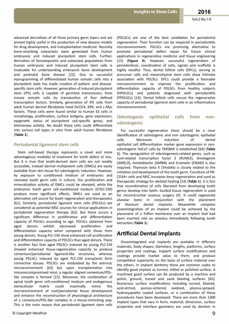
Dental pulp stromal cells (DPSC) are a promising sourceof stem cells for clinical regenerative therapy (Figure 3). Awider usage of DPSC used in transplantation requires large-scale expansion in vitro to equalize the supply and demand ofcell mass according to clinical requirement withoutcompromising current good manufacturing practice. For DPSCcell culture fetal bovine serum (FBS) is used as a nutritionalsupplement, but it is undesirable additive to cells because itcarries the risk of transmitting viral and prion diseases. Humanplatelet lysate (HPL) is also used as a substitute for FBS in alarge-scale expansion of DPSC in a shorter time period undercGMP conditions [25]. Adult dental pulp cells (DPCs) areisolated from third molars have the capability to differentiateinto keratocytes, cells of the corneal stoma (Figure 8). Afterinducing differentiation in vitro, DPCs expressed moleculescharacteristic of keratocytes, keratocan, and keratan sulfateproteoglycans at both the gene and the protein levels. DPCscultured on aligned nanofiber substrates generate tissue-engineered, corneal stromal-like constructs, recapitulating thetightly packed, aligned, parallel fibrillar collagen of nativestromal tissue. Similarly, bone marrow-derived cells are knownto contribute to wound healing, and are able to differentiate inmany different tissue-specific cell types [59] (Table 1). Theincreasing clinical demand for bone substitutes appropriatecell source and three-dimensional (3D) scaffolds that supportcell growth and enhance osteogenic potential are highlyrequired [60]. Mainly for wound healing of defective toothcell-scaffold constructs are implanted intraperitoneally inexperimental animals [61]. However, for in vivo implantation ofthe HDPSC 3D 45S5 Bioglass scaffolds having a sporadic wovenbone-like spicules and calcified tissue are implanted. Thesepromote bone-like tissue formation in vitro and in vivo andwere found promising candidate for clinical bone repair andregeneration [60] (Table 1).
Figure 8 Showing culture method for derivation of stemcells from dental pulp tissue for healing of wounds andrepairing of defective tooth
Polystyrene culture well plates
Human pluripotent stem cellsHuman pluripotent stem cells exhibit the essential
characteristics of embryonic stem cells. These possess normalkaryotypes, express telomerase activity, bear cell surfacemarkers and genes that characterize human ES cells. Thesecells possess development potential to differentiate into
Insights in Stem Cells
Vol.2 No.1:9
2016
8 This article is available from:http://stemcells.imedpub.com/
advanced derivatives of all three primary germ layers and areproved highly useful in the production of new disease modelsfor drug development, and transplantation medicine. Recentlybone-resorbing osteoclasts were generated from humanembryonic and induced pluripotent stem cells. Further,derivation of hematopoietic and osteoclast populations fromhuman embryonic and induced pluripotent stem cells isinvaluable for understanding embryonic bone developmentand postnatal bone disease [22]. Due to successfulreprogramming of differentiated human somatic cells into apluripotent state has made creation of patient- and disease-specific stem cells. However, generation of induced pluripotentstem (iPS) cells is capable of germline transmission, frommouse somatic cells by transduction of four definedtranscription factors. Similarly, generation of iPS cells fromadult human dermal fibroblasts need Oct3/4, Klf4, and c-Mycfactors. These cells were found similar to human ES cells inmorphology, proliferation, surface antigens, gene expression,epigenetic status of pluripotent cell-specific genes, andtelomerase activity. No doubt these cells could differentiateinto various cell types in vitro from adult human fibroblasts(Table 1).
Periodontal ligament stem cellsStem cell-based therapy represents a novel and more
advantageous modality of treatment for tooth defect or loss.But it is true that tooth-derived stem cells are not readilyaccessible, instead dermal multipotent cells (DMCs) are easilyavailable from skin tissue for odontogenic induction. However,by exposure to conditioned medium of embryonic andneonatal tooth germ cells in culture, the proliferation andmineralization activity of DMCs could be elevated, while theembryonic tooth germ cell-conditioned medium (ETGC-CM)produce more significant effects. DMCs are used as analternative cell source for tooth regeneration and therapeutics[61]. Similarly, periodontal ligament stem cells (PDLSCs) areconsidered as potential MSC sources for clinical applications inperiodontal regeneration therapy [62]. But there occurs asignificant difference in proliferation and differentiationcapacity of PDLSCs according to age. PDLSCs obtained fromaged donors exhibit decreased proliferation anddifferentiation capacity when compared with those fromyoung donors. Young PLC-CM show enhanced cell proliferationand differentiation capacity of PDLSCs than aged donors. Thereis another fact that aged PDLSCs induced by young PLC-CMshowed enhanced tissue-regenerative capacity to producecementum/periodontal ligament-like structures, whereasyoung PDLSCs induced by aged PLC-CM transplants formconnective tissues. PDLSCs are modulated by the extrinsicmicroenvironment [63] but upon transplantation intoimmunocompromised mice, a regular aligned cementum/PDL-like complex is formed [15]. Moreover, the combination ofapical tooth germ cell-conditioned medium and endogenousextracellular matrix could maximally mimic themicroenvironment of root/periodontal tissue developmentand enhance the reconstruction of physiological architectureof a cementum/PDL-like complex in a tissue-mimicking way.This is the main reason that periodontal ligament stem cells
(PDLSCs) are one of the best candidates for periodontalregeneration. Their function can be impaired in periodontitismicroenvironment. PDLSCs are promising alternative topromote periodontal defect repair for future clinicalapplications in regenerative medicine and tissue engineering[15] (Figure 4). However, successful regeneration ofperiodontium, coordination of cells, signals and scaffolds ishighly needful. Thus, dental follicle cells (DFCs), serving asprecursor cells and mesenchymal stem cells show intimateassociation with PDLSCs. DFCs could provide a favorablemicroenvironment to improve the proliferation anddifferentiation capacity of PDLSCs from healthy subjects(HPDLSCs) and patients diagnosed with periodontitis(PPDLSCs) [14]. Dental follicle cells rescue the regenerativecapacity of periodontal ligament stem cells in an inflammatorymicroenvironment.
Odontogenic epithelial cells from non-odontogenic
For successful regeneration there should be a clearidentification of odontogenic and non odontogenic epithelialcells. Moreover, induction of dentalepithelial cell differentiation marker gene expression in non-odontogenic HaCaT cells by TMSB4X is established [64] (Table1). The upregulation of odontogenesis-related genes, such asrunt-related transcription factor 2 (RUNX2), Amelogenin(AMELX), Ameloblastin (AMBN) and Enamelin (ENAM) is alsorequired. Thymosin beta 4 (Tmsb4x) is closely related to theinitiation and development of the tooth germ. Coculture of PB-CD34+ cells and MSC increases bony regeneration and used astherapeutic strategy for skeletal healing [14] (Table 1). It is factthat recombination of cells liberated from developing toothgerms develop into teeth. Guided tissue regeneration is usedfor reconstructive osseous surgery [4]. It could regeneratealveolar bone in conjunction with the placementof titanium dental implants. Meanwhile completeosseointegration of an implant could be achieved by theplacement of a Teflon membrane over an implant that hadbeen inserted into an alveolus immediately following toothextraction (Table 1).
Artificial Dental ImplantsOsseointegrated oral implants are available in different
materials, body shapes, diameters, lengths, platforms, surfaceproperties and coatings. Implant surface modifications andcoatings provide market value to them, and producecompetitive superiority on the basis of surface material overthe others. In implant dentistry there are common codes toidentify good implant as turned, milled or polished surface. Amachined good surface can be produced by a machine andpolish, ground, honed and sand blasting surfaces [65].Numerous surface modifications including turned, blasted,acid-etched, porous-sintered, oxidized, plasma-sprayed,hydroxyapatite coated surfaces, or a combination of theseprocedures have been developed. There are more than 1300implant types that vary in form, material; dimension, surfaceproperties and interface geometry are used by dentists in
Insights in Stem Cells
Vol.2 No.1:9
2016
© Copyright iMedPub 9
different countries [66]. Good implant surfaces are alsoprepared by embedding in polymethyl methacrylate resin.Surface modification of implant shapes or particular materialsis highly important because it can improve clinical results ofcommercially available implants. Un-decalcified sections areprepared with the sectioning-grinding technique. Thepercentage of bone contacting the implant surface can bemeasured with a self-designed histomorphometry methodusing a millimeter grid in a stereomicroscope.
Titanium-surfaced implant contains significantly higherpercentage of bone along the hydroxyapatite-coated material.Implant failure is a consequence of prosthetic loading in whichhealing is poorly developed. It could be resolved by measuringthe bone response around implants placed in the mandiblethat supported prostheses exhibiting two levels of fit and notloaded exclusively. A misfitting prostheses with high dynamicfunctional loads are superimposed with misfit loads alwaysprovides unsatisfactory results and is not suitable for clinicalapplications, because of fit does not alter the osseointegratedinterface. Finite element analysis (FEA) is considered a preciseand applicable method for evaluating dental implant systems.By means of FEA, a parasaggital model could be digitized withaddition of computed tomography (CT)-generated patient dataset, and various single-teeth, osseointegrated, two-dimensional dental implant models could be simulated forfinding a best fit. For successful implantation it is necessary toexamine the effect of implant diameter variation (3.8 mm-6.5mm) of both a press-fit, stepped cylindrical implant type and apress-fit, straight cylindrical implant type as osseointegrated inthe posterior mandible. In addition a comparison of stress-dissipating characteristics of the stepped implant versus thestraight implant design should be made to analyze bite forcedirection (vertical, horizontal, and oblique 45°) on implanttypes. It is true that using the widest diameter implant is notnecessarily the best choice when considering stressdistribution to surrounding bone, but within certainmorphological limits, for implant, an optimum dental implantexists for decreasing the stress magnitudes at the bone-implant interface. It could manage the stress and dissipate itthroughout the stepped cylindrical implant in comparison tothe straight implant. Therefore, it is highly important in FEA ofdental implants to consider not only axial forces (verticalloading) and horizontal forces (moment-causing loads), butalso to consider a combined load (oblique bite force). As it istrue that there is more realistic bite directions which canspread given force that will cause the highest localized stress incortical bone.
ConclusionRecent advancements made in regenerative techniques by
combining tissue culture and transplantation of stem cells forwound healing and replacement of defective teeth. Moreover,human periodontal ligament stem cells are considered as goodsource of physiological repair of periodontal tissues. MSCs areused in skeletal healing and are considered as promisingcandidate for bio-root engineering. Mesenchymal stem cellsand SSEA)-4 show plastic adherence, specific surface antigen
expression, and multipotent differentiation potential. Inaddition due to high proliferation property geneticallyengineered UC-MSCs are also used in bone tissue engineering.For successful development, framing, growth and regenerationof tooth stable and durable biodegradable polymer scaffoldsmaterials and cementum/periodontal-ligament complexformation are highly important. In addition,, inductionof enamel and formation of dentin complexes and removal ofdemineralized matrix, vascularization of engineered teeth,maintenance of microenvironment are important issues. Forsuccessful wound healing of tooth implantation ofcultured cells, adhesion and cementing molecules/factors arealso needed for stable attachment formationon dental implants. For adhesion of cells to periodontalconnective tissue amelogenin, bone sialoprotein and vimentinproteins are also needed. Osseointegrated oral implants areavailable in different materials, body shapes, diameters,lengths, platforms, surface properties and coatings.
Acknowledgements: Author is thankful to Prof. AshokKumar, Vice Chancellor, DDU Gorakhpur University, Gorakhpurfor his kind support.
References1. Wang Y, Preston B, Guan G (2012) Tooth bioengineering leads
the next generation of dentistry. Int J Paediatr Dent 22: 406-418.
2. Nie X, Jin Y, Long J, Wu L, Jing W, et al. (2008) Experimental studyon the development of tissue-engineered tooth germ withheterotopic allotransplantation. Sichuan Da Xue Xue Bao Yi XueBan 39: 279-282.
3. Scott MA, Levi B, Askarinam A, Nguyen A, Rackohn T, et al.(2012) Brief review of models of ectopic bone formation. StemCells Dev 21: 655-667.
4. Nyman S, Lang NP, Buser D, Bragger U (1990) Bone regenerationadjacent to titanium dental implants using guided tissueregeneration: a report of two cases. The International Journal ofOral & Maxillofacial Implants 5: 9-14.
5. Jin F, Zhang Y, Xuan K, He D, Deng T, et al. (2010) Establishmentof three-dimensional tissue-engineered bone constructs undermicrogravity-simulated conditions. Artif Organs 34: 118-25.
6. D'Antò V, Cantile M, Cillo C, Valletta A (2003) The cellular andmolecular bases of "tooth framing". Minerva Stomatol 52:489-506.
7. Nakata A, Kameda T, Nagai H, Ikegami K, Duan Y, et al. (2003)Establishment and characterization of a spontaneouslyimmortalized mouse ameloblast-lineage cell line. BiochemBiophys Res Commun 308: 834-839.
8. Han DS, Chang HK, Kim KR, Woo SM (2014) Consideration ofbone regeneration effect of stem cells: comparison of boneregeneration between bone marrow stem cells and adipose-derived stem cells. J Craniofac Surg 25: 196-201.
9. Tokiyasu Y, Takata T, Saygin E, Somerman M (2000) Enamelfactors regulate expression of genes associated withcementoblasts. J Periodontol 71: 1829-1839.
10. Honda MJ, Shinohara Y, Sumita Y, Tonomura A, Kagami H, et al.(2006) Shear stress facilitates tissue-engineered odontogenesis.Bone 39: 125-133.
Insights in Stem Cells
Vol.2 No.1:9
2016
10 This article is available from:http://stemcells.imedpub.com/
11. Craig RG, Kamer AR, Kallur SP, Inoue M, Tarnow DP (2006)Effects of periodontal cell grafts and enamel matrix proteins onthe implant-connective tissue interface: a pilot study in theminipig. J Oral Implantol 32: 228-236.
12. Ten Broek RW, Grefte S, von den Hoff JW (2010) Regulatoryfactors and cell populations involved in skeletal muscleregeneration. J Cell Physiol 224: 7-16.
13. Xu L, Tang L, Jin F, Liu XH, Yu JH, et al. (2009) The apical region ofdeveloping tooth root constitutes a complex and maintains theability to generate root and periodontium-like tissues. JPeriodontal Res 44: 275-282.
14. Liu J, Wang L, Liu W, Li Q, Jin Z, et al. (2014) Dental follicle cellsrescue the regenerative capacity of periodontal ligament stemcells in an inflammatory microenvironment. PLoS One 9:e108752.
15. Yang Z, Jin F, Zhang X, Ma D, Han C, et al. (2009) Tissueengineering of cementum/periodontal-ligament complex using anovel three-dimensional pellet cultivation system for humanperiodontal ligament stem cells. Tissue Eng Part C Methods 15:571-81.
16. Mrozik KM, Gronthos S, Menicanin D, Marino V, Bartold PM(2012) Effect of coating Straumann Bone Ceramic withEmdogain on mesenchymal stromal cell hard tissue formation.Clin Oral Investig 16: 867-878.
17. Ramis JM, Taxt-Lamolle SF, Lyngstadaas SP, Reseland JE, EllingsenJE, et al. (2012) Identification of early response genes toroughness and fluoride modification of titanium implants inhuman osteoblasts. Implant Dent 21: 141-9.
18. Abdollah S, Macias-Silva M, Tsukazaki T, Hayashi H, Attisano L, etal. (1997) J. Biol. Chem. 272.
19. Greenblatt MB, Kim JM, Oh H, Park KH, Choo MK, et al. (2015)p38α MAPK is required for tooth morphogenesis and enamelsecretion. J Biol Chem 290: 284-295.
20. Kanyama M, Shimo T, Sugito H, Nagayama M, Kuboki T, et al.(2013) Regulation of CCN2 gene expression and possible roles indeveloping tooth germs. Arch Oral Biol 58: 1659-1666.
21. Kaku M, Tai M, Kawata T, Fujita T, Motokawa M, et al. (2011)Mesenchymal stem cell-induced cranial suture-like gap in rats.Plast Reconstr Surg 127: 69-77.
22. Grigoriadis AE, Kennedy M, Bozec A, Brunton F, Stenbeck G, etal. (2010) Directed differentiation of hematopoietic precursorsand functional osteoclasts from human ES and iPS cells. Blood115: 2769-2776.
23. Li G, Wang X, Cao J, Ju Z, Ma D, et al. (2014) Coculture ofperipheral blood CD34+ cell and mesenchymal stem cell sheetsincrease the formation of bone in calvarial critical-size defects inrabbits. Br J Oral Maxillofac Surg 52: 134-139.
24. Vesterbacka M, Ringdén O, Remberger M, Huggare J, Dahllöf G(2012) Disturbances in dental development and craniofacialgrowth in children treated with hematopoietic stem celltransplantation. Orthod Craniofac Res 15: 21-29.
25. Govindasamy V, Ronald VS, Abdullah AN, Ganesan Nathan KR,Aziz ZA, et al. (2011) Human platelet lysate permits scale-up ofdental pulp stromal cells for clinical applications. Cytotherapy13: 1221-33.
26. Han C, Yang Z, Zhou W, Jin F, Song Y, et al. (2010) Periapicalfollicle stem cell: a promising candidate for cementum/periodontal ligament regeneration and bio-root engineering.Stem Cells Dev 19: 1405-1415.
27. Yang KC, Wang CH, Chang HH, Chan WP, Chi CH, et al. (2012)Fibrin glue mixed with platelet-rich fibrin as a scaffold seededwith dental bud cells for tooth regeneration. J Tissue Eng RegenMed 6: 777-785.
28. Liu Y, Jiang M, Hao W, Liu W, Tang L, et al. (2013) Skin epithelialcells as possible substitutes for ameloblasts during toothregeneration. J Tissue Eng Regen Med 7: 934-943.
29. Saito A, Saito E, Yoshimura Y, Takahashi D, Handa R, et al. (2011)Attachment formation after transplantation of teeth culturedwith enamel matrix derivative in dogs. J Periodontol 82: 1462-8.
30. Shioya K, Sawada T, Miake Y, Inoue S, Yanagisawa T (2009)Ultrastructural study of tissues surrounding replanted teeth anddental implants. Clin Oral Implants Res 20: 299-305.
31. Yang ZH, Jin F, Zhang XJ, Liu X, Zhang YF, et al. (2010) A novelpossible strategy based on self-assembly approach to achievecomplete periodontal regeneration. Artif Organs 34: 603-9.
32. Isogawa N, Terashima T, Nakano Y, Kindaichi J, Takagi Y, et al.(2004) The induction of enamel and dentin complexes bysubcutaneous implantation of reconstructed human and murinetooth germ elements. Arch Histol Cytol 67: 65-77.
33. Intini G, Andreana S, Buhite RJ, Bobek LA (2008) A comparativeanalysis of bone formation induced by human demineralizedfreeze-dried bone and enamel matrix derivative in rat calvariacritical-size bone defects. J Periodontol 79: 1217-24.
34. Mitsiadis TA, Tucker AS, De Bari C, Cobourne MT, Rice DP (2008)A regulatory relationship between Tbx1 and FGF signaling duringtooth morphogenesis and ameloblast lineage determination.Dev Biol 320: 39-48.
35. Takahashi S, Kawashima N, Sakamoto K, Nakata A, Kameda T, etal. (2007) Differentiation of an ameloblast-lineage cell line (ALC)is induced by Sonic hedgehog signaling. Biochem Biophys ResCommun 353: 405-411.
36. Hong L, Peptan IA, Colpan A, Daw JL (2006) Adipose tissueengineering by human adipose-derived stromal cells. CellsTissues Organs 183: 133-140.
37. Nait Lechguer A, Couble ML, Labert N, Kuchler-Bopp S, Keller L,et al. (2011) Cell differentiation and matrix organization inengineered teeth. J Dent Res 90: 583-589.
38. D'Souza RN, Aberg T, Gaikwad J, Cavender A, Owen M, et al.(1999) Cbfa1 is required for epithelial-mesenchymal interactionsregulating tooth development in mice. Development 126:2911-2920.
39. Nayak BN, Wiltshire WA, Ganss B, Tenenbaum H, McCulloch CA,et al. (2008) Healing of periodontal tissues followingtransplantation of cells in a rat orthodontic tooth movementmodel. Angle Orthod 78: 826-31.
40. Wang W (1993) Ectopic bone induction by human fetal enamelproteins. Zhonghua Kou Qiang Yi Xue Za Zhi 28: 362-364, 384.
41. Hölttä P, Hovi L, Saarinen-Pihkala UM, Peltola J, Alaluusua S(2005) Disturbed root development of permanent teeth afterpediatric stem cell transplantation. Dental root developmentafter SCT. Cancer 103: 1484-1493.
42. Liu J, Bian Z, Kuijpers-Jagtman AM, von den Hoff JW (2010) Skinand oral mucosa equivalents: construction and performance.Orthod Craniofac Res 13: 11-20.
43. Zhu WD, Hou JX, Liu KN, Meng HX, Tang XL. (2009) Clinical andradiographic evaluation of class III furcation defects in thetreatment using autogenous periodontal ligament cells with or
Insights in Stem Cells
Vol.2 No.1:9
2016
© Copyright iMedPub 11
without enamel matrix derivatives. Beijing Da Xue Xue Bao18;41: 56-61.
44. Numabe Y, Ito H, Hayashi H, Ryder MI, Kamoi K (1993) Epithelialcell kinetics with atelocollagen membranes: a study in rats. JPeriodontol 64: 706-712.
45. Inoue T, Shimono M (1992) Repair dentinogenesis followingtransplantation into normal and germ-free animals. Proc FinnDent Soc 88 Suppl 1: 183-194.
46. Caton J, Nyman S, Zander H (1980) Histometric evaluation ofperiodontal surgery. II. Connective tissue attachment levels afterfour regenerative procedures. J Clin Periodontol 7: 224-231.
47. Yang R, Liu Y, Kelk P, Qu C, Akiyama K, et al. (2013) A subset ofIL-17(+) mesenchymal stem cells possesses anti-Candidaalbicans effect. Cell Res 23: 107-121.
48. Wang B, Huang S, Pan L, Jia S (2013) Enhancement of boneformation by genetically engineered human umbilical cord-derived mesenchymal stem cells expressing osterix. Oral SurgOral Med Oral Pathol Oral Radiol 116: e221-229.
49. Jayaraman P, Nathan P, Vasanthan P, Musa S, Govindasamy V(2013) Stem cells conditioned medium: a new approach to skinwound healing management. Cell Biol Int 37: 1122-1128.
50. Rajan A, Eubanks E, Edwards S, Aronovich S, Travan S, et al.(2014) Optimized cell survival and seeding efficiency forcraniofacial tissue engineering using clinical stemcell therapy.Stem Cells Transl Med 3: 1495-503.
51. Kojima SI, Kaku M, Kawata T, Motokawa M, Sumi H, et al. (2015)Cranial suture-like gap and bone regeneration aftertransplantation of cryopreserved MSCs by use of a programmedfreezer with magnetic field in rats. Cryobiology 70: 262-8.
52. Sun Z, Tee BC, Kennedy KS, Kennedy PM, Kim DG, et al. (2013)Fields, H.W. Scaffold-based delivery of autologous mesenchymalstem cells for mandibular distraction osteogenesis: preliminarystudies in a porcine model. PLoS One 8: e74672.
53. Tee BC, Sun Z (2015) Mandibular distraction osteogenesisassisted by cell-based tissue engineering: a systematic review.Orthod Craniofac Res 18 Suppl 1: 39-49.
54. Evans MJ, Kaufman MH. (1981) Establishment in culture ofpluripotential cells from mouse embryos. Nature 292: 154-156.
55. Martin GR (1981) Isolation of a pluripotent cell line from earlymouse embryos cultured in medium conditioned by
teratocarcinoma stem cells. Proc Natl Acad Sci USA 78:7634-7638.
56. Thomson JA, Itskovitz-Eldor J, Shapiro SS, Waknitz MA, SwiergielJJ, et al. (1998) Embryonic stem cell lines derived from humanblastocysts. Science 282: 1145-1147.
57. Takahashi K, Tanabe K, Ohnuki M, Narita M, Ichisaka T, et al.(2007) Induction of pluripotent stem cells from adult humanfibroblasts by defined factors. Cell 131: 861-872.
58. Kawanabe N, Murata S, Fukushima H, Ishihara Y, Yanagita T, et al.(2012) Stage-specific embryonic antigen-4 identifies humandental pulp stem cells. Exp Cell Res 318: 453-463.
59. Verstappen J, van Rheden RE, Katsaros C, Torensma R, Von denHoff JW (2012) Preferential recruitment of bone marrow-derived cells to rat palatal wounds but not to skin wounds. ArchOral Biol 57: 102-108.
60. El-Gendy R, Yang XB, Newby PJ, Boccaccini AR, Kirkham J (2013)Osteogenic differentiation of human dental pulp stromal cells on45S5 Bioglass® based scaffolds in vitro and in vivo. Tissue EngPart A 19: 707-715.
61. Huo N, Tang L, Yang Z, Qian H, Wang Y, et al. (2010)Differentiation of dermal multipotent cells into odontogeniclineage induced by embryonic and neonatal tooth germ cell-conditioned medium. Stem Cells Dev 19: 93-104.
62. Zhao BJ, Liu YH (2014) Simvastatin induces the osteogenicdifferentiation of human periodontal ligament stem cells.Fundam Clin Pharmacol 28: 583-592.
63. Zheng W, Wang S, Wang J, Jin F (2015) Periodontitis promotesthe proliferation and suppresses the differentiation potential ofhuman periodontal ligament stem cells. Int J Mol Med 36:915-922.
64. Kiyoshima T, Fujiwara H, Nagata K, Wada H, Ookuma YF, et al.(2014) Induction of dental epithelial cell differentiation markergene expression in non-odontogenic human keratinocytes bytransfection with thymosin beta 4. Stem Cell Res 12: 309-322.
65. Stout KJ, Davis EJ, Sullivan P (1990) Atlas of Machined Surfaces,eBook ISBN 978-94-011-7772-6. Publisher Springer Netherlands.
66. Binon PP (2000) Implants and components: entering the newmillennium. Int J Oral Maxillofac Implants 15: 76-94.
Insights in Stem Cells
Vol.2 No.1:9
2016
12 This article is available from:http://stemcells.imedpub.com/
Recommended


![[Dental Implants Cost]](https://img.dokumen.tips/doc/110x75/55638cefd8b42ad2128b4ef9/dental-implants-cost-55849922c2925.jpg)