Research Paper 65
The molecular basis of nuclear genetic code change in ciliatesCatherine A. Lozupone, Robin D. Knight and Laura F. Landweber*
Background: The nuclear genetic code has changed in several lineages Address: Department of Ecology and EvolutionaryBiology, Princeton University, Princeton, Newof ciliates. These changes, UAR to glutamine and UGA to cysteine, implyJersey 08544that eukaryotic release factor 1 (eRF1), the protein that recognizes stop
codons and terminates translation, changes specificity. Here we test whetherCorrespondence: Laura F. Landweber
changes in eRF1 drive genetic code evolution. E-mail: [email protected]
Results: Database sequence analysis reveals numerous genetic code Received: 20 October 2000Revised: 26 November 2000alterations in ciliates, including UGA → tryptophan in BlepharismaAccepted: 27 November 2000americanum and the distantly related Colpoda. We sequenced eRF1 from
four ciliates: B. americanum, a heterotrich that independently derived thePublished: 23 January 2001same eRF1 specificity as Euplotes, and three spirotrichs, Stylonychia
lemnae, S. mytilus, and Oxytricha trifallax, that independently derived theCurrent Biology 2001, 11:65–74
same genetic code as Tetrahymena (UAR → glutamine). Distantly relatedciliates with similar codes show characteristic changes in eRF1. We used 0960-9822/01/$ – see front mattera sliding window analysis to test associations between changes in specific 2001 Elsevier Science Ltd. All rights reserved.eRF1 residues and changes in the genetic code. The regions of eRF1that display convergent substitutions are identical to those identified in arecently reported nonsense suppression mutant screen in yeast.
Conclusions: Genetic code change by stop codon reassignment issurprisingly frequent in ciliates, with UGA → tryptophan occurring twiceindependently. This is the first description of this code, previously foundonly in bacteria and mitochondria, in a eukaryotic nuclear genome. eRF1 hasevolved strikingly convergently in lineages with variant genetic codes. Thestrong concordance with biochemical data indicates that our methodologymay be generally useful for detecting molecular determinants of biochemicalchanges in evolution.
Background In a 1995 study, Baroin Tourancheau et al. [11] sequencedthe a-tubulin and phosphoglycerate kinase genes forThe genetic code was once thought to be universal among
all organisms: once fixed, any change – tantamount to members of 6 of the 10 currently recognized ciliate classesrewiring a keyboard – would cause deleterious changes and suggested that members of the class Litostomateain every protein [1]. We now know that the code can and the heterotrich Stentor coeruleus may use the standardchange; alternative genetic codes are found in most mito- genetic code, whereas the karyorelictid Loxodes striatus,chondrial genomes [2], the nuclear genomes of the eubac- the heterotrich Condylostoma magnum, and the nassopho-terium Mycoplasma [3], the yeast Candida [4], diplomonads rean Zosterograptus sp. appear to use UAA and UAG to[5], the green alga Acetabularia, and a variety of ciliates encode glutamine. These data indicate that alteration of[e.g., 6–10]. Ciliates are remarkable in this respect. Several the ciliate genetic code was not a single, ancient event,different code variants have arisen independently, even as initially supposed [14], but a relatively common eventwithin a single class [11]. For example, Tetrahymena and in ciliates [11].Paramecium, class Oligohymenophorea, and Oxytricha andStylonychia, class Spirotrichea, translate UAA and UAG as Changes in the genetic code involve changes in tRNAs,
tRNA-modifying enzymes, or release factors. These cru-glutamine (using only UGA as stop) [6, 12]. Euplotes, alsoa spirotrich, instead translates UGA as cysteine, using cial components of the translation apparatus have changed
in ciliates, conferring new meanings to specific codons.UAA and UAG for termination [13]. Blepharisma, classHeterotrichea, uses UAA to encode stop [8]: before this Altered tRNAs in organisms with variant genetic codes
have provided some insight into the mechanism of geneticstudy, the translation of UAG and UGA in this specieswas unknown. Although GenBank includes a separate code changes. In addition to normal tRNAGln, Tetrahymena
thermophila has two unusual glutamine-specific tRNAGlntranslation table for Blepharisma, called the “Blepharismacode,” in which UAA and UGA encode stop and UAG with anticodons complementary to UAA and UAG: these
unusual tRNAs arose by duplication and divergence ofencodes glutamine [8], we find no support for the transla-tion of UAG as glutamine or UGA as stop in this species. the canonical tRNAGln [7]. Euplotes octocarinatus has only

66 Current Biology Vol 11 No 2
one tRNACys that translates UGA efficiently, despite G:A (Figure 1); B. americanum has no intron at this position.The eRF1 gene has 2 in-frame UGA codons in B. ameri-mispairing at the first anticodon position [15]. This change
requires loss of release factor specificity for UGA and/or canum and numerous in-frame UAR codons in S. lemnae,S. mytilus and O. trifallax.high concentrations of tRNACys relative to other tRNAs.
Because all known ciliate code changes alter stop codon Genetic database analysisProtein sequence data were available in the database formeanings, the eukaryotic release factor 1 (eRF1) must
have evolved alternate specificities. In eukaryotes, eRF1 7 of the 10 currently recognized [21] classes of ciliatesand for a member of the order Amorphoridae classifiedrecognizes the three standard stop codons in mRNA at
the ribosomal A site and terminates translation by peptidyl as sedis mutabilis in the subphylum Intramacronucleata[21]. We analyzed, for the first time, data for memberstRNA hydrolysis. Archaea have an eRF1 homolog, aRF1,
which is highly conserved across domains, and aRF1 even of the class Colpodea and for Nyctotherus, of the orderAmorphoridea. In addition, we expanded analysis of thefunctions with eukaryotic ribosomes [16].six previously studied classes [11] to include more species
Although eRF1 sequences from organisms with altered allowing better definition within these groups. Figure 2termination are of particular interest, only one eRF1 se- is a composite tree assembled from the literature, usingquence was in GenBank for an organism with a nonstan- both 28S large subunit [11, 22] and 18S small subunit [23,dard genetic code (the ciliate Tetrahymena thermophila 24, 25] congruent rDNA phylogenies, for the purpose of[17]). However, this eRF1 sequence suggested a mecha- character mapping of genetic codes. It lists all of thenism for the specificity change: the NIKS motif, con- genera for which information on genetic code usage isserved across all eukaryotes, is NIKD in Tetrahymena. To- available, except for many of the Spirotrichs analyzed,gether with the recent crystal structure of human eRF1 which form a monophyletic group with Stylonychia, Oxytri-[18] and mutational evidence that changes adjacent to cha, and Urostyla, and all appear to use the same code.
These data support that the classes Oligohymenophorea,NIKS abolish stop codon recognition in vitro [19], weSpirotrichea, and Litostomatea are each monophyletic.suggested that this specific mutation might be the molec-The heterotrichs and the karyorelictids form an earlyular cause of Tetrahymena’s altered genetic code [20].branching monophyletic group, but the relationship ofthe rest of the classes is largely unresolved. There is someIn order to test this hypothesis and to further examine
the biochemical basis for altered stop codon recognition support, however, that the classes Nassophorea, Colpo-dea, and Oligohymenophorea form a monophyletic groupin ciliates, we sequenced the complete gene encoding
eRF1 in three spirotrichs, Stylonychia lemnae, S. mytilus, (Figure 2). Members of the classes Karyorelictea and Nas-sophorea are grouped together by morphological data onlyand Oxytricha trifallax, that independently derived the
same genetic code as Tetrahymena. For comparison, we [26, 27] and are placed on the tree based on the rRNAsequences of Zosterograptus and Loxodes, respectively. Ta-also sequenced the gene in Blepharisma americanum, an
early diverging ciliate that uses UAA as stop [8], and ble 1 summarizes the database analysis and supplementalTable 1 provides more detail (see Supplementary ma-found evidence that this species uses UGA to encode
tryptophan. Using a novel statistical approach that uses terial).sliding window analysis to associate changes in specific
We found evidence in two distantly related lineages forregions of the protein with changes in the genetic code,UGA reassignment to tryptophan (Figure 1 and supple-as well as mapping some of the convergent amino acidmental Figure 1). The gene for mitotic cyclin-like proteinsubstitutions in lineages that independently evolved thein Colpoda inflata contains an in-frame UGA in the positionsame eRF1 specificity onto the protein crystal structure,of a conserved tryptophan (supplemental Figure 1). Anal-we identify several candidate amino acid residues or re-ysis of five partial protein sequences revealed no in-framegions of the protein that may underlie the altered codonUAR codons in this class (Table 1). In the heterotrichs,specificity of eRF1 and genetic code change in ciliates.members of the genera Stentor and Eufolliculina both hadmultiple in-frame UGA codons, and Eufolliculina andResults
Isolation of eRF1 Blepharisma use UAA to encode stop (Table 1; [8]). Align-ment of the Stentor and Eufolliculina proteins containingWe determined the complete macronuclear sequence of
Stylonychia lemnae, S. mytilus, and Oxytricha trifallax eRF1 in-frame UGA codons with orthologs retrieved usingBLAST did not suggest which amino acid UGA was cod-gene-sized chromosomes, which are all predicted to en-
code proteins 445 amino acids long, as well as Blepharisma ing for because the alignment was in variable regions ofthe proteins. However, the B. americanum eRF1 sequenceamericanum eRF1, predicted to encode a 436 amino acid
protein. S. lemnae, S. mytilus, and O. trifallax each appear generated in this study had two in-frame UGA codons,both in the location of conserved tryptophan residuesto have a phase I intron, 32, 32, and 38 nucleotides long,
respectively, at position 78 in the amino acid alignment (Figure 1 and supplemental Figure 1). Based on the close
Research Paper Evolution of Ciliate Genetic Codes Lozupone et al. 67
Figure 1
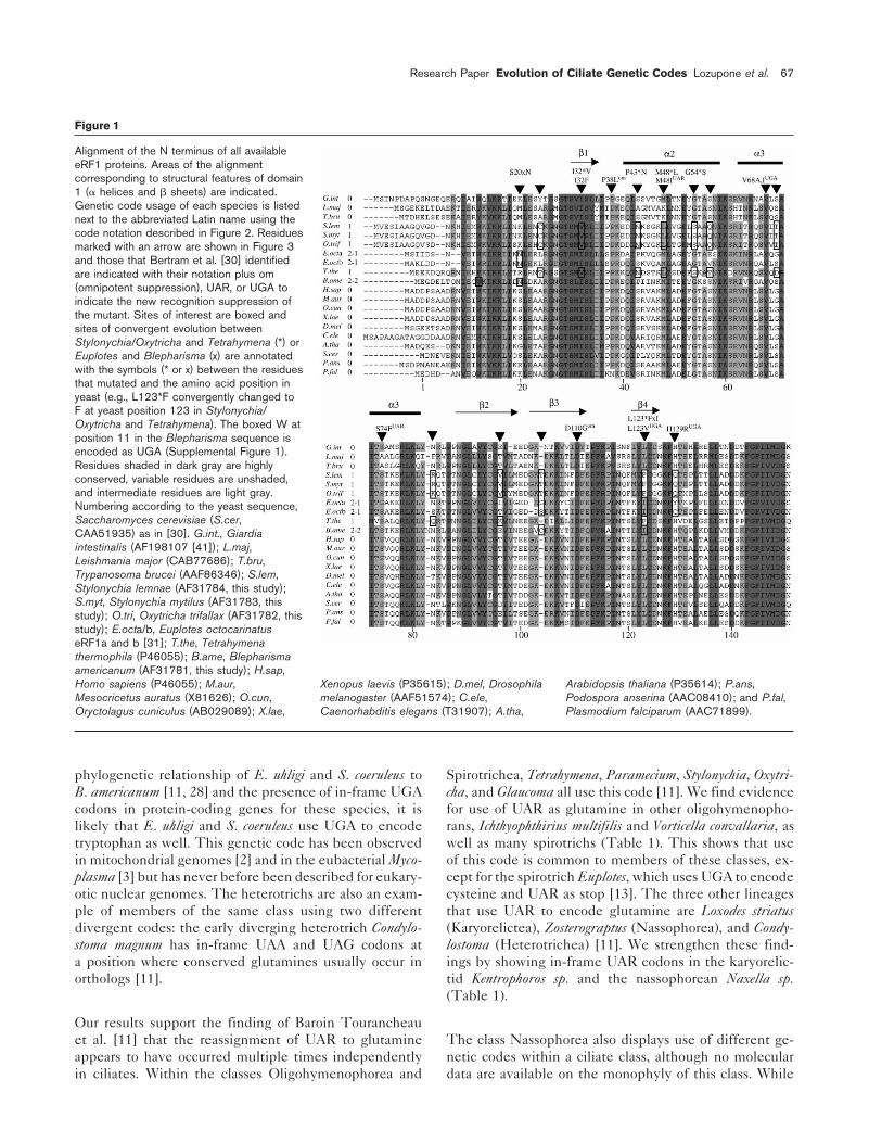
Alignment of the N terminus of all availableeRF1 proteins. Areas of the alignmentcorresponding to structural features of domain1 (a helices and b sheets) are indicated.Genetic code usage of each species is listednext to the abbreviated Latin name using thecode notation described in Figure 2. Residuesmarked with an arrow are shown in Figure 3and those that Bertram et al. [30] identifiedare indicated with their notation plus om(omnipotent suppression), UAR, or UGA toindicate the new recognition suppression ofthe mutant. Sites of interest are boxed andsites of convergent evolution betweenStylonychia/Oxytricha and Tetrahymena (*) orEuplotes and Blepharisma (x) are annotatedwith the symbols (* or x) between the residuesthat mutated and the amino acid position inyeast (e.g., L123*F convergently changed toF at yeast position 123 in Stylonychia/Oxytricha and Tetrahymena). The boxed W atposition 11 in the Blepharisma sequence isencoded as UGA (Supplemental Figure 1).Residues shaded in dark gray are highlyconserved, variable residues are unshaded,and intermediate residues are light gray.Numbering according to the yeast sequence,Saccharomyces cerevisiae (S.cer,CAA51935) as in [30]. G.int., Giardiaintestinalis (AF198107 [41]); L.maj,Leishmania major (CAB77686); T.bru,Trypanosoma brucei (AAF86346); S.lem,Stylonychia lemnae (AF31784, this study);S.myt, Stylonychia mytilus (AF31783, thisstudy); O.tri, Oxytricha trifallax (AF31782, thisstudy); E.octa/b, Euplotes octocarinatuseRF1a and b [31]; T.the, Tetrahymenathermophila (P46055); B.ame, Blepharismaamericanum (AF31781, this study); H.sap,Homo sapiens (P46055); M.aur, Xenopus laevis (P35615); D.mel, Drosophila Arabidopsis thaliana (P35614); P.ans,Mesocricetus auratus (X81626); O.cun, melanogaster (AAF51574); C.ele, Podospora anserina (AAC08410); and P.fal,Oryctolagus cuniculus (AB029089); X.lae, Caenorhabditis elegans (T31907); A.tha, Plasmodium falciparum (AAC71899).
phylogenetic relationship of E. uhligi and S. coeruleus to Spirotrichea, Tetrahymena, Paramecium, Stylonychia, Oxytri-cha, and Glaucoma all use this code [11]. We find evidenceB. americanum [11, 28] and the presence of in-frame UGA
codons in protein-coding genes for these species, it is for use of UAR as glutamine in other oligohymenopho-rans, Ichthyophthirius multifilis and Vorticella convallaria, aslikely that E. uhligi and S. coeruleus use UGA to encode
tryptophan as well. This genetic code has been observed well as many spirotrichs (Table 1). This shows that useof this code is common to members of these classes, ex-in mitochondrial genomes [2] and in the eubacterial Myco-
plasma [3] but has never before been described for eukary- cept for the spirotrich Euplotes, which uses UGA to encodecysteine and UAR as stop [13]. The three other lineagesotic nuclear genomes. The heterotrichs are also an exam-
ple of members of the same class using two different that use UAR to encode glutamine are Loxodes striatus(Karyorelictea), Zosterograptus (Nassophorea), and Condy-divergent codes: the early diverging heterotrich Condylo-
stoma magnum has in-frame UAA and UAG codons at lostoma (Heterotrichea) [11]. We strengthen these find-ings by showing in-frame UAR codons in the karyorelic-a position where conserved glutamines usually occur in
orthologs [11]. tid Kentrophoros sp. and the nassophorean Naxella sp.(Table 1).
Our results support the finding of Baroin Tourancheauet al. [11] that the reassignment of UAR to glutamine The class Nassophorea also displays use of different ge-
netic codes within a ciliate class, although no molecularappears to have occurred multiple times independentlyin ciliates. Within the classes Oligohymenophorea and data are available on the monophyly of this class. While

68 Current Biology Vol 11 No 2
Figure 2
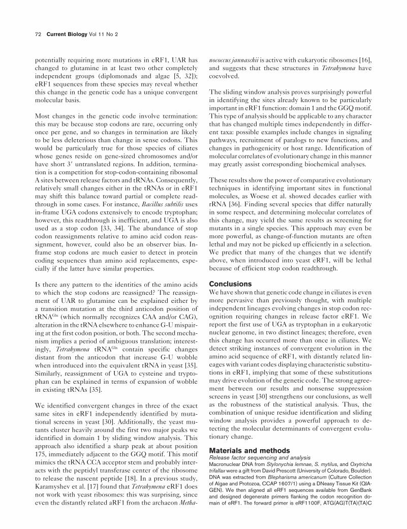
Mapping of genetic code changes (boxes)mapped onto a composite phylogenetic treeof ciliate genera compiled from congruentpublished analyses of 28S ([11] bootstrapvalues indicated above the line in boldface;[22] marked **) and 18S ([23] bootstrapvalues below the line in bold; [25] marked †;[24] ‡) rRNA sequences. Bootstrap valuesmarked § represent 28S and 18S studies,respectively, that grouped Blepharisma withStentor 92% [11] and with Eufolliculina 87%[28]. A single asterisk indicates lineages witha code change described for the first time inthis study.
in-frame UAR codons are present in Zosterograptus sp. and or both UAA and UAG. For each of these two types ofNaxella sp., three protein sequences in Pseudomicrothorax change, we have eRF1 sequences from representativesdubius have no in-frame stop codons and use UAG and of two different lineages in which the change has occurred:UAA to terminate translation. The coding of UGA in this the reassignment of UGA in Euplotes and Blepharismaspecies was ambiguous. The sequence of the hydrogenase and UAR in Stylonychia/Oxytricha and Tetrahymena. Theprotein in Nyctotherus ovalis, of the order Amorphorida, phylogenetic analysis indicates that both of these changeshas no in-frame stop codons and uses UAA to encode probably occurred independently. For the UGA change,stop. Additional DNA sequences from these two species it is strongly supported that Euplotes is more closely relatedshould be examined for evidence of UGA reassignment to many genera that use UAR to encode glutamine, suchto an amino acid. as Stylonychia, Oxytricha, and Tetrahymena, than it is to
Blepharisma (Figure 2). Blepharisma, likewise, is closelyThe class Litostomatea is the only group of ciliates for related to Condylostoma magnum, which also uses UAR towhich strong evidence suggests use of the standard ge- encode glutamine [11]. There is weaker evidence thatnetic code. None of the 55 protein sequences available the change of UAR to glutamine occurred independentlyfor litostomes had in-frame stop codons, and translation in the spirotrichs that use this code and the oligohymeno-was terminated with either UAA or UGA (Table 1 and phorans: Euplotes, which uses UGA to encode cysteine,supplemental Table 1). appears to have diverged early from a monophyletic lin-
eage that includes the other spirotrichs, and Colpoda, inwhich we find evidence of UGA use to encode tryptophan,Identification of convergent changesappears to form a monophyletic group with the oligohy-The changes in ciliate genetic codes involve two types
of altered stop codon recognition: recoding of either UGA menophorans (Figure 2). Both of these relationships are

Research Paper Evolution of Ciliate Genetic Codes Lozupone et al. 69
Table 1
Organism # 39 aa UAG UAA UGA stop Code
Nassophorea 6 0?,1Naxella 1 0 408 3 2 0 (UGA) 1Zosterograptus 1 0 382 0 6 0 1Pseudomicrothorax 3 3 1770 0 0 0 UAA,UAG 0?
Colpodea 4 2-2Colpoda 4 0 1253 0 0 1 2-2
Oligohymenophorea 61 1Vorticella 1 1 181 1 0 0 UGA 1Glaucoma 2 0 449 3 1 0 (UGA) 1Ichthyophthirius 2 1 517 1 23 0 UGA 1Lembadion 1 1 351 0 6 0 UGA 1
Litostomatea 55 0Entodinium 47 45 9447 0 0 0 UAA,UGA 0Epidinium 2 1 737 0 0 0 UAA 0Polyplastron 3 3 930 0 0 0 UAA 0Spathidium 3 0 1194 0 0 0 0
Armophorida 2 0?Nyctotherus 2 1 1742 0 0 0 UAA 0?
Spirotrichea 141 1, 2-1Pleurotricha 1 0 798 5 31 0 (UGA) 1Paraurostyla 1 0 966 10 24 0 (UGA) 1Uroleptus 1 0 973 10 26 0 (UGA) 1Urostyla 4 2 2606 15 33 0 UGA 1Histriculus 2 1 755 0 1 0 UGA 1Halteria 3 1 1697 7 5 0 UGA 1Holosticha 1 0 966 11 19 0 (UGA) 1Hypotrichida 1 0 373 0 0 0
Heterotrichea 32 1, 2-2Blepharisma 10 2 689 0 0 2 UAA 2-2Eufolliculina 12 12 3250 0 0 4 UAA 2-2?Stentor 8 0 1841 0 0 3 2-2?Condylostoma 1 0 380 1 4 0 (UGA) 1
Karyorelictea 2 1Kentrophoros 1 0 408 1 0 0 1Loxodes 1 0 380 6 0 0 1
A summary of ciliate class/genera, total number of protein sequences (codons in parentheses are inferred from the data); and the geneticavailable; number of sequences that were complete at the 39 end code used: (0) standard genetic code, (0?) UAA and UAG usedallowing determination of stop codon usage; total number of amino as stop and translation of UGA is unknown (1) UAR:glutamine, (2)acids (aa) analyzed; number of in-frame UAA, UAG, and UGA UGA:amino acid, (2-1) UGA:cysteine, (2-2) UGA:tryptophan.codons, the codon(s) used to terminate translation where available
supported in numerous 18S and 28S rRNA trees, generally encode glutamine, for instance, would potentially requireloss of extra glutamine tRNAs, eRF1 mutations that de-with greater bootstrap support for studies based on com-
plete 18S small subunit (e.g., [23, 24, 28]) than partial crease UAR readthrough and increase UGA readthrough,and mutations enabling a tRNACys to read UGA. For these28S large subunit [11, 22] sequences. Morphological data
also support the classification of Euplotes as a spirotrich reasons, we propose that the UAR-to-glutamine changemost likely emerged independently in the spirotrich and[29]. In addition, phylogenetic analysis of a-tubulin gene
sequences weakly supports both of these relationships, oligohymenophoran lineages. We next ask whether thereare convergent changes that could confer the altered spec-and an analysis of phosphoglycerate kinase gene se-
quences strongly supports the branching of Euplotes with ificity for both UAR and UGA reassignment. In otherwords, are there states of particular sites in eRF1 suchOxytricha (97% bootstrap support), though data for Colpoda
were unavailable [22]. that all and only ciliates with the same altered translationtermination share those particular states? This analysis isitself independent of the direction of change inferred inIf these phylogenetic inferences are correct, one could
still argue that an ancient change of UAR to glutamine Figure 2.in a common ancestor of the spirotrichs and oligohymeno-phorans might have been followed by UGA reassignment We wrote a C program (available from the authors) to
scan a set of sequences labeled according to arbitraryin Euplotes and Colpoda. It is much more likely, however,that the transition to these alternate genetic codes pro- criteria (in this case, the type of stop codons used by the
species) and to partition amino acid identities at each siteceeded from a standard code background. The evolutionof the Euplotes code from an ancestor that used UAR to of an aligned set of sequences according to which labels

70 Current Biology Vol 11 No 2
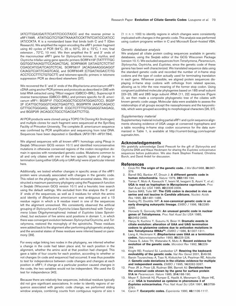
Figure 3have which sites. We find that the following states areunique to particular translation states (numbered ac-cording to yeast eRF1; amino acids to the left of a num-ber occur in organisms with the standard code and theamino acids to the right of the number occur in organismswith altered stop codon specificity): UAR 5 Stop:(KRGMS)20N, (.)103–104(CS), L123I, (NGT)213(DES),N262(ST), (AQ)279(ER), (DEIMQ)366(GV); UGA 5Stop: (AY)24(CT), I32V, (PSA)43N, (MK)48L, G54S,(ES)57(QT), (GS)70(DT), (NPT)83KQR, (ETD)96(KV),(.)103–104T, L123F, (SAGKQ)273T, (NDG)322E,(.NDGQ)332(.K), (DECQS)343(NT). Remarkably, 14 ofthese 22 convergent changes map to domain 1, the puta-tive codon recognition domain [18], and 4 of these changesin 3 different positions (in boldface) are the same changesrecently identified by mutational screens for enhancedtermination suppression in yeast [30]! Our alignmentspanned 417 residues: the probability that we would hit3 or more of the same residues that the mutational studyidentified by chance, if the changes were independent,is 0.014. (The mutational study identified 10 of 417 resi-dues as being functionally important. Of these, 9 were inthe region covered by our alignment. Since we identified20 sites as having states unique to particular genetic codes,the 2 3 2 contingency table has counts of 3, 6, 17, and
Human eRF1 structure of domain 1. The entire protein ribbon structure319 for identification in both studies, in [30], our study,of human eRF1 (inset) is shown with domain 1 boxed in blue and
and neither study respectively. The probability that these domains 2 and 3 in yellow (PDB accession code 1DT9 [18]).results are independent is very low [G test for indepen- Highlighted residues in domain 1 have been identified in this study
(blue text), mutational analysis in yeast (red text [30]), or both (purpledence applying the Williams correction: df 5 1, G 5 4.82,text). Numbers correspond to human eRF1, 13 from yeast eRF1P1-tailed 5 0.014]. Thus, our identification of several of theand Figure 1 (e.g., human M51 is homologous to yeast M48). Mutations
same sites is unlikely to be a coincidence.) in red residues are associated with UAR suppression; yellow, UGAsuppression; and orange, both.
Bertram et al. [30] identified ten changes in domain 1:three changes in mutants exhibiting UGA suppression
in [30] (Figure 3). As this position distinguishes UAR(V68I, L123V, and H129R) increased UGA readthrough,from UGA, we propose that these changes, in particular
and three changes in mutants with UAG suppression the convergent changes at position 123 (Figure 1), play(M48I, S74F, D110G) increased UAR readthrough. I32F a critical role in the evolution of release factor specificitywas a potential UGA suppressor and the remaining three in ciliates. There is only one additional convergent changeisolates were omnipotent suppressors. Our evolutionary in domain 1 of Euplotes and Blepharisma. In contrast, weapproach agrees surprisingly well with these mutational identified ten sites in domain 1 that changed in bothdata: the UGA suppressors Euplotes and Blepharisma have Stylonychia/Oxytricha and Tetrahymena. Of these, seven area convergent change from L to I in the homologous posi- in a helices 2 and 3 (Figures 1 and 3) and five mutate totion of L123V, and Euplotes [31] has a Y or C at the the same residue. Two convergent changes, I to V andposition of H129R, a conserved histidine in all taxa except M to L, are at the same site as I32F and M48I UARciliates with alternate codes (Figure 1). V68I is at a con- suppression mutations [30], and site 32 is conserved acrossserved valine across all ciliates, indicating that this site all taxa but the ones using this code. An important nexteither has not been explored by evolution or is fixed by step would be to engineer these changes into yeast eRF1other constraints. L123V is also a site of a convergent and test whether the resulting protein could rescue achange from L to F in Stylonychia/Oxytricha and Tetrahy- Tetrahymena eRF1 knockout.mena and is conserved in all other taxa; Stylonychia andOxytricha have a nonconservative change from H to Q at Sliding window analysis: associating eRF1 changessite H129R. The homologous residues L126 and H132 and termination changesin human eRF1 flank a hydrophobic pocket that may The analysis of individual residues is sometimes difficult
to interpret, because apparent associations between changerecognize the stop codon second position in the model

Research Paper Evolution of Ciliate Genetic Codes Lozupone et al. 71
Figure 4
Sliding window analysis of eRF1 and geneticcode change. This figure shows therobustness of peaks to changes in window size,showing windows of size 5 (gray line), 20(blue line), and 40 (black line). Peaks indicateregions in which changes are maximallyassociated with genetic code change. The xaxis is position in our alignment (Figure 1); they axis is relative G value (scaled relative tomaximum G value for that window size). Thefirst two peaks lie in domain 1, the third indomain 2 near the GGQ motif, and the fourthin domain 3, which binds eRF3 [18]. Red dotsmark mutations in yeast eRF1 that affect stopcodon readthrough (marked on the 40 residueline); UAR→Gln, yellow dots; UGA ? stop,orange boxes. An interactive Excel file thatallows the user to view the results withdifferent window sizes is available at ftp://rnaworld.princeton.edu/pub/export/windowresults2.xls
at a single site and change in the genetic code could occur in Euplotes and Blepharisma, which suggests either thatindependent substitutions may assist stop codon reassign-by chance. By pooling counts for a contiguous stretch of
sequence, it is possible to test for association between ment in these lineages or, more intriguingly, that UGAsuppression may be easier to effect than UAR suppres-changes in that region and changes in the genetic code.
This increases confidence that particular regions are im- sion, a conclusion that is also supported by the fact thatBertram et al.’s initial screen for unipotent suppressorportant, at the expense of making inferences about indi-
vidual sites. phenotypes only identified UGA suppressors [30]. Theisolation of UAR suppressors required a plasmid con-taining a mutant tRNASer with weak UAR nonsense sup-Figure 4 shows results for three window sizes (5, 20, and
40 residues) along with mutations found to affect decoding pressor activity.in yeast eRF1 [30] and those identified in this study.There are four peaks that are stable across a broad range We initially set out to test the hypothesis that a change
of S to D in Tetrahymena, within the otherwise universallyof window sizes. The first two of these peaks correspondto domain 1, and the third one corresponds to the region conserved NIKS motif of domain 1, was responsible for
altering the specificity of eRF1 in organisms with thisimmediately before the universal GGQ motif. Addition-ally, the decoding mutants in yeast and the convergent code. We found that Stylonychia and Oxytricha, which also
translate UAR as glutamine, do not have any deviationschanges identified in this study cluster around the firsttwo peaks. in the NIKS motif; nor does Blepharisma. One of the two
release factor genes in Euplotes octocarinatus, however, hasthe sequence SIKS in this region [31]. This indicates thatDiscussion
Our database analysis extends both the number of inde- changes in this motif are not necessary for altered stopcodon recognition, as we initially proposed, although theypendent code changes in ciliates and the types of code
change reported for eukaryotes. Both UAR and UGA have may be sufficient. However, several different changesadjacent to this region are heavily implicated in variantbeen reassigned independently many times in different
lineages of ciliates. Whether ciliates are particularly prone codes. Construction of specific mutants in yeast eRF1 willallow finer mapping of the critical residues and perhapsto changes in the genetic code or if they are they just
more diverse than other microbial eukaryotes remains indicate how many ways eRF1 can mutate to generatethe same changes in the genetic code.open. UGA has been reassigned to both cysteine and
tryptophan, showing that either of two tRNAs can expandits decoding ability to cover this codon. The UGA → Changes in ciliate termination may depend on less than
6 residue changes (for loss of UGA recognition) or lesstryptophan change also occurs in bacteria and mitochon-dria. Is UGA particularly prone to reassignment? than 15 residue changes (for loss of UAR recognition) in
eRF1 on the basis of convergent mutations in lineagesthat evolved the same changes independently. DespiteWe observed many fewer convergent eRF1 substitutions
72 Current Biology Vol 11 No 2
potentially requiring more mutations in eRF1, UAR has nococcus jannaschii is active with eukaryotic ribosomes [16],and suggests that these structures in Tetrahymena havechanged to glutamine in at least two other completely
independent groups (diplomonads and algae [5, 32]); coevolved.eRF1 sequences from these species may reveal whether
The sliding window analysis proves surprisingly powerfulthis change in the genetic code has a unique convergentin identifying the sites already known to be particularlymolecular basis.important in eRF1 function: domain 1 and the GGQ motif.This type of analysis should be applicable to any characterMost changes in the genetic code involve termination:
this may be because stop codons are rare, occurring only that has changed multiple times independently in differ-ent taxa: possible examples include changes in signalingonce per gene, and so changes in termination are likely
to be less deleterious than change in sense codons. This pathways, recruitment of paralogs to new functions, andchanges in pathogenicity or host range. Identification ofwould be particularly true for those species of ciliates
whose genes reside on gene-sized chromosomes and/or molecular correlates of evolutionary change in this mannermay greatly assist corresponding biochemical analyses.have short 39 untranslated regions. In addition, termina-
tion is a competition for stop-codon-containing ribosomalThese results show the power of comparative evolutionaryA sites between release factors and tRNAs. Consequently,techniques in identifying important sites in functionalrelatively small changes either in the tRNAs or in eRF1molecules, as Woese et al. showed decades earlier withmay shift this balance toward partial or complete read-rRNA [36]. Finding several species that differ naturallythrough in some cases. For instance, Bacillus subtilis usesin some respect, and determining molecular correlates ofin-frame UGA codons extensively to encode tryptophan;this change, may yield the same results as screening forhowever, this readthrough is inefficient, and UGA is alsomutants in a single species. This approach may even beused as a stop codon [33, 34]. The abundance of stopmore powerful, as change-of-function mutants are oftencodon reassignments relative to amino acid codon reas-lethal and may not be picked up efficiently in a selection.signment, however, could also be an observer bias. In-We predict that many of the changes that we identifyframe stop codons are much easier to detect in proteinabove, when introduced into yeast eRF1, will be lethalcoding sequences than amino acid replacements, espe-because of efficient stop codon readthrough.cially if the latter have similar properties.
ConclusionsIs there any pattern to the identities of the amino acidsWe have shown that genetic code change in ciliates is evento which the stop codons are reassigned? The reassign-more pervasive than previously thought, with multiplement of UAR to glutamine can be explained either byindependent lineages evolving changes in stop codon rec-a transition mutation at the third anticodon position ofognition requiring changes in release factor eRF1. WetRNAGln (which normally recognizes CAA and/or CAG),report the first use of UGA as tryptophan in a eukaryoticalteration in the tRNA elsewhere to enhance G-U mispair-nuclear genome, in two distinct lineages; therefore, evening at the first codon position, or both. The second mecha-this change has occurred more than once in ciliates. Wenism implies a period of ambiguous translation; interest-detect striking instances of convergent evolution in theingly, Tetrahymena tRNAGln contain specific changesamino acid sequence of eRF1, with distantly related lin-distant from the anticodon that increase G-U wobbleeages with variant codes displaying characteristic substitu-when introduced into the equivalent tRNA in yeast [35].tions in eRF1, implying that some of these substitutionsSimilarly, reassignment of UGA to cysteine and trypto-may drive evolution of the genetic code. The strong agree-phan can be explained in terms of expansion of wobblement between our results and nonsense suppressionin existing tRNAs [35].screens in yeast [30] strengthens our conclusions, as wellas the robustness of the statistical analysis. Thus, theWe identified convergent changes in three of the exactcombination of unique residue identification and slidingsame sites in eRF1 independently identified by muta-window analysis provides a powerful approach to de-tional screens in yeast [30]. Additionally, the yeast mu-tecting the molecular determinants of convergent evolu-tants cluster heavily around the first two major peaks wetionary change.identified in domain 1 by sliding window analysis. This
approach also identified a sharp peak at about positionMaterials and methods175, immediately adjacent to the GGQ motif. This motif Release factor sequencing and analysis
mimics the tRNA CCA acceptor stem and probably inter- Macronuclear DNA from Stylonychia lemnae, S. mytilus, and Oxytrichatrifallax were a gift from David Prescott (University of Colorado, Boulder).acts with the peptidyl transferase center of the ribosomeDNA was extracted from Blepharisma americanum (Culture Collectionto release the nascent peptide [18]. In a previous study,of Algae and Protozoa, CCAP 1607/1) using a DNeasy Tissue Kit (QIA-Karamyshev et al. [17] found that Tetrahymena eRF1 doesGEN). We then aligned all eRF1 sequences available from GenBank
not work with yeast ribosomes: this was surprising, since and designed degenerate primers flanking the codon recognition do-main of eRF1. The forward primer is eRF1100F, ATG(AG)T(TA)(TA)Ceven the distantly related aRF1 from the archaeon Metha-
Research Paper Evolution of Ciliate Genetic Codes Lozupone et al. 73
(ATC)TT(GA)(GA)T(TC)AT(TC)CC(TA)CC and the reverse primer is (1 # n # 100) to identify regions in which changes were consistentlyimplicated with changes in the genetic code. This analysis was performedeRF1799R, AT(KT)GC(TC)TGKTTKAA(AT)CCKTTNTC(AT)CC(AT)CC
(AT)CCKTA. K is a nonstandard base that binds both C and T (Glen using custom programs written in C and Microsoft Excel–hosted VBA.Research). We amplified the region encoding the eRF1 protein fragmentusing 40 cycles of PCR (948C, 25 s; 508C, 20 s; 728C, 1 min; final Genetic database analysisextension , 728C, 10 min). We then amplified the 59 and 39 ends of We analyzed all ciliate protein coding sequences available in geneticthe macronuclear eRF1 gene for Stylonychia lemnae, S. mytilus, and databases, using the Seqlab editor of the GCG Wisconsin PackageOxytricha trifallax using gene specific primers SORF415F [TATTTTTGC (version 10.1). We excluded sequences from Tetrahymena, Paramecium,GGTGGTAA(AG)TTCCAGACTGA], SORF680R [(AT)AGTCTCTTAT Stylonychia, Oxytricha, and Euplotes, since the genetic code of theseCGAGCAT(GA)TCAGTCTCA], SORF581F [ACAGAAAGGGAGGT genera has been well characterized. We translated sequence data usingCA(AG)TC(AT)TCAGTCAG], and SORF604R [CTGA(TA)GA(CT)TG the standard genetic code and recorded the number of in-frame stopACCTCCCTTTCTGTGCTT] and telomere-specific primers in telomere codons and the type of codon actually used for terminating translationsuppression PCR as described elsewhere [37]. in each gene. Wherever possible, we aligned protein sequences dis-
playing in-frame stop codons with orthologs from related species,We recovered the 59 and 39 ends of the Blepharisma americanum eRF1 allowing us to infer the new meaning of the former stop codon. UsingcDNA using anchor PCR primers and protocols as described in [38] with congruent published molecular phylogenies based on 18S small subunittotal RNA extracted using TRIzol reagent (GIBCO–BRL), Superscript II [23–25, 28] and 28S large subunit rRNA [11, 22] sequences (Figurereverse transcriptase (GIBCO–BRL), and primers specific for B. ameri- 2), we then made a composite phylogenetic tree of the ciliates withcanum eRF1: BGSP1F (TGCCAGGCTGCGTATGGAGTCC), BGSP known genetic code usage. Molecular data were available to assess the2F (CATTGCTGGGTCAGCTGAGTTC), BGSPRTR (AAATCAGACC relationships of all groups except the nassophoreans and the karyorelic-GTTTGCTGGGAG), BGSP1R (CGCTCTTTTGTGGAGGTAAGAG), tids, which were grouped according to morphological characters [26, 27].and BGSP2R (ACAGCCTGCCTGACGATTCTTG).
Supplementary materialAll PCR products were cloned using a TOPO TA Cloning Kit (Invitrogen) Supplementary material including partial eRF1 and cyclin sequence align-and multiple clones for each fragment were sequenced at the Syn/Seq ments showing evidence of UGA usage at conserved tryptophans andFacility of Princeton University. The complete B. americanum sequence a table recording in-frame stop codon occurrence for the data sum-was confirmed by PCR amplification and sequencing from total DNA. marized in Table 1, is available at http://current-biology.com/supmat/Sequences have been deposited in GenBank (AF31781–AF31784). supmatin.htm.
We aligned sequences with all known eRF1 homologs using Pileup in AcknowledgementsSeqlab (Wisconsin GCG version 10.1) and identified nonconservative We gratefully acknowledge David Prescott for the gift of Stylonychia andmutations in otherwise conserved regions of the codon recognition do- Oxytricha DNA and Klaus Heckmann for sharing the Euplotes octocarinatusmain in species with nonstandard genetic codes. Mutations common to sequences before publication. We also thank Stephen Freeland, Christina
Burch, and David Ardell for discussion.all and only ciliates with one of the two specific types of change intermination (using either UGA only or UAR only) were of particular interesthere. References
1. Crick FH: The origin of the genetic code. J Mol Biol1968, 38:367-379.Additionally, we tested whether changes in specific areas of the eRF1
2. Barrell BG, Bankier AT, Drouin J: A different genetic code inprotein were unusually associated with changes in the genetic code.human mitochondria. Nature 1979, 282:189-194.This relied on the phylogeny, with its inferred ancestral states. We con-
3. Yamao F, Muto A, Kawauchi Y, Iwami M, Iwagami S, Azumi Y, et al.:structed a maximum parsimony phylogeny using the Paupsearch functionUGA is read as tryptophan in Mycoplasma capricolum. Procin Seqlab (Wisconsin GCG version 10.1) and a heuristic tree search Natl Acad Sci USA 1985, 82:2306-2309.
using the default settings. We excluded from this analysis the 59 and 4. Santos MAS, Tuite MF: The CUG codon is decoded in vivo as39 ends of the sequences, as well as 2 residues for which the amino serine and not leucine in Candida albicans. Nucleic Acidsacid identity of Blepharisma americanum was ambiguous and a 13 Res 1995, 23:1481-1486.
5. Keeling PJ, Doolittle WF: A non-canonical genetic code in anresidue region in which a 9 residue insert in one of the sequencesearly diverging eukaryotic lineage. EMBO J 1996, 15:2285-left the alignment unresolved. We consistently observed the artificial2290.grouping of Stylonychia and Oxytricha (class Spirotrichea) with Tetrahy-
6. Horowitz S, Gorovsky MA: An unusual genetic code in nuclearmena (class Oligohymenophorea) instead of Euplotes (class Spirotri-genes of Tetrahymena. Proc Natl Acad Sci USA 1985,
chea), but exclusion of five amino acid positions in domain 1, in which 82:2452-2455.there was convergent evolution between Stylonychia/Oxytricha, and Tet- 7. Hanyu N, Kuchino Y, Susumu N, Beier H: Dramatic events inrahymena, restored the monophyly of the spirotrichs. These residues ciliate evolution: alteration of UAA and UAG terminationwere added back to the alignment after performing phylogenetic analysis, codons to glutamine codons due to anticodon mutations in
two Tetrahymena tRNAsGln. EMBO J 1986, 5:1307-1311.and the ancestral states of these residues were inferred based on parsi-8. Liang A, Heckmann K: Blepharisma uses UAA as a terminationmony [39].
codon. Naturwissenschaften 1993, 80:225-226.9. Osawa S, Jukes TH, Watanabe K, Muto A: Recent evidence forFor every edge linking two nodes in the phylogeny, we inferred whether
evolution of the genetic code. Microbiol Rev 1992, 56:229-a change in the code had taken place and, for each position in the 264.alignment, whether the amino acid at that position had changed. This 10. Knight RD, Freeland SJ, Landweber LF: Rewiring the keyboard:gave a table of four counts for each position, according to whether or evolvability of the genetic code. Nat Rev Genet 2001, 2:49-58.not changes (in code and sequence) had occurred. It was thus possible 11. Baroin Tourancheau A, Tsao N, Klobutcher LA, Pearlman RE, Adoutte
A: Genetic code deviations in the ciliates: evidence for multipleto test for independence between code changes and changes at eachand independent events. EMBO J 1995, 14:3262-3267.position in eRF1; if change at a particular position caused changes in
12. Preer JR Jr, Preer LB, Rudman BM, Barnett AJ: Deviations fromthe code, the two variables would not be independent. We used the Gthe universal code shown by the gene for surface proteintest for independence [40].51A in Paramecium. Nature 1985, 314:188-190.
13. Meyer F, Schmidt HJ, Plumper E, Hasilik A, Mersmann G, Meyer HE,Because there are relatively few sequences, individual residues typically et al.: UGA is translated as cysteine in pheromone 3 ofdid not give significant associations. In order to identify regions of se- Euplotes octocarinatus. Proc Natl Acad Sci USA 1991, 88:3758-quence associated with genetic code change, we performed sliding 3761.
14. Caron F: Eucaryotic codes. Experientia 1990, 46:1106-1117.window analysis, combining counts from contiguous regions of size n

74 Current Biology Vol 11 No 2
15. Grimm M, Brunen-Nieweler C, Junker V, Heckmann K, Beier H: The 37. Curtis EA, Landweber LF: Evolution of gene scrambling in ciliatemicronuclear genes. Ann NY Acad Sci 1999, 870:349-350.hypotrichous ciliate Euplotes octocarinatus has only one
38. Horton TL, Landweber LF: Evolution of four types of RNA editingtype of tRNACys with GCA anticodon encoded on a singlein myxomycetes. RNA 2000, 6:1339-1346.macronuclear DNA molecule. Nucleic Acids Res 1998,
39. Harvey PH, Pagel MD: The Comparative Method in Evolutionary26:4557-4565.Biology. Oxford: Oxford University Press; 1991.16. Dontsova M, Frolova L, Vassilieva J, Piendl W, Kisselev L, Garber M:
40. Sokal RR, Rohlf FJ: Biometry: The Principles and Practice of StatisticsTranslation termination factor aRF1 from the archaeonin Biological Research. New York: W. H. Freeman and Company;Methanococcus jannaschii is active with eukaryotic1995.ribosomes. FEBS Lett 2000, 472:213-216.
41. Inagaki Y, Doolittle WF: Evolution of the eukaryotic translation17. Karamyshev AL, Ito K, Nakamura Y: Polypeptide release factortermination system: origins of release factors. Mol Biol EvoleRF1 from Tetrahymena thermophila: cDNA cloning,2000, 17:882-889.purification and complex formation with yeast eRF3. FEBS
Lett 1999, 457:483-488.18. Song H, Mugnier P, Das AK, Webb HM, Evans DR, Tuite, MF,
Hemmings BA, Barford D: The crystal structure of humaneukaryotic release factor eRF1 – Mechanism of stop codonrecognition and peptidyl-tRNA hydrolysis. Cell 2000,100:311-321.
19. Mironova LN, Zelenaia OA, Ter-Avanesian MD: Nuclear-mitochondrial interactions in yeasts: mitochondrialmutations compensating the respiration deficiency of sup1and sup2 mutants. Genetika 1986, 22:200-208.
20. Knight RD, Landweber LF: The early evolution of the geneticcode. Cell 2000, 101:569-572.
21. Lynn DH, Small EB: A revised classification of the PhylumCiliophora Doflein, 1901. Rev Soc Mex Hist Nat 1997, 47:65-78.
22. Baroin Tourancheau A, Villalobo E, Tsau N, Torres A, Pearlman RE:Protein coding gene trees in ciliates: comparison with rRNA-based phylogenies. Mol Phylogenet Evol 1998, 10:299-309.
23. Stechmann A, Schlegel M, Lynn DH: Phylogenetic relationshipsbetween Prostome and Colpodean ciliates tested by smallsubunit rRNA sequences. Mol Phylogenet Evol 1998, 9:48-54.
24. Wright AG, Dehority BA, Lynn DH: Phylogeny of the rumenciliates Entodinium, Epidinium and Polyplastron(Litostomatea: Entodiniomorphida) inferred from smallsubunit ribosomal RNA sequences. J Eukaryot Microbiol 1997,44:61-67.
25. Struder-Kypke MC, Wright AG, Fokin SI, Lynn D: Phylogeneticrelationships of the subclass Peniculia(Oligohymenophorea, Ciliophora) inferred from small subunitrRNA gene sequences. J Eukaryot Microbiol 2000, 47:419-429.
26. Small EB, Lynn DH: A new macrosystem for the PhylumCiliophora Doflein, 1901. Biosystems 1981, 14:387-401.
27. Corliss JO: The Ciliated Protozoa. Characterization, Classification,and Guide to the Literature. London: Pergamon Press; 1979.
28. Hammerschmidt B, Schlegel M, Lynn DH, Leipe DD, Sogin ML,Raikov IB: Insights into the evolution of nuclear dualism inthe ciliates revealed by phylogenetic analysis of rRNAsequences. J Eukaryot Microbiol 1996, 43:225-230.
29. Lynn DH, Corliss JO: Ciliophora. In Microscopic Anatomy ofInvertebrates. Vol. 1: Protozoa. Edited by Corliss JO, Harrison FW.New York: Wiley-Liss, Inc.; 1991:333-467.
30. Bertram G, Bell HA, Ritchie DW, Fullerton G, Stansfield I:Terminating eukaryotic translation: domain 1 of releasefactor eRF1 functions in stop codon recognition. RNA 2000,6:1236-1247.
31. Liang A, Brunen-Nieweler C, Muramatsu T, Kuchino Y, Beier H,Heckmann K: The ciliate Euplotes octocarinatus expressestwo polypeptide release factors of the type eRF1. Gene 2001,262:161-168.
32. Schneider SU, Leible MB, Yang XP: Strong homology betweenthe small subunit of ribulose-1,5-bisphosphatecarboxylase/oxygenase of two species of Acetabularia andthe occurrence of unusual codon usage. Mol Gen Genet 1989,218:445-452.
33. Lovett PS, Ambulos NP Jr, Mulbry W, Noguchi N, Rogers, EJ: UGAcan be decoded as tryptophan at low efficiency in Bacillussubtilis. J Bacteriol 1991, 173:1810-1812.
34. Matsugi J, Murao K, Ishikura H: Effect of B. subtilis tRNA(Trp) onreadthrough rate at an opal UGA codon. J Biochem 1998,123:853-858.
35. Schultz DW, Yarus M: Transfer RNA mutation and themalleability of the genetic code. J Mol Biol 1994, 235:1377-1380.
36. Woese CR, Fox GE, Zablen L, Uchida T, Bonen L, Pechman K, etal.: Conservation of primary structure in 16S ribosomal RNA.Nature 1975, 254:83-86.
Recommended