Pier Giorgio Righetti
Politecnico di Milano,Department of Chemistry,Materials and Engineering Chemistry“Giulio Natta”,Milano, Italy
Received July 20, 2005Revised September 7, 2005Accepted September 8, 2005
Review
The Alpher, Bethe, Gamow of isoelectricfocusing, the alpha-Centaury ofelectrokinetic methodologies. Part I
The birth and evolution of IEF in conventional carrier ampholyte buffers is reviewedhere, from a shaky start during World War II, via desperate attempts of Svensson tocreate pH gradients by stationary electrolysis of salts, to the development of the IEFtheory and the solution of the steady-state equation. The remarkable synthetic processof Ampholines, as ingeniously devised by Vesterberg, is additionally recalled, with athorough description of the fundamental properties of these amphoteric buffers,creating and maintaining the pH gradient under strong electric fields. The review endswith a mention of the major contributions of B. J. Radola to this field, namely analyticaland preparative IEF in granulated Sephadex layers and the development of ultrathinIEF, in polyacrylamide gels as thin as 20–100 mm. The latter technique paved the way toDNA sequencing gels and to CZE. The symptoms of decay are here presented throughthe simulations of Mosher and Thormann, clearly indicating an isotachophoreticmechanism for pH gradient decay with time. The decay of IEF was the birth of IPGs.
Le donne, i cavalier, l’arme, gli amorile cortesie, l’audaci imprese io canto,che furo al tempo che passaro i Morid’Africa il mare, e in Francia nocquer tanto. . .
Ludovico Ariosto, Orlando Furioso, Ferrara, 1532
Keywords: Buffering power / Carrier ampholytes / Isoelectric focusing / Sephadexbeds / Ultrathin layer gels DOI 10.1002/elps.200500525
1 Introduction
I have always been puzzled by the lack, in the scientificliterature, of a kind of heroic perspective of some of themajor discoveries that have given impulse to the scienceof the epoch in which they were reported and haveshaped the future for quite a few decades afterwards.Such examples abound in the humanities, i.e. in thosebranches of learning connected with human thought andrelations, as distinguished from the sciences, such as lit-erature, philosophy, fine arts, history, and the like. Withoutdisturbing Homer, with the heroic epics of Iliad and
Odyssey, one could recall here a number of sagas thathave spurred our imagination, such as “The Quest of theHoly Graal” [1], “The Nibelungenlied” [2], “The VinlandSagas” [3], “The Buccaneers of America” [4], to name justa few. Even the four voyages of Christopher Columbusbecame a saga [5], as witnessed by the some50 000 books written about him and his deeds. There is nosuch counterpart in the scientific field. True, the mostimportant discoveries and their discoverers are oftengranted the Nobel Laurel [6], but rarely the accounts oftheir deeds are sculptured in the Hall of Fame andbecome a legend.
I am glad to be able to exploit this Special Issue ofElectrophoresis, in honor of Professor B. J. Radola, torecount the saga of IEF, the bright star that, like thea-Centaury, one of the brightest stars close to our solarsystem, illuminated the scenario of electrokinetic meth-odology since the early 70s and is not likely to set belowthe horizon even in today’s world. As one of the veterans
Correspondence: Professor Pier G. Righetti, Politecnico di Milano,Department of Chemistry, Materials and Engineering Chemistry “Giu-lio Natta”, Via Mancinelli 7, I-20131 Milano, ItalyE-mail: [email protected]: 139-02-23993080
Abbreviations: CA, carrier ampholyte; Hb, hemoglobin
Electrophoresis 2006, 27, 923–938 923
2006 WILEY-VCH Verlag GmbH & Co. KGaA, Weinheim www.electrophoresis-journal.com
924 P. G. Righetti Electrophoresis 2006, 27, 923–938
in the field, I think I should be able to offer a vivid scenarioof its development, for the young generation who has nothad a chance to follow the long march towards the“steady state” and beyond. As luck goes, most of theaccounts scattered in the literature have been written bypeople who have not had much to do with the develop-ment of the technique, and hardly know its intricacies.Their accounts most often have been obtained by lootingthe work of those who had been there since the very start.
2 The steady state
The long march towards the steady state, the trueessence of IEF, was studded with obstacles disseminatedalong the path of young Harry Svensson (who in 1968changed his name to Rilbe). That winter of 1942 musthave been the most dreadful one of his life. As a youngpupil, under the iron fist of Dom Arne Tiselius, he wasforced to spend endless days and nights trying to make adream of his boss come true, namely the creation of a pHgradient by stationary electrolysis of a salt solution.Among the innumerable problems encountered, onebecame immediately apparent: the ionic constituentswere completely swept to the opposite-charge electrode.Although equilibrium between ion transport and back dif-fusion ensued, at neutral pH, midway in the apparatuswhere a region of water almost completely devoid of ionsformed, ohmic resistance increased enormously, with theresult that the liquid almost boiled at this point. No usefulpH gradient could obviously ensue. When performing,under convection-free conditions, electrolysis of sodiumsulfate, only two portions of a pH course could be gener-ated: a pH 1.7–2.6 in the region of free H2SO4 and apH 11.5–12.3 in the zone of free NaOH [7], practicallyuseless for most protein/peptide separations.
Luckless Harry was chained to the bench adding saltdropwise from a burette, trying to fight the conductivityminimum. Add to this the fact that it was the peak of WorldWar II: Norway and Denmark were occupied by Germany;Finland was oppressed by neighboring Russia and Swe-den ducked low under a declaration of neutrality, butnevertheless under quite miserable conditions. Harrydreamed of escaping this agonizing life and paltry winter,repairing to England, to be drafted in the army and sent tothe African desert to fight the Italian–German coalition. Atleast he would have enjoyed sunny days and warmweather! As luck would have it, Tiselius relaxed his irongrip and let Harry present his Ph.D. thesis in 1946 (it wason “Electrophoresis by the Moving Boundary Method”)[8]. This was no ordinary thesis, mind you, for Harry’smentors were The Svedberg, a 1926 Nobel laureate andTiselius, a future Nobelist (1948).
Hapless Harry must have been obsessed for the re-mainder of his life by this idea of creating pH gradients.Already in 1956 he dreamed of a hypothetical biproticampholyte having two intrinsic pKa values both equal to7.0. If it were ever to exist, a solution of it in pure waterwould be a neutral buffer containing one-fourth of theamount in the cationic and one-fourth in the anionicform. A superb “carrier ampholyte” (CA), indeed, whichwould offer a substantial conductivity where it was mostneeded, i.e. in that terrible conductivity “Grand Canyon”located at pH 7, but would still be isoelectric and immo-bile in the electric field [9]. Although such a “superbuffer”could not possibly exist, 3 years later, during a leave ofabsence at Caltech, chez Linus Pauling, Svensson founda real chemical that came rather close to that: histidyl-histidine, with pKa values at 6.8 and 7.8, thus isoelectricat pH 7.3. It was used with hemoglobins (Hb) in the firstexperiments [10].
Once back home, as a freelance at the Karolinska Insti-tute, Harry worked hard on the theory of IEF and laiddown its theoretical foundations in a couple of, by now,classic papers [11, 12]. His secret battle cry as he wagedhis underground guerrilla warfare against the Maestro’smammoth instrumentation: “no more moving bound-aries!” He had a point. It had been foolish to try to createstable and stationary pH gradients in the presence of anelectric field with nonamphoteric compounds. These willvacate the grounds and leave an empty trail in their wakewith no soldiers to guard the battlefield. All buffers had tobe amphoteric and, in addition, they had to have decentbuffering power as ensured by not too large DpKa values.Only in this way would the zones of isoelectric CAs form acontinuous chain as the electric field would tie them totheir isoelectric zones while diffusion would cause themto broaden just enough to penetrate the neighboringampholytes, thus simultaneously ensuring buffer capacityand conductivity. The result was not just a few movingboundaries, à la Tiselius, but a horde of stationaryboundaries, each one standing guard against local pHchanges. It was too bad that this army was barely ahandful of soldiers, hardly able to cover the grounds in thepH 3–10 interval. Nonetheless, Svensson published in1964 some remarkable color pictures of unique Hbseparations in his 110-mL focusing column stabilized by asucrose density gradient. In these separations, Harryused protein lysates, notably of casein, albumin, Hb, andwhole blood, as background CAs. Peptides rich in Hisprovided the much needed buffering power and con-ductivity in the pH 6–7 gap [13].
This is a brief excursus of the many years of labor andintellectual challenge that haunted Harry. But how did hefinally get to the steady state? With a somersault, just like
2006 WILEY-VCH Verlag GmbH & Co. KGaA, Weinheim www.electrophoresis-journal.com
Electrophoresis 2006, 27, 923–938 General 925
any Italian would have done! He realized early enough inthe game that it would have been extremely difficult tosimulate and solve the “transient state”, i.e. the kineticbehavior of charged amphoteric molecules during theirtransport by the electric field in an IEF system. So, hewisely chose to solve the equation of the concentrationdistribution of the amphoteric ion in a stationary position,i.e. once this species had reached its pI along the pHgradient and was thus immobile in the field. At this point,the balance of electrophoretic and diffusional masstransports would give
cmi/qk = D(dC/dx)
where C, concentration of a component; m, electropho-retic mobility (cm2V21s21); i, electric current (A); q, cross-sectional area (cm2); k, medium conductance(ohm21cm21); D, diffusion coefficient (cm2/s); and x,separation distance.
The analytical solution of this equation gives a Gaussianconcentration distribution with inflection points at
xi ¼ �ffiffiffiffiffiffiffiffiffiffiffiffiffiffiffiffiffiffiffiffiffiffiffiðqkDÞ=ðpiÞ
p
where
p = 2du/dx = 2[du/d(pH)]? [d(pH)/dx]
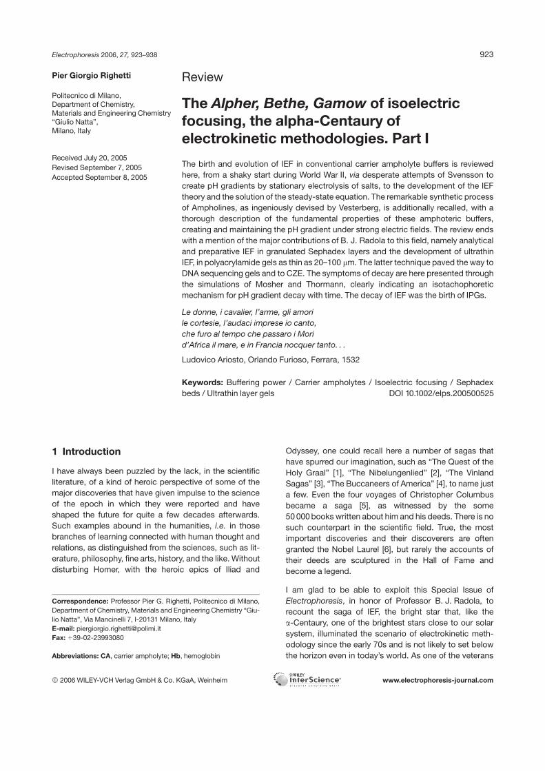
i.e. the ratio between the protein titration curve and theslope of the pH gradient over the separation axis. Figure 1illustrates the concentration profile of an amphotericspecies at its pI, together with the opposing forces actingon it, diffusion tending to spread uniformly the peak overthe separation axis and voltage gradient tending to focusit as a sharp, very thin zone symmetrically distributedabout its pI value (y = 0). Laying the foundation of IEF wastruly a major innovative step in all electrokinetic meth-odologies, and the year 1961 must be regarded as amilestone in the field of electrophoresis. But what aboutthe transient state? The solution to the law of movementof a particle in a pH gradient came much later, from Fru-min et al. [14], who exploited, for solving this problem, ananalogous equation for particle diffusion in the velocityspace, known in mathematical physics as Einstein-Smo-lukhovsky equation. Curiously, the solution to this equa-tion, for several functions of particle velocities, camealready in 1948 from the work of Chandrasekhar [15], inthe investigation of stellar dynamics! Sure enough, byassuming a case in which the sample is applied as a spikewith initial Gaussian concentration distribution, its move-ment along the pH gradient, toward the pI position, is thatof a Gaussian pulse which, from an initial broad distribu-tion, keeps narrowing and increasing in height till a limitingvalue at pH = pI. But what if the sample is uniformly dis-tributed along the migration path, as customarily done inmost IEF protocols? The situation here is even more
Figure 1. Illustration of the forces acting on a condensedzone in IEF. The focused zone is represented as a sym-metric Gaussian peak about its focusing point (pI; y = 0).Migration of sample towards the pI position is driven bythe voltage gradient and by the slope of the pH gradient(s is the SD of the peak).
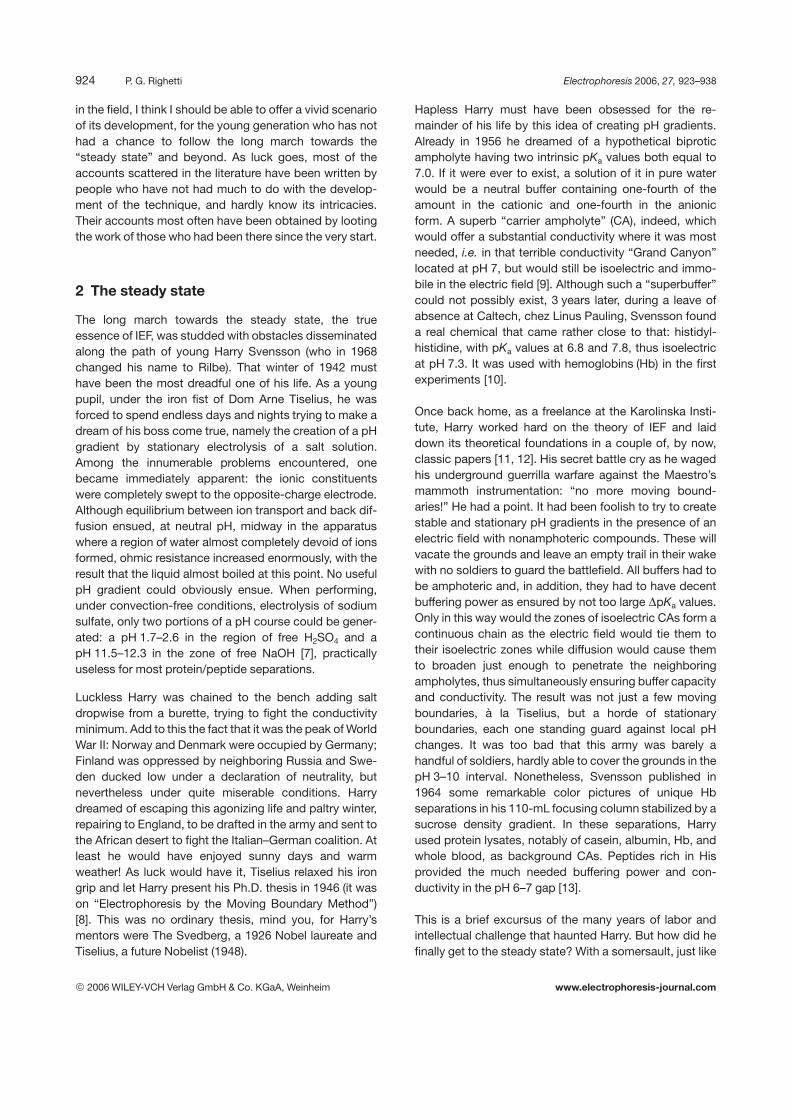
complex and it took years of hard work to Catsimpoolas[16] to solve the problem. Basically, if the compound hasbeen loaded in a uniform mode, two discernible peaks areseen arising at the two ends of the column (positive andnegative) until they merge into one at the pI (an elegantdemonstration of the focusing concept!). Here too,Gaussian profiles are hypothesized, even though theshape of the two peaks migrating toward each other, asseen by scanning the gel tubes in the UV, can hardly beclassified as Gaussian. Which brings up an interestingquestion: is there any such a thing as a true Gaussianprofile even in such a sophisticated technique as IEF, ableto correct distortions introduced during sample loadingand the run itself? Figure 2 seems to dispel this myth: asshown by Mosher et al. [17], a true Gaussian profile alongthe column length can only be obtained across neutrality!The simulation was conducted with CAs with DpKas 1, 2,or 3 and focusing in the acidic (pI 3.5), neutral (pI 7.0), orbasic (pI 10.5) regions. Note that, as the pI values areprogressively removed from neutrality, the peak shapeexhibits strong kurtosis, particularly pronounced in thepH 3–4 and 10–11 ranges. This is due to uneven con-ductivities underlying the zones, bringing about nonuni-form voltage gradients on the two sides of the peak. So,was Svensson-Rilbe’s theory wrong? Well, he was aknowledgeable physical chemist and made no errors;among the exemplifications adopted for solving the
2006 WILEY-VCH Verlag GmbH & Co. KGaA, Weinheim www.electrophoresis-journal.com
926 P. G. Righetti Electrophoresis 2006, 27, 923–938
Figure 2. A true Gaussian pro-file along the column length (cm)can only be obtained acrossneutrality! Simulated CAs withDpKas 1, 2, or 3 and pIs 3.5 (left),7.0 (center), or 10.5 (right). Notethe strong kurtosis in the pH 3–4and 10–11 ranges, due touneven conductivities (modifiedfrom Mosher et al., see [17]).
steady-state equation was the condition that the con-ductivity underlying the Gaussian peak should be con-stant! Honor saved, all around.
3 The fight for paternity
After such a long gestation period and hard trials, onewould think that by then Svensson would have enjoyedthe deserved glory and be placed in the Separation Sci-ence Hall of Fame. No way! As we have seen, early in1968, “Svensson” figuratively died, only to be resurrectedas “Rilbe”. We all thought that this was a gentleman’sgesture. Having remarried, we assumed that, to pay a tri-bute to the feminist movement, Harry had adopted hisnew wife’s last name. Well, it was a “nom de plume”, sinceneither of them would give up his/her identity! On Svens-son’s side, though, it must also have been a movedesigned to escape anonymity. When one travels toStockholm, and per chance looks up a name in the tele-phone directory, one notices that most of the populationthere seems to be the progeny of just two ancestors: Karland Sven. About half of the names in the phone book areKarlsson’s, the other half being equally attributed to the
Svensson’s. This is just fine, except that Svensson, lepauvre, made a fundamental mistake. From then on healso published his papers as Rilbe, confusing his fol-lowers and closing the door to immortality. In fact, as theIEF technique spread to the most remote villages in theworld (I went with LKB people to spread the IEF gospeljust about everywhere in the world – even as far as Mon-golia!), citations to Svensson skyrocketed and poor Rilbewas left with the remnants of the banquet. It was a greatdisappointment to most of us devout followers whosecretly hoped for a Nobel nomination – perhaps the thirdone in the series for the Uppsalienses, this unique team ofinventors.
Troubles came not just from within, but also from the otherside of the Atlantic as well. Unbeknownst to Svensson, anobscure physicist, a resident of Chicago, Illinois, hadpublished some papers in 1954–1955 [18, 19] with a keytheme: “the focusing of ions in a continuous pH gradient”.His separations were dubbed “isoelectric line spectra”and, sure enough, just to pay tribute to Tiselius, wereconducted in a U-tube, with detection by refractive indexgradients. In the mid seventies, Alexander Kolin enteredthe arena, coming regularly to our Separation Science
2006 WILEY-VCH Verlag GmbH & Co. KGaA, Weinheim www.electrophoresis-journal.com
Electrophoresis 2006, 27, 923–938 General 927
meetings and claiming his share of glory. In a couple ofvisionary articles [20, 21], he even went as far as propos-ing a rainbow of focusing effects under the general nameof “isoperichoric focusing” (the “perichoron” being theenvironment of the macromolecule, in Greek slang, whichattains the same physicochemical properties as the par-ticle under fractionation): isoconductivity, isomagnetic,isoparamagnetic, isodiamagnetic, and isodielectricfocusing.
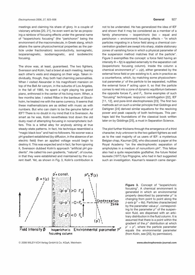
The show was, at least, guaranteed. The two fighters,Svensson and Kolin, had a brawl at each meeting, tearingeach other’s vests and stepping on their wigs. Taken in-dividually, though, they both had charming personalities.When I visited Alexander in his magnificent mansion ontop of the Bell Air canyon, in the suburbs of Los Angeles,in the fall of 1985, he spent a night playing his grandpiano, enthroned in the center of his living room. When, afew months later, I visited Rilbe in the banlieue of Stock-holm, he treated me with the same currency. It seems thatthese mathematicians are as skilled with music as withnumbers. But who can claim to be the genuine father ofIEF? There is no doubt in my mind that it is Svensson. Assmart as he was, Kolin nevertheless trod down the olddusty road of attempting focusing in nonamphoteric buf-fers. This is a lethal alley for anybody aiming at truesteady-state patterns. In fact, his technique resembled a“magic black box” and had no followers. No sooner was apH gradient established (by diffusion, in the absence of anelectric field) then an applied voltage would begin todestroy it. This was expected and in fact, far from ignoringit, Svensson dubbed Kolin’s approach “artificial pH gra-dients”. He called his own gradients, “natural”, of course,in that they were established and maintained by the cur-rent itself. Yet, as shown in Fig. 3, Kolin’s contribution is
not to be underrated. He has generalized the idea of IEFand shown that it may be considered as a member of afamily phenomena – isoperichoric (iso = equal andperichoron = environment) focusing effects – “in whichparticles migrating in a force field along a chemical con-centration gradient are swept into sharp, stable stationaryzones of vanishing force in which a physical parameter ofthe suspension method matches that of the particle”.Figure 3 exemplifies the concept: a force field having anintensity F = f(z) is applied externally to the separation cell(isoperichoric focusing column). Inside the column achemical environment p” = j(z), either generated by theexternal force field or pre-existing to it, acts in practice asa counterforce, which, by matching some physicochem-ical parameter p’ of the particle to be separated, nullifiesthe external force F acting upon it, so that the particlecomes to rest into a zone of dynamic equilibrium betweenthe opposite forces FU and FL. Some examples of such“focusing” techniques: isopycnic centrifugation [22], IEF[11, 12], and pore-limit electrophoresis [23]. The first twomethods act on such a similar principle that Giddings andDahlgren [24] derived similar equations for the resolvingpower and peak capacity of both techniques. This per-haps laid the foundations of the classical book writtenlater on by Giddings [25], a must in Separation Science.
The plot further thickens through the emergence of a thirdcharacter, truly unknown to the two gallant fighters as wellas to the vast majority of us users of IEF: a mysteriousBelgian chap, Kauman [26], who discussed a paper at theRoyal Academy “on the electrophoretic separation ofampholytes in a medium of nonuniform pH”. This fellowalso had a quite respectable godfather, the future Nobellaureate (1977) Ilya Prigogine, who had in fact suggestedsuch an investigation. Kauman’s research came danger-
Figure 3. Concept of “isoperichoricfocusing”. A chemical environment isgenerated in which an environmentalproperty described by parameter p” ischanging from point to point along thez-axis (p” = Fz). Particles characterizedby the parameter value p’, correspond-ing to the parameter p” of the suspen-sion fluid, are dispersed with an arbi-trary distribution in the fluid column. It isassumed that there is a point within thegradient of the p” distribution at whichp’ = p”, where the particle parameterequals the environmental parameter(from Kolin, see [20], by permission).
2006 WILEY-VCH Verlag GmbH & Co. KGaA, Weinheim www.electrophoresis-journal.com
928 P. G. Righetti Electrophoresis 2006, 27, 923–938
ously close to Svensson’s conclusions, which, however,were drawn only 4 years later. Kauman, too, related thedistributions of ampholytes to their pI values. To quotehim: “the distribution of the ampholyte at the stationarystate will vary approximately as a Gaussian error functionof distance, with the maximum at the pI”.
But then who can claim genuine fatherhood of IEF? As inthe good old plots of Agatha Christie, the role of the hero(or villain) is revealed in the last pages of the novel. Shallwe then crown Kauman with the laurel wreath and dumpKolin and Svensson, with their picturesque quarrels, in thegarbage bin? I do not think so. True, Kauman had reallydabbled in Svensson’s ideas and anticipated him by4 years, but he took the newborn and abandoned it in thebushes, offering no chance for survival. Svensson had thecourage to give birth to this idea and nurture it untiladulthood. Neither can Kolin, of course, claim paternityfor IEF as we know it today, for the reasons just outlinedabove.
4 The missing link: The CA chemicals
Although the theory had been laid down, experimentallyIEF was a failure, due to the lack of proper chemicals ableto control and dictate extended pH gradients. Even duringHarry’s stay at Caltech, in 1959, in L. Pauling lab, a6-month search on chemical catalogs gave meagerresults: some 40 amphoteric chemicals covering thepH 3–10 interval, with a big gap between pH 5 and 7, werethe only poor amphoteres that could be found [12]. Uponhis return from Caltech, Harry hired a medical student, OlofVesterberg, to help him devise a proper synthesis of the
much needed CAs. After 3 years of slow progress,though, Svensson became Professor of Physical Chem-istry at Gothenburg and the IEF team broke up. Vester-berg continued on this project in Stockholm, all alone. Inthe spring of 1964, Svensson received a phone call froman excited Vesterberg who appeared to have solved theproblem. Well, the chap had been moonlighting andpouring over textbooks of organic chemistry and surfacedwith a remarkable synthesis of the much wanted CAs: achaotic synthesis, to be sure, as chaotic as a medicalstudent could possibly devise. A most ingenious chaoticprocess, in fact, by which concoctions of oligoamines(from tetra- to hexa-amino groups) were reacted withlimiting amounts of an a-b-unsaturated acid, acrylic acid[27]. Chaos generated order! In a steep voltage gradient,this army of synthetic amphoteres joined arms in anorderly fashion, with each assuming a (quasi) Gaussiandistribution about its respective pI value [28, 29].
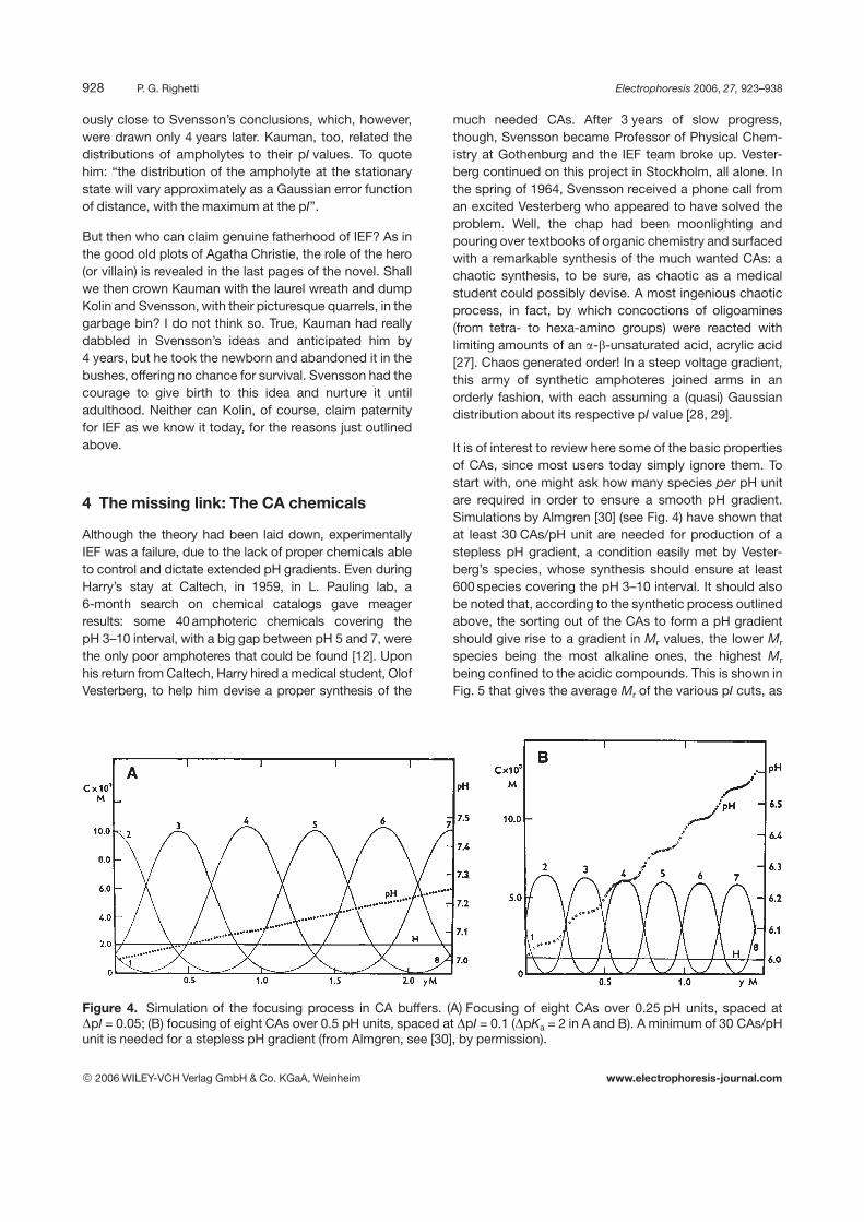
It is of interest to review here some of the basic propertiesof CAs, since most users today simply ignore them. Tostart with, one might ask how many species per pH unitare required in order to ensure a smooth pH gradient.Simulations by Almgren [30] (see Fig. 4) have shown thatat least 30 CAs/pH unit are needed for production of astepless pH gradient, a condition easily met by Vester-berg’s species, whose synthesis should ensure at least600 species covering the pH 3–10 interval. It should alsobe noted that, according to the synthetic process outlinedabove, the sorting out of the CAs to form a pH gradientshould give rise to a gradient in Mr values, the lower Mr
species being the most alkaline ones, the highest Mr
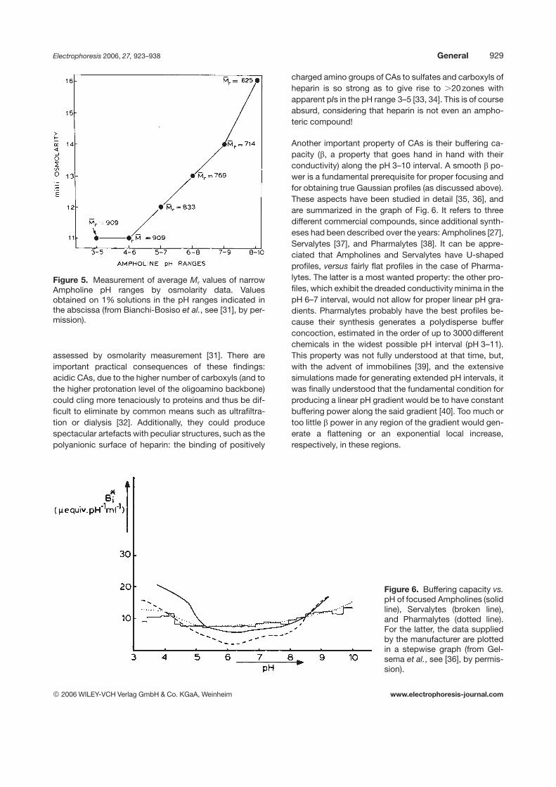
being confined to the acidic compounds. This is shown inFig. 5 that gives the average Mr of the various pI cuts, as
Figure 4. Simulation of the focusing process in CA buffers. (A) Focusing of eight CAs over 0.25 pH units, spaced atDpI = 0.05; (B) focusing of eight CAs over 0.5 pH units, spaced at DpI = 0.1 (DpKa = 2 in A and B). A minimum of 30 CAs/pHunit is needed for a stepless pH gradient (from Almgren, see [30], by permission).
2006 WILEY-VCH Verlag GmbH & Co. KGaA, Weinheim www.electrophoresis-journal.com
Electrophoresis 2006, 27, 923–938 General 929
Figure 5. Measurement of average Mr values of narrowAmpholine pH ranges by osmolarity data. Valuesobtained on 1% solutions in the pH ranges indicated inthe abscissa (from Bianchi-Bosiso et al., see [31], by per-mission).
assessed by osmolarity measurement [31]. There areimportant practical consequences of these findings:acidic CAs, due to the higher number of carboxyls (and tothe higher protonation level of the oligoamino backbone)could cling more tenaciously to proteins and thus be dif-ficult to eliminate by common means such as ultrafiltra-tion or dialysis [32]. Additionally, they could producespectacular artefacts with peculiar structures, such as thepolyanionic surface of heparin: the binding of positively
charged amino groups of CAs to sulfates and carboxyls ofheparin is so strong as to give rise to .20 zones withapparent pIs in the pH range 3–5 [33, 34]. This is of courseabsurd, considering that heparin is not even an ampho-teric compound!
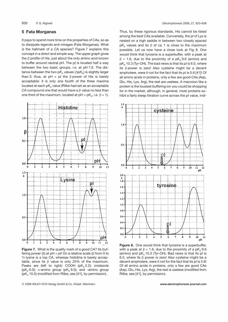
Another important property of CAs is their buffering ca-pacity (b, a property that goes hand in hand with theirconductivity) along the pH 3–10 interval. A smooth b po-wer is a fundamental prerequisite for proper focusing andfor obtaining true Gaussian profiles (as discussed above).These aspects have been studied in detail [35, 36], andare summarized in the graph of Fig. 6. It refers to threedifferent commercial compounds, since additional synth-eses had been described over the years: Ampholines [27],Servalytes [37], and Pharmalytes [38]. It can be appre-ciated that Ampholines and Servalytes have U-shapedprofiles, versus fairly flat profiles in the case of Pharma-lytes. The latter is a most wanted property: the other pro-files, which exhibit the dreaded conductivity minima in thepH 6–7 interval, would not allow for proper linear pH gra-dients. Pharmalytes probably have the best profiles be-cause their synthesis generates a polydisperse bufferconcoction, estimated in the order of up to 3000 differentchemicals in the widest possible pH interval (pH 3–11).This property was not fully understood at that time, but,with the advent of immobilines [39], and the extensivesimulations made for generating extended pH intervals, itwas finally understood that the fundamental condition forproducing a linear pH gradient would be to have constantbuffering power along the said gradient [40]. Too much ortoo little b power in any region of the gradient would gen-erate a flattening or an exponential local increase,respectively, in these regions.
Figure 6. Buffering capacity vs.pH of focused Ampholines (solidline), Servalytes (broken line),and Pharmalytes (dotted line).For the latter, the data suppliedby the manufacturer are plottedin a stepwise graph (from Gel-sema et al., see [36], by permis-sion).
2006 WILEY-VCH Verlag GmbH & Co. KGaA, Weinheim www.electrophoresis-journal.com
930 P. G. Righetti Electrophoresis 2006, 27, 923–938
5 Fata Morganas
It pays to spend more time on the properties of CAs, so asto dissipate legends and mirages (Fata Morganas). Whatis the hallmark of a CA species? Figure 7 explains thisconcept in a direct and simple way. The upper graph givesthe b profile of His, just about the only amino acid knownto buffer around neutral pH. The pI is located half a waybetween the two basic groups, i.e. at pH 7.6. The dis-tance between the two pKa values (DpKa) is slightly largerthan 3; thus, at pH = pI the b power of His is barelyacceptable: it is only one fourth of the three maximalocated at each pKa value (Rilbe had set as an acceptableCA compound one that would have a b value no less thanone third of the maximum, located at pH = pKa, i.e. b = 1).
Figure 7. What is the quality mark of a good CA? Its buf-fering power (b) at pH = pI! On a relative scale (b from 0 to1) lysine is a top CA, whereas histidine is barely accep-table, since its b value is only 25% of the maximum.Peaks are (left to right): COOH (pKa 2.2); imidazole(pKa 6.0); a-amino group (pKa 9.0); and -amino group(pKa 10.5) (modified from Rilbe, see [41], by permission).
Thus, by these rigorous standards, His cannot be listedamong the best CAs available. Conversely, the pI of Lys isnested on a high saddle in between two closely spacedpKa values and its b of ca. 1 is close to the maximumpossible. Let us now have a close look at Fig. 8. Onewould think that tyrosine is a superbuffer, with a peak atb = 1.6, due to the proximity of a pKa 9.6 (amino) andpKa 10.3 (Tyr-OH). The bad news is that its pI is 6.0, whereits b power is zero! Also cysteine might be a decentamphotere, were it not for the fact that its pI is 5.6 [41]! Ofall amino acids in proteins, only a few are good CAs (Asp,Glu, His, Lys, Arg), the rest are useless. A macroion like aprotein is the lousiest buffering ion you could be shoppingfor in the market, although, in general, most proteins ex-hibit a fairly steep titration curve across the pI value, indi-
Figure 8. One would think that tyrosine is a superbuffer,with a peak at b = 1.6, due to the proximity of a pKa 9.6(amino) and pKa 10.3 (Tyr-OH). Bad news is that its pI is6.0, where its b power is zero! Also cysteine might be adecent amphotere, were it not for the fact that its pI is 5.6!Of all amino acids in proteins, only a few are good CAs(Asp, Glu, His, Lys, Arg), the rest is useless (modified fromRilbe, see [41], by permission).
2006 WILEY-VCH Verlag GmbH & Co. KGaA, Weinheim www.electrophoresis-journal.com
Electrophoresis 2006, 27, 923–938 General 931
cating a decent buffering power (which is minimal on a perresidue basis, considering that the majority of residues aremonoamino monocarboxylic acids). There are importantreasons why these minimal theoretical concepts arebrought again to the limelight. Given the excellent theoreti-cal modeling and experimental work on the synthesis of CAbuffers, one would think that the struggle was over and thatfocusing in amphoteric buffers had assumed the role ofprima donna. Not quite! Led by a staunch defender of theimpossible (Chrambach, who, in 1978, still claimed “thepossibility to form natural pH gradients with nonamphotericbuffers”) [42], a couple of rebels proposed focusing on amixture of 47 buffers, mostly amphoteric, quite a few non-amphoteric [43]. Use of amphoteric buffers does not violateRilbe’s commandments (on the contrary, it is highly desir-able), except that all of them were Good’s buffers. Nothingis as bad as Good’s buffers when used in an improper en-vironment, i.e. for buffering in pH regions in which they arepowerless, such as at their apparent pI values. The DpKas,for these buffers, range from a minimum of 6 up to a max-imum of 9. Had Cuono and Chapo bothered to readSvensson’s papers (as elegantly summarized in his opusmagna that was the culmination of a life’s work) [41], theywould have learned that, with a DpKa = 6, the bufferingpower is zero for an amphoteric ion at pH = pI (and, for thatmatter, with a DpKa = 4, it is only 10% of the full value itwould display at pH = pKa), depriving it of its most wantedproperties, i.e. of being able to be a good conducting andbuffering species at its pI value (note that, for a good CA,these two properties go hand in hand, since at the pI valuethe relationship is b = 4a, where a is the degree of ioniza-tion). Note that, per se, use of nonamphoteric species is notincompatible with the creation of a pH gradient. For exam-ple, if a weak acid is used at pH values below its pKa, it cancreate a useful pH gradient spanning ca. 0.5 of a pH unit.However, for creating such a gradient, nine-tenths of theapparatus will be occupied by a concentration gradient ofthe weak acid, spanning one order of magnitude. If moreextended pH gradients are desired, mixtures of weak acidscan be exploited, as elegantly demonstrated by Petterson[44] and Stenman and Gräsbeck [45]. Such, gradients,however, to start with will be strongly acidic and, secondly,theycanneverbeexpected tobecame trulystationary, sincethe migration of the weaker acids will be stopped by proto-nation, due to the presence of the stronger acids. They willcontinue to diffuse, though, through the layers of the stron-ger acids, without any back transport in the electric field.
6 The journey continues
After such a long gestation period, and years and years offrustrations, Svensson and Vesterberg thought that theirordeals were over and that the technique would soon
spread to the four corners of the world. Well, with such asuperb technique (one of the few able to counteractentropy’s tendency to dissipate peaks via diffusion in thesurrounding medium by producing sharply focused zonesno matter how carelessly the experiment is carried out),one would think yes, all quiet on the western front! Well,things did not proceed so smoothly. Svensson and Ves-terberg were indeed convinced that a breakthrough hadbeen achieved, and they approached LKB Produkter AB,a Stockholm company, with a proposal for commercialproduction. As a safety measure, the two pilgrims weresent to the Mecca of Separation Science, Uppsala, toconsult with the high priest of electrophoresis, Arne, filiusquondam Tiselii (born Tiselius, in current slang). PoorArne, who had been witnessing the steady erosion of hisU-tube method, scolded them, infuriated at the notionthat they would dare to bring forward a technique surelybound for failure, considering that macroions, on theirapproach to pI values, would likely aggregate and pre-cipitate. There are rumors that he even went into secretnegotiations with NASA to send the two pilgrims intospace (one-way ticket, naturally!). As that was the year1967, he was hoping that the following year the two chapsmight have been left orbiting around the moon, togetherwith Borman, Lovell, Jr., and Anders. Arne in fact, tostrengthen the position of his method, had been pockinghis nose, a good 10 years back, into the department ofastrophysics, and stolen away one of their brilliant pupils,a chap called Stellan Hjertén (nomen omen, his first namesurely came from parents very fond of astronomy!). Thisdevout chap thoroughly dissected the moving boundarymethod; he even made the bold move to straighten theU-tube, something that not even Kolin in his magic blackbox of “isoelectric line spectra” dared to accomplish [46].
After such a discouraging pronouncement, the credit forfaith and persistence goes to Herman Haglund (likeSvensson, a former pupil of Tiselius), who was head of asmall team at LKB involved in separation techniques [47].Even though he knew of the discouraging assessment ofthe “Maestro” on the Svensson–Vesterberg experiments,nevertheless, with the help of his colleagues Holmströmand Davies, he decided to try to introduce into the marketthis novel, and quite revolutionary, methodology. He suc-cessfully squeezed from a reluctant LKB a bare60 000 SKr (roughly 5000 e in modern currency, a truepittance) and went into production of Svensson’s vertical-density-gradient columns and of Versterberg’s CAs. Thus,IEF was born as a preparative technique, requiring 110and 440 mL columns for operation. An entire experiment,including column setup, focusing, elution, and analysis ofhundreds of fractions, required a minimum of one week ofhard labor! These columns and CAs were at first offeredon a free trial basis to the scientific community. Although
2006 WILEY-VCH Verlag GmbH & Co. KGaA, Weinheim www.electrophoresis-journal.com
932 P. G. Righetti Electrophoresis 2006, 27, 923–938
during the sixties the growth of IEF was painfully slow, bythe beginning of the seventies, especially due to theintroduction of the analytical counterpart in polyacryl-amide gels [48], IEF enjoyed such a marked growth as tosoon become a leading separation technique in all fieldsof biological sciences. Of course, the technique receiveda big boost in 1975 when O’Farrell [49] described a thor-ough approach to 2-D mapping, in which IEF in CA buf-fers had a role of first dimension. For the first time in his-tory, the field of biochemical analysis was illuminated by astarry sky, some 1300 polypeptide chains disseminated inthe charge/mass plane.
7 Radola’s contributions
Since this Issue is dedicated to Professor Radola, it wouldbe unfair to close this humble review without a mention toat least some of his contributions to the field of IEF.Radola entered early in the game, probably just about atthe same time as I did, to the point at which we met eachother I believe at just about all meetings held around theworld on IEF (and concomitantly on ITP the other ragingtechnique of the seventies). He was in fact more activethan myself, since he organized several meetings on IEFand ITP, of which he regularly published the proceedings.Of course, he has devised many important novel tech-nologies in the field, but at least two are worth mention-ing: (a) analytical and preparative IEF in granulated gellayers; (b) analytical IEF in ultrathin polyacrylamide gels.These two major contributions will be reviewed below.
7.1 IEF in granulated gels
Quite early during the transition of IEF from liquid media(sucrose density gradients) to gel supports, Radolaunderstood the importance of adopting granulated gelmedia as opposed to continuous gels, as exemplified bypolyacrylamides. There were quite a few reasons for that:first of all, focusing of large proteins (.200 kDa) washampered by the sieving effects of polyacrylamide gels[50]; secondly, the same gels were known for generatingoxidative artefacts on proteins due to residual amounts ofcatalysts [51, 52], whose removal even by long pre-runscould not be completely guaranteed [53]. Granulated gels(especially of the Sephadex-type, that had been intro-duced only a few years earlier) [54, 55], when used asopen, horizontal layers, offered some distinct advan-tages, such as: (i) low content of ionic groups; (ii) absenceof chemical interaction between gel and separands;(iii) chemical stability; (iv) well-controlled shape and sizecharacteristics; (v) printing properties, and (vi) absence ofsieving and oxidative artefacts.
It is also true that granulated agarose [56] and porousglass [57] had been described as well, but they werenever found to be suitable for IEF, due to their high con-tent of charged groups and much pronounced electro-endo-osmotic flow (by contrast, Sephadex was reportedto contain less than 0.05 mequiv/g of dry powder, regard-less of the crosslinking). Other advantages of such open-bed thin granulated layers were soon apparent too:(i) excellent resolution; (ii) high loading capacity; (iii) greatflexibility with respect to the dimension of the gel layer;(iv) use for analytical and preparative runs in the samesystem; and (v) simple and quantitative elution of focusedspecies.
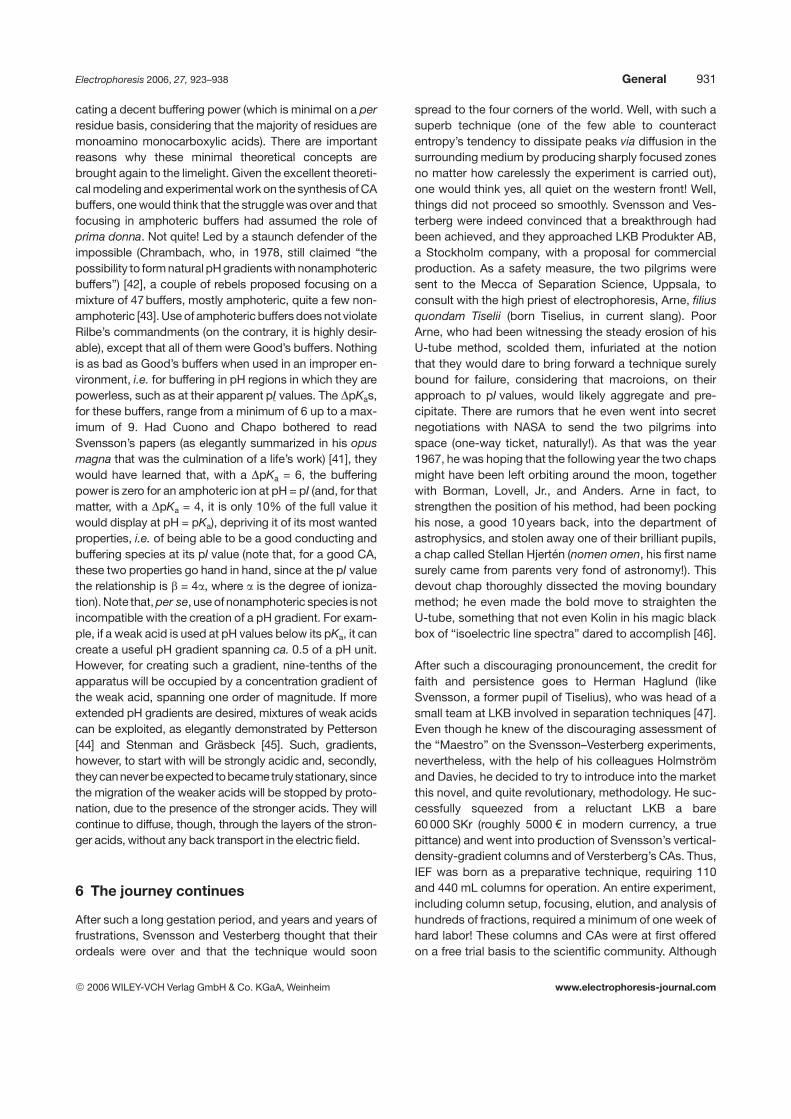
In a series of papers, Radola demonstrated the excellentproperties of this novel system, both for analytical andpreparative purposes [58–62]. Figure 9 gives an exampleof the excellent results that could be obtained by thistechnique: the analytical focusing in thin (0.6 mm thick)Sephadex layers of a set of pI markers produced indeed asharp array of zones, well visible in the scan as well as inthe paper print underneath. That was one of the remark-
Figure 9. Thin layer IEF of pH marker proteins in pH 3–10 CAs. Sephadex G-75 superfine, 0.6 mm thickness ofgel layer. Separation distance, 20 cm; Bottom : paperprint stained with CBB G-250. Upper graph: solid tracing:densitometric profile; open circles: pH gradient. Markerproteins: FER, ferritin, BSA; LAC, b-lactoglobulin; CON,conalbumin; MYH, horse myoglobin; MYW, sperm whalemyoglobin; RIB, ribonuclease; CYT, cytochrome C (fromRadola, see [62], by permission).
2006 WILEY-VCH Verlag GmbH & Co. KGaA, Weinheim www.electrophoresis-journal.com
Electrophoresis 2006, 27, 923–938 General 933
able expedients adopted, in fact: the only way to stain agranulated bed was to take a rapid print with filter paper,so as to allow a minimal amount of interstitial fluid (withprotein) to be conveyed to the contact paper, which, inturn, would be immediately stained so as to fix the trans-ferred protein and prevent diffusion and blurring. For thispurpose, one of the best granulated materials was foundto be Sephadex G-75 superfine. In case of overloadingthat would result in fusion of contiguous sample zones,Radola suggested taking a second print and staining itwith Coomassie Violet, a less sensitive stain than thewidely adopted CBB R-250.
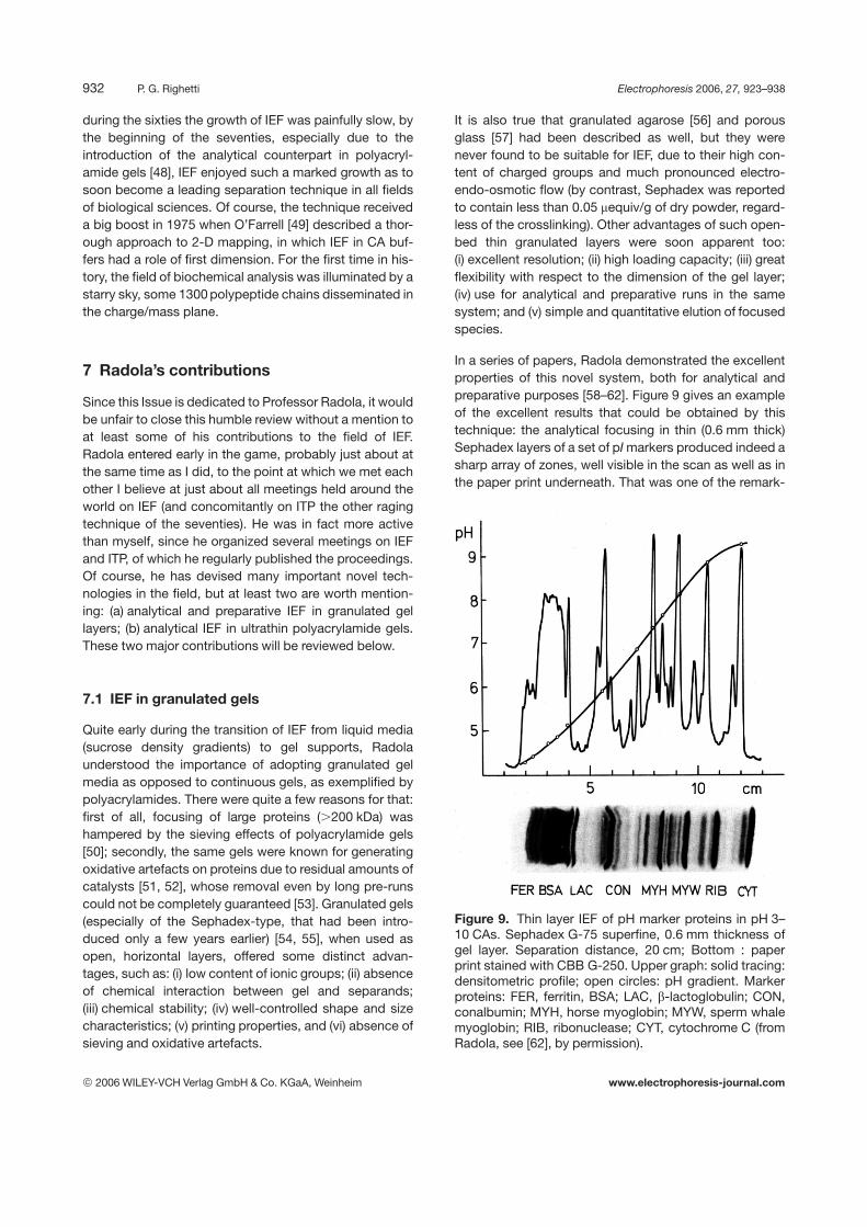
That was in fact another major advantage of such open-layer gels: the possibility of taking a few replicas from thesame gel for developing a series of specific stains, suchas for enzyme activities, or glyco- or lipoprotein stains, toname just a few. A nice example is given in Fig. 10: twodifferent Sephadex gels were run for generating IEF pat-terns of two different varieties of water-soluble barleyproteins (Bido and Union). The two Coomassie-tainedprints can be seen at the two lateral edges of the figures.Concomitantly, two additional prints were taken anddeveloped with a zymogram for esterases (with b-naphtylacetate and Fast Blue RR), the two matched prints beingdisplayed in the middle of the plate. It can be appreciated
that subtle differences are evident between the two vari-eties, not only in the Coomassie stains, but also in the re-spective zymograms, and all of this while maintaining topresolution in both types of stained patterns. A whole se-ries of zymograms were refined, by the paper print tech-nique on Sephadex layers, by Radola et al. [62], as sum-marized in Table III of one of his major reviews .
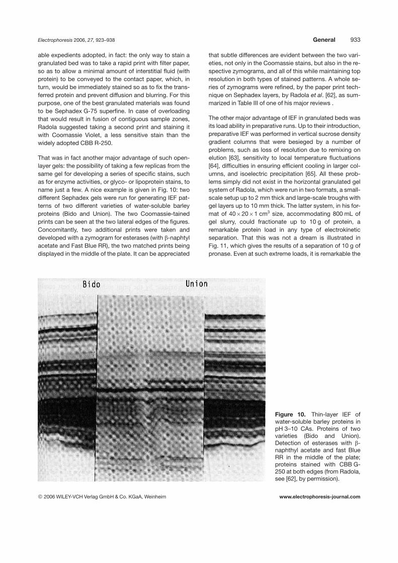
The other major advantage of IEF in granulated beds wasits load ability in preparative runs. Up to their introduction,preparative IEF was performed in vertical sucrose densitygradient columns that were besieged by a number ofproblems, such as loss of resolution due to remixing onelution [63], sensitivity to local temperature fluctuations[64], difficulties in ensuring efficient cooling in larger col-umns, and isoelectric precipitation [65]. All these prob-lems simply did not exist in the horizontal granulated gelsystem of Radola, which were run in two formats, a small-scale setup up to 2 mm thick and large-scale troughs withgel layers up to 10 mm thick. The latter system, in his for-mat of 4062061 cm3 size, accommodating 800 mL ofgel slurry, could fractionate up to 10 g of protein, aremarkable protein load in any type of electrokineticseparation. That this was not a dream is illustrated inFig. 11, which gives the results of a separation of 10 g ofpronase. Even at such extreme loads, it is remarkable the
Figure 10. Thin-layer IEF ofwater-soluble barley proteins inpH 3–10 CAs. Proteins of twovarieties (Bido and Union).Detection of esterases with b-naphthyl acetate and fast BlueRR in the middle of the plate;proteins stained with CBB G-250 at both edges (from Radola,see [62], by permission).
2006 WILEY-VCH Verlag GmbH & Co. KGaA, Weinheim www.electrophoresis-journal.com
934 P. G. Righetti Electrophoresis 2006, 27, 923–938
Figure 11. Preparative IEF of10 g of pronase E in a40620 cm2 trough. Thicknessof the gel layer, 1 cm. Sepha-dex G-75, pH 3–10 CAs. Loadcapacity: 12 mg protein/mL gelsuspension. Focusing: 400 V for16 h followed by 800 V for 6 h.Bottom: paper print stained withlight green SF. Top graph: solidtracing: densitometric profile;open circles: pH gradient (fromRadola, see [62], by permis-sion).
kind of separation that could be achieved. This experi-ment also illustrates a phenomenon that in those dayscould not quite be understood: in the gel layers wherelarge amounts of proteins focus, (e.g., the regions from 6to 12 cm and from 19 to 24 cm) the pH gradient suddenlychanges slope and is flattened. This was understood byus some 10 years later when simulating extended IPGs: inorder to obtain linear pH gradients, the b power along itscourse had be kept very constant [66–69]. Although pro-teins are poor buffers, in general, in zones of overloadingtheir b power overcomes the one of CA buffers and thisresults in a flattening of the pH gradient! The techniquehad become extremely popular in those days, to the pointthat LKB Produkter AB had released on the market atrough, a sample applicator and a fractionation grid,which would “guillotine” the focused gel slurry into20 fractions (for a more thorough description of the prin-ciple and the setup, see [32], pp. 129–136). There is also acute story behind it, as told to me by the LKB scientists:the Sephadex G-200 superfine sold in those days byPharmacia was contaminated by some material (perhapssalts or other chemicals), which severely interfered withproper focusing, generating wavy and distorted bands.So, they bought large quantities of Sephadex from thecompeting company, simply rinsed it thoroughly with dis-tilled water to remove the contaminants, dried it again andsold it (I suppose at higher prices) as their own materialunder the trade name of Ultrodex! It is nice that this bril-
liant idea of Radola has been resurrected recently by Görget al. [70] as a prefractionation step in proteome analysis:let us drink to that! Just to paraphrase a famous song byWoody Guthrie: “good ideas never die, they just fadeaway”.
7.2 IEF in ultrathin polyacrylamide layers
This was also a major innovation in the field of analyticalIEF. It was started by Görg et al. [71], who first reportedthin polyacrylamides (typically in the 200–300 mm thick-ness range), supported by cellophane foils. Early in thegame, Radola realized that these thin gels would not beenough for exploiting all possible benefits of such a tech-nique; therefore, he developed novel techniques forcasting ultrathin, gels, in the thickness range from as lowas 20 up to 100 mm [72–76]. Several advantages wereimmediately apparent: (i) markedly improved resolution;(ii) much more efficient heat transfer; (iii) extremely faststaining, destaining, and drying; and (iv) drastically low-ered demand for CAs and other reagents.
I suspect that Radola also invented a novel polyester foilfor proper attachment of these ultrathin gels, still sold byServa as Gel Fix: contrary to the Gel Bond PAG, the for-mer contains acrylic-type double bonds on the surface,so that attachment is fully guaranteed, with prevention of
2006 WILEY-VCH Verlag GmbH & Co. KGaA, Weinheim www.electrophoresis-journal.com
Electrophoresis 2006, 27, 923–938 General 935
peeling off, due to covalent bond formation. It turns outthat the idea of going ultrathin was fully right: when, yearslater, we simulated the heat transfer in IEF gels [77], withthe theory developed by Russian mathematicians in mylaboratory [78, 79], we found that, at the high wattagesadopted for rapid focusing and ultraresolution, the gelthickness had to be no greater than 50 mm! Needless tosay, Radola’s idea was immediately exploited for castingvery thin gels for DNA sequencing [80]. And, in nuce, hisidea of going ultrathin was at the basis of CZE, as imple-mented a few years later by Jorgenson and co-workers[81, 82]. With a proviso, though: in CZE, only a singlechannel could be exploited, which meant running onesample at a time, versus dozens of analytes run simulta-neously in these open-face gel slabs. Were this notenough, Radola also propounded the idea of washing,drying, and rehydrating such ultrathin gels, so as to get ridof all undesired chemicals trapped in the gel upon po-lymerization. The dried gels could be stored for years in
the frozen state and retrieved when needed for experi-ments. From this point of view, he was also a precursor ofthe Immobiline technique (IPG), in which washing anddrying of the IPG strips is now a routine [39].
8 The symptoms of decay
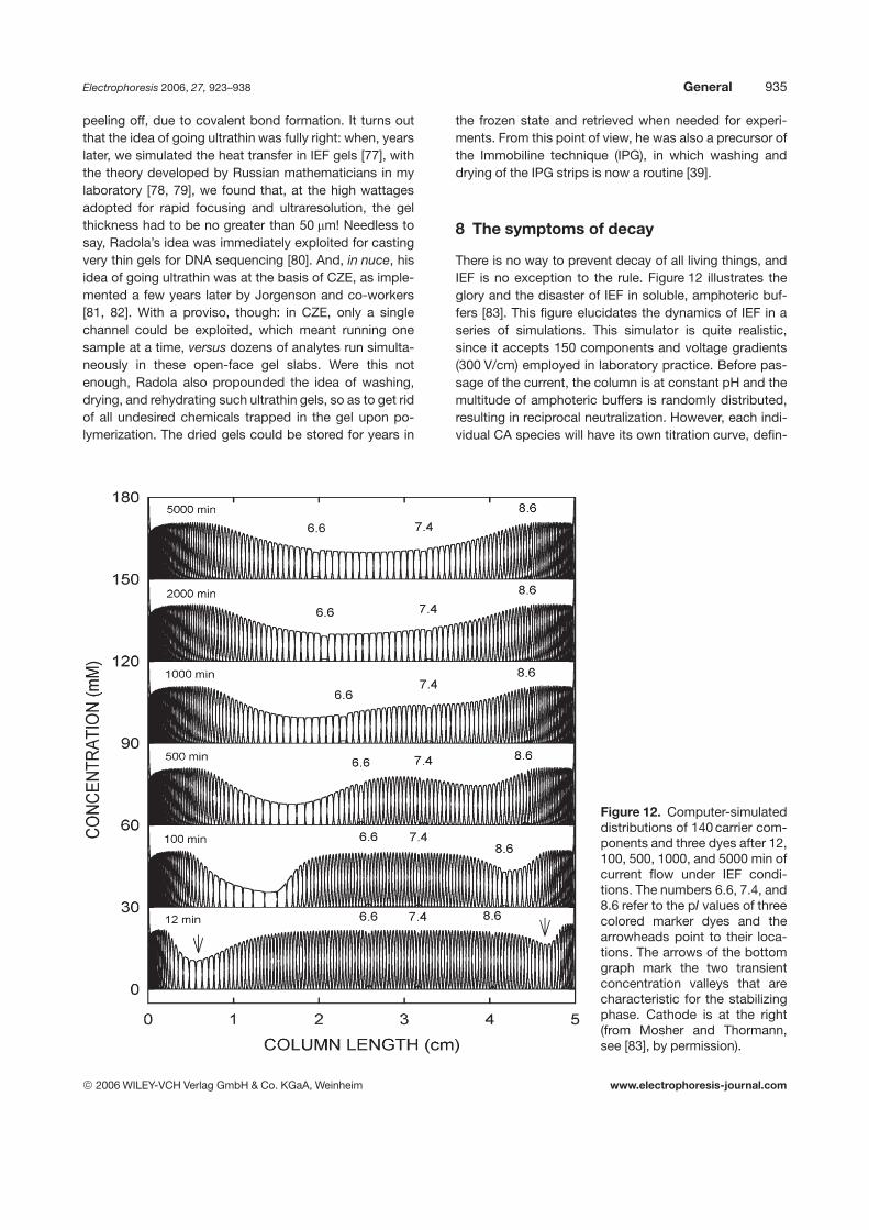
There is no way to prevent decay of all living things, andIEF is no exception to the rule. Figure 12 illustrates theglory and the disaster of IEF in soluble, amphoteric buf-fers [83]. This figure elucidates the dynamics of IEF in aseries of simulations. This simulator is quite realistic,since it accepts 150 components and voltage gradients(300 V/cm) employed in laboratory practice. Before pas-sage of the current, the column is at constant pH and themultitude of amphoteric buffers is randomly distributed,resulting in reciprocal neutralization. However, each indi-vidual CA species will have its own titration curve, defin-
Figure 12. Computer-simulateddistributions of 140 carrier com-ponents and three dyes after 12,100, 500, 1000, and 5000 min ofcurrent flow under IEF condi-tions. The numbers 6.6, 7.4, and8.6 refer to the pI values of threecolored marker dyes and thearrowheads point to their loca-tions. The arrows of the bottomgraph mark the two transientconcentration valleys that arecharacteristic for the stabilizingphase. Cathode is at the right(from Mosher and Thormann,see [83], by permission).
2006 WILEY-VCH Verlag GmbH & Co. KGaA, Weinheim www.electrophoresis-journal.com
936 P. G. Righetti Electrophoresis 2006, 27, 923–938
ing different mobilities in the electric circuit. After startingthe experiment, the focusing process appears to proceedin two phases, a relative fast separation phase followedby a very slow stabilizing phase during which a steadystate should be reached. As shown in the bottom tracing,a pH 3–10 gradient appears to be fully established within10–12 min, with the CAs forming Gaussian-like, over-lapping zones of high concentration (.100-fold increasecompared to the initial stage) with the three samples(dyes with pIs of 6.6, 7.4, and 8.6) well focused too. Closeto the column ends, large changes of CA concentrations(valleys) are marked with vertical arrowheads. This initialCAs distribution generates an almost linear pH profile.This simulation, however, shows also the major drawbackof all CA-based focusing processes: steady state can beachieved only upon extremely long focusing times (pre-dicted to be 8000 min in the present simulation). The twovalleys at the extremes migrate inwards, flattening thefocused CA from Gaussian into square-shaped peaks,and displacing them toward the terminal electrodes (seethe simulated profiles from 100 to 5000 min). As a result,the pH gradient keeps changing slopes, till it assumes asigmoidal profile, with marked flattening of the centralregion with inflection point around neutrality (called pla-teau phenomenon). This results in a continuous shift ofthe foci of the focused samples along the column length(see the diverging positions of the three dyes from bottomto top in Fig. 12). Although Fig. 12 does not show as yetthe decay phenomenon, other figures in [83] (notablyFigs. 11, 13) demonstrate the isotachophoretic decay ofthe stack of focused CAs in presence of anolyte andcatholyte. With this shock wave generated by the re-search of Mosher and Thormann [83], I will end this reviewand let you guess why IPGs had to be invented. But sincethe IPG epopee could easily fill up this entire issue, I willspare you the suffering.
9 Epilogue
“The ladies, the chevaliers, the weapons, the love stories,the courtesies, the daring deeds I sing, which occurredduring the time that the Moors crossed the Street ofGibraltar and pillaged France. . . So much for the verses ofLodovico Ariosto, a most famous renaissance poet at thecourt of the Este’s family in Ferrara. His Orlando Furiosoinspired generations of poets and artists the world over.
Now for the curious title. One might notice that Alpher,Bethe, and Gamow sound familiar and recall somehowthe Greek letters alpha, beta and gamma. The lettersclearly hint at the origin of this technique, much like theterm “alphabet” indicates the beginning and the sequel ofall letters of our language. This title was chosen as a tri-
bute to a brilliant astrophysicists, George Gamow, who, in1948, wrote a classic paper about the beginning of theuniverse, together with one of his students, Ralph Alpher.Since Gamow had a marked sense of humor, he invited asa coauthor the nuclear physicist Hans Bethe (a 1967Nobel laureate), so that the article would appear with thissequel of names [84]. No sequel could have been betterarranged, considering that the authors made the remark-able prediction that a background radiation, emitted atthe birth of the universe, should still exist, but with a tem-perature barely a few degrees above absolute zero(22737C). Such a prediction was fully verified later on byPenzias and Wilson [85] (both 1978 Nobel laureates inphysics).
10 Addendum
One of the reviewers did not seem to be happy with myhistorical reconstruction of the birth of IEF, and asked meto quote additional papers, notably the work of Schu-maker’s group. I am happy to recall it here [86, 87]. Inparticular, Friedli and Schumacher [88] described amethod, called electrophoretic focusing of ions (EFI,focusing ion exchange) for separation of rare-earth mix-tures in combined proton (pH) and ligand (pL) gradients.EFI spectra of La–Tb and Eu–Lu groups were obtained inthe course of 5 min. Not exactly focusing, but electro-phoresis perpendicular to a pre-established pH gradientwas practiced already on 1952 by Michl [89]: it was anembryonic form of “pH/mobility curves”, as later devel-oped by utilizing IEF in the first dimension (curiously, as Ipored over just about any possible paper published onconventional IEF, I seem to have been perhaps the onlyone ever quoting these two groups [90], as well as thework of Stahl [91, 92] who also described “titrationcurves” in TLC). But then this is a quite dangerous game,since the birth of ideas could be shifted back to someremote origins in the past, till complete “defocusing oftheir beginnings” (to quote a brilliant definition of Kolin).So, why do we not quote Ikeda and Suzuki as the realinventors of IEF, considering that they achieved aseparation of amino acids in plant hydrolysates in a three-chambered electrophoresis cell, generating a pH gradientalready in 1912 [93]? Or, for that matter, we could placethe origin of IEF in the year 1929, when Williams andWaterman [94] extended this previous work by designinga multichamber compartment, which gave better resolu-tion by reducing diffusion and convective disturbances.IEF was basically there!
Supported by the MIUR (FIRB 2001, Cod. RBNE01KJHT;PRIN, 40%, 2003 & 2005), Rome, and by a grant fromBanca Intesa (year 2006).
2006 WILEY-VCH Verlag GmbH & Co. KGaA, Weinheim www.electrophoresis-journal.com
Electrophoresis 2006, 27, 923–938 General 937
11 References
[1] De Troyes, C., Conte del Graal (The Quest of the Holy Graal,translated by Matarasso, P. M.), Penguin Books, Harmonds-worth, UK 1969.
[2] Hatto, A. T., The Nibelungenlied, Penguin Books, Har-mondsworth, UK 1965.
[3] Mgnusson, M., Palsson, H., The Vinland Sagas, PenguinBooks, Harmondsworth, UK 1965.
[4] Exquemelin, A. O., The Buccaneers of America, PenguinBooks, Harmondsworth, UK 1969.
[5] Cohen, J. M., The Four Voyages of Christopher Columbus,Penguin Books, Harmondsworth, UK 1969.
[6] Levinovitz, A. W., Ringertz, N., The Nobel Prize: The First 100Years, Imperial College Press, London 2001.
[7] Rilbe, H., in: Catsimpoolas, N. (Ed.), Isoelectric Focusing,Academic Press, New York 1976, pp. 13–52.
[8] Svensson, H., Electrophoresis by the Moving BoundaryMethod. A Theoretical and Experimental Study, Almqvist &Wiksells Boktryckeri, Stockholm 1946.
[9] Svensson, H., Sci. Tools 1956, 3, 30–35.
[10] Svensson, H., Arch. Biochem. Biophys. 1962, Suppl. 1, 132–140.
[11] Svensson, H., Acta Chem. Scand. 1961, 15, 325–341.
[12] Svensson, H., Acta Chem. Scand. 1962, 16, 456–466.
[13] Rilbe, H., Ann. N. Y. Acad. Sci. 1973, 209, 11–22.
[14] Frumin, L. L., Zilberstein, G. V., Peltek, S. E., J. Biochem.Biophys. Methods 2000, 45, 205–209.
[15] Chandrasekhar, S., Rev. Mod. Phys., 1943, 15, 1–89.
[16] Catsimpoolas, N., in: Catsimpoolas, N. (Ed.), IsoelectricFocusing, Academic Press, New York 1976, pp. 229–258.
[17] Mosher, R. A., Bier, M., Righetti, P. G., Electrophoresis 1986,7, 59–66.
[18] Kolin, A., J. Chem. Phys. 1954, 22, 1628–1629.
[19] Kolin, A., Proceedings of the National Academy of SciencesUSA 1955, 41, 101–110.
[20] Kolin, A., in: Electrofocusing and Isotachophoresis, Radola,B. J., Graesslin, D. (Eds.), de Gruyter, Berlin 1977, pp. 3–34.
[21] Kolin, A., in: Electrophoresis ’82, Stathakos, D. (Ed.), deGruyter, Berlin 1983, pp. 3–48.
[22] Meselson, M., Stahl, F. W., Vinogradov, J., Proceedings ofthe National Academy of Sciences USA 1957, 43, 581–585.
[23] Margolis, J., Kolin, K. J., Anal. Biochem. 1968, 25, 347–362.
[24] Giddings, J. C., Dahlgren, H., Separ. Sci. 1971, 6, 345–356.
[25] Giddings, J. C., Unified Separation Science, Wiley, New York1991.
[26] Kauman, W. G., Clas. Sci. Acad. Roy. Belg. 1957, 43, 854–868.
[27] Vesterberg, O., Acta Chem. Scand. 1969, 23, 2653–2666.
[28] Vesterberg, O., Ann. N. Y. Acad. Sci. 1973, 209, 23–33.
[29] Vesterberg, O., in: Catsimpoolas, N. (Ed.), IsoelectricFocusing, Academic Press, New York 1976, pp. 53–76.
[30] Almgren, M., Chem. Scripta 1971, 1, 69–75.
[31] Bianchi-Bosisio, A., Snyder, R. S., Righetti, P. G., J. Chro-matogr. 1981, 209, 265–272.
[32] Righetti, P. G., Isoelectric Focusing: Theory, Methodologyand Applications, Elsevier, Amsterdam 1983, pp. 306–313.
[33] Righetti, P. G., Brown, R. P., Stone, A. L., Biochim. Biophys.Acta 1978, 542, 232–244.
[34] Gianazza, E., Righetti, P. G., Biochim. Biophys. Acta 1978,540, 357–364.
[35] Haglund, H., in: Arbuthnott, J. P., Beeley, J. A. (Eds.), Iso-electric Focusing, Butterworths, London 1975, pp. 3–22.
[36] Gelsema, J. W., De Ligny, C. L., Van Der Veen, N. G., J.Chromatogr. 1978, 154, 161–174.
[37] Grubhofer, N., Borja, C., in: Radola, B. J., Graesslin, D.(Eds.), Electrofocusing and Isotachophoresis, de Gruyter,Berlin 1977, pp. 111–120.
[38] Söderberg, L., Buckley, D., Hagström, G., Bergström, J.,Prot. Biol. Fluids 1980, 27, 687–691.
[39] Bjellqvist, B., Ek, K., Righetti, P. G., Gianazza, E., Görg, A.,Westermeier, R., Postel, W., J. Biochem. Biophys. Methods1982, 6, 317–339.
[40] Righetti, P. G., Immobilized pH Gradients: Theory andMethodology, Elsevier, Amsterdam 1990, pp. 70–95.
[41] Rilbe, H., pH and Buffer Theory, Wiley, Chichester 1996,pp. 87–92.
[42] Chrambach, A., Nguyen, N. Y., in: Catsimpooloas, N. (Ed.),Electrophoresis ’78, Elsevier, Amsterdam 1978, pp. 3–18.
[43] Cuono, C. B., Chapo, G. A., Electrophoresis 1982, 3, 65–74.[44] Petterson, E., Acta Chem. Scand. 1969, 23, 2631–2635.[45] Stenman, U. K., Gräsbeck, R., Biochim. Biophys. Acta 1972,
286, 243–251.[46] Hjertén, S., Chromatogr. Rev. 1967, 9, 122–219.[47] Haglund, H., in: Glick, D. (Ed.), Methods of Biochemical
Analysis, Wiley Interscience, New York 1971, Vol. 19, pp. 1–104.
[48] Righetti, P. G., Drysdale, J. W., Biochim. Biophys. Acta 1971,236, 17–24.
[49] O’Farrell, P. H., J. Biol. Chem. 1975, 250, 4007–4021.[50] Righetti, P. G., Drysdale, J. W., J. Chromatogr. 1974, 88,
271–321.[51] Brewer, J. M., Science 1967, 156, 356–357.[52] Fantes, K. H., Furminger, I. J. S., Nature 1967, 215, 750–751.[53] Peterson, R. F., J. Agric. Food Chem. 1871, 19, 585–599.[54] Determan, H., Gel Chromatography, Springer, Berlin 1979.[55] Fischer, L., Gel Chromatography, Elsevier, Amsterdam 1969.[56] Hjertén, S., Biochim. Biophys. Acta 1964, 79, 393–398.[57] Haller, W., Nature 1965, 206, 693–697.[58] Radola, B. J., in: Peeters, H. (Ed.), Protides of the Biological
Fluids, Pergamon Press, Oxford 1971, Vol. 18, pp. 487–491.[59] Radola, B. J., Ann. N. Y. Acad. Sci. 1973, 209, 127–141.[60] Radola, B. J., Biochim. Biophys. Acta 1975, 386, 181–195.[61] Radola, B. J., in: Arbuthnott, J. P., Beeley, J. A. (Eds.), Iso-
electric Focusing, Butterworths, London 1975, pp. 182–197.[62] Radola, B. J., in: Catsimpoolas, N. (Ed.), Isoelectric Focus-
ing, Academic Press, New York 1976, pp. 119–171.[63] Vesterberg, O., Biochim. Biophys. Acta 1971, 243, 345–348.[64] Haglund, H., Methods Biochem. Anal. 1971, 19, 1–104.[65] Rilbe, H., in: Peeters, H. (Ed.), Protides of the Biological
Fluids, Pergamon Press, Oxford 1970, 17, 369–382.[66] Celentano, F., Gianazza, E., Dossi, G., Righetti, P. G., Che-
mometr. Intel. Lab. Syst. 1987, 1, 349–358.[67] Gianazza, E., Celentano, F., Magenes, S., Ettori, C., Righetti,
P. G., Electrophoresis 1989, 10, 806–808.[68] Tonani, C., Righetti, P. G., Electrophoresis 1991, 12, 1011–
1020.[69] Righetti, P. G., Tonani, C., Electrophoresis 1991, 12, 1021–
1027.[70] Görg, A., Boguth, G., Köpf, A., Reil, G., Parlar, H., Weiss, W.,
Proteomics 2002, 2, 1652–1657.[71] Görg, A, Postel, W., Westermeier, R., Anal. Biochem. 1978,
89, 60–70.
2006 WILEY-VCH Verlag GmbH & Co. KGaA, Weinheim www.electrophoresis-journal.com
938 P. G. Righetti Electrophoresis 2006, 27, 923–938
[72] Radola, B. J., in: Radola, B. J. (Ed.), Electrophoresis ’79, deGruyter, Berlin 1980, pp. 79–94.
[73] Radola, B. J., Electrophoresis 1980, 1, 43–56.[74] Radola, B. J., in: Jorgenson, J. W., Phillips, M. (Eds.), New
Directions in Electrophoretic Methods, ACS SymposiumSeries 335, Am. Chem. Soc., Washington DC, 1987, pp. 64–73.
[75] Radola, B. J., Kinzkofer, A., Frey, M., in: Allen, R. C., Arnaud,P. (Eds.), Electrophoresis ’81, de Gruyter, Berlin 1981,pp. 181–189.
[76] Frey, M., Atta, B., Radola, B. J., in: Neuhoff, V. (Ed.), Elec-trophoresis ’84, Verlag Chemie, Weinheim 1984, pp. 122–125.
[77] Righetti, P. G., Bello, M., Electrophoresis 1992, 13, 275–279.[78] Bello, M., Righetti, P. G., J. Chromatogr. 1992, 606, 95–102.[79] Bello, M., Righetti, P. G., J. Chromatogr. 1992, 606, 103–111.[80] Ansorge, W., De Meyer, L., J. Chromatogr. 1980, 202, 45–53.[81] Jorgenson, J. W., Lukacs, K. D., Anal. Chem. 1981, 53,
1298–1304.[82] Jorgenson, J. W., Lukacs, K. D., Science 1983, 222, 266–
268.
[83] Mosher, R. A., Thormann, W., Electrophoresis 2002, 23,1803–1814.
[84] Alpher, R., Bethe, H., Gamow, G., Phys. Reviews 1948, 73,803–815.
[85] Penzias, A. A., Wilson, R. W., Astrophys. J. 1965, 142, 419–430.
[86] Schumacher, E., Helv. Chim. Acta 1957, 40, 221–228.
[87] Schumacher, E., Helv. Chim. Acta 1957, 40, 2322–2340.
[88] Friedli, W., Schumacher, E., Helv. Chim. Acta 1961, 44,1829–1856.
[89] Michl, H., Monatsh. Chemie 1952, 83, 210–220.
[90] Righetti, P. G., Gianazza, E., in: Radola, B. J. (Ed.), Electro-phoresis ’79, de Gruyter, Berlin 1980, pp. 23–38.
[91] Stahl, E., Angew. Chem. Int. Ed. 1964, 3, 784–791.
[92] Stahl, E., J. Chromatogr. 1979, 165, 59–73.
[93] Ikeda, K., Suzuki, S., US Patent No 1, 015–891, 1912.
[94] Williams, R. R., Waterman, R. E., Proceedings of the Societyfor Experimental Biology and Medicine 1929, 27, 56–59.
2006 WILEY-VCH Verlag GmbH & Co. KGaA, Weinheim www.electrophoresis-journal.com
Recommended


![[Group 5] electrochemistry, electrophoresis, isoelectric focusing](https://img.dokumen.tips/doc/110x75/55c5bdefbb61eb5a3b8b458a/group-5-electrochemistry-electrophoresis-isoelectric-focusing.jpg)