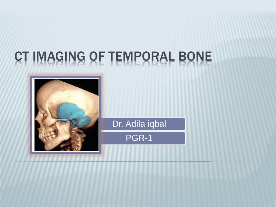
CT IMAGING OF TEMPORAL BONE
Dr. Adila iqbal
PGR-1
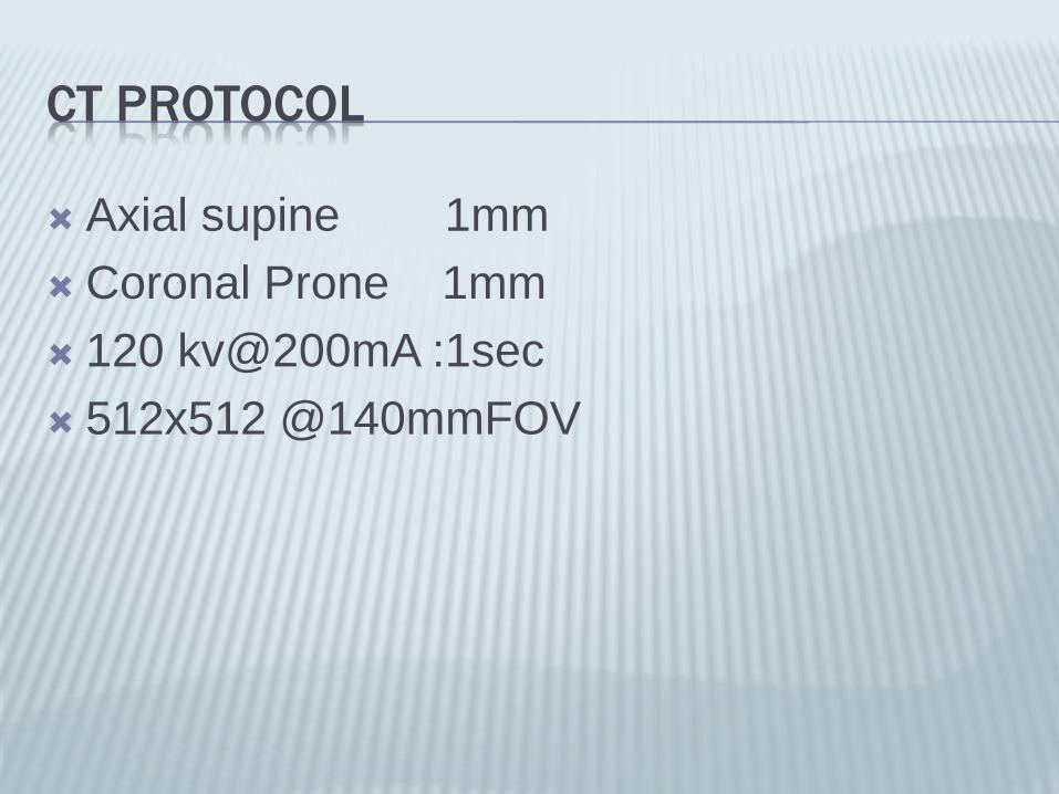
CT PROTOCOL
Axial supine 1mm
Coronal Prone 1mm
120 kv@200mA :1sec
512x512 @140mmFOV

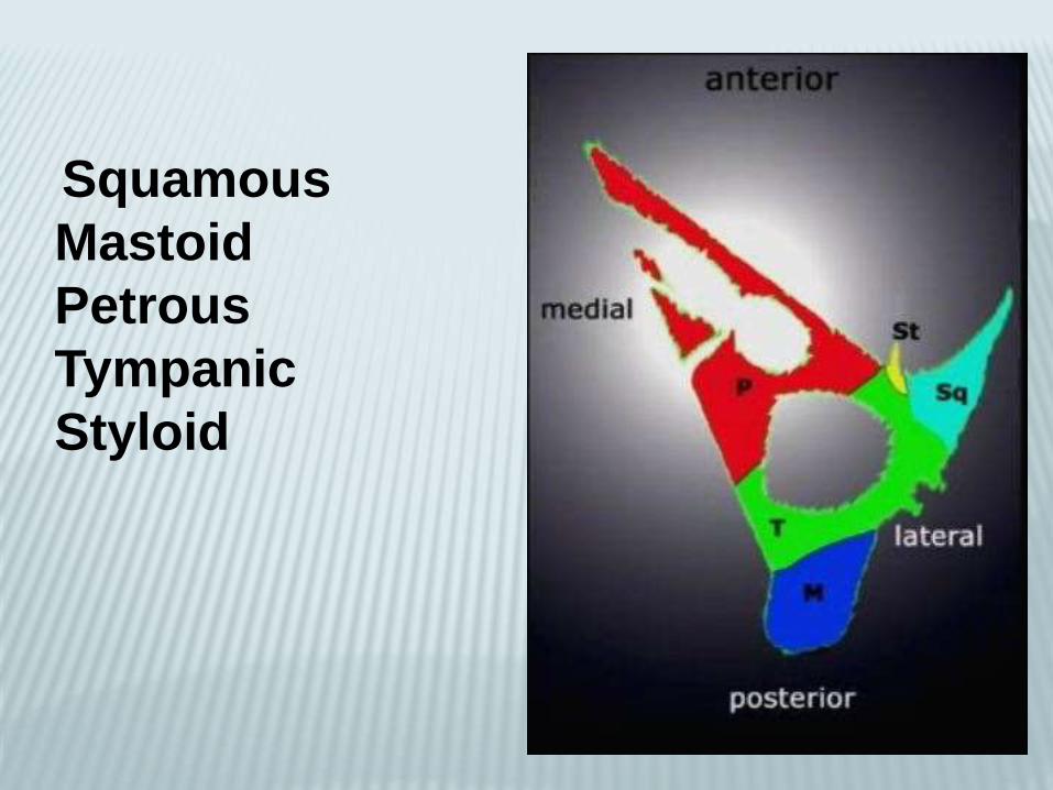
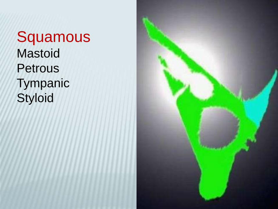
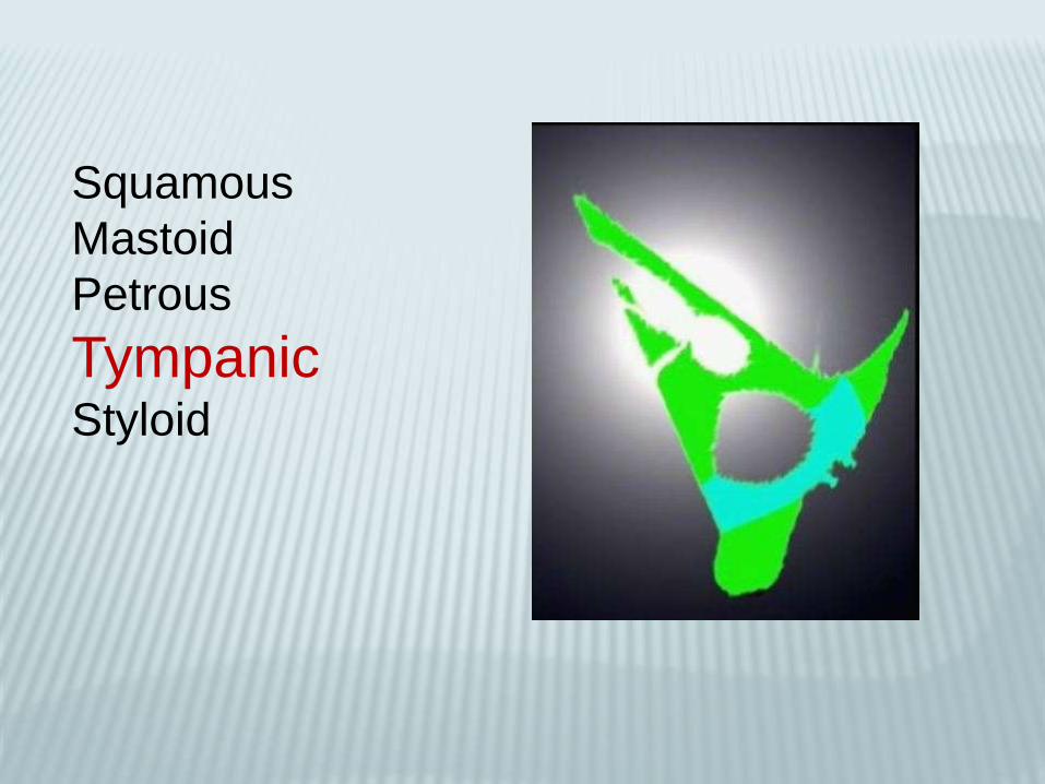
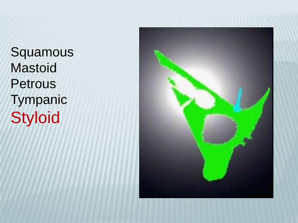
Squamous
Mastoid
Petrous
Tympanic
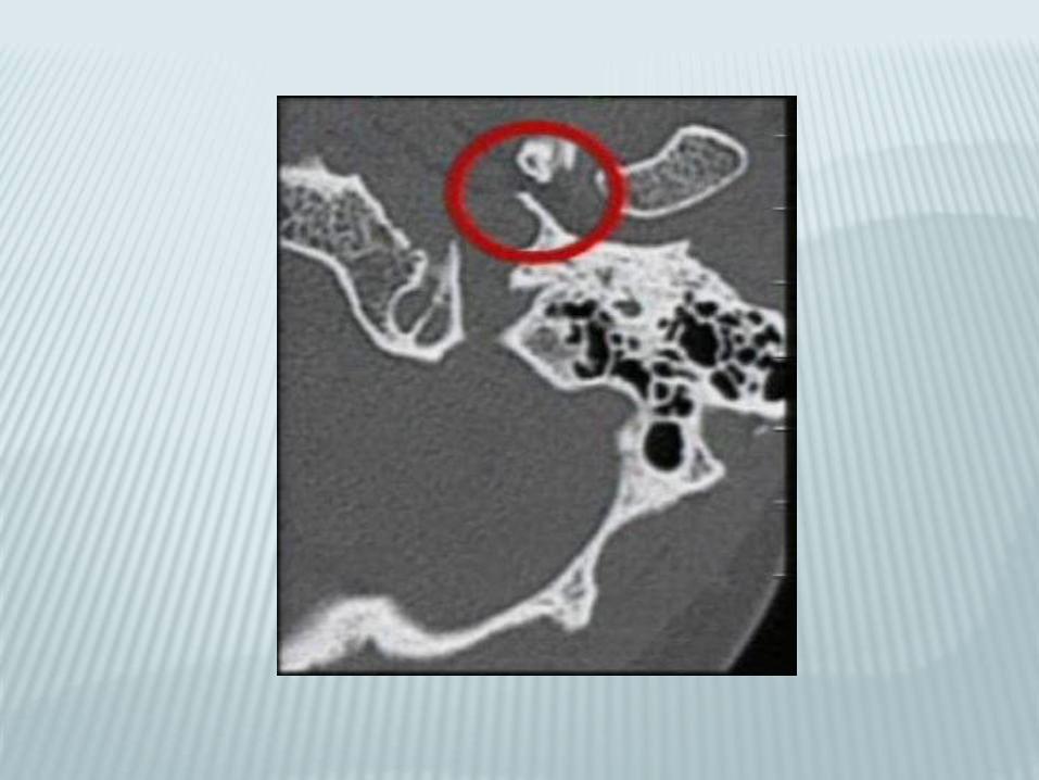
Styloid
SquamousMastoid
Petrous
Tympanic
Styloid


Squamous
MastoidPetrous
Tympanic
Styloid

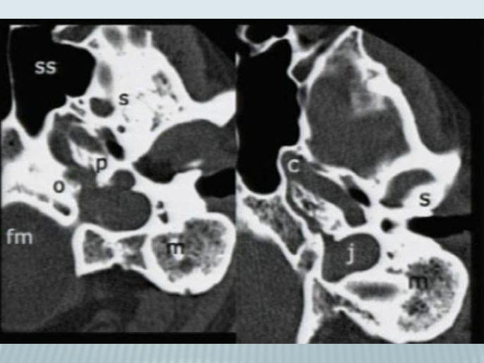


Petrous and its subsegments
•Pyramid and otic capsule
•Subsegments
1. Anterior
2. Posterior
3. Inferior
4. Apical

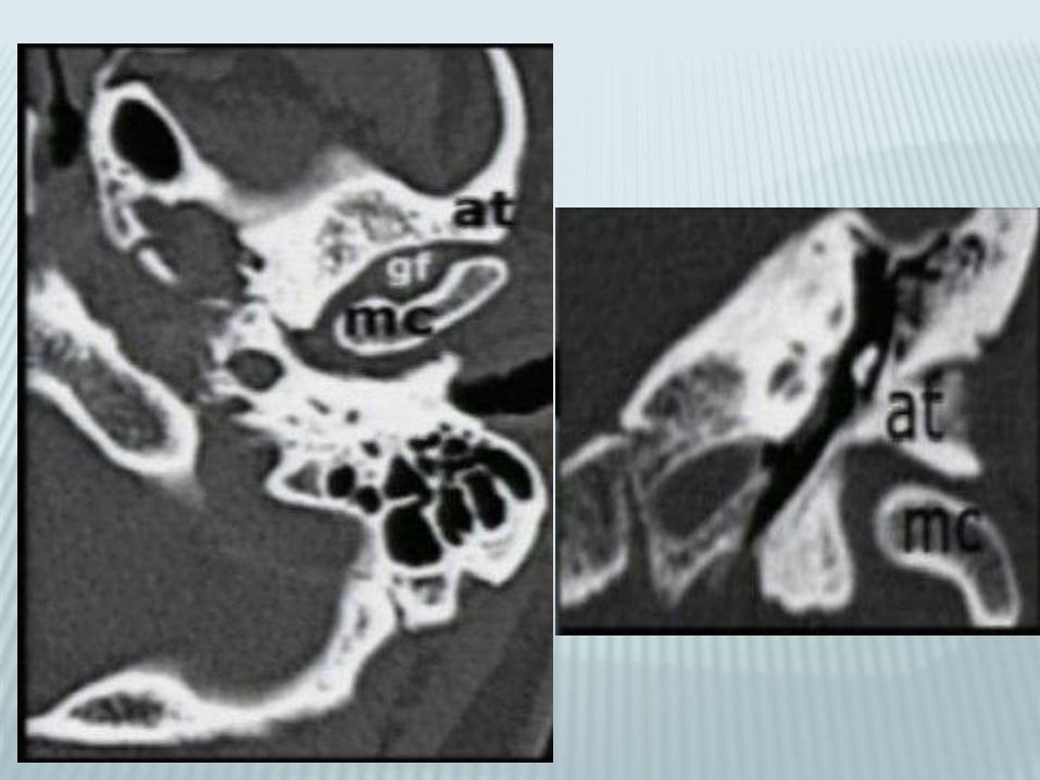
Anterior petrous subsegment
Tegmen tympani
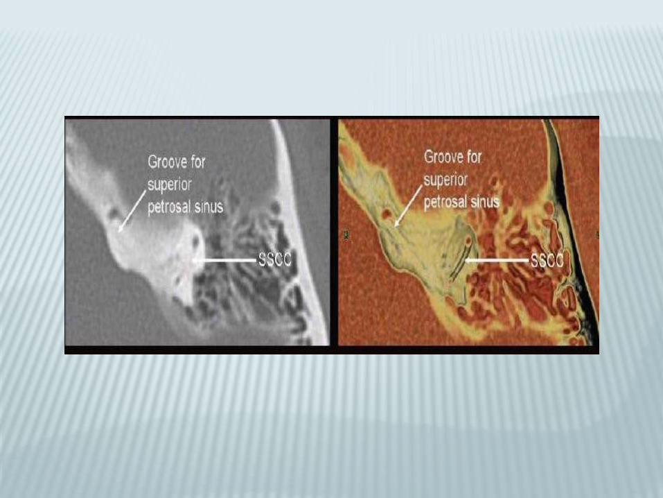
Arcuate eminenceProminence
over upper
SCC
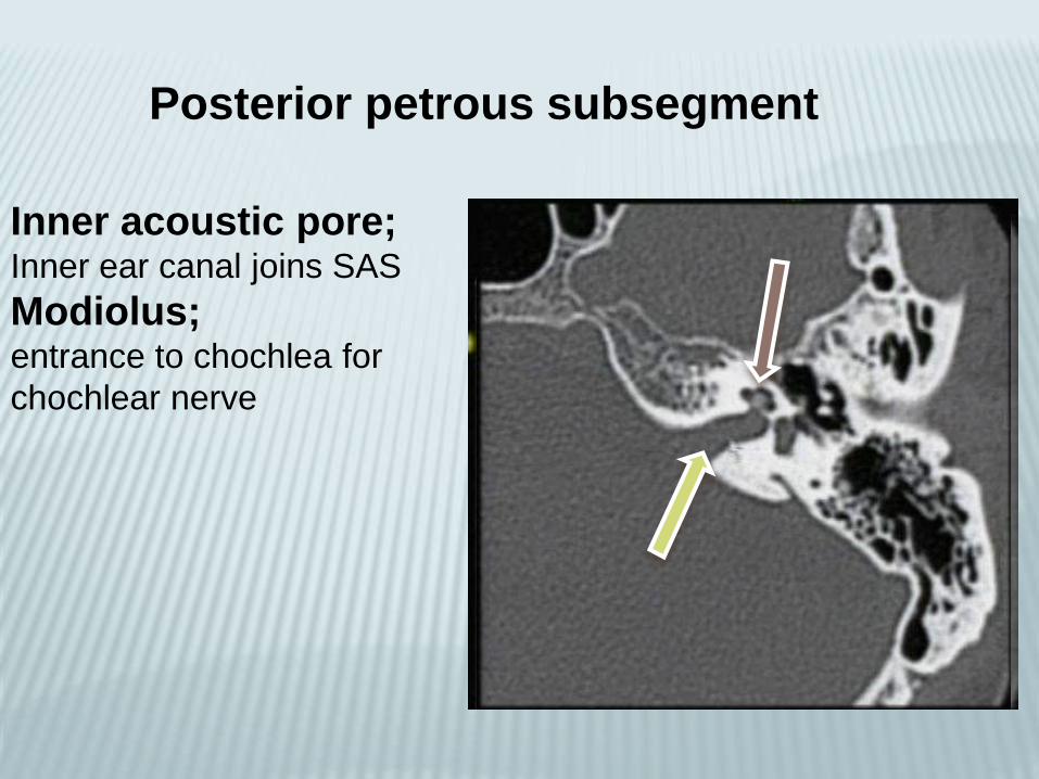
Posterior petrous subsegment
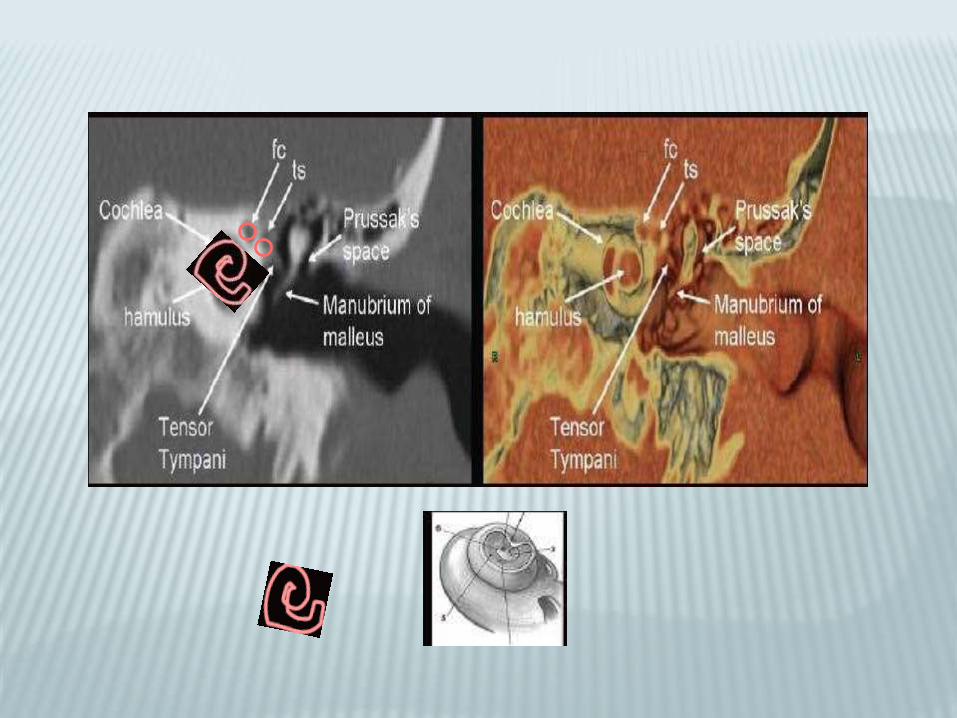
Inner acoustic pore;Inner ear canal joins SAS
Modiolus;entrance to chochlea for
chochlear nerve

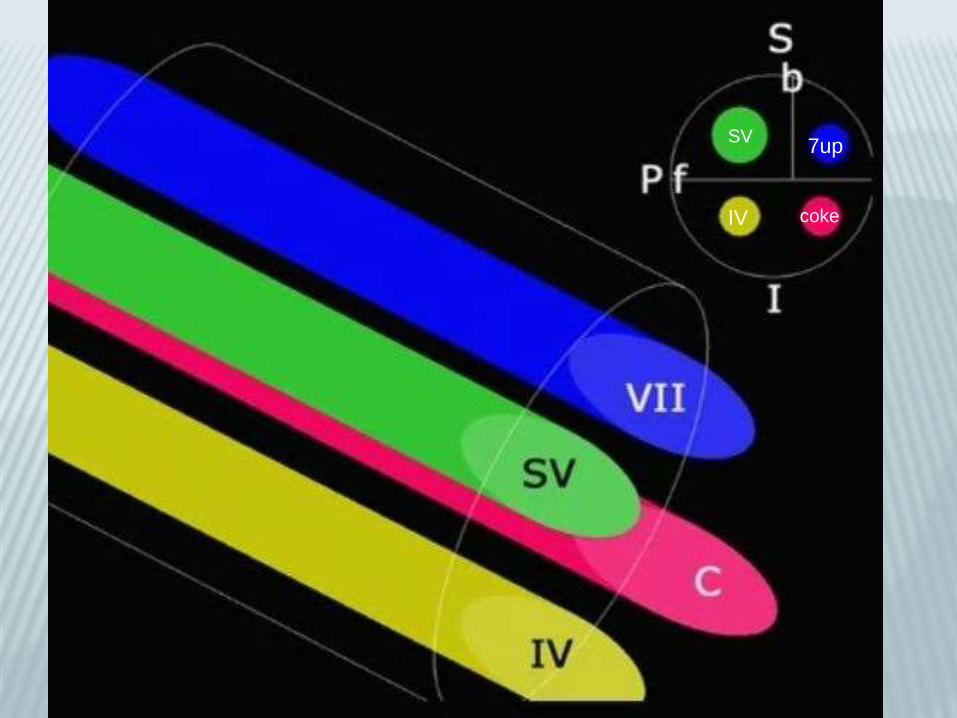
7up
coke
SV
IV
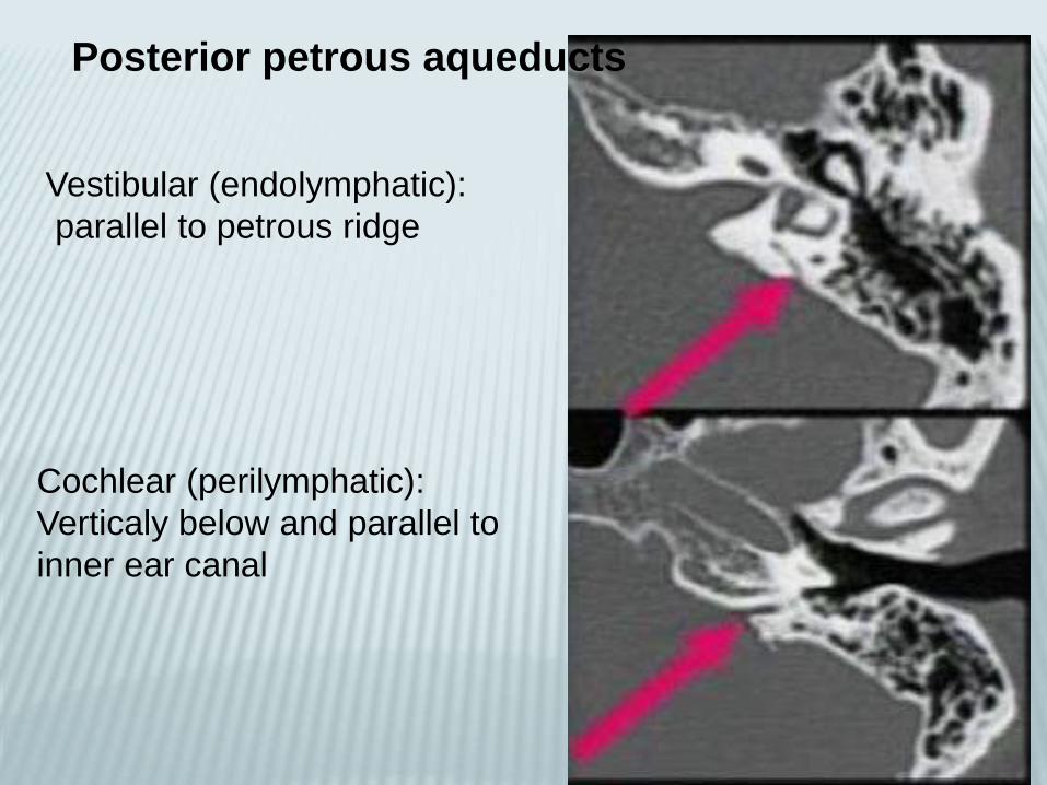
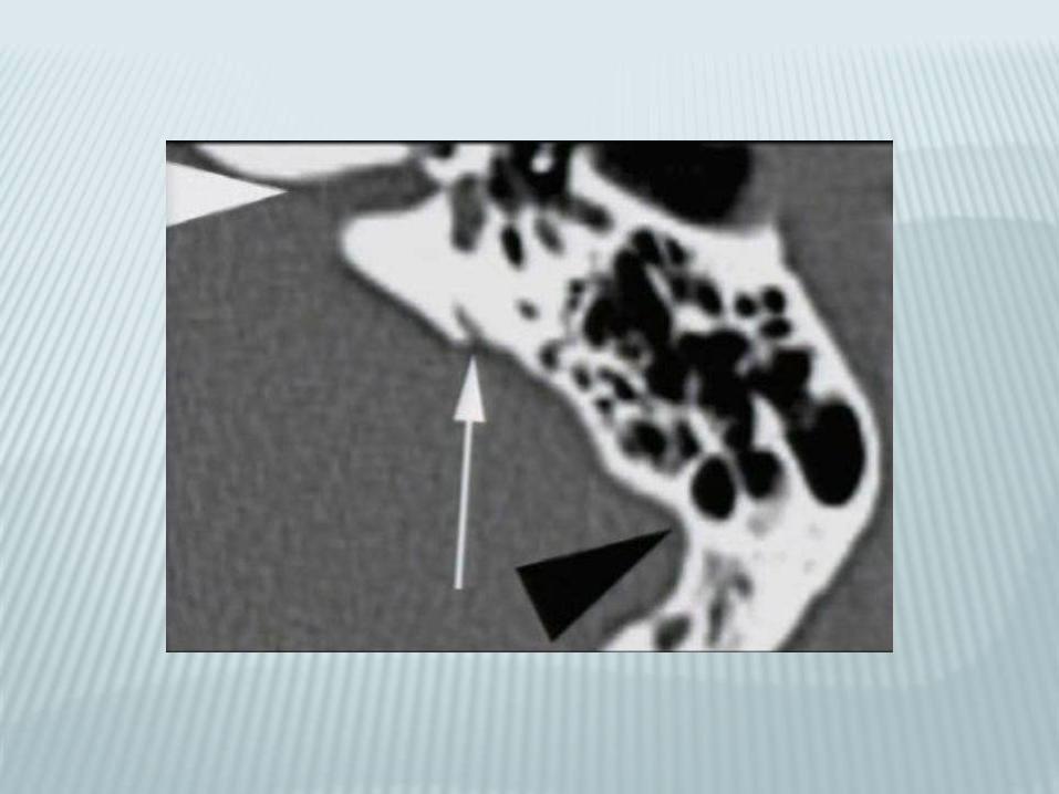
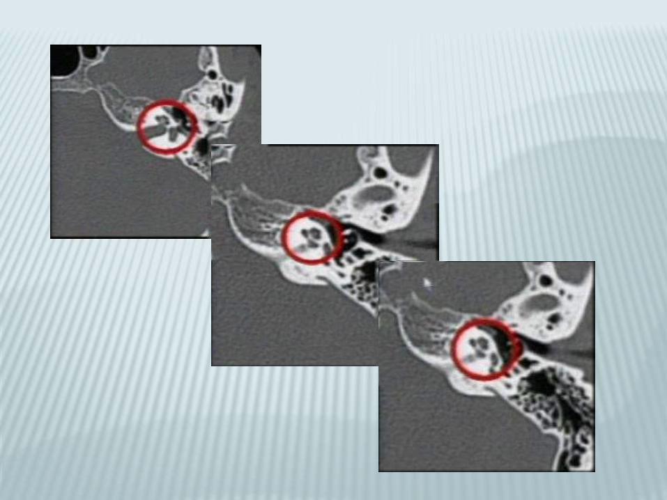
Posterior petrous aqueducts
Vestibular (endolymphatic):
parallel to petrous ridge
Cochlear (perilymphatic):
Verticaly below and parallel to
inner ear canal
VESTIBULAR AQUEDUCT
10 mm long endolymphatic duct
From common crus to post wall of petrous
pyramid
Joins endolymphatic sac nestled in leaves of
dura
Equilibration of endolymphatic fluid pressure
subdural

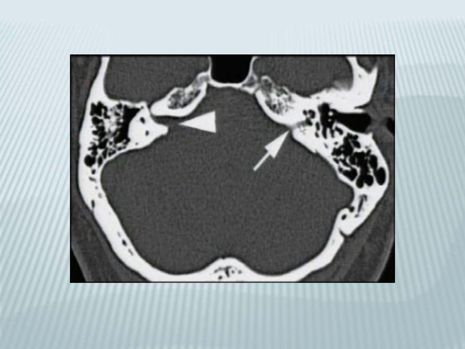
COCHLEAR AQUEDUCT
8 mm long perilymphatic aqueduct
From basal turn of cochlea to medial border
of jugular foramen
Regulation of CSF and perilymphatic fluid
pressure
Subarachnoid
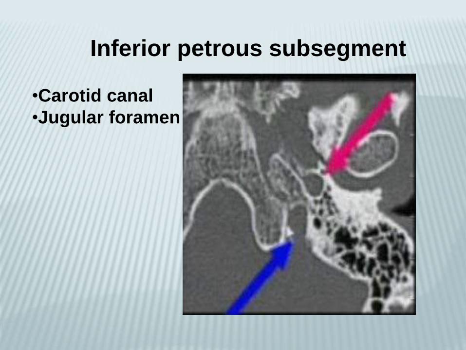
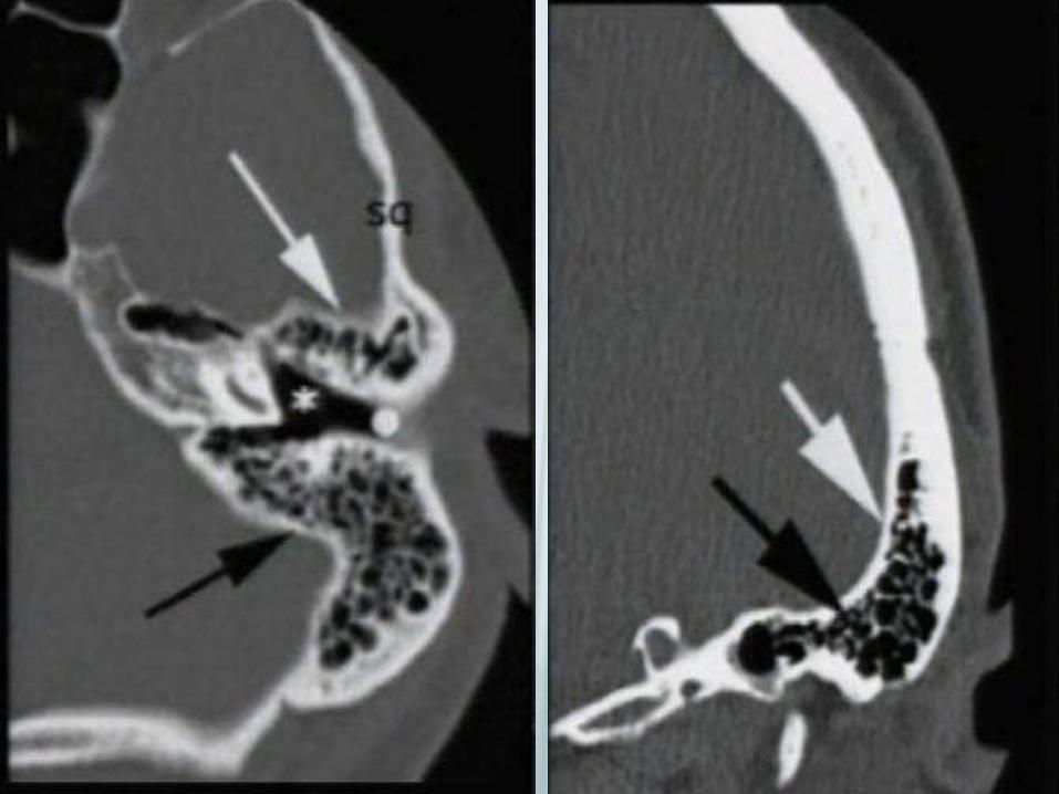
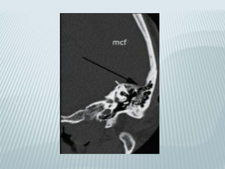
Inferior petrous subsegment
•Carotid canal
•Jugular foramen

Squamous
Mastoid
Petrous
TympanicStyloid
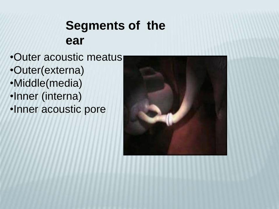
Segments of the
ear
•Outer acoustic meatus
•Outer(externa)
•Middle(media)
•Inner (interna)
•Inner acoustic pore
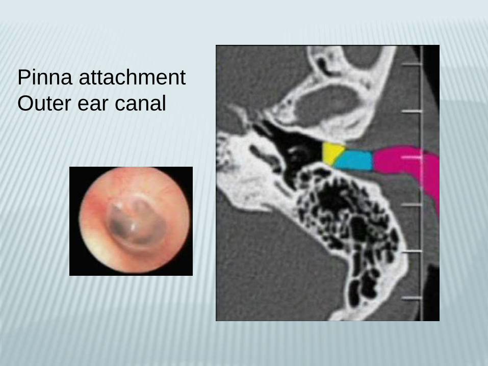
Pinna attachment
Outer ear canal
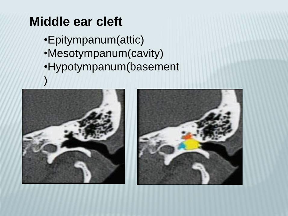
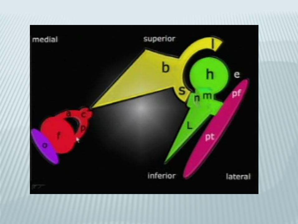
•Epitympanum(attic)
•Mesotympanum(cavity)
•Hypotympanum(basement
)
Middle ear cleft
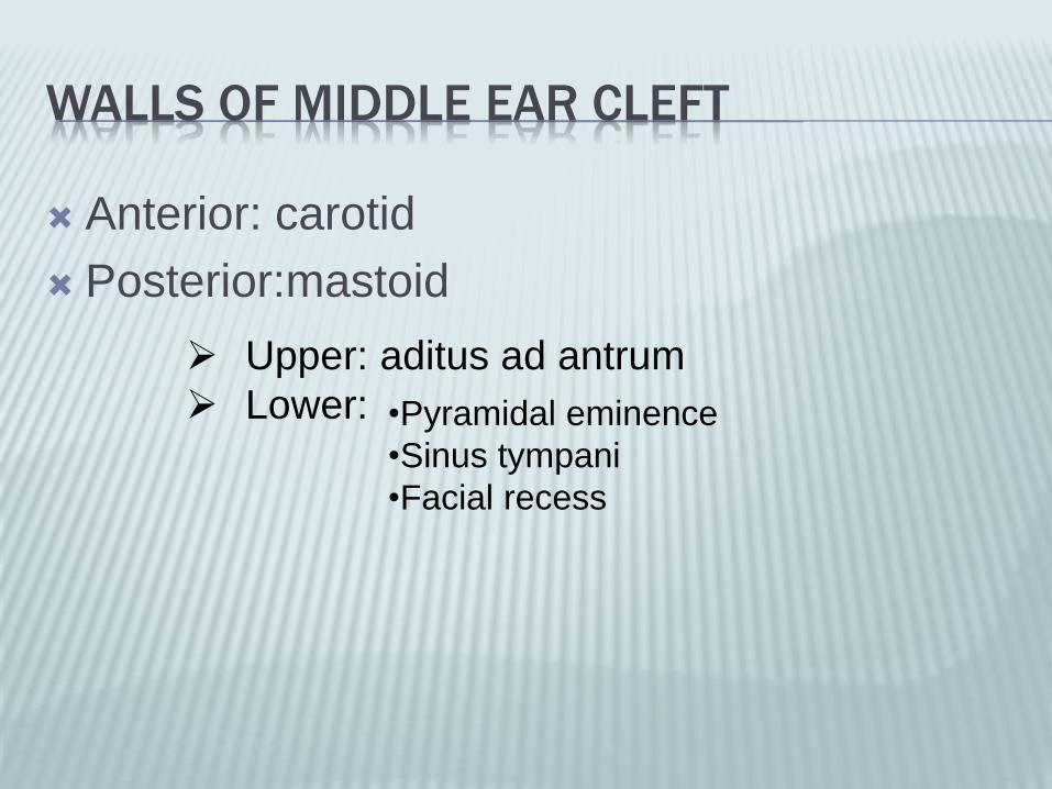
WALLS OF MIDDLE EAR CLEFT
Anterior: carotid
Posterior:mastoid
Upper: aditus ad antrum
Lower: •Pyramidal eminence
•Sinus tympani
•Facial recess
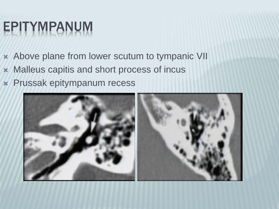
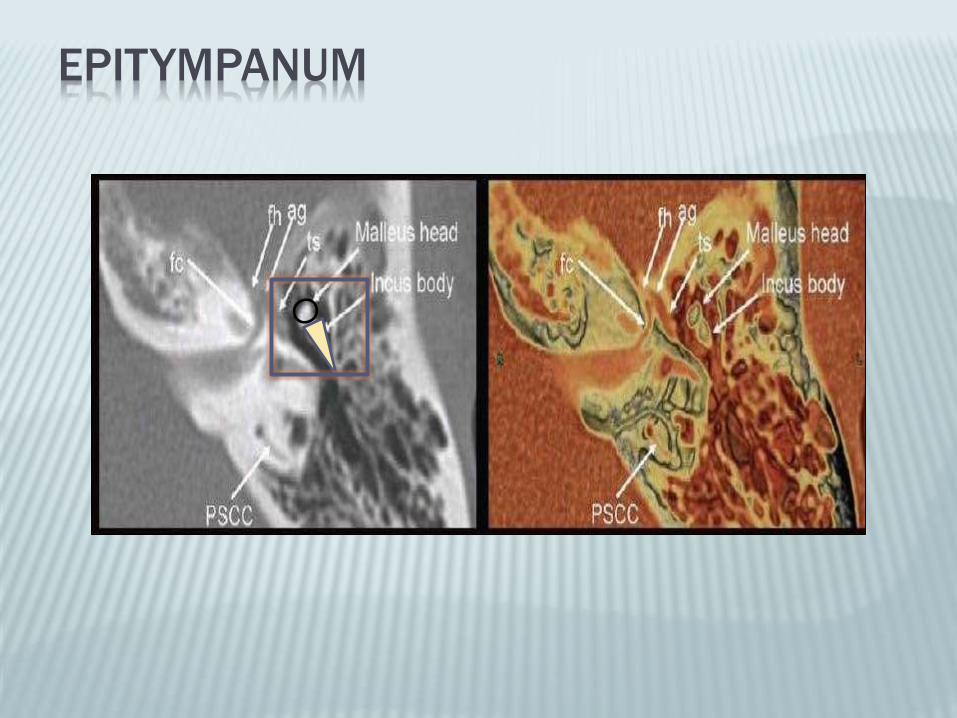
EPITYMPANUM
Above plane from lower scutum to tympanic VII
Malleus capitis and short process of incus
Prussak epitympanum recess
EPITYMPANUM

MESOTYMANUM
Tympanic cleft proper dampens sounds
Manubrium of malleus
Long process of incus
Superstructure and foot plate of stapes
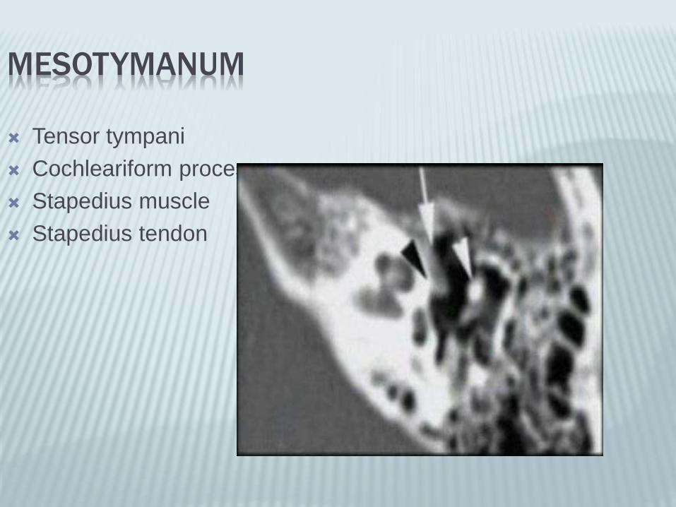
MESOTYMANUM
Tensor tympani
Cochleariform process
Stapedius muscle
Stapedius tendon
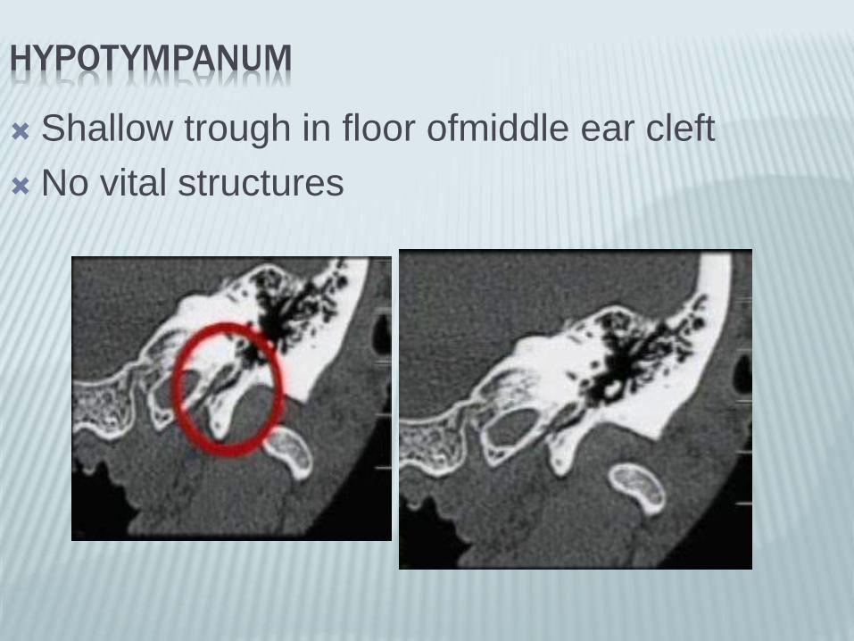
HYPOTYMPANUM
Shallow trough in floor ofmiddle ear cleft
No vital structures
Squamous
Mastoid
Petrous
Tympanic
Styloid
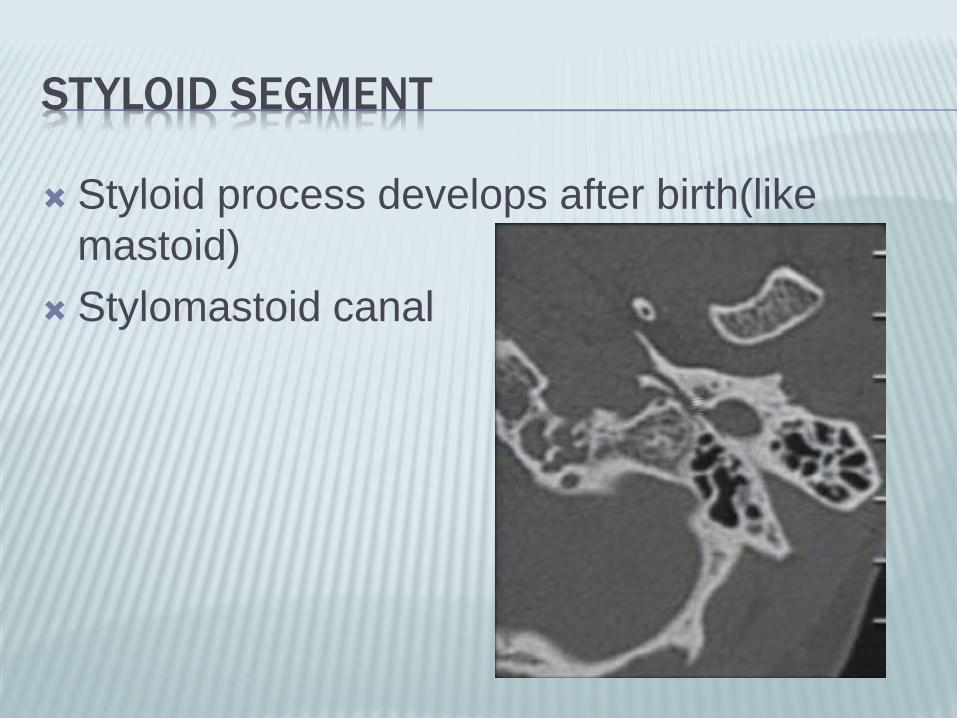
STYLOID SEGMENT
Styloid process develops after birth(like
mastoid)
Stylomastoid canal
INNER EAR
Membranous labyrinth
Osseus labyrinth
MEMBRANOUS LABYRINTH
Vestibule: utricle and saccule
Semicircular canals
OSSEUS(COCHLEAR) LABYRINTH
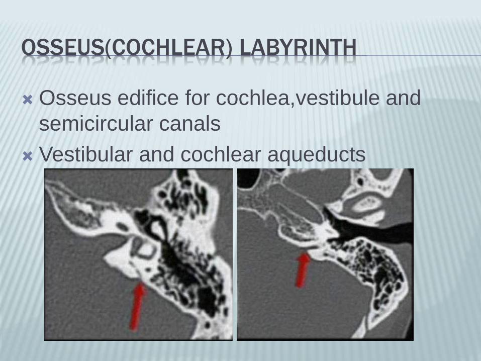
Osseus edifice for cochlea,vestibule and
semicircular canals
Vestibular and cochlear aqueducts
COCHLEA
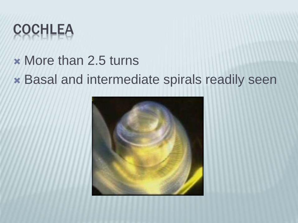
More than 2.5 turns
Basal and intermediate spirals readily seen

Basal turn opens post into round window
Cochlea encircles central osseus axis of
modiolus

SEMI CIRCULAR CANALS
Project off superior,posterior and lateral
aspects of vestibule
Upper margin of superior SCC forms
superior convexity on petrous pyramidal
roof(Arcuate eminence)
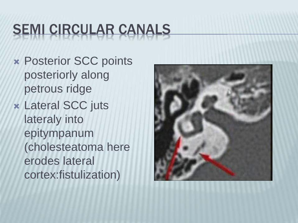
SEMI CIRCULAR CANALS
Posterior SCC points
posteriorly along
petrous ridge
Lateral SCC juts
lateraly into
epitympanum
(cholesteatoma here
erodes lateral
cortex:fistulization)

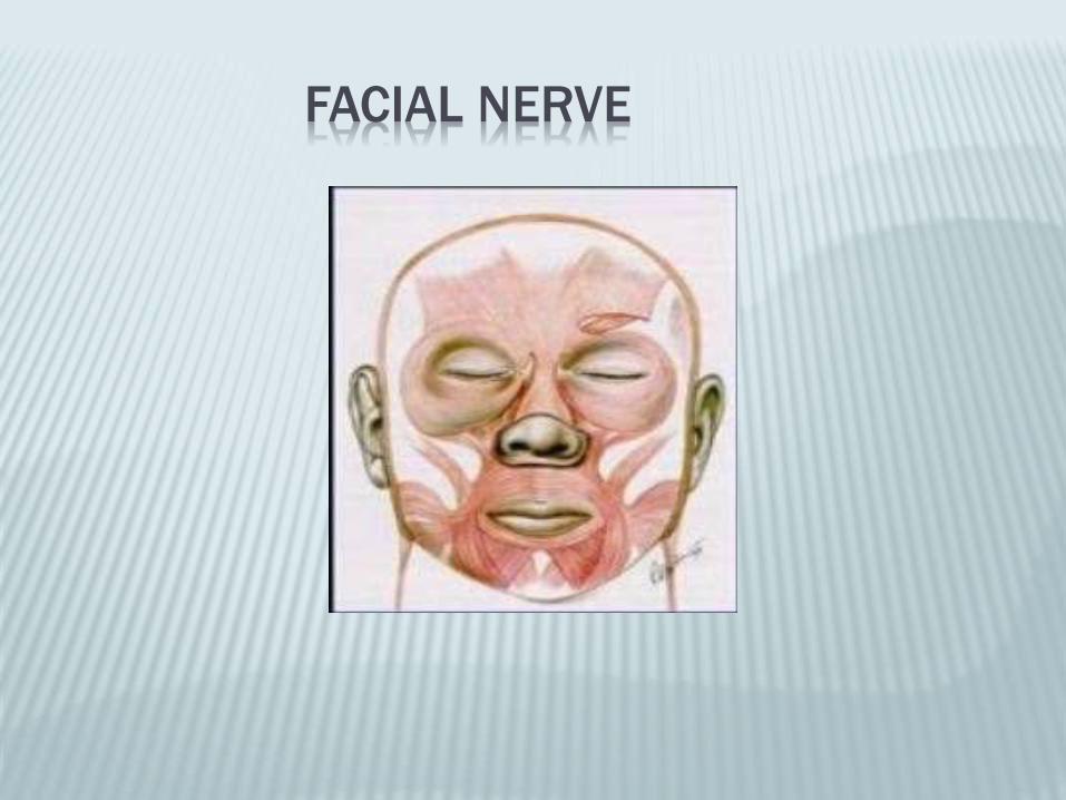
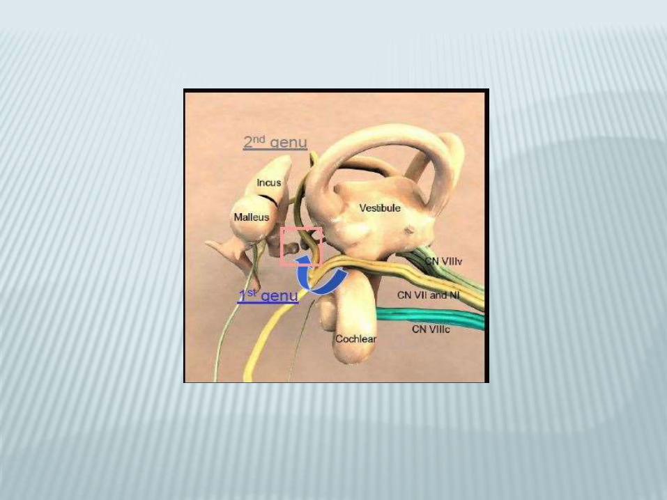
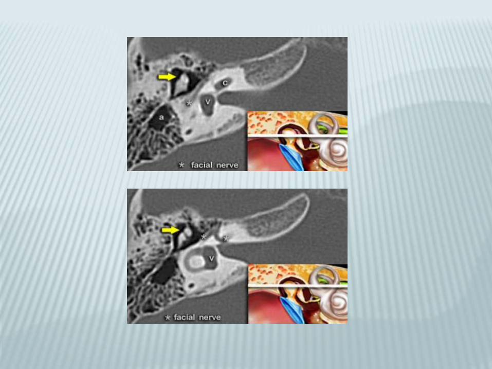
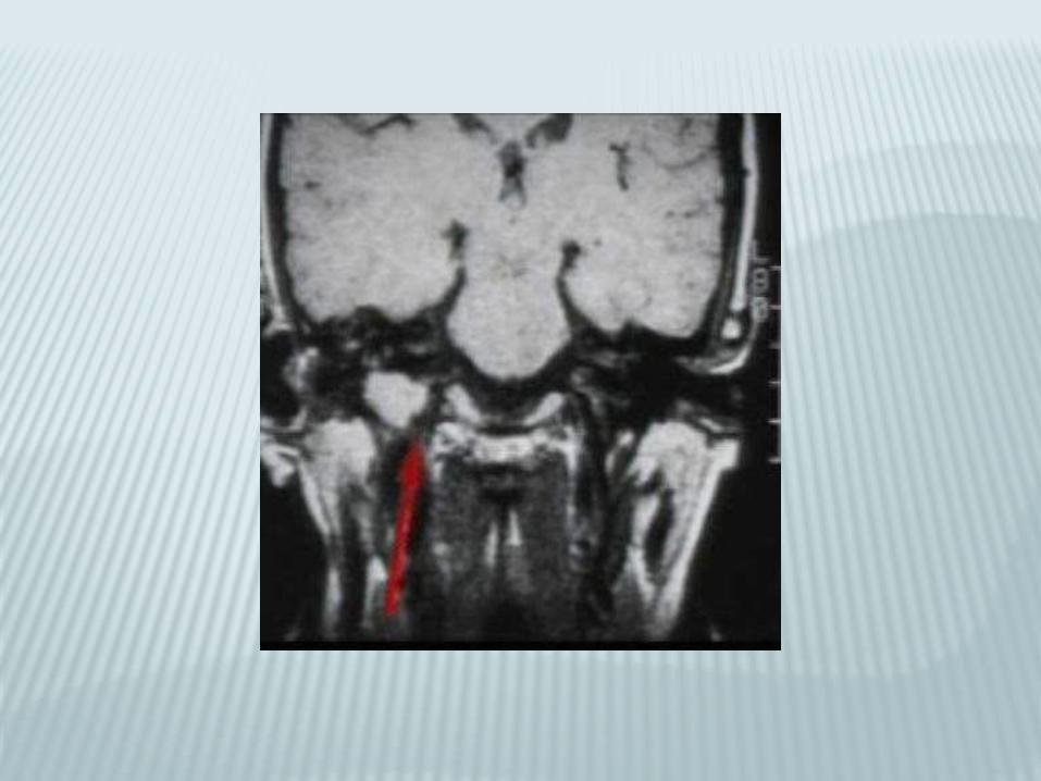
FACIAL NERVE

SEGMENTAL
TEMPORAL BONE ANALYSIS
Ear or anterior cerebellopontine sulcus Ear :outer,middle,inner
ACPA masses
Osseus destruction
Anatomic extent
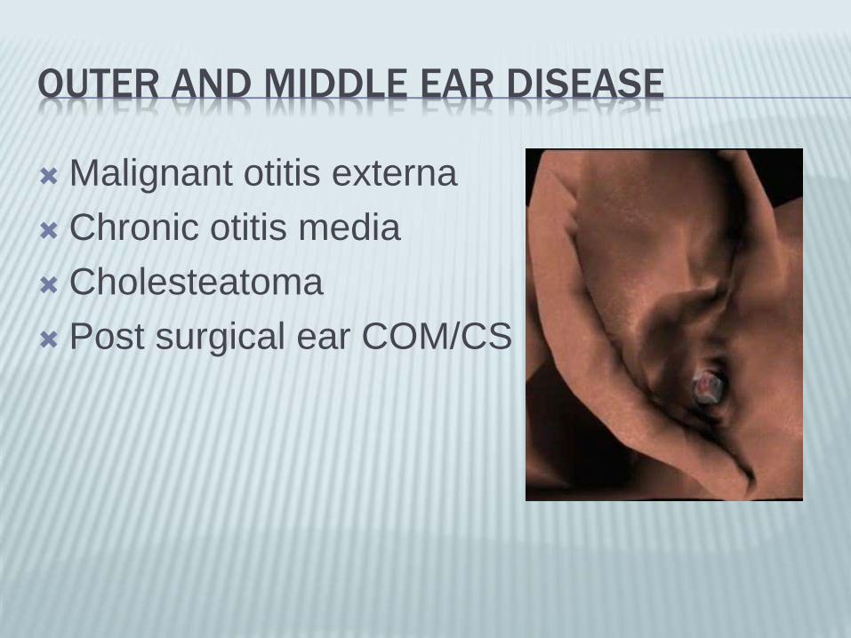
OUTER AND MIDDLE EAR DISEASE
Malignant otitis externa
Chronic otitis media
Cholesteatoma
Post surgical ear COM/CS
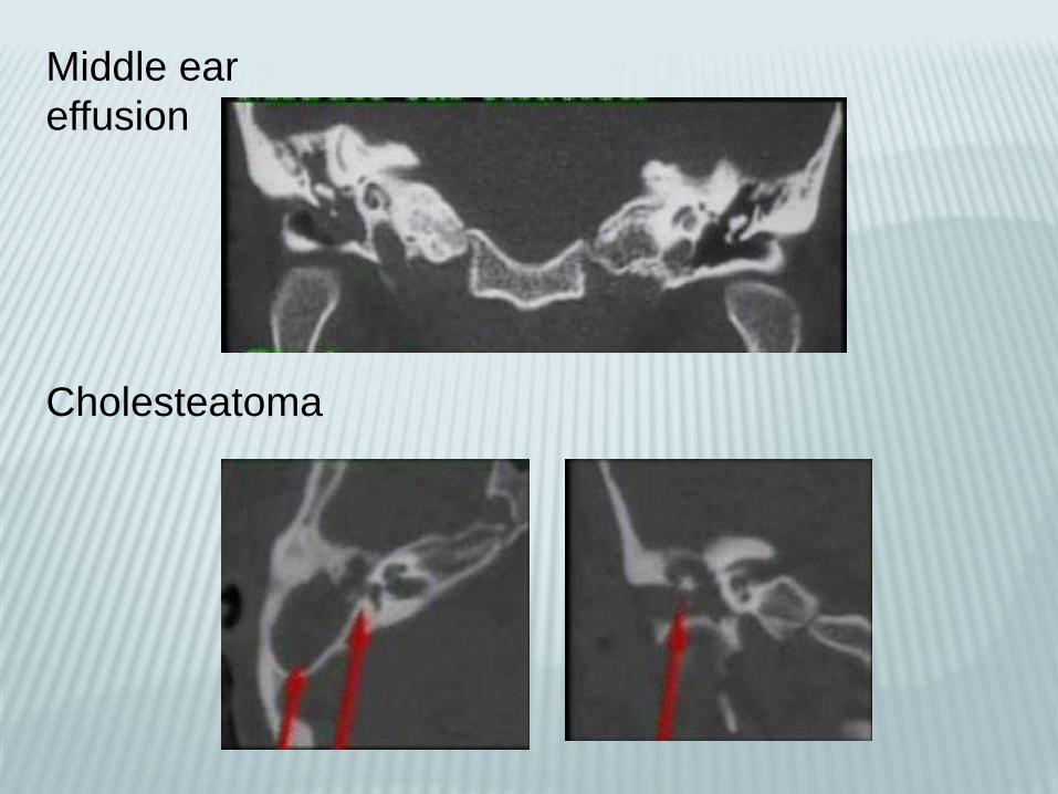
Middle ear
effusion
Cholesteatoma
ACPA masses
1. Neoplastic
•Schwanommas
•Meningeomas
•Epidermoid
2. Vascular• Vertebrobasilar dolichoectesias
• Glomus tumor
Symmetry
Patency
Outer ear canal
Middle ear cleft
Inner ear structures
Ossicular erosion
Aeration(mastoids,bones
Labyrinths
Nerves
Segmental Analysis

SEGMENTAL ANALYSIS
Outer ear canal
Middle ear cleft
Inner ear structures
Focal mass
•Destructive changes
•Ossicular inteagrity
•Focal enhancement
•Outer ear canal,Middle ear cleft,Inner
ear
Recommended