______________________________________________________________________ Guidelines for Maintenance Hemodialysis in India
STANDARD TREATMENT
GUIDELINES
HAEMODIALYSIS
Ministry of Health & Family Welfare
Govt. of India
______________________________________________________________________ Guidelines for Maintenance Hemodialysis in India
STANDARD OF CARE FOR
MAINTAINANCE
HEMODIALYSIS IN INDIA
Developed By
INDIAN SOCIETY OF NEPHROLOGY

______________________________________________________________________ Guidelines for Maintenance Hemodialysis in India
1.0 AUTHORS’DECLARATION
All the authors have no conflict of interest. The chapters were written at the request of Indian
Society of Nephrology. No remuneration was paid to the authors of the chapter. The chapters
were then given to Ms. Shachi Vyas, Medical Writer for converting the chapters into
standardized style without changing the original content.
Authors of this guideline were:
Sr. No Topic Author’s Signature& Date
1 Setting up of hemodialysis unit
for maintenance hemodialysis
Dr.Vijay Kher
Chairman
Deptt. of Nephrology &
Transplant Medicine
Medanta Kidney &
Urology Institute,
Medanta - The Medicity
Gurgaon,
Dr. Mohan Rajapurkar,
MD
Director, Postgraduate
Studies & Research,
Muljibhai Patel Urological
Hospital,
Nadiad
2 Personnel for hemodialysis unit
for maintenance hemodialysis
Dr. Abhijit Taraphder,
MD. DM
Chief Academics &
Research,
Senior Consultant,
Department of
Nephrology
Apollo Gleneagles
Hospital, Kolkata.
Dr. Dhananjay Ookalkar Consultant nephrologist
Ashwini Kidney & Dialysis
Centre,
Nagpur
3 Selection of Machine and
dialyser unit for maintenance
hemodialysis
Dr. Rajeev Annigeri
Consultant Nephrologist
Apollo Hospitals,
Chennai
4 Water treatment for hemodialysis
Dr. Mohan Rajapurkar, MD
Director, Postgraduate Studies & Research,
Muljibhai Patel Urological Hospital,
Nadiad
5 Vascular access
Dr. Valentine Lobo, MD.DNB.
Consultant Nephrologist
KEM Hospital, Pune

______________________________________________________________________ Guidelines for Maintenance Hemodialysis in India
6 Priming, connecting &
disconnecting dialyser
Dr. Amit Gupta
MD, DNB, FRCP
Professor of Nephrology,
SGPGIMS, Lucknow
7 Anti-coagulation in hemodialysis
Dr. Abhijit Taraphder,
MD. DM
Chief Academics &
Research,
Senior Consultant,
Department of
Nephrology
Apollo Gleneagles
Hospital, Kolkata.
Dr. Dhananjay Ookalkar Consultant nephrologist
Ashwini Kidney & Dialysis
Centre,
Nagpur
8 Dialyser reuse (Manual &
automated)
Dr. Valentine Lobo, MD.DNB.
Consultant Nephrologist
KEM Hospital, Pune
9 Dialysis dose/Adequacy
Dr. Vijay Kher
Chairman
Deptt. of Nephrology & Transplant Medicine
Medanta Kidney & Urology Institute, Medanta -
The Medicity
Gurgaon
10 Prevention of infections in
hemodialysis Unit
Dr. Vivekand Jha,MD,
DM
Secretary ISN
Additional Professor of
Nephrology
Postgraduate Institute of
Medical Education and
Research
Chandigarh
Dr. Amit Gupta
MD, DNB, FRCP
Professor of Nephrology,
SGPGIMS, Lucknow
11 Emergency services
Dr. Ravi Raju
President, ISN
Director of Medical Education
______________________________________________________________________ Guidelines for Maintenance Hemodialysis in India
Government of Andra Pradesh
Hyderabad
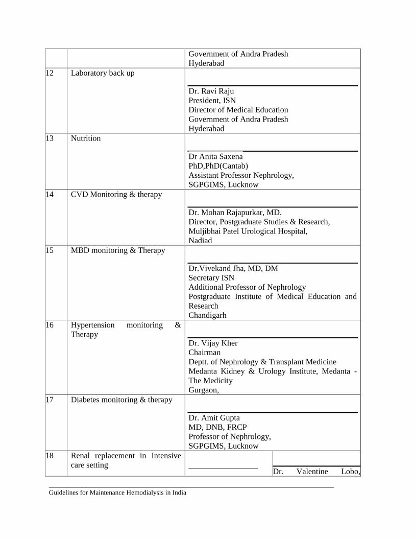
12 Laboratory back up
Dr. Ravi Raju
President, ISN
Director of Medical Education
Government of Andra Pradesh
Hyderabad
13 Nutrition
Dr Anita Saxena
PhD,PhD(Cantab)
Assistant Professor Nephrology,
SGPGIMS, Lucknow
14 CVD Monitoring & therapy
Dr. Mohan Rajapurkar, MD.
Director, Postgraduate Studies & Research,
Muljibhai Patel Urological Hospital,
Nadiad
15 MBD monitoring & Therapy
Dr.Vivekand Jha, MD, DM
Secretary ISN
Additional Professor of Nephrology
Postgraduate Institute of Medical Education and
Research
Chandigarh
16 Hypertension monitoring &
Therapy
Dr. Vijay Kher
Chairman
Deptt. of Nephrology & Transplant Medicine
Medanta Kidney & Urology Institute, Medanta -
The Medicity
Gurgaon,
17 Diabetes monitoring & therapy
Dr. Amit Gupta
MD, DNB, FRCP
Professor of Nephrology,
SGPGIMS, Lucknow
18 Renal replacement in Intensive
care setting
_________________
Dr. Valentine Lobo,
______________________________________________________________________ Guidelines for Maintenance Hemodialysis in India
Dr. Rajeev Annigeri
Consultant Nephrologist
Apollo Hospitals,
Chennai
MD.DNB.
Consultant Nephrologist
KEM Hospital, Pune
Please send feedback to [email protected], with a copy to [email protected]
______________________________________________________________________ Guidelines for Maintenance Hemodialysis in India
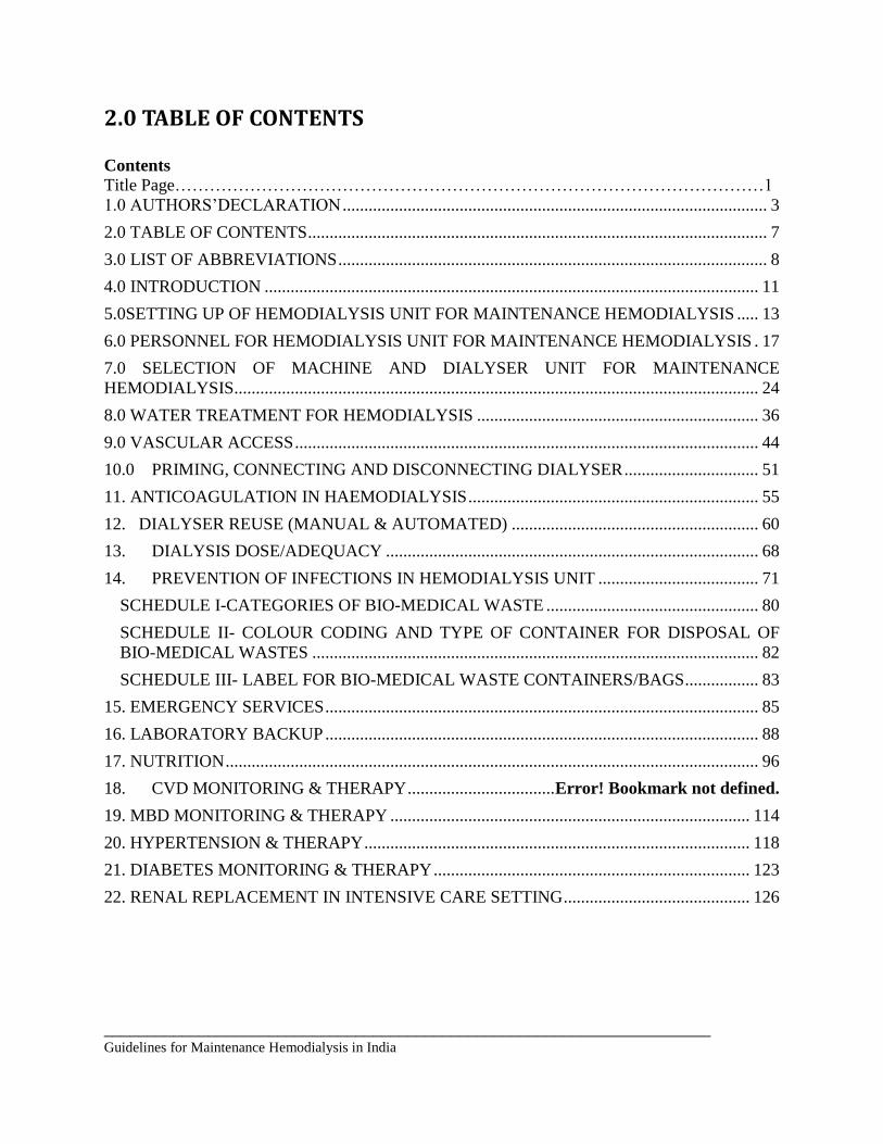
2.0 TABLE OF CONTENTS
Contents
Title Page…………………………………………………………………………………………1
1.0 AUTHORS’DECLARATION .................................................................................................. 3
2.0 TABLE OF CONTENTS .......................................................................................................... 7
3.0 LIST OF ABBREVIATIONS ................................................................................................... 8
4.0 INTRODUCTION .................................................................................................................. 11
5.0SETTING UP OF HEMODIALYSIS UNIT FOR MAINTENANCE HEMODIALYSIS ..... 13
6.0 PERSONNEL FOR HEMODIALYSIS UNIT FOR MAINTENANCE HEMODIALYSIS . 17
7.0 SELECTION OF MACHINE AND DIALYSER UNIT FOR MAINTENANCE
HEMODIALYSIS......................................................................................................................... 24
8.0 WATER TREATMENT FOR HEMODIALYSIS ................................................................. 36
9.0 VASCULAR ACCESS ........................................................................................................... 44
10.0 PRIMING, CONNECTING AND DISCONNECTING DIALYSER ............................... 51
11. ANTICOAGULATION IN HAEMODIALYSIS ................................................................... 55
12. DIALYSER REUSE (MANUAL & AUTOMATED) ......................................................... 60
13. DIALYSIS DOSE/ADEQUACY ...................................................................................... 68
14. PREVENTION OF INFECTIONS IN HEMODIALYSIS UNIT ..................................... 71
SCHEDULE I-CATEGORIES OF BIO-MEDICAL WASTE ................................................. 80
SCHEDULE II- COLOUR CODING AND TYPE OF CONTAINER FOR DISPOSAL OF
BIO-MEDICAL WASTES ....................................................................................................... 82
SCHEDULE III- LABEL FOR BIO-MEDICAL WASTE CONTAINERS/BAGS ................. 83
15. EMERGENCY SERVICES .................................................................................................... 85
16. LABORATORY BACKUP .................................................................................................... 88
17. NUTRITION ........................................................................................................................... 96
18. CVD MONITORING & THERAPY .................................. Error! Bookmark not defined.
19. MBD MONITORING & THERAPY ................................................................................... 114
20. HYPERTENSION & THERAPY ......................................................................................... 118
21. DIABETES MONITORING & THERAPY ......................................................................... 123
22. RENAL REPLACEMENT IN INTENSIVE CARE SETTING ........................................... 126
______________________________________________________________________ Guidelines for Maintenance Hemodialysis in India
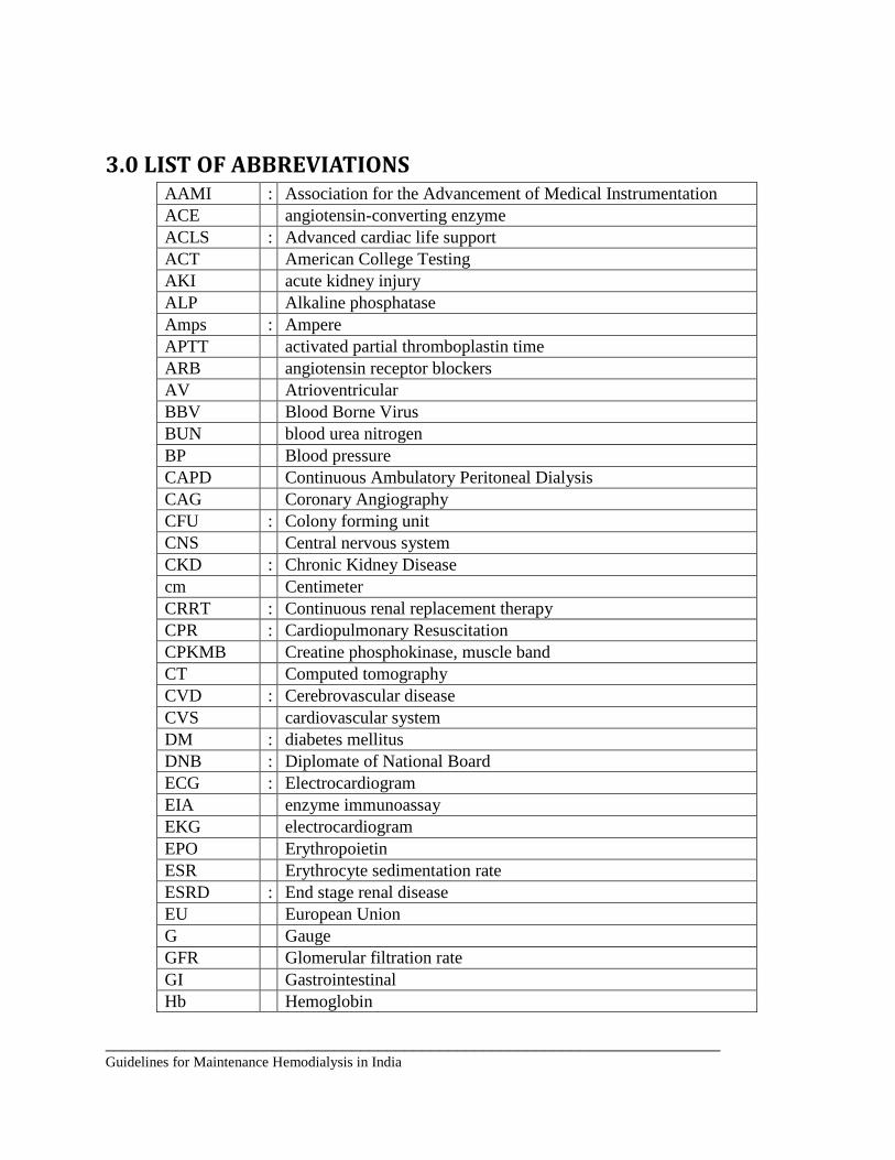
3.0 LIST OF ABBREVIATIONS AAMI : Association for the Advancement of Medical Instrumentation
ACE angiotensin-converting enzyme
ACLS : Advanced cardiac life support
ACT American College Testing
AKI acute kidney injury
ALP Alkaline phosphatase
Amps : Ampere
APTT activated partial thromboplastin time
ARB angiotensin receptor blockers
AV Atrioventricular
BBV Blood Borne Virus
BUN blood urea nitrogen
BP Blood pressure
CAPD Continuous Ambulatory Peritoneal Dialysis
CAG Coronary Angiography
CFU : Colony forming unit
CNS Central nervous system
CKD : Chronic Kidney Disease
cm Centimeter
CRRT : Continuous renal replacement therapy
CPR : Cardiopulmonary Resuscitation
CPKMB Creatine phosphokinase, muscle band
CT Computed tomography
CVD : Cerebrovascular disease
CVS cardiovascular system
DM : diabetes mellitus
DNB : Diplomate of National Board
ECG : Electrocardiogram
EIA enzyme immunoassay
EKG electrocardiogram
EPO Erythropoietin
ESR Erythrocyte sedimentation rate
ESRD : End stage renal disease
EU European Union
G Gauge
GFR Glomerular filtration rate
GI Gastrointestinal
Hb Hemoglobin

______________________________________________________________________ Guidelines for Maintenance Hemodialysis in India
HbA1C Glycosylated hemoglobin (HbA1c or HbA1c)
HBV : Hepatitis B virus
HCV : Hepatitis C virus
HD : Hemodialysis
HIT Heparin-Induced Thrombocytopenia
HIV Human immunodeficiency virus
Hr Hour
ICU Intensive care unit
IU/kg International unit/kilogram
IV : Intravenous
IVC Inferior vena cava
Kg/cm2 Kilogram/square centimeter
LAL Limulus Amebocyte Lysate
LDH Lactate dehydrogenase
LPS Lipopolysaccharide
LV Left ventricular
LVF Left ventricular failure
LVH Left ventricular hypertrophy
M.B.B.S : Bachelor of Medicine, Bachelor of Surgery degree
MBD : Mineral and Bone Disorder
MD : Doctor of Medicine
Mg/l milligrams per litre
MHD : Maintenance hemodialysis
MI Myocardial infarction
MIBI Methoxyisobutyl Isonitrile Stress
ml/min Milliliter per minute
mmol/L Milli moler per liter
mmHg millimetres of mercury
µ Micron
N Normal
NAT nucleic acid test
NKF National Kidney Foundation
PCR protein catabolic rate
pH potential of hydrogen
Psi per square inch
PTFE Polytetrafluoroethylene
PTH Parathyroid hormone
PVC : Polyvinylchloride
PT Prothrombin
R2A R2A Agar
RO Reversed osmosis
RRT Renal Replacement Therapy
______________________________________________________________________ Guidelines for Maintenance Hemodialysis in India
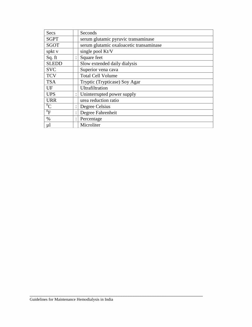
Secs Seconds
SGPT serum glutamic pyruvic transaminase
SGOT serum glutamic oxaloacetic transaminase
spkt v single pool Kt/V
Sq. ft : Square feet
SLEDD Slow extended daily dialysis
SVC Superior vena cava
TCV Total Cell Volume
TSA Tryptic (Trypticase) Soy Agar
UF Ultrafiltration
UPS : Uninterrupted power supply
URR urea reduction ratio 0C
: Degree Celsius 0F : Degree Fahrenheit
% : Percentage
µl Microliter

______________________________________________________________________ Guidelines for Maintenance Hemodialysis in India
4.0 INTRODUCTION
The burden of Chronic Kidney Disease (CKD) is increasing in alarming proportion all over the
world. In India due to lack of financial resources, lack of trained manpower & infrastructure
leads to severe strain on existing health policies in the light of the increasing burden of CKD.
Kidneys are probably the only vital organs which can be realistically replaced by artificial
means. Maintenance dialysis is a well-recognized modality of treating patients having end stage
renal disease. Several thousands of patients all over the world are surviving and achieving
reasonable quality of life on maintenance dialysis. The exact burden of CKD needing
maintenance dialysis and/ or renal transplantation is not known; however, from the existing
published data prevalence of CKD ranges between 0.7% to 1.4%. Whereas the incidence of end
stage renal disease was estimated to be 180 to 200 per million populations 1.
World over there is severe shortage of donor kidneys. In our country deceased donor
transplantation Programme is as yet in its infancy and because of breaking up of joint family
structure the live donor programme is not enough for the needs of ESRD patients. Therefore
several thousands of patients have to live on maintenance dialysis in India. Maintenance dialysis
importantly serves as a bridge to kidney transplantation.
In India the first Hemodialysis facility was established in 1961 2 at the CMC Vellore; soon it was
started at 3-4 major centers during that decade viz. CMC Vellore, KEM Mumbai, PGI
Chandigarh & All India Institute of Medical Sciences, New Delhi. Over the last three decades
many more dialysis facilities have been established in Government sector, in Charitable Trust
run institutions & by Private Nephrologist. There has been persistent improvement in the
numbers & quality of dialysis delivered over this period.
Good quality of life and survival on maintenance dialysis depends on following major factors
namely; i) The dose of dialysis delivered or solute removal achieved, ii) Time on dialysis, iii)
Adequacy of nutrition, iv) Family and socio-economic support, v) management of co-morbid
illnesses and vi) Prevention & management of infections.
Unfortunately in our country the quality of dialysis delivered to patients can vary from center to
center. The quality could range from very poor to as good as any center in world. This is because
there are no minimum defined standards of care of maintenance dialysis. The Indian Society of
Nephrology therefore set up core group to write these minimum standards that should be
followed in our country. It was realized by this group very early that if we make these standards
very stringent comparable to best centers in the world then it would be practically impossible to
follow by majority of dialysis units. However, for the practicality if we compromise on the
principles of good dialysis our patients will suffer. The general aim of these standards is
therefore to improve the general quality of dialysis delivered to our patients. Various members of
this group were asked to write on following topics/chapters: i) setting up of hemodialysis unit for
maintenance hemodialysis, ii) Personnel for hemodialysis unit for maintenance hemodialysis, iii)
Selection of Machine and dialyzer unit for maintenance hemodialysis, iv) Water treatment for
hemodialysis, v) Vascular access, vi) Priming, connecting & disconnecting dialyzer, vii) Anti-
coagulation for hemodialysis, viii) Dialyzer reuse (Manual & automated), ix) Dialysis
______________________________________________________________________ Guidelines for Maintenance Hemodialysis in India
dose/Adequacy, x) Prevention of infections in hemodialysis Unit, xi) Emergency services, xii)
Laboratory back up and xiii) Nutrition.
First manuscripts were then discussed & debated by the group collectively in meetings and on
emails. We have taken help from Ms. Shachi Vyas, a professional medical writer so that all
chapters would have uniform style but no change in the content was done. During the annual
conference of the Indian Society of Nephrology(ISN) held at Hyderabad; Dr.Vivekanand Jha had
declared that the document would be placed for public review on ISN website: www.isn-
india.org. These will be available on the ISN website for comments by the membership and after
incorporating suggestions these would be published as a supplement of the Indian Journal of
Nephrology. We hope that health authorities adopt these as minimum standard of care for
hemodialysis in our country.
1. Rajapurkar M and Dabhi M. Burden of disease – prevalence and incidence
of renal disease in India Clinical Nephrology,2010; 74 – Suppl.1(S9-S12)
2. Chugh K S Three decades of nephrology J Postgrad Med 1994;40:103-108

______________________________________________________________________ Guidelines for Maintenance Hemodialysis in India- Setting up of hemodialysis unit for maintenance hemodialysis
5.0SETTING UP OF HEMODIALYSIS UNIT FOR MAINTENANCE HEMODIALYSIS
Setting up of maintenance hemodialysis (MHD) unit could be a major challenge for a
nephrologist. The purpose of this guideline is to help design a new unit.
Rationale: The majority of patients in India who receive renal replacement therapy take in center
hemodialysis. The number of patients on Hemodialysis and the number of hospital based and
free standing units is steadily growing. A dialysis unit delivers patient care, and has specific
requirements of treated water, electricity, medical gases and plumbing for waste disposal. It
additionally requires to accommodate all the workers involved in patient care, allow emergency
and planned procedures, permit adequate hygiene and maintenance of specialized equipment.
The design and layout of a unit must take into account all the above features in order to function
smoothly and prevent development of complications. Proper planning of a dialysis unit prior to
construction is essential, and the following document aims to provide planners with the
necessary expertise.
It is recommended that Hemodialysis unit has the following facilities underlined in the text as
below
1. Hemodialysis area
We recommend that the hemodialysis area should have the following features
Each machine requires at least 11 x 10 ft. (100 to110 square feet)(figure 1).This is needed
because in case of an emergency, cardiac resuscitation equipment could be easily
wheeled on all four sides of the patient. Facilities for non-invasive blood pressure
monitoring of all patients and ECG monitoring of select patients are needed.
Each machine area should be easily observed from the nursing station which should be
included in this area.
Nursing station should have enough space for adequate number of nurses/technicians
depending on the number of dialysis machines (See chapter on personnel required for
hemodialysis), a computer terminal & working desk/bench.
Head end of each bed should have stable electrical supply (at least 3 outlet of 5/15 amps),
oxygen &vacuum outlet, treated water inlet & drainage facilities.
Air conditioning is strongly recommended to achieve 700
to 720 F temperatures & 55 to
60% humidity.
Areas for dialyzing patients having viral diseases (HBV/HCV) should be separated from
those patients not having any viral infections. These spaces should have independent
drainage, independent water supply, independent air handling & separate personnel
facilities.
Facilities for hand washing and Sterillium®
Or alcohol based hand rub/sterilent dispensers
should be available in each patient area.
2. Preparation, work & storage area
______________________________________________________________________ Guidelines for Maintenance Hemodialysis in India- Setting up of hemodialysis unit for maintenance hemodialysis
We recommend that the Preparation, work & storage area should have the following
features
Independent area is needed for reprocessing the dialyzers. This should have a work bench
with sink having side board & drainage. The work bench should be supplied with treated
as well as untreated water which are separately marked. Two sinks for the work bench
should be provided. The space should be sufficient for at least two persons working
simultaneously.
This preparation area should be physically separate for processing dialyzers from viral
infection patients versus those not having any viral infection.
For both areas stable electrical supply & drainage is needed for the work bench. There
should be space for dialyzer reprocessing machine(s) in this area.
1. There should be two storage areas, one for storage of new supplies and one for
reprocessed dialyzers.
The principle of dry storage area is to be able to store 3months supply of dialyzers,
tubings, hemodialysis concentrate solutions, IV fluids. It should also have space for
stationery, linen etc.
The wet storage is for reprocessed dialyzers & tubings.
The dry storage area should be separate from the wet storage.
2. A clean room with a work bench is needed for preparation of sterile trays for dialysis
startup kit & for preparation of injections & storage of emergency equipment.
3. This area should have a designated place for keeping wheelchair /trolleys for transporting
patients & weighing scale.
4. There should be an area for dirty utility. This area should be located in such a way that
personnel and material need not come from dirty utility to clean area of dialysis.
3. We recommend that there should be a consulting room for the doctor in-charge of the unit.
4. We recommend that there should be office area for nurses & technicians
5. Each patient is generally accompanied by two individuals; hence, we recommend a specially
designed area for their stay and some relaxation should be provided. Patients waiting to go
on dialysis & those who have recently completed dialysis could also utilize the same area.
6. We recommend that storage facility should be provided for individual patients belongings.
7. We recommend that there should be space for a water treatment unit1
8. We recommend that a Procedure room / operating room is required.
Equipment required at this place would be
a. Operating table
b. C-Arm imaging system
c. Ultrasound; preferably with a vascular probe for localizing & puncturing central
veins
d. Instrument storage facility
e. Clean & dirty utility.
This facility in general hospital could be shared but it should primarily be under control of
dialysis staff.
9. We recommend that there should be change rooms for male & female staff.
1Please refer Topic 5 Water treatment for hemodialysis

______________________________________________________________________ Guidelines for Maintenance Hemodialysis in India- Setting up of hemodialysis unit for maintenance hemodialysis
10. We recommend that there should be adequate toilets for consultants, technicians, patient &
patients’ relatives. Separate for men & women.
General Conditions
The general conditions should have the following
Air conditioning: All hemodialysis machine areas, consultants & technicians/nurses
rooms should have air conditioning. Treatment areas should have temperature700
to 720 F &
55 to 60% humidity.
Relative’s waiting / recreation area & reception should be well ventilated with fans or may
have air conditioning.
Electricity: Stable voltage continuous supply is required. Online UPS is recommended. It
should have a backup for at least 30 minutes. The power capacity of the UPS should be able
to support all functions of the dialysis machine2.
The electrical supply should be stable & uninterrupted, preferably a pure sine wave both
voltage and frequency regulated. The use of electrical surge protectors is necessary to protect
dialysis machine’s electronics.
Adequate capacity generator is recommended.
Plumbing & drainage:
All treated water pipelines should be stainless steel grade 316 or medical grade PVC. There
should be minimum bends & blind loops should be avoided.
All drainage should be connected directly to the main drainage line. There should be no
bends or blind loops.
There should be oxygen & vacuum outlets at the head end of each dialysis machine.
The ambience should be cheerful looking in terms of color used. It should be brightly lit so
that examination of patient or if procedures are required there is no difficulty. There should
be a facility to dim the lighting.
The system for record keeping should be preferably electronic for patient as well as unit
records. Depending on the authority & function of the person using it, the records system
should be accessible by username and password protection. There should be protection of
privacy of the patients.
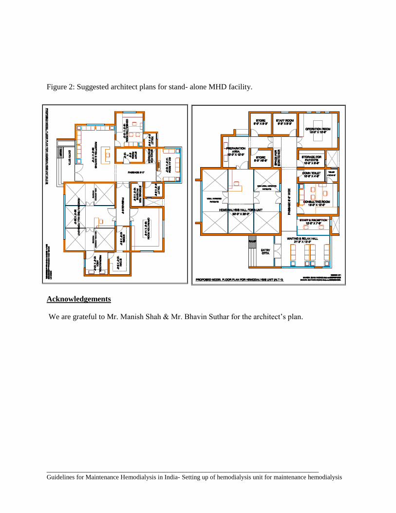
Please see figure 2 for architects plan for a stand-alone MHD unit. If the MHD unit is a part of a
general hospital set up then the areas for reception, waiting, records, consulting room and storage
could be shared.
Figure 1 : Showing typical hemodialysis machine
area:
Black dots Electricity outlets
Green dot : Oxygen outlet
Yellow dot: Vacuum outlet
Blue dot: treated water inlet
Ash dot: drainage outlet
2Please refer manufacturer’s manual for each machine
______________________________________________________________________ Guidelines for Maintenance Hemodialysis in India- Setting up of hemodialysis unit for maintenance hemodialysis
Figure 2: Suggested architect plans for stand- alone MHD facility.
Acknowledgements
We are grateful to Mr. Manish Shah & Mr. Bhavin Suthar for the architect’s plan.
______________________________________________________________________ Guidelines for Maintenance Hemodialysis in India- Personnel for hemodialysis unit for maintenance hemodialysis
6.0 PERSONNEL FOR HEMODIALYSIS UNIT FOR MAINTENANCE HEMODIALYSIS
We recommend that the hemodialysis facility should have sufficient specialist and support staff.
Rationale: The delivery of hemodialysis is carried out by both hospital based and freestanding
units. A hemodialysis unit is involved with patient care, record keeping, disposal of potentially
infectious and biohazardous and environmentally unfriendly waste. As standards of care continue
to change and the personnel in dialysis units are not constant, it becomes necessary for them to
participate in ongoing education in a unit. The responsibility for training of staff, maintaining
patient safety, efficacy of complete patient treatment, auditing performance and record
maintenance requires special skills in different disciplines. The following document outline the
job description, responsibilities and competence level required by the personnel responsible for
all the aspects of running a dialysis unit.
Proposed Minimum standards for personnel in Dialysis Facility
1. OUTLINE: It is recommended to have the following minimum staff-pattern for a proposed
dialysis unit:
a) Nephrologist
b) Dialysis doctors
c) Dialysis technicians
d) Dialysis nurses
e) Dialysis attendants
f) Medical social worker
g) Dietician (Optional)
h) Sweepers
2. PARTICULARS: Each category (A to D) of staff and the medical social worker & dietician
should satisfy the following:
a) Training
b) Job description / responsibilities (Dos and Don’ts)
c) Appraisal / Auditing
d) Updating
______________________________________________________________________ Guidelines for Maintenance Hemodialysis in India- Personnel for hemodialysis unit for maintenance hemodialysis
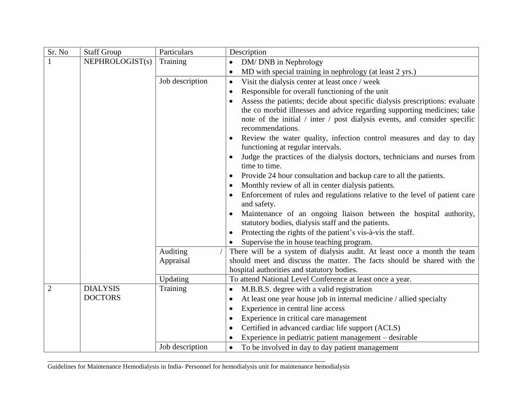
Sr. No Staff Group Particulars Description
1 NEPHROLOGIST(s) Training DM/ DNB in Nephrology
MD with special training in nephrology (at least 2 yrs.)
Job description
Visit the dialysis center at least once / week
Responsible for overall functioning of the unit
Assess the patients; decide about specific dialysis prescriptions: evaluate
the co morbid illnesses and advice regarding supporting medicines; take
note of the initial / inter / post dialysis events, and consider specific
recommendations.
Review the water quality, infection control measures and day to day
functioning at regular intervals.
Judge the practices of the dialysis doctors, technicians and nurses from
time to time.
Provide 24 hour consultation and backup care to all the patients.
Monthly review of all in center dialysis patients.
Enforcement of rules and regulations relative to the level of patient care
and safety.
Maintenance of an ongoing liaison between the hospital authority,
statutory bodies, dialysis staff and the patients.
Protecting the rights of the patient’s vis-à-vis the staff.
Supervise the in house teaching program.
Auditing /
Appraisal
There will be a system of dialysis audit. At least once a month the team
should meet and discuss the matter. The facts should be shared with the
hospital authorities and statutory bodies.
Updating To attend National Level Conference at least once a year.
2
DIALYSIS
DOCTORS
Training M.B.B.S. degree with a valid registration
At least one year house job in internal medicine / allied specialty
Experience in central line access
Experience in critical care management
Certified in advanced cardiac life support (ACLS)
Experience in pediatric patient management – desirable
Job description To be involved in day to day patient management
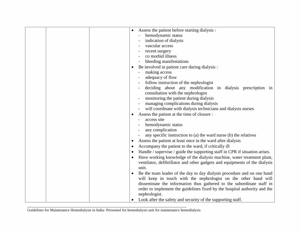
______________________________________________________________________ Guidelines for Maintenance Hemodialysis in India- Personnel for hemodialysis unit for maintenance hemodialysis
Assess the patient before starting dialysis :
- hemodynamic status
- indication of dialysis
- vascular access
- recent surgery
- co morbid illness
- bleeding manifestations
Be involved in patient care during dialysis :
- making access
- adequacy of flow
- follow instruction of the nephrologist
- deciding about any modification in dialysis prescription in
consultation with the nephrologist
- monitoring the patient during dialysis
- managing complications during dialysis
- will coordinate with dialysis technicians and dialysis nurses
Assess the patient at the time of closure :
- access site
- hemodynamic status
- any complication
- any specific instruction to (a) the ward nurse (b) the relatives
Assess the patient at least once in the ward after dialysis
Accompany the patient to the ward, if critically ill
Handle / supervise / guide the supporting staff in CPR if situation arises.
Have working knowledge of the dialysis machine, water treatment plant,
ventilator, defibrillator and other gadgets and equipments of the dialysis
unit.
Be the team leader of the day to day dialysis procedure and on one hand
will keep in touch with the nephrologist on the other hand will
disseminate the information thus gathered to the subordinate staff in
order to implement the guidelines fixed by the hospital authority and the
nephrologist.
Look after the safety and security of the supporting staff.
______________________________________________________________________ Guidelines for Maintenance Hemodialysis in India- Personnel for hemodialysis unit for maintenance hemodialysis
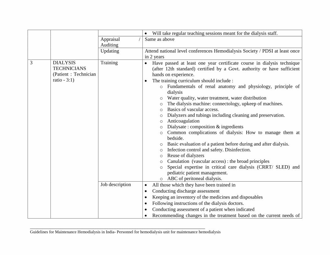
Will take regular teaching sessions meant for the dialysis staff.
Appraisal /
Auditing
Same as above
Updating Attend national level conferences Hemodialysis Society / PDSI at least once
in 2 years
3 DIALYSIS
TECHNICIANS
(Patient : Technician
ratio - 3:1)
Training Have passed at least one year certificate course in dialysis technique
(after 12th standard) certified by a Govt. authority or have sufficient
hands on experience.
The training curriculum should include :
o Fundamentals of renal anatomy and physiology, principle of
dialysis
o Water quality, water treatment, water distribution
o The dialysis machine: connectology, upkeep of machines.
o Basics of vascular access.
o Dialyzers and tubings including cleaning and preservation.
o Anticoagulation
o Dialysate : composition & ingredients
o Common complications of dialysis: How to manage them at
bedside.
o Basic evaluation of a patient before during and after dialysis.
o Infection control and safety. Disinfection.
o Reuse of dialyzers
o Canulation (vascular access) : the broad principles
o Special expertise in critical care dialysis (CRRT/ SLED) and
pediatric patient management.
o ABC of peritoneal dialysis.
Job description All those which they have been trained in
Conducting discharge assessment
Keeping an inventory of the medicines and disposables
Following instructions of the dialysis doctors.
Conducting assessment of a patient when indicated
Recommending changes in the treatment based on the current needs of
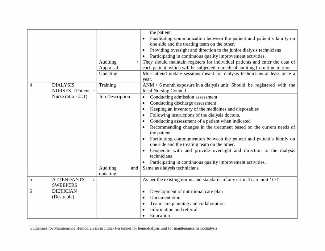
______________________________________________________________________ Guidelines for Maintenance Hemodialysis in India- Personnel for hemodialysis unit for maintenance hemodialysis
the patient
Facilitating communication between the patient and patient’s family on
one side and the treating team on the other.
Providing oversight and direction to the junior dialysis technicians
Participating in continuous quality improvement activities.
Auditing /
Appraisal
They should maintain registers for individual patients and enter the data of
each patient, which will be subjected to medical auditing from time to time.
Updating Must attend update sessions meant for dialysis technicians at least once a
year.
4 DIALYSIS
NURSES (Patient :
Nurse ratio - 3 :1)
Training ANM + 6 month exposure in a dialysis unit. Should be registered with the
local Nursing Council.
Job Description Conducting admission assessment
Conducting discharge assessment
Keeping an inventory of the medicines and disposables
Following instructions of the dialysis doctors.
Conducting assessment of a patient when indicated
Recommending changes in the treatment based on the current needs of
the patient
Facilitating communication between the patient and patient’s family on
one side and the treating team on the other.
Cooperate with and provide oversight and direction to the dialysis
technicians
Participating in continuous quality improvement activities.
Auditing and
updating
Same as dialysis technicians
5 ATTENDANTS /
SWEEPERS
As per the existing norms and standards of any critical care unit / OT
6 DIETICIAN
(Desirable)
Development of nutritional care plan
Documentation
Team care planning and collaboration
Information and referral
Education
______________________________________________________________________ Guidelines for Maintenance Hemodialysis in India- Personnel for hemodialysis unit for maintenance hemodialysis
Hospital facility planning activities
Ongoing nutrition assessment
Research
Medical review audit activities
Supervisory
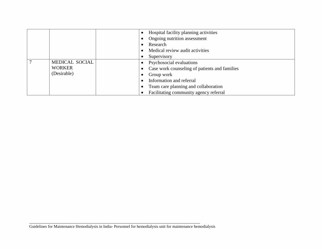
7 MEDICAL SOCIAL
WORKER
(Desirable)
Psychosocial evaluations
Case work counseling of patients and families
Group work
Information and referral
Team care planning and collaboration
Facilitating community agency referral

______________________________________________________________________ Guidelines for Maintenance Hemodialysis in India- Personnel for hemodialysis unit for maintenanc
Proposed Standard
Professional health care personnel shall be licensed or certified by appropriate
authority.
Adequate number of personnel shall be present so that patient staff ratio is
appropriate to the level of care being given and meets the needs of the patients.
The facility shall comply with all local, state and central regulations regarding
employment.
A formal system of staff evaluation and monitoring shall be established with
performance evaluation taking place at least annually.
Personnel records shall be maintained for all employees.
The organization shall have a well-defined organizational chart.
The organization shall have a well-documented disciplinary procedure
A grievance handling mechanism should exist in the organization.
The organization should address the health needs of the employees.
There has to be a process for collecting, verifying and evaluating the credentials of
medical professionals and other staff.
Suggested Personnel Records
A personnel record for each staff member of a facility shall include an application for
employment and a record of any disciplinary action taken.
Wage and salary information, time records, an authorization and record of leave shall
be maintained but may be kept in a separate location.
A job description shall be maintained which includes the employment requirements
and the job responsibilities for each facility staff position.
A personnel record shall be maintained which verifies that each employee meets the
respective employment requirements for the staff position held, including annual
verification of basic skills and annual evaluation of personnel performance. This
evaluation shall be in writing. There shall be documentation to verify that the
employee has reviewed the evaluation and has had an opportunity to comment on it.
Training and development activities which are appropriate in assisting the staff in
meeting the needs of the patients being served shall be provided for each staff
member including HIV and other communicable disease education. The provision of
such activities shall be evidenced by documentation in the facility records.
Direct services staff members shall be competent persons aged eighteen (18) years of
age or older.
All new employees, including volunteers, who have routine contact with patients
shall have a current health check-up and hepatitis B vaccination status prior to
employment or service. Complete hepatitis B vaccination after employment.
______________________________________________________________________ Guidelines for Maintenance Hemodialysis in India- Selection of Machine and dialyser unit for maintenance
hemodialysis
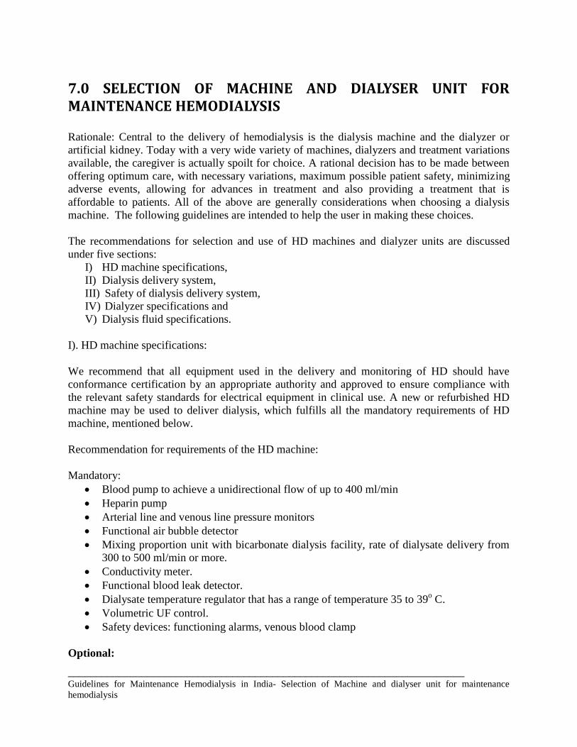
7.0 SELECTION OF MACHINE AND DIALYSER UNIT FOR MAINTENANCE HEMODIALYSIS
Rationale: Central to the delivery of hemodialysis is the dialysis machine and the dialyzer or
artificial kidney. Today with a very wide variety of machines, dialyzers and treatment variations
available, the caregiver is actually spoilt for choice. A rational decision has to be made between
offering optimum care, with necessary variations, maximum possible patient safety, minimizing
adverse events, allowing for advances in treatment and also providing a treatment that is
affordable to patients. All of the above are generally considerations when choosing a dialysis
machine. The following guidelines are intended to help the user in making these choices.
The recommendations for selection and use of HD machines and dialyzer units are discussed
under five sections:
I) HD machine specifications,
II) Dialysis delivery system,
III) Safety of dialysis delivery system,
IV) Dialyzer specifications and
V) Dialysis fluid specifications.
I). HD machine specifications:
We recommend that all equipment used in the delivery and monitoring of HD should have
conformance certification by an appropriate authority and approved to ensure compliance with
the relevant safety standards for electrical equipment in clinical use. A new or refurbished HD
machine may be used to deliver dialysis, which fulfills all the mandatory requirements of HD
machine, mentioned below.
Recommendation for requirements of the HD machine:
Mandatory:
Blood pump to achieve a unidirectional flow of up to 400 ml/min
Heparin pump
Arterial line and venous line pressure monitors
Functional air bubble detector
Mixing proportion unit with bicarbonate dialysis facility, rate of dialysate delivery from
300 to 500 ml/min or more.
Conductivity meter.
Functional blood leak detector.
Dialysate temperature regulator that has a range of temperature 35 to 39o C.
Volumetric UF control.
Safety devices: functioning alarms, venous blood clamp
Optional:
______________________________________________________________________ Guidelines for Maintenance Hemodialysis in India- Selection of Machine and dialyser unit for maintenance
hemodialysis
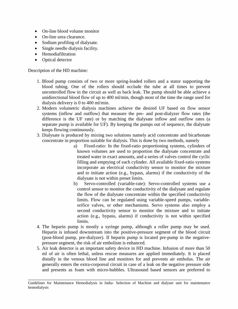
On-line blood volume monitor
On-line urea clearance.
Sodium profiling of dialysate.
Single needle dialysis facility.
Hemodiafiltration
Optical detector
Description of the HD machine:
1. Blood pump consists of two or more spring-loaded rollers and a stator supporting the
blood tubing. One of the rollers should occlude the tube at all times to prevent
uncontrolled flow in the circuit as well as back leak. The pump should be able achieve a
unidirectional blood flow of up to 400 ml/min, though most of the time the range used for
dialysis delivery is 0 to 400 ml/min.
2. Modern volumetric dialysis machines achieve the desired UF based on flow sensor
systems (inflow and outflow) that measure the pre- and post-dialyzer flow rates (the
difference is the UF rate) or by matching the dialysate inflow and outflow rates (a
separate pump is available for UF). By keeping the pumps out of sequence, the dialysate
keeps flowing continuously.
3. Dialysate is produced by mixing two solutions namely acid concentrate and bicarbonate
concentrate in proportion suitable for dialysis. This is done by two methods, namely
a) Fixed-ratio: In the fixed-ratio proportioning systems, cylinders of
known volumes are used to proportion the dialysate concentrate and
treated water in exact amounts, and a series of valves control the cyclic
filling and emptying of each cylinder. All available fixed-ratio systems
incorporate an electrical conductivity sensor to monitor the mixture
and to initiate action (e.g., bypass, alarms) if the conductivity of the
dialysate is not within preset limits.
b) Servo-controlled (variable-rate): Servo-controlled systems use a
control sensor to monitor the conductivity of the dialysate and regulate
the flow of the dialysate concentrate within the specified conductivity
limits. Flow can be regulated using variable-speed pumps, variable-
orifice valves, or other mechanisms. Servo systems also employ a
second conductivity sensor to monitor the mixture and to initiate
action (e.g., bypass, alarms) if conductivity is not within specified
limits.
4. The heparin pump is mostly a syringe pump, although a roller pump may be used.
Heparin is infused downstream into the positive-pressure segment of the blood circuit
(post-blood pump, pre-dialyzer). If heparin pump is located pre-pump in the negative-
pressure segment, the risk of air embolism is enhanced.
5. Air leak detector is an important safety device in HD machine. Infusion of more than 50
ml of air is often lethal, unless rescue measures are applied immediately. It is placed
distally in the venous blood line and monitors for and prevents air embolus. The air
generally enters the extra-corporeal circuit in case of a leak on the negative pressure side
and presents as foam with micro-bubbles. Ultrasound based sensors are preferred to
______________________________________________________________________ Guidelines for Maintenance Hemodialysis in India- Selection of Machine and dialyser unit for maintenance
hemodialysis
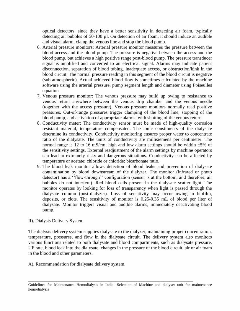
optical detectors, since they have a better sensitivity in detecting air foam, typically
detecting air bubbles of 50-100 µl. On detection of air foam, it should induce an audible
and visual alarm, clamp the venous line and stop the blood pump.
6. Arterial pressure monitors: Arterial pressure monitor measures the pressure between the
blood access and the blood pump. The pressure is negative between the access and the
blood pump, but achieves a high positive range post-blood pump. The pressure transducer
signal is amplified and converted to an electrical signal. Alarms may indicate patient
disconnection, separation of blood tubing, inadequate access, or obstruction/kink in the
blood circuit. The normal pressure reading in this segment of the blood circuit is negative
(sub-atmospheric). Actual achieved blood flow is sometimes calculated by the machine
software using the arterial pressure, pump segment length and diameter using Poiseulles
equation
7. Venous pressure monitor: The venous pressure may build up owing to resistance to
venous return anywhere between the venous drip chamber and the venous needle
(together with the access pressure). Venous pressure monitors normally read positive
pressures. Out-of-range pressures trigger clamping of the blood line, stopping of the
blood pump, and activation of appropriate alarms, with shutting of the venous return.
8. Conductivity meter: The conductivity sensor must be made of high-quality corrosion
resistant material, temperature compensated. The ionic constituents of the dialysate
determine its conductivity. Conductivity monitoring ensures proper water to concentrate
ratio of the dialysate. The units of conductivity are millisiemens per centimeter. The
normal range is 12 to 16 mS/cm; high and low alarm settings should be within ±5% of
the sensitivity settings. External readjustment of the alarm settings by machine operators
can lead to extremely risky and dangerous situations. Conductivity can be affected by
temperature or acetate: chloride or chloride: bicarbonate ratio.
9. The blood leak monitor allows detection of blood leaks and prevention of dialysate
contamination by blood downstream of the dialyzer. The monitor (infrared or photo
detector) has a ‘‘flow-through’’ configuration (sensor is at the bottom, and therefore, air
bubbles do not interfere). Red blood cells present in the dialysate scatter light. The
monitor operates by looking for loss of transparency when light is passed through the
dialysate column (post-dialyzer). Loss of sensitivity may occur owing to biofilm,
deposits, or clots. The sensitivity of monitor is 0.25-0.35 mL of blood per liter of
dialysate. Monitor triggers visual and audible alarms, immediately deactivating blood
pump.
II). Dialysis Delivery System
The dialysis delivery system supplies dialysate to the dialyzer, maintaining proper concentration,
temperature, pressures, and flow in the dialysate circuit. The delivery system also monitors
various functions related to both dialysate and blood compartments, such as dialysate pressure,
UF rate, blood leak into the dialysate, changes in the pressure of the blood circuit, air or air foam
in the blood and other parameters.
A). Recommendation for dialysate delivery system.
______________________________________________________________________ Guidelines for Maintenance Hemodialysis in India- Selection of Machine and dialyser unit for maintenance
hemodialysis
We recommend a single patient, single pass system or central delivery system may be
used.
Description:
Single patient, single pass systems discharge dialysate to drain after one passage through the
dialyzer and are used to deliver dialysate to one patient at a time. Dialysate is produced from
proportioning dialysate concentrate and purified water. Normally, single patient systems, also
called "negative pressure systems," maintain a sub atmospheric (negative) dialysate pressure in
order to accomplish fluid removal. The central delivery system maintains a single "central
dialysate proportioner” which prepares dialysate for a number of bedside consoles or bedside
stations.
Both the single patient/single pass systems and the multi-patient/single pass systems require a
continuous supply of purified water and a continuous source of concentrate. Spent (discarded)
dialysate is discarded to the drain after it has made a single pass through the dialyzer.
B). Recommendations for maintenance of HD machines:
We suggest that machines should be replaced after between five and ten years’ service or
after completing between 15,000 and 40,000 hours of use for HD, depending upon an
assessment of machine condition and specifications provided by the manufacturer.
Routine servicing of the machines should be done at regular intervals by the qualified
engineers or technicians. These designated technicians may be located in-house or may
be stationed outside. Maintenance of records of routine servicing of machines should be
maintained.
There must be a provision for emergency electric power supply for life-saving equipment
in case of power failure. An uninterrupted power supply (UPS) backup of up to 15
minutes is desirable for each machine in case of power failure.
Fire precautions must be taken and fire escapes should be clearly visible (mandatory for
large HD units, optional for small HD units).
C). Recommendations for disinfection of the HD machine:
After an episode of blood leak in to the dialysate.
If surveillance cultures show high cfu or endotoxin levels
Regular disinfection at least once a week.
After each dialysis session or once a day (optional).
Bleach or Citrosteril( Combination of Citric, maleic and oxalic acid) or heat or a
combination may be used for disinfection of HD machines.
Description:
Disinfection of the HD machine is mandatory to prevent transmission of infections between
patients. The disinfection of the machine may be performed using either bleach or citrosterilor
heat. Disinfection with bleach is recommended after each blood leak in to the dialysate or at a

______________________________________________________________________ Guidelines for Maintenance Hemodialysis in India- Selection of Machine and dialyser unit for maintenance
hemodialysis
regular interval of at least one week. Disinfection with citrosteril may be performed after each
dialysis session or at least once daily. The steps for sterilization are detailed below. With
standard disinfectant fitted to the rear of the machine, bleach must be administered via the pickup
stick (Concentrate connectors) at the front of the machine. The disinfection procedure is
performed by the designated personnel in the dialysis unit. Gloves and protective glasses must be
worn during the procedure by the operator.
Steps of bleach disinfection:
1. Bleach (Sodium hypochlorite 5%) is used for disinfecting the machine.
2. Bleach should not be heated.
3. If bleach disinfection is required for the blood leak, rinse machine for 15 minutes.
4. Ensure that power and water supply to the machine are operational.
5. Turn on the machines.
6. Press cleaning key.
7. Use up/down arrow keys to select “Cleaning (font supplied)”
8. Select Treatment/Rinse – select chemical mode – confirm.
9. Machine alarm “! Connect disinfectant” displayed.
10. Place PICKUP STICK Concentrate connectors into sodium hypochlorite at the front of
the machine.
11. Press conf key, “Please Wait” displayed.
12. On completion “Mandatory rinse end” displayed.
13. Test for residual bleach using Chlorine test strips on completion of cycle.
Steps of citrosteril disinfection:
1. Following conditions/reminder must be fulfilled before activating the cleaning program:
a. The dialysate lines are connected to the shunt (Rinse bridge)..
b. The shunt door is closed.
c. The concentrate suction tubes are in the appropriate rinse ports.
d. The interlock plate of the bigbag® connector (option) is closed.
e. The optical detector does not sense blood.
2. Citrosteril should be fitted to the rear of the machine
3. Citrosteril should be heated (≥60oC) for efficient results.
4. Ensure that power and water supply to the machine are operational.
5. Turn on the machines.
6. Ensure the basic conditions/reminder (mentioned above) has been reviewed.
7. Press cleaning key.
8. Use up/down key to select the desired program- “Hot Disinfection”.
9. Press conf key, “-F-HDIS- or –F-HDIS-M-HR-“displayed.
10. On completion “Mandatory Rinse End” will be displayed.
11. It is not necessary to test residual citric acid if Citrosteril is used, since it is a decaying agent
which is formulated in a non-toxic solution.
III). Safety of Dialysis Delivery System:
______________________________________________________________________ Guidelines for Maintenance Hemodialysis in India- Selection of Machine and dialyser unit for maintenance
hemodialysis
Patient safety is the most important goal that should never be compromised during HD.
1) Alarms: Various ‘‘alarms’’ built into the HD system can signal impending or ongoing system
malfunction.
Recommendations for HD machine alarm system:
Alarms should never be taken lightly and disarming of alarms should never be practiced.
The range and sensitivity of the alarms should be internally set as default and the operator
should only be able to operate within the set range without being able to alter these
settings, especially while HD is in progress.
Alarms should be visible clearly from at least 2 meters, but also easily audible (70 dB).
All blood alarms (air detector, arterial, venous, blood leak, transmembrane pressure,
blood pump torque) should automatically shut off the blood pump, clamp the venous
return line, and stop UF, thus isolating the patient.
Equipment should programmed to automatically switch to ‘‘safe mode,’’ thus essentially
isolating the patient from the HD machine.
2). Monitoring and Evaluation of HD machine.
Monitoring and evaluation of the HD machine performance at periodic interval should be
performed to enhance safety and reduce the level of risk of patient injury due to incidents related
to malfunction and or improper use of dialysis delivery systems. The actual numerical reading of
each test or the result of a test should be recorded after each test and initials of the person
performing the test should be noted.
Recommendations for evaluation and monitoring on daily basis.
Conductivity of the final dialysate being delivered to the dialyzer should be checked before
every treatment. According to manufacturers' instructions, the conductivity should be
checked with an independent reference meter which is known to be properly calibrated.
Conductivity must be within the manufacturer's stated specifics.
When used, the pH of bicarbonate dialysate should also be confirmed before each treatment.
If the pH is below 6.5 or above 7.5, dialysis should not be started, even when conductivity is
within acceptable limits. The pH can be checked with a similar pH meter.
Temperature should also be within the manufacturer's specifications. Temperature may be
checked with an independent reference meter or with a reference thermometer.
Absence of residual germicide should be verified on all delivery systems connected to a
single water treatment “loop" before dialysis begins. Such testing must be performed with an
assay known to detect the minimum standard level.
A test of proper functioning of the air/foam detector should be performed before dialysis is
initiated. This test should be a direct test of function of the alarm, causing interruption of the
blood pump and actuation of the blood line clamp, either by introducing air into the venous
level detector or by removing the tubing so that air is sensed by the detector as recommended
by the device manufacturer.
______________________________________________________________________ Guidelines for Maintenance Hemodialysis in India- Selection of Machine and dialyser unit for maintenance
hemodialysis
The blood detector must be checked for proper armed status according to the method
recommended by the manufacturer.
The user should perform applicable tests of the UF control system as prescribed by the
manufacturer.
All other alarms must be tested according to the manufacturer's instructions for use before
every treatment including low and high conductivity alarm, low and high temperature alarm,
dialysate pressure alarm, water pressure alarm, etc. Documentation of that testing should be
performed. If the particular delivery system is equipped with a "self-alarm check” mode, it is
important that the user understand that, most often, it is a check of the electronic circuitry,
and not a confirmation of some of the vital functions of specific alarms.
Observation of dialysate flow should be made while the machine is in a "dialyzing” mode.
Absence of dialysate flow should be confirmed when the machine is in "bypass” mode
actuated by both manual setting of the machine to bypass or via any of the alarm functions
that will cause the machine to enter a bypass mode.
The automatic “self-test” should be performed if this facility is available prior to each HD
treatment to confirm proper performance of operative and protective functions of the
machine and should never be bypassed.
Recommendation for once monthly evaluation and monitoring:
Microbiological monitoring: water for production of dialysate and actual dialysate
proportioned and exiting the dialyzer should be monitored for bacterial levels on no less than
a monthly basis. Microbiological monitoring is performed to establish ongoing validation of
proper disinfection protocols. The sampling should be done at the termination of dialysis at
the point where dialysate exits the dialyzer. Results for total microbial counts shall not
exceed 2,000 colony forming units per ml.
Assessing trends: Pertinent information, i.e., bacterial levels, conductivity and pH readings,
etc., should be logged on a chart across a page so that readings can be examined and
compared over an extended period of time. This tool makes it possible to compare current
readings to those taken during the past several days/weeks/months.
C). Prevention measures.
It is desirable to take measures to prevent HD malfunction, so that safety of the HD therapy is
ensured.
Recommendations for preventive measures:
All electrical and other equipment used in the facility should be maintained free of defects to
prevent potential hazard to patients or personnel.
Each manufacturer provides comprehensive directions pertaining to preventative
maintenance requirements for the entire dialysis delivery system and these should be
followed.
______________________________________________________________________ Guidelines for Maintenance Hemodialysis in India- Selection of Machine and dialyser unit for maintenance
hemodialysis
A master schedule of all preventative maintenance should be developed. Such a master
schedule will list every machine by serial number (or other identifier) and identify when
preventative maintenance is required.
Dialysis units should have an established and agreed upon plan of action for repair and
trouble shooting of HD machines.
Description:
1. Maintenance: The schedules and procedures established for preventive maintenance should
be followed. Maximum time internals either in number of hours of operation of the system or
in calendar days between preventative maintenance procedures are also specified.
2. Recordkeeping: A history record of all repairs and maintenance for each piece of equipment
should be maintained in a separate file. This file describes all technical operations performed
on the equipment, parts used, actions taken, tests performed to assure proper functioning
before and after maintenance/ repair. The dates and the personnel performing
maintenance/repair should also be documented. A log of all maintenance and repair work for
each piece of equipment should be kept at the front of the "history file." This log includes a
very brief description of the maintenance/repair (e.g., "300 hr maintenance” or "adjusted
conductivity" or "repaired inoperable blood pump," etc.), date, and person performing action.
Such a log provides a trend analysis of any problems related to the delivery system, as well
as a quick confirmation of maintenance being performed according to schedule.
3. Repair & Troubleshooting: Despite proper maintenance of the machines, rarely entire
machine or a component of the system may fail. Although these failures cannot be foreseen
and occur very infrequently, when they do occur, it is important that patient is not put at risk.
To counteract these events, dialysis units should have an established and agreed upon plan of
action. This plan should be approved by the medical director and communicated to all facility
staff. Repair and maintenance on a delivery system should be performed by "qualified
personnel." The definition of "qualified personnel may differ from facility to facility, and that
definition is the final responsibility of the medical director.
D). Quality Assurance for Dialysis Delivery Systems: It consists of several components such
as policies and procedures, staff training and continuing education, and monitoring and
evaluation.
Recommendations for Quality Assurance for Dialysis Delivery Systems.
Each dialysis units should have “policies and procedures” for delivery of dialysis delivery
system, which should be developed as per the need, implemented and evaluated periodically.
Staff training and education should include operation and proper use and monitoring of
dialysis delivery system.
Description:
1). Policies and Procedures
______________________________________________________________________ Guidelines for Maintenance Hemodialysis in India- Selection of Machine and dialyser unit for maintenance
hemodialysis
An essential step in designing the facility's quality assurance program is the development,
implementation, and evaluation of policies and procedures for dialysis delivery systems. All
standards previously described must be incorporated into these policies and procedures.
Specifically, the policies and procedures must address the scope of care and therapeutic choices,
equipment, disposables, and supplies used in the dialysis facility. Comprehensive policies and
procedures must also address the interrelationships of each component.
Policies and procedures must also address safe and effective operation of the delivery system.
This includes the following factors:
1 Basic technical operation
2 Set up and use of equipment and related components
3 Safety checks
4 Preventative maintenance
5 Cleaning and disinfection
6 Trouble shooting and repair
7 Record keeping
8 patient monitoring
2). Staff Training and Continuing Education.
Responsibilities for operation and use of the delivery system including preventative maintenance,
troubleshooting and repairs, daily or per-treatment safety and other system checks and
recordkeeping should be clearly defined. Each responsibility should stem from a specific policy
or procedure. Staff training should be a well-defined and organized program. Content should be
clearly defined for the learner and should be based on behavioral objectives. The behavioral
objectives can be used to accurately and objectively measure learning.
IV). Dialyzer (filter) specifications:
The hollow fiber dialyzer forms the central component of dialysis deliver system, where in actual
process of transfer of solutes and water occurs across a semi-permeable membrane. A large array
of dialyzers is available for clinical use with several permutations and combinations based on
biocompatibility, flux and surface area of the dialyzer. Most often a single type of dialyzer may
be sufficient in most patients in a dialysis unit. However, some patients may have specific needs
and may require change in the dialyzer specifications. Hence, dialyzers with specifications other
than that generally used in the dialysis unit may also be routinely stocked or should be made
available at a short notice when the need arises.
Recommendations for dialyzer use in HD:
Biocompatible, synthetic (e.g., polysulfone, polyacrilonitrile, polymethylmethacrylate) or
modified cellulose membrane (e.g., cellulose acetate) should be preferred over unmodified
______________________________________________________________________ Guidelines for Maintenance Hemodialysis in India- Selection of Machine and dialyser unit for maintenance
hemodialysis
cellulose membranes (e.g., cuphraphan). Cupraphane membranes should used only when
other more bio-compatible membranes are not available or patient is intolerant to all others.
Either low flux or high flux biocompatible membrane may be used for regular HD.
High flux dialyzers may be preferred over low flux dialyzers to provide HD under specific
situations such as 1) incident patients, who have lower serum albumin concentrations (<40
g/L) or have diabetes mellitus, and 2) prevalent patients, who have been on HD for more than
4 years or have dialysis related amyloidosis.
High flux dialyzer should be used only in facilities where a very high quality of dialysate is
ensured at all times.
Surface area of the dialyzers should be chosen based on the required dialysis dose and the
body size of the patient. Large surface area dialyzers should be avoided in pediatric patients
and adult patients with small body size.
An allergic reaction to a specific dialyzer is rarely encountered in some patients. In such
situation, the particular dialyzer should be avoided and this should be specifically written in
bold letters on the dialysis folder of the patient to prevent its inadvertent use.
V). Dialysis fluid specifications:
Dialysate, or dialysis fluid, is a non-sterile aqueous solution with an electrolyte composition near
that of normal extracellular fluid. Its electrolyte composition is designed to correct the metabolic
imbalance that occurs as a result of uremia. Dialysate concentrates are manufactured
commercially in liquid or powder form. The chemicals present in the dialysate have access, via
the dialyzer, to the bloodstream of patients undergoing dialysis. Hence, the proper concentration
of all of these chemicals as well as the quality of the concentrate and the water used to dilute the
concentrate is critical.
1).Recommendations for dialysis fluid use in HD.
Commercially produced concentrates are classified as medical devices and should be
approved for clinical use by appropriate authority.
The dialysate should contain bicarbonate as the buffer and acetate as a buffer may be used
only when bicarbonate based dialysate is not available.
The final diluted dialysate should be analyzed every 6 months, with every new batch of
dialysate and after each major servicing/repair of dialysis machine.
Water used to prepare the dialysate must have a bacteriological colony count of less than
200/ml.
Electrolyte content of dialysate includes sodium, potassium, chloride, magnesium, calcium,
glucose (optional), and bicarbonate (or acetate) as a buffer. The concentration of HD
solutions should be such that after dilution to the stated volume the final concentrations of
the ions expressed as mmol/L are usually in the following ranges: Sodium 135-145,
Potassium 0-4, Calcium 1.0-2.0, Magnesium 0.25-1.0, bicarbonate (acetate equivalent of
bicarbonate) 32-40, Chloride 95-110.
Sodium concentration may be adjusted to levels outside the range of 135-140 mmol/L by HD
machines with variable sodium capabilities only when prescribed by physician in charge.
______________________________________________________________________ Guidelines for Maintenance Hemodialysis in India- Selection of Machine and dialyser unit for maintenance
hemodialysis
Bacteriological analysis of the dialysate shall be carried out at least 2 monthly, preferably
every 15 days.
The colony count in dialysate samples collected at the termination of dialysis a) in a single
pass system or b) in a re-circulating single pass system at the periphery of the recirculating
chamber containing the dialyzer shall be less than 2000 colony-forming units/ml.
Dialysate containing glucose at 100- 200 mg/dl concentration is preferable to glucose free
solution.
Description:
1. Sodium concentration of dialysate varies from 135-140 mmol/L in routine HD. However, a
wide range (130-155 mmol/L) of sodium concentration is dialysis fluid may be achieved in
modern HD machine. Sodium concentration may vary in certain circumstances depending on
serum sodium level, cardiovascular instability and need for higher UF. An option of variable
dialysate sodium concentration during a single HD session (sodium modeling) is available in
most modern machines, which may be used in patients with cardiovascular instability who
need larger UF.
2. Glucose free dialysate is associated with increased risk of hypoglycemia especially in
diabetics, cachectic and septic patients and increased catabolism. We recommend
isoglycemic (100 mg/dl) or mildly hyperglycemic (200 mg/dl) dialysis fluid for HD.
3. Routine use of acetate as a buffer should be avoided. However in an emergency situation
when bicarbonate buffer is not available, acetate buffer may be used for HD. The conversion
of acetate to bicarbonate is limited especially in patients with low muscle mass. Acetate
dialysis is associated with increased risk of hypotension, cardiac dysfunction, hypoxemia,
nausea, headache and fatigue.
4. A standard potassium concentrate of 2 mmol/L is recommended for routine HD to keep
predialysis serum potassium below 6 mmol/L. However, dialysate potassium concentration
varying from 0-4 may be used depending on patient need. When dialysis fluid potassium is
other than standard one, a label indicating the concentration should be displayed.
5. The physician in charge shall be responsible for arranging for the analysis of the dialysate. Its
chemical composition shall be clearly labeled. The results of analysis, bearing the name of
the centre and officer analyzing the dialysate shall be made available on request as and when
required.
2) Recommendations for storing and mixing dialysis concentrates:
Store and dispense dialysate concentrates as though they were drugs.
Ensure that all personnel in your unit are aware of the types of dialysate concentrates
available, even if you currently use only one type.
Develop a policy, management, and storage system that will effectively control the mixing
and dispensing of all concentrates. Storing concentrates according to type, composition, and
proportioning ratios should reduce the risk of mismatching concentrates. Prohibit access to
storage areas and allow only authorized, specially trained personnel to mix and dispense
concentrates.
______________________________________________________________________ Guidelines for Maintenance Hemodialysis in India- Selection of Machine and dialyser unit for maintenance
hemodialysis
Double-check and record concentrate formulas on the patient's record. Consider a procedure
for countersigning patient and storage records.
Do not dispense concentrates from large containers into smaller ones without a "keyed"
dispensing system. Whenever possible, purchase concentrates in single-treatment (2½-
gallon) containers (optional).
Always dispose of concentrates remaining from the previous treatment. Do not pour
remaining concentrate into another container or use in the next treatment. Replace empty or
partially full containers with full ones.
Whenever possible, standardize equipment so that only one bicarbonate concentrate system is
used.
Description:
1. Bicarbonate dialysis requires mixing two concentrates acid and bicarbonate with treated
water. Bicarbonate concentrate is typically supplied in powder form, to be mixed with treated
water immediately preceding dialysis. Acid concentrate, containing an electrolyte
composition similar to that of acetate concentrates but at a lower pH, is supplied in liquid
form. The availability of acid/bicarbonate concentrates with varying ionic contents and
proportioning ratios increases the probability of an inappropriate dialysate. The problem is
further compounded by the availability of two types of HD machines with different
proportioning systems: fixed-ratio and servo-controlled (variable-rate).
2. Risks and Hazards related to Dialysate: Approximately 50% of patient complications
related to dialysis concentrate are related to the quality of water used for preparing dialysate
and the concentrate when delivered from the manufacturer. The remaining 50% were related
to user error or machine malfunctions.
Briefly, the problems related to manufacturers include the following:
a) Minimal bacterial growth in liquid bicarbonate concentrate.
b) Actual electrolyte content of the concentrate was different than described on the label.
c) Foreign matter in liquid bicarbonate concentrate.
d) High levels of aluminum contaminating acetate concentrate
Incidents related to user error, or machine malfunction include:
a) Improper sodium concentrations due to mis-calibration of or improper proportioning by the
dialysis delivery systems.
b) Use of wrong concentrates or improper mixing of concentrates due to staff misreading labels.
c) Bacterial problems related to improper disinfection of storage containers or use of water
containing excess bacteria.
______________________________________________________________________ Guidelines for Maintenance Hemodialysis in India- Water treatment for hemodialysis
8.0 WATER TREATMENT FOR HEMODIALYSIS Rationale: The average hemodialysis patient is exposed to approximately 25 times the amount of
water normally ingested by an individual. In addition he is deprived of the protective barrier of
the gastrointestinal tract and the detoxification function of the kidneys, increasing the risk several
fold of toxicity caused by the numerous chemical and microbiological contaminants in the water.
The final quality of the water is dependent on the configuration of the treatment system and the
quality of the feed water which itself may be highly variable. As processes of hemodialysis
evolve with use of high flux dialyzers and hemodiafiltration probably becoming increasingly
used many countries in the world have made the use of ultrapure water the goal of every dialysis
unit. This requires a unit to design a system capable of delivering this very high quality from the
worst feed water and a monitoring system and quality assurance to prevent breakdown of the
system. The following guidelines describe the setting up, maintenance and monitoring of a
system designed to reliably provide ultrapure water
1. Water treatment to achieve the following water quality (AAMI standards) is mandatory for
all hemodialysis units. (Table 1)
2. It is recommended that the hemodialysis units should try to achieve European Standards of
purity of water (Table 2)
______________________________________________________________________ Guidelines for Maintenance Hemodialysis in India- Water treatment for hemodialysis
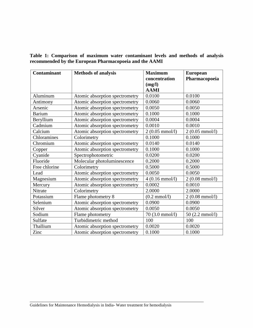
Table 1: Comparison of maximum water contaminant levels and methods of analysis
recommended by the European Pharmacopoeia and the AAMI
Contaminant Methods of analysis Maximum
concentration
(mg/l)
AAMI
European
Pharmacopoeia
Aluminum Atomic absorption spectrometry 0.0100 0.0100
Antimony Atomic absorption spectrometry 0.0060 0.0060
Arsenic Atomic absorption spectrometry 0.0050 0.0050
Barium Atomic absorption spectrometry 0.1000 0.1000
Beryllium Atomic absorption spectrometry 0.0004 0.0004
Cadmium Atomic absorption spectrometry 0.0010 0.0010
Calcium Atomic absorption spectrometry 2 (0.05 mmol/l) 2 (0.05 mmol/l)
Chloramines Colorimetry 0.1000 0.1000
Chromium Atomic absorption spectrometry 0.0140 0.0140
Copper Atomic absorption spectrometry 0.1000 0.1000
Cyanide Spectrophotometric 0.0200 0.0200
Fluoride Molecular photoluminescence 0.2000 0.2000
Free chlorine Colorimetry 0.5000 0.5000
Lead Atomic absorption spectrometry 0.0050 0.0050
Magnesium Atomic absorption spectrometry 4 (0.16 mmol/l) 2 (0.08 mmol/l)
Mercury Atomic absorption spectrometry 0.0002 0.0010
Nitrate Colorimetry 2.0000 2.0000
Potassium Flame photometry 8 (0.2 mmol/l) 2 (0.08 mmol/l)
Selenium Atomic absorption spectrometry 0.0900 0.0900
Silver Atomic absorption spectrometry 0.0050 0.0050
Sodium Flame photometry 70 (3.0 mmol/l) 50 (2.2 mmol/l)
Sulfate Turbidimetric method 100 100
Thallium Atomic absorption spectrometry 0.0020 0.0020
Zinc Atomic absorption spectrometry 0.1000 0.1000
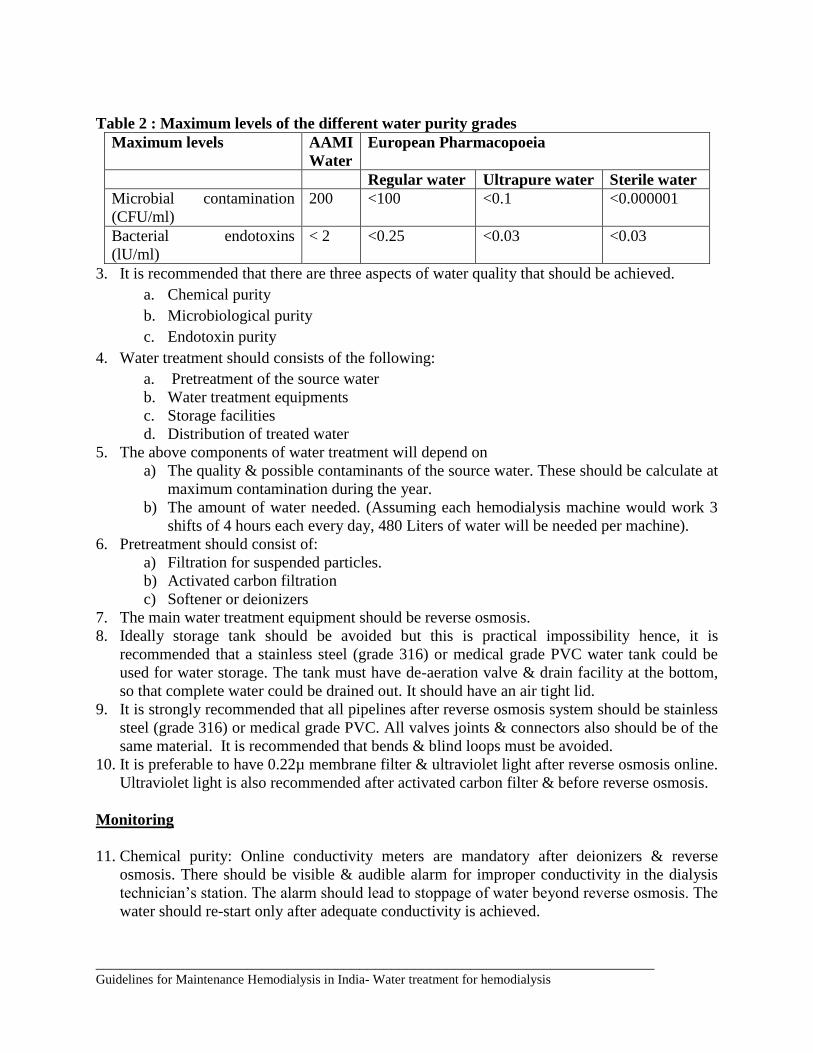
______________________________________________________________________ Guidelines for Maintenance Hemodialysis in India- Water treatment for hemodialysis
Table 2 : Maximum levels of the different water purity grades Maximum levels AAMI
Water
European Pharmacopoeia
Regular water Ultrapure water Sterile water
Microbial contamination
(CFU/ml)
200 <100 <0.1 <0.000001
Bacterial endotoxins
(lU/ml)
< 2 <0.25 <0.03 <0.03
3. It is recommended that there are three aspects of water quality that should be achieved.
a. Chemical purity
b. Microbiological purity
c. Endotoxin purity
4. Water treatment should consists of the following:
a. Pretreatment of the source water
b. Water treatment equipments
c. Storage facilities
d. Distribution of treated water
5. The above components of water treatment will depend on
a) The quality & possible contaminants of the source water. These should be calculate at
maximum contamination during the year.
b) The amount of water needed. (Assuming each hemodialysis machine would work 3
shifts of 4 hours each every day, 480 Liters of water will be needed per machine).
6. Pretreatment should consist of:
a) Filtration for suspended particles.
b) Activated carbon filtration
c) Softener or deionizers
7. The main water treatment equipment should be reverse osmosis.
8. Ideally storage tank should be avoided but this is practical impossibility hence, it is
recommended that a stainless steel (grade 316) or medical grade PVC water tank could be
used for water storage. The tank must have de-aeration valve & drain facility at the bottom,
so that complete water could be drained out. It should have an air tight lid.
9. It is strongly recommended that all pipelines after reverse osmosis system should be stainless
steel (grade 316) or medical grade PVC. All valves joints & connectors also should be of the
same material. It is recommended that bends & blind loops must be avoided.
10. It is preferable to have 0.22µ membrane filter & ultraviolet light after reverse osmosis online.
Ultraviolet light is also recommended after activated carbon filter & before reverse osmosis.
Monitoring
11. Chemical purity: Online conductivity meters are mandatory after deionizers & reverse
osmosis. There should be visible & audible alarm for improper conductivity in the dialysis
technician’s station. The alarm should lead to stoppage of water beyond reverse osmosis. The
water should re-start only after adequate conductivity is achieved.
______________________________________________________________________ Guidelines for Maintenance Hemodialysis in India- Water treatment for hemodialysis
Once in 3 months treated water sample must be sent for detailed chemical analysis to an
independent laboratory having adequate instrumentation for testing (see table 1). The results
should be mandatory part of the record system.
12. Microbiological purity: This should be checked once every 15 days to achieve the standards
as per table 2. It is strongly recommended that pour plate method on nutrient poor medium
should be used for cultures of treated water. Incubation should be at room temperature (200
C to 240 C) for 7 days. (See annexure)
13. Endotoxin levels: should be checked once in every 15 days to achieve the standard as per
table 2. (Please see annexure)
Sterilization
14. It is strongly recommended that each component of the water treatment system must be
thoroughly cleaned & sterilized as per the manufacturer’s recommendation. The process of
sterilization should be carried out once every 15 days. After sterilization it is essential that
the sterilent is completely removed before the treated water is used for dialysis.
15. Cleaning & sterilization of the water storage tank & plumbing is necessary once every 15
days.
Maintenance of Water Treatment System:
16. A log should be maintained documenting the performance of the water treatment system
components and indicating the maintenance done on each component. These are shown in
Table 3 below

______________________________________________________________________ Guidelines for Maintenance Hemodialysis in India- Water treatment for hemodialysis
Table 3 : Maintenance of A water Treatment System
Component Monitoring
Parameter
Maintenance
required
Recommended Frequency
Depth Filter Pressure drop
across filter
Backwashing &
Rinsing
Twice a week and daily
during the monsoon or when
water is contains extra
suspended
particles/impurities.
Activated
Carbon Filter
Pressure drop
across filter
Backwashing &
Rinsing
Twice a week
Activated
Carbon Filter
Chlorine in
product water
Changing of
charcoal
If > 0.1 µg/ml.
Softener Hardness Regeneration Failure to achieve 10 fold
decrease.
Membrane
filters
Pressure drop
across filter
Change of filter 25%
Reverse
Osmosis
membranes
Inlet, Reject
and Permeate
pressures &
flows
Increase in inlet
pressure > 25%.,
or decrease in
permeate flow by
25%
Cleaning of membranes
offline
Reverse
Osmosis
membranes
Conductivity Increase by 50%
from baseline
Cleaning of membranes
offline or Replacement
Deioniser Conductivity or
resistivity
< 1megaohm or >
0.5 micromhos
Regeneration with acid and
alkali
Storage tank
and Pipeline
Bacterial counts >50% increase
over baseline.
Cleaning & Disinfection
Cleaning of Reverse Osmosis Membranes:
Membranes should be taken offline and the system shut down during the process.
The flow of reject should gradually increase if the process is successful.
Membranes should be backwashed at low pressure using an external pump and flow in the same
direction as normal operation. Each membrane should be cleaned individually. 2 cleaning
solutions are generally required.
a) Sodium tripolyphosphate and Sodium edetate at a pH adjusted to > 10 to remove
calcium scales and low level organic foulants.
b) 2% Citric acid (no pH adjustment required) which removes calcium carbonate, metal
oxides and inorganic colloidal compounds and also provides disinfection.
A water wash is recommended between the cleaning solutions. A final disinfection of the
membranes using 1% peracetic acid or 2% formalin (to be treated for at least 6 hours) is
recommended.
After a cleaning and disinfection cycle; the first 200 to 250 liters of water or permeate over 1
hour should be discarded. The pH of the permeate should be confirmed before using.

______________________________________________________________________ Guidelines for Maintenance Hemodialysis in India- Water treatment for hemodialysis
Cleaning and disinfection of the Distribution system.
The storage tank should be filled with around 50 to 100 liters of 1% sodium hypochlorite. After
allowing a contact time of 30 minutes the solution is circulated in the loop for 20 minutes and
drained followed by a complete rinsing with water which is discarded until a negative test with a
starch iodide paper or a conductivity equal to that of the feed water is obtained. Inline steam or
Ozone are alternatives to sodium hypochlorite in some systems.
Figure 1:
ANNEXURE
LAL TEST
METHODOLOGY-Steps
1. Add 0.1 ml. of reconstituted (as instructed in package insert) pyrotell
(C.No.G2003/G2125) (0.03/0.125 EU/ml.) .
2. Added 0.1ml test specimen or control (C.No.E0005- Positive control or C. No. W0504
LRW- Lal Reagent Water- Negative control).

______________________________________________________________________ Guidelines for Maintenance Hemodialysis in India- Water treatment for hemodialysis
3. Mixed vigorously (vortex) for 20-30 secs.
4. Place the reaction tubes at 37+1 0C water bath for 60 minutes.
5. Remove reaction tubes & invert the tubes in one smooth motion.
6. A positive test is indicated by the formation of a gel which does not collapse when the
tube is inverted by 180 Degree.
Contact Address :
Ashish Parikh Product manager,
Nexus, Division of Span Diagnostics
173-B New Industrial Estate
Udna, Surat – 394 210.
Mob.No. 9825831746
Fax.No. 91-261-28679319
Manufacturer: Associates of CAPE CODE Incorporated,
124 Bernard Saint Jean Drive,
East Falmouth, MA 02536-4445, USA
Tel: (888) 395-2221 Fax: (508) 540-8680,
(508) 540-3444 URL: www.acciusa.com
MICROBIAL CULTURE:
1. 100 ml of treated water is collected in sterile & pyrogen free container.
2. Pour 1ml sample of water on Soya triptone agar (nutrient deficient)-(Himedia),
McConkey agar-(Himedia), and Nutrient Agar-(HiMedia) plates.
Address for procurement: Himedia, General Trade Agency, 416, Business Center, Nr.
LIC Building, Relief Road, Ahmedabad 380001.
Phone No.079- 25508143
Address of Hi-Media in Mumbai:
HiMedia Laboratories Limited.
A-406, Bhaveshwar Plaza
LSB Marg, Mumbai-400 086, India
3. The agar plates are incubated at 37 0C & Room Temperature.
4. Checked for the bacterial colony formation after 48hr and 7 days.
5. Colonies are counted in the plates with positive growth and expressed as the colony
forming unit (cfu) per ml.
Test for microelements in RO water:
1. Hundred ml of treated water is collected in a glass stoppered bottle (BOROSIL)
previously washed in the following way for proper decontamination from microelements.
2. First the bottle is washed twice with mild detergent solution followed by repeated
washing with treated/ Pyrogen free water.
3. Washed bottle is oven dried.
4. Rinse bottle with hydrochloric acid diluted in treated/ Pyrogen free water and then keep
overnight under 0.1N HCl.
5. Repeatedly wash with treated/ Pyrogen free water.
6. Wash the bottle with the sample RO water for ten times with vigorous shaking.
7. Drain out the washing water.
8. Collect the sample of treated water (100 ml) in bottle for analysis. Close the stopper
immediately.
9. The sample is sent to analytic laboratory capable of the tests as per table 1.
______________________________________________________________________ Guidelines for Maintenance Hemodialysis in India- Water treatment for hemodialysis
10. Estimation is done for the following analytes: Aluminium(Al), Arsenic(As), Barium(Ba),
Berilium(Be), Bismuth(Bi), Calcium(Ca), Cadmium(Cd), Cobalt(Co), Chromium(Cr),
Copper(Cu), Iron(Fe), Mercury(Hg), Potassium(K), Lithium(Li), Magnesium(Mg),
Manganese(Mn), Nickel(Ni), Phosphorus(P), Lead(Pb), Sodium(Na), Palladium(Pd),
Rubidium(Rb), Antimony(Sb), Tin(Sn), Titanium(Ti), Vanadium(V), Zinc(Z), Chloride,
Thalium(Tl), Nitate, Fluoride and Sulphate.
______________________________________________________________________ Guidelines for Maintenance Hemodialysis in India- Anti-coagulation for hemodialysis
9.0 VASCULAR ACCESS
Rationale: Vascular access has been described as the “Achilles Heel” of hemodialysis. Although
a working A V fistula should be ideally constructed prior to starting dialysis a large number of
patients first come to attention as end stage renal disease when they require emergency dialysis.
These patients start and sometimes continue dialysis with catheters, running all the attendant
risks of infection and central vein thrombosis. The regular monitoring of an AV fistula and early
intervention for stenosis can prolong the life of these accesses. Comprehensive care of a vascular
access involves a team approach involving the patient and several caregivers in order to obtain
the best possible results and a basic approach to this is set down here.
The responsibility of evaluating a patient, creating and preserving a vascular access should be a
team effort and should include the following members each of whom have specific
responsibilities.
Dialysis Nurse / technician
Resident doctor/medical officer
Nephrologist
Vascular/General surgeon/Urologist
Interventional Radiologist/Nephrologist.
Types of Access
Permanent (Fistulae & grafts)
o Radio cephalic AV fistula
o Sephaneous vein forearm grafts
o Brachiocephalic AV fistula
o Brachiobasilic fistula with transposed vein
o Upper arm autologous sephaneous vein grafts
o PTFE grafts (straight or U) in any site.
An attempt should be made to provide every patient on Hemodialysis with a permanent vascular
access in the order listed above.
Recommendation: - 65% or more of patients starting HD should do so with an AV fistula,
and 90% or more of prevalent patients should dialyze with an AV fistula.
As more than 50% of patients are diagnosed as CKD V < 90 days before starting HD it is
recommended that these patients may begin dialysis with a temporary access and an
attempt to construct a permanent access should be made within the 1st 3 months on HD.
Autologous Sephaneous vein grafts may be preferred to PTFE grafts because of lower costs and
thrombogenicity.
Responsibility – Nephrologist.
Temporary
Double lumened uncuffed non tunneled soft catheters inserted in the Internal Jugular vein
should be the temporary access of choice either when
1. A permanent access has been created and is expected to mature within 90 days.
______________________________________________________________________ Guidelines for Maintenance Hemodialysis in India- Anti-coagulation for hemodialysis
2. A period of waiting before the permanent access can be created is anticipated.
3. Partial or complete recovery is expected.
The sub-clavian vein should not be cannulated as a temporary access unless the internal jugular
is unusable and no permanent access is possible ipsilaterally. Even a single cannulation is
associated with a 35% risk of stenosis.
The femoral vein on the left side may be used as a temporary vascular access with rigid single
lumened cannulae in an emergency situation only. Cannulae in the femoral vein should not be
retained for longer than 5 – 7 days and should never be used in the outpatient setting. Right sided
femoral vein cannulation should be avoided if the patient is to undergo a future renal
transplantation.
Semi-Permanent
Cuffed tunneled biluminal soft catheters inserted in the internal jugular vein with an exit
site on the anterior chest wall should be utilized as semi-permanent access. The cuff
should be placed subcutaneously above the clavicle and at a distance of 3 – 4 cm from the
exit site.
Responsibility - Nephrologist
Selection and Site of vascular access.
All patients of CKD should have an AV fistula constructed at Stage IV or when the
serum creatinine is > 5 mg% or the anticipated start of HD is < 6 months away.
Configuration:- The radiocephalic AV fistula on the non-dominant upper limb should be
the 1st choice of AV fistula.
Responsibility – Nephrologist & Surgeon
The “ Rule of 6” should be followed in deciding when to use an AV fistula,
o 6 weeks from the time of creation,
o a vein of at least 6 mm in diameter with clearly distinguishable margins,
o a cannulation length of at least 6 cm from the anastomosis,
o flow of at least 600 ml/min and a depth of not more than 6 mm from the skin.
Numerous collateral veins should not be visible and there should be no evidence
of venous hypertension.
Responsibility – Dialysis Nurse/medical Officer/ Nephrologist
Initial cannulation should be with 17G needles equipped with a “back eye”. Flows of
upto 200 ml/min can be obtained with a 17G needle. Subsequent cannulation should be
with a 16G needle to obtain flows of 300 ml/min and with a 15G needle to obtain flows
of > 300 ml/min.
Venous grafts, both autologous and PTFE may be used within three weeks of
construction.
Responsibility – Dialysis Nurse/technician/Medical Officer
Design and performance of temporary accesses.

______________________________________________________________________ Guidelines for Maintenance Hemodialysis in India- Anti-coagulation for hemodialysis
The diameter of the cannula and the length are the 2 criteria responsible for the blood flow
obtained.
Single lumened femoral cannulae should be at least 19 cm long to reach the IVC. Flows
of > 200 ml/min are not obtained with standard femoral single lumened cannulae.
The length of a cannula in the right Internal jugular vein should be around 13.5 cm for an
adult Indian patient, while that of a left internal jugular cannula should be around 16 cm,
that of a right sub-clavian cannula should be 15 cm and a left subclavian vein cannula
around 16 cm.
The cannulae should be at least 12 French to obtain flows of 300 ml/min and 14 french if
higher flows are to be obtained.
8 & 10 French cannulae can be used in children.
Cuffed tunneled cannulae should have a total length of at least 35 cm for right internal
jugular cannulation and a length of 44 cm for left internal jugular cannulation.
Responsibility – Nephrologist/Interventionist
Patient preparation and Evaluation prior to access
A history should be obtained regarding previous central venous cannulation, arterial
cannulation, previous attempted AV fistulae, and failure including the time and possible
cause of access failure, presence of severe cardiac disorders, malignancy and
prothrombotic tendency or anticoagulation.
Physical examination of both upper extremities should be carried out
o Physical examination should include
a. Examination of peripheral pulses
b. Bilateral upper extremity blood pressure measurement.
c. Allens test & Modified Allens Test
Allen's test assesses collateral circulation in the hand, in 2 steps.
Step 1 occludes the radial artery for several minutes and compares the hand color
to the other hand. The hand is said to have sufficient collateral circulation through
the ulnar artery if there is no change in color.
Step 2 occludes the ulnar artery. A change in hand color means the potential for
radial artery occlusion is high. That is a positive Allen's test,
which contraindicates radial-artery use for an AV fistula
Modified Allen's Test The procedure for performing a modified Allen's Test is as follows:
o Instruct the patient to clench his/her fist, or if the patient is unable, close
the hand tightly.
o Apply occlusive pressure with the fingers to both the ulnar and radial
arteries. This maneuver obstructs blood flow to the hand.
o While applying occlusive pressure to both the arteries, have the patient
relax his/her hand. Blanching of the palm and fingers should occur. If it
does not, you have not completely occluded the arteries with your finger.
o Release the occlusive pressure on the ulnar artery. Flushing of the hand
should occur within 5 to 15 seconds. This denotes that the ulnar artery if
patent and has good blood flow. This normal flushing of the hand is
considered to be a positive modified Allen's test. A negative modified
______________________________________________________________________ Guidelines for Maintenance Hemodialysis in India- Anti-coagulation for hemodialysis
Allen's test is one in which the hand does not flush within the specified
time period. This indicates that ulnar circulation is inadequate or
nonexistent. The radial artery supplying arterial blood to that hand should
not be used for an AV fistula.
4. Presence of edema.
5. Presence of collateral veins
6. Collapsibility.
Responsibility – Nephrologist/Access Surgeon.
Pre op Imaging/mapping
Ultrasonographic mapping of venous drainage of the extremity is recommended in
difficult cases. A tourniquet should be applied to the upper arm and the vein diameter
measured. The vein diameter should be between 2 & 2.5 mm. This assessment should be
done when patient has achieved near ideal volume status.
In cases of previous 1 or more fistula failure and history of central vein Cannulation
venography should be done for patency & adequacy of peripheral & central veins.
Patients with 1 or more previous fistulae lost due to early thrombosis should have a
thrombotic screen done.
Responsibility – Nephrologist/ Access Surgeon
Preservation of peripheral and central veins
Patients with CKD IV or V should not have venipunctures or peripheral cannulae inserted
in the forearm or above the wrist once a decision to create an A V fistula for dialysis has
been taken.
Patients admitted in hospital should be provided with bracelets labeled “No
Venipuncture” to be worn during admission.
Out patients should be educated about preserving of forearm veins.
Responsibility – Nephrologist/Dialysis Nurse
The subclavian vein should not be cannulated as a temporary access unless the internal
jugular is unusable and no permanent access is possible ipsilaterally. Even a single
cannulation is associated with a 35% risk of stenosis.
Responsibility - Nephrologist
Cannulation and Use of a Vascular Access
Every vascular access should be examined at each hemodialysis session prior to starting
dialysis.
o Fistulae should be examined for a low pitched continuous bruit, and a thrill,
absence of edema, normal limb temperature, absence of ischemia, steal and large
collateral veins.
o Fistulae should not have a water hammer pulse on examination.
o Veins should collapse upon raising the arm above the level of the heart.
The entire arm should be cleaned with 2% alcoholic chlorhexidine prior to needle
insertion.
A railroading technique rather than a buttonhole technique should be followed for
cannulation.
______________________________________________________________________ Guidelines for Maintenance Hemodialysis in India- Anti-coagulation for hemodialysis
o Railroading – At each dialysis session, puncture of the fistula should be done 1 to
2 mm away from the previous point and a return to the original site should occur
after 6 – 7 sessions.
o Buttonhole – Every puncture is done through an identical point. This eventually
leads to decreased pain sensation at the site but also to weakening of the vein wall
and aneurismal dilatation.
o The “arterial needle” should point towards the anastomosis and the “venous
needle” should point away from the anastomosis.
The skin at the site of puncture should be infiltrated with 2% Xylocaine using a 26G needle.
Alternatively the entire region should have been applied with a Lignocaine-Prilocaine gel, at
least 30 minutes prior to puncture to minimize pain.
Removal of needles should be accompanied by dusting of an antibacterial powder.
Firm digital pressure over a sterile guaze should be given to the site of needle insertion
for 10 minutes after removal of the needle followed by application of a sterile adhesive
dressing.
Tight tourniquets should not be applied to a fistula limb.
Responsibility – Dialysis Nurse/technician/Medical officer
Care of a vascular access
Dressings of a vascular access should be transparent, occlusive, and strong enough to
resist the weight of the dialysis cannula. Micropore or Tegaderm is a useful dressing
The skin around the exit site of the access site should be clipped of hair, and tincture
benzoin should be applied to the area prior to application of the dressing.
Mupirocin ointment should be applied to the exit site of both cuffed and uncuffed
cannulae.
Mupirocin ointment should also be applied to the external nares, axilla and groin in
patients using cuffed tunneled cannulae as a vascular access, who have been found to
be staphylococcal carriers.
Surveillance cultures should be carried out using nasal swabs of all patients and dialysis
personnel once a year and nasal staphylococcal carriers treated.
Patients and the attendants should wear a disposable surgical mask during any
manipulation of the cannula, dressing changes and connection and disconnection to the
dialysis machine.
Dressings should be changed weekly and whenever wet, visibly soiled or stained with
blood or other material. Cannulae should not be unnecessarily manipulated.
The hubs of the cannulae should be cleaned with sterile swabs soaked in 2% alcoholic
chlorhexidine, the connection to blood tubings done without touching the hubs or
connectors, and the joint wrapped with a swab soaked in 2% alcoholic chlorhexidine or
10% povidone iodine for 10 minutes.
The cannula should be flushed with sterile saline till free of blood prior to anticoagulant
instillation after each dialysis.
Responsibility – Patient/Dialysis Nurse/Technician.
Monitoring and detection of Complications
______________________________________________________________________ Guidelines for Maintenance Hemodialysis in India- Anti-coagulation for hemodialysis
The maximum blood flow obtained from the access should be documented at each
dialysis.
A progressive drop in the flow obtained with needles of the same guage properly
positioned should prompt further investigation of the access for stenosis.
Venous pressure should not be used to monitor stenosis in an AV fistula vein but may be
used to monitor stenosis in an A V graft.
The venous pressure should be measured using 17G needles within the first 5 minutes of
dialysis at a blood flow of 200 ml/min. Serial readings are more useful than single. An
increase of more than 20% or an absolute value persistently > 120 mm of Hg is indicative
of a graft outflow stenosis.
Fistula and graft stenosis should be investigated by fistulograms. Ultrasonography is an
alternative but is highly operator dependant and can give fallacious readings due to deep
collateral veins.
A fistulogram or CT fistulogram should evaluate the AV anastomosis, the draining veins,
and the central veins. (subclavian & SVC).
Fever or rigors during hemodialysis in patients with indwelling cannulae should prompt
evaluation of the vascular access as a source of infection.
Responsibility – Nephrologist/Interventional Radiologist
Measures to prevent Access dysfunction
Antibiotic locks may be used in patients with cuffed tunneled cannulae. Citrate which has
antibacterial properties may be used alone as an alternative to heparin as a locking
solution in a 4% strength.
Prophylactic antibiotic lock solutions should never include drugs like Vancomycin which
are to be retained for therapeutic use.
Stenosis of AV fistulae should be treated to prevent the risk of thrombosis however
overall life of the AV fistula has not been shown to be increased by pre emptive
intervention.
Responsibility – Dialysis nurse/Nephrologist
Preparation of Antibiotic Cannula Locks
Trisodium citrate is commercially available as a 46% solution. This may be diluted 10 times
with sterile water for injection to produce a 4.5% solution which is independently sufficient
as an antibacterial prophylactic lock solution.
Gentamicin – Citrate Lock solution:- 46% Trisodium citrate is diluted with sterile water 1:5
to produce a 9.2% solution. 1 ml of this solution is mixed with 0.5 ml of 10mg/ml
Gentamicin injection and the resulting 1.5 ml injected into each limb of the cannula. The
final solution contains 6.1% citrate and 3.3mg/ml of Gentamicin can be used as either a
prophylactic or therapeutic lock solution.
Gentamicin – heparin solution - 1 ml of gentamicin inj containing 10 mg is mixed with 4 ml
of Heparin containing 1000 units/ml and up to 1.5 ml of the solution should be injected into
each limb of the cannula. The final solution contains 800 units/ml of Heparin and 2mg/ml of
______________________________________________________________________ Guidelines for Maintenance Hemodialysis in India- Anti-coagulation for hemodialysis
gentamicin, which are physic-chemically compatible. Stronger concentrations should not be
used and care should be taken not to exceed the volume of the cannula to avoid systemic
toxicity.
Treatment of permanent Access Complications
Surgical revision or percutaneous intervention should be attempted to salvage a stenosed
AV fistula or graft before attempting to construct a new access.
Thrombolysis or surgical thrombectomy should be attempted in case of an early acute
access thrombus.
Surgical thrombectomy is rarely successful in cases of late thrombus formation which are
usually due to an underlying stenosis.
Responsibility- Nephrologist/Surgeon/Interventionist
Treatment of temporary Access Complications
Temporary cannulae in the femoral vein should always be removed if suspected to be
infected.
Temporary cannulae in the internal jugular vein may be retained for a 24 to 48 hour
period while systemic antibiotics are administered, but should be removed if fever
persists for longer than that and subsequently replaced at a fresh site.
Cuffed tunneled cannulae may be retained for 72 hours or longer while antibiotic therapy
according to culture reports is administered. Systemic antibiotics should be accompanied
by local antibiotic lock solutions, the concentration of which can be several times higher
than that of the MIC reported for blood cultures.
Cannulae may be changed over a guidewire if fever persists for > 48 hours.
Cannulae should be removed and a fresh cannula inserted if
Bloodstream infection is accompanied by exit site & tunnel infection or abscess. (Fat
necrosis should be distinguished from pus)
Culture grows:
Staphylococcus aureus
Candida species
Gram negative bacilli.
Infection is accompanied by diminished cannula performance.
Decreased flows or high venous pressures should be investigated with a catheterogram,
which should include visualization of the SVC for intraluminal thrombosis, migration or
formation of a fibrin sheath.
Local thrombolysis may be attempted for cannula thrombus or luminal thrombus.
Catheter change over a guidewire may be required for fibrin sheath formation.
______________________________________________________________________ Guidelines for Maintenance Hemodialysis in India- Anti-coagulation for hemodialysis
10.0 PRIMING, CONNECTING AND DISCONNECTING DIALYSER
Rationale: The connection, starting stopping and disconnection of a patient from the
extracorporeal circuit is an integral part of the treatment and one which can give rise to
complications both early and delayed if improperly handled. The starting and stopping of dialysis
also includes assessment of the patient as well as noting developments during dialysis and in the
interdialytic period. Because this is the period when both patients and unit staff are under
pressure to speed up the entire procedure strict adherence to a protocol and perhaps the use of
checklists is necessary to prevent complications. The following guidelines provide a protocol for
the procedure, designed to optimize treatment.
Rinsing & priming of dialyzer:
Thorough rinsing of dialyzer is important since it will reduce the incidence or severity of
anaphylactic reactions by removal of leachable allergens.
For new dialyzer it is advisable to rinse blood compartment with 1 litre of normal saline and
with a dialyzer that is being reused using 2 litres of normal saline. This is done to eliminate
all the air and residual sterilant from the dialyzer, blood lines and for priming of the circuit.
The last 500 ml of normal saline is heparinized with 1000 units of heparin.
Dialysate compartment of dialyzer is rinsed with dialysate for at least 5 minutes before
initiating dialysis.
A label mentioning the name of the patient along with the hospital registration number of the
patient should be put on the new dialyser being used. In case a previously used dialyser is
being reused the name and registration number of the patient has to be checked by 2 persons
and recorded in a register by the individuals doing the activity.
Check all alarms
1. Blood circuit
a) Inflow (pre- pump) pressure monitor: Inflow pressure is 80 to -200 mm Hg. If
there is poor blood flow from the vascular access the alarm will beep and the blood
pump will stop. Once the pump stops the suction is relieved and the alarm is
deactivated.
Important causes for excessive suction could be either a thrombus or fibrin plug at
catheter tip (venous catheter) or improperly placed arterial needle or clotting of
arterial needle (AV fistula), drop in patient’s BP, kinking of arterial line, use of a too
small needle.
b) Outflow (venous) pressure monitor : It is usually +50 to +250 mm Hg. Causes
could be clotting of the venous blood line filter or venous line/needle, high blood
flow rate when using a small venous needle, kinked venous line, stenosis at the
venous limb, improperly placed venous needle.
c) Air detector: Important alarm to prevent air embolism which can be fatal. Common
sites for air entry include the region around the arterial needle, via leaky tubing
connections, via broken blood tubings, via saline infusion tubing
d) Blood line kinking & hemolysis alarm
2. Dialysis solution circuit monitors
______________________________________________________________________ Guidelines for Maintenance Hemodialysis in India- Anti-coagulation for hemodialysis
a) Conductivity : Most common causes of reduced conductivity are empty concentrate
container or defect in the proportioning pump
b) Temperature is automatically limited between 35 & 39oC for most machines in
the dialysis mode. A common cause of low temperature alarms could be either a
loose heater cable, a tripping of the safety switch or a faulty sensor in some
machines
c) Blood leak: A blood leak alarm should be confirmed by testing the effluent dialysate
with a test strip used for detecting hemoglobin in the urine. If leak is confirmed, the
dialysate compartment pressure should be set to -50 mm Hg or lower to minimize
entry of bacteria from the dialysis solution into the blood side of the extracorporeal
circuit. The blood should be returned and dialysis should be discontinued. Use new
dialyser to restart dialysis.
Patient assessment
It is recommended to follow the below mentioned steps.
1. Record weight of patient
2. Measure Blood Pressure in lying and standing position
3. Assess patient for any new symptoms and examine patient
4. Plan target UF and assess dry weight of patient
Vascular access
Percutaneous venous cannula:
1. Aspirate residual heparin or clot from each catheter lumen
2. Check patency of catheter lumina by irrigating with heparinised saline (100 units/ml)
3. During catheter connect and disconnect procedures, both dialysis staff and patient should
wear surgical masks. Face shield should not be used without surgical mask.
4. The lumen and catheter tips should never remain open to air. A cap or syringe should
always be placed on or in the catheter lumen while maintaining a clean field under the
catheter connectors.
5. Caps should be soaked in povidone-iodine and kept wrapped in gauze soaked in povidone
iodine for the entire length of the dialysis. Alternatively the caps can be sterilised with
ethylene oxide autoclaving during the dialysis and can be reused after the dialysis is
completed.
6. Catheter lumens must be kept sterile. Interdialytic infusions through the catheter are
forbidden.
7. Always inspect the exit site for any evidence of infection (redness or purulent discharge)
8. If any evidence of exit site infection is seen a swab culture should be taken and sent to the
laboratory
9. The exit site should be cleaned with betadine and then dried before it is dressed.
10. If there are infection appropriate systemic antibiotics either oral or parenteral can be
started.
11. Local antiseptic ointment such as Mupirocin can be applied to the exit site
12. Exit site should never be immersed in bath water. Showering is best avoided but if the
patient showers it should be done prior to coming for dialysis where a new dressing and
antibacterial ointment can be promptly applied.
______________________________________________________________________ Guidelines for Maintenance Hemodialysis in India- Anti-coagulation for hemodialysis
Arteriovenous fistula:
1. Check the fistula for patency and function after tying tourniquet
2. Both needles are placed in the vein downstream to the anastomosis
3. Arterial needle is placed distally as compared to the venous needle
4. If the patients has a poorly distended venous limb, briefly apply a tourniquet to define the
location
5. A 16 or 15 gauge needle should be used in adults
6. Prepare the needle insertion site with povidone iodine for a full 10 mts
7. Arterial needle is inserted first 3 cm from the anastomosis site. The needle is inserted
bevel up at a 45 degree angle pointing either upstream or downstream
8. The venous needle is inserted at a 45 degree angle pointing downstream (usually towards
the heart)
9. The insertion point of the venous needle should be at least 3-5 cm downstream to the
arterial needle to minimize recirculation.
Arteriovenous graft:
1. Guidelines for placing the needles are similar to that of AV fistula
Initial heparin administration:
1. If heparin is used, heparin loading dose is administered into the venous port and flushed
with saline
2. After 3 minutes of administration of heparin the blood flow is started (Some centres
administer the heparin into the arterial line leading to the dialyzer and start the blood flow
immediately)
Initiating dialysis: 1. Blood flow rate is initially set at 50 ml/min, then 100 ml/min until the entire blood circuit
fills with blood.
2. The priming fluid in the dialyzer can either be given to the patient in case the BP is low
or disposed off to the drain
3. Ensure proper blood levels in the venous drip chamber
4. Promptly increase blood flow rate to close to 250-300 ml/min
5. Record the pressure levels at inflow and outflow monitor
6. Set the pressure limits slightly above and below (10-20 mm Hg) the operating pressure to
ensure that the blood pump will stop in case of any change of operating pressure beyond
the limits set
7. Dialysis solution flow is initiated
8. Enter the UF volume desired
Monitoring of patient:
The patient’s BP should be monitored and recorded as often as necessary. In an unstable patient
the BP should be checked every 15 minutes. In a stable patient BP is checked every 30-60
minutes.
In diabetic patients attempts should be made to measure the capillary blood glucose levels to
detect any episode of hypoglycaemia.
______________________________________________________________________ Guidelines for Maintenance Hemodialysis in India- Anti-coagulation for hemodialysis
Termination of dialysis:
1. Blood in the extracorporeal circuit is returned using saline or air
2. If saline is used patient receives 100-200 ml of this fluid during the rinse back procedure
3. If air is used,
a) The blood pump is first switched off
b) The arterial blood line is clamped close to the patient.
c) The arterial blood line is disconnected just distal to the clamp, opening it to air.
d) The blood pump is restarted at a reduced rate of 20-50 ml/min and the air is allowed
to displace the blood in the dialyzer.
e) When the air reaches the venous air trap or when the air bubbles are first seen in the
venous blood line, the venous line is clamped
f) The blood pump stopped and the return procedure terminated.
Closure of vascular access:
1. AV fistula
a) Remove the needles from the AV fistula and apply gauze.
b) Tie the tourniquets at the sites of puncture over the gauze pieces
c) Patient advised to loosen the tourniquet straps after 4 to 6 hours and remove the
tourniquets is there is no oozing from the puncture sites
2. Venous catheters
a) After each dialysis session, the dead space of each lumen is filled with heparin
through the injection ports using 1000-5000 units/ml. Do not use higher concentration
of heparin than suggested since it may result in significant systemic anticoagulation.
b) The dead space of each catheter lumen varies among different manufacturers and also
depends on the length of the catheter. The required volume of heparin is usually
labelled on the catheter hub. It should be recorded on the patient’s chart. Do not inject
a volume of heparin solution than necessary as it may be hazardous in patients who
are at risk for bleeding.
c) After each dialysis, catheter hubs or blood line connectors should be soaked in
povidone-iodine for 3-5 minutes, then dried prior to separation.
d) The catheter should be covered with a sterile dry dressing. Nonbreathable or non
porous transparent film dressings should be avoided since they pose a greater threat
of exit site colonization than dry dressings.
Post dialysis monitoring:
1. Measure blood pressure
2. Record the UF done
3. Measure post dialysis weight
______________________________________________________________________ Guidelines for Maintenance Hemodialysis in India- Anti-coagulation for hemodialysis
1. ANTICOAGULATION IN HAEMODIALYSIS
Rationale: The patient receiving hemodialysis requires anticoagulation of the extracorporeal
circuit both to prevent blood clotting during a session and also to prolong the life of the dialyser
therby improving reuse. Although unfractionated heparin has been traditionally used for
anticogulation in hemodialysis, low molecular weight heparin, trisodium citrate, Fondaparinux,
and prostacyclin, which are now available, may have specific uses in individual patients on
hemodialysis, but are more expensive. The safety of these agents has also not been completely
esablished in chronic kidney disease. Over-anticoagulation is associated with both short and long
term bleeding complications. This guideline attempts to provide an outline of the rational use of
anticoagulation, monitoring and special precautions for their use.
Background:
Clotting in the extracorporeal circuit is a major challenge in carrying out haemodialysis. The
hemodialysis circuit represents a large extracorporeal surface area and the simple passage of
blood through the circuit could potentially lead to the deposition and activation of plasma
coagulation proteins thus initiating clotting.
The factors predisposing clotting of the extracorporreal circuit are given in table 1:
Consequences of clotting in the exrtacorporeal circuit:
The clotting in the extracorporeal circuit leads to blood loss and reduced solute clearance and
ultrafiteration due to reduction in dialyser surface area. Hence it is important to prevent clotting
and assess the adequacy of anticoagulation. The clues which help in assessing anticoagulation
during dialysis are given in table 2.
Table 2: Assessing coagulation during dialysis
1. Visual inspection
Extremely dark blood
Shadows or black streaks in the dialyser
Foaming with subsequent clot formation in the drip chambers and venous trap
Rapid filling of transducer monitors with blood
Tethering (blood in the post dialyser venous line segment that is unable to continue in
the venous chamber but falls back into the line segment )
Table 1: Factors favoring clotting of extracorporal circuit
1. Low blood flow
2. High hematocrit
3. High ultrafiltration rate
4. Dialysis access recirculation
5. Intra dialytic blood and blood product transfusion
6. Intra dialytic lipid infusion
7. Use of drip chambers (air exposure , foam formation , turbulence )
______________________________________________________________________ Guidelines for Maintenance Hemodialysis in India- Anti-coagulation for hemodialysis
Presence of clot at the arterial side header
2. Extra corporial circuit pressures
3. Measurement of residual dialyser volume
Guideline 1: The operator will make attempts to minimize clotting in the extracorporeal circuit
by paying attention to the following factors:
Technical or operator induced factors resulting in clotting
1. Dialyser priming
a. Following the correct priming technique and adequate priming to prevent retained air
in dialyser. (Refer guideline on priming technique).
b. Ensuring adequate priming of heparin infusion line
2. Heparin administration
a. Correct loading dose
b. Correct heparin pump setting for constant infusion
c. In time starting of heparin pump
d. Ensuring timely release of heparin line clamp
e. Adequate time interval after loading dose for systemic heparinisation to occur
3. Vascular access
a. Ensuring adequate blood flow by correct needle and catheter position.
b. Correct needle position to prevent recirculation.
c. Adequate uninterrupted blood flow by preventing repeated machine alarm situation
Guideline 2: Heparin administration techniques
The operator shall decide regarding the correct heparinisation schedule taking into
consideration the risk of haemorrhage and other comorbidities.
I. Routine anti coagulation with unfractionated heparin-
1. Indication – Those patients who do not have increased risk of hemorrhage or co
morbidities like CNS bleed, GI haemorrhage, uremic pericarditis are routinely treated
with full dose heparinisation.
2. Delivery techniques
a. Intermittent bolus: Bolus loading dose 35-55units/kg followed by intermittent
maintenance dose of 10-20 IU/kg boluses
b. Constant infusion: Bolus loading dose of 35- 55units/kg followed by constant
infusion
3. Dose of unfractionated heparin
a. Body weight between 50 – 90 kg : no change in dose
b. Body weight outside these limits : bolus dose 75-100 units per kg;
Infusion dose 750-1000 units per hour
4. Termination of heparin infusion
a. AV fistula-One hour before end of dialysis
b. Venous catheters- at the end of dialysis
5. Reversal of over heparinisation
______________________________________________________________________ Guidelines for Maintenance Hemodialysis in India- Anti-coagulation for hemodialysis
Injection protamine 1 mg for every 100 units heparin
6. Target clotting times during dialysis
ACT test baseline value 120 -150 seconds
Routine heparinisation: During dialysis desired range +80% (200-250
seconds); At the end of dialysis +40 % (170-190 seconds)
Tight heparinisation: During dialysis desired range +40% (170-190 seconds);
At the end of dialysis +40 % (170-190 seconds)
II. Tight heparinisation
1. Indication –
a. Patient at slight risk of bleeding
b. Heparin free dialysis unsuccessful due to frequent clotting
2. Delivery technique a. Bolus dose followed by constant infusion
b. Do not try intermittent boluses as it will lead to rising and falling
clotting times
3. Dose
a. Initial bolus dose : 750 units
b. Heparin infusion rate : 600 units per hour
c. Monitor and keep ACT at baseline +40
4. Termination of heparin infusion
a. Continue till end of dialysis
5. Target clotting times during dialysis
ACT test baseline value 120 -150 seconds
Tight heparinisation: During dialysis desired range +40% (170-190
seconds); At the end of dialysis +40 % (170-190 seconds)
III. Heparin free dialysis
1. Indication
a. Pericarditis
b. Recent surgery with bleeding complications or risks
I. Vascular and cardiac surgery
II. Eye surgery ( retinal and cataract)
III. Renal transplant
IV. Brain surgery
c. Coagulopathy
d. Thrombocytopenia
e. Intracerebral hemorrhage
f. Active bleeding
2. Technique a. Heparin rinse (avoid in case of thrombocytopenia) – rinse with saline
containing 3000 units heparin per litre
b. Drain out heparin containing saline by filling extracorporeal circuit with
patients blood or unheparinised saline at the start of dialysis
c. Keep blood flow to 400 ml per minute. In case high blood flow is not
possible due to small patient size, very high predialysis plasma urea level
______________________________________________________________________ Guidelines for Maintenance Hemodialysis in India- Anti-coagulation for hemodialysis
may use small surface area dialyser or reduce dialysate flow or shorten
treatment session.
d. Periodic saline rinse allows inspection of dialyser for evidence of clotting.
In case clotting detected, consider changing dialyser or terminating
dialysis. Remove amount of saline infused by adjusting ultra filtration.
Guideline 3: Anticoagulation in case Heparin use is contraindicated
In situations where the use of heparin is contraindicated and heparin free dialysis is not
advisable, the operator may choose alternative anticoagulants like
I. Bicarbonate dialysis solution with low concentration citrate
1. Indication
a. When heparinisation is contraindicated and heparin free dialysis not possible
b. To increase dialyser reuse
2. Technique: Dialysis solution contains 0.8 millimoles per litre citrate
II. Regional citrate (high concentration anti coagulation)
1. Indication – When systemic heparinisation Is not desirable
2. Technique
1. Infuse tri sodium citrate in arterial blood line
2. Use dialysate containing no calcium
3. Infuse calcium chloride in venous blood line
Advantage over heparin free dialysis –
1. Blood flow rate need not be kept high
2. Clotting rarely occurs
Disadvantage of Citrate –
1. Possibility of metabolic alkalosis- used with caution in patients with liver disease
2. chronic citrate used may result in aluminium overload
III. Low molecular weight heparin
1. Dose- a. Loading dose : 125-250 aXaU IU/kg’
b. No intermittent bolus or infusion required.
2. Reversal –
a. Protamine of no use
b. Use plasma if needed
Advantages-
1. Less osteoporosis
2. Better lipid profile
3. Less hyperkalemia
4. Montioring not required
Complications-
1. Bleeding complications – seen in patients receiving clopidrel and aspirin
2. Anaphylactic reactions
IV. Heparinoids- heparin mixtures
Types-
______________________________________________________________________ Guidelines for Maintenance Hemodialysis in India- Anti-coagulation for hemodialysis
1. danaparoid
2. fondaparinux
Use-
1. in patients with HIT
Guideline 4: The operator shall monitor the potential complications of use of heparin.
Heparin use may be associated with complications like heparin induced thrombocytopenia (HIT)
[Table 3], drug drug interaction [Table 4], bleeding events and osteopenia.
Table 3 : Heparin induced thrombocytopenia-
Characteristics HIT type 1 HIT type 2
Frequency 10 – 20 % 2-3%
Timing 1-4 days 5-10 days
Platelet Count 100 x 10 12
/L 30 -50 x 10 12
/L
Antibody No yes
Thrombosis No yes
Skin necrosis No yes
Repeated circuit clotting No yes
Access thrombosis No yes
Management Observe Withdraw Heparin
Table 4: Potential hazardous Drug interactions of heparin
1. Analgesics – Increased risk of bleeding with NSAID – avoid concommited use with
IV diclophenac, Increased risk of hemorrhage with ketorolac
2. Nitrates- Anti coagulant effect reduced by infusion of gyceryltrinitrate
3. Use with care in patients receiving oral anti couagulants, aspirin ,dextran
______________________________________________________________________ Guidelines for Maintenance Hemodialysis in India- Dialyser reuse (Manual & automated) Page 60 of 58
12. DIALYSER REUSE (MANUAL & AUTOMATED) Rationale: Reuse of dialyzers remains among the most controversial aspects of hemodialysis.
While being banned by law in certain countries in the world, it is widely carried out in others for
cost saving, and has in fact been validated by the manufacturers and regulatory authorities for
certain devices. The reuse of certain other devices, tubings, and end caps is even more
controversial. The method of reprocessing, manual vs automated, the use of different chemicals
and disinfectants is also highly variable as is the use of tests of performance. This guideline
attempts to provide a comprehensive outline of all the above controversies with a basis for the
practices recommended.
Introduction. Reprocessing of Dialysers is carried out by most units in India1. The objective of
reprocessing dialysers is cost saving, but should not compromise the quality and safety of
patients undergoing hemodialysis. An additional concern is the relative environmental load of
reprocessing chemicals vs that of discarded dialysers.
Recommendation1:- Hollow fibre Dialysers may be reprocessed in order to reduce
the cost of the hemodialysis procedure. Prior to reprocessing the dialyzer should be
checked for the manufacturers labeling it for single or multiple uses.
The working group of the NKF has recommended that dialyser reuse may be carried out
provided an adequate protocol of reprocessing and a reliable system of monitoring is carried out.
The recommended protocol and monitoring systems are addressed further in this chapter. Studies
from India have also assessed the adequacy of dialysis with reused dialysers.2,3, ,
. In a survey
carried out in Western India among 52 centres, 51 routinely reprocessed dialysers. In addition the
dialyzers approved for reuse are listed in Table 1. These have been validated and are labeled by
the manufacturers for multiple uses.
Recommendation 2:- There is no improvement in biocompatibility of reprocessed
dialysers made of synthetic material.
In dialyzers made of unsubstituted cellulose, the adherence of plasma proteins to the membrane
led to decreased exposure to hydroxyl groups, and free radical generation. With the widespread
use of dialysers made of synthetic material, there is no longer a significant effect of reuse on
biocompatibility. On the contrary a decrease in the clearance of middle molecules with dialyzers
reprocessed with peracetic acid based chemicals, not including a bleach cycle was demonstrated.
However this effect was demonstrated with a manual technique but not with automated
reprocessing.
Recommendation 3:- The process has been associated with safe and effective dialysis
provided the process is strictly adhered to and monitored.
An analysis of reuse practices adjusted for covariates, concluded that dialyzer reuse per se was
not associated with increased mortality.7
Recommendation 4:- Where possible automated reprocessing should be used,
however for cost reasons manual reprocessing only may be feasible.
______________________________________________________________________ Guidelines for Maintenance Hemodialysis in India- Dialyser reuse (Manual & automated) Page 61 of 58
As has already been mentioned an effect on clearance with manually reprocessed dialyzers, but
not automated reprocessing has been demonstrated. The automated technique may eliminate
human error, making the process more reproducible. It also provides an accurate estimation of
fibre bundle volume, leak testing and in vitro measurement of the ultrafiltration coefficient. The
number of reuses obtained by automated reprocessing may be consistently higher than by manual
reprocessing, however studies with the manual method have also shown similar reuse numbers
where a protocol was rigidly adhered to and monitored.
Recommendation 5:- Chemical disinfectant reprocessing is the usual method,
Polysulfone dialysers may be processed by heat.
Recommendation 6:- Separate Reprocessing areas should be utilized for dialysers of
patients who are infected with Hepatitis C.
Dialysers of patients with Hepatitis B infection should not be reprocessed.
Reprocessing of dialyzers of patients infected with HIV has been shown to be safe, and
not associated with increased risks of transmission.
The areas utilized for reprocessing should be equipped with a hood and preferably with
an exhaust fan.
Sinks used for reprocessing should have a depth of at least 45 cm with a drainage mesh at
a depth of around 20 cm to prevent the dialyzer and tubing resting in the effluent.
This is particularly useful in protecting the operators from exposure to infective material and
limiting the spread of infection.
Recommendation 7:- Performance testing as mentioned in the description of
reprocessing should be performed for all reused dialyzers. Visual impressions should not be used as the sole criteria for continuing to reuse a
dialyzer. Studies have demonstrated that dialyzers which appeared normal on inspection
delivered an inadequate dose of dialysis.8
Recommendation 8:- Tubings, end caps, O rings and dialyser headers may be
reused, however no tests of performance are available to monitor the process.
Venous and arterial transducer protectors should not be reused.
Recommendation 9:- Operators concerned with the actual reprocessing procedure
should wear appropriate protective gear for all reprocessing procedures.
The procedure involves a high risk of mucosal splash with contaminated material/ effluent. The
risk of transmission of hepatitis B is approximately 30% after a mucosal exposure, around 3%
for Hepatitis C and 0.3% for HIV.
Recommendation 10:- The process of reusing dialyzers should be monitored for
efficacy and safety. This is the duty of the attending nephrologist.
Although the process of reprocessing and reusing dialyzers has been validated in large studies
using tests of performance, as surrogate markers for solute clearance, occasional studies have
shown abnormal performances of reused dialyzers due to dialysate flow streaming, despite
normal tests of performance. Additionally outbreaks of infection by rare organisms like M.
______________________________________________________________________ Guidelines for Maintenance Hemodialysis in India- Dialyser reuse (Manual & automated) Page 62 of 58
chelonei have been reported, hence an ongoing surveillance procedure for microbial
contamination is warranted.
Responsibilities of Personnel concerned with reusing dialyzers.
Returning blood from extracorporeal circuit and starting the reprocessing procedure –
Dialysis technician or dialysis nurse.
Fixing protocol and decision to use dialyzer singly or multiple times. – Nephrologist in
charge of Unit.
Adherence to reprocessing protocol - Dialysis technician or dialysis nurse.
Documentation of tests of performance – Dialysis technician /dialysis nurse.
Decision to discard dialyzer - Dialysis technician or dialysis nurse as per protocol,
Nephrologist in charge in case of protocol deviation.
Verification of identity by label on dialyzer prior to starting dialysis - Dialysis technician
or dialysis nurse AND concerned patient.
Monitoring of adequacy and safety of the reuse procedure - Nephrologist in charge of
Unit.
Maintaining of records relevant to the reuse procedure – Senior technician or Sister in
charge of the Unit.
Requirements for reprocessing dialyzers
A. Manual
Water – This should be of AAMI standard, or preferably of ultrapure quality. The water line
pressure should be 1.3 kg/cm2 (20 psi), and every washing area should be equipped with 2 outlets
or a T connection. 2 different fittings will be required on the water line at each reprocessing area,
a standard tubing to clean the blood compartment and a Hansen connector for backwashing the
dialysate compartment. Fittings should be of 316 SS, or medical grade PVC only.
Cleaning and disinfecting agents:- These should preferably be available online in the
reprocessing areas. Overhead tanks containing the chemicals maybe of 25 to 50 litres capacity,
and should be refilled with fresh solutions every week, after cleaning. All tanks and piping for
sodium hypochlorite should be composed of medical grade PVC, and those for formaldehyde,
glutaraldehyde and peracetic acid should be composed of 316SS.
Sodium Hypochlorite 1- 2 %. Commercially available cans are 10% and will require to be
diluted to the above concentration. An un substantiated fear exists regarding the use of
hypochlorite for dialyzer cleaning, as various studies have shown that preservation of fibre
bundle volume, ultrafiltration coefficient and number of reuses are all enhanced with the use of
sodium hypochlorite, provided the protocol for use is adhered to.
Hydrogen peroxide – Should be available as solution. This is instilled in the dialysate
compartment of the dialyzer rather than the blood compartment. Though hydrogen peroxide has
been shown to have a lower potency than hypochlorite, 2 Indian studies showed good reuse with
this agent.
______________________________________________________________________ Guidelines for Maintenance Hemodialysis in India- Dialyser reuse (Manual & automated) Page 63 of 58
Formaldehyde 4% - Commercially available as 40%, this can be diluted with the water used for
reprocessing to give a final strength of 4%
Glutaraldehyde 2% - This has to be freshly prepared and activated. The chemical potency of the
solution may be tested with Schiffs reagent which produces a magenta color similar to that seen
with Formaldehyde. Glutaraldehyde should be used for preserving the end caps, universal
connectors, O rings and dialyzer caps when not in use. The solution should be replaced at
intervals of not less than 10 days. Small containers containing Glutaraldehyde should be
available both at the dialysis stations and at the reprocessing areas.
Peracetic Acid (Renalin/Hemoclean etc) The undiluted solution should be diluted to prepare 2
solutions of 2% (200 ml in 10 liters of water) as a cleaning agent and 3.5% ( 350 ml in 10 liters
of water) as a disinfectant.
Measuring Cylinder:- Scientific Laboratory grade with a capacity of 100 ml, 200 ml and 1000
ml. Should have a least count of 2 ml or preferably 1 ml. Covered tray for transferring dialyser
and tubings to the reprocessing areas.
Protective gear:- All chemicals used for reprocessing are associated with potential corrosive
action and other toxicity on skin exposure. In addition the risk of hepatitis transmission from
mucous membrane splash is small but tangible. It is recommended that protective gear be used
by the personnel carrying out the reprocessing procedure. This should include waterproof apron,
goggles or spectacles.
B. Automated.
The machines for automated reprocessing should be capable of performing the 3 tests of
performance recommended, viz estimation of TCV, ultrafiltration co efficient and
pressure leak testing.
In addition the machine should be capable of carrying out a disinfection cycle of its
hydraulics.
The chemicals required for cleaning and disinfection cycles should be connected to the
machines as specified by the manufacturer.
Periodic changing of the chemicals should be carried out with checks for exhaustion.
Procedure of reprocessing (Manual Rinse Sequence)4
1. Returning of Blood at the end of dialysis. – Should be done using the machines blood
pump and 0.9% N. Saline. Air should not be allowed to enter the blood tubings or the
dialyser. Some machines will display a message that blood is no longer detected in the
extracorporeal circuit. If not, around 200 ml of Saline will generally suffice to return
most of the blood from the circuit. It is advisable to then add around 1000 units to the
saline bottle and further fill the circuit after disconnecting it completely from the patient.
Following this step the arterial and venous tubings are joined with a universal connector
and heparinised saline is circulated in the extracorporeal circuit for about 5 minutes. The
pressure leak test described below may be performed at this time.
2. Pre Rinsing – The dialyser and tubings are removed from the machine and carried to the
reprocessing area in a covered tray to avoid blood spills. The tubings are disconnected
______________________________________________________________________ Guidelines for Maintenance Hemodialysis in India- Dialyser reuse (Manual & automated) Page 64 of 58
and the blood compartment of the dialyser is connected to the water source. The blood
compartment is rinsed with water till the effluent is clear.
3. Cleaning – This step is optional, however we strongly recommend it be followed. 1%
Hypochlorite should be instilled into the blood compartment till it is completely filled
and allowed to act for not more than 2 minutes. Immediate rinse out of the cleaning agent
from the blood compartment is recommended. Hydrogen peroxide is used, it should be
instilled in the the dialysate compartment and backwashing or reverse ultrafiltration
started after 1 – 2 minutes. Peracetic acid based agents usually also contain hydrogen
peroxide and should therefor also be instilled in the dialysate compartment.
4. Visual inspection - At this point the dialyzer is inspected for a large no of discolored
fibres ( > 20%), large clots in the header, generalized blackening , change in color or
aesthetically unpleasing appearance. If the clots in the headers appear small and friable
the header may be removed from the dialyser to be cleaned separately.
If the header is removed special care should be taken to check the O ring and replace it
properly. Improper placement of the O ring or failure to replace it will result in a blood leak
when the dialyzer is next used.
A better examination of the fibers is possible when the headers are removed. The headers and
the O rings should be placed in glutaraldehyde while the dialyzer is being reprocessed. If the
dialyzer or the header cannot be made free of clots or too many fibres appear blackened it
should be discarded.
5. Rinsing - The cleaning agents should be rinsed out of the dialyzer with water.
6. Backwashing or Reverse Ultrafiltration - 1 end of the blood compartment is connected to
the water supply, which is turned off, while the other end is left open. 1 end of the
dialysate compartment is capped, while the other is connected to a water supply with a
pressure of 1 to 1.3 bar through a Hansens connector. The water should enter the
dialysate compartment and exit through the blood compartment. This step is the most
critical and is carried out for at least 15 minutes with periodic 1 – 2 minute rinsing of the
blood compartment. The direction of flow should be reversed at 5 minute intervals.
7. Tests of Performance – The blood and dialysate compartment are both filled with water
and both openings of the dialysate compartment are capped. The dialyzer is placed over a
scientific measuring cylinder and the water from the blood compartment expelled into the
cylinder with a sphygmomanometer bulb or a large syringe. This is the total cell volume
(TCV), or the fiber bundle volume (FBV) of the dialyzer. The dialyzer should be
discarded if the TCV is < 80% of its initial value.
This implies that all dialyzers should be tested before the 1st use and over reliance not placed
on the stated values.
Pressure leak testing – This can be performed at the time of priming the dialyzer using
the dialysis monitor or by using a vaccum guage. The venous bubble trap is filled with
saline upto 2/3 of its volume and connected to the venous pressure transducer. The
venous outflow line is clamped and the blood pump run at a speed of 100 to 150 ml/min,
until the venous pressure rises to 400 mm of Hg. The blood pump is then turned off. The
pressure should decrease slowly by around 1 mm/sec. If the pressure drops abruptly,
there is likely to be a leak due to rupture of some of the fibres and the dialyzer should be
discarded.
Where automated techniques are being followed the above tests as also measurement of the the
UF coefficient are measured by the machine and a report generated.
______________________________________________________________________ Guidelines for Maintenance Hemodialysis in India- Dialyser reuse (Manual & automated) Page 65 of 58
8. Filling with disinfectant – The air from the blood compartment is once again rinsed out
with water, and the dialyzer filled with the disinfectant from below, allowing the
disinfectant to displace water. Care should be taken that both the blood and the dialysate
compartment are completely filled with the disinfectant.
9. Labelling & Storage – The patients name, the TCV, the reuse number and the date should
be marked in indelible ink and affixed to the dialyzer. The dialyzer should be placed in a
sealed polyethene bag and stored in a rack with separate compartments for each dialyzer.
The minimum period of storage at ambient temperature should be 24 hours, for complete
action of the disinfectant. If the dialyzer is stored for 7 days prior to the subsequent use, it
should be refilled with disinfectant at this point in time. Verification of the name on the
label should be confirmed by both the dialysis personnel and also the patient prior to the
start of the subsequent dialysis.
10. Priming and checking for residual disinfectant - The dialyzer should be primed with at
least 2000 ml of 0.9% Normal saline using the dialysis machine blood pump at a speed of
150 ml/min. The dialysate lines should be connected and the dialysate compartment filled
with dialysate flowing at 500 ml/min prior to starting the priming procedure. Failure to
“dialyze” the disinfectant out may result in inadequate removal and reactions after
starting dialysis. The pressure leak test may also be performed at this time. After 2000 ml
of saline priming the effluent from the venous line should be checked for the presence of
residual disinfectant. This involves using a commercial (Formacure) test strip or Schiffs
reagent which gives a magenta colour if the concentration of Formalin is > 5 ppm.
Similar testing with starch iodide paper may be done for peracetic acid and sodium
hypochlorite and absence of color change with litmus or pH papers for citric acid.
Prior to priming, the patient and the technician or the dialysis nurse should verify the identity
of the patient and the label on the dialyzer.
Automated reprocessing techniques usually follow the same sequence of steps or a slightly
modified cycle
Procedure for heated Citric acid reprocessing.5 This method has only been validated for
Polysulfone dialyzers
1. Preparation of the citric acid solution. – A 1.5% citric acid solution is prepared by
dissolving 150 gm of anhydrous citric acid in 10 litres of water of AAMI or purer
standard. The concentration of the citric acid solution can be verified by testing its
conductivity on a meter which should give a conductivity of 2875 micro Siemens /cm at
21oC.
2. The steps of pre rinsing, cleaning, inspection, backwashing and performance testing are
carried out as per the procedure described earlier. The dialyzer is wiped dry with a sterile
gauze pad and the blood compartment filled with citric acid. The dialysate compartment
is filled 4/5 with citric acid and capped. The dialyzer is labeled as above and also with a
heat sensitive strip which changes color on exposure to heat, and placed in a sealed
polythene bag.
3. The dialyzer is placed in a hot air oven at 95oC continuously for 20 hours. The dialyzer is
removed from the oven checked for leak of citric acid, and exposure to heat by change in
color of the heat sensitive paper. The presence of citric acid in the dialyzer is confirmed
by a pH of 2.2 on a pH meter or pH paper strips.
______________________________________________________________________ Guidelines for Maintenance Hemodialysis in India- Dialyser reuse (Manual & automated) Page 66 of 58
4. Performing a pressure leak test is mandatory for dialyzer reprocessed by heat. When the
blood compartment is pressurized to 400 mm of Hg for 1 minute, a drop of > 40 mm of
Hg is considered as an indication to discard the dialyzer. The absence of citric acid in the
effluent is confirmed by testing the effluent saline from the venous line with a pH paper
strip. A pH of 6 to 8 confirms the absence of citric acid.
Reprocessing of Tubings:- No literature supports the reuse of blood tubings however they are
reused in most units. The tubings are washed free of blood by treated water of AAMI or EU
standard, and then with a 1.6% solution of sodium hypochlorite. The arterial and venous bubble
chambers are gently tapped to release clots and the side tubings are all cleared by clamping the
outlets to dislodge any adherent material. The tubings are again rinsed with water and then
connected to a supply of 4% formaldehyde, which is allowed to completely displace water and
air from the tubings.
Tests of performance :- No objective tests of performance are available for blood tubings. The
tubings are discarded if the normal elasticity appears to be lost, if there are visible cracks, a
change from the normal transparent appearance, or damage to any of the hubs. A test sometimes
useful is to compare the elasticity of the pump segment with that of new tubing on a blood pump.
Failure to give 90% of the flow obtained with a new tubing segment, or “slipping” of the tubing
or a “slapping sound” from the the rollers may indicate a malfunction of the pump segment of
the tubing and require it to be discarded.
Reuse of Dialyzers and Tubings of Patients with AKI:-
No separate procedure is required to be followed for reprocessing dialyzers or tubings in patients
with AKI.
Wherever possible single use of dialyzers should be followed in AKI. A much lower
rate of reuse may be expected in patients undergoing SLEDD, extended sessions and
anticoagulant free dialysis sessions which is often required in AKI.
Monitoring of Outcomes and Quality control in Reprocessing of Dialyzers.
The 2 outcome measures suggested for monitoring are the efficacy and safety of reused
dialyzers.
Recommendation 11:- The spKt/V with new and reused dialyzers should be
monitored at least once a month.
Recommendation 12:- Rigors, fever and hypotension on dialysis should be
investigated keeping in mind infection caused by failure of the reprocessing
technique, and hemolysis caused by the chemical disinfectants.
Recommendation 13 In case of rigors, fever and hypotension or a visible change in
the color of the blood in the tubing, the dialysis should be stopped. Blood should not
be returned to the patient.
Samples should be sent for culture, LDH, and smear examination.
The dialyser should be rinsed with sterile normal saline and the effluent tested for
1. Residual disinfectants as described above
2. Cultured on TSA and R2A at 25oC and 37
oC.
3. Endotoxin by Gel clot LAL assay.

______________________________________________________________________ Guidelines for Maintenance Hemodialysis in India- Dialyser reuse (Manual & automated) Page 67 of 58
Dialysate should be tested for LPS endotoxin by LAL gel clot assay.
Table 1:- Dialyzers validated and Approved for reuse6
Manufacturer Models approved for multiple use
Asahi medical Co Ltd AM-R series, APS series
Baxter Healthcare Corp CA 90, CA 110, CA 130, CA 150, CA 170, CA 190, CA 210
CAHP 110, CAHP 130, CAHP 150, CAHP 170, CAHP 210
CT 190, CT 210
PSN 130, PSn 150, PSn 170, PSN 210
Althin Althin Altraflux 200/Altra Nova 200
Fresenius Medical Care F4, F5, F6, F7, F8
F60M, F70M, F80M
F60A, F70A, F80A
F60B, F70B, F80B Optiflux 200A
Gambro Healthcare Polyflux 17R, Polyflux 21R
Minntech Corp Primus 100, Primus 1350, Primus 2000
Terumo Medical Corp CLIRANS T-series
1. Kirpalani A L, Current practice in water treatment of hemodialysis in India. Ind J Nephrol 2000, 11: S10
2. Kamat NM Kolekar V, Gandhi B V, Reuse of dialyzers- Experience at an urban center Ind J Nephrol
(1999), 9:80 – 83
3. Lobo V, Gang S, Ganju A, Rajapurkar M, Acharya V, Effect of reuse of hollow fibe dialyzers upon Kt/V:
A prospective study , Ind J Nephrol (2001), 12:40 -46.
4. Kant K, Pollak V, Multiple use of dialyzers;safety and efficiency Kid Int (1981), 19:728 -38
5. Levine N,Parnell S, Prince H et al, The use of heated citric acid for dialyzer reprocessing,. J. Am. Soc
Nephrol (1995);6(6): 1578- 1585
6. Brown C, Current Opinions and Controversies of dialyzer reuse. Saudi J Kid Dis & transplant
(2001);12(3):352 – 363
______________________________________________________________________ Guidelines for Maintenance Hemodialysis in India- Dialysis dose/Adequacy Page 68 of 74
13. DIALYSIS DOSE/ADEQUACY Rationale: Hemodialysis provides only a fraction of some of the numerous functions performed
by the native kidneys. Patients receiving inadequate dialysis suffer from malnutrition,
inflammation, and a poor quality of life. Additionally these conditions may lead to accelerated
atherosclerosis. Since 1981 inadequate dialysis has been shown to adversely affect survival,
while increasing frequency and duration of dialysis improved both survival quality of life and
medication amounts. No single clinical or laboratory parameter can assess adequacy of dialysis
and overt signs may develop very late, hence it is necessary to develop a comprehensive
monitoring system of measuring adequacy of dialysis which is also easy to use and reproducible.
Some of these guidelines are set down in the following document.
Recommendations
Recommendations / guidelines suggested are based on International data and local experience.
Patients on MHD must receive 3 times dialysis in a week for at least 4 hrs each time with
blood flows of 300 ml/min and dialysate flows of 500 ml/min.
MHD less than twice a week (less than 8 hrs in 2 sessions) is not recommended.
For those on twice a week dialysis, may be given longer dialysis for 6 hrs i.e at night.
Residual renal function must be monitored every 3 months. Should be determined by
average of 24 hrs urea and creatinine clearance.
It is recommended that kt/v or urea reduction ratio (URR) be used as a measure of
dialysis prescription.
URR is a simpler method to determine dialysis adequacy. URR should be targeted to >
65 %. Post dialysis sample should be taken 2 minutes after dialysis or during slowing
pump speed to 100 ml/min and sample taken 15 seconds later.
Kt/v of 1.4 – 1.6 should be achieved in each dialysis.
It is recommended that the minimally adequate dose of dialysis can be reduced among
patients with residual kidney function of greater than 2 mL/min per 1.73 m2, although the
minimum single-pool Kt/V should be no lower than 60 percent of the minimum target for
those without residual renal function.
An assessment of the dialysis dose in stable hemodialysis patients should be performed
once per month. More frequent measurements may be required in patients not doing
well on dialysis.
The search for causes of a low Kt/V or URR should be done and these are:
o An assessment of fistula integrity
o Treatment duration
o Possible technical errors in the method of obtaining BUN samples
o Dialysis machine and patient specific variables such as:
Inadequate machine calibration
Low blood flow rates
Hypotensive episodes that require changes in treatment
Overestimation of dialyzer clearance.
Measures should be incorporated to improve effective hemodialysis treatment times,
improve blood flows, correct errors in blood sampling, or improve dialyzer clearance.
______________________________________________________________________ Guidelines for Maintenance Hemodialysis in India- Dialysis dose/Adequacy Page 69 of 74
Dialysis adequacy is related also to other related variables. Lower death risk in dialysis
patients is associated with:
o Dialysis time greater than four hours
o Pre dialysis BUN between 70-90 mg/dl with adequate protein catabolic rate
(PCR).
o Low requirement for erythropoietin & antihypertensive drugs.
o Plasma albumin greater than 4 gms/dl.
o Plasma cholesterol between
200-300 mg/dl
o Pre dialysis creatinine greater
than 12.5 mg/dl
Dialysis adequacy also needs to be assessed in addition by the clinical well-being of the
patient assessed by a regular monthly clinical checkup of dialysis patients in the clinic
along with monthly hematology and biochemistry reports & other tests as required.
Nocturnal daily dialysis or daily dialysis has been shown to improve dialysis outcomes
even further. These need to be validated in more studies.
Currently routine use of the online Kt/V monitors cannot be recommended. However as
increasing number of machines are being fitted with this module, it can be used as an
adjunct to monthly urea based measurements. It offers the advantage of being possible on
each and every dialysis session, does not require a lag time, and no blood collection is
required. As the machine software uses the Watson formula to calculate V, it is often
overestimated and consequently Kt/V measured by online sodium or conductivity
monitoring underestimates urea based measurements by around 0.03.
Explanation & Discussion
Assessment of adequacy of hemodialysis is important. Monitoring the patient’s
symptoms alone as is commonly done in India, is insufficient as dialysis & erythropoietin
to correct anemia may eliminate most symptoms for many months even when patients are
underdialyzed. Following BUN & creatinine is insufficient because a low BUN &
creatinine may reflect malnutrition & poor muscle mass rather than sufficient dialytic
removal, also a common clinical state in our dialysis population. Protein catabolic rate
(PCR) and timed average urea concentration have been shown to be important
determinants of morbidity & mortality as shown by National cooperative dialysis study
(NCDS).
Urea clearance has been used mechanistically in a formula kt/v & shown to reflect the
amount of dialysis prescribed & delivered. Kt/v is defined as dialyzer clearance of urea
(k obtained from manufacturer of dialyzer & is available as ml/min), multiplied by
duration of dialysis and divided by volume of distribution of urea in the body (v in ml),
which is approximately equal to total body water. Individualizing dialysis prescription
is a useful method to achieve a cost effective dialysis treatment. Dialysis dose can be
measured by Kinetic urea modeling (kt/v) or by simple urea reduction ratios (URR).
There is no universally accepted target value for the Kt/V. It is recommended that target
single-pool Kt/V of approximately 1.4 to 1.6 be achieved. These levels are consistent
with the 2006 K/DOQI guidelines for hemodialysis patients with minimal residual renal
function (less than 2 mL/min per 1.73 m2).
Reflect well-
nourished
individuals with
greater muscle
mass
______________________________________________________________________ Guidelines for Maintenance Hemodialysis in India- Dialysis dose/Adequacy Page 70 of 74
Residual renal function facilitates the regulation of fluid and electrolyte balance, and may
enhance survival. The 2006 K/DOQI clinical practice guidelines. It is recommended that
the minimally adequate dose of dialysis can be reduced among patients with residual
kidney function of greater than 2 mL/min per 1.73 m2, although the minimum single-
pool Kt/V should be no lower than 60 percent of the minimum target for those without
residual renal function.
Hemo study established that the risk of death (primary outcome) and secondary
outcomes of combined hospitalization & death were not different between high dose vs
standard dose or high flux vs low flux dialysis. In India, majority of patients on MHD
receive twice a week dialysis. Dialysis dose received by patients is mostly not measured
& we do not have studies to provide any specific guidelines based on Indian data.
However such patients should have more frequent measurements of residual renal
function and if it is less than 2 ml/min/1.73 m2 then thrice a week dialysis should be
recommended.
______________________________________________________________________ Guidelines for Maintenance Hemodialysis in India-.Prevention of infections in Hemodialysis Unit
14. PREVENTION OF INFECTIONS IN HEMODIALYSIS UNIT Rationale: The hemodialysis patient is particularly susceptible to several infections both bacterial
occasioned by the decreased immunity and blood borne viral infections. Studies have shown that
bacterial infections in addition to carrying a higher short term mortality also increase the risk of
long term cardiovascular complications. Viral infections like Hepatitis B and C progress to liver
cirrhosis and increase the morbidity and mortality on hemodialysis. In addition the staff of a
dialysis unit are uniquely at risk of contracting these viral infections from contaminated blood
and dialysate. Preventing the transmission of infections involves several links in the chain
involving the patients, the dialysis procedure and ancillary care, the staff of the unit and various
administrative and waste disposal protocols. A comprehensive infection preventive protocol
includes hygiene measures, vaccination, dialyzer reprocessing and disposal of biohazardous
materials as set out in the following guideline.
Description:
The number of patients on maintenance hemodialysis is increasing rapidly in India. Chronic
hemodialysis patients have an increased infection risk. HD facility is very conducive for
transmission of infection since multiple patients receive dialysis concurrently. Transmission can
occur directly or indirectly via contaminated devices, equipment and supplies, environmental
surfaces, or hands of personnel. Even in the developed world, there are substantial deficiencies in
infection control practices. These suggested reasons include lack of awareness of the practices
and their importance, and lack of clarity of difference between universal precautions
(recommended for all health-care settings) and the additional precautions necessary in the
hemodialysis setting.
The important infections that develop in these patients include viral infections such as hepatitis B
and C, HIV and bacterial infections, especially those involving vascular access. The prevalence
of antimicrobial-resistant bacteria has increased rapidly in health-care settings, including
hemodialysis units in recent years. Multi-resistant organisms (MRO) are defined as bacteria that
are resistant to one or more classes of antimicrobial agents. These include Methicillin Resistant
Staphylococcus aureus (MRSA), Vancomycin Resistant Enterococci (VRE), Extended Spectrum
β-lactamase (ESBL)-producing Klebsiella pneumonia, Carbapenem-resistant Acinetobacter
baumannii (CRAB) and Clostridium difficile (antibiotic associated diarrhoea). Antimicrobial use
and direct contact transmission of resistant strains are the two main factors that have contributed
to this significant increase.
Infection control guidelines and surveillance system for infections in hemodialysis centers has
been implemented in most advanced countries to cut down infection risk and to determine the
frequency and risk factors for these complications. The suggested guidelines have been prepared
by combining essential features from several documents, and are meant to guide infection control
implementation and surveillance in HD units.
Units should establish written protocols for all procedures including cleaning and disinfecting
surfaces and equipment in the dialysis unit
______________________________________________________________________ Guidelines for Maintenance Hemodialysis in India-.Prevention of infections in Hemodialysis Unit
Hand Hygiene
Staff should cover any cuts and abrasions with waterproof dressings. Staff who has
extensive untreated cuts or chronic skin disease, such as eczema, should not work in
dialysis units when their skin lesions are active.
Unwashed hands of healthcare workers are the major route of transmission of
microorganisms in healthcare settings.
Hand hygiene is includes hand washing with soap and water, and/or applying an alcohol-
based hand rub (e.g. sterilium)
Hands should be washed with soap and water when visibly dirty or contaminated with
proteinaceous material (e.g. blood or other body fluids).
If hands are not visibly soiled, an alcohol-based hand rub can be used.
Hand hygiene should be performed
o before and after patient contact
o after contact with a source of microorganisms (body fluids and substances,
mucous membranes, non-intact skin, or inanimate objects that are likely to be
contaminated)
o after removing gloves
Hand hygiene facilities should be located as close as possible to the point of contact with
patients and dialysis equipment.
One hand wash basin should be provided for every 2-3 dialysis stations in the main
dialysis area and a minimum of one in an isolation room.
Soap solution must be provided in dispensers with disposable cartridges or single-use
bottles, to prevent bacterial contamination of the product.
Alcohol-based hand rubs should be placed at the point of contact, for example:
- Next to or attached to the frame of dialysis bed or chair
- At points of entry and exit of dialysis room
- At staff stations or chart and medication trolleys.
Use of gloves
Clean, non-sterile gloves should be worn when contact with blood or body fluids is
anticipated; this includes contact with patients and dialysis equipment.
Gloves must be changed and hands cleaned between patients and/or stations. ·
Gloves must also be changed and hands cleaned between different activities on the same
patient (e.g. moving from a contaminated to a clean body site).
Gloves should be worn for any cleaning activities.
Hands should be decontaminated or washed after removing gloves.
Gloves should not be washed or reused.
Personal protection
Face protection (eyewear/goggles, masks) is required to protect the mucous membranes
of the eyes, nose and mouth when performing procedures that may generate splashes or
sprays of blood or body fluids (e.g. during initiation and termination of dialysis).
______________________________________________________________________ Guidelines for Maintenance Hemodialysis in India-.Prevention of infections in Hemodialysis Unit
Personal eyeglasses and contact lenses are not considered adequate eye protection.
Plastic aprons are indicated to prevent contamination of clothing with blood, body fluids,
and other potentially infectious material.
A long-sleeved, fluid-barrier (impervious) gown should be worn if exposed areas of the
body e.g. arms, body front, are likely to be contaminated by blood or body fluids.
All personal protection equipment (with the exception of eyewear/goggles unless soiled)
must be changed and hands cleaned
o between attending different patients.
o if it becomes splashed with blood or body fluids
o on leaving the work area.
Environmental Issues including Equipment and Consumables
Storage of equipment close to dialysis machines and patients should be minimized.
Where possible, regularly used equipment such as adhesive tapes, tourniquets, blood
pressure cuffs and clamps should be designated to each patient.
Consumables taken to the patient’s station should be used only for that patient and should
not be returned to a common clean area or used on other patients.
Cleaning of dialysis machines and chairs/beds
Dialysis machines should be internally disinfected, externally cleaned (and disinfected if
indicated), and dried after each patient.
The exterior of the machine should be effectively cleaned using protocols following
manufacturer’s instructions.
Special attention should be given to cleaning control panels on the dialysis machines and
other surfaces that are frequently touched and potentially contaminated with patients’
blood.
Cleaning of non-critical surfaces (e.g. dialysis bed or chair, countertops, external surfaces
of dialysis machines and equipment) should be done with neutral detergent and warm
water.
The following procedure should be adopted for any surface/item that is visibly
contaminated with blood OR following dialysis of a patient infected with bloodborne
virus:
o Clean with neutral detergent and water, and then
o Disinfect with sodium hypochlorite 1% (1,000 ppm available chlorine; 1:10
dilution).
o Remove chlorine residues from metallic surfaces with water as sodium
hypochlorite in high concentrations (>500 ppm) is corrosive to metals.
External transducer protectors
o should be fitted to the pressure lines of extracorporeal circuit.
o should be replaced if the filter becomes wet.
o Using a syringe to clear the flooded line may damage the filter and increase the
possibility of blood passing into the dialysis machine.

______________________________________________________________________ Guidelines for Maintenance Hemodialysis in India-.Prevention of infections in Hemodialysis Unit
The machine should be decommissioned if spillage occurs at inaccessible locations, such
as behind the blood pump until proper cleaning and disinfection are done.
The following practices should be avoided
o Blood tubing draped or clipped to waste containers,
o Use of attached waste containers during priming of dialyzers
o Placing items on tops of machines for convenience (e.g., dialyzer caps and
medication vials).
Due to the instability of chlorine compounds all diluted solutions should be discarded at the end
of the day.
Disinfection of Haemodialysis Machines
Dialysis units must follow the manufacturer’s recommendations in relation to
management of haemodialysis machines
Manufacturers producing dialysis machines each recommend a different procedure for
decontamination, but they concentrate only on bacterial kill. It is recommended that
efficacy of decontamination procedure should additionally take into account level of
biofilm and endotoxin removal.
The development of bacterial biofilms in the hydraulic circuit of haemodialysis machines
can be prevented by frequent use of chemical and heat disinfection strategies.
Disinfection should include the following
o Heat disinfection (80°C to 90°C) after each dialysis
o Citric acid and heat disinfection at the end of the day
o Bleaching (5% chlorine) once a month.
Frequent bleaching is not recommended because of possible damage to the machine.
Dialysates
Liquid bicarbonate dialysate concentrate can support rapid bacterial proliferation, and
hence it not be used more than 24 hours after opening.
Bottles containing unused dialysate should be immediately capped and the exterior of the
bottle wiped over with detergent and water as part of the overall procedure of cleaning
the haemodialysis machine.
The date and time of opening should be recorded on the bottle using an indelible pen.
Opened bottles containing unused fluid should be discarded after 24 hours.
Unfinished bottles used for infected patients must be discarded immediately after the
dialysis session.
Medications
• Medications (including multiple dose vials) or supplies (syringes, swabs, etc) taken to the
patient’s station should be used only for that patient and should not be returned to a
common clean area or used on other patients.
• Wherever possible, multiple dose vials should be used for the same patient.
______________________________________________________________________ Guidelines for Maintenance Hemodialysis in India-.Prevention of infections in Hemodialysis Unit
• Bags or bottles of intravenous solution should not be used as a common source of supply
for multiple patients
• When multiple dose medication vials (e.g., heparin, vials containing diluents) or solution
bags are used for multiple patients, individual patient doses should be prepared in a clean,
centralised area away from dialysis stations and delivered separately to each patient.
• Do not carry medication vials from station to station.
• Do not carry vials, syringes, swabs or other supplies in pockets.
• If trays are used to deliver medications to individual patients, they must be cleaned
between patients.
• Clean areas should be clearly designated for the preparation, handling and storage of
medications, supplies and equipment.
• Do not handle and store medications or clean supplies in the same or an adjacent area to
that where used equipment or blood samples are handled.
Needle and sharps
All needles and sharps must be disposed of into an approved closed, unbreakable
container according to the biomedical waste management rules.
Needles should not be manually recapped
No-touch technique should be used to drop the needle into the container, as it is likely to
have a contaminated surface.
These containers should be located as close as possible to the point of generation either
attached to a trolley or on a mobile stand.
Containers should be large enough to accommodate the types of devices being used in the
area.
They should be closed and sealed when 2/3 full and disposed off in approved manner.
Blood spills
• For minor spills on surfaces (e.g. benches, counter tops):
o Wipe up with paper towel soaked in undiluted 1% sodium hypochlorite and then
wash with neutral detergent and hot water and allow to dry.
• For major blood spills
o Cover with chlorine powder (10,000 ppm available chlorine) and leave for two
minutes OR Limit spread using paper towels and slowly flood contaminated area with
undiluted sodium hypochlorite 1% (5,000 - 10,000 ppm); leave for two minutes
before cleaning up.
o This should be followed by washing with neutral detergent
• Common equipment including weighing scales should be cleaned after use with detergent
and water at least daily and when they become visibly soiled or come in contact with
body fluids.
Blood Borne Virus Screening and Management
All patients should be tested for HBV, HCV and HIV on admission to the dialysis unit
including after transfer from another unit
______________________________________________________________________ Guidelines for Maintenance Hemodialysis in India-.Prevention of infections in Hemodialysis Unit
All maintenance dialysis patients should be retested at regular every 6 months for HBV,
HCV and HIV infection.
All HBsAg-negative patients must be vaccinated against hepatitis B using approved
protocol.
Anti-HBs titers should be checked 4 weeks after the last dose and at 6 monthly intervals
thereafter.
Non-responders (anti-HBs titers < 10 IU/ml) should receive 3 more doses of the vaccine.
All staff members should be vaccinated against hepatitis B, have their anti-HBs titer
tested and be aware of their serostatus, i.e., whether or not they have titers >10 U/ml
Testing of staff and carers for HCV or HIV is only recommended following a needlestick
injury or body fluid exposure
Patients with different bloodborne virus infections should be managed separately.
HBsAg, HBeAg and HBV DNA positive patients should be dialysed in a separate room.
Units with high (>20%) prevalence of HCV infection should strongly consider dialyzing
anti-HCV positive patients in a separate room.
Where there are no isolation facilities, positive patients should be separated from
susceptible patients (negative for HBsAg, anti-HBs, anti-HBc, anti-HCV, or anti-HIV),
and undergo dialysis on dedicated machines.
Patients with anti-HBs ≥10 mIU/mL may undergo dialysis in the same area as HBsAg-
positive patients. In case HBV patients are not dialyzed in a separate area, these patients
should be placed as buffer between HBsAg-positive and negative patients.
When a room/area/machine has been used for dialyzing infected patients, it should be
used for uninfected patients only after cleaning and disinfection.
Dialysis staff members caring for positive patients should not care for susceptible patients
at the same time (e.g. during the same shift or during patient change-over), but may
change in different shifts.
If staff members must care for both positive and negative patients during the same shift,
they must change their gown and gloves, and clean their hands in between patients.
Close contacts of positive patients should be tested for HBsAg and anti-HBs testing and
if necessary, vaccination.
If a staff member or carer experiences a needlestick injury or exposure to blood or
potentially blood-contaminated secretions from an infected patient, specialist opinion
should be sought for management.
Vaccinations
All patients over 5 years old should receive pneumococcal vaccine (23vPPV).
Optional
Influenza vaccine should be given annually before the beginning of the influenza season
Non-immune future transplant candidates should receive varicella vaccine.
Multi-Resistant Organism (MRO) Screening
______________________________________________________________________ Guidelines for Maintenance Hemodialysis in India-.Prevention of infections in Hemodialysis Unit
Dialysis units should institute measures to preventing transmission of MROs. These
include
o Access to good clinical microbiology laboratory to ensure prompt detection of
MROs including antimicrobial susceptibility.
o Appropriate antimicrobial stewardship (optimal selection, dose, and duration of
treatment)
o active surveillance cultures (screening) to identify patients colonised or infected
with MROs
o decolonisation therapy where appropriate
Management of Patients Infected or Colonised with a MRO
Contact Precautions should be considered for the management of patients with the
following because of increased risk of transmitting a MRO:
o an infected/colonised wound that cannot be covered by a dressing
o urinary incontinence
o uncontrolled faecal incontinence or diarrhoea or enterostomies;
o exfoliative skin conditions (e.g. dermatitis, psoriasis) and burns
Contact precautions should be considered for ALL patients with a MRO when
o the incidence of a MRO is increasing despite correct adherence to infection
control precautions
o First case or outbreak of an MRO in the unit.
For isolation, the following precautions should be followed in the given order of
preference
1. Dialyse MRO-positive patients in a separate room designated only for MRO-positive
patients.
2. In a separate area in the main unit.
3. The main unit with ≥1 metre separation between beds/chairs
Staff caring for these patients must wear a gown and clean non-sterile gloves for all
interactions that with the patient or potentially contaminated areas in the patient’s
environment.
Patients with different MROs should be managed separately.
The room where MRO-positive patients have previously been dialysed may be used for
negative patients only after cleaning and the area is dry.
Transport equipment (e.g. wheelchairs, trolleys) should be cleaned with detergent and water
or detergent or alcohol-impregnated wipes after use.
Prophylaxis for Staphylococcus aureus infection
The prevalence of S. aureus nasal carriage in many dialysis patients is higher than the
normal population (≥50%) and increases with duration of dialysis.
It is desirable that units should make efforts to ascertain rates of S. aureus nasal carriage
amongst patients in their units by performing surveillance cultures of anterior nares.
______________________________________________________________________ Guidelines for Maintenance Hemodialysis in India-.Prevention of infections in Hemodialysis Unit
If the prevalence is found to be high, unit should institute regular surveillance for S.
aureus carriage and treatment of positive patients with twice a day intranasal mupirocin
for 7 days, repeated every 3 months.
Routine use of mupirocin in dialysis patients to prevent S. aureus carriage is not
recommended because of risk of developing resistance
There should a prominent display at entry to the unit or reception requesting that patients
and individuals accompanying the patient promptly inform the staff if there are any
symptoms of a respiratory infection (e.g. cough, flu-like illness); gastroenteritis (e.g.
diarrhoea, nausea, vomiting); skin rash; or known exposure to a infectious disease (e.g.
chickenpox, measles, pertussis).
Source containment measures should be implemented to prevent transmission of
respiratory infections. Coughing patients should be asked to wear a surgical mask or
cover their cough.
All patients should perform hand hygiene as part of basic personal hygiene, including the
use of alcohol-based hand rubs
Preparing the Access for Cannulation
1. Wash hands.
2. Wash (or ask the patient to wash) the access site with antimicrobial or plain soap and
water.
3. Apply clean gloves.
4. Cleanse the skin by applying any one of the following:
- 0.5 - 2% chlorhexidine gluconate in 70% ethyl or isopropyl alcohol
- alcoholic chlorhexidine (0.5% - 2% chlorhexidine gluconate in 70% ethyl or
isopropyl alcohol).
- 70% isopropyl alcohol using sterile swabs
5. Cleanse in a circular, rubbing motion from the centre outwards, for 1 minute immediately
prior to cannulation. Do not use a backward and forward movement.
6. Wear sterile gloves for cannulation if the skin needs to be re-palpated.
7. Gloves should be changed if contaminated.
In patients being dialyzed using central venous catheters, topical antimicrobial ointments
(e.g. povidone-iodine and 2% mupirocin) should be applied to the exit site
Staff Training
All staff in dialysis units should be trained in infection prevention and control practices
including
o Proper hand hygiene technique
o Appropriate use of personal protection equipment
o Modes of transmission for BBV, pathogenic bacteria, and other microorganisms
o Infection Control Precautions for Dialysis Units
o Rationale for segregating patients
o Correct techniques for initiation, care, and maintenance of dialysis access sites.
______________________________________________________________________ Guidelines for Maintenance Hemodialysis in India-.Prevention of infections in Hemodialysis Unit
New and inexperienced staff should be supervised until they are considered competent to
practice safely on their own.
Surveillance
All units should develop methods to monitor, review and evaluate all infection data
including
o Rates of infection with blood borne viruses and bacterial infections overall and
individually
o Results of serological testing for blood borne viruses.
o They should calculate incidence and conversion rates for blood borne viruses.
Unit in charge should regularly review adherence to infection control practices annually
and more frequently if there is significant staff turnover.
Waste management
Wastes generated by the hemodialysis facility should be considered infectious and handled
accordingly. These solid medical wastes should be disposed of properly in an incinerator or
sanitary landfill, according to and regulations governing medical waste disposal (Bio-Medical
Waste (Management & Handling) Rules, 1998).
Some relevant sections have been appended.

______________________________________________________________________ Guidelines for Maintenance Hemodialysis in India-.Prevention of infections in Hemodialysis Unit
SCHEDULE I-CATEGORIES OF BIO-MEDICAL WASTE
Option Waste Category Treatment & Disposal
Category
No. I
Human Anatomical Waste
(human tissues, organs, body parts)
Incineration @/deep
burial*
Category
No. 2 Animal Waste (animal tissues, organs, body parts carcasses,
bleeding parts, fluid, blood and experimental animals
used in research, waste generated
by veterinary hospitals colleges, discharge from
hospitals, animal) houses)
Incineration @ / deep
burial*
Category
No 3 Microbiology & Biotechnology Waste (wastes from laboratory cultures, stocks or specimens
of micro-organisms live or attenuated vaccines,
human and animal cell culture used in research and
infectious agents from research and industrial
laboratories, wastes from production of biologicals,
toxins, dishes and devices used for transfer of
cultures)
local autoclaving / micro-
waving / incineration@
Category
No 4 Waste sharps (needles, syringes, scalpels, blades, glass, etc. that
may cause puncture and cuts. This includes both used
and unused sharps)
disinfection (chemical
treatment @ 01/auto
claving / micro- waving
and mutilation/
shredding"
Category
No 5 Discarded Medicines and Cytotoxic drugs
(wastes comprising of outdated, contaminated and
discarded medicines)
Incineration @/destruct
ion and drugs disposal in
secured landfills drugs
disposal in secured
Category
No 6 Solid Waste (Items contaminated with blood, and body fluids
including cotton dressings, soiled plaster casts, lines,
beddings, other material
contaminated with blood)
Incineration @
autoclaving / micro-
waving
Category
No. 7 Solid Waste (wastes generated from disposable items other than
the waste shaprs such as tubings, catheters,
intravenous sets etc).
disinfection by chemical
treatment @ @
autoclaving/micro-
waving and mutilation/
shredding##
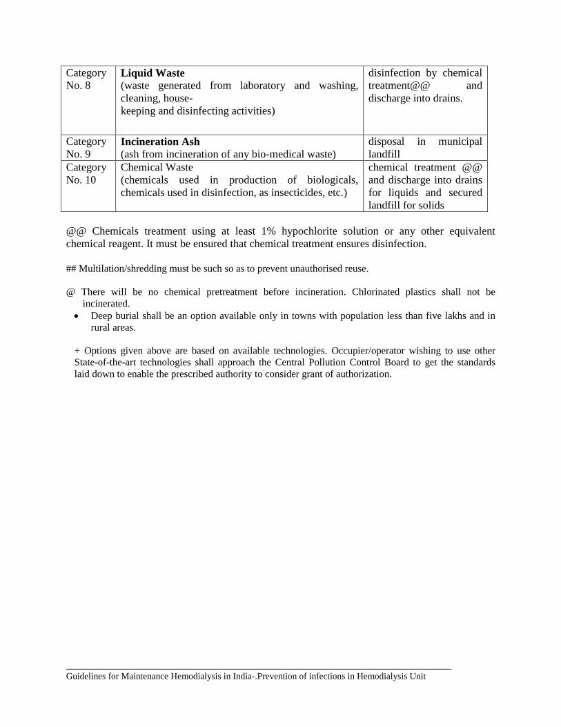
______________________________________________________________________ Guidelines for Maintenance Hemodialysis in India-.Prevention of infections in Hemodialysis Unit
Category
No. 8 Liquid Waste (waste generated from laboratory and washing,
cleaning, house-
keeping and disinfecting activities)
disinfection by chemical
treatment@@ and
discharge into drains.
Category
No. 9 Incineration Ash (ash from incineration of any bio-medical waste)
disposal in municipal
landfill
Category
No. 10
Chemical Waste
(chemicals used in production of biologicals,
chemicals used in disinfection, as insecticides, etc.)
chemical treatment @@
and discharge into drains
for liquids and secured
landfill for solids
@@ Chemicals treatment using at least 1% hypochlorite solution or any other equivalent
chemical reagent. It must be ensured that chemical treatment ensures disinfection.
## Multilation/shredding must be such so as to prevent unauthorised reuse.
@ There will be no chemical pretreatment before incineration. Chlorinated plastics shall not be
incinerated.
Deep burial shall be an option available only in towns with population less than five lakhs and in
rural areas.
+ Options given above are based on available technologies. Occupier/operator wishing to use other
State-of-the-art technologies shall approach the Central Pollution Control Board to get the standards
laid down to enable the prescribed authority to consider grant of authorization.
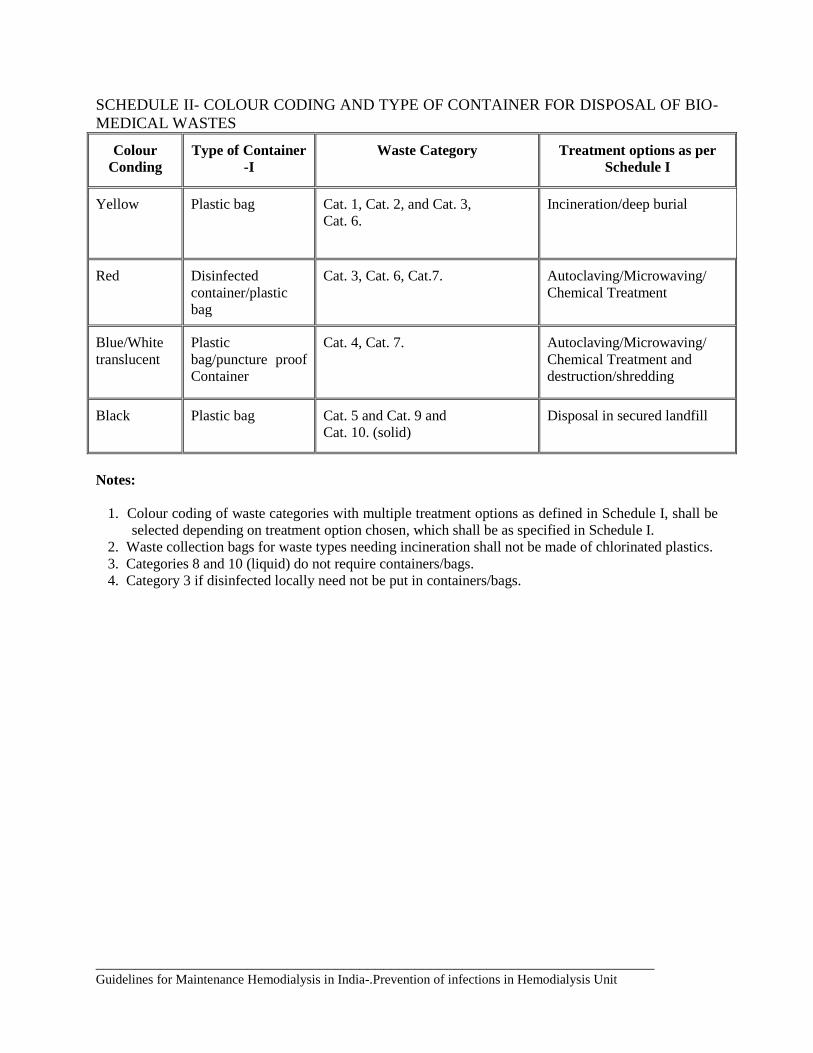
______________________________________________________________________ Guidelines for Maintenance Hemodialysis in India-.Prevention of infections in Hemodialysis Unit
SCHEDULE II- COLOUR CODING AND TYPE OF CONTAINER FOR DISPOSAL OF BIO-
MEDICAL WASTES
Colour
Conding
Type of Container
-I
Waste Category Treatment options as per
Schedule I
Yellow Plastic bag Cat. 1, Cat. 2, and Cat. 3,
Cat. 6.
Incineration/deep burial
Red Disinfected
container/plastic
bag
Cat. 3, Cat. 6, Cat.7. Autoclaving/Microwaving/
Chemical Treatment
Blue/White
translucent
Plastic
bag/puncture proof
Container
Cat. 4, Cat. 7.
Autoclaving/Microwaving/
Chemical Treatment and
destruction/shredding
Black Plastic bag Cat. 5 and Cat. 9 and
Cat. 10. (solid)
Disposal in secured landfill
Notes:
1. Colour coding of waste categories with multiple treatment options as defined in Schedule I, shall be
selected depending on treatment option chosen, which shall be as specified in Schedule I.
2. Waste collection bags for waste types needing incineration shall not be made of chlorinated plastics.
3. Categories 8 and 10 (liquid) do not require containers/bags.
4. Category 3 if disinfected locally need not be put in containers/bags.
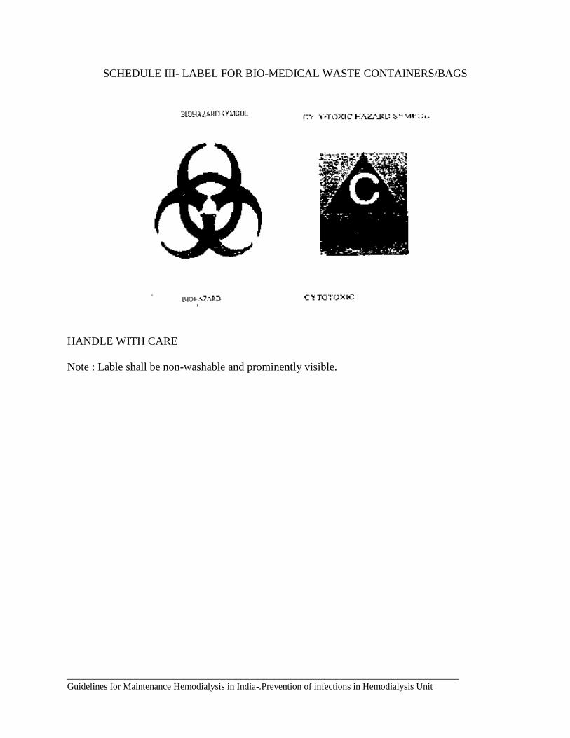
______________________________________________________________________ Guidelines for Maintenance Hemodialysis in India-.Prevention of infections in Hemodialysis Unit
SCHEDULE III- LABEL FOR BIO-MEDICAL WASTE CONTAINERS/BAGS
HANDLE WITH CARE
Note : Lable shall be non-washable and prominently visible.
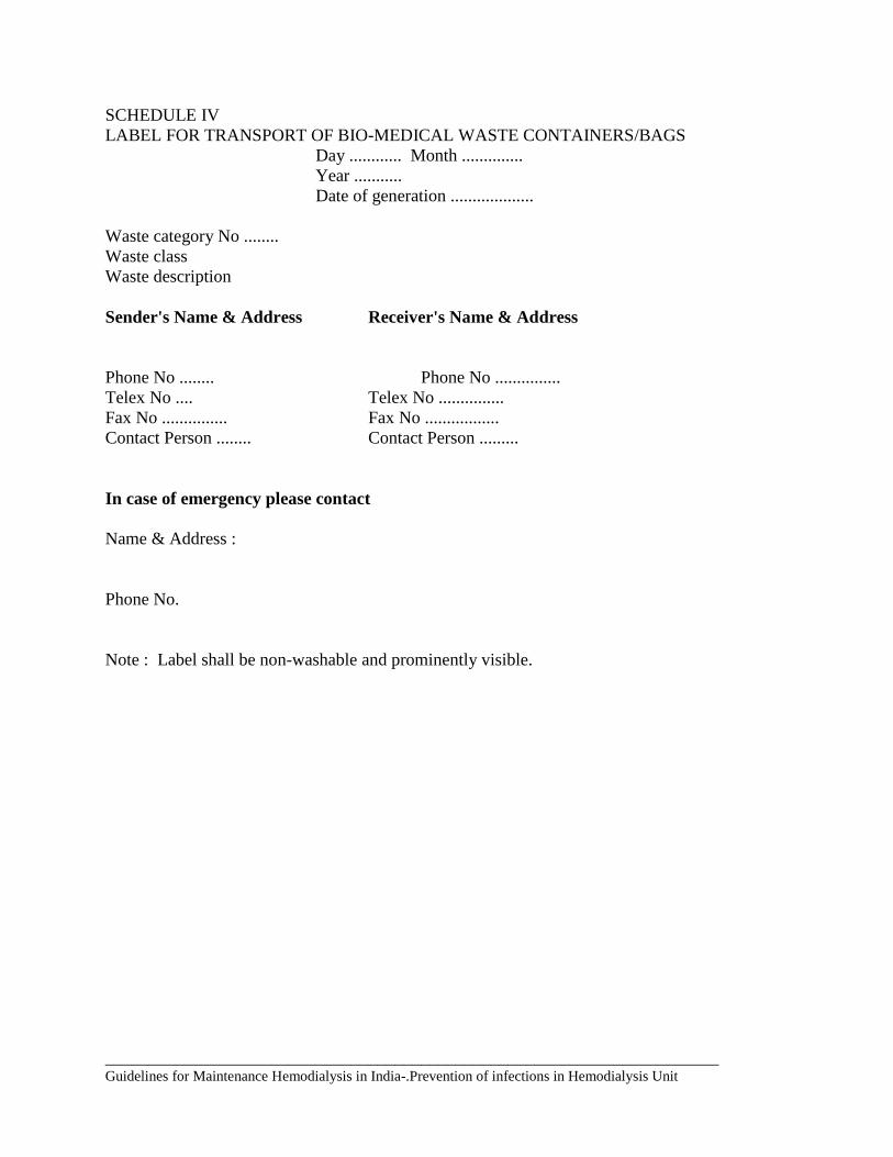
______________________________________________________________________ Guidelines for Maintenance Hemodialysis in India-.Prevention of infections in Hemodialysis Unit
SCHEDULE IV
LABEL FOR TRANSPORT OF BIO-MEDICAL WASTE CONTAINERS/BAGS
Day ............ Month ..............
Year ...........
Date of generation ...................
Waste category No ........
Waste class
Waste description
Sender's Name & Address Receiver's Name & Address
Phone No ........ Phone No ...............
Telex No .... Telex No ...............
Fax No ............... Fax No .................
Contact Person ........ Contact Person .........
In case of emergency please contact
Name & Address :
Phone No.
Note : Label shall be non-washable and prominently visible.

______________________________________________________________________ Guidelines for Maintenance Hemodialysis in India- Emergency services Page 85 of 74
15. EMERGENCY SERVICES
Rationale: The process of hemodialysis is akin to a major surgical operation. At any given time a
fixed amount of blood is in the extracorporeal circuit which is not under the physiological
control and the normal feedback mechanisms of the patient. Although most dialysis machines are
equipped with a fail safe mode, a self-test, alarms and a safety profile of less than 1 event per
100 million treatments, emergencies related to personal error, and patients intrinsic condition
ranging from minor discomfort to cardiac arrests on hemodialysis have been reported in dialysis
units. The following guideline elaborates the personnel protocols and equipment required for
managing emergencies in the dialysis unit.
Description:
Hemodialysis unit may be located in the premises of a hospital or it may be a standalone HD
unit. In either case emergency equipment, personnel and medicines are to be kept ready in the
unit for urgent use before the patient is shifted to ICU.
The common haemodialysis emergencies are:
1. Hypotension
2. Dialyzer reactions
a. Type A (anaphylactic reaction)
b. Type B (non-specific reaction)
3. Haemolysis
4. Air embolism
5. Disequilibrium syndrome
6. Chest pain, MI
7. Arrhythmias
8. Sudden cardiac arrest
Personnel
1. Nephrologist
2. Resident doctors-One per shift in three shifts
3. Dialysis technologists – one per two machine-- in three shifts
4. Nurses—one per three patients—in three shifts
5. Anaesthesiologists during day time and on call
6. Anaesthesia technician
Nephrologist DM/DNB should be available roud the clock, atleast on call, for managing
emergencies. Resident Doctors should be trained in identifying cardiac arrhythmias,
cardioversion and intubation technique. Nurses should be able to handle EKG machine and
cardiac monitors. All should be ACLS certified. An Anesthesiologist should be available on
call.
______________________________________________________________________ Guidelines for Maintenance Hemodialysis in India- Emergency services Page 86 of 74
Equipment required to prevent and treat these emergencies
1. Accurate weighing scale to exactly measure weight.
2. Dialysis machines with ultrafiltration controller and sodium modelling to prevent
hypotension. Spare HD machine is advisable
3. Micro-haematocrit tube for manual measurement
4. Activated clotting time machine.
5. Glucometer.
6. Multichannel cardiac monitor, Signal-averaged ECG (SAECG) and defibrillator.
7. Laryngoscopes, Endotracheal tubes, Suction apparatus or wall mounted suction, Central
oxygen supply & suction tubes, mouth gag and Ambu Bag
8. Ryles tube.
9. Arterial blood gas analysis machine.
10. 24 hour emergency power generator to ensure uninterrupted power supply.
Equipment not required for an emergency, but useful in preventing an emergency
1. Implantable cardioverter-defibrillator.
2. Ambulatory blood pressure monitor.
3. Portable ultrasound for abdominal emergencies.
4. Hand held doppler device for vascular access assessment.
These are optional equipments and may be made available depending on the size of the unit.
Medicines to be available for emergency use
1. Ionotropes: Injections: Dopamine, Dobutamine, Nor-adrenaline, vasopressin
2. Solutions: 25% dextrose; 3% saline; 5% dextrose
3. Injection Protamine
4. Injections: Lignocaine, amiodarone
5. Injection Hydrocortisone
6. Injection Adrenaline
7. Injection Atropine
8. Injection and tablet Pheniramine maleate
9. Capsule and tablet Nifedepine
10. Tablets: Clonidine, paracetamol, sorbitrate
11. Injection Nitroglycerine
12. Injections: Ondansetron, metoclopramide, pantoprazole, ranitidine
13. Injection vitamin K
14. Anti-convulsants Medozolam, Dilantin
15. Salbutamol
All medicines are to be stacked in Crash Carts in adequate quantities depending on
patient load.
Expiry dates of medicines to be verified periodically.
Stocks are to be verified every morning and replaced.
______________________________________________________________________ Guidelines for Maintenance Hemodialysis in India- Emergency services Page 87 of 74
An Intensive Care and a Respiratory Care Unit are to be within the reach so that a
critically ill patient may be shifted there, without delay.
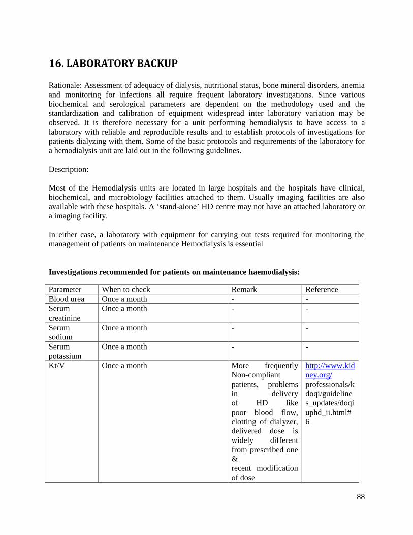
88
16. LABORATORY BACKUP
Rationale: Assessment of adequacy of dialysis, nutritional status, bone mineral disorders, anemia
and monitoring for infections all require frequent laboratory investigations. Since various
biochemical and serological parameters are dependent on the methodology used and the
standardization and calibration of equipment widespread inter laboratory variation may be
observed. It is therefore necessary for a unit performing hemodialysis to have access to a
laboratory with reliable and reproducible results and to establish protocols of investigations for
patients dialyzing with them. Some of the basic protocols and requirements of the laboratory for
a hemodialysis unit are laid out in the following guidelines.
Description:
Most of the Hemodialysis units are located in large hospitals and the hospitals have clinical,
biochemical, and microbiology facilities attached to them. Usually imaging facilities are also
available with these hospitals. A ‘stand-alone’ HD centre may not have an attached laboratory or
a imaging facility.
In either case, a laboratory with equipment for carrying out tests required for monitoring the
management of patients on maintenance Hemodialysis is essential
Investigations recommended for patients on maintenance haemodialysis:
Parameter When to check Remark Reference
Blood urea Once a month - -
Serum
creatinine
Once a month - -
Serum
sodium
Once a month - -
Serum
potassium
Once a month - -
Kt/V Once a month More frequently
Non-compliant
patients, problems
in delivery
of HD like
poor blood flow,
clotting of dialyzer,
delivered dose is
widely different
from prescribed one
&
recent modification
of dose
http://www.kid
ney.org/
professionals/k
doqi/guideline
s_updates/doqi
uphd_ii.html#
6
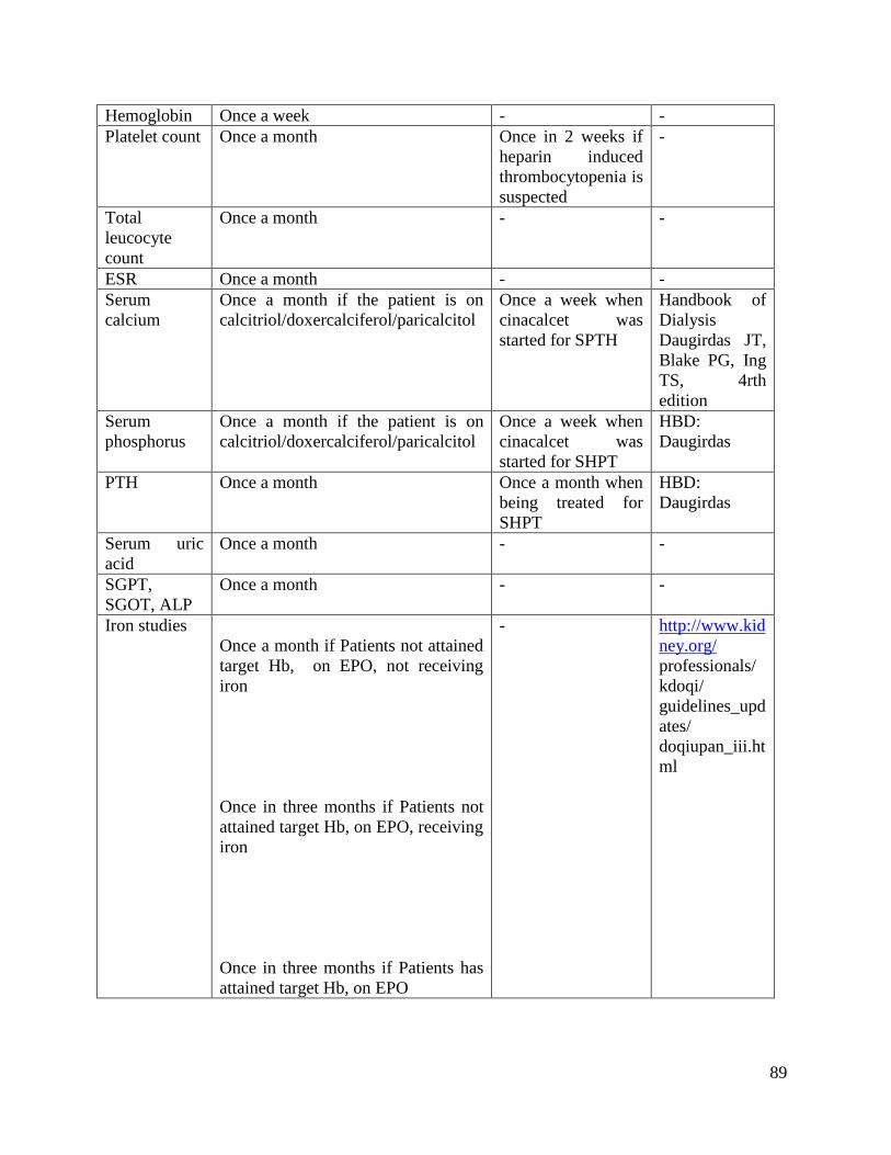
89
Hemoglobin Once a week - -
Platelet count Once a month Once in 2 weeks if
heparin induced
thrombocytopenia is
suspected
-
Total
leucocyte
count
Once a month - -
ESR Once a month - -
Serum
calcium
Once a month if the patient is on
calcitriol/doxercalciferol/paricalcitol
Once a week when
cinacalcet was
started for SPTH
Handbook of
Dialysis
Daugirdas JT,
Blake PG, Ing
TS, 4rth
edition
Serum
phosphorus
Once a month if the patient is on
calcitriol/doxercalciferol/paricalcitol
Once a week when
cinacalcet was
started for SHPT
HBD:
Daugirdas
PTH Once a month Once a month when
being treated for
SHPT
HBD:
Daugirdas
Serum uric
acid
Once a month - -
SGPT,
SGOT, ALP
Once a month - -
Iron studies
Once a month if Patients not attained
target Hb, on EPO, not receiving
iron
Once in three months if Patients not
attained target Hb, on EPO, receiving
iron
Once in three months if Patients has
attained target Hb, on EPO
- http://www.kid
ney.org/
professionals/
kdoqi/
guidelines_upd
ates/
doqiupan_iii.ht
ml
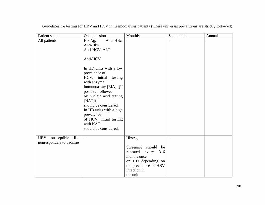
90
Guidelines for testing for HBV and HCV in haemodialysis patients (where universal precautions are strictly followed)
Patient status On admission Monthly Semiannual Annual
All patients HbsAg, Anti-HBc,
Anti-Hbs,
Anti-HCV, ALT
Anti-HCV
In HD units with a low
prevalence of
HCV, initial testing
with enzyme
immunoassay [EIA]; (if
positive, followed
by nucleic acid testing
[NAT])
should be considered.
In HD units with a high
prevalence
of HCV, initial testing
with NAT
should be considered.
- - -
HBV susceptible like
nonresponders to vaccine
- HbsAg
Screening should be
repeated every 3–6
months once
on HD depending on
the prevalence of HBV
infection in
the unit
-
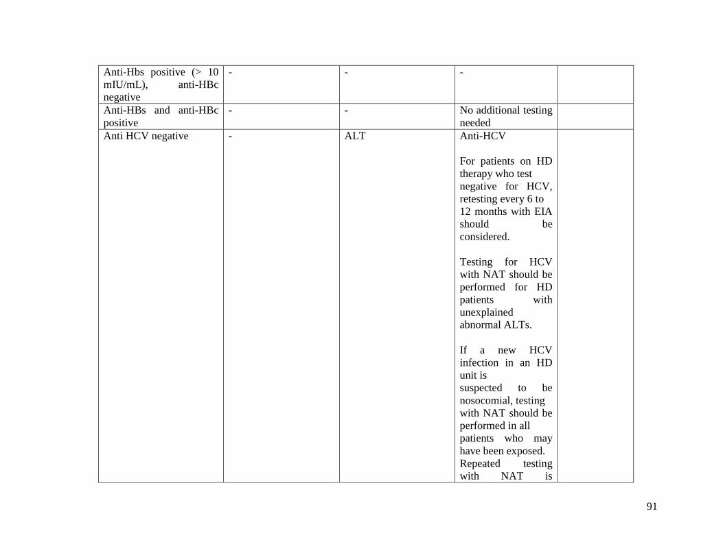
91
Anti-Hbs positive (> 10
mIU/mL), anti-HBc
negative
- - -
Anti-HBs and anti-HBc
positive
- - No additional testing
needed
Anti HCV negative - ALT Anti-HCV
For patients on HD
therapy who test
negative for HCV,
retesting every 6 to
12 months with EIA
should be
considered.
Testing for HCV
with NAT should be
performed for HD
patients with
unexplained
abnormal ALTs.
If a new HCV
infection in an HD
unit is
suspected to be
nosocomial, testing
with NAT should be
performed in all
patients who may
have been exposed.
Repeated testing
with NAT is
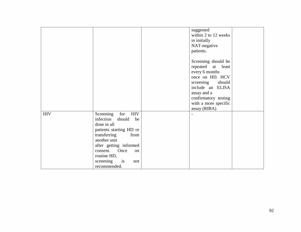
92
suggested
within 2 to 12 weeks
in initially
NAT-negative
patients.
Screening should be
repeated at least
every 6 months
once on HD. HCV
screening should
include an ELISA
assay and a
confirmatory testing
with a more specific
assay (RIBA)
HIV Screening for HIV
infection should be
done in all
patients starting HD or
transferring from
another unit
after getting informed
consent. Once on
routine HD,
screening is not
recommended.
-
93
Equipment required for investigations:
A. Biochemistry
1. Semi auto analyser / Bench Top auto analyser
2. Electrolyte Analyser (Ion selective electrode)
3. Semi auto coagulation analyser
B. Imaging facility.
1. 500 ma X-ray
2. Ultra Sound, Echocardiography, Doppler
94
C. Clinical Pathology
1. Cell counter
2. Urine analyser / Binocular Microscope
3. pH meter
D. Microbiology
1. Manual Elisa Reader / Immunoassay analyser
2. Bacterial cultures
E. Blood Bank
Rationale:
A.1.
Auto analyzers are recommended as analysis by enzymatic method is accurate compared to
calorimetric method. Bench top analyzers are good and less expensive, whereas semi auto / auto
analyzers are more expensive. Units may choose the equipment depending on the work load and
expertise available. Analysers and kits for doing all the tests listed below have to be made
available in the attached laboratories.
A.2.
Electrolyte analysis with Ion selective analyzer is accurate and reliable. Flame photometers are
not recommended.
A.3.
For coagulation test like clotting time, PT and APTT, manual methods may be used. However,
depending on workload, semi auto coagulation analyser may be installed which is preferable.
B.1.
500ma X-ray machine is necessary for radiological evaluation.
B.2.
Ultra sound ,echo and Doppler are required for cardiovascular and other organ evaluation .
C.
Cell counters for quick and accurate cell counts and urine analyser for various urine test are
important. However, a good binocular microscope and routine equipments for cell counts and
urine microscopy is also adequate.
D.1.
Manual Elisa readers for screening for HBs Ag. HCV and HIV are good and adequate.
However, depending on the work load and need for other investigations, Immunoassay analyser
may be used in place of Elisa readers
95
E.
HD centres must have access to Blood Bank which has facilities for component separation.
References:
1. CDC guidelines; accessed on
http://www.cdc.gov/mmwr/preview/mmwrhtml/rr5005a1.htm#
2. European Best Practice Guidelines for Haemodialysis (Part 1)
Nephrol Dial Trans Volume 17 suppl 7 July 2002
3. Gordon CE, Balk EM, Becker BN, Crooks PA, Jaber BL, Johnson CA, Michael MA,
Pereira BJ, Uhlig K, Levin A, US Commentary on the KDIGO Clinical Practice
Guideline for the Prevention, Diagnosis, Evaluation, and Treatment of Hepatitis C in
CKD Am J Kidney Dis 52:811-825, 2008
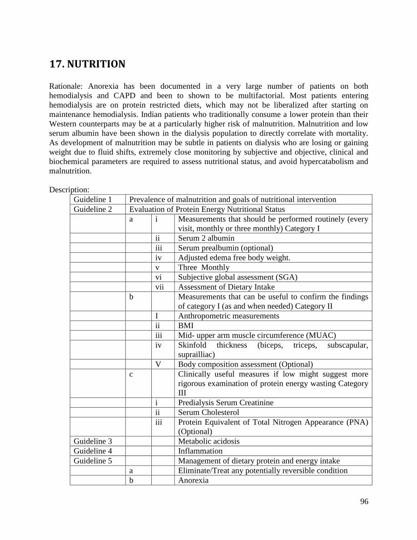
96
17. NUTRITION
Rationale: Anorexia has been documented in a very large number of patients on both
hemodialysis and CAPD and been to shown to be multifactorial. Most patients entering
hemodialysis are on protein restricted diets, which may not be liberalized after starting on
maintenance hemodialysis. Indian patients who traditionally consume a lower protein than their
Western counterparts may be at a particularly higher risk of malnutrition. Malnutrition and low
serum albumin have been shown in the dialysis population to directly correlate with mortality.
As development of malnutrition may be subtle in patients on dialysis who are losing or gaining
weight due to fluid shifts, extremely close monitoring by subjective and objective, clinical and
biochemical parameters are required to assess nutritional status, and avoid hypercatabolism and
malnutrition.
Description:
Guideline 1 Prevalence of malnutrition and goals of nutritional intervention
Guideline 2 Evaluation of Protein Energy Nutritional Status
a i Measurements that should be performed routinely (every
visit, monthly or three monthly) Category I
ii Serum 2 albumin
iii Serum prealbumin (optional)
iv Adjusted edema free body weight.
v Three Monthly
vi Subjective global assessment (SGA)
vii Assessment of Dietary Intake
b Measurements that can be useful to confirm the findings
of category I (as and when needed) Category II
I Anthropometric measurements
ii BMI
iii Mid- upper arm muscle circumference (MUAC)
iv Skinfold thickness (biceps, triceps, subscapular,
suprailliac)
V Body composition assessment (Optional)
c Clinically useful measures if low might suggest more
rigorous examination of protein energy wasting Category
III
i Predialysis Serum Creatinine
ii Serum Cholesterol
iii Protein Equivalent of Total Nitrogen Appearance (PNA)
(Optional)
Guideline 3 Metabolic acidosis
Guideline 4 Inflammation
Guideline 5 Management of dietary protein and energy intake
a Eliminate/Treat any potentially reversible condition
b Anorexia
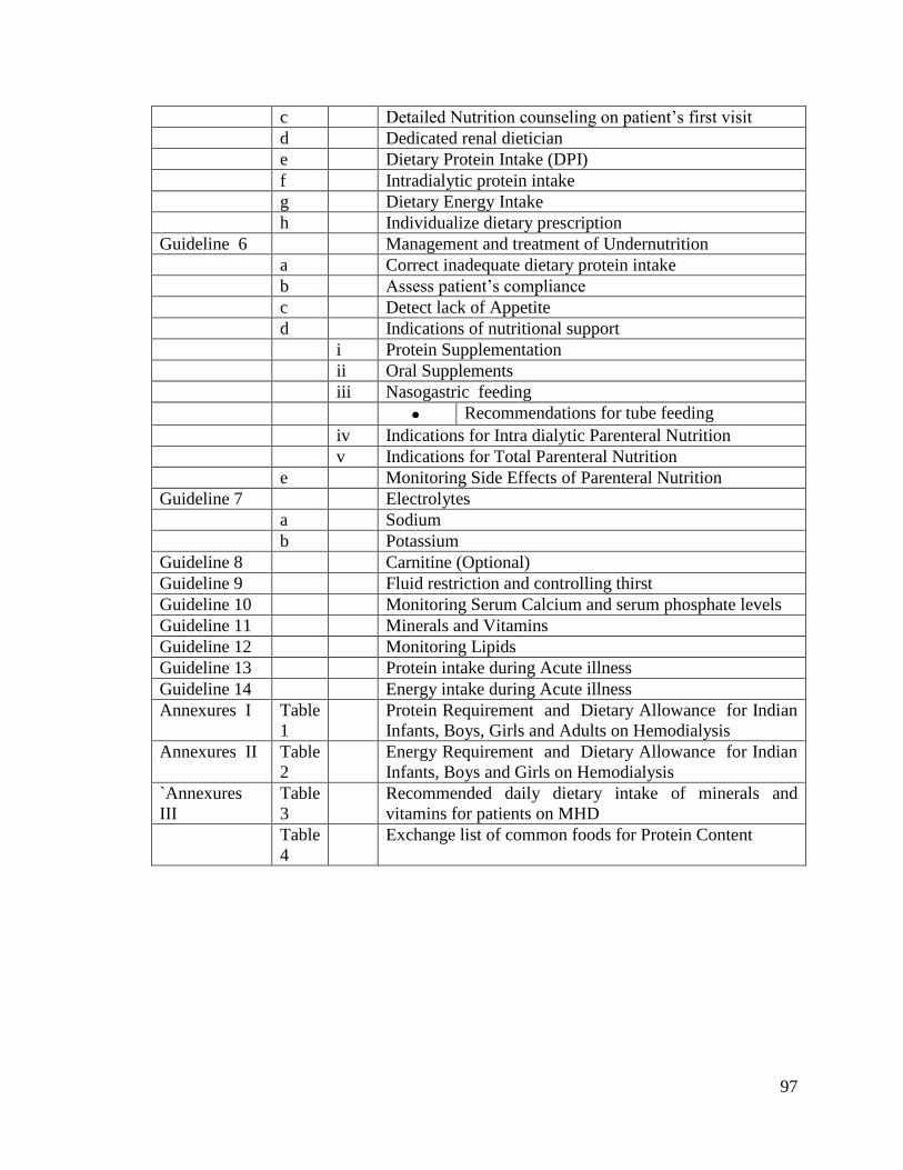
97
c Detailed Nutrition counseling on patient’s first visit
d Dedicated renal dietician
e Dietary Protein Intake (DPI)
f Intradialytic protein intake
g Dietary Energy Intake
h Individualize dietary prescription
Guideline 6 Management and treatment of Undernutrition
a Correct inadequate dietary protein intake
b Assess patient’s compliance
c Detect lack of Appetite
d Indications of nutritional support
i Protein Supplementation
ii Oral Supplements
iii Nasogastric feeding
Recommendations for tube feeding
iv Indications for Intra dialytic Parenteral Nutrition
v Indications for Total Parenteral Nutrition
e Monitoring Side Effects of Parenteral Nutrition
Guideline 7 Electrolytes
a Sodium
b Potassium
Guideline 8 Carnitine (Optional)
Guideline 9 Fluid restriction and controlling thirst
Guideline 10 Monitoring Serum Calcium and serum phosphate levels
Guideline 11 Minerals and Vitamins
Guideline 12 Monitoring Lipids
Guideline 13 Protein intake during Acute illness
Guideline 14 Energy intake during Acute illness
Annexures I Table
1
Protein Requirement and Dietary Allowance for Indian
Infants, Boys, Girls and Adults on Hemodialysis
Annexures II Table
2
Energy Requirement and Dietary Allowance for Indian
Infants, Boys and Girls on Hemodialysis
`Annexures
III
Table
3
Recommended daily dietary intake of minerals and
vitamins for patients on MHD
Table
4
Exchange list of common foods for Protein Content
98
Guideline 1. Prevalence of malnutrition and goals of
nutritional intervention Protein energy malnutrition (protein energy wasting (PEW)) is common (18%-70%) among
patients on maintenance hemodialysis (MHD). PEW is the strongest predictor of morbidity and
mortality, therefore, goals of nutritional therapy in dialysis patients are i) to prevent malnutrition,
ii) improve nutritional status, iii) build up body stores for good transplant outcome (if planned)
and iv) improve quality of life.
Hemo study has demonstrated that progressive renal insufficiency is associated with a
spontaneous decline in protein intake. Predialysis patients appear to have a spontaneous protein
intake of <0.7 g/kg/day, which is below the minimal recommended daily intake so by the time
patient starts hemodialysis, patient is already malnourished. Therefore, nutritional status should
be assessed at the start of maintenance haemodialysis (MHD), PEW should be avoided during
MHD because of poor patient outcome, and in the absence of malnutrition, nutritional status
should be monitored every 2 months in patients <50 years of age and in patients >50 years of age
nutritional status should be monitored every month.
In India, protein intake of an average middle class individual is less than 0.7g/kgbw/day. Also
recommended cut offs for Asian population for BMI are lower than those of Western population
because of difference in body size. Although recommendations for dietary protein intake are
based on recommended dietary allowances for Indian population, which are quite similar to those
of western population, but for an Indian patient to switch over from moderately low protein diet
to a very high protein diet may be difficult because of i) body may not accept such high quantity
of protein instantneously, ii) it may affect residual renal function, iii) patient can get uremic
because of excessively high protein intake and iv) affordability. Therefore, although, patient
should be put on high protein diet, but the target should be achieved gradually over a period of
time depending upon how much the patient can easily tolerate and digest. Patient should be
given high calorie diet to ensure proper utilization of protein.
Guideline 2 – Evaluation of Protein Energy Nutritional Status
Nutritional status in maintenance dialysis patients should be assessed with a combination of valid
complementary measures rather than by any single measure alone because no single measure
provides a comprehensive indication of nutritional status. Measures of intake, visceral and
somatic protein stores, body composition, and functional status identify different aspects of
nutrition status. Therefore, using combination of nutritional measures provides an assessment of
visceral and somatic protein body pools, body weight and hence fat mass and nutrient intake.
These can be divided into three categories: Category I: Measurements that should be performed
routinely (every visit, monthly or three monthly), Category II: Measures that can be useful to
confirm the findings of category I (as and when needed) and Category III: Clinically useful
measures if low might suggest more rigorous examination of protein energy wasting:
2a ) Measurements that should be performed routinely (every visit, monthly or three
monthly) Category I
99
2a i.) Every visit Assessment: Protocol should include a) medical history, b) physical
examination (including deficiency signs on skin), c) hydration status, 4) difference between
actual weight and dry weight. Malnutrition may be identified with greater sensitivity and
specificity using a combination of factors including calculating weight loss <85% of ideal
weight.
Stabilized serum albumin, percent of usual edema free post dialysis weight, dietary diaries
(dietician’s role) should be evaluated monthly.
2a ii) Serum albumin is a valid and clinically useful measure of protein energy nutritional status
in maintenance dialysis patients. Serum albumin is recommended for routine measurement
because there is a large body of literature that defines the normal albumin values. Predialysis or
stabilized serum albumin should be checked monthly, maintained at ≥ 4.0g/dL using Bromcresol
Green (BCG) method. Since half life of albumin is ~20 days, it should evaluated once month.
However, the limitations of using serum albumin as a nutritional marker are that the levels
decrease in Inflammation & Infection.
2 a iii) Serum prealbumin (optional): Serum prealbumin is a valid and clinically useful
measure of protein energy nutritional status in maintenance dialysis patients. In Indian scenario
cost inhibits regular use of serum prealbumin as a nutritional marker though its advantage is a
much shorter half life ~2-3 days. However, depending upon affordability, serum prealbumin can
be used as a marker of nutritional status. Prealbumin should be greater than 30 mg/dL
Serum prealbumin is a negative acute phase reactant protein, as its levels decrease in
Inflammation & stress, so therefore prealbumin may not correlate well with other nutritional
parameters. Also levels are increased in renal disease due to impaired degradation by kidneys.
2 a iv) Adjusted edema free body weight. Body weight should be obtained post dialysis. For
individuals whose edema free body weight is between 95th
and 115th
percent of median standard
weight, the actual edema free body weight may be used. Following equation can be used to
calculate edema free adjusted body weight (aBWef)
aBW ef= BWef + SBW-BWef x 0.25
Where BW ef is the actual edema free body weight and SBW is standard body weight as
determined from NHANES II data. Because of interdialytic weight gain, aBW ef should be
calculated based on post dialysis values.
2 a v.) Three Monthly Assessments: Clinician must examine fat deposits/ depletion, muscle
mass (Mid-upper arm circumference (cm)/3.14-triceps skinfold mm)
2 a vi) Subjective global assessment (SGA) is recommended because it gives a comprehensive
overview of nutritional intake and body composition including a rough assessment of both
muscle and fat mass and because it is correlated with mortality rates. It is recommended that
SGA scoring be determined by 4 item, 7- point scale used in CANUSA Study. SGA score correlates with objective measures (albumin/weight/intake/anthropometry). Change in SGA
rating by 1 point decreases relative risk of death by 25%. A higher SGA score is associated with
a lower RR of death and fewer hospitalized days/year (CANUSA study) SGA should be six
monthly.
100
Recommendations for future Research: Should BMI, Serum albumin and serum TIBC be
included in SGA scoring as is done in malnutrition inflammation scores (MIS).
2 a vii) Assessment of Dietary Intake should be done using 1.) Diet History Questionnaires, 2.)
Food weighing and 3.) Observation. Food weighing is difficult for patients who visit dialysis
units on out patient basis. Assessment of food intake using diet history questionnaires is
therefore more appropriate for such patients. Dietary interviews and diaries can be used to
assess intake not only of protein and energy but also of a variety of other nutrients as well as
pattern and frequency of meals. Dietary interviews and diaries should be followed once in 3
months.
2 b) Measures that can be useful to confirm the findings of category I (as and when needed)
2b i) Anthropometric measurements are valid and clinically useful indicators of protein
energy nutritional status in maintenance dialysis patients. These measures include, percent usual
body weight, percent standard body weight, height, Body mass index (BMI), Mid- upper arm
muscle circumference (MUAC), skinfold thickness (biceps, triceps, subscapular, suprailliac) and
waist/hip ratio. For anthropometric calculations, post dialysis actual edema free body weight
should be used. Patients in the lower 50th
percentile of weight for height clearly have a reduced
survival rate. Differences in anthropometric measurements among MD patients and normal
individuals may indicate a nutritional disorder or other clinical abnormality. Standard
international methods should be followed for performing anthropometry and calculating body
composition from measurements.
2b ii) BMI: Because height may decrease with aging in MHD patients particularly those with
bone disease, height should be measured once annually. Skeletal frame size must also be
determined to calculate individual’s standard body weight percent (%SBW).
2b iii) Mid- upper arm muscle circumference (MUAC) should preferablybe measured every three
months.
2 b iv) Skinfold thickness (biceps, triceps, subscapular, suprailliac) should preferably be
measured every six months.
2 b v) Body composition assessment (Optional): Depending upon availability of the equipment,
bioelectrical impedance analysis (BIA), Infrared reactance, or DEXA can be used to assess long
term adequacy of protein-energy nutritional status. Measurement can be repeated every six
months. Accurate data on body composition are helpful to. Although whole body DEXA is less
influenced by abnormalities in hydration status which are common in HD patients, it does not
distinguish between intracellular and extracellular water compartments. Every dialysis centre
may not have DEXA because of prohibitive cost and also it is not a bed side tool. Routine use of
DEXA is not recommended.
2c) Clinically useful measures if low might suggest more rigorous examination of protein
energy wasting: Predialysis creatinine, blood urea nitrogen, cholesterol, serum and urine
electrolytes , serum and Urine Urea Nitrogen, serum and visceral protein. A low predialysis or
stabilized serum urea level may indicate a low intake of protein and amino acids. Depending
upon availability of test facility and reliability and financial affordability: serum and blood cell
vitamin levels, plasma amino acid levels (Essn/NonEssn & valine/glycine) (Optional).
101
2c i) Predialysis Serum Creatinine:
Low stabilized predialysis creatinine of between 2.0-4.5 mg/dL with negligible renal function
should be investigated for low dietary protein intake and skeletal muscle wasting and risk for
high mortality
This is a direct indicator of protein intake and muscle mass. It is directly proportional to skeletal
muscle mass & dietary muscle intake According to NKF/KDOQI guideline 5 predialysis Serum
Creatinine of <10mg/dL is considered as high risk for Protein Energy Wasting (PEW) which
warrants thorough evaluation of Protein Energy Wasting (PEW). Mortality risk increases with
creatinine below 9-11 mg/dL in patients on MHD or PD. But these data are based on Western
population with significantly different body frame and size.
A predialysis serum creatinine of higher than 10mg/dL is not tolerated by Indian patients and
they become highly uremic (anorexia, vomiting, loss of taste) . Therefore, the threshold levels
of predialysis serum creatinine for Indian are much lower than those recommended by
NKF/KDOKi guidelines.
A low predialysis or stabilized serum creatinine level in MHD patients suggests decreased
skeletal muscle mass and or low dietary protein intake. Therefore in Indian patients, low
stabilized predialysis creatinine of between 2-4.5 mg/dL with negligible residual renal function
should be investigated for low dietary protein intake and skeletal muscle wasting and risk for
high mortality.
Recommendations for future research: To create threshold for predialysis creatinine for
Indian patients for evaluation of PEW.
2 c ii) Serum Cholesterol is a valid and clinically useful measure of protein energy nutritional
status in maintenance dialysis patients may be influenced by comorbid conditions.
Hypocholesterolemia is associated with chronic protein-energy intake/deficits and or the
presence of co morbid conditions including inflammation. Individuals with low normal <150-
180 mg/dL or declining serum cholesterol concentrations should be evaluated for nutritional
deficit and indicate as they have increased mortality risk. In stable patients the recommended
dietary intake of cholesterol is <200mg/d.
2c iii) Protein Equivalent of Total Nitrogen Appearance (PNA) (Optional):
Protein catabolic rate (PCR) is a valid and clinically useful measure of net protein degradation
and protein intake in maintenance dialysis patients. In a clinically stable patient PNA provides a
valid estimate of protein intake.
There are a number of technical problems with measuring PNA in individuals undergoing HD.
PNA approximates protein intake only when the patient is in zero nitrogen equilibrium. PNA
may fluctuate from day to day as a function of protein intake and a single measurement may not
reflect usual protein intake. When dietary protein intake (DPI) is high total nitrogen appearance
(TNA) underestimates protein intake. PNA may overestimate DPI when protein intake is less
than 1 g/kg/d possibly due to endogenous protein catabolism. Finally normalizing PNA to body
weight can be misleading in obese, malnourished and edematous patients.
Guideline 3. Management of Acid-Base Status

102
Guideline 3a Measurement of Serum Bicarbonate: Serum bicarbonate should be measured in
maintenance hemodialysis (MHD) patients once monthly. Low serum bicarbonate
concentrations in MHD patient almost always indicate metabolic acidosis. Acidemia associated
with metabolic acidosis is associated with increased oxidation of branched chain amino acids
(valine, leucine and isoleucine), increased protein degradation and PNA and decreased albumin
synthesis.
Guideline 3b Treatment of Low Serum Bicarbonate: Predialysis or stabilized serum
bicarbonate levels should be maintained at or above 22 mmol/L. Normalization of predialysis or
stabilized serum bicarbonate concentrations can be achieved by higher basic anion
concentrations in the dialysate and/or by oral supplementation with bicarbonate salts. Higher
concentrations of bicarbonate in hemodialysate (38mmo/L) have been shown to safely increase
predialysis serum bicarbonate concentrations. Oral dose of sodium bicarbonate usually about 2 to
4 g/d or 25 to 50 mEq/d can be used to increase bicarbonate concentration. Correction of
academia due to metabolic acidosis increases serum albumin and decreases protein degradation
rates. Most trials report that normalizing predialysis or stabilized serum bicarbonate
concentrations is beneficial for protein, amino acid and bone metabolism and protein-energy
nutritional status.
Guideline 4 Inflammation
Many patients undergoing HD show evidence of chronic inflammation with intermittent or
persistently elevated levels of acute phase proteins. C Reactive protein (CRP) levels should be
checked every 3 months. An elevated CRP is often associated with reduced serum albumin levels
secondary to impaired albumin synthesis. In this context hypoalbuminemia is an inflammatory
marker, rather than an index of poor dietary intake.
Guideline 5. Management of dietary protein and energy intake
Guideline 5a. Eliminate/Treat any potentially reversible or treatable condition (anemia)
or medication that might interfere with appetite or cause malnutrition.
Guideline 5b. Anorexia: Major proportion of patients treated with HD consume less protein and
energy than is recommended due to loss of appetite. Factors that contribute to anorexia are i)
underdialysis (switch over to thrice weekly dialysis in place of twice weekly dialysis therapy), ii)
comorbidity, iii) medication (in such circumstances discontinuing phosphate binders and iron
and vitamin supplements for a short period of time helps improve appetite) and iv) psychosocial
factors . These factors should eliminated.
Guideline 5c Dialysis regimen should be regularly monitored and modified to treat
intensification of the patient’s uremic state that is caused by superimposed illness. Maintain
KT/V of 1.2 in HD patients.
Guideline 5c) Detailed Nutrition counseling on patient’s first visit. New patients require proper counseling on disease, its causes, what has caused disease in the
patient, progression of disease, how to control progression, importance of nutritional counselling.
Clinician’s personal involvement in nutritional counseling is important for better compliance.
103
Guideline 5d) Dedicated renal dietician: Each center should have a dedicated renal dietician
who can follow up the patients. Patient must visit dietician regularly. Telephonic follow-ups:
Dietician should contact patients telephonically in order to motivate them to improve
compliance. Regular telephonic follow-up results in better nutritional status and QOL
(KDQOL).
Dietician should advise patients to maintain dietary diaries. Dietician should evaluate nutritional
status which should be evaluated every month. Even small decrease in nutritional indices
together with decrease in protein and energy intake strongly suggest need for frequent nutritional
monitoring (MDRD Study). Dietician should evaluate changes in body composition, loss of
muscle mass and weight with longitudinal anthropometry.
Guideline 5e) Dietary Protein Intake (DPI): Dietary protein intake for clinically stable MHD
patients should be 1.2 g/kgbw/d (Table 1). This amount is necessary to ensure neutral or positive
nitrogen balance. At least 50% of protein should be of high biological value (HBV)Proteins of
HBV have an amino acid composition that is similar to human protein and is likely to be utilized
more efficiently by humans to conserve , body proteins. Egg white, fish, chicken, milk and milk
products (curd, chenna/paneer), dehusked (without outer covering to prevent
hyperphosphatemia) lentils kidney beans, soy protein (milk and cheese marketed as Tofu) are
good sources of protein with HBV. Include two cereals in one meal eg:- rice and wheat.to
improve protein quality the ratio of cereal protein to pulse protein should be 4:1.
Guideline 5f) Intradialytic protein intake: Patients should be advised to eat high protein food
(high protein snack/chenna/curd/egg whites/protein biscuits etc) during dialysis to prevent
protein catabolism and to make up for losses due to dialysis procedure. Protein snacks should be
taken anytime after half an hour of initiation of dialysis.
Guideline 5g) Dietary Energy Intake: Recommended energy intake for MHD patients is 35
kcal/kg b.w if the patient is less than 60 years of age and 30 kcal/kg bw if the patient is more
than 60 year. Recommendations for children are based RDA for chronological age (Table 2) It is
recommended that 50-60% of total calories should come from carbohydrate, 30% of total
calories should come from fat (saturated fats <7%), and 20% of total calories should come from
protein. Energy intake of patients having diabetes mellitus should be 25 to 30 Kcal/kg/d. Blood
sugar levels should be monitored to avoid hyperglycemia.
Guideline 5h) Individualize dietary prescription: Renal diet has numerous restrictions
therefore adherence to such a diet can be difficult and stressful. Prescribed diets should be
individualized to help accommodate each patient’s unique circumstances in terms of palatability,
cost, comorbid medical conditions and cultural eating habits.
Guideline 6) Management and treatment of Undernutrition
Guideline 6a) Correct inadequate dietary protein intake: Patients who do not have adequate
DPI should first receive dietary counseling and education. If DPI remains inadequate oral
104
supplementation should be prescribed. If oral supplements are not tolerated or effective and
protein malnutrition is present consider tube feeding to increase protein intake.
Guideline 6b) Assess patient’s compliance: Assessment of patient compliance to dietary
prescription and nutritional intervention should be done on every visit. Patients who do not have
adequate dietary Intake should first receive dietary counseling and education. Meal plan should
be Individualized Patient should be motivated to eat enough calories for proper utilization of
protein.
Guideline 6c) Detect Lack Of Appetite: Appetite assessment tools are a valid and clinically
useful measure of estimating nutritional intake. It is recommended to use one or more of these
tools. (i) Appetite and diet assessment Tools (ADAT) for appropriate nutritional intervention.
Rating is based on prompt questions like during the past week how would you rate your appetite?
Rating may be1) Very good, 2) good, 3) fair, 4) poor and 5) very poor. (ii) Subjective Global
Assessment (SGA). Scores are based on prompt question “how would you grade your appetite in
the last week? Scoring may be 1) Good, 2) sometimes bad, 3) often bad, and 4) always
bad.Dietary Intake is assessed in terms of 1) overall change, 2) no change, 3) change and 4)
duration of change in weeks. Type of change is assessed in terms of 1) suboptimal solid diet, 2)
hypocaloric diet, 3) full liquid diet, 4) starvation.(iii) Kidney Disease Quality of Life-Short Form
(KDQL)Rating is based on question “To what extent were you bothered during past four weeks
by lack of appetite”? Scoring may be 1) Not at all, 2) somewhat, 3) moderately, 4) very much 5)
extremely.
Guideline 6 d) Indications of nutritional support: Patients who are unable to meet
protein/energy requirements with food for an extended period of time should receive nutrition
support. Extended period is defined as days to 2 weeks depending upon the severity of patient’s
clinical condition, degree of malnutrition,and degree of inadequacy of their nutritional intake.
Complete nutritional assessment is needed before intervention.
Guideline 6 d i) Protein Supplementation: In dialysis patients if DPI remains inadequate oral
supplementation should be prescribed.
Guideline 6 d ii) Oral Supplements: 1). Special Calorie Dense Commercial Formulas provide
2kcal/ml with high protein and low electrolytes. 2). Provide smaller water load than intravenous
feeds. Use of Alpha Keto-analogues (optional) may improve protein utilizationand reduce degree
of catabolism. Standard recommended dose of ketoacid dosage is 6 to 14 g daily. However,high
cost of Alpha Keto-analogues deters their use.
In case of children, supplemental nutritional support should be considered when a patient is not
growing normally or fails to consume the RDA for protein and/or energy. Supplementation by
oral route is preferred followed by enteral tube feeding.
Guideline 6d iii) Nasogastric feeding: If oral supplements are not tolerated or effective and
malnutrition is present consider tube feeding should be considered as it provides balanced
nutrients. But who decides regarding tube feeding? In India, most of the patients want to escape tube
feeding and because patient is reluctant to go for tube feeding because of discomfort caused by
105
Ryle’s tube, malnutrition worsens. It is the clinician’s responsibility to explain the potential
risks of worsening malnutrition and convince and motivate the patient and his attendants
for tube feeding.With tube feeding, overnight enteral supplements can improve nutritional
status. Tube feeding provides smaller water load than intravenous feeds, lowers risk of infection
than TPN , is less expensive, overnight supplementation improves nutritional status.
Guideline 6 diii
Recommendations for Tube feeding. 1. Start with 50-100 ml feeds every 6 hours and
gradually increase to 300-400ml per feeding. 2) If continuous feedings are started, then
start feeding from 20-50ml/hr, then increase 20ml every 2-8hrs until requirement is
reached.
Guideline 6d iv) Indications for Intra dialytic Parenteral Nutrition: If tube feedings are not
used then intra dialytic parenteral nutrition (IDPN) should be considered. In any case IDPN
should be given if spontaneous intake of energy is >20 & <25 kcal/kgIBW and if protein is >
0.8g but < 1g/kg/IBW. Consider regular use of IDPN during hemodialysis or SLED in anuric or
oliguric patients as because of fluid restriction IV nutrition cannot be used aggressively. An
equivoluminous degree of ultrafiltration should be added to regular UF rate to maintain fluid
balance. Minerals : Include sodium, potassium and Magnesium in the IDPN/TPN solution as per
patient’s requirement.
Guideline 6d v) Indications for Total Parenteral Nutrition: If combination of Oral and IDPN
is insufficient then total parenteral nutrition (TPN) should be considered. In any case TPNshould
be given if spontaneous intake is <20 kcal/kgIBW and < 0.8g protein/kgIBW.
Guideline 6e ) Monitoring Side Effects of Parenteral Nutrition:It is recommended to monitor
side effects of parenteral nutrition. 15-25% of the patients may get nausea and vomiting when
IDPN is initiated. In such cases, 1) decrease infusion rate, 2) reduce total IDPN by half for 1 to 2
weeks. Intradialytic cramping may occur in rare cases of low plasma osmolality if sodium
profiling is not used. It is recommended that 1gNaCl/250ml of infusion should be added to
IDPN. 3) Glucose metabolism should be checked. 4)Prevent hyperglycemia (>300mg/dL) by
administer ing 2-6 units short acting insulin.
Guideline 7 Electrolytes:
Guideline 7a Sodium: It is recommended that patients on HD should restrict sodium intake to
no more than 2 g/d. Patients with limited residual renal function and uncontrolled
hypertensionshould restrict its use to 1.5 grams/d. In case of hyponatremia (Na < 135 mmol/dL),
depending upon deficit, salt capsules made out of measured quantity of salt (prescribed for
correction) should be advised. In case of hypernatremia, correct hydration and rule out all
medical conditions which can cause hypernatremia. Restrict foods with high salt content
(papadams, pickles, chutney, sauce), dry fruits, popcorns, coconut water.
Guideline 7b Potassium: Potassium intake for a patient on HD should be 1mEq/kg BW/d.
Patients should be advised to leach potassium from green vegetables. Fruit juices and vegetable
soup should be avoided. Patients on HD should be allowed to take fruits with low potassium
106
content (<100mg/100g). Recommended fruits are apple, banana, pineapple, pear, orange, guava
and papaya (approximately 50-60 g/d). Patients should avoid green leafy vegetables and
vegetables with very high (>300mg/g) potassium content. Diabetic patients should not take
banana and orange. Hypokalemia may develop in patients who are on diuretics or who have low
protein intake. To correct hypokalemia, 60-80 mEq (or as per s. potassium level) of potassium
should be administered either orally or through intravenous route (if deficit is large). Anuric
patients on HD should have stricter control of potassium and advised to stop fruit intake if serum
potassium level approaches 4.9 mEq. Potassium binders should be prescribed in case of
hyperkalemia.
Guideline 8 Carnitine (Optional)
Administration of L-Carnitine may improve subjective symptoms such as malaise, muscle
weakness, intradialytic cramps, and hypotension, and quality of life in HD patients. It should,
therefore, be used keeping in mind patients’ condition. However, the totality of evidence is
insufficient to recommend its routine use. Carnitine may enhance responsiveness to
erythropoietin stimulating agents (ESA) in erythropoietin resistant anemia.
Guideline 9 Fluid restriction and controlling thirst
Recommended fluid intake for HD patients is 24 hour urine output + 500 ml for insensible. Fluid
includes all liquids (for example water, tea, milk, curd) consumed by the patient. However, if
the patient is in volume overload, the +500 ml for insensible losses should be reduced and
diuretic therapy should be started. However if the patient is anuric, diuretics should be avoided to
prevent. Electrolytes should be closely monitored. Salt should be restricted in order to control
thirst. Rinsing mouth when ever patient feels thirsty may bring down fluid intake.
Guideline 10 Monitoring Serum Calcium and serum phosphate
levels
Hypocalcemia should be treated with IV or oral calcium supplements. Stop calcium based
phosphate binders in hypercalcemia and shift patient on non-calcium based phosphate binders.
Consider discontinuing phosphate binders for few weeks if i) patient’s serum levels are within
normal range with strict instructions for avoiding foods rich in phosphates and ii) in case of loss
of appetite.
Guideline 11 Minerals and Vitamins
Recommended daily dietary intake of minerals and vitamins is given in Table 3.0. Zinc needs
special mention. Zinc supplements are recommended for patients having proteinuria.
In children 100% of the recommended dietary allowance is a reasonable starting point for water
soluble vitamin (thiamine, pyridoxine B12 and folic acid) requirement in children on MHD.
Nutritional status of water soluble vitamins be monitored. Supplementation should be considered
if dietary intake alone does not meet or exceed the RDA, if measured blood vitamin levels are
107
below normal values (monitor 4-6 months), or if clinical evidence of deficiency is present (low
folic acid or Vitamin B12 levels giving rise poor responsiveness to recombinant human
erythropoetin). An intake of 100 % of RDA should be the goal for vitamins A,C,E, K, zinc and
copper. Supplements of fat soluble vitamins should be avoided due to reduced renal clearance.
Vitamin K supplementation may be considered during antibiotic therapy.
Guideline 12 Monitoring Lipids
Restrict dietary fat and sugar intake. The therapeutic goal should be to achieve a low density
lipoprotein (LDL) cholesterol of <100 mg/dL and a fasting triglyceride level of <500 mg/dL.
Therapeutic life style changes diet, weight reduction, increased physical activity and treatment of
hyperglycemia if present. Diet should contain < 7% saturated fats, with polyunsaturated fat
<10% of total calories and monounsaturated fat <20% of total calories and with total fat at 20-
30% of total calories. Carbohydrates should not exceed 60% of total calories. In HD patients 20-
30g of fibre per day should be consumed to reduce dyslipedemia. If required drug therapy
should be started.
Guideline 13 Protein intake during Acute illness
Acutely ill patients on HD should receive at least 1.2-1.3g/kg/d depending upon catabolic rate.
Patient may require nasogastric feeds along with parenteral nutrition. Fluid overload should be
checked in patients on TPN.
In case of acute pancreatitis, oral intake should be stopped and IV fluids and parenteral
formulations containing medium chain triglycerides (MCTs) should be administered. In diabetic
patients blood sugars must be monitored and dose of insulin adjusted as per the requirement.
Guideline 14 Energy intake during Acute illness
Recommended energy intake for a maintenance dialysis patient who is acutely ill is at least 35
kcal/kg/d for those who are below 60 years and 30-35 kcal/d for those above 60 years of age. In
diabetic patients blood sugar levels should be monitored to avoid hyperglycemia.
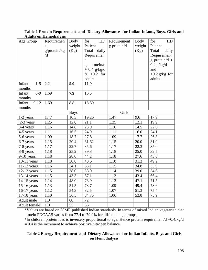
108
Table 1 Protein Requirement and Dietary Allowance for Indian Infants, Boys, Girls and
Adults on Hemodialysis
Age Group Requiremen
t
g/protein/kg
/d
Body
weight
(Kg)
for HD
Patient
Total daily
Requiremen
t
g protein/d
+ 0.4 g/kg/d
& +0.2 for
adults
Requirement
g protein/d
Body
weight
(Kg)
for HD
Patient
Total daily
Requirement
g protein/d +
0.4 g/kg/d
and
+0.2.g/kg for
adults
Infant 1-5
months
2.2 5.0 11.0
Infant 6-9
months
1.69 7.9 16.5
Infant 9-12
months
1.69 8.8 18.39
Boys Girls
1-2 years 1.47 10.3 19.26 1.47 9.6 17.9
2-3 years 1.25 12.8 21.1 1.25 12.1 19.9
3-4 years 1.16 14.8 23.0 1.16 14.5 22.6
4-5 years 1.11 16.5 24.9 1.11 16.0 24.1
5-6 years 1.09 18.7 27.8 1.09 17.7 26.3
6-7 years 1.15 20.4 31.62 1.15 20.0 31.0
7-8 years 1.17 22.7 35.6 1.17 22.3 35.0
8-9 years 1.18 25.2 39.8 1.18 25.0 39.5
9-10 years 1.18 28.0 44.2 1.18 27.6 43.6
10-11 years 1.18 30.8 48.6 1.18 31.2 49.2
11-12 years 1.16 34.1 53.1 1.15 34.8 53.9
12-13 years 1.15 38.0 58.9 1.14 39.0 54.6
13-14 years 1.15 43.3 67.1 1.13 43.4 66.4
14-15 years 1.14 48.0 73.9 1.12 47.1 71.5
15-16 years 1.13 51.5 78.7 1.09 49.4 73.6
16-17 years 1.12 54.3 82.5 1.07 51.3 75.4
17-18 years 1.10 56.5 84.75 1.06 52.8 75.9
Adult male 1.0 60 72
Adult female 1.0 55 66
*Values are based on ICMR published Indian standards. In terms of mixed Indian vegetarian diet
protein PDCAAS varies from 77.4 to 79.0% for different age groups.
*In children protein loss is inversely proportional to age. Hence protein requirement/d +0.4/kg/d
= 0.4 is the increment to achieve positive nitrogen balance.
Table 2 Energy Requirement and Dietary Allowance for Indian Infants, Boys and Girls
on Hemodialysis
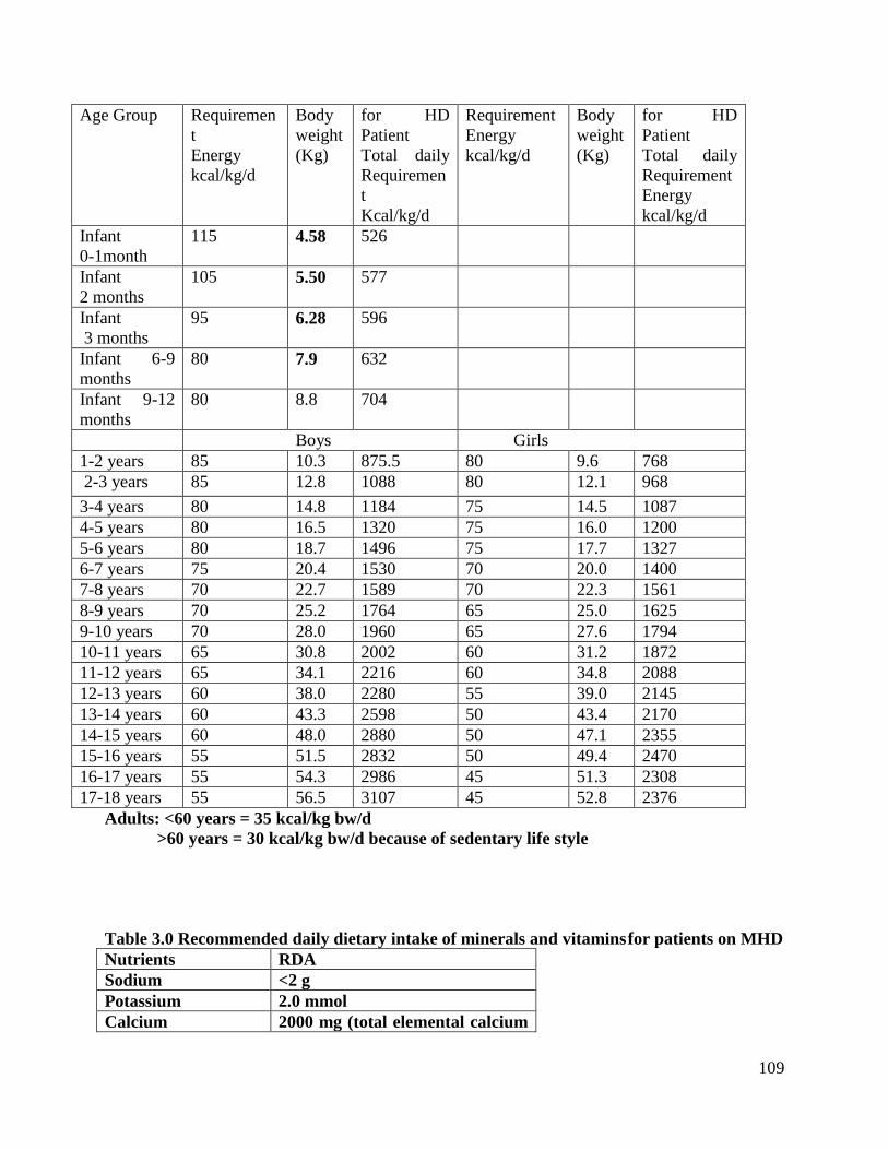
109
Age Group Requiremen
t
Energy
kcal/kg/d
Body
weight
(Kg)
for HD
Patient
Total daily
Requiremen
t
Kcal/kg/d
Requirement
Energy
kcal/kg/d
Body
weight
(Kg)
for HD
Patient
Total daily
Requirement
Energy
kcal/kg/d
Infant
0-1month
115 4.58
526
Infant
2 months
105 5.50
577
Infant
3 months
95 6.28
596
Infant 6-9
months
80 7.9 632
Infant 9-12
months
80 8.8 704
Boys Girls
1-2 years 85 10.3 875.5 80 9.6 768
2-3 years 85 12.8 1088 80 12.1 968
3-4 years 80 14.8 1184 75 14.5 1087
4-5 years 80 16.5 1320 75 16.0 1200
5-6 years 80 18.7 1496 75 17.7 1327
6-7 years 75 20.4 1530 70 20.0 1400
7-8 years 70 22.7 1589 70 22.3 1561
8-9 years 70 25.2 1764 65 25.0 1625
9-10 years 70 28.0 1960 65 27.6 1794
10-11 years 65 30.8 2002 60 31.2 1872
11-12 years 65 34.1 2216 60 34.8 2088
12-13 years 60 38.0 2280 55 39.0 2145
13-14 years 60 43.3 2598 50 43.4 2170
14-15 years 60 48.0 2880 50 47.1 2355
15-16 years 55 51.5 2832 50 49.4 2470
16-17 years 55 54.3 2986 45 51.3 2308
17-18 years 55 56.5 3107 45 52.8 2376
Adults: <60 years = 35 kcal/kg bw/d
>60 years = 30 kcal/kg bw/d because of sedentary life style
Table 3.0 Recommended daily dietary intake of minerals and vitamins for patients on MHD
Nutrients RDA
Sodium <2 g
Potassium 2.0 mmol
Calcium 2000 mg (total elemental calcium
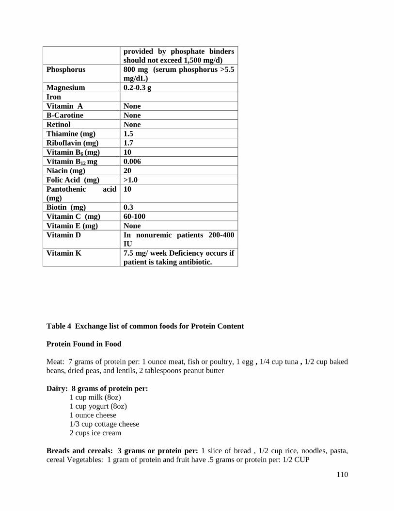
110
provided by phosphate binders
should not exceed 1,500 mg/d)
Phosphorus 800 mg (serum phosphorus >5.5
mg/dL)
Magnesium 0.2-0.3 g
Iron
Vitamin A None
Β-Carotine None
Retinol None
Thiamine (mg) 1.5
Riboflavin (mg) 1.7
Vitamin B6 (mg) 10
Vitamin B12 mg 0.006
Niacin (mg) 20
Folic Acid (mg) >1.0
Pantothenic acid
(mg)
10
Biotin (mg) 0.3
Vitamin C (mg) 60-100
Vitamin E (mg) None
Vitamin D In nonuremic patients 200-400
IU
Vitamin K 7.5 mg/ week Deficiency occurs if
patient is taking antibiotic.
Table 4 Exchange list of common foods for Protein Content
Protein Found in Food
Meat: 7 grams of protein per: 1 ounce meat, fish or poultry, 1 egg , 1/4 cup tuna , 1/2 cup baked
beans, dried peas, and lentils, 2 tablespoons peanut butter
Dairy: 8 grams of protein per: 1 cup milk (8oz)
1 cup yogurt (8oz)
1 ounce cheese
1/3 cup cottage cheese
2 cups ice cream
Breads and cereals: 3 grams or protein per: 1 slice of bread , 1/2 cup rice, noodles, pasta,
cereal Vegetables: 1 gram of protein and fruit have .5 grams or protein per: 1/2 CUP
111
8. CVD MONITORING & THERAPY
Rationale: The largest contribution to mortality in Chronic Kidney disease patients on dialysis is
from Cardiovascular Disease. A large number of patients especially those dialyzing in free
standing units may not attend outpatient clinics and cannot be completely assessed during a
dialysis session. Hemodialysis itself may impose an additional stress on the patient with pre
existing cardiovascular disease and patients on dialysis have both traditional and non traditional
risk factors. It therefore becomes necessary that every patient on dialysis be adequately
monitored and treated early for Cardiovascular disease. This document attempts to provide
guidelines on the optimum time and methods of monitoring patients for cardiovascular disease.
Description:
The burden of Cardio-vascular disease (CVD) in chronic kidney disease (CKD) is very high in
HD population. Beyond, traditional risk factors like diabetes mellitus, hypertension, smoking,
dyslipidemia, obesity in HD population many more non-traditional risk factors like volume
overload, anemia, mineral and bone disease, inflammation, oxidative stress etc contribute
significantly to very high prevalence of CVD.
In patients on MHD common types of CVD are; atherosclerotic vascular disease especially
involving coronary and cerebro-vascular arteries and Left Ventricular Hypertrophy. Other
common CVD manifestations are Congestive Heart Failure and peripheral vascular disease. The
CVD is major cause of morbidity and mortality in these patients. It is therefore important that
CVD is diagnosed and adequately monitored in a proactive manner.
ALL PATIENTS STARTING MHD MUST HAVE FOLLOWING DIAGNOSTIC
EVALUATION FOR CVD
On entry into HD unit:
Patients who are initiated for the first time, usually they are having significant hemodynamic,
metabolic, biochemical and volume imbalance. Hence, the investigation for CVD should be
deferred till the based weight, appropriate Hb, volume, electrolyte and divalent balance is
achieved.
It is mandatory to perform baseline 12 lead ECG, Chest X-ray and Echocardiogram in all
patients to assess their baseline status of CVS and to identify and stratify their risk for future
CVD.
Electrocardiography (Mandatory)
All dialysis patients should have baseline ECG at the start of maintenance hemodialysis and then
it should be ordered as and when clinically indicated. Interpretation of ECG should take into
account the shifts of volume, changes in electrolytes and anemia.

112
Echocardiography (Mandatory)
Two-dimensional and M mode echocardiography provides a noninvasive assessment of left
ventricular structure and function, together with imaging of valves and pericardium. Systolic
dysfunction, diagnosed by low fractional shortening or ejection fraction can be determined, as
can LV geometry and LV hypertrophy. The degree of hypertrophy can be identified by increased
LV wall thickness or by calculating LV mass index according to various formulae. LV mass
measurement varies over the course of a hemodialysis session by as much as 25 g/m. Therefore
where possible, imaging should be carried out when the patient has achieved their “base dry
weight”. Diastolic LV function can be assessed noninvasively using pulsed Doppler analysis of
flow across the mitral valve during diastole. Baseline and annual assessment of
echocardiography should be followed in all dialysis patients.
Dobutamine stress echocardiography (Optional) could be utilized as a screening tool for
ischemic heart disease in HD patients. It can also be used in patients with valvular disease
or impaired systolic function to assess underlying systolic reserve. Since the sensitivity
and specificity are operator dependent the interpretation should be correlated with clinical
context.
OR
Nuclear Scintigraphic Scanning(Optional)
Nuclear scintigraphy can be used both for assessment of myocardial systolic function and for
ischemia. The predominant role for nuclear scanning techniques, however, is in the assessment
of myocardial ischemia. Exercise-based studies as well as the use of dipyridamole to enhance
vasodilatation are commonly used, together with one or other of Tc-labelled thallium,
methoxyisobutylisonitrile (MIBI), or metaiodobenzylguanidine (MIBG).
Inherent problems with scintigraphy must be taken into consideration. Blood pressure may be too
high or too low to permit safe administration of a vasodilatory agent; high endogenous
circulating levels of adenosine may blunt the efficacy of dipyridamole; coronary flow reserve
may be reduced due to LV hypertrophy and small vessel disease; and symmetrical coronary
disease and/or a blunted tachycardic response due to autonomic neuropathy can mask significant
pathology. Both on-site expertise and the recognized testing limitations in patients with HD
negatively influence the utility of nuclear scanning.
Electron-Beam Ultrafast Computed Tomography(Optional)
Electron-beam ultrafast computed tomography (EBCT)-derived coronary artery calcification is a
reliable surrogate for significant coronary atherosclerosis. Evidence is accumulating that
increased calcium content per se is a poor prognostic sign. It should be utilized for diagnosis and
monitoring the coronary calcifications load and should influence the treatment of MBD including
P binders, Vit D or its analogues, calcium supplementations, cinacalcet etc.

113
During an Acute Event:
During on acute event it is mandatory to perform ECG 12 lead and biochemical markers of
ischemia. Once the event is controlled, every patient with acute event should be considered stress
test and CAG.
Stress test and CAG should be considered whenever the other tests during baseline evaluation
and/or follow up assessment suggest significant CVD and/or warrants intervention.
1. Coronary Angiography (Only to be done for clinical indication such as acute coronary
syndrome and angina)
Coronary Angiography remains the gold standard for diagnosis of coronary artery disease. It
should be ordered whenever there is CAD is clinically suspected and non invasive tests are
positive or inconclusive. In situations where non invasive tests like ECG, DSE are negative the
clinical suspicion should determine regarding CAG.
2. Biochemical Markers of Ischemia
The elevated levels of CPK MB, Troponin T or I, LDH in the serum can be used to assess acute
ischemia. Troponin I is more specific in CKD and HD group. Interpretation should be cautious in
asymptomatic individuals as these enzyme levels go up in HD population by 1 to 3 times. These
enzymes should be ordered when there is acute coronary ischemia is suspected; they have no role
in routine screening for coronary artery disease.
Annual Monitoring:
It is desirable to perform 12 lead ECG, Chest X-ray and Echo cardiogram in all HD patients on
regular annual basis. Whenever, these tests are inconclusive or negative with strong clinical
suspicion stress test (stress ECG, Echo or Scintigraphy) should be considered.
Advanced Testing:
Various investigations like Duplex Ultrasonography and Doppler Color-Flow Imaging,
Intravascular Ultrasound, Plethysmography and Brachial Artery Reactivity, Ankle-Brachial
Index etc should be considered depending on the clinical condition and suspicious, availability of
resources and expertise. These tools are indicator of quantitative load of vascular disease rather
than one time event hence these investigations should be followed on regular basis and before
and after any therapeutic intervention.
114
9. MBD MONITORING & THERAPY
Rationale: Disorders of Mineral metabolism in patients of Chronic kidney disease are highly
vaable. They may overlap, and treatment of one form may worsen the other, and are ultimately
associated with vascular calcification and worsening of ischemic heart disease and peripheral
vascular disease. In childhood they may also be associated with growth retardation and gait
abnormalities. All disorders of bone mineral metabolism require intensive monitoring, frequent
treatment adjustments and especially vascular surveillance. The guideline attempts to provide a
comprehensive and easy to follow protocol for monitoring bone and mineral disorders in CKD.
Description:
Abnormalities of mineral metabolism in CKD include hyperphosphatemia, hypocalcemia,
elevated parathyroid hormone (PTH), and reduced 25-hydroxyvitamin D, 1,25-dihydroxyvitamin
D, and other vitamin D metabolites and growth hormone. Though they start early, abnormalities
are very obvious in patients on dialysis (CKD-MBD). The abnormal mineral and endocrine
functions critically impair the regulation of both bone formation, and structure and function and
bone abnormalities universal in this population. Deranged mineral and bone metabolism as well
as the therapy used to correct these abnormalities can lead to extraskeletal calcification. There is
a strong association between CKD-MBD and bony fractures, cardiovascular disease, and
mortality. The latter two have received increased attention recently. It is therefore, important to
identify and correct these abnormalities in dialysis patients.
Evaluation for detecting CKD-MBD
Biochemical assessment
Serum levels of calcium, phosphorus, parathyroid hormone (PTH), and alkaline
phosphatase should be monitored regularly
The frequency of monitoring should be based on the presence and magnitude of
abnormalities. For stable levels, the monitoring should be at longer intervals and for
unstable levels or after any change in treatment, more frequent monitoring intervals are
recommended to monitor for trends and treatment efficacy and side-effects
a. Serum calcium and phosphorus 1-3 months
b. Alkaline phosphatase 3 months
c. PTH 6-12 months.
25 (OH) Vitamin D should be monitored at baseline and then annually
It is important to make therapeutic decisions on the basis of trends rather than on a single
laboratory value and the entire clinical picture should be taken into consideration rather
than just the one parameter.
The use of calcium-phosphorus product should be abandoned for making therapeutic
decisions
So far as possible, services of a laboratory that the nephrologist is familiar with should be
used both for following a patient longitudinally and for comparing readings between
patients.
115
Nephrologists should be aware of the methodologies used by the laboratories and request
the labs to inform them of any change in assay method.
The person in-charge of drawing samples should be familiar with the requirement of
sample source (whole blood, plasma or serum), drawing requirements (e.g. vacutainers,
needle size etc.) and sample handling specifications (such as storage and transport
conditions, temperature etc.) to prevent inappropriate interpretation.
In case of inconsistent or variable results that are not consistent with the clinical picture,
the laboratory should be contacted and the test repeated before making a therapeutic
decision.
Imaging
1. Bone mineral density testing should not be performed for assessment of CKD-MBD.
2. Use of any test other than the ones mentioned here is not recommended for evaluation
of CKD-MBD
3. An abdominal X-ray (lateral view) should be obtained at baseline and repeated
annually.
4. If available, echocardiogram should be done to detect the presence or absence of
valvular calcification
Growth should be evaluated in children on dialysis.
1. For infants, the length should be measured at least once every 3 months
2. Older children should be assessed for linear growth at least annually.
Management of CKD-MBD
Goals of treatment
o If the serum phosphorus levels are elevated, the goal should be to reduce them
towards the reference range using phosphate binding agents.
o Serum calcium levels should be maintained in the reference range.
o The iPTH levels should be maintained in the range of approximately 2-9 times of
the upper limit for the assay.
Treatment of hyperphosphatemia
1. All patients with hyperphosphatemia (values above the reference range) should be
started on dietary phosphate binders
2. Patients should be expressly instructed to take phosphate binders with meals.
3. Calcium containing phosphate binders should be used as first choice agents unless
there are contraindications (see below).
4. Non-calcium containing binders can be used either alone or in combination with each
other or with calcium containing binders.
5. Calcium-based phosphate binders should be avoided in patients who exhibit persistent
hypercalcemia or vascular/valvular calcification.
6. The use of aluminum-containing phosphate binders should be restricted to
exceptional situations and be restricted to a maximum of 6-8 weeks.
7. Dialysate water should be treated adequately to ensure removal of aluminum.
116
8. The dialysate calcium concentration should be kept between 5 and 6 mg/dl to allow
optimal use of calcium-containing phosphate binders.
9. Limiting dietary phosphate intake for treatment of hyperphosphatemia should be
considered only in patients who do not show evidence of malnutrition.
10. If the serum phosphate levels do not come down with maximal doses of phosphate
binders, the dialysis duration and/or frequency should be increased to ensure dialytic
removal.
All patients with 25 (OH) vitamin D levels in insufficient or deficient range should
be treated with oral cholecalciferol.
Treatment of hyperparathyroidism
1. In patients with persistently high serum PTH levels, treatment vitamin D analogs
(calcitriol or doxercalciferol) should be initiated.
2. The iPTH levels should be maintained in the range of approximately 2-9 times of the
upper limit for the assay. However, treatment should be initiated if the levels show a
consistent increase or decrease in one direction, even when whey are within this
range. This is suggested to avoid progression to levels outside of this range.
3. In patients who fail to show a decline or show an increase in PTH levels despite use
of vitamin D analogues, calcimimetics may be started, either alone or in combination
with vitamin D analogs.
4. In selected patients, it might be appropriate to start cinacalcet as a first line therapy
for hyperparathyroidism.
5. The dose of phosphate-binder dosage should be adjusted to appropriately control
changes in phosphorus and calcium levels that may occur following institution of
vitamin D analogs or calcimimetics.
Caution
1. Vitamin D analog use should be avoided in patients with hypercalcemia and/or if
serum PTH levels are persistently low.
2. Vitamin D analogues should be reduced or stopped in patients with persistent
hyperphosphatemia.
3. Vitamin D analogs, and/or calcimimetics should be reduced or stopped if iPTH levels
decrease to less than two times upper limit of normal.
4. Calcimimetics be reduced or stopped in patients with hypocalcemia, especially if it is
severe and/or clinical signs and symptoms appear
Patients who continue to exhibit high iPTH levels despite adequate vitamin D and/or
calcimimetics should be worked up to evaluate for development of parathyroid adenoma
using ultrasound and/or CT scan and MIBI scan.
o Patient who continue to exhibit high iPTH levels and are found to have a solitary
adenoma should be treated with alcohol injection by an experienced operator
and/or surgical removal.
o Patients with persistently high iPTH levels who do not show an adenoma should
undergo a parathyroidectomy.

117
Children and adolescents who continue to experience height deficits despite correction of
malnutrition and biochemical abnormalities of CKD-MBD should be treated with
recombinant human growth hormone when additional growth is desired.
118
20. HYPERTENSION & THERAPY Rationale: Of all modifiable risk factors for mortality and especially cardiac deaths, the greatest
benefit has been shown for blood pressure lowering. Blood pressure in patients on hemodialysis
possesses several unique features, and wide fluctuations occur during dialysis and in the
interdialytic interval. Benefits have also been shown for different classes of drugs used to control
blood pressure. The aim of treatment outlined in this guideline includes optimizing blood
pressure control avoiding hypotension and obtaining maximum cardiovascular benefits and
quality of life.
Recommendations
All dialysis patients benefit by a comprehensive evaluation of their cardiovascular status.
Blood pressure control is an important component for this. MHD patients should have
frequent interdialysis blood pressure monitoring, predialysis & post dialysis B.P.
monitoring. Blood pressure should be monitored every hour during every dialysis session
in stable patients & more frequently in unstable patients.
The target goals should generally be realized based upon individual patient
characteristics, with the lowest target BP values being consistent with patient well-being
and the absence of intradialytic hypotension.
Post dialysis blood pressure target of < 140 / 90 mmHg is recommended. This correlates
better with interdialysis blood pressure. Wherever possible monitor interdialysis blood
pressure which should also be less than 140 / 90 mmHg. In patients with variable blood
pressure levels during dialysis, ambulatory blood pressure monitoring is recommended
whose mean should be targeted at <135/85 mmHg during day time & < 120/80 mmHg at
night time.
To attain this level of control, the following measures may be utilized:
If the blood pressure remains elevated despite the attainment of ‘dry weight’,
antihypertensive medications should be administered. The choice of drug is based upon
the benefits and adverse effect profile, but an antihypertensive agent is preferably
administered during the evening with a once per day dosing schedule. ACE inhibitors or
angiotensin II receptor blockers may be preferred because they may provide greater
benefits, such as relatively more LVH regression and cardiovascular benefits.
Patients should be adequately dialyzed. (Please see dialysis adequacy)
Large interdialytic weight gains must be discouraged. Management of increased fluid
accumulation should be accomplished in consultation with dietician to achieve a low
sodium intake, increased ultrafiltration, and/or increased dialysis treatments.
Low initial doses of subcutaneous erythropoietin should be administered, and the target
hematocrit should be slowly achieved
To achieve recommended target blood pressure levels in MHD following recommendations:
Based on clinical assessment of volume status including edema, fluid in lungs, JVP and
serous cavities, blood pressure, chest x-ray, echocardiography, the clinician must try to
set a ‘target dry weight’ and achieve that weight over 3-6 weeks in young adults and 12-
14 weeks in older individuals & those with vascular pathology.
119
Antihypertensive drugs used before initiation of dialysis may have to be titrated &
reduced or withdrawn depending on blood pressure control. Once dialysis is started and
hypervolumenia is improved.
For patients who have difficulty in achieving ‘target dry weight’ or whose blood
pressures are labile or develop LVF despite achieving dry weight, should be evaluated
by echocardiography, bioimpedence, plethysomography, inferior vena cava diameters,
plasma or brain natriuretic peptide levels & and true dry weight determined.
Dry weight is dynamic and frequent clinical assessment must be made to reassess dry
weight.
Explanation & Discussion
Hypertension is common in dialysis patients. Based upon multiple studies, over 50 to 60 percent
of hemodialysis patients (up to 85 percent in some reports) and nearly 30 percent peritoneal
dialysis patients are hypertensive. These values are lower than the 80 percent incidence of
hypertension at the initiation of dialysis, due largely to better volume control in most patients.
Clinicians should strive for an even better blood pressure control rate. Since poorly controlled
hypertensive hemodialysis patients are more likely to have large interdialytic and excessive
weight gains, persistent hypertension often reflects volume control that remains imperfect despite
the initiation of dialysis.
Poor blood pressure control has also been reported in children undergoing dialysis. In one study,
approximately one-half of children had uncontrolled hypertension after one year of dialysis
therapy.
Uncontrolled hypertension is perhaps the most important risk factor for increased cardiovascular
disease in dialysis patient.
Pathogensis
The single most important cause of hypertension in CKD & dialysis patients is:
Sodium and volume excess due to diminished sodium excretory capacity
Other are:
Activation of the renin-angiotensin-aldosterone system due to primary vascular disease or
to regional ischemia induced by scarring.
Increased activity of the sympathetic nervous system.
Calcification of the arterial tree.
An increase in endothelium-derived vasoconstrictors (such as endothelin) or a reduction
in endothelium-derived vasodilators (such as nitric oxide).
The administration of erythropoietin may be associated with increase in blood pressure or
poor control of blood pressure.
Preexistent essential hypertension.
120
Method of blood pressure measurement
A reliance upon immediate predialysis and/or postdialysis BP measurements alone to detect
hypertension in patients undergoing hemodialysis may be misleading. The predialysis systolic
BP may overestimate the mean interdialytic SBP by 10 mmHg, while the postdialysis systolic
BP may underestimate the mean systolic BP by 7 mmHg. Some studies, however, have
suggested that the postdialysis BP may be more reflective of interdialytic BP.
Continuous monitoring is therefore warranted in patients suspected of poor control (such as those
with large interdialytic weight gain). The results with ambulatory blood pressure monitoring
appear to be more reproducible.
Optimal blood pressure
Current blood pressure target ranges for dialysis patients have been extrapolated from those
suggested for the non-dialysis patient population. For some dialysis patients, we suggest that
goal BP levels be a predialysis value of below 140/90 mmHg and a postdialysis value of below
130/80 mmHg. If clinical characteristics permit and ambulatory pressures are measured, a
"normal" BP, defined as a mean ambulatory BP less than 135/85 mmHg during the day and less
than120/80 mmHg by night, is a reasonable target.
The target goals should generally be based upon individual patient characteristics. In some
younger patients, the target BP may even be set as low as 120/80 mmHg.
Treatment
Control of volume status — Control of volume status can either normalize the BP or make the
hypertension easier to control in the great majority of dialysis patients. Heavy reliance is placed
on the dialysis procedure to gradually remove fluid over a period of days to weeks until a stable
"dry weight" is achieved.
Accurate setting of the "dry weight" — The ‘dry weight’ is the weight below which further
ultrafiltration will always produce hypotension and unacceptable symptoms such as cramps,
nausea & vomiting. The ‘dry weight’ is highly variable in many patients, and can fluctuate with
intercurrent illnesses (such as diarrhea or infection) as well as nutritional status.
Numerous attempts have been made to utilize alternative methods to more accurately assess dry
weight. These include bioimpedance plethysmography, and measurement of the inferior cava
diameter, plasma natriuretic peptide (particularly atrial and brain) concentrations, blood volume,
and other parameters. However, these methods are frequently impractical, are not necessarily
accurate, and a large prospective study has not yet been performed that compares these methods
to clinical assessment alone. The clinician must therefore define the dry weight and goal blood
pressure for each dialysis patient based upon his or her best clinical judgment.
Prolonged and/or more frequent hemodialysis — Patients in a large dialysis center in Tassin,
France and some home hemodialysis patients undergo long, slow hemodialysis in which the
standard regimen is eight hours, three times per week. This regimen is associated with the
maintenance of normotension without medications in almost all patients. Although these results
have been largely attributed to optimal volume control, other factors may also contribute, such as
121
more complete control of uremia which, as noted above, may decrease afferent renal nerve
activity and efferent sympathetic activation.
Nocturnal hemodialysis, — a procedure in which dialysis is performed six or seven nights a
week during sleep for a variable amount of time based upon the length of sleep desired (usually 6
to 12 hours in total), is also associated with excellent blood pressure control. Almost all patients
become normotensive without medications. To achieve this, the "target weight" is progressively
decreased until all antihypertensive agents are discontinued.
The 2007 European Best Practice Guidelines recommend that the treatment time and/or
frequency of dialysis should be increased in patients with hypertension despite optimal volume
removal.
Antihypertensive medications
Therapy with antihypertensive drugs is primarily indicated in the 25 to 30 percent of dialysis
patients in whom hypertension persists despite seemingly adequate volume control. Some
evidence suggests that the administration of such agents may provide significant clinical
benefits, including improved mortality.
Calcium channel blockers — Calcium channel blockers are both effective and well tolerated in
dialysis patients, even in those who are volume expanded. The only randomized prospective
study found that amlodipine, compared with placebo, improved overall mortality among
hypertensive dialysis patients.
Calcium channel blockers are particularly useful in patients with left ventricular hypertrophy and
diastolic dysfunction. Calcium channel blockers do not require supplementary postdialysis
dosing.
ACE inhibitors — ACE inhibitors are well tolerated and are particularly effective in patients
with heart failure due to systolic dysfunction and in many patients after an acute myocardial
infarction. The 2006 K/DOQI guidelines also suggest that these agents and/or angiotensin II
receptor blockers are preferred in dialysis patients with significant residual renal function. These
agents may help preserve native kidney function.
ACE inhibitors and angiotensin II receptor blockers [ARBs], are associated with a decrease in
left ventricular mass among hemodialysis patients. There are conflicting data concerning the
effect of ACE inhibitors on mortality among hypertensive patients undergoing maintenance
dialysis.
ARBs — A number of angiotensin II receptor blockers (ARBs) are currently available. Among
hemodialysis patients, ARBs (and ACE inhibitors) are associated with a decrease in left
ventricular mass.
Beta blockers — Beta blockers are particularly indicated in patients who have had a recent
myocardial infarction. As in nonuremic subjects, patients with end-stage renal disease who have
heart failure due to systolic dysfunction may also benefit from therapy with a beta blocker. Such
therapy should be initiated at very low doses to minimize the risk of hemodynamic deterioration.
122
Potential side effects include central nervous system depression (an effect that may be more
prominent with lipid-soluble drugs that cross the blood-brain barrier), hyperkalemia (particularly
with non-selective beta blockers), bradycardia, and possible exacerbation of heart failure. In
addition, beta blockers should be used cautiously in patients also taking a calcium channel
blocker, since there are often additive negative chronotropic and inotropic actions.
Central sympathetic agonists — The central sympathetic agonists, such as methyldopa and
clonidine, are used less frequently because of their adverse effects involving the central nervous
system.
Reduced dialysate sodium concentration — A variable dialysate sodium concentration may
result in lower antihypertensive medication requirements and decrease in postdialysis blood
pressure. A fixed lower dialysate sodium concentration in combination with dietary salt
restriction may also help control hypertension.
Refractory hypertension
Some dialysis patients are resistant to both volume control and antihypertensive medications.
Factors to be considered in this setting are concurrent use of a medication that can raise the BP
(such as nonsteroidal antiinflammatory drugs), renovascular hypertension, noncompliance to
medical regimen, and expanding cyst size in polycystic kidney disease. If a treatable cause
cannot be found, minoxidil may be effective in reducing the BP.
Patients undergoing hemodialysis who are noncompliant and in whom volume status and
hypertension cannot be controlled may also benefit by switching to peritoneal dialysis. Nearly all
peritoneal dialysis patients can become normotensive with strict adherence to volume control.
The efficacy of peritoneal dialysis in controlling blood pressure in refractory patients is related to
its smoother volume removal and its more consistent maintenance of dry weight.
Hypertension during hemodialysis
Although hypotension during hemodialysis is a frequent complication, some patients develop
paradoxical hypertension in the later stages of dialysis, a time at which most of the excess fluid
has already been removed. This problem is intermittent in a given patient with a widely variable
frequency. The pathogenesis is unclear, although some evidence suggests that altered nitric
oxide/endothelin-1 balance may contribute.
Various medical modalities (such as the administration of an angiotensin converting enzyme
inhibitor or an alpha-blocker at the time of hypertension, or pretreatment with antihypertensive
medications to lower the blood pressure before dialysis) have not been predictably effective.
Sometimes, the administration of isotonic saline to presumably treat hypovolemia-induced
excessive reflex sympathetic activation may be helpful. Limited observational evidence suggests
that this increase in blood pressure is associated with adverse outcomes.
123
21. DIABETES MONITORING & THERAPY
Rationale: Diabetes mellitus is the single largest contributor to chronic kidney disease (31.3% in
the 2010 annual report of the CKD Registry of India). Patients on dialysis often require low or no
antidiabetic medication. However other systemic complications of Diabetes may continue even
after a patient has reached ESRD. Glucose control and monitoring may retard or prevent other
complications. Several oral drugs may have to be discontinued in CKD or have major
modifications in the dosages used. The targets of HbA1c, and blood glucose in CKD(5) may
have to be adjusted taking into account the risks of hypoglycemia, the contributions of CKD to
AGEs and the possible benefits of strict blood glucose control. Guidelines presented here
elaborate the available drugs and their use in CKD along with the methods to be used for
monitoring.
Description
It is well known that blood glucose levels need to be well controlled in ESRD patients. Several
observational studies have shown that high levels of HbA1C are associated with higher death
rates
The recommendations for control of blood sugar in diabetic patients on dialysis are as follows
1. Glycemic control & monitoring in ESRD are complex
2. Patients with ESRD are susceptible to hypoglycaemia and therefore drug therapy
requires special caution
3. ESRD patients need ongoing diabetes education with an emphasis on how to
recognize and treat hypoglycaemia
4. All patients should have a baseline measurement of their blood sugar levels (fasting
& postprandial) and HbA1C at the time of initiation of dialysis therapy
5. Therapy for control of blood sugar should be individualized. The targets of therapy
are HbA1C between 6-7%, fasting blood sugar < 140 mg% and post prandial blood
sugar < 200 mg%
6. Patients can be maintained on Insulin (preferably) or oral hypoglycaemic agents.
However the risk of hypoglycaemia is more with OHAs.
7. Long acting insulin (glargine or NPH) for basal requirements along with rapid acting
insulin before meals two to three times daily
8. Newer basal insulin (glargine) and rapid acting insulin analogues (lispro or aspart)
insulin are more favourable than NPH and regular insulin but are more expensive
9. Some patients may prefer a premixed insulin combination for convenience of dosing.
In that case NPH + lispro insulin may be better than NPH + regular insulin
10. For Type 1 diabetes, insulin therapy should be started at 0.5 IU/kg which is half the
dose in patients without renal failure
11. For ESRD patients with ype 2 diabetes, insulin therapy should be started at a total
dose of 0.25 IU/kg
12. Further adjustments to the regimen should be individualized based on the self
monitored blood glucose testing
13. Of the oral antidiabetic drugs available, glipizide, gliclazide, sitagliptin and
saxagliptin may be used in ESRD patients. Glipizide starting 2.5 mg daily should be

124
reserved for ESRD patients with HbA1C value less than 8.5 %. Maximum dose used
is 10 mg. Avoid sustained release forms of the drug. The usual dose of Sitagliptin is
100 mg orally once daily, with reduction to 50 mg for patients with a GFR of 30 to 50
mL/min, and 25 mg for patients with a GFR less than 30 mL/min. Sitagliptin may be
used at doses of 25 mg daily in ESRD, irrespective of dialysis timing. Other drugs of
this class are being developed. Saxagliptin can be used at a dosage of 2.5 mg daily
after dialysis. Valdagliptin is not to be used.
14. Thiazolidinediones (Pioglitazone & Rosigliyazone) do not need any dose
adjustments. The main adverse effect is oedema and fluid overload and thus should
be avoided in ESRD since they may precipitate heart failure.
15. It is recommended that diabetic ESRD patients should have a fixed schedule for
dialysis since the blood sugars are affected by dialysis.
16. It is advisable to consult an endocrinologist with expertise in managing diabetes in
ESRD
Insulin preparations:
INSULIN
PREPARATION
ONSET OF
ACTION
PEAK
ACTION
EFFECTIVE
DURATION
DOSING CHANGE IN
RENAL FAILURE
Rapid-acting
Reduce dose by 25% when
glomerular filtration rate (GFR)
is 10–50 mL/min, and by 50%
when GFR < 10 mL/min
Regular 30–60 min 2–3 hr 8–10 hr
Lispro (Humalog) 5–15 min 30–90 min 4–6 hr
Aspart (NovoLog) 5–15 min 30–90 min 4–6 hr
Long-acting Reduce dose by 25% when GFR
is 10–50 mL/min, and by 50%
when GFR is less than 10
mL/min
Neutral protamine
Hagedorn (NPH) 2–4 hr 4–10 hr 12–18 hr
Glargine (Lantus) 2–4 hr None 20–24 hr
Detemir (Levemir) 3–4 hr 3–14 hr 6–23 (19.9) hr
Premixed
Reduce dose by 25% when GFR
is 10–50 mL/min, and by 50%
when GFR is less than 10
mL/min
70/30 human mix 30–60 min 3–12 hr 12–18 hr
70/30 aspart mix 5–15 min 30–90 min 12–18 hr
75/25 lispro mix 5–15 min 30–90 min 12–18 hr
125
Discussion & Rationale Blood glucose levels can fluctuate widely due to various effects of uremia and dialysis
The HbA1C levels can be falsely high in ESRD but is still a reasonable measure of glycemic
control
Most anti diabetic drugs are at least in part excereted by the kidneys, so patients with ESRD are
at greater risk of developing hypoglycaemia
Insulin is the main drug used for control of diabetes since most oral anti diabetic drugs are
contraindicated or not recommended.
A variety of insulin preparations are available, including rapid-acting, intermediate-acting, and
long-acting forms and premixed combinations, each with its specific onset, peak, and duration of
action.
Studies have shown that the pharmacokinetic profile of insulin lispro (Humalog), which has a
short onset of action and a short duration of action, may not only facilitate the correction of
hyperglycemia but may also decrease the risk of late hypoglycemic episodes, which is of
increased relevance in hemodialysis patients.
On the basis of the available evidence it is recommended to use a long-acting insulin such as
insulin glargine (Lantus) or NPH insulin for basal requirements, along with a rapid-acting insulin
analogue such as lispro or insulin aspart (NovoLog) before meals two or three times daily.
When the GFR drops to between 10 and 50 mL/min, the total insulin dose should be reduced by
25%; once the filtration rate is below 10 mL/min, as in ESRD, the insulin dose should be
decreased by 50% from the previous amount.
126
22. RENAL REPLACEMENT IN INTENSIVE CARE SETTING
Rationale: The patient with acute kidney Injury (AKI) in the intensive care unit (ICU) may have
hemodynamic instability a circulation supported by inotropic agents, large fluid requirements for
nutrition and drugs, coagulopathy and is often anuric. Many modalities of renal replacement
therapy are available for such patients yet the individual patient may require a treatment “tailor
made” for his condition at different stages of his illness. The different therapies vary in their
labor intensiveness, costs, availability and centre expertise often producing a dilemma when
choosing the best modality. In addition the critically ill patient may have frequent treatment
interruptions for other life saving procedures, a temporary vascular access, drug dosing requiring
adjustment, and intensive monitoring. Guidelines presented here attempt to elaborate the
indications, timing of initiation, modality choice and intensity of treatment required for
managing the ICU patient with AKI often in the setting of multi organ failure.
Description:
Acute kidney injury is common in intensive care unit, occurring in about 50% of patients.
Dialysis support is needed in up to a quarter of AKI patients in ICU. Dialysis dependent AKI in
ICU carries high mortality in excess of 50%. AKI patients in ICU differ from end-stage renal
disease (ESRD) patients on maintenance hemodialysis in that 1) the rate as well as the amount of
nitrogenous waste products generated is much higher due to increased catabolic rate and 2)
cardiovascular instability is more likely in them. The choice of renal replacement therapy (RRT)
in them should take in to account these factors.
Recommendations for initiation of RRT in AKI in ICU.
RRT in the intensive care setting may need to be initiated irrespective of the serum creatinine if
any of the following conditions exist. Formulae for estimating GFR should not be used in the
setting of AKI.
Azotemia (BUN > 100 mg/dl or blood urea >200 mg/dl).
Clinically significant organ edema not responding to high dose diuretics.
Weight gain of more than 10% above baseline in AKI, which does not respond well to
frusemide.
Severe acidemia due to metabolic acidosis. ( pH < 7.1)
Hyperkalemia (plasma K+ > 6.5 meq/L or rapidly rising).
Uremic organ involvement. (pericarditis, encephalopathy, neuropathy, myopathy)
Progressive severe dysnatremia (Na+ > 160 or < 115 meq/L).
Non-obstructive oliguria (< 200 ml/12 hrs) or anuria ( < 50 ml/12 hrs).
Malignant Hyperthermia.
Overdose with a dialysable drug.
Coagulopathy requiring large amounts of blood products in patients at risk of pulmonary
edema or ARDS
127
Type of initial modality of RRT in ICU:
The main four modalities of RRT available for patients in ICU are intermittent hemodialysis
(IHD), continuous renal replacement therapy (CRRT), hybrid therapy such as sustained low
efficiency dialysis (SLED) and peritoneal dialysis (PD).
Recommendations for initial choice of type of RRT in ICU.
Adult patients should receive eitherIHD, SLED or CRRT as the initial modality of RRT,
unless all of these are contraindicated.
Adult patients who are septic and hypercatabolic may not achieve adequate small solute
clearance with PD and may carry an increased risk of mortality. Hence PD should not be
the initial modality of RRT in them. However if all other modalities of RRT are not
available or contraindicated, PD may be used for RRT in them.
Pediatric patients in an intensive care setting may be managed with either PD or a form of
Hemodialysis or CRRT
Neonatal and pediatric patients weighing < 10 kg should probably be managed with PD
except where expertise is available to perform CRRT.
Hemodynamically stable patients should be managed with IHD, which is effective and
provides a standardized urea clearance of around 20 ml/min with standard 4 hour
treatments. The frequency of dialysis will be determined by the ultrafiltration requirement
and the catabolic state of the patient.
Patients with hypotension or who require vasopressor drugs to maintain their circulation
should be treated with either SLED or CRRT.
The duration of SLED should be determined by the requirement of ultrafiltration which
should probably not exceed 300 ml/hr.
A continuous form of SLED known as C- SLEDD may be used to achieve ultrafiltration
targets while maintaining hemodynamic stability.
The form of CRRT either veno-venous hemofiltration, (CVVH), veno-venous
hemodialysis (CVVHD), or a combination (CVVHDF), should be chosen according to
clinician expertise available and the capabilities of the machines available.
For patients who do not require biochemical clearance but only ultrafiltration, either
isolated UF on a hemodialysis machine or slow continuous ultrafiltration (SCUF) should
be used.
All therapies should be veno-venous and pump driven using dedicated machines with
safety devices including blood leaks, transmembrane pressure and air detection alarms.
Arteriovenous techniques should not be used as efficacy & safety monitoring may be
inadequate.
Recommendations for vascular access for dialysis in ICU:
The vascular access may be inserted in either the femoral, internal jugular or subclavian
veins.
The access should be a dedicated cannula of a minimum of 12F in adults, and should not
be used for administering drugs or TPN or measurement of central venous pressure.
128
If high volume hemofiltration or hemodiafiltration is being delivered a 14F cannula or 2
cannulae in different veins should be used.
The vascular access should be sited distant from vascular cannulae delivering TPN,
antibiotics or catecholamine infusions.
Antibiotic lock such as gentamicin may be used in patients who are at increased risk of
catheter related infection.
Recommendations for selection and use of machines for ICU RRT
Standard Hemodialysis machines used for maintenance hemodialysis can be used for
dialysis in the intensive care settings. They should be equipped with accurate volumetric
controlled UF, and slow flow options for SLEDD, both parameters should be easily and
frequently calibrated. Most machines deliver minimal dialysate flow rate of 300 ml/min,
ArrT plus delivers flows of 200 ml/min.
Machines capable of generating fluid for online fluid may be used for Hemodiafiltration
and SLEDD-f. These should be equipped with at least 2 and preferably a 3rd
ultrafilter in
the replacement fluid delivery circuit. All machines should be of a “fail safe” design.
All machines used for CRRT should be dedicated, with their own disposables. The
machines should have a minimum of 3 and preferably 4 or more pumps to make all forms
of treatment possible. The machines should have an error of < 2.5% in measurement of
ultrafiltration volumes. Gravimetric balancing scales are preferable to flow
measurements. A few of the available machines are shown in Table 1.
Recommendations for Dialysate & Replacement Fluids
For IHD and SLED, bicarbonate based dialysate should be used. Acetate or Lactate based
dialysate should be avoided for IHD or SLED.
Bicarbonate or Lactate based dialysate may be used in CRRT.
Isotonic bicarbonate based fluid may be preferred as replacement fluid in CRRT. If
commercially made bicarbonate replacement fluid is not available or if is too expensive
for routine use, a custom made replacement fluid may be used instead. This could be
0.9% saline of isotonic fluid with sodium concentration equal to or very close plasma
sodium, as shown in Table 2. Fluid recommended for intravenous use alone should used
as replacement fluid.
Lactate based dialysate for CRRT may be avoided in patients with liver failure and
cachectic patients with poor muscle mass.
Potassium concentrate in the dialysate fluid and replacement fluid may vary from 0-4
mmol/L depending on the need. Cardiac patients and those with arrhythmias should have
dialysate and replacement fluids with K+ of 4 meq/L.
Where online hemodiafiltration or SLEDD-f is carried out the water used for dialysis
should be of ultrapure standard. Dry powder concentrates should be used for these
therapies. Where possible, endotoxin levels should be carried out prior to starting
therapy.
Recommendations for Dialyzers & Hemofilters for RRT in ICU.
Standard intermittent hemodialysis and SLEDD may be carried out with low flux
dialyzers.
129
SLEDD-f and CHFD (continuous high flux dialysis) may require high flux, ultra flux or
special high porosity dialyzers.
All dialyzers should be of biocompatible materials, either fully substituted cellulose or
synthetic material. Bio-incompatible Cupraphane dialyzers should be avoided in ICU
since they carry increased risk of mortality compared to bio-compatible membranes.
CRRT requires special hemofilter sets, usually as part of a complete set, each of which is
compatible with specific machines only.
Blood flow and ultrafiltration Rates
The total ultrafiltration rate should be predetermined by the patients requirement and the
blood flow set thereafter so that the filtration fraction is < 15% of the blood flow. (
specially applicable to hemofiltration and hemodiafiltration)
For intermittent hemodialysis blood flows should be around 200– 300 ml/min to achieve
adequate clearances.
For SLED blood flow rate is set at approximately 150 - 200 ml/min in anticoagulant free
sessions.
Anticoagulation for ICU RRT
Unfractionated heparin should be the anticoagulant of choice during intermittent
hemodialysis and CRRT when not contraindicated.
Heparin should be administered as a continuous infusion in patients on CRRT with aPTT
monitoring every 4-6 hours and the dose adjusted. After an initial loading dose of 1500 to
2000 units, an infusion of 250 to 500 units per hour may be initiated and adjusted
according to the aPTT reports. The aPTT should be maintained at between 45-60
seconds or around 1.5 times the control value.
In patients receiving LMWH as DVT prophylaxis the same can be continued with the
doses given just before a session of IHD or SLED. Monitoring is not possible and the risk
of bleeding is increased with the use of these agents in AKI.
In patients with DIC or who are bleeding, IHD and SLEDD can be carried out without
any anticoagulation with little compromise of duration and efficacy.
A dialysate containing citrate may be useful in enhancing dialyzer life.
CRRT almost always requires anticoagulation and in patients with DIC, regional
anticoagulation with citrate and calcium or heparin and protamine may be used. If the
risk of bleeding is high, CRRT may be performed without anti-coagulation. In such a
situation periodic saline flushes (150 ml every 4 hours) may be given to increase the life
span of the filter.
In regional citrate anticoagulation the blood levels of ionic Calcium require to be
monitored 4 hourly and the infusion rates adjusted.
In heparin protamine regional anticoagulation the aPTT of both the patient and the
extracorporeal circuit between the sampling ports should be monitored. For an infusion of
500 units /hour of heparin a neutralizing infusion of 5 mg /hour of protamine will be
required.
Adequacy of RRT in AKI

130
Thrice weekly intermittent hemodialysis giving a Kt/V of ≥1.2 per session may be
sufficient in most cases. This is equivalent to a urea clearance of 21 ml/min, when
normalized to a week
Increased frequency of dialysis may be required to achieve appropriate fluid balance or
acidosis, hyperkalemia correction or for urea clearance if extremely hypercatabolic.
SLED may be performed thrice a week with 8 – 12 hourly sessions to achieve a clearance
of around 33 ml/min. More frequent or longer sessions will be determined by the need for
ultrafiltration and acidosis correction rather than enhanced urea clearance.
The prescribed times may be slightly higher to account for failure to achieve the time
prescribed, the blood flow with temporary access and the risk of circuit clotting.
In CRRT, a total effluent (dialysate plus ultrafiltration) rate of 20-25 ml/kg/min is
generally sufficient in most cases of AKI. However in an extremely catabolic or septic
patient, a higher dose may be used.
The proportion of diffusive and convective clearance in CRRT may vary. A proportion of
50:50 or 65:35 for diffusion (dialysate) and convection (ultrafiltration) may be generally
preferred for regular CRRT.
Replacement fluid may be administered either pre-filter or post-filter. However if rate of
replacement fluid administration is more than one third of blood flow rate, it is better to
administer it post-filtration to prevent excessive dilution of pre-processed blood.
However this strategy may increase the risk of filter clotting.
Other Modalities of Extracorporeal Therapy
No generalized recommendations can be made for the following therapies which are currently
under research, and which may have to be individualized for certain patients.
High Volume Hemofiltration
Coupled plasma filtration hemadsorption
Cascade hemofiltration
Polymixin B hemoperfusion for Endotoxin removal.
Table 1:- Machines for ICU dialysis & SLEDD
Company Machine for each modality
Intermittent hemodialysis SLEDD CRRT
Fresenius 4008 Series H & S Arrt plus online Multifiltrate
B. Braun Dialog plus Dialog plus Diapact
Baxter/Edwards Tina Tina BM 25
Nikisso DBB series Nikisso DBB series Acquarius
Minntech - - HF 400
Gambro AK series AK 200 ultra Prisma & Prisma plus
Medica - - Equasmart
Nx stage - - Nx stage
Nipro Surdial & Diamax Surdial & Diamax -
Table 2:-Replacement Fluids for CRRT
Component Lactated
Ringers
Dianeal
(Peritoneal
Hemasol
(Gambro)
In house prepared*

131
Solution dialysis
fluid)
Glucose - 1360mg% - May be added if risk of
hypoglycemia is present.
Na+ 130 meq/L 132 140 134.5
K+ 4 meq/L - - 4
Cl- 109 meq/L 96 109.5 87
Ca++ 2.7 meq/L 3.5 3.5 2.6
Mg++ - 0.5 1 -
Lactate 28 meq/L 40 3 14
Bicarbonate - 32 37.5
*The above solution is prepared by adding 75 ml of NaHCO3 (3 ampoules) of 7.5% to
825 ml of 0.45% saline along with 2 ml of 15% KCL and 100 ml of 50% dextrose.
This solution is alternated with equal amounts of lactated Ringers solution to which 10 ml
of 10% Calcium gluconate is added.
Extreme asepsis must be maintained while mixing the above components.
Calcium should never be added to any solution containing bicarbonate.
Recommendations for pediatric CRRT.
CRRT is being used increasingly in pediatric ICU world over, and is the preferred modality of
RRT in the developed world. No clear evidence based recommendations are available for pediatric
CRRT, but some recommendations may be made based on the experience of units doing pediatric
CRRT. Recommendations for use of CRRT in children are summarized in Table 3.
Table 3.
Weight of the child Filter (Surface area) Blood flow (ml/min) Total effluent volume
(ml/hour)
Up to 5 kg M10 (0.04 m2) 25-30 100-150
5-20 kg FX20 (0.2 m2) 30-75 150-500
20-40 kg M60 (0.62 m2) 75-100 500-1000
>40 kg M100 (0.95 m2) ≥100 ≥1000
Femoral catheter is preferred to jugular catheter in small children.
F5 catheter may be used for small children of ≤20 kg weight.
There are no clear recommendations for the dose of dialysis in children. A Kt/V of 1 per
day may be a reasonable target dose in them. If For example, if the weight of the child is
20 kg, total daily effluent may be equal to total body water, i.e., 20X0.6= 12 liters per day
or 0.5 liters per hour.
Recommendations for goals of RRT.
Steady level in CRRT and pre-dialysis level in IHD or SLED of BUN of ≤60mg/dl
(Blood urea ≤120 mg/dl) should be achieved within 48 hours after initiating dialysis.
Volume status as close to euvolemia as possible.
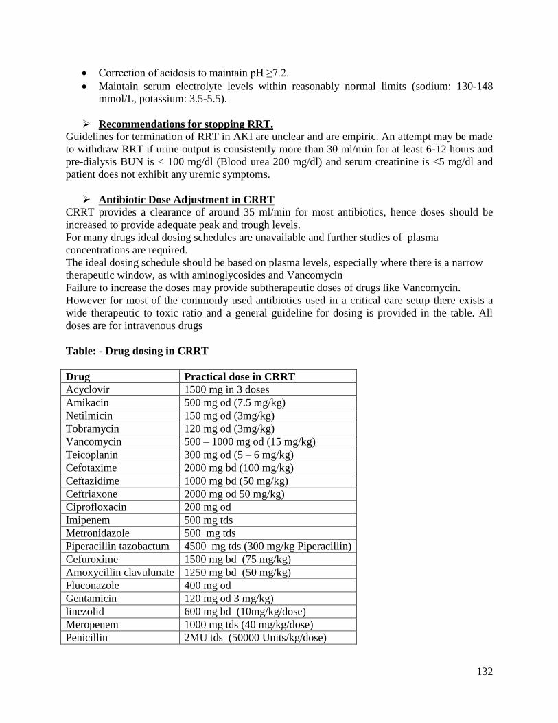
132
Correction of acidosis to maintain pH ≥7.2.
Maintain serum electrolyte levels within reasonably normal limits (sodium: 130-148
mmol/L, potassium: 3.5-5.5).
Recommendations for stopping RRT.
Guidelines for termination of RRT in AKI are unclear and are empiric. An attempt may be made
to withdraw RRT if urine output is consistently more than 30 ml/min for at least 6-12 hours and
pre-dialysis BUN is < 100 mg/dl (Blood urea 200 mg/dl) and serum creatinine is <5 mg/dl and
patient does not exhibit any uremic symptoms.
Antibiotic Dose Adjustment in CRRT
CRRT provides a clearance of around 35 ml/min for most antibiotics, hence doses should be
increased to provide adequate peak and trough levels.
For many drugs ideal dosing schedules are unavailable and further studies of plasma
concentrations are required.
The ideal dosing schedule should be based on plasma levels, especially where there is a narrow
therapeutic window, as with aminoglycosides and Vancomycin
Failure to increase the doses may provide subtherapeutic doses of drugs like Vancomycin.
However for most of the commonly used antibiotics used in a critical care setup there exists a
wide therapeutic to toxic ratio and a general guideline for dosing is provided in the table. All
doses are for intravenous drugs
Table: - Drug dosing in CRRT
Drug Practical dose in CRRT
Acyclovir 1500 mg in 3 doses
Amikacin 500 mg od (7.5 mg/kg)
Netilmicin 150 mg od (3mg/kg)
Tobramycin 120 mg od (3mg/kg)
Vancomycin 500 – 1000 mg od (15 mg/kg)
Teicoplanin 300 mg od (5 – 6 mg/kg)
Cefotaxime 2000 mg bd (100 mg/kg)
Ceftazidime 1000 mg bd (50 mg/kg)
Ceftriaxone 2000 mg od 50 mg/kg)
Ciprofloxacin 200 mg od
Imipenem 500 mg tds
Metronidazole 500 mg tds
Piperacillin tazobactum 4500 mg tds (300 mg/kg Piperacillin)
Cefuroxime 1500 mg bd (75 mg/kg)
Amoxycillin clavulunate 1250 mg bd (50 mg/kg)
Fluconazole 400 mg od
Gentamicin 120 mg od 3 mg/kg)
linezolid 600 mg bd (10mg/kg/dose)
Meropenem 1000 mg tds (40 mg/kg/dose)
Penicillin 2MU tds (50000 Units/kg/dose)
Recommended