Sporadic Visual Acuity Loss in the Comparison ofAge-RelatedMacularDegenerationTreatmentsTrials (CATT)
BENJAMIN J. KIM, GUI-SHUANG YING, JIAYAN HUANG, NICOLE E. LEVY, ANDMAUREEN G. MAGUIRE, ONBEHALF OF THE CATT RESEARCH GROUP
� PURPOSE: To evaluate transient, large visual acuity(VA) decreases, termed sporadic vision loss, duringanti–vascular endothelial growth factor treatment forneovascular age-related macular degeneration (AMD).� DESIGN: Cohort within a randomized clinical trial.� METHODS: SETTING: Comparison of Age-RelatedMacular Degeneration Treatments Trials (CATT).STUDY POPULATION: Total of 1185 CATT patients. MAIN
OUTCOME MEASURES: Incidence of sporadic vision loss andodds ratio (OR) for association with patient and ocularfactors. Sporadic vision loss was a decline of ‡15 lettersfrom the previous visit, followed by a return at the nextvisit to no more than 5 letters worse than the visit beforethe VA loss.� RESULTS: There were 143 sporadic vision loss eventsin 122 of 1185 patients (10.3%). Mean VA at 2 yearsfor those with and without sporadic vision loss was 58.5(w20/63) and 68.4 (w20/40) letters, respectively(P< .001). Among patients treated pro re nata, no injec-tion was given for 27.6% (27/98) of sporadic vision lossevents. Multivariate analysis demonstrated that baselinepredictors for sporadic vision loss included worse baselineVA (OR 2.92, 95% confidence interval [CI]:1.65–5.17for £20/200 compared with ‡20/40), scar (OR 2.21,95% CI:1.22–4.01), intraretinal foveal fluid on opticalcoherence tomography (OR 1.80, 95% CI:1.11–2.91),and medical history of anxiety (OR 1.90, 95%CI:1.12–3.24) and syncope (OR 2.75, 95% CI:1.45–5.22). Refraction decreased the likelihood of sporadicvision loss (OR 0.62, 95%CI: 0.42–0.91).� CONCLUSIONS: Approximately 10% of CATTpatients had sporadic vision loss. Baseline predictorsincluded AMD-related factors and factors independentof AMD. These data are relevant for clinicians in prac-tice and those involved in clinical trials. (Am JOphthalmol 2014;158:128–135. � 2014 by ElsevierInc. All rights reserved.)
Supplemental Material available at AJO.com.Accepted for publication Apr 1, 2014.
From the Scheie Eye Institute, Perelman School of Medicine,University of Pennsylvania, Philadelphia, Pennsylvania.
Inquiries to Benjamin J. Kim, Scheie Eye Institute, 51 N 39th St,Philadelphia, PA 19104; e-mail: [email protected]
128 � 2014 BY ELSEVIER INC.
VISUAL ACUITY (VA) HAS BEEN THE PRIMARY
outcome measure for every major clinical trial forneovascular age-related macular degeneration
(AMD).1–7 Previous studies have established that VAmeasurement administered under a standard protocolthat includes refraction provides a reliable outcomemeasure.8,9 Still, VA scores can be affected by multiplefactors, some of which have little to do with the conditionof the eye. Health issues that are not primarily ocular,such as depression and neurologic disease, can impactVA measurement or visual function.10–17 In addition,clinicians occasionally see patients in follow-up who havea worse VA measurement without any change on clinicalexamination.As part of their analysis of vision loss during the Mini-
mally Classic/Occult Trial of the Anti-VEGF AntibodyRanibizumab in the Treatment of Neovascular AMD(MARINA) and Anti-VEGF Antibody for the Treatmentof Predominantly Classic Choroidal Neovascularizationin AMD (ANCHOR) trials, Wolf and associates identi-fied patients that had acute loss of >_15 letters within any1-month period.18 A total of 106 of 758 ranibizumab-treated patients (13.9%) experienced an acute loss of visionduring the first year, and several had more than 1 episode ofacute vision loss. Although they concluded that continuedtreatment was beneficial, there was no clear relationshipbetween patient characteristics and acute vision loss,including an analysis of study eye adverse events (AEs) orserious adverse events (SAEs). It is possible that other fac-tors in addition to progressive AMD disease were involvedin some of these acute vision loss events.Given that significant resources are devoted to studying a
treatment’s effects on VA inAMDpatients, we have soughtfurther understanding of factors that influence this outcomemeasurement. The Comparison of Age-Related MacularDegeneration Treatments Trials (CATT) was a 2-yearstudy that evaluated the efficacy of ranibizumab comparedwith bevacizumab, as well as monthly compared withas-needed treatments.6,19 The CATT database providesan unprecedented opportunity to investigate AMDpatients as it expands on MARINA and ANCHOR data,providing treatment regimen, drug, and optical coherencetomography (OCT) correlations. We previously reportedthe frequency of sustained VA loss and its associatedfactors within CATT.20 Here, we report similarly for spo-radic VA loss within CATT. Rather than studying patients
0002-9394/$36.00http://dx.doi.org/10.1016/j.ajo.2014.04.004
ALL RIGHTS RESERVED.
with only an acute loss of >_15 letters, we were interested inpatients who had a decline of >_15 letters from the previousvisit, followed by a return of vision at the next visit.Although changes of 5 and occasionally 10 letters arewithin test-retest variability,9 little is known about thecauses of transient VA losses of >_15 letters for AMDpatients.
METHODS
THIS STUDY WAS A SECONDARY ANALYSIS OF A COHORT
within a randomized clinical trial (CATT). PreviousCATT reports provide a detailed summary of the CATTstudy design.6,19 CATT is registered with http://www.clinicaltrials.gov (NCT00593450). Design features relevantto this report are described here.
� STUDY PATIENTS: Study patients provided writteninformed consent to participate in CATT. The Institu-tional Review Board of each study site prospectivelyapproved the CATT study protocol, and the study is inaccordance with the Health Insurance Portability andAccountability Act regulations. The inclusion criteriawere age >_50 years, untreated choroidal neovascularization(CNV) from AMD in the study eye, VA of 20/25–20/320,and neovascularization or its sequelae at the foveal center.Baseline medical history was obtained from all patients.
Patientswere randomized at study entry to 1 of 4 treatmentarms: ranibizumabmonthly, bevacizumabmonthly, ranibizu-mab pro re nata (PRN), and bevacizumab PRN. At 1 year,study patients in the monthly groups were randomized again1:1 to continuedmonthly treatment or PRN treatment. PRNtreatment was given when there were signs of active neovas-cularization, defined as fluid onOCT,hemorrhage, decreasedVA compared with the prior visit, or leakage or increasedlesion size on fluorescein angiography.
All patients had monthly VA measurements using anelectronic VA testing system by certified VA examinerswho were masked to the patients’ treatment assignment.9
Protocol refraction before measurement of VA wasrequired at baseline and at weeks 4, 12, 24, 36, 52, 64,76, 88, and 104. For efficiency, refractions were notroutinely performed at every visit.
� IMAGING PROCEDURES: Stereoscopic color fundusphotography and fluorescein angiography were per-formed by certified photographers at baseline, 52 weeks,and 104 weeks. Stratus (version 4.0 or higher) time-domain OCT systems (Carl Zeiss Meditec, Dublin, Cali-fornia, USA) were used for first-year visits and mostsecond-year visits. Spectral-domain OCT images wereobtained for 23% of second-year visits. OCT imageswere obtained monthly in the PRN arms. Certified tech-nicians masked to the patients’ treatment assignment
VOL. 158, NO. 1 SPORADIC VISION LOSS I
followed standardized procedures and performed OCTimaging with macular thickness maps and fast macularthickness maps. OCT scans were independently analyzedby 2 certified OCT readers at the CATT OCT ReadingCenter, and photographs were analyzed by 2 certifiedreaders at the CATT Photography Reading Center.Details about image acquisition and analysis by thereading centers are previously described.21–23
� DATA ANALYSIS: Sporadic vision loss required VA datafrom 3 consecutive visits (ie, VA1, VA2, and VA3). Spo-radic vision loss was defined as a decline of >_15 lettersfrom the previous visit (ie, VA1 � VA2 >_ 15 letters),followed by a return at the next visit to no more than 5 let-ters worse than the visit before the VA loss (ie, VA3 �VA1 >_ �5 letters). Five letters was chosen for the latterpart of this definition since 89% of test-retest electronicVAmeasurements reportedly are within 5 letters.9 Sporadicvision loss of 30 letters was defined as a decline of >_30 lettersfrom the previous visit, followed by a return at the next visitto no more than 5 letters worse than the visit before theVA loss.The incidence of sporadic vision loss loss was calculated
as the proportion of eyes with sporadic vision loss within 2years among all CATT patients. Mean VA during the studywas compared between eyes with sporadic vision loss andall other study eyes. VA, fundus photograph features, andOCT features were compared at 2 years between eyeswith and without sporadic vision loss. As noted, the2-group t test or the paired t test was used for comparisonof means. Fisher exact test or McNemar test was used forcomparison of proportions.For the 27 events of sporadic vision loss that did not
coincide with an injection, investigators for these eventswere queried about the possible cause of vision loss,whether new hemorrhage at the macula was present, andwhy no injection was given.For the evaluation of baseline medical history associations
with sporadic vision loss, we focused on neurologic and psy-chological histories because of their potential effects on visualfunction measurements.10–17 Additionally, a FunctionalComorbidity Index was used to determine if patients withmore comorbidities in their baseline history had anincreased risk for sporadic vision loss. The FunctionalComorbidity Index is an established measure of comorbiddisease that correlates with physical function as theoutcome of interest.24 This index contains 18 items such asvisual impairment, congestive heart failure, arthritis, asthma,depression, anxiety, and neurologic disease. The FunctionalComorbidity Index is scored by summing the number of spe-cific comorbidities in a patient’s medical history. A score of0 indicates no relevant comorbidities, while a score of 18indicates the highest number of comorbid illnesses.The association of sporadic vision loss and nonocular
SAEs was investigated using nonocular SAEs reportedwithin 30 days (before or after) of the time of sporadic
129N AMD TREATMENT
FIGURE. Mean (±standard error) visual acuity over 2 yearsamong patients with and without sporadic vision loss in theComparison of Age-Related Macular Degeneration TreatmentsTrials. Study visits with refraction are represented by solidsymbols.
vision loss events. These time frames were chosen as wewere interested in knowing if sporadic vision loss has anassociation with a patient that is still recovering from arecent systemic SAE or that is becoming systemically illand about to have an SAE. To investigate these potentialassociations, we matched sporadic vision loss patients(cases) with patients without sporadic vision loss (con-trols). The matching criteria were: drug, regimen, age(63 years), Functional Comorbidity Index score (62points), and the number of visits with measured VA. Tomaximize the use of the controls, we allowed 1 case tohave more than 1 control if available (ie, 1:n matching).
The evaluation of factors associated with sporadic visionloss was first performed by univariate analysis usingrepeated measures logistic regression models to accommo-date patients with more than 1 event of sporadic visionloss. Multivariate analyses started with the factors with aP < .20 in univariate analysis, and the final multivariateanalysis model was developed using a backward selectionprocedure by keeping only predictors with P < .05, withthe exception of the drug and regimen groups. Adjustedodds ratios (OR) of sporadic vision loss and the 95% con-fidence intervals (95% CI) were calculated from the finalmultivariate logistic regression model for repeated mea-sures. All data analyses were performed using SASv9.2 (SAS Inc, Cary, North Carolina, USA). Two-sidedP < .05 was considered statistically significant.
RESULTS
� INCIDENCE AND VISUAL ACUITY: Over 2 years, 122 ofthe 1185 patients (10.3%) had at least 1 event of sporadicvision loss. There were 143 sporadic vision loss events.One hundred and two of 122 patients (83.6%) had only1 sporadic vision loss event; 19 (15.6%) had 2 events;and 1 patient (0.82%) had 4 events. There were 10patients (0.8%) of the 1185 patients who developed spo-radic vision loss of 30 letters, including 1 patient who had2 events of this. The time to first sporadic vision loss eventwas evenly distributed across the entire duration of thestudy. For 59 of 143 sporadic vision loss events (41.3%),the patient had a VA of 20/40 or better at the study visitpreceding the sporadic vision loss. At all time pointsthroughout the study, the patients with sporadic visionloss had a worse mean VA than patients without sporadicvision loss (Figure).
� OPTICAL COHERENCE TOMOGRAPHY FEATURES ANDINJECTIONS AROUND THE TIME OF SPORADIC VISIONLOSS AMONG EYES TREATED PRO RE NATA: Sporadicvision loss events occurred 98 times in 83 eyes treatedaccording to the PRN dosing regimen. OCT analysis ofthe 98 events showed that the mean retinal thickness was169 mm at the visit before the sporadic vision loss,183 mm at the time of sporadic vision loss, and 151 mm
130 AMERICAN JOURNAL OF
at the visit afterwards (Table 1). Among all CATTpatients, a change in OCT retinal thickness had a weakcorrelation with a change in VA (data not shown), andonly 13 of these 98 sporadic vision loss events (13.3%)among eyes treated PRN coincided with an increase ofretinal thickness of 50 mm or more. Foveal fluid was seenin 25 of the 98 events (25.5%) before the sporadic visionloss, in 37 (37.8%) at the time of sporadic vision loss,and in 17 (17.4%) afterwards. Subretinal fluid at the foveawas seen in 12 events (12.2%) before the sporadic visionloss, in 14 (14.3%) at the time of sporadic vision loss,and in 5 (5.1%) afterwards.Of the 98 events among eyes treated PRN, 43 (43.9%)
had a study injection at the prior visit and 71 (72.4%)had an injection at the visit when sporadic vision losswas noted. Among the 27 patients that were not treatedat the time of sporadic vision loss, 6 (22.2%) had intrareti-nal or subretinal fluid at the fovea. Investigators werequeried about these 27 events, and responses for 21 of theseevents were received. No identifiable cause for vision losswas found for 11 of these 21 events. For the remainingevents, the cause of sporadic vision loss was thought tobe related to a change in systemic health (3/21), progres-sion of non-neovascular AMD (3/21), dry eyes (2/21), cata-ract (1/21), and increased subretinal fluid from neovascularAMD (1/21). The 1 patient that that had increased subre-tinal fluid refused treatment on that day, and the otherpatients were not treated because the investigator did notthink there were signs of neovascular AMD activity.None of the responding investigators indicated that therewas new hemorrhage at the macula.
� TWO-YEAR VISUAL ACUITY AND MORPHOLOGICFEATURES ASSOCIATED WITH SPORADIC VISION LOSS: Atotal of 113 patients that had sporadic vision loss wereavailable for data analysis of 2-year VA and morphology.
JULY 2014OPHTHALMOLOGY

TABLE 1. Comparison of Treatment Status and Optical Coherence Tomography Features Before, At, and After Sporadic Vision LossAmong Eyes Treated Pro Re Nata in the Comparison of Age-Related Macular Degeneration Treatments Trials (83 Eyes, 98 Events)a—
Patients in Pro Re Nata Arm for 2 Years or Switchers in the Second Year
4 Weeks Before
Sporadic Vision Loss
At Sporadic
Vision Loss
4 Weeks After
Sporadic Vision Loss
P Valueb
(At vs Before Sporadic
Vision Loss)
P Valueb
(At vs After Sporadic
Vision Loss)
P Valueb
(Before vs After Sporadic
Vision Loss)
Events with injections in
pro re nata groups, n (%)
43 (43.9%) 71 (72.4%) 38 (38.8%) <.001 <.001 .45
Retinal thickness at foveal center (mm)
<120 24 (24.5%) 21 (21.4%) 28 (28.6%) .28 .006 .43
120–212 55 (56.1%) 50 (51.0%) 58 (59.2%)
>212 17 (17.4%) 25 (25.5%) 12 (12.2%)
Mean (SE) 169 (8) 183 (10) 151 (6) .15 <.001 .007
Retinal fluid at foveal center
No 69 (70.4%) 59 (60.2%) 79 (80.6%) .08 <.001 .07
Yes 25 (25.5%) 37 (37.8%) 17 (17.4%)
Subretinal fluid at foveal center
No 82 (83.7%) 80 (81.6%) 90 (91.8%) .78 .007 .01
Yes 12 (12.2%) 14 (14.3%) 5 (5.1%)
SE ¼ standard error.aThe totals may not add to 98 because of missing values in less than 5%.bMcNemar test for comparing proportions, paired t test for comparing means.
At 2 years, the mean VA of sporadic vision loss patientswas 58.5 letters (w20/63), as compared with 68.4 letters(w20/40) for those patients without sporadic vision loss(P< .001) (Table 2). The mean VA change from baselinewas 3.1 letters for patients with sporadic vision loss,compared with 6.7 letters for patients without sporadicvision loss (P ¼ .03). Forty-four of 113 patients (38.9%)with sporadic vision loss were 20/40 or better, as comparedwith 610 of 921 patients (66.2%) without sporadic visionloss (P < .001). Fifty-six of 113 sporadic vision losspatients (49.6%) had a scar, as compared with 371of 921 patients (40.3%) without sporadic vision loss(P ¼ .04). Sixteen of 113 sporadic vision loss patients(14.2%) had no pathology at the foveal center, comparedwith 188 of 921 patients (20.4%) without sporadic visionloss (P¼ .02). Also, patients with sporadic vision loss hada larger total area of CNV lesion (9.97 mm2 vs 8.04 mm2,P ¼ .02). The presence of geographic atrophy was notsignificantly associated with sporadic vision loss (P ¼.27). OCT analysis showed that the percent with fluidand the mean retinal thickness were not associated withsporadic vision loss (P > .05).
� ASSOCIATION OF SERIOUS ADVERSE EVENTS WITHSPORADIC VISION LOSS: There were 11 events (10patients) of sporadic vision loss of 30 letters, which metcriteria for an ocular SAE. The causes reported by the inves-tigator were related toAMD (5/11), related to central retinalvein occlusion (1/11), and possibly related to systemichealth condition (3/11). There was no clear cause statedfor the vision loss in 2 of these 11 events. Furthermore, an
VOL. 158, NO. 1 SPORADIC VISION LOSS I
evaluation of ocular and systemic AEs did not show any sig-nificant associations (data not shown).Using amatched case-control approach, we also evaluated
whether a nonocular SAEwithin 30days (before or after)wasassociatedwith sporadic vision loss.Among94matchedcase-control pairs that met criteria for analysis, 6 of 94 patients(6.4%)with sporadic vision loss had a nonocular SAEwithin30 days compared with 9 of 199 matched controls (4.5%)without sporadic vision loss (P ¼ .48). Similarly, 47 of 122patients (38.5%) with sporadic vision loss had a nonocularSAE during the 2 years of the trial, compared with 377 of1063 patients (35.5%)without sporadic vision loss (P¼ .55).
� BASELINE MEDICAL HISTORY AND OCULAR PREDIC-TORS OF SPORADIC VISION LOSS: The univariate analysis(Supplemental Table 1, available at AJO.com) showed thata baseline neurologic history and a baseline psychological his-tory were risk factors for sporadic vision loss. Sixty-five of 546patients (11.9%) with a neurologic history had sporadicvision loss, compared with 57 of 639 patients (8.9%) withouta neurologic history (P ¼ .04). Thirty-two of 232 patients(13.8%) with a psychological history had sporadic visionloss, compared with 90 of 953 patients (9.4%) without a psy-chological history (P ¼ .02). Within the broad category ofpsychological disorders, subcategory analysis showed an ‘‘anx-iety’’ history for 12 of 122 (9.8%) sporadic vision loss patientsand only 43 of 1063 patients (4.1%) without sporadic visionloss (P¼ .01) (SupplementalTable 2, available atAJO.com).Additionally, the neurologic history subcategory of ‘‘syncope’’was present for 10 of 122 patients (8.2%)with sporadic visionloss, compared with 28 of 1063 patients (2.6%) without
131N AMD TREATMENT
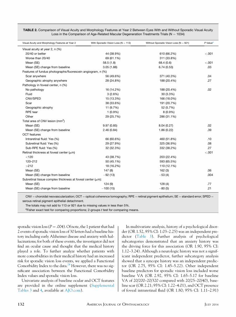
TABLE 2. Comparison of Visual Acuity and Morphology Features at Year 2 Between Eyes With and Without Sporadic Visual AcuityLoss in the Comparison of Age-Related Macular Degeneration Treatments Trials (N ¼ 1034)
Visual Acuity and Morphology Features at Year 2 With Sporadic Vision Loss (N ¼ 113) Without Sporadic Vision Loss (N ¼ 921) P Valuea
Visual acuity at year 2, n (%)
20/40 or better 44 (38.9%) 610 (66.2%) <.001
Worse than 20/40 69 (61.1%) 311 (33.8%)
Mean (SE) 58.5 (1.8) 68.4 (0.6) <.001
Mean (SE) change from baseline 3.05 (1.88) 6.74 (0.53) .03
Features of fundus photographs/fluorescein angiogram, n (%)
Scar anywhere 56 (49.6%) 371 (40.3%) .04
Geographic atrophy anywhere 28 (24.8%) 188 (20.4%) .27
Pathology in foveal center, n (%)
No pathology 16 (14.2%) 188 (20.4%) .02
Fluid 3 (2.6%) 30 (3.3%)
CNV/SPED 15 (13.3%) 166 (18.0%)
Scar 38 (33.6%) 191 (20.7%)
Geographic atrophy 11 (9.7%) 52 (5.7%)
RPE tear 1 (0.9%) 8 (0.9%)
Other 29 (25.7%) 286 (31.1%)
Total area of CNV lesion (mm2)
Mean (SE) 9.97 (0.85) 8.04 (0.27) .02
Mean (SE) change from baseline 2.46 (0.84) 1.86 (0.22) .39
OCT features
Intraretinal fluid: Yes (%) 66 (60.6%) 460 (51.8%) .10
Subretinal fluid: Yes (%) 29 (27.9%) 325 (36.9%) .08
Sub-RPE fluid: Yes (%) 32 (32.3%) 332 (38.2%) .27
Retinal thickness at foveal center (mm) <.001
<120 43 (38.7%) 203 (22.4%)
120–212 50 (45.1%) 593 (65.5%)
>212 18 (16.2%) 110 (12.1%)
Mean (SE) 147 (8) 162 (3) .06
Mean (SE) change from baseline �92 (13) �53 (4) .004
Subretinal tissue complex thickness at foveal center (mm):
Mean (SE) 124 (9) 128 (4) .77
Mean (SE) change from baseline �100 (15) �80 (5) .21
CNV¼ choroidal neovascularization; OCT¼ optical coherence tomography; RPE¼ retinal pigment epithelium; SE¼ standard error; SPED¼serous retinal pigment epithelial detachment.
The totals may not add to 113 or 921 due to missing values in less than 5%.aFisher exact test for comparing proportions; 2-groups t test for comparing means.
sporadic vision loss (P¼ .004).Ofnote, the 1 patient that had2 events of sporadic vision loss of 30 letters had a baseline his-tory including early Alzheimer disease and anxiety with hal-lucinations; for both of these events, the investigator did notfind an ocular cause and thought that the medical historyplayed a role. To further analyze whether patients withmore comorbidities in their medical history had an increasedrisk for sporadic vision loss events, we applied a FunctionalComorbidity Index to the data.24 However, there was no sig-nificant association between the Functional ComorbidityIndex values and sporadic vision loss.
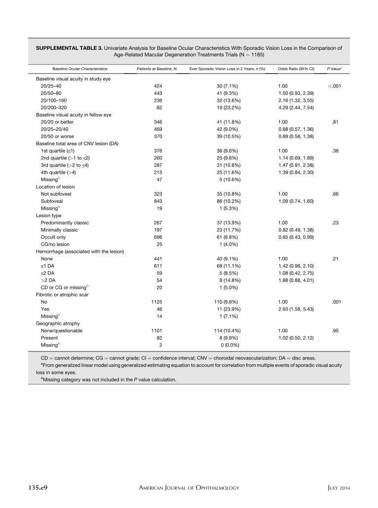
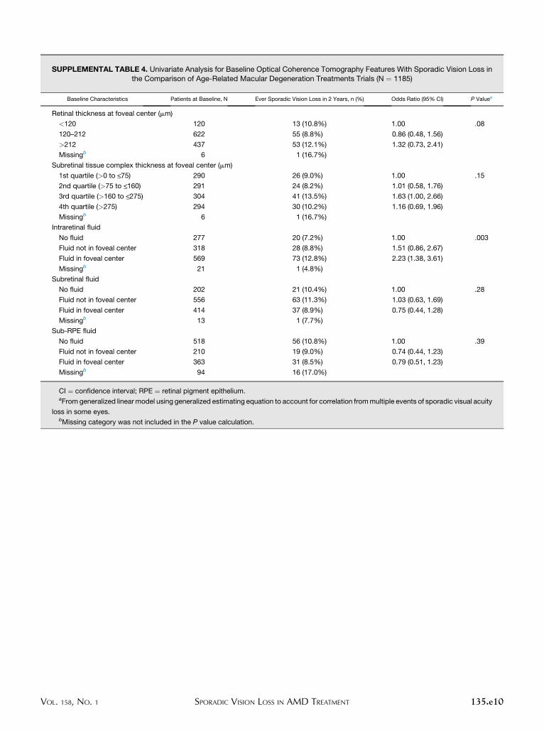
Univariate analysis of baseline ocular and OCT featuresare provided in the online supplement (SupplementalTables 3 and 4, available at AJO.com).
132 AMERICAN JOURNAL OF
In multivariate analysis, history of a psychological disor-der (OR 1.52, 95%CI: 1.03–2.25) was an independent pre-dictor (Table 3). Further analysis of psychologicalsubcategories demonstrated that an anxiety history wasthe driving force for this association (OR 1.90, 95% CI:1.12–3.24). Although a neurologic history was not a signif-icant independent predictor, further subcategory analysisshowed that a syncope history was an independent predic-tor (OR 2.75, 95% CI: 1.45–5.22). Other independentbaseline predictors for sporadic vision loss included worsebaseline VA (OR 2.92, 95% CI: 1.65–5.17 for baselineVA of 20/200–20/320 compared with 20/25–20/40), base-line scar (OR 2.21; 95%CI: 1.22–4.01), and OCT presenceof foveal intraretinal fluid (OR 1.80; 95% CI: 1.11–2.91)
JULY 2014OPHTHALMOLOGY
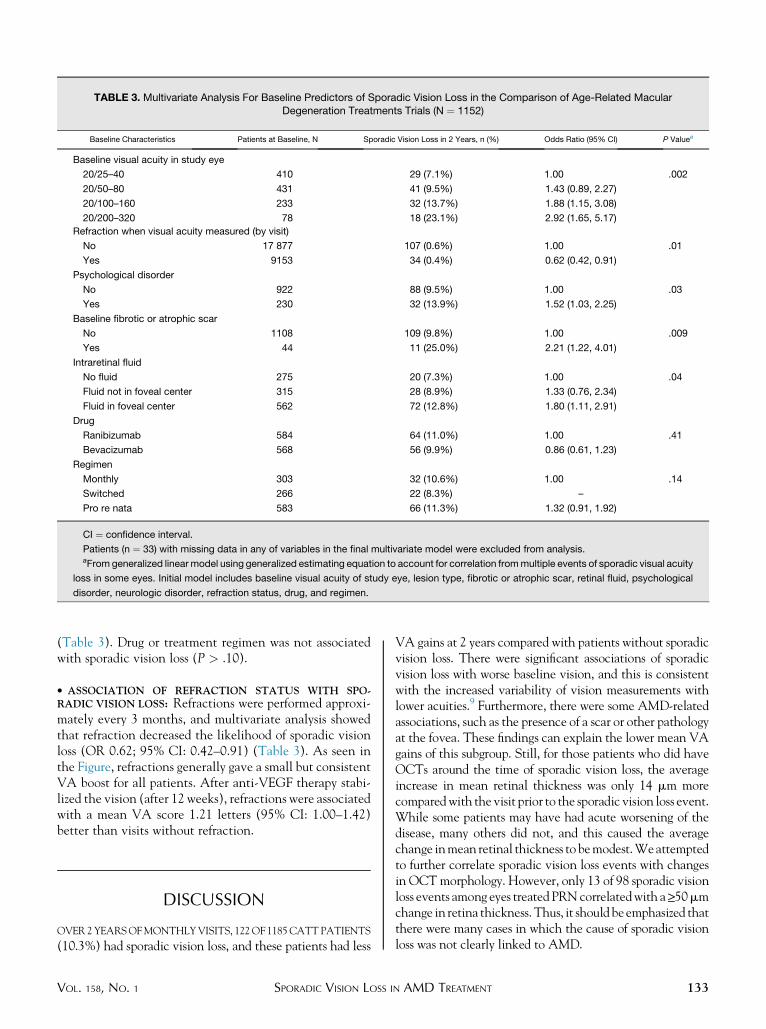
TABLE 3. Multivariate Analysis For Baseline Predictors of Sporadic Vision Loss in the Comparison of Age-Related MacularDegeneration Treatments Trials (N ¼ 1152)
Baseline Characteristics Patients at Baseline, N Sporadic Vision Loss in 2 Years, n (%) Odds Ratio (95% CI) P Valuea
Baseline visual acuity in study eye
20/25–40 410 29 (7.1%) 1.00 .002
20/50–80 431 41 (9.5%) 1.43 (0.89, 2.27)
20/100–160 233 32 (13.7%) 1.88 (1.15, 3.08)
20/200–320 78 18 (23.1%) 2.92 (1.65, 5.17)
Refraction when visual acuity measured (by visit)
No 17 877 107 (0.6%) 1.00 .01
Yes 9153 34 (0.4%) 0.62 (0.42, 0.91)
Psychological disorder
No 922 88 (9.5%) 1.00 .03
Yes 230 32 (13.9%) 1.52 (1.03, 2.25)
Baseline fibrotic or atrophic scar
No 1108 109 (9.8%) 1.00 .009
Yes 44 11 (25.0%) 2.21 (1.22, 4.01)
Intraretinal fluid
No fluid 275 20 (7.3%) 1.00 .04
Fluid not in foveal center 315 28 (8.9%) 1.33 (0.76, 2.34)
Fluid in foveal center 562 72 (12.8%) 1.80 (1.11, 2.91)
Drug
Ranibizumab 584 64 (11.0%) 1.00 .41
Bevacizumab 568 56 (9.9%) 0.86 (0.61, 1.23)
Regimen
Monthly 303 32 (10.6%) 1.00 .14
Switched 266 22 (8.3%) –
Pro re nata 583 66 (11.3%) 1.32 (0.91, 1.92)
CI ¼ confidence interval.
Patients (n ¼ 33) with missing data in any of variables in the final multivariate model were excluded from analysis.aFromgeneralized linear model using generalized estimating equation to account for correlation frommultiple events of sporadic visual acuity
loss in some eyes. Initial model includes baseline visual acuity of study eye, lesion type, fibrotic or atrophic scar, retinal fluid, psychological
disorder, neurologic disorder, refraction status, drug, and regimen.
(Table 3). Drug or treatment regimen was not associatedwith sporadic vision loss (P > .10).
� ASSOCIATION OF REFRACTION STATUS WITH SPO-RADIC VISION LOSS: Refractions were performed approxi-mately every 3 months, and multivariate analysis showedthat refraction decreased the likelihood of sporadic visionloss (OR 0.62; 95% CI: 0.42–0.91) (Table 3). As seen inthe Figure, refractions generally gave a small but consistentVA boost for all patients. After anti-VEGF therapy stabi-lized the vision (after 12 weeks), refractions were associatedwith a mean VA score 1.21 letters (95% CI: 1.00–1.42)better than visits without refraction.
DISCUSSION
OVER2YEARSOFMONTHLYVISITS, 122OF1185CATTPATIENTS
(10.3%) had sporadic vision loss, and these patients had less
VOL. 158, NO. 1 SPORADIC VISION LOSS I
VA gains at 2 years compared with patients without sporadicvision loss. There were significant associations of sporadicvision loss with worse baseline vision, and this is consistentwith the increased variability of vision measurements withlower acuities.9 Furthermore, there were some AMD-relatedassociations, such as the presence of a scar or other pathologyat the fovea. These findings can explain the lower mean VAgains of this subgroup. Still, for those patients who did haveOCTs around the time of sporadic vision loss, the averageincrease in mean retinal thickness was only 14 mm morecomparedwith the visit prior to the sporadic vision loss event.While some patients may have had acute worsening of thedisease, many others did not, and this caused the averagechange inmean retinal thickness to bemodest.Weattemptedto further correlate sporadic vision loss events with changesin OCTmorphology. However, only 13 of 98 sporadic visionloss events among eyes treatedPRNcorrelatedwith a>_50mmchange in retina thickness.Thus, it shouldbeemphasized thatthere were many cases in which the cause of sporadic visionloss was not clearly linked to AMD.
133N AMD TREATMENT
Data from the eyes treated PRN further support theconclusion that many cases of sporadic vision loss were notdirectly linked to worsening of AMD. Of particular interestis the finding that investigators did not give an injectionfor 27 of 98 (27.6%) sporadic vision loss events in eyestreated PRN, even though vision loss was an indication forPRN treatment. Six of these 27 patients (22.2%) had fluidat the fovea based on OCT reading center evaluation, andthis also was a treatment indication. It was previouslyreported that approximately 30% of patients in PRN groupsdid not receive an injection even though the reading centerfound fluid on theOCT.6 For these untreated sporadic visionloss cases with fluid, the investigator may not have noticed asmall amount of fluid or, less likely, thought that the fluidwasnot significant enough to warrant treatment. When investi-gators for these 27 eventswere queried, 11 of the 21 responsesindicated that therewas no identifiable cause and 3 indicatedthat the eventmay be related to a change in systemic health.Only 1 of 21 responses indicated that there was a worseningof neovascular AMD.Our data suggest that there were othercauses for sporadic vision loss, including a low baseline VA,syncope history, anxiety history, or absence of refraction.
Previous reports have highlighted the role that depres-sion plays on visual function in AMD patients.10–13
While we did not find that a baseline history of depres-sion specifically is associated with sporadic vision loss,our multivariate analysis showed that a psychiatrichistory generally increases the odds of sporadic visionloss. Further analysis showed that a history of anxiety,rather than depression, was the driving force behind thesignificance of a psychiatric history. Additionally, theneurologic subcategory of syncope was a significantpredictor of sporadic vision loss. Among the elderlypopulation, the most common causes of syncope areorthostatic hypotension, volume depletion, cardiovascularevents, vasovagal reflex, and idiopathic.25,26 These datasuggest that acute changes in mental health as well as thosefactors that lead to syncope may lead to sporadic visionloss. One may wonder if patients who are ‘‘sicker’’ overall atbaseline are more likely to have sporadic vision loss, but wecould not find a clear association of this through our use ofa Functional Comorbidity Index. We also could not findany associations between SAEs or AEs with sporadic visionloss. This is consistent with Wolf and associates’ analysisof acute vision loss in the MARINA and ANCHORstudies,18 although they looked at ocular adverse eventsand did not specifically focus on transient vision loss. Given
134 AMERICAN JOURNAL OF
the paramount importance of vision measurements, it maybe worthwhile for investigators to consider these findingswhen enrolling patients for clinical trials. This is emphasizedby a study patientwith a history of anxiety andhallucinationswho had 2 events of sporadic vision loss of 30 letters.In an effort to increase efficiency, some clinical trials do
not perform refracted VA at every study visit. These datafrom CATT showed that refraction slightly boosted themean VA measurements, and absence of refraction wasassociated with sporadic vision loss. Although the visiondifference on average was small, the data demonstrate theimportant role of study visit refractions. Some studieshave defined visits with refractions and a protocol stipula-tion for the visits without routine refractions. If the VA haschanged by 10 or more letters since the last visit, then arefraction should be performed.27
There are several limitations of this secondary analysis. Inthe CATT, OCT was not required at every visit for themonthly treatment patients. Thus, we had OCT data fromthe time of all sporadic vision loss events for PRN-treatedpatients but not for monthly-treated patients. Fundus photoswere performed only at the baseline, 1-year, and 2-year visits.Although we recognize that an image characteristic at thelast study visit may not have been present at the time ofthe sporadic vision loss event, we did investigate the differ-ences to understand why sporadic vision loss patients had alower meanVA at 2 years. It should be noted that we cannotexclude the possibility of hemorrhage at the macula at thetime of sporadic vision loss in some patients, since photo-graphs were not available at every visit. However, investiga-tors for the 27 events in PRN eyes that were not treated werequeried about the sporadic vision loss and the decision not totreat. None of the responses indicated that there was newhemorrhage at the macula. Although hemorrhage at themacula could explain some of the 143 events of sporadicvision loss, the data suggest that there were several other fac-tors involved in sporadic vision loss as well.In summary, approximately 10% of CATT patients had a
sporadic vision loss event during the trial, and 27.6% of spo-radic vision loss events in PRN groups did not coincide withan injection. Although there is some expected relationshipbetween acute worsening of AMD and sporadic vision loss,there certainly were other associations with these aberrantVAmeasurements, including worse baseline vision, psychi-atric history, syncope history, and lack of refraction. Webelieve that these data are valuable for clinicians, thoseplanning clinical trials, and trial investigators.
ALL AUTHORSHAVE COMPLETED AND SUBMITTED THE ICMJE FORM FOR DISCLOSUREOF POTENTIAL CONFLICTS OF INTEREST.The authors indicate the following financial disclosures: B.J.K. has been on advisory boards for Thrombogenics, Eyetech, and Allergan. The other authorshave no financial disclosures. Supported by cooperative agreements U10 EY017823, U10 EY017825, U10 EY017826, and U10 EY017828 from theNational Eye Institute, National Institutes of Health, Department of Health and Human Services, Bethesda, Maryland, USA. Contributions of authors:design and conduct of the study (B.J.K., G.-S.Y., M.G.M.); collection and management of data (G.-S.Y., J.H., N.E.L.); analysis and interpretation of data(B.J.K., G.-S.Y., J.H., N.E.L., M.G.M.); preparation, review, approval of manuscript (B.J.K., G.-S.Y., J.H., M.G.M.).
The members of the CATT Research Group are listed in the Appendix (Supplemental material, available at AJO.com).
JULY 2014OPHTHALMOLOGY
REFERENCES
1. Argon laser photocoagulation for senile macular degenera-tion. Results of a randomized clinical trial. Arch Ophthalmol
1982;100(6):912–918.2. Bressler NM. Treatment of Age-Related Macular Degenera-
tion with Photodynamic Therapy Study Group. Photody-namic therapy of subfoveal choroidal neovascularization inage-related macular degeneration with verteporfin: two-yearresults of 2 randomized clinical trials-TAP report 2. ArchOphthalmol 2001;119(2):198–207.
3. Gragoudas ES, Adamis AP, Cunningham ET Jr, Feinsod M,Guyer DR, VISiON Clinical Trial Group. Pegaptanib forneovascular age-related macular degeneration. N Engl J Med
2004;351(27):2805–2816.4. Rosenfeld PJ, Brown DM, Heier JS, et al. Ranibizumab for
neovascular age-related macular degeneration. N Engl J Med
2006;355(14):1419–1431.5. Brown DM, Kaiser PK, Michels M, et al. Ranibizumab versus
verteporfin for neovascular age-related macular degeneration.N Engl J Med 2006;355(14):1432–1444.
6. Comparison of Age-related Macular Degeneration Treat-ments Trials Research Group, Martin DF, Maguire MG,et al. Ranibizumab and bevacizumab for treatment of neovas-cular age-related macular degeneration: two-year results.Ophthalmology 2012;119(7):1388–1398.
7. Heier JS, Brown DM, Chong V, et al. Intravitreal aflibercept(VEGF trap-eye) in wet age-related macular degeneration.Ophthalmology 2012;119(12):2537–2548.
8. BlackhurstDW,MaguireMG.Reproducibility of refraction andvisual acuitymeasurement under a standard protocol.TheMac-ular PhotocoagulationStudyGroup.Retina 1989;9(3):163–169.
9. Beck RW, Moke PS, Turpin AH, et al. A computerizedmethod of visual acuity testing: adaptation of the early treat-ment of diabetic retinopathy study testing protocol. Am JOphthalmol 2003;135(2):194–205.
10. Rovner BW, Casten RJ, Massof RW, Leiby BE, Tasman WS,Wills Eye AMD Study. Psychological and cognitive determi-nants of vision function in age-related macular degeneration.Arch Ophthalmol 2011;129(7):885–890.
11. Rovner BW, Casten RJ, Tasman WS. Effect of depression onvision function in age-related macular degeneration. ArchOphthalmol 2002;120(8):1041–1044.
12. Zhang X, Bullard KM, Cotch MF, et al. Association betweendepression and functional vision loss in persons 20 years ofage or older in the United States, NHANES 2005-2008.JAMA Ophthalmol 2013;131(5):573–581.
13. Carriere I, Delcourt C, Daien V, et al. A prospective study ofthe bi-directional association between vision loss and depres-sion in the elderly. J Affect Disord 2013;151(1):164–170.
14. Armstrong RA. Visual symptoms in Parkinson’s disease.Parkinsons Dis 2011;2011:908306.
15. Kirby E, Bandelow S, Hogervorst E. Visual impairment inAlzheimer’s disease: a critical review. J Alzheimers Dis 2010;21(1):15–34.
16. Jackson GR, Owsley C. Visual dysfunction, neurodege-nerative diseases, and aging. Neurol Clin 2003;21(3):709–728.
17. Moschos MM, Markopoulos I, Chatziralli I, et al. Struc-tural and functional impairment of the retina and opticnerve in Alzheimer’s disease. Curr Alzheimer Res 2012;9(7):782–788.
18. Wolf S, Holz FG, Korobelnik JF, et al. Outcomes followingthree-line vision loss during treatment of neovascular age-related macular degeneration: subgroup analyses fromMARINA and ANCHOR. Br J Ophthalmol 2011;95(12):1713–1718.
19. Group CR, Martin DF, Maguire MG, et al. Ranibizumab andbevacizumab for neovascular age-related macular degenera-tion. N Engl J Med 2011;364(20):1897–1908.
20. Ying GS, Kim BJ, Maguire MG, et al. Sustained visual acuityloss in the Comparison of AMD Treatments Trials (CATT).JAMA Ophthalmol (forthcoming).
21. Ying GS, Huang J, Maguire MG, et al. Baseline predictors forone-year visual outcomes with ranibizumab or bevacizumabfor neovascular age-related macular degeneration. Ophthal-
mology 2013;120(1):122–129.22. Grunwald JE, Daniel E, Ying GS, et al. Photographic assess-
ment of baseline fundus morphologic features in the Compar-ison of Age-Related Macular Degeneration TreatmentsTrials. Ophthalmology 2012;119(8):1634–1641.
23. DeCroos FC, Toth CA, Stinnett SS, et al. Optical coherencetomography grading reproducibility during the Comparisonof Age-related Macular Degeneration Treatments Trials.Ophthalmology 2012;119(12):2549–2557.
24. Groll DL, To T, Bombardier C,Wright JG. The developmentof a comorbidity index with physical function as the outcome.J Clin Epidemiol 2005;58(6):595–602.
25. Kenny RA, Bhangu J, King-Kallimanis BL. Epidemiology ofsyncope/collapse in younger and older Western patient popu-lations. Prog Cardiovasc Dis 2013;55(4):357–363.
26. Khera S, Palaniswamy C, Aronow WS, et al. Predictors ofmortality, rehospitalization for syncope, and cardiac syncopein 352 consecutive elderly patients with syncope. J Am MedDir Assoc 2013;14(5):326–330.
27. Age-Related Eye Disease Study Research Group. A random-ized, placebo-controlled, clinical trial of high-dose supple-mentation with vitamins C and E, beta carotene, and zincfor age-related macular degeneration and vision loss:AREDS report no. 8. Arch Ophthalmol 2001;119(10):1417–1436.
VOL. 158, NO. 1 135SPORADIC VISION LOSS IN AMD TREATMENT
APPENDIX. CREDIT ROSTER FOR THECOMPARISON OF AMD TREATMENTS
TRIALS
Clinical Centers (Ordered by Number of PatientsEnrolled)
Certified Roles at Clinical Centers: Clinic Coordinator(CC), Data Entry Staff (DE), Participating Ophthalmologist(O), Ophthalmic Photographer (OP); Optical CoherenceTomography Technician (OCT); Principal Investigator(PI); Refractionist (R); Visual Acuity Examiner (VA).
VitreoRetinal Surgery, PA (Edina, MN): David F.Williams, MD (PI); Sara Beardsley, COA (VA/R); StevenBennett, MD (O); Herbert Cantrill, MD (O); CarmenChan-Tram, COA (VA/R); Holly Cheshier, CRA, COT,OCTC (OP); Kathyrn Damato, COT (VA); John Davies,MD (O); Sundeep Dev, MD (O); Julianne Enloe, CCRP,COA (CC); Gennaro Follano (OP/OCT); Peggy Gilbert,COA (VA/R); Jill Johnson, MD (O); Tori Jones, COA(OCT); Lisa Mayleben, COMT (CC/VA/R/OCT); RobertMittra, MD (O); Martha Moos, COMT, OSA (VA/R);Ryan Neist, COMT (VA/R); Neal Oestreich, COT(CC); Polly Quiram, MD (O); Robert Ramsay, MD (O);Edwin Ryan, MD (O); Stephanie Schindeldecker, OA(VA/R); John Snater, COA (VA); Trenise Steele, COA(VA); Dwight Selders, COA (VA/R); Jessica Tonsfeldt,AO (OP/OCT); Shelly Valardi, COT (VA/R).
Texas Retina Associates (Dallas, TX): Gary Edd Fish,MD (PI); Hank A. Aguado, CRA (OP/OCT); Sally Arce-neaux (CC/VA/R); Jean Arnwine (CC); Kim Bell, COA(VA/R); Tina Bell (CC/OCT); Bob Boleman (OP); Patri-cia Bradley, COT (CC); David Callanan, MD (O); LoriCoors, MD (O); Jodi Creighton, COA (VA/R); TimothyCrew, COA (OCT); Kimberly Cummings (OP/OCT);Christopher Dock (OCT); Karen Duignan, COT (VA/R); Dwain Fuller, MD (O); Keith Gray (OP/OCT); BetsyHendrix, COT, ROUB (OCT); Nicholas Hesse (OCT);Diana Jaramillo, COA (OCT); Bradley Jost, MD (O);Sandy Lash (VA/R); Laura Lonsdale, CCRP (DE); MichaelMackens (OP/OCT); Karin Mutz, COA (CC); MichaelPotts (VA/R); Brenda Sanchez (VA/R); William Snyder,MD (O); Wayne Solley, MD (O); Carrie Tarter (VA/R);Robert Wang, MD (O); Patrick Williams, MD (O).
Southeastern Retina Associates (Knoxville, TN):Stephen L. Perkins, MD (PI); Nicholas Anderson, MD(O); Ann Arnold, COT (VA/R); Paul Blais (OP/OCT);Joseph Googe, MD (O); Tina T. Higdon (CC); CecileHunt (VA/R); Mary Johnson, COA (VA/R); James Miller,MD (O); Misty Moore (VA/R); Charity K. Morris, RN(CC); Christopher Morris (OP/OCT); Sarah Oelrich,COT (OP/OCT); Kristina Oliver, COA (VA/R); VickySeitz, COT (VA/R); Jerry Whetstone (OP/OCT).
Retina Vitreous Consultants (Pittsburgh, PA):Bernard H. Doft (PI); Jay Bedel, RN (CC); Robert Bergren,MD (O); Ann Borthwick (VA/R); Paul Conrad, MD, PHD
135.e1 AMERICAN JOURNAL OF
(O); Amanda Fec (OCT); Christina Fulwylie (VA/R);Willia Ingram (DE); Shawnique Latham (VA/R); GinaLester (VA/R); Judy Liu, MD (O); Louis Lobes, MD (O);Nicole M. Lucko (CC); Holly Mechling (CC); LoriMerlotti, MS, CCRC (CC); Keith McBroom (OCT);Karl Olsen, MD (O); Danielle Puskas, COA (VA/R);Pamela Rath, MD (O); Maria Schmucker (CC); LynnSchueckler (OCT); Christina Schultz (CC/VA/R); HeatherShultz (OP/OCT); David Steinberg, CRA (OP/OCT);Avni Vyas, MD (O); Kim Whale (VA/R); KimberlyYeckel, COA, COT (VA/R).Ingalls Memorial Hospital/Illinois Retina Associates
(Harvey, IL): David H. Orth, MD (PI); Linda S. Arre-dondo, RN (CC/VA); Susan Brown (VA/R); Barbara J.Ciscato (CC/VA); Joseph M. Civantos, MD (O); CelesteFigliulo (VA/R); Sohail Hasan, MD (O); Belinda Kosinski,COA (VA/R); Dan Muir (OP/OCT); Kiersten Nelson(OP/OCT); Kirk Packo, MD (O); John S. Pollack, MD(O); Kourous Rezaei, MD (O); Gina Shelton (VA); Shan-nya Townsend-Patrick (OP/OCT); Marian Walsh, CRA(OP/OCT).West Coast Retina Medical Group, Inc (San Francisco,
CA): H. Richard McDonald, MD (PI); Nina Ansari (VA/R/OCT); Amanda Bye, (OP/OCT);Arthur D. Fu,MD (O);Sean Grout (OP/OCT); Chad Indermill (OCT); Robert N.Johnson, MD (O); J. Michael Jumper, MD (O); SilviaLinares (VA/R); Brandon J. Lujan,MD (O);AmesMunden(OP/OCT);Meredith Persons (CC); Rosa Rodriguez (CC);Jennifer M. Rose (CC); Brandi Teske, COA (VA/R);Yesmin Urias (OCT); Stephen Young (OP/OCT).Retina Northwest, P.C. (Portland, OR): Richard F.
Dreyer, MD (PI); Howard Daniel (OP/OCT); MicheleConnaughton, CRA (OP/OCT); Irvin Handelman, MD(O); Stephen Hobbs (VA/R/OCT); Christine Hoerner(OP/OCT); Dawn Hudson (VA/R/OCT); Marcia Kopfer,COT (CC/VA/R/OCT); Michael Lee, MD (O); CraigLemley, MD (O); Joe Logan, COA (OP/OCT); ColinMa, MD (O); Christophe Mallet (VA/R); Amanda Milli-ron (VA/R); Mark Peters, MD (O); Harry Wohlsein,COA (OP).Retinal Consultants Medical Group, Inc (Sacramento,
CA): Joel A. Pearlman, MD, PHD (PI); Margo Andrews(OP/OCT); Melissa Bartlett (OCT); Nanette Carlson(CC/OCT); Emily Cox (VA/R); Robert Equi, MD (O);Marta Gonzalez (VA/R/OCT); Sophia Griffin (OP/OCT); Fran Hogue (VA/R); Lance Kennedy (OP/OCT);Lana Kryuchkov (OCT); Carmen Lopez (VA/R); DannyLopez (OP/OCT); Bertha Luevano (VA/R); ErinMcKenna, (CC); Arun Patel, MD (O); Brian Reed, MD(O); Nyla Secor (CC/OCT); Iris R. Sison (CC); TonyTsai, MD (O); Nina Varghis (CC); Brooke Waller(OCT); Robert Wendel, MD (O); Reina Yebra (OCT).Retina Vitreous Center, PA (New Brunswick, NJ):
Daniel B. Roth, MD (PI); Jane Deinzer, RN (CC/VA/R);Howard Fine, MD MHSC (O); Flory Green (VA/R); Stu-art Green, MD (O); Bruce Keyser, MD (O); Steven Leff,
JULY 2014OPHTHALMOLOGY
MD (O); Amy Leviton (VA/R); Amy Martir (OCT); Kris-tin Mosenthine (VA/R/OCT); Starr Muscle, RN (CC);Linda Okoren (VA/R); Sandy Parker (VA/R); JonathanPrenner, MD (O); Nancy Price (CC); Deana Rogers(OP/OCT); Linda Rosas (OP/OCT); Alex Schlosser(OP/OCT); Loretta Studenko (DE); Thea Tantum (CC);Harold Wheatley, MD (O).
Vision Research Foundation/Associated Retinal Con-sultants, P.C. (Royal Oak, MI): Michael T. Trese, MD(PI); Thomas Aaberg, MD (O); Tina Bell (VA/R/OP/OCT); Denis Bezaire, CRA (OP/OCT); Craig Bridges,CRA (OP/OCT); Doug Bryant, CRA (OP/OCT); Anto-nio Capone, MD (O); Michelle Coleman, RN (CC);Christina Consolo, CRA, COT (OP/OCT); Cindy Cook,RN (CC); Candice DuLong (VA/R); Bruce Garretson,MD (O); Tracy Grooten (VA/R); Julie Hammersley, RN(CC); Tarek Hassan, MD (O); Heather Jessick (OP/OCT); Nanette Jones (VA/R/OP/OCT); Crystal Kinsman(VA/R); Jennifer Krumlauf (VA/R); Sandy Lewis, COT(VA/R/OP/OCT); Heather Locke (VA/R); AlanMargherio, MD (O); Debra Markus, COT (CC/VA/R/OP/OCT); Tanya Marsh, COA (OP/OCT); Serena Neal(CC); Amy Noffke, MD (O); Kean Oh, MD (O); ClarencePence (OP/OCT); Lisa Preston (VA/R); Paul Raphaelian,MD (O); Virginia R. Regan, RN, CCRP (VA/R); PeterRoberts (OP/OCT); Alan Ruby, MD (O); Ramin Sarrafiza-deh, MD, PHD (O); Marissa Scherf (OP/OCT); SaritaScott (VA/R); Scott Sneed, MD (O); Lisa Staples (CC);Brad Terry (VA/R/OP/OCT); Matthew T. Trese (OCT);Joan Videtich, RN (VA/R); George Williams, MD (O);Mary Zajechowski, COT, CCRC (CC/VA/R).
Barnes Retina Institute (St. Louis, MO): Daniel P. Jo-seph, MD (PI); Kevin Blinder, MD (O); Lynda Boyd, COT(VA/R); Sarah Buckley (OP/OCT); Meaghan Crow (VA/R); Amanda Dinatale (OCT); Nicholas Engelbrecht, MD(O); Bridget Forke (OP/OCT); Dana Gabel (OP/OCT);Gilbert Grand, MD (O); Jennifer Grillion-Cerone (VA/R); Nancy Holekamp, MD (O); Charlotte Kelly, COA(VA/R); Ginny Nobel, COT (CC); Kelly Pepple (VA/R); Matt Raeber (OP/OCT); P. Kumar Rao, MD (O);Tammy Ressel, COT (VA/R); Steven Schremp (OCT);Merrilee Sgorlon (VA/R); Shantia Shears, MA (CC);Matthew Thomas, MD (O); Cathy Timma (VA/R);Annette Vaughn (OP/OCT); Carolyn Walters, COT(CC/VA/R); Rhonda Weeks, CRC (CC/VA/R); JarrodWehmeier (OP/OCT); Tim Wright (OCT).
The Retina Group of Washington (Chevy Chase,MD): Daniel M. Berinstein, MD (PI); Aida Ayyad (VA/R); Mohammed K. Barazi, MD (O); Erica Bickhart (CC/VA/R); Tracey Brady (OCT); Lisa Byank, MA (CC);Alysia Cronise, COA (VA/R); Vanessa Denny (VA/R);Courtney Dunn (VA/R); Michael Flory (OP/OCT); Rob-ert Frantz (OP/OCT); Richard A. Garfinkel, MD (O);Wil-liam Gilbert, MD (O); Michael M. Lai, MD, PHD (O);Alexander Melamud, MD (O); Janine Newgen (VA/R);Shamekia Newton (CC); Debbie Oliver (CC); Michael
VOL. 158, NO. 1 SPORADIC VISION LOSS I
Osman, MD (O); Reginald Sanders, MD (O); Manfredvon Fricken, MD (O).Retinal Consultants of Arizona (Phoenix, AZ): Pravin
Dugel, MD (PI); Sandra Arenas (CC); Gabe Balea (OCT);Dayna Bartoli (OP/OCT); John Bucci (OP/OCT); JenniferA. Cornelius (CC); Scheleen Dickens (CC); Don Doherty(OP/OCT); Heather Dunlap, COA (VA/R); David Gold-enberg, MD (O); Karim Jamal, MD (O); Norma Jimenez(OP/OCT); Nicole Kavanagh (VA/R); Derek Kunimoto,MD (O); John Martin (OP/OCT); Jessica Miner, RN(VA/R); Sarah Mobley, CCRC (CC/VA/R); DonaldPark, MD (O); Edward Quinlan, MD (O); Jack Sipperley,MD (O); Carol Slagle (R); Danielle Smith (OP/OCT);Miguelina Yafchak (OCT); Rohana Yager, COA (OP/OCT).Casey Eye Institute (Portland, OR):Christina J. Flaxel,
MD (PI); Steven Bailey, MD (O); Peter Francis, MD, PHD(O); Chris Howell, (OCT); Thomas Hwang, MD (O);Shirley Ira, COT (VA/R); Michael Klein, MD (O);Andreas Lauer, MD (O); Teresa Liesegang, COT (CC/VA/R); Ann Lundquist (CC/VA/R); Sarah Nolte (DE);Susan K. Nolte (VA/R); Scott Pickell (OP/OCT); SusanPope, COT (VA/R); Joseph Rossi (OP/OCT); MitchellSchain (VA/R); Peter Steinkamp, MS (OP/OCT);Maureen D. Toomey (CC/VA/R); Debora Vahrenwald,COT (VA/R); Kelly West (OP/OCT).Emory Eye Center (Atlanta, GA): Baker Hubbard, MD
(PI); Stacey Andelman, MMSC, COMT (CC/VA/R);Chris Bergstrom, MD (O); Judy Brower, COMT (CC/VA/R); Blaine Cribbs, MD (O); Linda Curtis (VA/R);Jannah Dobbs (OP/OCT); Lindreth DuBois, MED,MMSC, CO, COMT (CC/VA/R); Jessica Gaultney(OCT); Deborah Gibbs, COMT, CCRC (VA/R); DeboraJordan, CRA (OP/OCT); Donna Leef, MMSC, COMT(VA/R); Daniel F. Martin, MD (O); Robert Myles, CRA(OP); Timothy Olsen, MD (O); Bryan Schwent, MD(O); Sunil Srivastava, MD (O); Rhonda Waldron,MMSC, COMT, CRA, RDMS (OCT).Charlotte Eye, Ear, Nose & Throat Associates/South-
east Clinical Research (Charlotte, NC): Andrew N.Antoszyk, MD (PI); Uma Balasubramaniam, COA(OCT); Danielle Brooks, CCRP (VA/R); Justin Brown,MD (O); David Browning, MD, PHD (O); Loraine Clark,COA (OP/OCT); Sarah Ennis, CCRC (VA/R); SusannahHeld (OCT); Jennifer V. Helms, CCRC (CC); JennaHerby, CCRC (CC); Angie Karow, CCRP (VA/R); PearlLeotaud, CRA (OP/OCT); Caterina Massimino (OCT);Donna McClain, COA (OP/OCT); Michael McOwen,CRA (OP/OCT); Jennifer Mindel, CRA, COA (OP/OCT); Candace Pereira, CRC (CC); Rachel Pierce,COA (VA/R); Michele Powers (OP/OCT); Angela Price,MPH, CCRC (CC); Jason Rohrer (CC); Jason Sanders,MD (O).California Retina Consultants (Santa Barbara, CA):
Robert L. Avery, MD (PI); Kelly Avery (VA/R); JessicaBasefsky (CC/OCT); Liz Beckner (OP); Alessandro
135.e2N AMD TREATMENT

Castellarin, MD (O); Stephen Couvillion, MD (O); JackGiust (CC/OCT); Matthew Giust (OP); Maan Nasir,MD (O); Dante Pieramici, MD (O); Melvin Rabena(VA/R); Sarah Risard (VA/R/OCT/DE); Robert See, MD(O); Jerry Smith (VA/R); Lisha Wan (VA/R).
Mayo Clinic (Rochester, MN): Sophie J. Bakri, MD(PI); Nakhleh Abu-Yaghi, MD (O); Andrew Barkmeier,MD (O); Karin Berg, COA (VA/R); Jean Burrington,COA (VA/R); Albert Edwards, MD (O); ShannonGoddard, COA (OP/OCT); Shannon Howard (VA/R);Raymond Iezzi, MD (O); Denise Lewison, COA (OP/OCT); Thomas Link, CRA (OP/OCT); Colin A. McCan-nel, MD (O); Joan Overend (VA/R); John Pach, MD (O);Margaret Ruszczyk, CCRP (CC); Ryan Shultz, MD (O);Cindy Stephan, COT (VA/R); Diane Vogen (CC).
Dean A. McGee Eye Institute (Oklahoma City, OK):Reagan H. Bradford Jr, MD (PI); Vanessa Bergman,COA, CCRC (CC); Russ Burris (OP/OCT); AmandaButt, CRA (OP/OCT); Beth Daniels, COA (CC); ConnieDwiggins, CCRC (CC); Stephen Fransen, MD (O);Tiffany Guerrero (CC/DE); Darin Haivala, MD (O);Amy Harris (CC); Sonny Icks (CC/DE); Ronald Kingsley,MD (O); Lena Redden (VA/R); Rob Richmond (OP/OCT); Brittany Ross (VA/R); Kammerin White, CCRC(VA/R); Misty Youngberg, COA, CCRC (VA/R).
Ophthalmic Consultants of Boston (Boston, MA):Trexler M. Topping, MD (PI); Steve Bennett (OCT);Sandy Chong (VA/R); Mary Ciotti, COA (CC); TinaCleary, MD (O); Emily Corey (VA/R); Dennis Donovan(OP/OCT); Albert Frederick, MD (O); Lesley Freese(CC/VA/R); Margaret Graham (OP/OCT); Natalya Gud,COA (VA/R); Taneika Howard (VA/R); Mike Jones(OP/OCT); Michael Morley, MD (O); Katie Moses (VA/R); Jen Stone (VA/R); Robin Ty, COA (VA/R); TorstenWiegand, PHD, MD (O); Lindsey Williams (CC); BethWinder (CC).
Tennessee Retina, P.C. (Nashville, TN):Carl C. Awh,MD (PI); Michelle Amonette (OCT); Everton Arrindell,MD (O); Dena Beck (OCT); Brandon Busbee, MD (O);Amy Dilback (OP/OCT); Sara Downs (VA/R); AllisonGuidry, COA (VA/R); Gary Gutow, MD (O); JackeyHardin (VA/R); Sarah Hines, COA (CC); Emily Hutchins(VA/R); Kim LaCivita, MA (OP/OCT); Ashley Lester(OP/OCT); Larry Malott (OP/OCT); MaryAnn McCain,RN, CNOR (CC); Jayme Miracle (VA/R); KennethMoffat, MD (O); Lacy Palazzotta (VA/R); Kelly Robinson,COA (VA/R); Peter Sonkin, MD (O); Alecia Travis (OP/OCT); Roy Trent Wallace, MD (O); Kelly J. Winters,COA (CC); Julia Wray (OP/OCT).
Retina Associates Southwest, P.C. (Tucson, AZ):April E. Harris, MD (PI); Mari Bunnell (OCT); KatrinaCrooks (VA/R); Rebecca Fitzgerald, CCRC (CC/OCT);Cameron Javid, MD (O); Corin Kew (VA/R); Erica Kill,VAE (VA/R); Patricia Kline (VA/R); Janet Kreienkamp(VA/R); Maricruz Martinez (CC/OCT); Roy Ann Moore,OMA (CC/OCT); Egbert Saavedra, MD (O); LuAnne
135.e3 AMERICAN JOURNAL OF
Taylor, CSC (CC/OCT); Mark Walsh, MD (O); LarryWilson (OP).Midwest Eye Institute (Indianapolis, IN): Thomas A.
Ciulla, MD (PI); Ellen Coyle, COMT (VA/R); TonyaHarrington, COA (VA/R); Charlotte Harris, COA (VA/OCT); Cindi Hood (OCT); Ingrid Kerr, COA (VA/R);Raj Maturi, MD (O); Dawn Moore (OCT); StephanieMorrow, COA (OP); Jennifer Savage, COA (VA);Bethany Sink, COA (CC/VA/R); Tom Steele, CRA(OP); Neelam Thukral, CCRC (CC/OCT); JanetWilburn, COA (CC).National Ophthalmic Research Institute (Fort Myers,
FL): Joseph P. Walker, MD (PI); Jennifer Banks (VA/R);Debbie Ciampaglia (OP/OCT); Danielle Dyshanowitz(VA/R); Jennifer Frederick, CRC (CC); A. Tom Ghuman,MD (O); Richard Grodin, MD (O); Cheryl Kiesel, CCRC(CC); Eileen Knips, RN, CCRC, CRA (OP/OCT); Jona-than McCue (VA/R); Maria Ortiz (VA/R); Crystal Peters,CCRC (CC); Paul Raskauskas, MD (O); Etienne Schoe-man (OP/OCT); Ashish Sharma, MD (O); Glenn Wing,MD (O), Rebecca Youngblood (CC).University of Wisconsin Madison (Madison, WI):
Suresh R. Chandra, MD (PI); Michael Altaweel, MD(O); Barbara Blodi, MD (O); Kathryn Burke, BA (VA/R); Kristine A. Dietzman (CC); Justin Gottlieb, MD (O);Gene Knutson (OP/OCT); Denise Krolnik (OP/OCT);T. Michael Nork, MD (O); Shelly Olson (VA/R); JohnPeterson, CRA (OP/OCT); Sandra Reed (OP/OCT);Barbara Soderling (VA/R); Guy Somers (VA/R); ThomasStevens, MD (O); Angela Wealti (CC).Duke University Eye Center (Durham, NC): Srilaxmi
Bearelly, MD (PI); Brenda Branchaud (VA/R); Joyce W.Bryant, COT, CPT (CC/VA/R); Sara Crowell (CC/VA);Sharon Fekrat, MD (O); Merritt Gammage (OP/OCT);Cheala Harrison, COA (VA/R); Sarah Jones (VA);Noreen McClain, COT, CPT, CCRC (VA/R); BrooksMcCuen, MD (O); Prithvi Mruthyunjaya, MD (O); JeanneQueen, CPT (OP/OCT); Neeru Sarin, MBBS (VA/R);Cindy Skalak, RN, COT (VA/R); Marriner Skelly, CRA(OP/OCT); Ivan Suner, MD (O); Ronnie Tomany (OP/OCT); Lauren Welch (OP/OCT).University of California-Davis Medical Center (Sacra-
mento, CA): Susanna S. Park, MD, PHD (PI); AllisonCassidy (VA/R); Karishma Chandra (OP/OCT); IdalewGood (VA/R); Katrina Imson (CC); Sashi Kaur (OP/OCT);HelenMetzler,COA,CCRP(CC/VA/R); LawrenceMorse, MD, PHD (O); Ellen Redenbo, ROUB (OP/OCT);Marisa Salvador (VA/R); David Telander, MD (O); MarkThomas, CRA (OCT); Cindy Wallace, COA (CC).University of Louisville School of Medicine (Louis-
ville, KY): Charles C. Barr, MD (PI); Amanda Battcher(VA/R); Michelle Bottorff, COA (CC/OCT); Mary Chas-teen (VA/R); Kelly Clark (VA/R); Diane Denning, COT(OCT); Debra Schoen (OP); Amy Schultz (OP); EvieTempel, CRA, COA (OP); Lisa Wheeler, COT (VA/R);Greg K. Whittington, MPS, PSY (CC).
JULY 2014OPHTHALMOLOGY
Retina Associates of Kentucky (Lexington, KY):Thomas W. Stone, MD (PI); Todd Blevins (OP/OCT);Michelle Buck, COT (VA/R/OCT); Lynn Cruz, COT(CC); Wanda Heath (VA/R); Diana Holcomb (VA/R);Rick Isernhagen, MD (O); Terri Kidd, COA (OCT);John Kitchens, MD (O); Cathy Sears, CST, COA (VA/R); Ed Slade, CRA, COA (OP/OCT); Jeanne Van Arsdall,COA (VA/R); Brenda VanHoose, COA (VA/R); JennyWolfe, RN (CC); William Wood, MD (O).
Colorado Retina Associates (Denver, CO): John Zilis,MD (PI); Carol Crooks, COA (VA/R); Larry Disney(VA/R); Mimi Liu, MD (O); Stephen Petty, MD (O);Sandra Sall, ROUB, COA (CC/VA/R/OP/OCT).
University of Iowa Hospitals & Clinics (Iowa City,IA): James C. Folk, MD (PI); Tracy Aly, CRA (OP/OCT); Abby Brotherton (VA); Douglas Critser, CRA(OP/OCT); Connie J. Hinz, COT, CCRC (CC/VA/R);Stefani Karakas, CRA (OP/OCT); Valerie Kirschner(VA); Cheyanne Lester (VA/R); Cindy Montague, CRA(OP/OCT); Stephen Russell, MD (O); Heather Stockman(VA/R); Barbara Taylor, CCRC (VA/R); Randy Verdick,FOPS (OP/OCT); Jean Walshire (CC).
Retina Specialists (Towson, MD): John T. Thompson,MD (PI); Barbara Connell (VA/R); Maryanth Constantine(CC); John L. Davis Jr (VA/R); Gwen Holsapple (VA/R);Lisa Hunter (OP/OCT); C. Nicki Lenane (CC/VA/R/OP/OCT); Robin Mitchell (CC); Leslie Russel, CRA (OP/OCT); Raymond Sjaarda, MD (O).
Retina Consultants of Houston (Houston, TX): DavidM. Brown, MD (PI); Matthew Benz, MD (O); LlewellynBurns (OCT); JoLene G. Carranza, COA, CCRC (CC);Richard Fish, MD (O); Debra Goates (VA/R); ShaylaHay (VA/R); Theresa Jeffers, COT (VA/R); Eric Kegley,CRA, COA (OP/OCT); Dallas Kubecka (VA/R); StacyMcGilvra (VA/R); Beau Richter (OCT); Veronica Sneed,COA (VA/R); Cary Stoever (OCT); Isabell Tellez (VA/R); Tien Wong, MD (O).
Massachusetts Eye and Ear Infirmary/HarvardVanguard Medical Associates (Boston, MA): IvanaKim, MD (PI); Christopher Andreoli, MD (O); LeslieBarresi, CRA, COA, OCT-C (VA/OP/OCT); Sarah Brett(OP); Charlene Callahan (OP); Karen Capaccioli (OCT);William Carli, COA (VA/R/OCT); Matthew Coppola,COA (VA); Nicholas Emmanuel (CC); Claudia Evans,OD (VA/R); Anna Fagan, COA (VA/R); Marcia Grillo(OCT); John Head, CRA, OCT-C (OP/OCT); TroyKieser, COA, OCT-C (CC/VA/R); Elaine Lee, COA(VA); Ursula Lord, OD (VA/R); Edward Miretsky (CC);Kate Palitsch (OP/OCT); Todd Petrin, RN (OCT); LizReader (CC); Svetlana Reznichenko, COA (VA); MaryRobertson, COA (VA); Justin Smith, OD (VA/R); Deme-trios Vavvas, MD, PHD (O).
Palmetto Retina Center (West Columbia, SC): JohnWells, MD (PI); Cassie Cahill (VA/R); W. Lloyd Clark,MD (O); Kayla Henry (VA/R); David Johnson, MD (O);Peggy Miller (CC/VA/R); LaDetrick Oliver, COT
VOL. 158, NO. 1 SPORADIC VISION LOSS I
(OP/OCT); Robbin Spivey (OP/OCT); Tiffany Swinford(VA/R); Mallie Taylor (CC).Retina and Vitreous of Texas (Houston, TX):Michael
Lambert, MD (PI); Kris Chase (OP/OCT); DebbieFredrickson, COA (VA/R); Joseph Khawly, MD, FACS(O); Valerie Lazarte (VA/R); Donald Lowd (OP/OCT);Pam Miller (CC); Arthur Willis, MD (O).Long Island Vitreoretinal Consultants (Great Neck,
NY): Philip J. Ferrone, MD (PI); Miguel Almonte(OCT); Rachel Arnott, (CC); Ingrid Aviles (VA/R/OCT); Sheri Carbon (VA/R); Michael Chitjian (OP/OCT); Kristen DAmore (CC); Christin Elliott (VA/R);David Fastenberg, MD (O); Barry Golub, MD (O);Kenneth Graham, MD (O); AnnMarie Lavorna (CC);Laura Murphy (VA/R); Amanda Palomo (VA/R); Chris-tina Puglisi (VA/R); David Rhee, MD (O); Juan Romero,MD (O); Brett Rosenblatt, MD (O); Glenda Salcedo(OP/OCT); Marianne Schlameuss, RN (CC); Eric Shakin,MD (O); Vasanti Sookhai (VA/R).Wills Eye Institute (Philadelphia, PA): Richard Kaiser,
MD (PI); Elizabeth Affel, MS, OCT-C (OCT); GaryBrown, MD (O); Christina Centinaro (CC); DeborahFine, COA (OCT); Mitchell Fineman, MD (O); MicheleFormoso (CC); Sunir Garg, MD (O); Lisa Grande (VA/R); Carolyn Herbert (VA/R); Allen Ho, MD (O); JasonHsu, MD (O); Maryann Jay (OCT); Lisa Lavetsky(OCT); Elaine Liebenbaum (OP); Joseph Maguire, MD(O); Julia Monsonego (OP/OCT); Lucia O’Connor(OCT); Lisa Pierce (CC); Carl Regillo, MD (O); MariaRosario (DE); Marc Spirn, MD (O); James Vander, MD(O); Jennifer Walsh (VA/R).Ohio State University Eye Physicians & Surgeons-
Retina Division (Dublin, OH): Frederick H. Davidorf,MD (PI); Amanda Barnett (OP/OCT); Susie Chang, MD(O); John Christoforidis, MD (O); Joy Elliott (CC); Heath-er Justice (VA/R); Alan Letson, MD (O); KathryneMcKinney, COMT (CC); Jeri Perry, COT (VA/R); JillA. Salerno, COA (CC); Scott Savage (OP); StephenShelley (OCT).Retina Associates of Cleveland (Beachwood, OH):
Lawrence J. Singerman, MD (PI); Joseph Coney, MD(O); John DuBois (OP/OCT); Kimberly DuBois, LPN,CCRP, COA (VA/R); Gregg Greanoff, CRA (OP/OCT); Dianne Himmelman, RN, CCRC (CC); Mary Ilc,COT (VA/R); Elizabeth Mcnamara (VA/R/OP); MichaelNovak, MD (O); Scott Pendergast, MD (O); Susan Rath,PA-C (CC); Sheila Smith-Brewer, CRA (OP/OCT);Vivian Tanner, COT, CCRP (VA/R); Diane E. Weiss,RN (CC); Hernando Zegarra, MD (O).Retina Group of Florida (Fort Lauderdale, FL):
Lawrence Halperin, MD (PI); Patricia Aramayo (OCT);Mandeep Dhalla, MD (O); Brian Fernandez, MD (OP/OCT); Cindy Fernandez, MD (CC); Jaclyn Lopez (CC);Monica Lopez (OCT); Jamie Mariano, COA (VA/R);Kellie Murphy, COA (OCT); Clifford Sherley, COA(VA/R); Rita Veksler, COA (OP/OCT).
135.e4N AMD TREATMENT
Retina-Vitreous Associates Medical Group (BeverlyHills, CA): Firas Rahhal, MD (PI); Razmig Babikian(DE); David Boyer, MD (O); Sepideh Hami (DE); JeffKessinger (OP/OCT); Janet Kurokouchi (CC); SabaMukarram (VA/R); Sarah Pachman (VA/R); Eric Protacio(OCT); Julio Sierra (VA/R); Homayoun Tabandeh, MD,MS, FRCP (O); Adam Zamboni (VA/R).
Elman Retina Group, P.A. (Baltimore, MD): MichaelElman, MD (PI); Jennifer Belz (CC); Tammy Butcher(CC); Theresa Cain (OP/OCT); Teresa Coffey, COA(VA/R); Dena Firestone (VA/R); Nancy Gore (VA/R);Pamela Singletary (VA/R); Peter Sotirakos (OP/OCT);JoAnn Starr (CC).
University of North Carolina at Chapel Hill (ChapelHill, NC): Travis A. Meredith, MD (PI); Cassandra J.Barnhart, MPH (CC/VA/R); Debra Cantrell, COA (VA/R/OP/OCT); RonaLyn Esquejo-Leon (OP/OCT); OdetteHoughton, MD (O); Harpreet Kaur (VA/R); FatoumattaNDure, COA (CC).
Ophthalmologists Enrolling Patients but No LongerAffiliated with a CATT Center: Ronald Glatzer, MD(O); Leonard Joffe, MD (O); Reid Schindler, MD (O).
RESOURCE CENTERS
Chairman’s Office (Cleveland Clinic, Cleveland, OH):Daniel F. Martin, MD (Chair); Stuart L. Fine, MD (Vice-Chair; University of Colorado, Denver, CO); MarilynKatz (Executive Assistant).
Coordinating Center (University of Pennsylvania,Philadelphia, PA): Maureen G. Maguire, PhD (PI); MaryBrightwell-Arnold, SCP (Systems Analyst); RuchiraGlaser, MD (Medical Monitor); Judith Hall (ProtocolMonitor); Sandra Harkins (Staff Assistant); Jiayan Huang,MS (Biostatistician); Alexander Khvatov, MS (SystemsAnalyst); Kathy McWilliams, CCRP (Protocol Monitor);Susan K. Nolte (Protocol Monitor); Ellen Peskin, MA,CCRP (Project Director); Maxwell Pistilli, MS, Med(Biostatistician); Susan Ryan (Financial Administrator);Allison Schnader (Administrative Coordinator); Gui-Shuang Ying, PhD (Senior Biostatistician).
OCT Reading Center (Duke University, Durham,NC): Glenn Jaffe, MD (PI); Jennifer Afrani-Sakyi(CATT PowerPoint Presentations); Brannon Balsley(OCT Technician Certifications); Linda S. Bennett (Proj-ect Manager); Adam Brooks (Reader/SD-Reader); Adri-enne Brower-Lingsch (Reader); Lori Bruce (DataVerification); Russell Burns (Senior Technical Analyst/Se-nior Reader/SD Reader/OCT Technician Certifications);Dee Busian (Reader); John Choong (Reader); LindseyCloaninger (Reader Reliability Studies/Document Crea-tion/CATT PPT Files); Francis Char DeCroos (ResearchAssociate); Emily DuBois (Data Entry); Mays El-Dairi
135.e5 AMERICAN JOURNAL OF
(Reader/SD-Reader); Sarah Gach (Reader); Katelyn Hall(ProjectManager/Reader Reliability Studies/Data Verifica-tion/Document Creation); Terry Hawks (Reader); Cheng-Chenh Huang (Reader); Cindy Heydary (Senior Reader/Quality Assurance Coordinator/SD Reader/Data Verifica-tion); Alexander Ho (Reader, Transcription); ShashiKini (Data Entry/Transcription); Michelle McCall (DataVerification); Daaimah Muhammad (Reader Feedback);Jayne Nicholson (Data Verification); Jeanne Queen(Reader/SD-Reader); Pamela Rieves (Transcription);Kelly Shields (Senior Reader); Cindy Skalak (Reader);Adam Specker (Reader); Sandra Stinnett (Biostatistician);Sujatha Subramaniam (Reader); Patrick Tenbrink(Reader); Cynthia Toth, MD (Director of Grading); AaronTowe (Reader); Kimberly Welch (Data Verification);Natasha Williams (Data Verification); Katrina Winter(Senior Reader); Ellen Young (Senior Project Manager).Fundus Photograph Reading Center (University of
Pennsylvania, Philadelphia, PA): Juan E. Grunwald,MD (PI); Judith Alexander (Director); Ebenezer Daniel,MBBS, MS, MPH, PhD (Director); Elisabeth Flannagan(Administrative Coordinator); E. Revell Martin (Reader);Candace Parker (Reader); Krista Sepielli (Reader); TomShannon (Systems Analyst); Claressa Whearry (DataCoordinator).National Eye Institute, National Institutes of Health
(Bethesda, MD):Maryann Redford, DDS, MPH (ProgramOfficer).
COMMITTEES
Executive Committee: Daniel F. Martin, MD (chair);Robert L. Avery, MD; Sophie J. Bakri, MD; EbenezerDaniel, MBBS, MS, MPH; Stuart L. Fine, MD; Juan E.Grunwald, MD; Glenn Jaffe, MD; Marcia R. Kopfer,BS, COT;Maureen G.Maguire, PhD; Travis A. Meredith,MD; Ellen Peskin, MA, CCRP; Maryann Redford, DDS,MPH; David F. Williams, MD.Operations Committee: Daniel F. Martin, MD (chair);
Linda S. Bennett; Ebenezer Daniel, MBBS, MS, MPH;Frederick L. Ferris III, MD; Stuart L. Fine, MD; Juan E.Grunwald, MD; Glenn Jaffe, MD; Maureen G. Maguire,PhD; Ellen Peskin, MA, CCRP; Maryann Redford, DDS,MPH; Cynthia Toth, MD.Clinic Monitoring Committee: Ellen Peskin, MA,
CCRP (chair); Mary Brightwell-Arnold, SCP; JoanDuPont; Maureen G. Maguire, PhD; Kathy McWilliams,CCRP; Susan K. Nolte.Data and Safety Monitoring Committee: Lawrence M.
Friedman, MD (chair); Susan B. Bressler, MD; David L.DeMets, PhD; Martin Friedlander, MD, PhD; Mark W.Johnson, MD; Anne Lindblad, PhD; Douglas W. Losordo,MD, FACC; Franklin G. Miller, PhD.
JULY 2014OPHTHALMOLOGY

Biosketch
Dr Benjamin J. Kim is an Assistant Professor of Ophthalmology at the Scheie Eye Institute of the University of
Pennsylvania. Dr Kim completed his residency training at the Massachusetts Eye and Ear Infirmary and a fellowship in
medical and surgical retina at the Wilmer Eye Institute. Dr Kim’s interests are in clinical trials, age-related macular
degeneration, and diabetes.
VOL. 158, NO. 1 135.e6SPORADIC VISION LOSS IN AMD TREATMENT
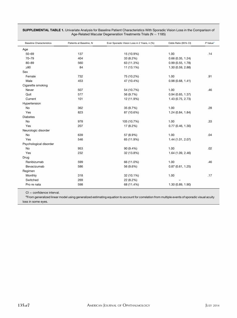
SUPPLEMENTAL TABLE 1. Univariate Analysis for Baseline Patient Characteristics With Sporadic Vision Loss in the Comparison ofAge-Related Macular Degeneration Treatments Trials (N ¼ 1185)
Baseline Characteristics Patients at Baseline, N Ever Sporadic Vision Loss in 2 Years, n (%) Odds Ratio (95% CI) P Valuea
Age
50–69 137 15 (10.9%) 1.00 .14
70–79 404 33 (8.2%) 0.66 (0.35, 1.24)
80–89 560 63 (11.3%) 0.99 (0.55, 1.78)
>_90 84 11 (13.1%) 1.30 (0.59, 2.88)
Sex
Female 732 75 (10.2%) 1.00 .91
Male 453 47 (10.4%) 0.98 (0.68, 1.41)
Cigarette smoking
Never 507 54 (10.7%) 1.00 .46
Quit 577 56 (9.7%) 0.94 (0.65, 1.37)
Current 101 12 (11.9%) 1.43 (0.75, 2.73)
Hypertension
No 362 35 (9.7%) 1.00 .28
Yes 823 87 (10.6%) 1.24 (0.84, 1.84)
Diabetes
No 978 105 (10.7%) 1.00 .33
Yes 207 17 (8.2%) 0.77 (0.46, 1.30)
Neurologic disorder
No 639 57 (8.9%) 1.00 .04
Yes 546 65 (11.9%) 1.44 (1.01, 2.07)
Psychological disorder
No 953 90 (9.4%) 1.00 .02
Yes 232 32 (13.8%) 1.64 (1.09, 2.46)
Drug
Ranibizumab 599 66 (11.0%) 1.00 .46
Bevacizumab 586 56 (9.6%) 0.87 (0.61, 1.25)
Regimen
Monthly 318 32 (10.1%) 1.00 .17
Switched 269 22 (8.2%) –
Pro re nata 598 68 (11.4%) 1.30 (0.89, 1.90)
CI ¼ confidence interval.aFromgeneralized linear model using generalized estimating equation to account for correlation frommultiple events of sporadic visual acuity
loss in some eyes.
135.e7 JULY 2014AMERICAN JOURNAL OF OPHTHALMOLOGY
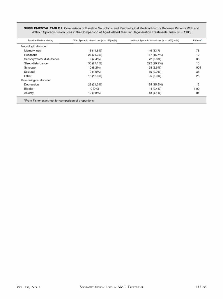
SUPPLEMENTAL TABLE 2. Comparison of Baseline Neurologic and Psychological Medical History Between Patients With andWithout Sporadic Vision Loss in the Comparison of Age-Related Macular Degeneration Treatments Trials (N ¼ 1185)
Baseline Medical History With Sporadic Vision Loss (N ¼ 122) n (%) Without Sporadic Vision Loss (N ¼ 1063) n (%) P Valuea
Neurologic disorder
Memory loss 18 (14.8%) 146 (13.7) .78
Headache 26 (21.3%) 167 (15.7%) .12
Sensory/motor disturbance 9 (7.4%) 72 (6.8%) .85
Sleep disturbance 33 (27.1%) 222 (20.9%) .13
Syncope 10 (8.2%) 28 (2.6%) .004
Seizures 2 (1.6%) 10 (0.9%) .35
Other 15 (12.3%) 95 (8.9%) .25
Psychological disorder
Depression 26 (21.3%) 165 (15.5%) .12
Bipolar 0 (0%) 4 (0.4%) 1.00
Anxiety 12 (9.8%) 43 (4.1%) .01
aFrom Fisher exact test for comparison of proportions.
VOL. 158, NO. 1 135.e8SPORADIC VISION LOSS IN AMD TREATMENT
SUPPLEMENTAL TABLE 3. Univariate Analysis for Baseline Ocular Characteristics With Sporadic Vision Loss in the Comparison ofAge-Related Macular Degeneration Treatments Trials (N ¼ 1185)
Baseline Ocular Characteristics Patients at Baseline, N Ever Sporadic Vision Loss in 2 Years, n (%) Odds Ratio (95% CI) P Valuea
Baseline visual acuity in study eye
20/25–40 424 30 (7.1%) 1.00 <.001
20/50–80 443 41 (9.3%) 1.50 (0.93, 2.39)
20/100–160 236 32 (13.6%) 2.16 (1.32, 3.55)
20/200–320 82 19 (23.2%) 4.29 (2.44, 7.54)
Baseline visual acuity in fellow eye
20/20 or better 346 41 (11.8%) 1.00 .81
20/25–20/40 469 42 (9.0%) 0.88 (0.57, 1.36)
20/50 or worse 370 39 (10.5%) 0.89 (0.58, 1.38)
Baseline total area of CNV lesion (DA)
1st quartile (<_1) 376 36 (9.6%) 1.00 .38
2nd quartile (>1 to <_2) 260 25 (9.6%) 1.14 (0.69, 1.89)
3rd quartile (>2 to <_4) 287 31 (10.8%) 1.47 (0.91, 2.38)
4th quartile (>4) 215 25 (11.6%) 1.39 (0.84, 2.30)
Missingb 47 5 (10.6%)
Location of lesion
Not subfoveal 323 35 (10.8%) 1.00 .66
Subfoveal 843 86 (10.2%) 1.09 (0.74, 1.60)
Missingb 19 1 (5.3%)
Lesion type
Predominantly classic 267 37 (13.9%) 1.00 .23
Minimally classic 197 23 (11.7%) 0.82 (0.49, 1.38)
Occult only 696 61 (8.8%) 0.65 (0.43, 0.99)
CG/no lesion 25 1 (4.0%)
Hemorrhage (associated with the lesion)
None 441 40 (9.1%) 1.00 .21
<_1 DA 611 68 (11.1%) 1.42 (0.96, 2.10)
<_2 DA 59 5 (8.5%) 1.08 (0.42, 2.75)
>2 DA 54 8 (14.8%) 1.88 (0.88, 4.01)
CD or CG or missingb 20 1 (5.0%)
Fibrotic or atrophic scar
No 1125 110 (9.8%) 1.00 .001
Yes 46 11 (23.9%) 2.93 (1.58, 5.43)
Missingb 14 1 (7.1%)
Geographic atrophy
None/questionable 1101 114 (10.4%) 1.00 .95
Present 82 8 (9.8%) 1.02 (0.50, 2.12)
Missingb 2 0 (0.0%)
CD ¼ cannot determine; CG ¼ cannot grade; CI ¼ confidence interval; CNV ¼ choroidal neovascularization; DA ¼ disc areas.aFromgeneralized linear model using generalized estimating equation to account for correlation frommultiple events of sporadic visual acuity
loss in some eyes.bMissing category was not included in the P value calculation.
135.e9 JULY 2014AMERICAN JOURNAL OF OPHTHALMOLOGY
SUPPLEMENTAL TABLE 4. Univariate Analysis for Baseline Optical Coherence Tomography Features With Sporadic Vision Loss inthe Comparison of Age-Related Macular Degeneration Treatments Trials (N ¼ 1185)
Baseline Characteristics Patients at Baseline, N Ever Sporadic Vision Loss in 2 Years, n (%) Odds Ratio (95% CI) P Valuea
Retinal thickness at foveal center (mm)
<120 120 13 (10.8%) 1.00 .08
120–212 622 55 (8.8%) 0.86 (0.48, 1.56)
>212 437 53 (12.1%) 1.32 (0.73, 2.41)
Missingb 6 1 (16.7%)
Subretinal tissue complex thickness at foveal center (mm)
1st quartile (>0 to <_75) 290 26 (9.0%) 1.00 .15
2nd quartile (>75 to <_160) 291 24 (8.2%) 1.01 (0.58, 1.76)
3rd quartile (>160 to <_275) 304 41 (13.5%) 1.63 (1.00, 2.66)
4th quartile (>275) 294 30 (10.2%) 1.16 (0.69, 1.96)
Missingb 6 1 (16.7%)
Intraretinal fluid
No fluid 277 20 (7.2%) 1.00 .003
Fluid not in foveal center 318 28 (8.8%) 1.51 (0.86, 2.67)
Fluid in foveal center 569 73 (12.8%) 2.23 (1.38, 3.61)
Missingb 21 1 (4.8%)
Subretinal fluid
No fluid 202 21 (10.4%) 1.00 .28
Fluid not in foveal center 556 63 (11.3%) 1.03 (0.63, 1.69)
Fluid in foveal center 414 37 (8.9%) 0.75 (0.44, 1.28)
Missingb 13 1 (7.7%)
Sub-RPE fluid
No fluid 518 56 (10.8%) 1.00 .39
Fluid not in foveal center 210 19 (9.0%) 0.74 (0.44, 1.23)
Fluid in foveal center 363 31 (8.5%) 0.79 (0.51, 1.23)
Missingb 94 16 (17.0%)
CI ¼ confidence interval; RPE ¼ retinal pigment epithelium.aFromgeneralized linear model using generalized estimating equation to account for correlation frommultiple events of sporadic visual acuity
loss in some eyes.bMissing category was not included in the P value calculation.
VOL. 158, NO. 1 135.e10SPORADIC VISION LOSS IN AMD TREATMENT
Recommended