Screening of Fungi for Decolorization of Dye Wastewater
Merih Kıvanc+, Mine Doğruer Özen
Anadolu University, Faculty of Science,Department of Biology, Eskişehir, TURKEY
Abstract. A total of 40 fungi were screened for their ability to decolorize Xiron orange RHD (FW),
Tobactive scarlet P2R (Kimsa). Microorganisms having the ability of decolourization of organic colorants
were isolated from Porsuk stream, soil and wastewater of textile factory. Four strains of Fusarium, Penicillim
expansum, P. citreo-viride, Aspergillus flavipes, Trichoderma harzianum Paecilomyes variotii have been
chosen for this study. In addition to these strains four different strains of Myrothecium were also used in this
study. By using the those strains, maximum decolourisation was observed at 25oC pH 7.0.Among these,
maximum decolourisation was obtained by A. flavipes. It was followed by T. harzianum, Fusarium sp2.
These strains has decreased B.O.D. degree of was the wastewater of textile factory on the rates of 17.7% and
24.2%.
Keywords: texile dye, fungi, decolourization
1. Introduction
Azo compounds are used extensively in the food, pharmaceutical, cosmetic and textile industries [1].
Aromatic azo groups are not synthesized in nature, azo dyes are considered to be xenobiotics [2]. As
consequence azo dyes are recalcitrant in aerobic wastewater treatment plants. However, provided that the
proper conditions and microorganisms are used, biodegrading of azo dyes is possible [3].
Wastewater treatment systems generally do not remove the dyes, wastewaters from textile industry result
in pollution of the environment. The elimination of collared effluents in wastewater is based mainly on
physical or chemical methods. Although these methods are effective, they suffer from such shortcomings as
high cost, formation of hazardous by-products and intensive energy requirements. Therefore, as a better
alternative, microbial biodegradation methods are receiving attention. The use of white-rot fungi has
attracted increasing attention as these organisms have the ability to metabolize a diverse range of polluting
compounds. Phanerochaete chrysosporium, the most extensively studied white-rot fungus, has been shown
to metabolize and decolorize textile dyes [4], [5].
Bio-decolourization of lignin-containing pulp and paper wastewater using white rot fungi
P.chrysosporium and Tictoporia sp. Due to high oxidative potential of many of the enzymes associated with
white rot fungi, e.g. ligninase, laccase, Mn-peroxidase [6], [7]. Several other dye decolorizing fungal species
have been reported, which include Aspergillus niveus 2 ve Fusarium moniliforme [8].
It is thus not surprising those efforts to isolate from nature microorganisms utilizing azo dyes as carbon
sources where unsuccessful. However, adaptation experiments in chemostats and carefully adjusted selective
pressure let to bacterial cultures which mineralised the carboxylated azo dyes.
As for dye colour removal, review [9], [10] described the ability of Rhodococcus, Bacillus cereus and
Plasmiomonas/Achromobacter to degrade soluble dyes, acid red dye and five azo-dyes, respectively.
On the other hand, textile dyes were found strongly adsorbed and held by wastewater treatment plant
sludge that was land filled. This suggests that adsorption may play another key role in bio- decolourization.
Corresponding author. Tel.: + 905374326098
E-mail address: [email protected]
International Proceedings of Chemical, Biological and Environmental Engineering, V0l. 100 (2017)
DOI: 10.7763/IPCBEE. 2017. V100. 1
1
There is not much information about the effects of thenon-white-rot fungi on decolourization of azo dyes.
In this research, isolated fungi were used and the ability of these organisms to decolorize Xiron orange
RHD(FW),Tobactive scarlet P2R(Kimsa)tested.
2. Materials And Methods
2.1. Microorganisms
Myrothecium leutricum, M.penicilloides, M.masonii obtained from Norten Research Center, USA, Peoria
IL. Fusarium sp.1, Fusarium sp2, Fusarium sp 3, Fusarium sp4, Penicillium expansum Aspergillus flavipes,
P.sitreo-viride, Tricoderma harzianum and Paesilomises variotti was isolated in our laboratory.
They were maintained through periodic transfers on sabauroud dextrose agar at +4oC. Subculters were
made every 3 to 4 weeks.
2.2. Dyes
Xiron orange RHD (FW), Tobactive scarlet P2R (Kimsa) were obtained from textile fabric, Eskişehir,
Turkey.
2.3. Wastewater
A textil dye factory in Eskişehir provided the wastewater. From a textile dyeing factory,
Sample I wastewater from dyeing, pH 7.5 dark green
Sample II wastewater from dyeing, pH 7.0
Sample III wastewater from dyeing, pH 8.5 brown, flom
Sample IV wastewater from dyeing, pH 7.5 dark
Sample V wastewater from dyeing, pH 8.5 Lila
2.4. Isolation of Fungi
Water samples were collected from Porsuk river in Eskişehir (Turkey) that is heavily polluted by textile
wastewater. And soil samples were collected textile fabric in Eskişehir. Nutrient agar and potato dextrose
agar petri plates supplemented with dyes (1%) used to screen soil, water and waste water samples for
colonies circlet by a clear decolorized zone. Isolates were identified by using the methods and identification
keys for fungi (Hasenekoğlu 1991).
These fungi were then tested for dye decolourization under submerged culture condition at near ambient
temperature for up to two weeks.
2.5. Decolourisation of Dyes with Fungi
Cultures were cultured at 25oC in malt extract broth. After one week of incubation, they were filtered
with Whatman no 1 filter paper then weighed.
Wet cell cake were mixed with specific aqueous dye solution (0.1%) in 1:3 weight ratio and incubated
for 1 and 2 week. OD measured after 1 and 2 week.
Decolorizing activity of Tobactive scarlet P2R. Xiron orange RHD were assayed by measurement of the
decrease in colour density at 663nm, 514nm and 490nm respectively. The decolorizing yield was expressed
as the degree of the decrease in absorbance at the same wavelength.
Each treatment was carried out in duplicate and results obtained are given as the arithmetic mean.
2.6. Effect of Dye Concentration
The culture of Fusarium sp2, A.flavipes and T. harzianum was gradually exposed to increasing
concentration of dye (0.1mg/l, 1mg/l,10mg/l). Decolorizing activity of Tobactive scarlet P2R, Xiron orange
RHD were assayed by measurement of the decrease in colour density at 663nm and 514nm respectively.
2.7. Effect of pH
2
The pH of the individual culture was adjusted to 3.0, 5.0, 6.5 and 7.0 and all cultures were incubated at
25oC.Decolorization of dyes were monitored. The absorbance was measured at 663nm and 514nm to
determine the concentration of Tobactive scarlet P2R. Xiron orange RHD.
2.8. Effect of Temperature
Individual cultures were incubated at 5, 25 and 35oC. Decolorization of Xiron orange RHD (FW)
(Orange 13), Tobactive scarlet P2R (Kimsa) (Red mix) were determined the cell free supernatant. Its
absorbance was read at OD663 Tobactive scarlet P2R OD514 Xiron orange RHD OD490.
Decolourization of textile dyes were determined as follows;
Decolorization (%)=Initial absorbance- absorbance x100
Initial absorbance
2.9. Decolorization of Dye Containing Wastewater by Fungi
Real dye containing wastewater samples were added to fungi and its effectiveness in dye color removal
evaluated. They were each mixed with 5 days old mycelia in roughly 2.5:1 weight ratio and observed after 1-
2 weeks static.
3. Result And Discussion
Xiron orange RHD (FW), Tobactive scarlet P2R (Kimsa) which is monoazo dye has seen extensive in
textile dying. Fusarium sp., P.expansum, P. citreo-viride, A.flavipes, T. harzianum, P.variotii were capable
of decolorizing Xiron orange RHD (FW), Tobactive scarlet P2R (Kimsa) produced clear zones surrounding
its colonies on the agar plates. Mou et al [11] and Karaca ve Kıvanç [8] also reported similar results. Mou et
al [11] reported the decolourization activity of the Myrothecium and Ganoderma culture filtrate. Maximal
decolorizing activity of A. flavipes are within 14 days. Bio-decolourisation was evident and effective all
turned colourless to naked eyes. T. harzianum also showed high decolorizing ability for Tobactive scarlet
P2R. The Trichoderma species were degrade aromatic pollutants [12]. A. niger, F. oxysporum and
Trichoderma lignorum were degrade textile dye [13].
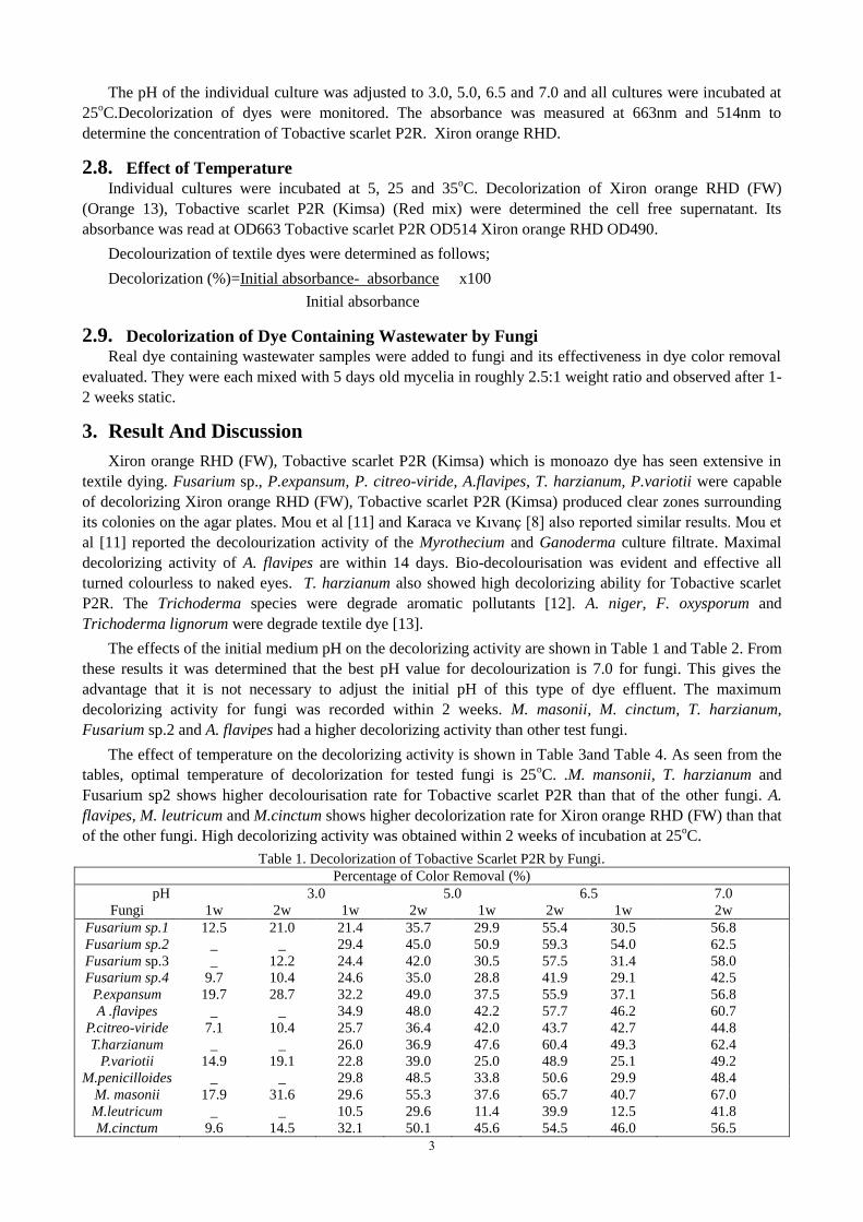
The effects of the initial medium pH on the decolorizing activity are shown in Table 1 and Table 2. From
these results it was determined that the best pH value for decolourization is 7.0 for fungi. This gives the
advantage that it is not necessary to adjust the initial pH of this type of dye effluent. The maximum
decolorizing activity for fungi was recorded within 2 weeks. M. masonii, M. cinctum, T. harzianum,
Fusarium sp.2 and A. flavipes had a higher decolorizing activity than other test fungi.
The effect of temperature on the decolorizing activity is shown in Table 3and Table 4. As seen from the
tables, optimal temperature of decolorization for tested fungi is 25oC. .M. mansonii, T. harzianum and
Fusarium sp2 shows higher decolourisation rate for Tobactive scarlet P2R than that of the other fungi. A.
flavipes, M. leutricum and M.cinctum shows higher decolorization rate for Xiron orange RHD (FW) than that
of the other fungi. High decolorizing activity was obtained within 2 weeks of incubation at 25oC.
Table 1. Decolorization of Tobactive Scarlet P2R by Fungi.
Percentage of Color Removal (%)
pH 3.0 5.0 6.5 7.0
Fungi 1w 2w 1w 2w 1w 2w 1w 2w
Fusarium sp.1 12.5 21.0 21.4 35.7 29.9 55.4 30.5 56.8
Fusarium sp.2 _ _ 29.4 45.0 50.9 59.3 54.0 62.5
Fusarium sp.3 _ 12.2 24.4 42.0 30.5 57.5 31.4 58.0
Fusarium sp.4 9.7 10.4 24.6 35.0 28.8 41.9 29.1 42.5
P.expansum 19.7 28.7 32.2 49.0 37.5 55.9 37.1 56.8
A .flavipes _ _ 34.9 48.0 42.2 57.7 46.2 60.7
P.citreo-viride 7.1 10.4 25.7 36.4 42.0 43.7 42.7 44.8
T.harzianum _ _ 26.0 36.9 47.6 60.4 49.3 62.4
P.variotii 14.9 19.1 22.8 39.0 25.0 48.9 25.1 49.2
M.penicilloides _ _ 29.8 48.5 33.8 50.6 29.9 48.4
M. masonii 17.9 31.6 29.6 55.3 37.6 65.7 40.7 67.0
M.leutricum _ _ 10.5 29.6 11.4 39.9 12.5 41.8
M.cinctum 9.6 14.5 32.1 50.1 45.6 54.5 46.0 56.5
3
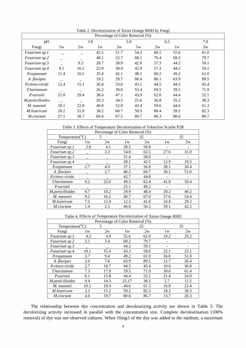
Table 2. Decolorization of Xiron Orange RHD by Fungi
Percentage of Color Removal (%)
pH 3.0 5.0 6.5 7.0
Fungi 1w 2w 1w 2w 1w 2w 1w 2w
Fusarium sp.1 _ _ 42.5 51.7 54.2 60.2 55.6 61.0
Fusarium sp.2 _ _ 48.1 52.7 68.1 76.4 69.2 79.7
Fusarium sp.3 _ 9.2 28.7 39.9 42.9 57.3 44.2 59.1
Fusarium sp.4 9.1 10.2 25.9 38.4 42.9 57.3 44.2 59.1
P.expansum 11.4 16.5 35.4 42.1 48.1 60.2 49.2 61.0
A .flavipes _ _ 19.2 29.7 60.4 86.1 63.9 89.5
P.citreo-viride 12.4 15.1 30.4 33.0 43.1 44.5 44.5 45.4
T.harzianum _ _ 26.2 39.0 55.4 69.5 59.5 71.9
P.variotii 21.0 29.4 38.4 47.1 43.9 52.0 44.4 52.1
M.penicilloides _ _ 20.3 34.5 25.6 36.8 25.2 38.3
M. masonii 18.1 22.8 40.8 52.8 43.4 59.6 44.6 61.3
M.leutricum 20.2 31.0 38.2 60.7 58.9 80.4 59.2 82.3
M.cinctum 27.1 38.7 60.4 67.5 80.7 86.3 80.6 86.7
Table 3. Effects of Temperature Decolorization of Tobactive Scarlet P2R
Percentage of Color Removal (%)
Temperature(oC) 5 25 35
Fungi 1w 2w 1w 2w 1w 2w
Fusarium sp.1 2.8 4.1 30.5 56.8 - -
Fusarium sp.2 _ 2.3 54.0 62.5 27.6 31.0
Fusarium sp.3 _ _ 31.4 58.0 - -
Fusarium sp.4 _ _ 29.1 42.5 12.9 19.5
P.expansum 2.7 4.9 37.1 56.8 28.1 30.4
A .flavipes _ 2.7 46.2 60.7 36.1 51.0
P.citreo-viride _ _ 42.7 44.8 - -
T.harzianum 9.2 22.0 49.3 62.4 41.8 50.4
P.variotii _ _ 25.1 49.2 - -
M.penicilloides 9.7 10.2 29.9 48.4 29.2 40.2
M. masonii 9.2 16.2 40.7 67.0 37.6 54.4
M.leutricum 7.5 12.4 12.5 41.8 10.8 29.3
M.cinctum 1.4 2.5 46.0 56.5 39.1 42.2
Table 4. Effects of Temperature Decolorization of Xiron Orange RHD
Percentage of Color Removal (%)
Temperature(oC) 5 25 35
Fungi 1w 2w 1w 2w 1w 2w
Fusarium sp.1 4.2 4.9 55.6 61.0 29.2 29.2
Fusarium sp.2 3.1 3.4 69.2 79.7 - -
Fusarium sp.3 _ _ 44.2 59.1 - -
Fusarium sp.4 10.1 15.4 43.3 58.0 22.1 22.1
P.expansum 3.7 9.4 49.2 61.0 36.0 51.0
A .flavipes 2.0 7.6 63.9 89.5 12.7 26.4
P.citreo-viride 2.7 18.7 44.5 45.4 10.6 36.8
T.harzianum 7.3 17.9 59.5 71.9 30.6 61.4
P.variotii 6.1 15.8 44.4 52.1 21.4 24.9
M.penicilloides 0.4 14.3 25.17 38.3 2.7 11.5
M. masonii 10.1 18.4 44.6 61.3 16.8 21.4
M.leutricum 2.3 15.2 59.2 82.3 18.2 38.3
M.cinctum 4.0 19.7 80.6 86.7 13.7 26.3
The relationship between dye concentration and decolourizing activity are shown in Table 5. The
decolorizing activity increased in parallel with the concentration size. Complete decolourisation (100%
removal) of dye was not observed cultures. When 10mg/l of the dye was added to the medium, a maximum
4
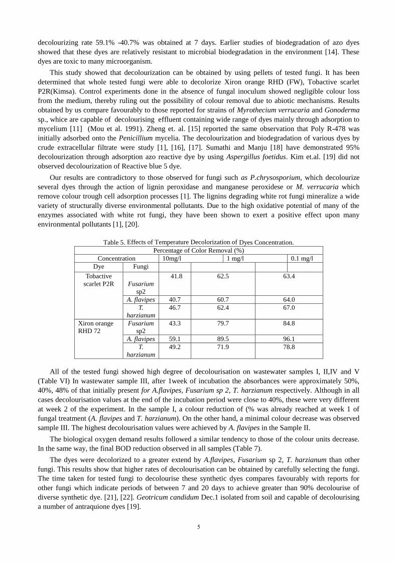
decolourizing rate 59.1% -40.7% was obtained at 7 days. Earlier studies of biodegradation of azo dyes
showed that these dyes are relatively resistant to microbial biodegradation in the environment [14]. These
dyes are toxic to many microorganism.
This study showed that decolourization can be obtained by using pellets of tested fungi. It has been
determined that whole tested fungi were able to decolorize Xiron orange RHD (FW), Tobactive scarlet
P2R(Kimsa). Control experiments done in the absence of fungal inoculum showed negligible colour loss
from the medium, thereby ruling out the possibility of colour removal due to abiotic mechanisms. Results
obtained by us compare favourably to those reported for strains of Myrothecium verrucaria and Gonoderma
sp., whice are capable of decolourising effluent containing wide range of dyes mainly through adsorption to
mycelium [11] (Mou et al. 1991). Zheng et. al. [15] reported the same observation that Poly R-478 was
initially adsorbed onto the Penicillium mycelia. The decolourization and biodegradation of various dyes by
crude extracellular filtrate were study [1], [16], [17]. Sumathi and Manju [18] have demonstrated 95%
decolourization through adsorption azo reactive dye by using Aspergillus foetidus. Kim et.al. [19] did not
observed decolourization of Reactive blue 5 dye.
Our results are contradictory to those observed for fungi such as P.chrysosporium, which decolourize
several dyes through the action of lignin peroxidase and manganese peroxidese or M. verrucaria which
remove colour trough cell adsorption processes [1]. The lignins degrading white rot fungi mineralize a wide
variety of structurally diverse environmental pollutants. Due to the high oxidative potential of many of the
enzymes associated with white rot fungi, they have been shown to exert a positive effect upon many
environmental pollutants [1], [20].
Table 5. Effects of Temperature Decolorization of Dyes Concentration.
Percentage of Color Removal (%)
Concentration 10mg/l 1 mg/l 0.1 mg/l
Dye Fungi
Tobactive
scarlet P2R
Fusarium
sp2
41.8 62.5 63.4
A. flavipes 40.7 60.7 64.0
T.
harzianum
46.7 62.4 67.0
Xiron orange
RHD 72
Fusarium
sp2
43.3 79.7 84.8
A. flavipes 59.1 89.5 96.1
T.
harzianum
49.2 71.9 78.8
All of the tested fungi showed high degree of decolourisation on wastewater samples I, II,IV and V
(Table VI) In wastewater sample III, after 1week of incubation the absorbances were approximately 50%,
40%, 48% of that initially present for A.flavipes, Fusarium sp 2, T. harzianum respectively. Although in all
cases decolourisation values at the end of the incubation period were close to 40%, these were very different
at week 2 of the experiment. In the sample I, a colour reduction of (% was already reached at week 1 of
fungal treatment (A. flavipes and T. harzianum). On the other hand, a minimal colour decrease was observed
sample III. The highest decolourisation values were achieved by A. flavipes in the Sample II.
The biological oxygen demand results followed a similar tendency to those of the colour units decrease.
In the same way, the final BOD reduction observed in all samples (Table 7).
The dyes were decolorized to a greater extend by A.flavipes, Fusarium sp 2, T. harzianum than other
fungi. This results show that higher rates of decolourisation can be obtained by carefully selecting the fungi.
The time taken for tested fungi to decolourise these synthetic dyes compares favourably with reports for
other fungi which indicate periods of between 7 and 20 days to achieve greater than 90% decolourise of
diverse synthetic dye. [21], [22]. Geotricum candidum Dec.1 isolated from soil and capable of decolourising
a number of antraquione dyes [19].
5
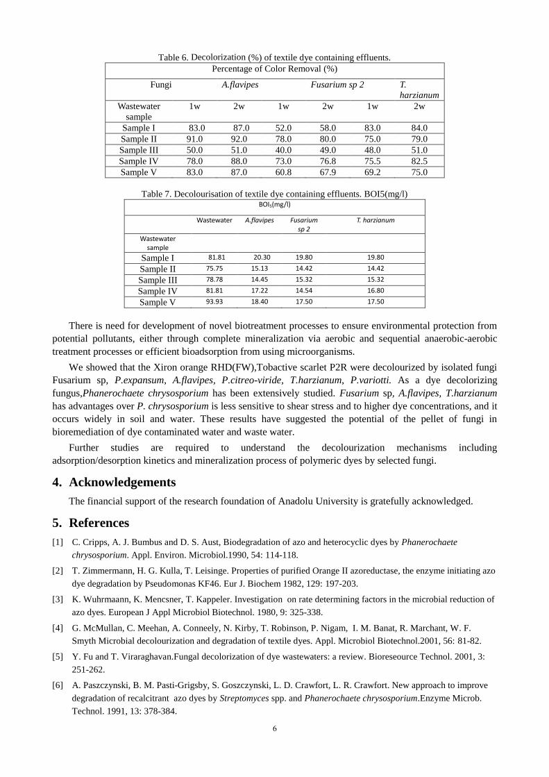
Table 6. Decolorization (%) of textile dye containing effluents.
Percentage of Color Removal (%)
Fungi A.flavipes Fusarium sp 2 T.
harzianum
Wastewater
sample
1w 2w 1w 2w 1w 2w
Sample I 83.0 87.0 52.0 58.0 83.0 84.0
Sample II 91.0 92.0 78.0 80.0 75.0 79.0
Sample III 50.0 51.0 40.0 49.0 48.0 51.0
Sample IV 78.0 88.0 73.0 76.8 75.5 82.5
Sample V 83.0 87.0 60.8 67.9 69.2 75.0
Table 7. Decolourisation of textile dye containing effluents. BOI5(mg/l) BOI5(mg/l)
Wastewater A.flavipes Fusarium sp 2
T. harzianum
Wastewater sample
Sample I 81.81 20.30 19.80 19.80
Sample II 75.75 15.13 14.42 14.42
Sample III 78.78 14.45 15.32 15.32
Sample IV 81.81 17.22 14.54 16.80
Sample V 93.93 18.40 17.50 17.50
There is need for development of novel biotreatment processes to ensure environmental protection from
potential pollutants, either through complete mineralization via aerobic and sequential anaerobic-aerobic
treatment processes or efficient bioadsorption from using microorganisms.
We showed that the Xiron orange RHD(FW),Tobactive scarlet P2R were decolourized by isolated fungi
Fusarium sp, P.expansum, A.flavipes, P.citreo-viride, T.harzianum, P.variotti. As a dye decolorizing
fungus,Phanerochaete chrysosporium has been extensively studied. Fusarium sp, A.flavipes, T.harzianum
has advantages over P. chrysosporium is less sensitive to shear stress and to higher dye concentrations, and it
occurs widely in soil and water. These results have suggested the potential of the pellet of fungi in
bioremediation of dye contaminated water and waste water.
Further studies are required to understand the decolourization mechanisms including
adsorption/desorption kinetics and mineralization process of polymeric dyes by selected fungi.
4. Acknowledgements
The financial support of the research foundation of Anadolu University is gratefully acknowledged.
5. References
[1] C. Cripps, A. J. Bumbus and D. S. Aust, Biodegradation of azo and heterocyclic dyes by Phanerochaete
chrysosporium. Appl. Environ. Microbiol.1990, 54: 114-118.
[2] T. Zimmermann, H. G. Kulla, T. Leisinge. Properties of purified Orange II azoreductase, the enzyme initiating azo
dye degradation by Pseudomonas KF46. Eur J. Biochem 1982, 129: 197-203.
[3] K. Wuhrmaann, K. Mencsner, T. Kappeler. Investigation on rate determining factors in the microbial reduction of
azo dyes. European J Appl Microbiol Biotechnol. 1980, 9: 325-338.
[4] G. McMullan, C. Meehan, A. Conneely, N. Kirby, T. Robinson, P. Nigam, I. M. Banat, R. Marchant, W. F.
Smyth Microbial decolourization and degradation of textile dyes. Appl. Microbiol Biotechnol.2001, 56: 81-82.
[5] Y. Fu and T. Viraraghavan.Fungal decolorization of dye wastewaters: a review. Bioreseource Technol. 2001, 3:
251-262.
[6] A. Paszczynski, B. M. Pasti-Grigsby, S. Goszczynski, L. D. Crawfort, L. R. Crawfort. New approach to improve
degradation of recalcitrant azo dyes by Streptomyces spp. and Phanerochaete chrysosporium.Enzyme Microb.
Technol. 1991, 13: 378-384.
6

[7] A. Paszczynski, L. R. Crawfort. Degradation of azo dyes by the ligninase from Phanerochaete
chrysosporium:Involvement of veratryl alcohol.Biochemical and Biophysical Research Communications.1992,
136: 220-227.
[8] H. Karaca and M. Kivanç. Decolorızatıon of blue 13 with Aspergillus nıveus and Fusarıum monıliforme.Anadolu
Unıversity journal of Science and Technology –CLife Sciences and Biotechnology 2012, 2(1): 21-30.
[9] T. Marimuthu, S. Rajendran, M. Manivannan. A review on bacterial degradation of textile dyes. J Chem Chem Sci.
2013, 3: 201-212.
[10] T. OGAWA and C. YATOME, C., Biodegradation of azo dyes in multistage rotating biological contator
immobilized by assimilating bacteria. Bull. Environ. Contam. Toxicol. 1990, 44, 561–566.
[11] D. G. Mou, K. K. Lim, and H. P. Shen. Microbial agents for decolorization of dye wastewater.Biotech Adv. 1991,
9: 613-622.
[12] A. Katayama and F. Matsumura. Degradation of organochlorine pesticides, particularly endosulfan, by
Trichoderma harzianum. Environ Toxicol. Chem. 1993, 12: 1059-1065.
[13] A. Shahid, J. Singh, S. Bisht, P. Teotia, V. Kumar. Biodegradation of textile dyes by fungi isolated from North
Indian field soil. Env. Asia. 2013, 6 (2): 51–57.
[14] U, Meyer. Biodegradation of synthetic organic colorants, In, T. Leisinger, T. Cook, J. Nuesch and R. Huffer,
(Eds),Microbial degradation of xenobiotics and Recalcitrant compounds. Academic Press, London. 1981, pp. 371-
385.
[15] Z. Zheng, R. E. Levin, J. L. Pinkham, K. Shetty. Decolorization of polymeric dyes by a novel Penicillium isolate.
Process Biochem. 1999, 34(1): 31-34.
[16] B. M. Pasti-Grigsby, A. Paszczynski, S.Goszczynski, L. R. Crawfort Influence of aromatic substation patterns on
azo dye degradability by Streptomyces spp. and Phanerochaete chrysosporium.Appl. Environ Microbiol. 1992, 58:
3605-3613.
[17] T. J. Sparado, H. M. Gold, V. Renganathan. Degradation of azo dyes by the lignindegrading fungus
Phanerochaete chrysosporium. Appl. Environ Microbiol. 1992, 58: 2397-2401.
[18] S. Sumathi and BS. Manju. Uptake of reactive textile dyes by Aspergillus foetidus. Enzyme and Microbial
Technology. 2000, 27: 347-355.
[19] J. S. Kim, K. Ishıkawa M. Hirai, M. Shoda. 1995. Characteristics of a newly isolated fungus, Geotrichum
candidum Dec 1, whice decolorizes various dyes. J. of Fermentation and Bioengineering.1995, 79: 601-607.
[20] E. Esposito, P. V. Canhos and N. Duran 1991. Screening of lignin degrading fungi for removal of colour from
Kraft mill wastewater with no additional carbon source. Biotechnology letters, 1991, 13: 571-576.
[21] C. A. Reddy. 1995. The potential for white-rot fungi in the treatment of pollutants.Curr. Opin.Biotechnol, 1995, 6,
320-328.
[22] N. Kirby, R. Marchant, G. McMullan, G. Decolourisation of synthetic textile dyes by Phlebia tremellosa. Fems
Microbiol Lett. 2000, 188, 93-96.
7
Recommended