1
Case Report
Recurrence of a Refractory Chronic Subdural Hematoma after Middle Meningeal Artery Embolization That Required Craniotomy
Hideo Chihara, Hirotoshi Imamura, Takenori Ogura, Hidemitsu Adachi, Yukihiro Imai, and Nobuyuki Sakai
Department of Neurosurgery, Kobe City Hospital Organization, Kobe City Medical General Hospital, Kobe, Hyogo
Received: November 6, 2013; Accepted: December 16, 2013
NMC Case Report Journal 2014; 1: 1–5 DOI: 10.2176/nmccrj.2013-0343
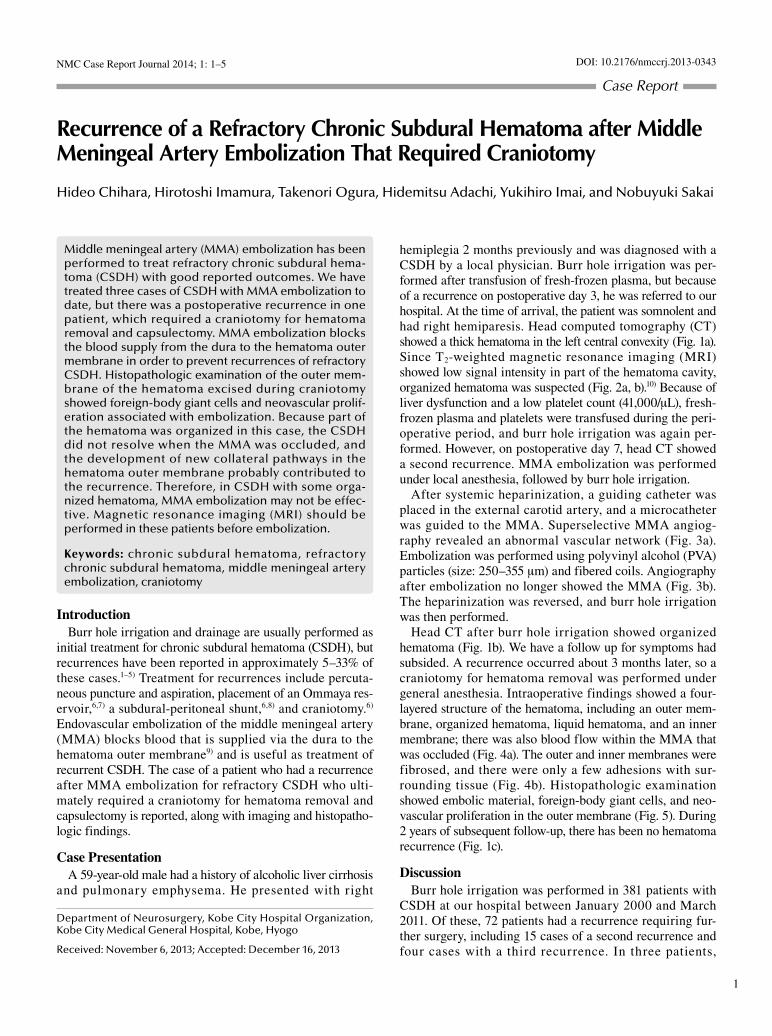
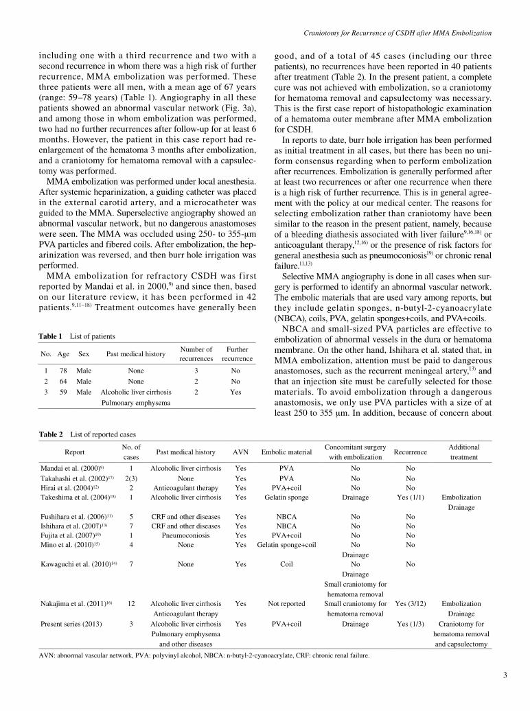
hemiplegia 2 months previously and was diagnosed with a CSDH by a local physician. Burr hole irrigation was per-formed after transfusion of fresh-frozen plasma, but because of a recurrence on postoperative day 3, he was referred to our hospital. At the time of arrival, the patient was somnolent and had right hemiparesis. Head computed tomography (CT) showed a thick hematoma in the left central convexity (Fig. 1a). Since T2-weighted magnetic resonance imaging (MRI) showed low signal intensity in part of the hematoma cavity, organized hematoma was suspected (Fig. 2a, b).10) Because of liver dysfunction and a low platelet count (41,000/μL), fresh-frozen plasma and platelets were transfused during the peri-operative period, and burr hole irrigation was again per-formed. However, on postoperative day 7, head CT showed a second recurrence. MMA embolization was performed under local anesthesia, followed by burr hole irrigation.
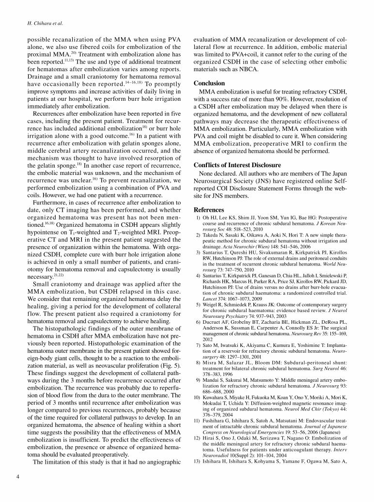
After systemic heparinization, a guiding catheter was placed in the external carotid artery, and a microcatheter was guided to the MMA. Superselective MMA angiog-raphy revealed an abnormal vascular network (Fig. 3a). Embolization was performed using polyvinyl alcohol (PVA) particles (size: 250–355 μm) and fibered coils. Angiography after embolization no longer showed the MMA (Fig. 3b). The heparinization was reversed, and burr hole irrigation was then performed.
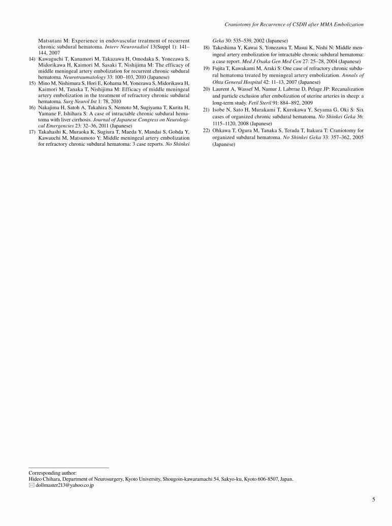
Head CT after burr hole irrigation showed organized hematoma (Fig. 1b). We have a follow up for symptoms had subsided. A recurrence occurred about 3 months later, so a craniotomy for hematoma removal was performed under general anesthesia. Intraoperative findings showed a four-layered structure of the hematoma, including an outer mem-brane, organized hematoma, liquid hematoma, and an inner membrane; there was also blood flow within the MMA that was occluded (Fig. 4a). The outer and inner membranes were fibrosed, and there were only a few adhesions with sur-rounding tissue (Fig. 4b). Histopathologic examination showed embolic material, foreign-body giant cells, and neo-vascular proliferation in the outer membrane (Fig. 5). During 2 years of subsequent follow-up, there has been no hematoma recurrence (Fig. 1c).
DiscussionBurr hole irrigation was performed in 381 patients with
CSDH at our hospital between January 2000 and March 2011. Of these, 72 patients had a recurrence requiring fur-ther surgery, including 15 cases of a second recurrence and four cases with a third recurrence. In three patients,
Middle meningeal artery (MMA) embolization has been performed to treat refractory chronic subdural hema-toma (CSDH) with good reported outcomes. We have treated three cases of CSDH with MMA embolization to date, but there was a postoperative recurrence in one patient, which required a craniotomy for hematoma removal and capsulectomy. MMA embolization blocks the blood supply from the dura to the hematoma outer membrane in order to prevent recurrences of refractory CSDH. Histopathologic examination of the outer mem-brane of the hematoma excised during craniotomy showed foreign-body giant cells and neovascular prolif-eration associated with embolization. Because part of the hematoma was organized in this case, the CSDH did not resolve when the MMA was occluded, and the development of new collateral pathways in the hematoma outer membrane probably contributed to the recurrence. Therefore, in CSDH with some orga-nized hematoma, MMA embolization may not be effec-tive. Magnetic resonance imaging (MRI) should be performed in these patients before embolization.
Keywords: chronic subdural hematoma, refractory chronic subdural hematoma, middle meningeal artery embolization, craniotomy
IntroductionBurr hole irrigation and drainage are usually performed as
initial treatment for chronic subdural hematoma (CSDH), but recurrences have been reported in approximately 5–33% of these cases.1–5) Treatment for recurrences include percuta-neous puncture and aspiration, placement of an Ommaya res-ervoir,6,7) a subdural-peritoneal shunt,6,8) and craniotomy.6) Endovascular embolization of the middle meningeal artery (MMA) blocks blood that is supplied via the dura to the hematoma outer membrane9) and is useful as treatment of recurrent CSDH. The case of a patient who had a recurrence after MMA embolization for refractory CSDH who ulti-mately required a craniotomy for hematoma removal and capsulectomy is reported, along with imaging and histopatho-logic findings.
Case PresentationA 59-year-old male had a history of alcoholic liver cirrhosis
and pulmonary emphysema. He presented with right

2
H. Chihara et al.
Fig. 1 a: Plain head computed tomography (CT) before emboliza-tion shows a chronic left subdural hematoma with an internal area of isodensity. b: Plain head CT 6 months after craniotomy for hema-toma removal and capsulectomy shows no recurrence of the hema-toma. c: During 2 years of subse-quent follow-up, there has been no hematoma recurrence.
Fig. 2 a, b: T1-weighted (a) and T2-weighted (b) head magnetic reso-nance imaging images before embolization show a mixture of a liquid hematoma component (T1- and T2-weighted: high signals) and an orga-nized component (T1-weighted: isodense signal, T2-weighted: low signal).
Fig. 5 Outer membrane of the hematoma (×100 magnification). Small branches from the middle meningeal artery that feed the dura are embolized with polyvinyl alcohol, and there are foreign-body giant cells (arrow). In addition, there is proliferation of new blood vessels (neovascularization) with a fragile structure (arrowhead).
a b c
a b
Fig. 3 a: Selective middle meningeal artery (MMA) angiography before embolization. There is an abnormal vascular network extending along the dura from the anterior and posterior branches. b: External carotid angiography after embolization. The MMA is completely embolized with polyvinyl alcohol and coils.
a
b
Fig. 4 a: Craniotomy findings. There is no recanalization of the middle meningeal artery where embolization was performed. b: Dural incision findings. The hematoma has a four-layered structure, including an outer membrane, organized hematoma, liquid hematoma, and inner membrane.
a b
3
Craniotomy for Recurrence of CSDH after MMA Embolization
Table 1 List of patients
No. Age Sex Past medical historyNumber of recurrences
Further recurrence
1 78 Male None 3 No
2 64 Male None 2 No
3 59 Male Alcoholic liver cirrhosis 2 Yes
Pulmonary emphysema
Table 2 List of reported cases
ReportNo. of cases
Past medical history AVN Embolic materialConcomitant surgery
with embolization Recurrence
Additional treatment
Mandai et al. (2000)9) 1 Alcoholic liver cirrhosis Yes PVA No No
Takahashi et al. (2002)17) 2(3) None Yes PVA No NoHirai et al. (2004)12) 2 Anticoagulant therapy Yes PVA+coil No NoTakeshima et al. (2004)18) 1 Alcoholic liver cirrhosis Yes Gelatin sponge Drainage Yes (1/1) Embolization
DrainageFushihara et al. (2006)11) 5 CRF and other diseases Yes NBCA No NoIshihara et al. (2007)13) 7 CRF and other diseases Yes NBCA No NoFujita et al. (2007)19) 1 Pneumoconiosis Yes PVA+coil No NoMino et al. (2010)15) 4 None Yes Gelatin sponge+coil No
DrainageNo
Kawaguchi et al. (2010)14) 7 None Yes Coil NoDrainage
Small craniotomy for hematoma removal
No
Nakajima et al. (2011)16) 12 Alcoholic liver cirrhosisAnticoagulant therapy
Yes Not reported Small craniotomy for hematoma removal
Yes (3/12) Embolization Drainage
Present series (2013) 3 Alcoholic liver cirrhosis Pulmonary emphysema
and other diseases
Yes PVA+coil Drainage Yes (1/3) Craniotomy for hematoma removal and capsulectomy
AVN: abnormal vascular network, PVA: polyvinyl alcohol, NBCA: n-butyl-2-cyanoacrylate, CRF: chronic renal failure.
including one with a third recurrence and two with a second recurrence in whom there was a high risk of further recurrence, MMA embolization was performed. These three patients were all men, with a mean age of 67 years (range: 59–78 years) (Table 1). Angiography in all these patients showed an abnormal vascular network (Fig. 3a), and among those in whom embolization was performed, two had no further recurrences after follow-up for at least 6 months. However, the patient in this case report had re-enlargement of the hematoma 3 months after embolization, and a craniotomy for hematoma removal with a capsulec-tomy was performed.
MMA embolization was performed under local anesthesia. After systemic heparinization, a guiding catheter was placed in the external carotid artery, and a microcatheter was guided to the MMA. Superselective angiography showed an abnormal vascular network, but no dangerous anastomoses were seen. The MMA was occluded using 250- to 355-μm PVA particles and fibered coils. After embolization, the hep-arinization was reversed, and then burr hole irrigation was performed.
MMA embolization for refractory CSDH was first reported by Mandai et al. in 2000,9) and since then, based on our literature review, it has been performed in 42 patients.9,11–18) Treatment outcomes have generally been
good, and of a total of 45 cases (including our three patients), no recurrences have been reported in 40 patients after treatment (Table 2). In the present patient, a complete cure was not achieved with embolization, so a craniotomy for hematoma removal and capsulectomy was necessary. This is the first case report of histopathologic examination of a hematoma outer membrane after MMA embolization for CSDH.
In reports to date, burr hole irrigation has been performed as initial treatment in all cases, but there has been no uni-form consensus regarding when to perform embolization after recurrences. Embolization is generally performed after at least two recurrences or after one recurrence when there is a high risk of further recurrence. This is in general agree-ment with the policy at our medical center. The reasons for selecting embolization rather than craniotomy have been similar to the reason in the present patient, namely, because of a bleeding diathesis associated with liver failure9,16,18) or anticoagulant therapy,12,16) or the presence of risk factors for general anesthesia such as pneumoconiosis19) or chronic renal failure.11,13)
Selective MMA angiography is done in all cases when sur-gery is performed to identify an abnormal vascular network. The embolic materials that are used vary among reports, but they include gelatin sponges, n-butyl-2-cyanoacrylate (NBCA), coils, PVA, gelatin sponges+coils, and PVA+coils.
NBCA and small-sized PVA particles are effective to embolization of abnormal vessels in the dura or hematoma membrane. On the other hand, Ishihara et al. stated that, in MMA embolization, attention must be paid to dangerous anastomoses, such as the recurrent meningeal artery,13) and that an injection site must be carefully selected for those materials. To avoid embolization through a dangerous anastomosis, we only use PVA particles with a size of at least 250 to 355 μm. In addition, because of concern about
4
H. Chihara et al.
possible recanalization of the MMA when using PVA alone, we also use fibered coils for embolization of the proximal MMA.20) Treatment with embolization alone has been reported.11,13) The use and type of additional treatment for hematomas after embolization varies among reports. Drainage and a small craniotomy for hematoma removal have occasionally been reported.14–16,18) To promptly improve symptoms and increase activities of daily living in patients at our hospital, we perform burr hole irrigation immediately after embolization.
Recurrences after embolization have been reported in five cases, including the present patient. Treatment for recur-rence has included additional embolization18) or burr hole irrigation alone with a good outcome.16) In a patient with recurrence after embolization with gelatin sponges alone, middle cerebral artery recanalization occurred, and the mechanism was thought to have involved resorption of the gelatin sponge.18) In another case report of recurrence, the embolic material was unknown, and the mechanism of recurrence was unclear.16) To prevent recanalization, we performed embolization using a combination of PVA and coils. However, we had one patient with a recurrence.
Furthermore, in cases of recurrence after embolization to date, only CT imaging has been performed, and whether organized hematoma was present has not been men-tioned.16,18) Organized hematoma in CSDH appears slightly hypointense on T1-weighted and T2-weighted MRI. Preop-erative CT and MRI in the present patient suggested the presence of organization within the hematoma. With orga-nized CSDH, complete cure with burr hole irrigation alone is achieved in only a small number of patients, and crani-otomy for hematoma removal and capsulectomy is usually necessary.21,22)
Small craniotomy and drainage was applied after the MMA embolization, but CSDH relapsed in this case. We consider that remaining organized hematoma delay the healing, giving a period for the development of collateral flow. The present patient also required a craniotomy for hematoma removal and capsulectomy to achieve healing.
The histopathologic findings of the outer membrane of hematoma in CSDH after MMA embolization have not pre-viously been reported. Histopathologic examination of the hematoma outer membrane in the present patient showed for-eign-body giant cells, thought to be a reaction to the emboli-zation material, as well as neovascular proliferation (Fig. 5). These findings suggest the development of collateral path-ways during the 3 months before recurrence occurred after embolization. The recurrence was probably due to reperfu-sion of blood flow from the dura to the outer membrane. The period of 3 months until recurrence after embolization was longer compared to previous recurrences, probably because of the time required for collateral pathways to develop. In an organized hematoma, the absence of healing within a short time suggests the possibility that the effectiveness of MMA embolization is insufficient. To predict the effectiveness of embolization, the presence or absence of organized hema-toma should be evaluated preoperatively.
The limitation of this study is that it had no angiographic
evaluation of MMA recanalization or development of col-lateral flow at recurrence. In addition, embolic material was limited to PVA+coil, it cannot refer to the curing of the organized CSDH in the case of selecting other embolic materials such as NBCA.
ConclusionMMA embolization is useful for treating refractory CSDH,
with a success rate of more than 90%. However, resolution of a CSDH after embolization may be delayed when there is organized hematoma, and the development of new collateral pathways may decrease the therapeutic effectiveness of MMA embolization. Particularly, MMA embolization with PVA and coil might be disabled to cure it. When considering MMA embolization, preoperative MRI to confirm the absence of organized hematoma should be performed.
Conflicts of Interest DisclosureNone declared. All authors who are members of The Japan
Neurosurgical Society (JNS) have registered online Self-reported COI Disclosure Statement Forms through the web-site for JNS members.
References 1) Oh HJ, Lee KS, Shim JJ, Yoon SM, Yun IG, Bae HG: Postoperative
course and recurrence of chronic subdural hematoma. J Korean Neu-rosurg Soc 48: 518–523, 2010
2) Takeda N, Sasaki K, Oikawa A, Aoki N, Hori T: A new simple thera-peutic method for chronic subdural hematoma without irrigation and drainage. Acta Neurochir (Wien) 148: 541–546, 2006
3) Santarius T, Qureshi HU, Sivakumaran R, Kirkpatrick PJ, Kirollos RW, Hutchinson PJ: The role of external drains and peritoneal conduits in the treatment of recurrent chronic subdural hematoma. World Neu-rosurg 73: 747–750, 2010
4) Santarius T, Kirkpatrick PJ, Ganesan D, Chia HL, Jalloh I, Smielewski P, Richards HK, Marcus H, Parker RA, Price SJ, Kirollos RW, Pickard JD, Hutchinson PJ: Use of drains versus no drains after burr-hole evacua-tion of chronic subdural haematoma: a randomized controlled trial. Lancet 374: 1067–1073, 2009
5) Weigel R, Schmiedek P, Krauss JK: Outcome of contemporary surgery for chronic subdural haematoma: evidence based review. J Neurol Neurosurg Psychiatry 74: 937–943, 2003
6) Ducruet AF, Grobelny BT, Zacharia BE, Hickman ZL, DeRosa PL, Anderson K, Sussman E, Carpenter A, Connolly ES Jr: The surgical management of chronic subdural hematoma. Neurosurg Rev 35: 155–169, 2012
7) Sato M, Iwatsuki K, Akiyama C, Kumura E, Yoshimine T: Implanta-tion of a reservoir for refractory chronic subdural hematoma. Neuro-surgery 48: 1297–1301, 2001
8) Misra M, Salazar JL, Bloom DM: Subdural-peritoneal shunt: treatment for bilateral chronic subdural hematoma. Surg Neurol 46: 378–383, 1996
9) Mandai S, Sakurai M, Matsumoto Y: Middle meningeal artery embo-lization for refractory chronic subdural hematoma. J Neurosurg 93: 686–688, 2000
10) Kuwahara S, Miyake H, Fukuoka M, Koan Y, Ono Y, Moriki A, Mori K, Mokudai T, Uchida Y: Diffusion-weighted magnetic resonance imag-ing of organized subdural hematoma. Neurol Med Chir (Tokyo) 44: 376–379, 2004
11) Fushihara G, Ishihara S, Satoh A, Matsutani M: Endovascular treat-ment of intractable chronic subdural hematoma. Journal of Japanese Congress on Neurological Emergencies 19: 53–56, 2006 (Japanese)
12) Hirai S, Ono J, Odaki M, Serizawa T, Nagano O: Embolization of the middle meningeal artery for refractory chronic subdural haema-toma. Usefulness for patients under anticoagulant therapy. Interv Neuroradiol 10(Suppl 2): 101–104, 2004
13) Ishihara H, Ishihara S, Kohyama S, Yamane F, Ogawa M, Sato A,
Craniotomy for Recurrence of CSDH after MMA Embolization
5
Matsutani M: Experience in endovascular treatment of recurrent chronic subdural hematoma. Interv Neuroradiol 13(Suppl 1): 141–144, 2007
14) Kawaguchi T, Kanamori M, Takazawa H, Omodaka S, Yonezawa S, Midorikawa H, Kaimori M, Sasaki T, Nishijima M: The efficacy of middle meningeal artery embolization for recurrent chronic subdural hematoma. Neurotraumatology 33: 100–103, 2010 (Japanese)
15) Mino M, Nishimura S, Hori E, Kohama M, Yonezawa S, Midorikawa H, Kaimori M, Tanaka T, Nishijima M: Efficacy of middle meningeal artery embolization in the treatment of refractory chronic subdural hematoma. Surg Neurol Int 1: 78, 2010
16) Nakajima H, Satoh A, Takahira S, Nemoto M, Sugiyama T, Kurita H, Yamane F, Ishihara S: A case of intractable chronic subdural hema-toma with liver cirrhosis. Journal of Japanese Congress on Neurologi-cal Emergencies 23: 32–36, 2011 (Japanese)
17) Takahashi K, Muraoka K, Sugiura T, Maeda Y, Mandai S, Gohda Y, Kawauchi M, Matsumoto Y: Middle meningeal artery embolization for refractory chronic subdural hematoma: 3 case reports. No Shinkei
Geka 30: 535–539, 2002 (Japanese)18) Takeshima Y, Kawai S, Yonezawa T, Masui K, Nishi N: Middle men-
ingeal artery embolization for intractable chronic subdural hematoma: a case report. Med J Osaka Gen Med Cen 27: 25–28, 2004 (Japanese)
19) Fujita T, Kawakami M, Araki S: One case of refractory chronic subdu-ral hematoma treated by meningeal artery embolization. Annals of Ohta General Hospital 42: 11–13, 2007 (Japanese)
20) Laurent A, Wassef M, Namur J, Labrrae D, Pelage JP: Recanalization and particle exclusion after embolization of uterine arteries in sheep: a long-term study. Fetil Steril 91: 884–892, 2009
21) Isobe N, Sato H, Murakami T, Kurokawa Y, Seyama G, Oki S: Six cases of organized chronic subdural hematoma. No Shinkei Geka 36: 1115–1120, 2008 (Japanese)
22) Ohkawa T, Ogura M, Tanaka S, Terada T, Itakura T: Craniotomy for organized subdural hematoma. No Shinkei Geka 33: 357–362, 2005 (Japanese)
Corresponding author: Hideo Chihara, Department of Neurosurgery, Kyoto University, Shougoin-kawaramachi 54, Sakyo-ku, Kyoto 606-8507, Japan. * [email protected]
Recommended