SECTION VSPECIALIZED TECHNIQUES
UNIT 1.16Recombineering: Genetic Engineering inBacteria Using HomologousRecombination
Lynn C. Thomason,1 James A. Sawitzke,2 Xintian Li,2 Nina Costantino,2
and Donald L. Court2
1Basic Science Program, GRCBL-Molecular Control & Genetics Section, Frederick NationalLaboratory for Cancer Research, Leidos Biomedical Research, Inc., Frederick, Maryland2Molecular Control and Genetics Section, Gene Regulation and Chromosome Biology, NationalCancer Institute at Frederick, National Institutes of Health, Frederick, Maryland
ABSTRACT
The bacterial chromosome and bacterial plasmids can be engineered in vivo by homol-ogous recombination using PCR products and synthetic oligonucleotides as substrates.This is possible because bacteriophage-encoded recombination proteins efficiently re-combine sequences with homologies as short as 35 to 50 bases. Recombineering allowsDNA sequences to be inserted or deleted without regard to location of restriction sites.This unit first describes preparation of electrocompetent cells expressing the recom-bineering functions and their transformation with dsDNA or ssDNA. It then presentssupport protocols that describe several two-step selection/counter-selection methods ofmaking genetic alterations without leaving any unwanted changes in the targeted DNA,and a method for retrieving onto a plasmid a genetic marker (cloning by retrieval) fromthe Escherichia coli chromosome or a co-electroporated DNA fragment. Additional pro-tocols describe methods to screen for unselected mutations, removal of the defectiveprophage from recombineering strains, and other useful techniques. Curr. Protoc. Mol.Biol. 106:1.16.1-1.16.39. C© 2014 by John Wiley & Sons, Inc.
Keywords: recombineering � bacteria � homologous recombination � bacteriophage λ� λ Red system � RecET � Rac prophage � selection/counter-selection
INTRODUCTION
Over the past decade, an in vivo technology has emerged that is precise, rapid, efficient,and more practical than standard in vitro recombinant genetic engineering techniques.This technology, termed recombineering, allows DNA sequences to be modified withoutregard to the presence or location of restriction sites, which are an essential componentof classical genetic engineering. The bacterial chromosome as well as bacterial episomescan be directly engineered in vivo by homologous recombination using linear DNAdonor substrates, such as PCR products or synthetic single-stranded oligonucleotides(ssDNA oligos). Gene knockouts, replacements, deletions, and point mutations can bemade; genes can be modified with tags, reporter fusions, and almost any imaginedconstruction. This in vivo genetic engineering is possible because bacteriophage-encodedrecombination functions efficiently recombine sequences with homologies as short as50 nucleotides (nt). The linear DNA donor substrate containing the desired changeis introduced by electroporation into bacterial strains that express the recombinationfunctions.
Current Protocols in Molecular Biology 1.16.1-1.16.39, April 2014Published online April 2014 in Wiley Online Library (wileyonlinelibrary.com).DOI: 10.1002/0471142727.mb0116s106Copyright C© 2014 John Wiley & Sons, Inc.
Escherichia coli,Plasmids, andBacteriophages
1.16.1
Supplement 106
step
1: s
elec
t dru
g re
sist
ance
or
prot
otro
phy
step
2. s
elec
t aga
inst
co
unte
r-se
lect
able
mar
ker
gene
rate
poi
nt m
utat
ion
or s
mal
l cha
nge
with
hig
h ef
ficie
ncy
inse
rt n
on-s
elec
tabl
eD
NA
retr
ieve
DN
A o
nto
a pl
asm
id v
ecto
rin
sert
sel
ecta
ble
mar
ker
goal
fo
r m
odify
ing
the
bact
eria
l chr
omos
ome,
use
Red
on
a pr
opha
ge o
r a
pSIM
pla
smid
fo
r hi
gh-e
ffici
ency
poi
nt m
utat
ions
use
a m
utS
str
ain
or a
n ol
igo
with
mul
tiple
cha
nges
fo
r m
odify
ing
plas
mid
s, u
se R
ed o
n th
e pr
opha
ge
fo
r m
odify
ing
BA
Cs,
use
eith
er D
Y38
0 or
SW
102
use
Red
for
chro
mos
omal
re
trie
val a
nd R
ecE
T fo
r lin
ear
dsD
NA
ch
oose
reco
mbi
neer
ing
syst
em
gene
rate
lin
ear
subs
trat
eD
NA
with
hom
olog
y
dsD
NA
(P
CR
pro
duct
)
with
sel
ecta
ble
mar
ker
~70
-nt s
ingl
e-st
rand
edol
igon
ucle
otid
e co
ntai
ning
des
ired
chan
ge(s
)
two-
step
sel
ectio
nco
unte
r-se
lect
ion
step
1: d
sDN
A c
ount
er-
sele
ctab
le c
asse
tte
step
2: d
sDN
A o
r ss
DN
A
olig
o co
ntai
ning
des
ired
non
-sel
ecta
ble
chan
ge
dsD
NA
with
pla
smid
or
igin
and
sel
ecta
ble
mar
ker,
and
dsD
NA
to
be r
etrie
ved,
or
dsD
NA
with
pla
smid
or
igin
to
ret
rieve
dru
g m
arke
r an
d ad
jace
nt D
NA
indu
ce R
ed o
r R
ecE
T r
ecom
bina
tion
gene
sin
trod
uce
subs
trat
e D
NA
(s)
into
bac
teria
l cel
ls b
y el
ectr
opor
atio
n
prep
are
cells
intr
oduc
esu
bstr
ate
DN
A
outg
row
an
d se
lect
sele
ct d
rug
resi
stan
cese
lect
or
scre
en b
y M
AM
A P
CR
tech
niqu
ese
lect
dru
g re
sist
ance
or
prot
otro
phy
reco
mbi
nant
conf
irm r
ecom
bina
nt b
y P
CR
, seq
uenc
ing,
and
/or
rest
rictio
n an
alys
isco
nfirm
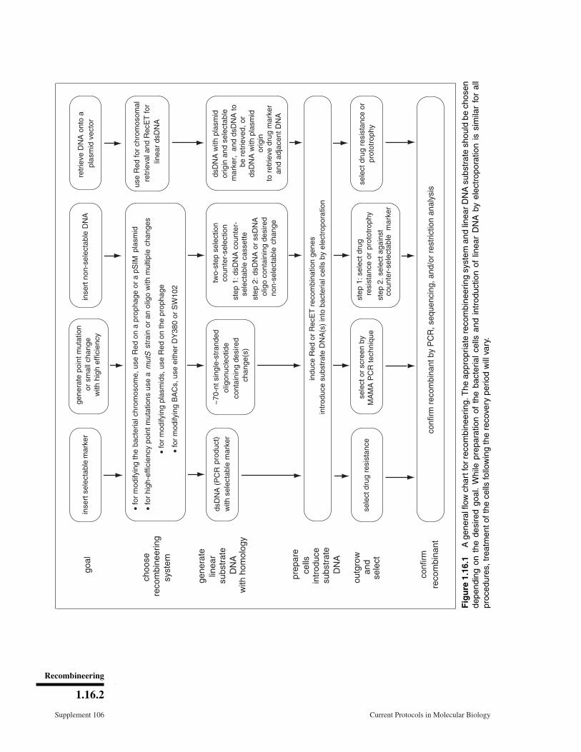
Fig
ure
1.16
.1A
gene
ralfl
owch
artf
orre
com
bine
erin
g.T
heap
prop
riate
reco
mbi
neer
ing
syst
eman
dlin
ear
DN
Asu
bstr
ate
shou
ldbe
chos
ende
pend
ing
onth
ede
sire
dgo
al.
Whi
lepr
epar
atio
nof
the
bact
eria
lce
llsan
din
trod
uctio
nof
linea
rD
NA
byel
ectr
opor
atio
nis
sim
ilar
for
all
proc
edur
es,t
reat
men
toft
hece
llsfo
llow
ing
the
reco
very
perio
dw
illva
ry.
Recombineering
1.16.2
Supplement 106 Current Protocols in Molecular Biology

The general flow chart in Figure 1.16.1 will help the researcher determine which recombi-neering pathway is most appropriate for the desired construct. The steps are summarizedbelow. First, define the goal of the recombineering project, which may be to insert a se-lectable marker, create point mutations or other small alterations, insert a non-selectablesegment of dsDNA, or retrieve dsDNA onto a plasmid vector. Depending on the goal,choose the appropriate recombineering system and generate the appropriate linear DNAsubstrate. Linear dsDNA substrates are generally made using PCR, while linear single-stranded substrates are synthetic oligos �70 nt in length with the desired alteration(s)centrally located. The linear DNA is introduced into electrocompetent bacteria (E. coli)pre-induced for the recombination functions. After electroporation, the cells are allowedto recover for a period of time before looking for recombinants. Recombinant coloniesmay be identified by a selection or simply by screening.
In this unit, Basic Protocol 1 describes preparation of electrocompetent cells pre-inducedfor the recombination functions. The Court laboratory phage λ–based system uses theλ pL promoter to express these functions. The competent cells are then transformedwith dsDNA or ssDNA (see Basic Protocol 1). Support Protocol 1 describes severalvariations of a two-step selection/counter-selection method of making precise genomicalterations without leaving any unwanted changes. In this support protocol, the firstof the two steps is a recombineering reaction that inserts a selectable DNA cassetteeither consisting of, or accompanied by, a counter-selectable marker near or within thesequence to be modified. The second step is a subsequent recombination that replaces thecounter-selectable cassette with the desired genetic alteration. This leaves the engineeredsequence otherwise unaltered and thus allows the same selection and counter-selectionto be reused to make further modifications. Support Protocol 2 describes a method forretrieving a genetic marker (cloning) from the E. coli chromosome or a co-electroporatedDNA fragment onto a plasmid. Whereas the above protocols generally use selection toidentify the recombinants, Support Protocols 3 and 4 describe methods to screen forunselected mutations. Support Protocols 5 and 6 detail modifications necessary whenusing recombineering to introduce changes into multicopy plasmids.
Basic Protocol 2 describes the removal of the defective prophage that expresses therecombineering functions in Basic Protocol 1. Basic Protocol 3 is for the plasmid-based phage systems. It describes a simple method for curing temperature-sensitiverecombineering plasmids after engineering is complete. Finally, Alternate Protocols 1and 2 present methods for recombineering with an intact prophage and for introducingmutations onto bacteriophage λ, respectively.
STRATEGIC PLANNING
General Considerations and Key Points
First, before attempting to modify the E. coli chromosome, a plasmid, or a bacterialartificial chromosome (BAC), the design of the desired final construct should be deter-mined. A DNA-analysis program such as Gene Construction Kit (GCK; Textco Software;http://www.textco.com/) or Vector NTI (Invitrogen) is invaluable for this task. Havingthe sequence of both the original genome arrangement and the desired final construct aselectronic files facilitates design of oligonucleotides to be used as primers for PCR re-combination substrates or as ssDNA substrates. Possession of the desired DNA sequencein silico also allows rapid design of primers to analyze and verify potential recombinants.
During the design process, the researcher should be aware of complications in the finalconstruct due to gene regulation issues. Bringing in a promoter with an antibiotic cas-sette can help in establishing drug resistance; however, transcription from this promotercan extend beyond the drug marker and affect distal genes. The authors have designed
Escherichia coli,Plasmids, andBacteriophages
1.16.3
Current Protocols in Molecular Biology Supplement 106
several drug cassettes with their promoter, open reading frame, and transcription ter-minator region, as described in Yu et al. (2000); primers for amplification are listed inTable 1.16.3. During the design process, the researcher should also be careful of possiblepolarity effects caused by premature termination within a coding sequence and inhibitionof expression of downstream genes in a multicistronic transcript (Adhya and Gottesman,1978; Bubuenenko et al., 2007). The researcher should also be careful to design the finalconstruct in a way that avoids creating unwanted fusion proteins.
Second, the investigator must decide whether to use dsDNA or ssDNA linear substratesto make the desired construct. Generally, insertion or removal of segments of DNA longerthan �20 nt requires a means of selection, since the efficiency of such reactions is nothigh enough to screen for recombinants: the frequency of such recombinants is �10–4-to 10–5. For example, to knock out a gene on the E. coli chromosome or on a BAC, adsDNA cassette encoding drug resistance is routinely used. On the other hand, if pointmutations or changes of only a few bases are desired, a synthetic oligo of �70 to 100 ntcan be used to create them. Creating these small changes with ssDNA recombineering inwild-type E. coli containing the Court laboratory defective λ prophage gives efficienciesof 0.1% to 1% (Ellis et al., 2001; Costantino and Court, 2003), and if host methyl-directedmismatch repair (MMR) is inactivated, either by mutating the mismatch repair system orby using oligos that escape MMR, a 25% to 50% recombination frequency is achievable(Costantino and Court, 2003; Sawitzke et al., 2011). Such a high frequency means that,for oligo recombination, it is possible to create recombinants without a selection andfind them by screening methods, such as using a PCR technique. A mutS mutation isroutinely used to eliminate MMR, although we have also created and tested mutH, mutL,and uvrD mutants, which give similarly increased recombination frequencies (Costantinoand Court, 2003). These four genes encode various components of the MMR system(Schofield and Hsieh, 2003).
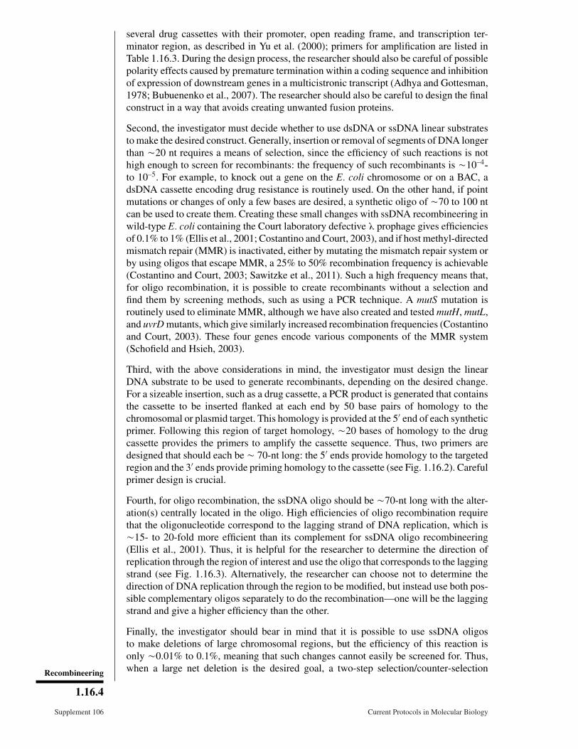
Third, with the above considerations in mind, the investigator must design the linearDNA substrate to be used to generate recombinants, depending on the desired change.For a sizeable insertion, such as a drug cassette, a PCR product is generated that containsthe cassette to be inserted flanked at each end by 50 base pairs of homology to thechromosomal or plasmid target. This homology is provided at the 5′ end of each syntheticprimer. Following this region of target homology, �20 bases of homology to the drugcassette provides the primers to amplify the cassette sequence. Thus, two primers aredesigned that should each be � 70-nt long: the 5′ ends provide homology to the targetedregion and the 3′ ends provide priming homology to the cassette (see Fig. 1.16.2). Carefulprimer design is crucial.
Fourth, for oligo recombination, the ssDNA oligo should be �70-nt long with the alter-ation(s) centrally located in the oligo. High efficiencies of oligo recombination requirethat the oligonucleotide correspond to the lagging strand of DNA replication, which is�15- to 20-fold more efficient than its complement for ssDNA oligo recombineering(Ellis et al., 2001). Thus, it is helpful for the researcher to determine the direction ofreplication through the region of interest and use the oligo that corresponds to the laggingstrand (see Fig. 1.16.3). Alternatively, the researcher can choose not to determine thedirection of DNA replication through the region to be modified, but instead use both pos-sible complementary oligos separately to do the recombination—one will be the laggingstrand and give a higher efficiency than the other.
Finally, the investigator should bear in mind that it is possible to use ssDNA oligosto make deletions of large chromosomal regions, but the efficiency of this reaction isonly �0.01% to 0.1%, meaning that such changes cannot easily be screened for. Thus,when a large net deletion is the desired goal, a two-step selection/counter-selectionRecombineering
1.16.4
Supplement 106 Current Protocols in Molecular Biology
upstream primer downstream primer
primer fordrug cassette~20 nt
5′homologyto target40-50 nt
homologyto target40-50 nt
primer fordrug cassette~20 nt
antibiotic cassette
amplify drug cassette orsequence of choice
upstreamhomologyto target
40-50 nt
downstreamhomologyto target
40-50 nt
target geneor sequenceto bereplacedbyrecombineering
in chromosome or on plasmid
target gene
antibiotic cassette
final constructin chromosome or on plasmid
5′
antibiotic cassette
Figure 1.16.2 Targeting of an antibiotic cassette. Two primers with 5′ homology to the target areused to PCR amplify the antibiotic cassette. The PCR product is introduced by electroporation intocells induced for the Red recombineering functions. The Red functions catalyze the insertion ofthe cassette at the target site, which may be on the bacterial chromosome or on a plasmid.
lagging strand
5
leading strand
Beta-bound oligo
replication fork
3
Figure 1.16.3 A schematic of the DNA replication fork. The leading and lagging strands areindicated. A DNA oligonucleotide, coated by the λ Beta protein, is shown annealed to the laggingstrand template at a gap in the discontinuously replicated lagging strand. Escherichia coli,
Plasmids, andBacteriophages
1.16.5
Current Protocols in Molecular Biology Supplement 106
Gene
oriamp
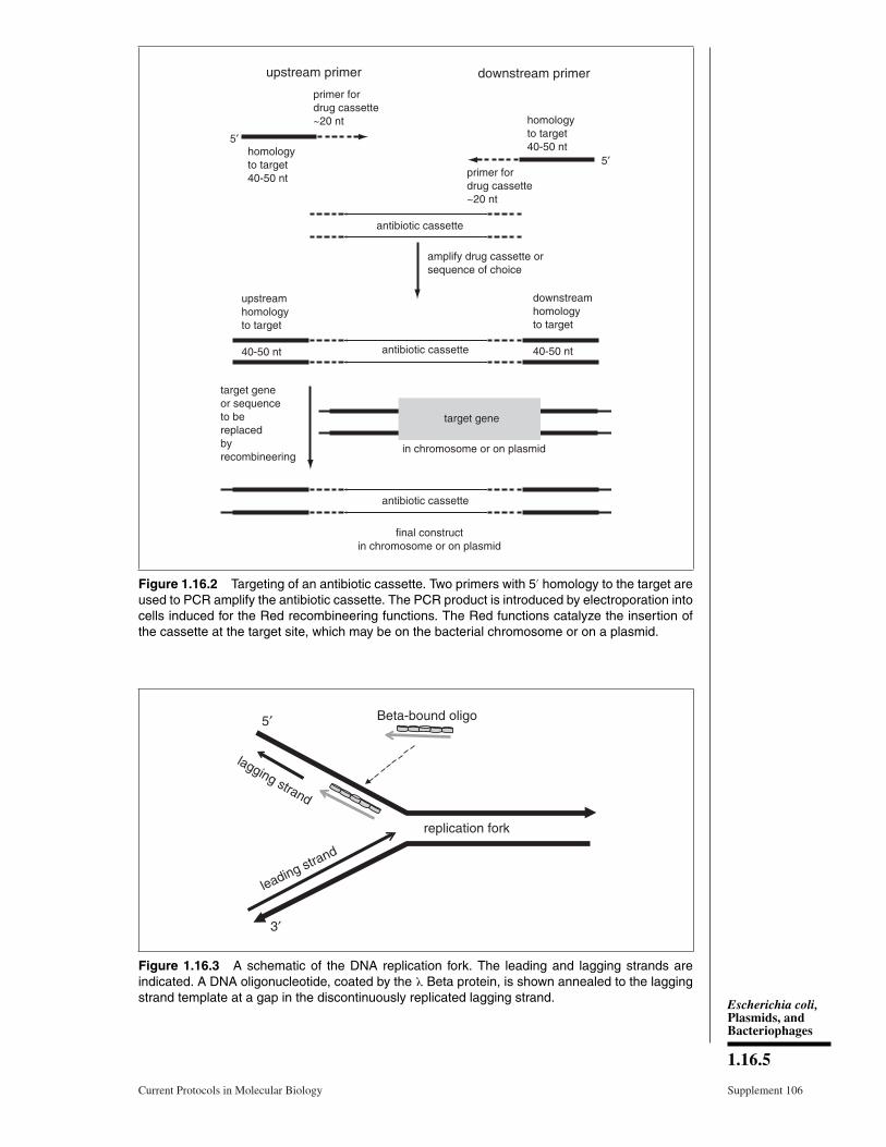
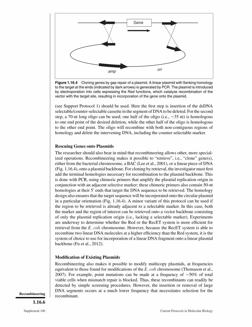
Figure 1.16.4 Cloning genes by gap repair of a plasmid. A linear plasmid with flanking homologyto the target at the ends (indicated by dark arrows) is generated by PCR. The plasmid is introducedby electroporation into cells expressing the Red functions, which catalyze recombination of thevector with the target site, resulting in incorporation of the gene onto the plasmid.
(see Support Protocol 1) should be used. Here the first step is insertion of the dsDNAselectable/counter-selectable cassette in the segment of DNA to be deleted. For the secondstep, a 70-nt long oligo can be used; one half of the oligo (i.e., �35 nt) is homologousto one end point of the desired deletion, while the other half of the oligo is homologousto the other end point. The oligo will recombine with both non-contiguous regions ofhomology and delete the intervening DNA, including the counter-selectable marker.
Rescuing Genes onto Plasmids
The researcher should also bear in mind that recombineering allows other, more special-ized operations. Recombineering makes it possible to “retrieve”, i.e., “clone” gene(s),either from the bacterial chromosome, a BAC (Lee et al., 2001), or a linear piece of DNA(Fig. 1.16.4), onto a plasmid backbone. For cloning by retrieval, the investigator must firstadd the terminal homologies necessary for recombination to the plasmid backbone. Thisis done with PCR, using chimeric primers that amplify the plasmid replication origin inconjunction with an adjacent selective marker; these chimeric primers also contain 50-nthomologies at their 5′ ends that target the DNA sequence to be retrieved. The homologydesign also ensures that the target sequence will be incorporated onto the circular plasmidin a particular orientation (Fig. 1.16.4). A minor variant of this protocol can be used ifthe region to be retrieved is already adjacent to a selectable marker. In this case, boththe marker and the region of interest can be retrieved onto a vector backbone consistingof only the plasmid replication origin (i.e., lacking a selectable marker). Experimentsare underway to determine whether the Red or the RecET system is more efficient forretrieval from the E. coli chromosome. However, because the RecET system is able torecombine two linear DNA molecules at a higher efficiency than the Red system, it is thesystem of choice to use for incorporation of a linear DNA fragment onto a linear plasmidbackbone (Fu et al., 2012).
Modification of Existing Plasmids
Recombineering also makes it possible to modify multicopy plasmids, at frequenciesequivalent to those found for modifications of the E. coli chromosome (Thomason et al.,2007). For example, point mutations can be made at a frequency of �50% of totalviable cells when mismatch repair is blocked. Thus, these recombinants can readily bedetected by simple screening procedures. However, the insertion or removal of largeDNA segments occurs at a much lower frequency that necessitates selection for therecombinant.Recombineering
1.16.6
Supplement 106 Current Protocols in Molecular Biology

Recombineering with multicopy plasmids requires some modification of Basic Proto-col 1. Different plasmids vary widely in their intracellular copy number, ranging fromlow-copy vectors with only a few molecules per cell, such as pSC101, to pUC-basedplasmids that routinely have �500 copies per cell. After recombineering, cells containingrecombinant plasmids will also contain unmodified parental plasmids. In all cases, therecombinant class of molecules can be separated from the unmodified parental class byplasmid DNA isolation and retransformation at a DNA concentration of less than onemolecule per cell; this creates pure clonal populations of the recombinant and parentalplasmid species in different cells. If the desired plasmid recombinant is designed sothat it lacks a restriction site present on the parent, the investigator can enrich for therecombinant class by destroying the parental population by restriction digestion prior totransformation.
Another complexity arising when plasmids are modified with recombineering is thatcircular multimeric plasmid species can be formed during the recombination reaction.In the absence of linear substrate DNA, expression of the Red proteins alone does notcause plasmids to multimerize under the conditions given here (Thomason et al., 2007),but when linear single-stranded or double-stranded substrate DNA modifies the targetsequence, generating the recombinant, circular multimeric species are often formed.Multimer formation is thus a hallmark of a recombinant plasmid molecule (and could, inprinciple, be used to identify recombinant species). A host defective in RecA-mediatedrecombination eliminates host-mediated plasmid-by-plasmid recombination, and thusone route to multimer formation, so it is useful to use recA mutant strains for plasmidengineering.
For plasmid modification by recombineering, a decision should be made as to whether theplasmid will be introduced into the Red-expressing cells by co-electroporation with thelinear DNA after the recombination functions are induced, or whether the plasmid shouldalready be established in the bacterial strain. Co-electroporation offers the advantage thatit helps control plasmid copy number at the time of recombineering and minimizesopportunities for plasmid multimer formation. However, for very large plasmids of lowcopy number, co-electroporation may not introduce plasmids into enough cells to befeasible.
Recombineering Expression Systems
To perform recombineering, a bacterial strain that expresses a bacteriophage generalizedrecombination system is required. Phage λ Red and RecET from the endogenous E. coliRac prophage are two such systems used for engineering E. coli. The two systems donot yield similar recombination efficiencies for a given particular type of recombinationreaction, and so should not be substituted for each other without careful consideration;restated, each system has an optimal use. While the λ Red system is superior for targetingthe E. coli chromosome and episomes replicating in E. coli (Datta et al., 2006), the RecETsystem is superior for recombining two linear DNA molecules to generate a new, intactplasmid (Fu et al., 2012). The Court laboratory makes available bacterial strains allowingwork with each system (see Table 1.16.1).
Each recombination system uses proteins that perform similar functions. Both λ Exo andRac RecE are 5′→3′ double-stranded DNA (dsDNA)-dependent exonucleases (Little,1967; Carter and Radding, 1971; Joseph and Kolodner, 1983). These nucleases willinitiate degradation from 5′ ends of a linear dsDNA, creating either a partially duplexmolecule with 3′ single-stranded DNA (ssDNA) overhangs, or if the dsDNA is shortenough, a ssDNA molecule in which the complementary strand is entirely degraded.These ssDNAs are the substrates of the λ Beta and Rac RecT proteins, which aressDNA-annealing proteins (Muniyappa and Radding, 1986; Hall et al., 1993). During
Escherichia coli,Plasmids, andBacteriophages
1.16.7
Current Protocols in Molecular Biology Supplement 106
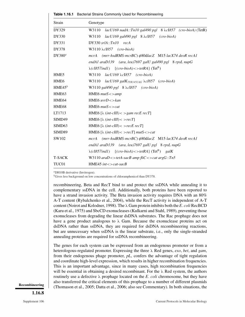
Table 1.16.1 Bacterial Strains Commonly Used for Recombineering
Strain Genotype
DY329 W3110 �lacU169 nadA::Tn10 gal490 pgl�8 λcI857�(cro-bioA) (TetR)
DY330 W3110 �lacU169 gal490 pgl�8 λcI857 �(cro-bioA)
DY331 DY330 srlA::Tn10 �recA
DY378 W3110 λcI857 �(cro-bioA)
DY380a mcrA �(mrr-hsdRMS-mcrBC) φ80dlacZ�M15 lacX74 deoR recA1
endA1 araD139 �(ara, leu)7697 galU gal490 pgl�8 rpsL nupG
λ(cI857ind1) �{(cro-bioA)<>tetRA} (TetR)
HME5 W3110 �lacU169 λcI857 �(cro-bioA)
HME6 W3110 �lacU169 galKTYR145UAG λcI857�(cro-bioA)
HME45b W3110 gal490 pgl�8 λcI857 �(cro-bioA)
HME63 HME6 mutS<>amp
HME64 HME6 uvrD<>kan
HME68 HME6 mutS<>cat
LT1713 HME6 [λ (int-cIII)< >gam recE recT]
SIMD49 HME6 [λ (int-cIII)< >recT]
SIMD63 HME6 [λ (int-cIII)< >recE recT]
SIMD89 HME6 [λ (int-cIII)< >recT] mutS<>cat
SW102 mcrA �(mrr-hsdRMS-mcrBC) φ80dlacZ�M15 lacX74 deoR recA1
endA1 araD139 �(ara, leu)7697 galU pgl�8 rpsL nupG
λ(cI857ind1) �{(cro-bioA)<>tetRA} (TetR) �galK
T-SACK W3110 araD<>tetA-sacB amp fliC<>cat argG::Tn5
TUC01 HME45 int<>cat-sacB
aDH10B derivative (Invitrogen).bGives less background on low concentrations of chloramphenicol than DY378.
recombineering, Beta and RecT bind to and protect the ssDNA while annealing it tocomplementary ssDNA in the cell. Additionally, both proteins have been reported tohave a strand invasion activity. The Beta invasion activity requires DNA with an 80%A-T content (Rybalchenko et al., 2004), while the RecT activity is independent of A-Tcontent (Noirot and Kolodner, 1998). The λ Gam protein inhibits both the E. coli RecBCD(Karu et al., 1975) and SbcCD exonucleases (Kulkarni and Stahl, 1989), preventing thoseexonucleases from degrading the linear dsDNA substrates. The Rac prophage does nothave a gene product analogous to λ Gam. Because the exonuclease proteins act ondsDNA rather than ssDNA, they are required for dsDNA recombineering reactions,but are unnecessary when ssDNA is the linear substrate, i.e., only the single-strandedannealing proteins are required for ssDNA recombineering.
The genes for each system can be expressed from an endogenous promoter or from aheterologous-regulated promoter. Expressing the three λ Red genes, exo, bet, and gam,from their endogenous phage promoter, pL, confers the advantage of tight regulationand coordinate high-level expression, which results in higher recombination frequencies.This is an important advantage, since in many cases, high recombination frequencieswill be essential in obtaining a desired recombinant. For the λ Red system, the authorsroutinely use a defective λ prophage located on the E. coli chromosome, but they havealso transferred the critical elements of this prophage to a number of different plasmids(Thomason et al., 2005; Datta et al., 2006; also see Commentary). In both situations, theRecombineering
1.16.8
Supplement 106 Current Protocols in Molecular Biology

Table 1.16.2 Plasmids Containing the Red System Under cI857 Controla
Plasmid designation Drug resistance Plasmid origin Copy number/cell
pSIM5 Chloramphenicol pSC101ts 16
pSIM6 Ampicillin
pSIM7 Chloramphenicol pBBR1 30-40
pSIM8 Ampicillin
pSIM9 Chloramphenicol pRK2ts 20-40
pSIM10 Hygromycin
pSIM17 Blasticidin pSC101ts 16
pSIM18b Hygromycin
pSIM19 Spectinomycin
aNote that plasmids require higher drug concentrations for retention than those for single copy insertions given in BasicProtocol 1.bBe aware that this plasmid has homology to AmpR (but is not AmpR). This can result in a background of incorrectrecombinants if targeting an Amp-cassette into cells containing this plasmid.
phage recombination functions are under control of the bacteriophage λ CI repressor,which affords extremely tight regulation. Three operator sites are present at each of theλ pL and pR promoters flanking the repressor gene; the repressor protein, CI, bindscooperatively to these operator sites and blocks transcription from these promoters.CI-mediated looping between the two sets of operators provides an additional level ofrepression and in combination with autoregulation ensures tight and uniform repression.The prophage λ constructs used for recombineering, whether resident on the E. colichromosome or on plasmids, encode a temperature-sensitive version of this CI repressor,CI857, which represses expression from the powerful λ pL promoter at low temperatures(30°C to 32°C) by binding to both sets of operators. To express the recombination proteinsat high levels, the temperature of the bacterial culture is quickly but temporarily raisedto 42°C, resulting in rapid inactivation of the repressor and expression of recombinationfunctions. Lowering the temperature after a 15-min induction allows CI to renature withconsequent restoration of tight repression. This burst of expression is sufficient to catalyzehigh levels of recombination while minimizing unnecessary expression, perpetuationof long-term recombination activity, and stress on the cells from the continuing hightemperatures.
In the other λ Red expression systems, the recombination genes are expressed fromnon-λ promoters on multicopy plasmids. One such plasmid is pKM208 (Murphy andCampellone, 2003), which has the Red genes expressed from pTAC. Plasmid pKM208contains the bla gene for ampicillin drug selection, lacI to lower expression until de-pression of the lac repressor with IPTG, and a temperature-sensitive origin of DNAreplication to allow isolation of cells that lack the plasmid (“curing”) after use by growthat high temperature. The authors have found that pKM208 yields similar recombinationfrequencies to those obtained with the prophage system, although repression of this plas-mid is incomplete, with consequent constitutive recombination activity. Other plasmidsusing the arabinose-inducible pBAD promoter are also used to express the Red proteins(Datsenko and Wanner, 2000). In the authors’ laboratory, the Datsenko and Wannerplasmids give about ten-fold lower recombination levels than the prophage constructs(Datta et al., 2006). Most of the plasmid constructs from the authors (Table 1.16.2) alsohave temperature-sensitive origins of DNA replication to facilitate curing. The plasmid-based systems have the advantage of portability to different strains—they can be trans-ferred among different E. coli strains, or to Salmonella typhimurium, and presumably toother Gram-negative bacteria. Still, using the prophage system located on the bacterial
Escherichia coli,Plasmids, andBacteriophages
1.16.9
Current Protocols in Molecular Biology Supplement 106
chromosome requires no drug-resistance markers and is the system of choice if the objectis to engineer a plasmid.
By contrast, since expression from the endogenous Rac prophage gives only low-level recombination, the required enzymes for the RecET system are usually expressedfrom multicopy plasmids and under the control of heterologous promoters, such as thearabinose-inducible pBAD promoter (Zhang et al., 1998). The Court laboratory has re-placed the Red genes with the full-length recE and recT genes (including λ gam) on thedefective prophage. A similar strain carrying only recT also exists (Datta et al., 2008)(Table 1.16.1).
Cleaning Up After Recombineering
For the Court laboratory’s phage-based systems, after the desired construct is obtained,the defective prophage carrying the recombination genes can be removed, either by re-combination as described in Basic Protocol 2 or by P1 transduction (UNIT 1.17), using anon-lysogenic donor, selecting for growth in the absence of biotin (Bio+). Alternatively,engineered alleles on the chromosome can be moved into a different host by P1 transduc-tion (UNIT 1.17), provided there is a selection for them. For plasmid-borne systems, plasmidreplication is temperature-sensitive and growth at high temperature causes plasmid lossas detailed in Basic Protocol 3.
BASICPROTOCOL 1
MAKING ELECTROCOMPETENT CELLS AND TRANSFORMING WITHLINEAR DNA
This protocol describes the preparation of electrocompetent cells that are preinducedfor the recombination functions and transformation with appropriate DNA to createthe desired genetic change. As noted in Strategic Planning, the phage recombinationfunctions are repressed by the phage λ temperature-sensitive CI857 repressor, so thatthey are not expressed when the cells are grown at low temperatures (30° to 32°C) butare highly expressed when the culture temperature is shifted to 42°C. See Commentaryfor additional considerations before performing the procedure.
Materials
Purified PCR product with �50 bases of flanking homology or oligonuclotideprimers with �35 bases of flanking homology on either side of desired change(also see UNIT 15.1)
Bacterial strain expressing the defective lambdoid prophage recombination systemλ Red (Table 1.16.1; strains available from Court Laboratory Website:http://redrecombineering.ncifcrf.gov/)
LB medium and plates (UNIT 1.1), without antibioticMedium lacking carbon source: M9 medium (UNIT 1.1) or 1× TM buffer (APPENDIX 2)Selective plates (UNIT 1.1)—minimal plates if selecting for prototrophy or rich plates
containing antibiotic (depending on drug cassette used):
30 μg/ml ampicillin30 μg/ml kanamycin10 μg/ml chloramphenicol12.5 μg/ml tetracycline50-100 μg/ml spectinomycin
30° to 32°C incubator30° to 32°C shaking incubator or roller32° and 42°C shaking water baths125- and 250-ml Erlenmeyer flasks, preferably baffledRefrigerated, low-speed centrifuge with Sorvall SA-600 rotor (or equivalent)Recombineering
1.16.10
Supplement 106 Current Protocols in Molecular Biology
35- to 50-ml plastic centrifuge tubes1.5-ml microcentrifuge tubesRefrigerated microcentrifuge0.1-cm electroporation cuvettes (Bio-Rad), chilledElectroporator (e.g., Bio-Rad E. coli Pulser)Micropipettor and 1000-μl pipet tips18 × 150-mm sterile borosilicate glass culture tubes15-ml sterile culture tubes, optional
Additional reagents and equipment for PCR (UNIT 15.1), agarose gel electrophoresisof DNA (UNITS 2.5A & 2.6), purification of DNA by ethanol precipitation (UNIT 2.1A;optional; commercially available PCR cleanup kit may be substituted),electroporation (UNIT 1.8), isolation of bacterial colonies by streaking (UNIT 1.3),restriction enzyme digestion (UNIT 3.1), and DNA sequencing (Chapter 7)
Prepare DNA for transformation
1. Design and procure the oligos to use for PCR-mediated generation of a dsDNAproduct, or for use in single-stranded oligo engineering.
The sequences of the primers used to amplify the common drug cassettes are listed inTable 1.16.2. Remember to add the homologous targeting sequence to the 5′ ends of theoligos.
UNIT 15.1 describes general considerations for primer design.
2. Make the PCR product (UNIT 15.1) and examine it by agarose gel electrophoresis (UNIT
2.5A).
Gel purifying the DNA is not recommended. If it is essential that the DNA be gel pu-rified, avoid exposing it to ultraviolet light, which will damage it and result in lowerrecombination frequencies.
3. Clean up the PCR product by ethanol precipitation or using a commercially availablekit (e.g., Qiagen) to remove salt (UNIT 2.1A).
A strain designated T-SACK available from the Court laboratory facilitates constructionof tetracycline, kanamycin, ampicillin, and chloramphenicol cassettes as well as theTetA-SacB dual cassette. From this strain, custom cassettes can be amplified using colonyPCR. In colony PCR, a PCR reaction is prepared without allowing any volume for thetemplate DNA; a fresh colony of the appropriate E. coli K-12 strain is then touchedwith a sterile inoculating loop and mixed into the PCR reaction. Too many cells willinhibit the PCR reaction. Use of this strain to make cassettes rather than a plasmidtemplate is encouraged, since if a plasmid template is used to construct a PCR-amplifieddrug cassette, any intact circular plasmid remaining will transform the cells extremelyefficiently and give unwanted background, hindering recombinant identification. Suchbackground can be minimized by using a linear plasmid template for the PCR and bydigesting the completed PCR reaction with DpnI before using it for electroporation;however, using the bacterial template is still recommended. If linear plasmid templatesare used, always use as little as possible and include a control reaction of uninducedcells transformed with the PCR product to give a measure of any unwanted intact plasmidbackground.
Prepare bacterial cultures
4. Inoculate the suitable bacterial strain (Table 1.16.1) from frozen glycerol stock or asingle colony into 3 to 5 ml LB medium. Shake the culture overnight at 30° to 32°C.
Most of the authors’ strains containing the defective lambdoid prophage are W3110derivatives; however, the prophage can be moved into other backgrounds by P1 trans-duction. See Commentary for details. Similarly, plasmids expressing the recombination Escherichia coli,
Plasmids, andBacteriophages
1.16.11
Current Protocols in Molecular Biology Supplement 106
functions can be put into any strain of choice. Add antibiotic as appropriate to maintainplasmid selection during growth.
Either 30° or 32°C is acceptable for the low temperature throughout the procedure,since either temperature allows good repression by the CI857 repressor. The repressionis tighter at 30°C but the cultures will grow more rapidly at 32°C.
5. Add �0.5 ml of the overnight culture to 35 ml of LB medium in a 250-ml (baffled)Erlenmeyer flask.
This is a 70-fold dilution. Ensure that the dilution is at least 70-fold. Higher (more dilute)dilutions will also work, but the cells will take longer to grow to the appropriate density.If targeting the recombineering to a plasmid, or expressing the recombineering proteinsfrom a plasmid, do not add antibiotic for plasmid maintenance when sub-culturing cells.It has been observed that antibiotics may inhibit the recombination. If an alternativeto temperature shift is used to induce the recombination functions (i.e., addition ofarabinose or IPTG), the cells are grown at 37°C and inducer should be added to themedium. Expression of the λ Red functions from the plasmids of Datsenko and Wanner(2000) is enhanced by use of 10 mM arabinose (for Ara+ strains) (see Datta et al., 2006).If using an inducer, remember to include an additional flask containing an uninducedculture as a negative control.
6. Place the flask into a 32°C shaking water bath and grow cells for �2 hr at 32°C withshaking.
The time will vary with different strains and dilutions. The cells are ready when the A600
is between 0.4 and 0.6. It is important not to over grow the cells, since stationary phasecells do not work well for recombineering with the Red system, which benefits from activereplication forks.
Induce recombination functions
7. Transfer half of the culture to a 125-ml (baffled) Erlenmeyer flask and place flask intothe 42°C water bath. Shake 15 min at 220 rpm to induce. Leave the remainder of theculture at 32°C (this will be used as the uninduced control that lacks recombinationactivity). While the cells are inducing, fill an ice bucket with an ice-water slurry.
8. Immediately after inducing for 15 min at 42°C, rapidly cool the flask in the ice-water slurry with gentle swirling. Leave on ice for �5 min. Follow the same coolingprotocol with the uninduced 32°C culture. While the cells are on ice, precool thecentrifuge to 4°C and chill the necessary number of 35- to 50-ml plastic centrifugetubes, labeled for induced and uninduced cells.
As mentioned above, a temperature shift is unnecessary when a chemical inducer likearabinose or IPTG is used.
Make electrocompetent cells
9. Transfer both the induced and uninduced cultures to the appropriately labeled chilled35- to 50-ml centrifuge tubes. Centrifuge 7 min at 4600 × g (6700 rpm in a SorvallSA-600 rotor), 4°C. Aspirate or pour off supernatant.
10. Add 1 ml ice-cold distilled water to the cell pellet in the bottom of each tube andgently resuspend cells with a large pipet tip (do not vortex). Add an additional30 ml ice-cold distilled water to each tube, seal, and gently invert to mix (withoutvortexing). Centrifuge tubes again as in step 9.
All subsequent resuspensions of cells through step 16 should be done gently and withoutvortexing. Preparation of the cells for electroporation washes out any added chemicalinducing agent.
11. Very carefully decant the supernatant from the soft pellet in each tube and suspendeach cell pellet in 1 ml ice-cold distilled water.Recombineering
1.16.12
Supplement 106 Current Protocols in Molecular Biology
Remove tubes from the centrifuge promptly. The pellet is very soft and care should betaken not to dislodge it, especially when processing multiple tubes.
12. Transfer suspended cells to 1.5-ml microcentrifuge tubes. Microcentrifuge tubes for30 to 60 sec at maximum speed, 4°C. Carefully aspirate off the supernatant. In eachof the tubes, resuspend the cell pellet in 200 μl ice-cold distilled water, which willprovide enough material for four or five electroporations.
For routine procedures when optimal recombination frequency is not necessary, e.g.,when selection is used to find recombinants, electrocompetent cells induced for therecombination functions can be used later after storage of the cell pellet, suspended in15% (v/v) glycerol at –80°C. However, for highest efficiency, use freshly processed cells.
Introduce DNA by electroporation
13. Chill the desired number of 0.1-cm electroporation cuvettes on ice. Turn on theelectroporator and set to 1.80 kV.
Brands of cuvettes and electroporators other than that available from Bio-Rad may work,but have not been tested in the authors’ laboratory. Larger 0.2-cm cuvettes have not beenused successfully, and they may require different electroporation conditions (consultelectroporator instruction manual) and standardization to obtain optimal recombinationfrequencies.
14. In microcentrifuge tubes on ice, mix 100 to 150 ng of salt-free PCR fragment (fromstep 3) or 1 pmol of single-stranded oligonucleotide with 50 to 100 μl of the inducedor uninduced cell suspensions (from step 12). Rapidly mix and perform subsequentelectroporation; do not leave the DNA/cell mixes on ice for extended periods. Besure to include the following electroporation reactions and controls:
a. Induced cells plus DNA.
This is the culture that should yield the designed recombinants.
b. Induced cells without DNA.
This is a control to identify contamination, determine the reversion frequency, and obtainsome idea of the efficiency of the selection.
c. Uninduced cells plus DNA.
This control tells whether there is some contaminating factor in the DNA that is con-tributing to the selected colonies (e.g., intact plasmid template from the PCR reactionwill give rise to drug-resistant colonies here). If the recombination functions are partiallyconstitutive, this control will give colonies.
15. Introduce the DNA into the cells by electroporation (UNIT 1.8).
The time constant should be >5 msec for optimal results. Low time constants indicateproblems with the cells, the DNA, the electroporation cuvettes, or even the equipment.
16. Immediately after electroporation, add 1 ml LB medium to the cuvette using amicropipettor with a 1000-μl pipet tip. If transforming with a drug cassette, transferthe electroporation mix to 15-ml sterile culture tubes and incubate tubes >2 hr withshaking at 30° to 32°C to allow expression of the antibiotic-resistance gene.
If not selecting for drug resistance, it is still recommended that the cells be allowedto recover in broth from the shock of electroporation for at least 30 min. It has beenobserved in the authors’ laboratory that omitting this recovery period greatly reducesthe cell viability.
An alternative, and in the author’s experience more reliable, method for outgrowth isas follows. After the 30-min recovery period, spread appropriate dilutions of cells (seestep 18 below) onto a sterile 82-mm diameter nitrocellulose filter atop a rich LB plate.Incubate this plate for 3 to 4 hr at 30° to 32°C. After the incubation, transfer the filter
Escherichia coli,Plasmids, andBacteriophages
1.16.13
Current Protocols in Molecular Biology Supplement 106
to the appropriate selective drug plate using sterile forceps. This method is preferredbecause all of the recombinants are independent, the cells are less dense, and moreefficient outgrowth is achieved.
Determine cell titers
17. Prepare serial 1:10 dilutions of the electroporation mix through 10–6 using M9medium or 1× TM buffer, dispensing 0.9 ml of M9 or TM buffer and 0.1 ml of thecell suspension per tube.
The dilutions can be made in rich medium if a selection for antibiotic resistance isapplied.
18. To determine total viable cell count, spread 100 μl of 10–4, 10–5, and 10–6 dilutionson LB plates (rich plates without drug). Incubate plates 1 to 2 days at 30° to 32°C,depending on the growth requirements of the recipient strain.
19. To determine recombinant cell count, plate cells on selective plates as followsdepending on the anticipated recombinant frequency.
a. If efficient recombination is expected, spread both 10 and 100 μl of the 10–1 and10–2 dilutions.
b. If low numbers of recombinants are expected, spread 100 μl each of a 1:10dilution and the undiluted culture. Pellet the remaining cells, suspend in 100 μland plate.
The authors routinely obtain ten-fold or higher recombinant yields with the prophagesystem than with the Datsenko and Wanner pBAD arabinose inducible system.
c. For the no-DNA and uninduced controls, plate 200 μl directly onto selectiveplates.
Since targeting to the chromosome results in a lower copy number of the drug cassettethan is present with a multicopy plasmid, antibiotic concentrations must be adjustedaccordingly. The authors routinely use the drug concentrations recommended in thematerials list for single copy chromosomal constructs. Selections based on nutritionalprototrophy require appropriate minimal plates.
20. Incubate plates at the appropriate temperature (30° to 32°C).
At 30°C, colonies may take 2 days to develop on LB plates and 3 to 4 days on minimalplates. Candidates should be purified by streaking for single colonies on the selectiveplates and retested for the appropriate phenotype.
Analyze recombinants
21. Once recombinant clones are identified, confirm the presence of the desired mu-tation(s) by PCR analysis (UNIT 15.1) followed by DNA sequencing (Chapter 7) orrestriction digestion analysis (UNIT 3.1), if appropriate.
The design of the PCR primers depends on the changes to be made (see Strategic Plan-ning). The recombinant junctions should be confirmed with two flanking primers and twoadditional primers pointing outwards from the cassette (all four should have compatibleannealing temperatures); use one primer flanking the cassette and one internal cassetteprimer to amplify the unique junctions created by the recombination reaction. The primerpair that hybridizes to the external flanking sequences on each side rather than the in-sertion itself can also be used to demonstrate loss of the target sequences and presenceof the insertion. Unwanted mutations can be introduced by heterologies (variations) inthe synthetic primer population (Oppenheim et al., 2004); therefore, it is important toconfirm the final construct by sequence analysis, especially the regions derived from theoriginal primers.
Recombineering
1.16.14
Supplement 106 Current Protocols in Molecular Biology

SUPPORTPROTOCOL 1
SELECTION/COUNTER-SELECTIONS FOR GENE REPLACEMENT
This protocol describes several variations on two-step methods to create precise geneticchanges without otherwise altering the DNA sequence. First, the selectable/counter-selectable cassette of choice is placed in the stretch of DNA to be altered; second, thiscassette is replaced with the desired alteration in a second recombineering event. The finalconstruct will have neither the cassette nor a drug marker. All these procedures requirespecial media for the counter-selection step. The flow chart in Fig. 1.16.5 defines the
choose selection
counter-selection
I IIIII
requires special strain
amplify linear dsDNAcassett use
appropriate templateand primers
strainTUC01
strainT-SACK
BACwith
induce Red recombination functionsintroduce selectable counter-selectable dsDNA cassette
into bacterial cells by electroporation
prophageor
pSIM
Red-expressing strain for
recombineeringwith
prophagepSIM
select on Cmconfirm sucrose sensitive
select on Tet
confirm sucrose sensitive
select on
minimal galactose
select on sucrose at
C
select on DOG at
select on Tet-SacB
induce Red recombination genesintroduce linear DNA with desired mutation by electroporation
prepare cellsintroducesubstrate
DNA
outgrow select and confirm
prepare cellsintroducesubstrate
DNA
confirm recombinant by PCR, sequencing, and/or restriction analysis
counter-selection 30 -32 C
SW102
Figure 1.16.5 A flow chart for selection/counter-selections. Three procedures are provided, twousing the sacB counter-selectable gene linked to either cat or tetA, and one using galK designedspecifically for BAC modification.
Escherichia coli,Plasmids, andBacteriophages
1.16.15
Current Protocols in Molecular Biology Supplement 106
three types of selection counter-selections for which protocols are given. Other possibleselection/counter-selections are described in the Commentary.
Additional Materials (also see Basic Protocol 1)
Bacterial strain for specific selection/counter-selection:
cat-sacB: any recombineering strain can be usedtetA-sacB: the Red functions should be expressed from a heat curable
plasmid rather than from a prophagegalK: use strain SW102, available from Frederick National Laboratory for
Cancer Research:http://ncifrederick.cancer.gov/research/brb/recombineeringInformation.aspx
Appropriate template for amplification of selectable/counter-selectable cassette:
cat-sacB: use bacterial strain TUC01 (DY329 with a cat-sacB insertion onthe E. coli chromosome), available from the Court Laboratory Website:http://redrecombineering.ncifcrf.gov/
tetA-sacB: E. coli strain T-SACK, available from the Court LaboratorygalK: the galK expression cassette has been cloned onto BAC pBeloBAC11,
also available from Frederick National Laboratory for Cancer Research
Hybrid primers to amplify cassettes—all hybrid primers should have 50 bases of 5′homology for targeting to the desired location:
cat-sacBPrimer L sacB: 5′-homology sequence-ATC AAA GGG AAA ACT GTC
CAT AT-3′
Primer R cat: 5′-homology sequence-TGT GAC GGA AGA TCA CTTCG-3′
tetA-sacBPrimer L sacB: same as for cat-sacBPrimer R tetA: 5′-homology sequence- TCC TAA TTT TTG TTG ACA
CTC TA-3′
galKGalK forward primer: 5′-homology sequence-CCT GTT GAC AAT TAA
TCA TCG GCA-3′
GalK reverse primer: 5′-homology sequence-TCA GCA CTG TCC TGCTCC TT-3′
Invitrogen Platinum High Fidelity enzymeSelective media for inserting and removing cassettes:
cat-sacB selection, use LB-Cm plates: LB plates (UNIT 1.1) containing10 μg/ml chloramphenicol; for counter-selection, use LB plates (UNIT 1.1)lacking NaCl but containing 6% (w/v) sucrose
tetA-sacB selection, use LB-Tet plates: LB plates (UNIT 1.1) containing12.5 μg/ml tetracycline; for counter-selection, use Tet/SacB plates (seerecipe)
galK selection, use M63 minimal D-galactose plates containing biotin andleucine, with chloramphenicol for BAC selection (see recipe); forscreening, use MacConkey galactose plates (see recipe); forcounter-selection, use M63 minimal glycerol with 2-deoxygalactose(DOG), biotin and leucine, and chloramphenicol for BAC selection (seerecipe)
Thermal cycler (MJ Research PTC-100)Recombineering
1.16.16
Supplement 106 Current Protocols in Molecular Biology
Amplify the selectable/counter-selectable element for recombineering
1. Amplify the cassette from the appropriate template with the Invitrogen Platinum HighFidelity enzyme and an MJ Research thermal cycler, using 50 pmol of each primerand one of the following cycling programs:
For cat-sacB or tet-sacB cycling (�3.5-kb product):1 cycle: 2 min 94°C (denaturation)9 cycles: 15 sec 94°C (denaturation)
30 sec 55°C (annealing)3.5 min 68°C (extension)
19 cycles: 15 sec 94°C (denaturation)30 sec 55°C (annealing)3.5 min (adding 5 sec/cycle) 68°C (extension)
1 cycle: 7 min 68°C (extension)1 cycle: indefinite 4°C (hold).
For galK cycling (�1.3-kb product):1 cycle: 2 min 94°C (denaturation)30 cycles: 15 sec 94°C (denaturation)
30 sec 60°C (annealing)1.5 min 68°C (extension)
1 cycle: 7 min 68°C (extension)1 cycle: indefinite 4°C (hold).
The PCR primers used to amplify the selection/counter-selection elements are at the 3′ endof chimeric primers that include 5′ segments of bacterial target homology �50 nt in length.Hence, each primer is �70-nt long. Since the PCR products for cat-sacB and tetA-sacBare �3.5 kb, they can be difficult to amplify. The above conditions are used successfully inthe authors’ laboratory.
2. Visualize the PCR product on an agarose gel. Purify the PCR product to remove salt(UNIT 2.1A).
Perform electroporation and recombination to insert the element at the desiredlocation
3. Insert the selectable/counter-selectable cassette into the chromosome as described inBasic Protocol 1, steps 4 to 21 (also see Background Information for important tips)using the following techniques specific for each cassette.
a. Selection for cat-sacB or tetA-sacB: select drug-resistant colonies, CmR for cat-sacB or TcR for tetA-sacB, and purify on LB-plates containing drug to isolate singlecolonies. For galK: select GalK+ chloramphenicol-resistant (CmR) colonies on theminimal galactose plates containing supplements and purify on the same plates toisolate single colonies.
The em7 promoter that is amplified along with the galK gene results in constitutive ex-pression of galK. Since bacteria grow more slowly on minimal medium, it will take severaldays for colonies to grow. Select on plates with chloramphenicol to ensure maintenanceof the BAC.
b. Confirmation for cat-sacB or tetA-sacB: test several drug resistant isolates for sen-sitivity to sucrose on LB plates lacking NaCl but containing 6% sucrose (Blomfieldet al., 1991). Use the targeted strain lacking a sacB insertion as a sucrose-resistantcontrol, and use TUC01 or T-SACK as a SucS control. Determine that the insertionis correct before proceeding with the next step of the procedure.
The sucrose-resistant parent serves as a control for growth in the presence of sucrose.Sucrose sensitivity needs to be tested because in a fraction of the clones the PCR processgenerates mutations that inactivate sacB. Occasionally, one orientation of the dual cassettedoes not give good expression of the sacB cassette. In this case, all drug-resistant colonies
Escherichia coli,Plasmids, andBacteriophages
1.16.17
Current Protocols in Molecular Biology Supplement 106
will also be SucR. If so, then different homologies can be designed to either invert thecassette or move it.
For galK: Purify candidate colonies on MacConkey galactose indicator plates toverify that they have become Gal+. Include SW102 containing the BAC as anegative control. Colonies should grow overnight on this indicator mediumand positive clones will be bright pink or red. Be careful not to lose the BAC.SW102 will not turn red on this indicator.
Perform electroporation and recombination to replace theselectable/counter-selectable cassette with chosen allele
4. Use a confirmed candidate from step 3 as the starting bacterial strain for a secondround of recombineering by carrying out Basic Protocol 1, but at step 16, suspendthe electroporated cells in a final volume of 10 ml instead of 1 ml of LB medium,and incubate with aeration (applied via shaking in a shaking water bath) for 4 hr toovernight at 30° to 32°C for outgrowth following electroporation.
The higher dilution promotes better cell recovery and allows complete segregation ofrecombinant chromosomes that no longer carry the counter-selectable cassette from non-recombinant sister chromosomes that still contain it. If outgrowth is inadequate and sisterchromosomes are not fully segregated, the presence of the cassette on one chromosome willconfer sensitivity to the entire cell, thus preventing recovery of recombinants. This is gener-ally true in counter-selection experiments and illustrates a common problem encounteredwhen using them.
5. Centrifuge cells 7 min at 4600 × g, 4°C. Remove supernatant, then wash the cellstwo times, each time by suspending in l ml minimal medium lacking a carbon source,such as M9, centrifuging again as before, and removing the supernatant. Suspendand dilute the cells for plating, and spread appropriate dilutions of the cells on theappropriate solid medium as described below. Be sure to also similarly dilute andplate the cells without added DNA for comparison of titers.
a. For cat-sacB: plate 100 μl of a 10–1, 10–2, and 10–3 dilutions on LB-sucrose platesat 30° to 32°C.
The frequency of spontaneous sucrose-resistant cells is normally �1 in 104. Thus, inthe recombination experiment, the sucrose-resistant colonies that arise are of two types,spontaneous mutants like those found in the control, and deletions caused by replacingcat-sacB by recombination. The frequency of the latter is optimally 10- to 100-fold greaterthan that of spontaneous mutation. However, colonies should be analyzed even whenno increase in titer with respect to the control without DNA is observed, since correctrecombinants will usually be found.
b. For tetA-sacB: plate 100 μl of the undiluted culture and the 10–1 and 10–2 dilutionson Tet/SacB plates at 42°C.
The tetA counter-selection works best at 42°C. The tetA-sacB counter-selection givesmuch lower background than either of the other counter-selections, since two independentcounter-selections are being applied.
c. For galK: use minimal glycerol plates containing DOG and supplements at 30° to32°C.
Like sucrose-resistance, the frequency of DOG-resistance is �1 in 104, and the com-ment above also applies to DOG-resistance. In parallel, be sure to include a control ofelectroporated cells to which no PCR product was added, to determine the frequency ofspontaneous DOG-resistant mutants.
6. Purify �12 colonies by streaking to isolate single colonies (UNIT 1.3) on the appropriatemedium and test as follows:
Recombineering
1.16.18
Supplement 106 Current Protocols in Molecular Biology
a. For cat-sacB and tetA-sacB: purify on sucrose plates and test for drug sensitivityby patching to a drug plate.
Spontaneous mutants will be drug resistant while the recombinants will be sensitive.
b. For galK: purify on MacConkey galactose plates—recombinants will be white,i.e., GalK−.
7. Screen the colonies by PCR (see Basic Protocol 1, step 21), then sequence to confirmthe presence of the desired change.
Screen a minimum of twelve colonies. If high-efficiency recombination is not achieved,more colonies will need to be screened. Note that the frequency of spontaneous mutantsremains relatively constant and provides an internal control for determining the efficiencyof the recombination.
SUPPORTPROTOCOL 2
RETRIEVAL OF ALLELES ONTO A PLASMID BY GAP REPAIR
Often it is desirable to retrieve (i.e., rescue) DNA sequence from the bacterial chro-mosome onto a plasmid. For example, a researcher might want to do this to clone andamplify a gene, to express a gene under a given promoter, or to create a gene or operonfusion. To do this with recombineering, a PCR product with homology to the target atthe ends is made from a linearized plasmid DNA and introduced into cells expressingthe Red system. This homology will allow recombination with the sequence to be re-trieved, yielding a circular plasmid containing the sequence. It is important to linearizethe plasmid DNA used as template. This retrieval method works with ColEI and p15A(pACYC) replicons, but not with pSC101 (Lee et al., 2001). Retrieval is illustrated inFigure 1.16.4.
If the desired gene is not present on the chromosome, it can be provided as a PCRproduct and introduced into the cells along with the PCR-amplified linear vector DNAby co-electroporation. It has been reported (Fu et al., 2012) and confirmed by the Courtlaboratory that the RecET system (including the full-length RecE protein) promotes thisreaction at a 100-fold higher level than does the λ Red system. Always provide flankinghomology so that the linear plasmid fragment can recombine with the additional PCRproduct (it may be easier to provide the homology on the product to be cloned rather thanon the vector).
Additional Materials (also see Basic Protocol 1)
Synthetic chimeric primers providing homology to sequence flanking gene ofchoice and to the plasmid sequence to be amplified
Plasmid onto which sequence of choice is to be rescuedRestriction enzyme(s) (UNIT 3.1) that do not cut within plasmid region to be
amplifiedBacterial strain SIMD63 or other RecET expression system (with full length recE)
for recombining two PCR products
Amplify linear plasmid PCR product with homology to the target
1. Design primers with homology that flanks the desired target.
Drawing a sketch of the plasmid as a gapped circle interacting with the target sequencewill help one visualize the recombination reaction (see Fig. 1.16.4). Both of the primerswill have �50 nt of sequence homology at the 5′ ends linked to 3′ plasmid sequence.It is not necessary to amplify the entire plasmid. Any portion can be amplified as longas the minimal requirements of a selectable marker and an origin of DNA replicationare met. If the DNA to be retrieved is adjacent to an antibiotic-resistance gene, only aplasmid replication origin need be amplified; the origin can be used to retrieve both thedesired sequence and the nearby drug marker. Avoid having other regions of the plasmid
Escherichia coli,Plasmids, andBacteriophages
1.16.19
Current Protocols in Molecular Biology Supplement 106
that are homologous to the bacterial chromosome (e.g., lac); these may lead to unwantedrearrangements.
2. To minimize background, digest the plasmid with one or more restriction enzyme(s)that do not cut within the region to be amplified. Amplify the linear plasmid by PCRusing the primers (reaction conditions must be established empirically).
The amplified product will be a linear-gapped plasmid with flanking homology to eitherside of the allele to be rescued from the chromosome. Use the least amount of plas-mid DNA possible for the PCR template, to minimize the background of false positives.Digestion of the completed PCR reaction mix with DpnI will help remove the templateplasmid.
3. Purify the PCR product to remove salt before proceeding.
Transform induced cells with linear plasmid and select recombinants
4a. Introduce the linear plasmid PCR product into the strain (see Basic Protocol 1, steps4 to 21). If necessary, also add the PCR product to be co-electroporated, in this case,be sure to use the RecET system; the bacterial strain SIMD63 has this system on thedefective prophage under temperature control. Select for the marker on the plasmid,and transform the uninduced cells with the linear plasmid PCR product, to determinethe background of intact plasmid present. Purify candidate colonies and screen themwith PCR. Isolate the recombinant plasmids and use them to retransform a standardcloning strain such as DH5α or XL-2 Blue, to generate pure clones.
Transformation into a recA mutant host ensures that the newly engineered plasmid doesnot undergo additional rearrangement.
4b. Alternatively, if the DNA of interest is linked to a drug marker and it is retrievedwith a replication origin lacking a drug marker, dilute the electroporation mix into10 ml LB medium and grow the culture overnight non-selectively. The next day,isolate plasmid DNA and transform into a high-efficiency cloning strain, selectingfor the drug resistance of the rescued marker. The transformation should be donewith a low concentration of DNA to minimize uptake of multiple plasmids into thesame cell. The advantage of using only the plasmid origin for retrieval is that anypossible background of circular vector is eliminated.
The RecET system is able to recombine two linear DNA molecules, e.g., to form an intactplasmid, with a frequency of >104 to 105 per 108 viable cells. This type of linear by linearrecombination is less efficient when performed with the Red system, where the maximalfrequency achieved with Red in the authors’ laboratory is �103 recombinants per 108
viable cells. (This may be because λ Exo degrades one of the two complementary DNAchains of a dsDNA completely while RecE may not). A possible side reaction is joiningof the plasmid ends without incorporation of the chromosomal marker. This is caused bysmall (>5 base) repeats near the linear ends (Zhang et al., 2000); it is likely that the Redfunctions facilitate the short repeat recombination that generates this background. Theshort repeat recombination can be reduced or eliminated by designing primers that arefree of such repeated sequences.
SUPPORTPROTOCOL 3
SCREENING FOR OLIGO RECOMBINANTS BY PCR
Point mutations and small sequence changes can be created with oligo recombination athigh efficiency in a methyl-directed mismatch repair proficient host. The method worksbecause the user designs oligos with mispairs that are refractory to this repair system(Sawitzke et al., 2011). A procedure for creating and identifying such recombinants isgiven below and how it works is described in more detail in the Commentary.
Recombineering
1.16.20
Supplement 106 Current Protocols in Molecular Biology

Additional Materials (also see Basic Protocol 1)
70-mer oligo containing desired mutation
1. Obtain a 70-mer oligo containing the desired mutation centered in the oligo. Addadditional base changes adjacent to the desired change to create a 5-base alteration,or add 4 to 5 wobble changes (i.e., alter the third base in 4 to 5 adjacent codons),either flanking or to one side of the desired mutation.
The clustered bases can be changed to anything as long as they differ from the originalsequence. The wobble changes should still code for the original amino acids. The mismatchrepair system does not act efficiently on either distribution of mismatches so both thesetypes of oligos will generally give efficient recombination in a Mut+ host.
2. Perform Basic Protocol 1, steps 1 through 16, however, after recombineering, allowcells to recover for only 30 min.
3. Prepare 1:10 serial dilutions of the culture in M9 buffer out to 10–6.
4. Plate the diluted cells non-selectively on LB-agar plates at 32°C to obtain singlecolonies, i.e., plate 100 μl of 10−4, 10−5, and 10−6 dilutions and plate several platesof each dilution.
5. To detect the recombinant, test 25 single colonies from step 4 using colony PCR anda pair of short (�20 to 24 base) primers designed to amplify a small PCR product(�200 to 500 bp in length) with one of the primers having a 3′ end consisting of thebases that were altered.
The latter primer will prime only the mutant sequence. The desired Tm of this primer pairshould be �64°C. Make a grid of tested colonies, since the positive isolates will be returnedto. Generally, it has been found that 1/2 to 1/10 of the total colonies contain recombinants.These colonies are genetically mixed, containing both recombinant and parental cells ina single colony.
6. Once a colony with the desired change has been identified, further purify this mixedcolony to isolate genetically clonal single colonies from it, either by streaking forsingle colonies or by suspending the colony in broth, diluting, and again plating forsingle colonies. From this second round of plating, test 20 to 25 colonies using PCRand the same primer pair.
If the mutation will give a slow growth phenotype, it is preferable to dilute and plate thepositive colony rather than to simply streak it.
It is critical to keep track of candidate colonies by returning to positive isolates. In thissecond round, it has been found that at least 1/10 of the colonies have the desired mutation.
7. Confirm the desired mutation by sequencing.
Once the purified colony with the change has been identified, it is possible to do a secondround of recombineering with another oligo that leaves only the desired mutation, restoringall the other changes back to wild type. As before, this will be a 4 to 5 base change, thusa high-efficiency event, so a mutation in mutS will not be necessary. Again, use PCR todetect the changes, with a primer designed to specifically target the desired sequence. Asbefore, two rounds of PCR will be necessary—first screening unpurified colonies platednon-selectively after a 30-min recovery period, followed by a second round of screeningfor purified positive colonies.
There are rare instances when this method does not work well; in this case, a straindefective for MMR (i.e., containing a mutS or uvrD mutation, see Table 1.16.1) should beused.
Escherichia coli,Plasmids, andBacteriophages
1.16.21
Current Protocols in Molecular Biology Supplement 106

SUPPORTPROTOCOL 4
OTHER METHODS OF SCREENING FOR UNSELECTED RECOMBINANTS
When recombinant frequencies approach 1/1000, direct screening can often be used tofind recombinant colonies from total viable cells plated out non-selectively on LB. Theauthors have successfully used the nonradioactive Roche DIG (digoxigenin) system forcolony hybridization (UNIT 6.3) to detect the recombinant bacterial colonies (Thomason,unpub. observ.). For this method to be feasible, the sequence inserted by recombineeringmust be unique to the recombinant and absent in the starting strain. The authors have useda 21-nt long DIG-labeled oligonucleotide probe to detect insertion of the same sequence.For larger insertions or fusion proteins such as GFP derivatives, a labeled PCR productor gel-purified fragment could also be used as a probe. Both oligo probes and larger DNAfragments could be radioactively labeled with 32P.
The authors have also successfully isolated unselected recombinants when the geneticchange confers a slow growth phenotype by looking for colonies that grow more slowlythan the majority class. This screening method has been used to isolate recA mutations.
Additional Materials (also see Basic Protocol 1)
Flanking primers for PCR analysis of mutation of interest (also see UNIT 15.1)
Additional reagents and equipment for colony hybridization (e.g., UNIT 6.6)
1. Introduce the genetic change of interest (see Basic Protocol 1, steps 1 to 18).
Possible changes include small in-frame deletions or a protein tag. For subtle changes,it is helpful to engineer a restriction-site change that can be detected in a PCR product.For detection by hybridization, confirm beforehand that the sequence does not exist in theparental bacterial strain—e.g., using a BLAST search (UNIT 19.3; Altschul et al., 1990).
2. Assuming an expected viable cell count in the transformation mix of �108, after a30-min outgrowth, plate the cells non-selectively on rich plates so that the expectednumber of colonies per plate is �500.
Six plates should be adequate if the recombination is efficient. More crowded plates willmean that fewer plates must be screened, but if the plates are too crowded it will be moredifficult to locate positive clones.
3. Screen for recombinants by performing colony hybridization using established proce-dures (e.g., UNIT 6.6). If using a nonradioactive labeling method, follow the conditionssuggested by the manufacturer.
The required length of oligonucleotide probes will depend on the sensitivity of the system.
The authors have detected positives colonies at frequencies as low as 5 × 10⁻² to 1 ×10⁻³.
4. Streak positive candidates to obtain pure clones and retest.
Since the recombination only alters one of several copies of the chromosome existingin a single cell, the colony from that cell will be heterozygous for the allele. Thus, onre-streaking, some fraction of the colonies will not give a positive signal. The signal canagain be detected by colony hybridization. If a new restriction site has been designed intothe construct, perform PCR and cut the product with the appropriate restriction enzymeto detect the recombinant (also see UNIT 3.1).
SUPPORTPROTOCOL 5
MODIFYING MULTICOPY PLASMIDS WITH RECOMBINEERING
Multicopy plasmids can be modified by recombineering with efficiencies similar tothose obtained when targeting the E. coli chromosome. This protocol optimizes thebasic procedure to deal with additional complexities arising when plasmids are targeted(see Commentary). Addition of both the plasmid and the linear substrate DNA, either
Recombineering
1.16.22
Supplement 106 Current Protocols in Molecular Biology
double- or single-stranded, by co-electroporation allows better control over the initialratio of plasmid molecules to number of cells and minimizes opportunity for plasmidmultimer formation. After recombineering, the recombinant species will be contaminatedwith unmodified parental plasmid and the two species must be separated.
Additional Materials (also see Basic Protocol 1)
Plasmid to be modified: monomer species, freshly isolated from recA mutant hostsuch as DH5α; determine plasmid DNA concentration by A260
recA mutant bacterial strain expressing Red functions (e.g., DY331 or DY380; seeTable 1.16.1)
Appropriate selective plates, if needed (when selecting for drug resistance, usedrug concentrations appropriate for the multicopy plasmid used)
Additional reagents and equipment for plasmid miniprep (UNIT 1.6) and agarose gelelectrophoresis (UNIT 2.5A)
1. Follow Basic Protocol 1, steps 1 to 13, using a recA mutant strain containing the Redfunctions.
2. In Basic Protocol 1, step 14, mix the plasmid DNA and the linear DNA, either single-stranded or double-stranded, with the cells. The DNAs will be introduced into thecells in one electroporation reaction. Include the following electroporation reactionsand controls:
a. Induced cells plus plasmid and modifying DNA.
This is the culture that should yield the recombinant plasmids.
b. Induced cells without plasmid or linear DNA.
This is a control to identify contamination in the bacterial culture.
c. Induced cells plus plasmid only.
This control will provide a measure of the plasmid transformation efficiency and serve asa negative control for recombinant selection or screening.
d. Uninduced cells plus plasmid and linear DNA.
This control will confirm that the “recombinants” are dependent on expression of the Redsystem.
If the plasmid is introduced by co-electroporation with the linear substrate, enoughmolecules of plasmid must be added to obtain a high transformation frequency, sincerecombineering will occur only in cells receiving the plasmid. In most cases, �10 ng ofplasmid DNA per electroporation is sufficient, but it may be necessary to determine empir-ically the appropriate amount of plasmid DNA to add, since the transformation efficiencyof the plasmid being used may differ. Ideally, each cell should receive about one plasmidmolecule.
If the plasmid is already resident in the cell, add only the modifying linear DNA at thisstep.
3. Perform electroporation as in Basic Protocol 1, step 15. After electroporation, followstep 16 for outgrowth, using the longer 2-hr time.
4. Add 9 ml LB medium and the appropriate amount of antibiotic for plasmid selection;allow the culture to grow overnight at 30° to 32°C.
5. The following day, isolate plasmid DNA from the culture with a standard miniprepprocedure (UNIT 1.6).
If the recombinant has been designed to remove a unique restriction site, digest several mi-croliters of the DNA with this enzyme. This will help to eliminate the inevitable backgroundof unmodified parental plasmid, thus enriching for the recombinant population.
Escherichia coli,Plasmids, andBacteriophages
1.16.23
Current Protocols in Molecular Biology Supplement 106
6. Transform the plasmid miniprep DNA or the clean restriction digest into a standardrecA cloning strain such as DH5α at a low DNA concentration (less than one DNAmolecule/cell).
Electroporation is highly recommended, since it is much more efficient than chemicaltransformation.
7. After a 2-hr outgrowth, plate for single colonies on the appropriate medium.Plate selectively to select recombinant colonies, or non-selectively on LB plates(maintaining plasmid selection) and screen colonies for the desired phenotype.Isolate the recombinant plasmid DNA from about ten candidate colonies andscreen for a monomer species by visualization with agarose gel electrophoresis(UNIT 2.5A).
Possible methods of screening include restriction digestion, identification of plasmids withaltered size as assayed by migration on agarose gels, sequencing, and PCR analysis (i.e.,MAMA-PCR; Cha et al., 1992).
8. If a multimeric plasmid that contains the desired modification has been identified,convert it to a monomer species by first digesting it with a unique restriction enzymeand subsequently ligating under conditions favoring an intramolecular reaction (i.e.,a low DNA concentration) (UNIT 3.14). Re-transform and verify the presence of thedesired modification on the monomer plasmid.
SUPPORTPROTOCOL 6
SCREENING FOR UNSELECTED PLASMID RECOMBINANTS
With the extremely high oligonucleotide recombination frequencies obtainable in theabsence of mismatch repair (20% to 25% of total viable cells), direct sequencing ofunselected plasmid clones can be used to find recombinants. It is helpful if the mutationto be inserted creates a restriction site change that can be monitored by digestion of aPCR-amplified fragment covering the region of interest.
Additional Materials (also see Basic Protocol 1)
Cells expressing Red but mutant for host mismatch repair system: e.g., HME63,HME64, and/or HME68 (see Table 1.16.1) or cells expressing Red andcompetent for host mismatch repair and an oligonucleotide that createsmismatches refractory to mismatch repair when annealed to target DNA strand
High-efficiency cloning strain (lacking the Red system)
1. Perform Basic Protocol 1, introducing both the plasmid and the oligonucleotide byco-electroporation into cells mutant for the host mismatch repair system, or usingan oligonucleotide that escapes mismatch repair when annealed to the target DNAstrand.
2. After the outgrowth, dilute the electroporation mix into 10 ml broth containing theappropriate antibiotic for plasmid selection and grow the culture overnight.
3. Isolate plasmid DNA from this culture.
4. Using a low DNA concentration, transform the engineered plasmid into a high-efficiency cloning strain (lacking the Red system), selecting for drug resistance.Purify colonies.
Use a low DNA concentration to minimize uptake of more than one molecule/cell.
5. Screen to find the mutation, either by direct sequencing or, if there is a restrictionsite change, by amplifying a fragment with PCR and digesting it with the appropriateenzyme(s).
The authors recommend looking at 25 to 50 colonies. This procedure has been successfullyused for plasmid mutagenesis (Thomason, unpub. observ.).
Recombineering
1.16.24
Supplement 106 Current Protocols in Molecular Biology
BASICPROTOCOL 2
REMOVAL OF THE PROPHAGE BY RECOMBINEERING
Once the mutational changes are introduced, the defective λ prophage can be removed ifnecessary. This is done by another Red-mediated recombination reaction. Alternatively,the prophage can be removed by P1 transduction. In both cases, the desired recombinantcan be selected on minimal plates since it will grow in the absence of biotin. Be sure toprovide supplements for any auxotrophies in the specific strain used.
Materials
Oligonucleotide primers for amplifying the bacterial attB site:
5′ GAG GTA CCA GGC GCG GTT TGA TC 3′
5′ GTT GCC GAT GTG CGC GTA CTG 3′
E. coli K12 strain lacking the prophage (e.g., W3110), but containing the attB andbiotin (bio) genes
M63 minimal glucose plates (see recipe) with and without biotin
Additional reagents and equipment for recombineering (see Basic Protocol 1)
1. Amplify the bacterial attB site by PCR using 50 pmol of each of the oligonucleotideprimers with an E. coli K12 strain lacking the prophage (e.g., MG1655 or W3110) astemplate (the PCR product is �1.0 kb), and the following program:
1 cycle: 2 min 94°C (denaturation)9 cycles: 15 sec 94°C (denaturation)
30 sec 60°C (annealing)1 min 68°C (extension)
19 cycles: 15 sec 94°C (denaturation)30 sec 60°C (annealing)1 min (adding 5 sec/cycle) 68°C (extension)
1 cycle: 7 min 68°C (extension)1 cycle: indefinite 4°C (hold).
The technique used here is colony PCR in which the PCR reaction is prepared withoutallowing any volume for the template DNA; a fresh colony of the E. coli K12 strain is thenpicked with a sterile inoculating loop and mixed into the PCR reaction.
2. Delete the prophage region by recombineering (see Basic Protocol 1, steps 1 to 18)using the attB PCR product for recombination.
3. Wash cells in minimal salts two times and resuspend them in the same medium forplating. Select for the desired recombinant on minimal glucose plates lacking biotinbut containing vitamin B1 by incubating 4 hr at 32°C then shifting to 42°C untilcolonies appear.
LB has trace amounts of biotin, enough to complement a bio mutant for growth; therefore,the cells must be washed free of LB for this selection to be successful. Those cells in whichthe prophage has been deleted will be bio+, since the PCR product brings in a wild-type(wt) biotin gene, and will grow at 42°C, since the removal of the prophage makes the cellstemperature resistant. As a control, confirm that the prophage-containing strain, subjectedto the same washes, does not plate on minimal glucose plates lacking biotin, but does platewhen biotin is present.
The 32°C incubation allows time for the recombinant chromosomes to segregate away fromthose still containing the prophage, which expresses a killing function at high temperature(see Commentary). Streak the recombinant colonies to purify them. The same PCR primerscan be used to confirm the presence of the 2.5-kb band in the purified strain, as an additionalconfirmation that the prophage has been deleted. Escherichia coli,
Plasmids, andBacteriophages
1.16.25
Current Protocols in Molecular Biology Supplement 106
BASICPROTOCOL 3
CURING RECOMBINEERING PLASMIDS CONTAINING ATEMPERATURE-SENSITIVE REPLICATION FUNCTION
A temperature-sensitive plasmid used to express the recombineering functions can beeliminated from the strain by propagation at high temperature after the recombinant hasbeen confirmed.
Materials
Overnight culture of confirmed recombinant strainRich broth
50- or 125-ml baffled Erlenmeyer flask37° to 42°C shaking water bath
Additional reagents and equipment for recombineering (see Basic Protocol 1)
1. Grow a fresh overnight culture of the confirmed recombinant strain.
It is unnecessary to add drug to maintain plasmid selection.
2. Dilute the strain 1000-fold into 5 or 10 ml rich broth in a baffled Erlenmeyer flask.Allow the strain to grow at least 4 to 5 hr or to saturation in a shaking 37° to 42°Cwater bath.
The temperature-sensitive replication protein encoded by the plasmid will be inactive atthis temperature range. The high dilution and long growth are necessary to allow adequatecellular duplication for plasmid-free cells to predominate in the culture. An air shaker maynot allow sufficient heat transfer to cure the plasmid efficiently.
3. Follow Basic Protocol 1, steps 17 and 18, to plate for total viable cells and obtainsingle colonies.
4. From the most dilute plate, patch �20 colonies onto both a drug plate (of the resistanceencoded by the plasmid) and a non-selective rich plate to find a clone that has lost theplasmid.
Most, if not all, of these colonies should have lost the recombineering plasmid.
ALTERNATEPROTOCOL 1
RECOMBINEERING WITH AN INTACT λ PROPHAGE
Basic Protocol 1 describes a method for recombineering using a defective lambdoidprophage. It is also possible to recombineer using an intact λ prophage (Court et al.,2003). If the phage of interest has a temperature-sensitive cI857 repressor gene, intact exo,beta, and gam genes, and is able to lysogenize the host, it can exist in the prophage stateand can be induced by high temperature to express the recombination functions. Such aphage will provide the necessary functions for recombineering; it can also itself serve as atarget for such engineering. Since the recombineering efficiency is lower with this methodthan with the defective prophage, a selection for the recombinants should be applied. Torecombineer using an intact phage, construct and confirm a phage λ bacterial lysogen(Arber et al., 1983) with the phage of choice. Once the phage exists in the prophage state,it can be induced by a temperature shift, as described below, and the PCR product or oligocontaining the desired genetic change can be introduced by electroporation. The proce-dure will vary slightly depending on whether the desired target is the bacterial genome orthe prophage itself, and for the latter, whether a λ lysogen or a stock of modified phage isdesired.
Recombineering
1.16.26
Supplement 106 Current Protocols in Molecular Biology
Additional Materials (also see Basic Protocol 1)
Bacterial lysogen carrying the λ cI857 bacteriophage of choice as a prophageChloroform1× TM buffer (APPENDIX 2)
82-mm nitrocellulose filters, sterileSterile forceps39°C shaking water bath
Additional reagents and equipment for plating λ phage to generate plaques (plaquepurification; UNIT 1.11), PCR (UNIT 15.1), and DNA sequencing (Chapter 7)
1. Grow the host bacterial strain (lysogen) to mid-log phase at 32°C (see Basic Protocol1, step 6).
2. Induce recombination functions, electroporate cells, and plate to determine viablecell counts (see Basic Protocol 1, steps 7 to 18, but reduce the induction time to 4 to5 min in step 7).
The induction time must be shorter to prevent lytic phage replication and resultant cellkilling. The shorter induction time means that lower levels of the recombination functionsare produced. If a modified prophage or a modified host genome still carrying the prophageis desired, use the shorter induction time (see below). The only situation in which the 15-min induction time should be used is when changes are being targeted to the bacteriophagechromosome itself (steps 3b and 4a) and a phage lysate is desired.
If the mutation of interest is targeted to the bacterial chromosome or to modify theprophage and maintain the lysogenic state
3a. Select for recombinants by plating an entire electroporation mix on one selectiveplate (because recombinant levels are reduced by the lower induction time). Use asterile 82-mm diameter nitrocellulose filter atop a rich (LB) plate and incubate 3 to4 hr at 30° to 32°C, then transfer the filter to the appropriate drug plate using sterileforceps.
The number of recombinants is generally <500 per electroporation mix. Usually, ap-proximately half of the surviving cells will have spontaneously lost the prophage. It ispossible to screen for non-lysogens by testing candidate colonies for their ability to plateλ, since the prophage renders the cells immune to phage infection; the cured cells willalso be viable at 42°C while those containing the prophage will not (see Commentary).
4a. Use PCR (UNIT 15.1) and subsequent DNA sequencing (Chapter 7) to confirm themutation.
If mutations have been targeted to the bacteriophage itself and the free phage isdesired, the 15-min induction time can be used
3b. Dilute the electroporation mix into 10 ml LB medium and aerate by shaking 90 minin a 39°C shaking water bath. Add 0.25 ml chloroform to complete cell lysis andrelease the phage particles. Centrifuge the lysate to remove debris. Dilute the lysateand plate for single plaques (see UNIT 1.1). Identify positive plaques by the expectedphenotype. To check by PCR, suspend a plaque in 50 μl of sterile water and use 20μl of this suspension as template for a PCR reaction (UNIT 15.1; reduce the amount ofwater accordingly).
4b. Plaque-purify positive candidates (UNIT 1.11). Suspend another plaque from the sameplate in 1 ml of 1× TM buffer to grow a stock. Reconfirm the mutation after growingthe stock. Escherichia coli,
Plasmids, andBacteriophages
1.16.27
Current Protocols in Molecular Biology Supplement 106
ALTERNATEPROTOCOL 2
TARGETING AN INFECTING PHAGE λ WITH THE DEFECTIVEPROPHAGE STRAINS
Occasionally, it may be useful to target genetic changes to bacteriophage λ derivatives.A strain carrying the Red system on the prophage or a plasmid can be infected with aphage and recombineering can then be targeted to the incoming phage chromosome. Aprocedure for this method is described. Ideally, the construction should be designed sothat the plaque morphology of the recombinant phage will differ from that of the parent.For example, a PCR product able to both introduce the mutation of interest and correcta known mutation (such as an amber or temperature-sensitive allele) can be recombinedonto a phage containing the known mutation to be corrected. In this case, the investigatorwould select for correction of the known mutation and screen among these recombinantsfor the mutation of interest. If no selection exists, plaque hybridization can sometimesbe used to identify recombinant phages. While the authors have only tested this methodfor phage λ, theoretically, it may be possible to introduce genetic changes onto thechromosome of any phage able to propagate in the defective prophage host (Oppenheimet al., 2004).
Additional Materials (also see Basic Protocol 1)
Host strain with defective prophage10% (w/v) maltose stock solution, filter sterilized1× TM buffer (APPENDIX 2)High-titer lysate of bacteriophage to be engineeredPCR product with desired sequence changes and flanking homology to the target
on the phage chromosomeChloroformLambda plates and lambda top agar (see UNIT 1.1, adjust NaCl to 5 g per liter in both
plates and top agar)Appropriate bacterial indicator strain
Additional reagents and equipment for working with λ bacteriophages (UNITS
1.9-1.13)
1. Grow host strain with defective prophage to mid-log phase at 32°C (see Basic Protocol1, step 6), except supplement the LB medium with 0.4% maltose.
Maltose induces the phage receptor on the bacterial cell surface, ensuring efficient ad-sorption of the phage to the host cells.
2. Harvest cells by centrifuging 7 min at 4600 × g, 4°C. Resuspend pellet in 1 ml of1× TM buffer. Infect cells with the phage to be engineered at a multiplicity of one tothree phages per cell.
The cells will be at �1 × 10⁸/ml before concentration. UNITS 1.9-1.13 contain protocols forworking with λ bacteriophages.
3. Allow the phage to adsorb the cells for 15 min at room temperature.
Phages other than λ may require different adsorption conditions.
4. Transfer infected cells to 5 ml of 42°C LB medium and shake vigorously for 15 min.At end of incubation, chill the culture rapidly on ice.
This serves to both induce the Red functions and allow phage infection to proceed.
5. Prepare electrocompetent cells and introduce transforming DNA (i.e., introduce PCRproduct with mutation of interest; see Basic Protocol 1, steps 8 to 14).
Recombineering
1.16.28
Supplement 106 Current Protocols in Molecular Biology
6. Dilute electroporation mix into 5 ml warm LB medium and shake vigorously for 90min at 42°C to allow the phage to complete a lytic cycle. Add 0.25 ml chloroform tocompletely lyse infected cells. Plate phage on lambda plates with lambda top agar (seeUNIT 1.11) using the appropriate bacterial indicator strain as a host. Apply a selection,if possible, or plate non-selectively and screen for the desired mutation with plaquehybridization.
Amber mutations can be specifically selected (Oppenheim et al., 2004).
REAGENTS AND SOLUTIONSUse Milli-Q purified water or equivalent in all recipes and protocol steps. For common stocksolutions, see APPENDIX 2; for suppliers, see APPENDIX 4.
M63 minimal galactose plates
Per 1 liter, add:3 g KH2PO4
7 g K2HPO4
2 g (NH4)SO4
0.5 ml 1 mg/ml FeSO4
0.2% (v/v) galactose0.001% (w/v) D-biotin1 ml 1% (w/v) vitamin B1 (thiamine)1 ml 1 M MgSO4
Add chloramphenicol to 12.5 μg/ml for BAC retention15 g agarPour 40 ml of the agar-containing medium per plateStore plates for 2 months at 4°C
M63 minimal glucose plates
Per 1 liter, add:3 g KH2PO4
7 g K2HPO4
2 g (NH4)SO4
0.5 ml 1 mg/ml FeSO4
0.2% (v/v) glucose0.001% (w/v) D-biotin (omit for control)1 ml 1% (w/v) vitamin B1 (thiamine)1 ml 1 M MgSO4
15 g agarPour 40 ml of the agar-containing medium per plateStore plates for 2 months at 4°C
M63 minimal glycerol plates with 2-deoxy-galactose (DOG)
Per 1 liter, add:3 g KH2PO4
7 g K2HPO4
2 g (NH4)SO4
0.5 ml 1 mg/ml FeSO4
0.2% (v/v) glycerol0.2% (v/v) 2-deoxy-galactose (DOG)0.001% (w/v) biotin1 ml 1% (w/v) vitamin B1 (thiamine)1 ml 1 M MgSO4
continued
Escherichia coli,Plasmids, andBacteriophages
1.16.29
Current Protocols in Molecular Biology Supplement 106
Add chloramphenicol to 12.5 μg/ml for BAC retention15 g agarPour 40 ml of the agar-containing medium per plateStore plates for 2 months at 4°C
MacConkey galactose indicator plates
Per 1 liter, add:40 g Difco MacConkey agar base1% galactose
Heat with frequent agitation and boil for 1 min to completely dissolve the powders.Autoclave mixture 15 min at 121°C. After cooling the solution to �50°C, addchloramphenicol to 12.5 μg/ml for BAC retention. Store plates for 2 months at 4°C.
Tet/SacB counter-selection agar
Per 1 liter, add:4 g tryptone4 g yeast extract15 g Difco agarMix the above three ingredients, add 400 ml dH2O, and autoclave 15 min at 121°C8 g NaCl8 g NaH2PO4·H2OMix the salts, add 400 ml dH2O, and autoclave 15 min at 121°CAutoclave 100 ml of a 60% sucrose stock solution in dH2O separately.
After autoclaving the above three solutions separately, mix them together. Allowsolution to cool to 55°C.
After cooling, add the following supplements:0.5 ml of a 48 mg/ml fusaric acid stock solution in ethanol (store at −20°C in a
lightproof container)32 ml of a 25 mM ZnCl2 stock (filter sterilize, do not autoclave)
Bring final volume to 1 liter with sterile dH2O. Pour 40 ml agar solution into each100-mm petri plate. After plates solidify, wrap in aluminum foil and store up to 2months at 4°C.
COMMENTARY
Background InformationBacteriophage λ encodes three genes im-
portant for recombineering. The exo andbet genes, respectively, encode a 5′ to 3′
double-stranded exonuclease, Exo, and asingle-stranded annealing protein, Beta, whichtogether can recombine a double-strandedPCR product with short flanking homologiesinto the desired genetic target. The λ gamgene encodes a protein, Gam, which inhibitsthe RecBCD enzyme that will otherwise de-grade linear DNA introduced into the bacte-rial cell. Only the Beta single-stranded anneal-ing function is required to recombine single-stranded oligos containing the desired alter-ations. Unlike some other in vivo genetic en-gineering methods (Russell et al., 1989), re-combineering does not require the host recA
function (Sawitzke et al., 2011). The abilityto perform the recombination in a strain mu-tant for recA provides more controllable re-combination, since the strain is recombina-tion proficient only when the phage functionsare induced. Although it is not completely un-derstood how dsDNA recombineering works,there is evidence that it may proceed througha single-stranded intermediate (Maresca et al.,2010; Mosberg et al., 2010). An older, moredetailed discussion of the molecular mecha-nism of Red-mediated recombineering can befound in Court et al. (2002).
While the Court laboratory routinely uses adefective prophage resident on the E. coli chro-mosome to express the recombineering func-tions, others have also moved the λ Red genesto several plasmids (Table 1.16.3; DatsenkoRecombineering
1.16.30
Supplement 106 Current Protocols in Molecular Biology
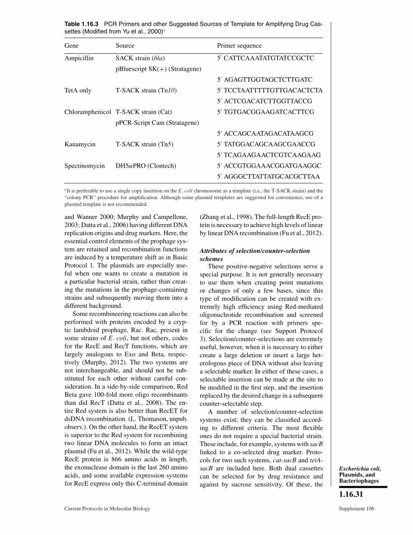
Table 1.16.3 PCR Primers and other Suggested Sources of Template for Amplifying Drug Cas-settes (Modified from Yu et al., 2000)a
Gene Source Primer sequence
Ampicillin SACK strain (bla) 5′ CATTCAAATATGTATCCGCTC
pBluescript SK(+) (Stratagene)
5′ AGAGTTGGTAGCTCTTGATC
TetA only T-SACK strain (Tn10) 5′ TCCTAATTTTTGTTGACACTCTA
5′ ACTCGACATCTTGGTTACCG
Chloramphenicol T-SACK strain (Cat) 5′ TGTGACGGAAGATCACTTCG
pPCR-Script Cam (Stratagene)
5′ ACCAGCAATAGACATAAGCG
Kanamycin T-SACK strain (Tn5) 5′ TATGGACAGCAAGCGAACCG
5′ TCAGAAGAACTCGTCAAGAAG
Spectinomycin DH5αPRO (Clontech) 5′ ACCGTGGAAACGGATGAAGGC
5′ AGGGCTTATTATGCACGCTTAA
aIt is preferable to use a single copy insertion on the E. coli chromosome as a template (i.e., the T-SACK strain) and the“colony PCR” procedure for amplification. Although some plasmid templates are suggested for convenience, use of aplasmid template is not recommended.
and Wanner 2000; Murphy and Campellone,2003; Datta et al., 2006) having different DNAreplication origins and drug markers. Here, theessential control elements of the prophage sys-tem are retained and recombination functionsare induced by a temperature shift as in BasicProtocol 1. The plasmids are especially use-ful when one wants to create a mutation ina particular bacterial strain, rather than creat-ing the mutations in the prophage-containingstrains and subsequently moving them into adifferent background.
Some recombineering reactions can also beperformed with proteins encoded by a cryp-tic lambdoid prophage, Rac. Rac, present insome strains of E. coli, but not others, codesfor the RecE and RecT functions, which arelargely analogous to Exo and Beta, respec-tively (Murphy, 2012). The two systems arenot interchangeable, and should not be sub-stituted for each other without careful con-sideration. In a side-by-side comparison, RedBeta gave 100-fold more oligo recombinantsthan did RecT (Datta et al., 2008). The en-tire Red system is also better than RecET fordsDNA recombination. (L. Thomason, unpub.observ.). On the other hand, the RecET systemis superior to the Red system for recombiningtwo linear DNA molecules to form an intactplasmid (Fu et al., 2012). While the wild-typeRecE protein is 866 amino acids in length,the exonuclease domain is the last 260 aminoacids, and some available expression systemsfor RecE express only this C-terminal domain
(Zhang et al., 1998). The full-length RecE pro-tein is necessary to achieve high levels of linearby linear DNA recombination (Fu et al., 2012).
Attributes of selection/counter-selectionschemes
These positive-negative selections serve aspecial purpose. It is not generally necessaryto use them when creating point mutationsor changes of only a few bases, since thistype of modification can be created with ex-tremely high efficiency using Red-mediatedoligonucleotide recombination and screenedfor by a PCR reaction with primers spe-cific for the change (see Support Protocol3). Selection/counter-selections are extremelyuseful, however, when it is necessary to eithercreate a large deletion or insert a large het-erologous piece of DNA without also leavinga selectable marker. In either of these cases, aselectable insertion can be made at the site tobe modified in the first step, and the insertionreplaced by the desired change in a subsequentcounter-selectable step.
A number of selection/counter-selectionsystems exist; they can be classified accord-ing to different criteria. The most flexibleones do not require a special bacterial strain.These include, for example, systems with sacBlinked to a co-selected drug marker. Proto-cols for two such systems, cat-sacB and tetA-sacB are included here. Both dual cassettescan be selected for by drug resistance andagainst by sucrose sensitivity. Of these, the
Escherichia coli,Plasmids, andBacteriophages
1.16.31
Current Protocols in Molecular Biology Supplement 106
tetA-sacB counter-selection is unique and haslower background than cat-sacB since tetAcan also be selected against (Bochner et al.,1980; Maloy and Nunn, 1981; Li et al., 2013).Another class of selection/counter-selectionsuses single genes that can be either selectedfor or against, for example, tetA and galK.Some of these selections/counter-selectionsrequire specialized strains with specific mu-tations; among these is the galK system,designed specifically for BAC modificationand included in this unit. Other counter-selections that require specialized strains andhave been used successfully by others includerpsL, thyA, and tolC. A promising new ap-proach using a combination of recombineeringand Cas9/CRISPR to reduce non-recombinantbackground has been reported by Jiang et al.(2013). Here, protocols for three selectioncounter-selections are provided in this unit andare described in detail below.
The cat-sacB and tetA-sacB dual cassettesA versatile selection/counter-selection sys-
tem in wide use is cat-sacB. The primaryadvantage of cat-sacB and similar sacB cas-settes is that they do not require a specialbacterial strain. Insertion of the dual cas-sette is by selection for drug resistance, i.e.,CmR; the counter-selection is against the sacBgene on sucrose medium (Gay et al., 1985),as the levansucrase SacB converts sucroseto a toxic polymer, levan, which accumu-lates in the E. coli periplasm. Dual cassetteswith sacB linked to a different drug maybe created. However, a disadvantage of thesacB counter-selection is often a high back-ground of sucrose-resistant cells. To lower thisbackground, a tetA-sacB selection/counter-selection system has been recently developed.One of the earliest counter-selections reportedused the tetAR genes of the Tn10 transpo-son (Bochner et al., 1980; Maloy and Nunn,1981). TetR is a repressor of tetA and is inac-tivated by tetracycline. The TetA protein en-ables both a selection for TetR and counter-selection against it. The TetA protein insertsinto the bacterial membrane where it serves asan efflux pump for tetracycline, thus allowingselection for tetracycline resistance. However,the TetA protein also causes the cells to be-come sensitive to lipophilic chelating agents,such as fusaric acid. Therefore, fusaric acidand similar chelating compounds can be usedto select against tetracycline-resistant strains,as first reported by Bochner et al. (1980) andmodified by Maloy and Nunn (1981). Separat-ing tetR from tetA allows its constitutive ex-
pression and use as a selectable cassette. Thecombination of tetA and sacB allows selectionfor cassette insertion by TetR, and counter-selection against tetA and/or sacB (Li et al.,2013). The use of these cassettes is describedin Support Protocol 1. The presence of TetA inthe membrane enhances the counter-selectionagainst SacB on sucrose plates.
galKAnother selection/counter-selection com-
monly used for BAC work utilizes GalK(Warming et al., 2005). To use thisselection/counter-selection, a special bacterialstrain, SW102, is required; this DH10B deriva-tive contains the defective λ prophage anda precise deletion of the galK gene. For thegalK system, a wild-type galK gene drivenby a constitutive promoter is amplified by thePCR with chimeric primers and targeted tothe region to be modified. The galK inser-tion is selected on minimal plates with galac-tose as the sole carbon source, supplementedwith biotin and leucine to complement aux-otrophies in SW102 and chloramphenicol (orother appropriate drug) for BAC maintenance.During the counter-selection step, the galKgene is replaced with the genetic modificationof choice. Here selection is against the galKgene on plates containing a galactose ana-logue, 2-deoxy-galactose (DOG) (Alper andAmes, 1975). In the cell, the GalK enzymephosphorylates DOG, resulting in the buildupof a toxic compound, 2-deoxy-galactose-1-phosphate. The primary advantage of theGalK selection/counter-selection is that galKis smaller than a dual cassette such as cat-sacB,thus easier to amplify. Strain SW102 and thepgalK plasmid template for galK amplificationare available from the Biological ResourcesBranch of the Frederick National Labora-tory for Cancer Research (http://ncifrederick.cancer.gov/RESEARCH/BRB/).
rpsLYet another selection/counter-selection
system utilizes the rpsL gene, encoding theS12 ribosomal subunit. Mutations exist in rpsLthat make the strain resistant to streptomycin,and a few commonly used E. coli bacterialstrains have this mutation, notably DH10B.The selection/counter-selection cassette con-tains a wild-type rpsL gene linked to a drugmarker. When both rpsL alleles are expressedin the same cell, that cell becomes sensitive tostreptomycin, since the wild-type gene is dom-inant. Therefore, once the dual cassette is tar-geted to a region, replacement of the cassette
Recombineering
1.16.32
Supplement 106 Current Protocols in Molecular Biology
with the DNA of choice will remove the wild-type copy of rpsL and restore Strep resistance.The rpsL gene has been linked to numerousselectable drug markers, including neo, bla,and tetA (Bird et al., 2011). A drawback tothis system is that the cells necessarily havea mutant copy of the rpsL gene, which willalso be a target for recombination with thedual cassette, thus some fraction of the re-combination will occur there, restoring thewild-type allele. In recA+ cells, the rpsL+andrpsLStr alleles can also recombine via the E.coli generalized recombination system; this isnot an issue in the recA mutant DH10B. ForRecA+ strains, to circumvent adventitious re-combination problems, an rpsL gene from P.luminescens has been substituted for E. coliwild-type rpsL (Bird et al., 2011). This gene isdifferent enough in sequence from the rpsLStr
gene to prevent homologous recombination.
thyALike galK, the single gene thyA can also
be used for counter-selection. The thyA geneencodes thymidylate synthase, which is nec-essary for conversion of dUMP to dTTP. AthyA mutant will not grow in minimal mediumunless it is supplemented with thymine, thusthe thyA cassette can be selected on minimalmedium lacking thymine. Counter-selectionagainst ThyA+ is possible on minimal mediumcontaining thymine and trimethoprim becausethe enzyme requires a cofactor, tetrahydrofo-late (THF), which is converted to dihydrofo-late (DHF) during the reaction. THF is re-generated from DHF by dihydrofolate reduc-tase, which is inhibited by trimethoprim. Thus,when a functional thyA gene is present, thethymidylate synthase enzyme will utilize theTHF, depleting it from the cell. Inhibitionof dihydrofolate reductase by trimethoprimwill prevent THF regeneration and thus cellgrowth, since THF is required for a numberof cellular reactions. A thyA null strain con-taining the defective prophage, QW1, is avail-able from the Huang laboratory (Wong et al.,2005).
tolCA selection/counter-selection was devel-
oped for the channel protein TolC (DeVito,2008). The killing function colicin E1 requiresa functional TolC channel for access to the cy-toplasmic membrane, thus tolC mutants arecolicin-tolerant. For this system to work, thetolC gene was deleted from DY329 to makeRS206, which became colicin-tolerant. A tolCcassette was targeted to a number of genes
in this background, making cells sensitive tothe colicin; the cassette was subsequently re-moved with either an ssDNA oligo or a ds-DNA PCR, selecting in the presence of colicinE1. The major disadvantage of this particularselection/counter-selection is that colicin E1 isno longer commercially available.
Ampicillin enrichmentWhile removing a bacteriostatic drug
marker (such as tetracycline), enrichment forrecombinants is possible using ampicillin.Nonrecombinant bacteria will still express re-sistance to the tet marker encoded on the chro-mosome; when the electroporated culture ispropagated in the presence of both tetracyclineand ampicillin, only nonrecombinant cells willgrow; these nonrecombinants will be killed bythe ampicillin (Murphy et al., 2000). Recombi-nant cells will not divide because they lack theTetR marker, and because they do not divide,they are not killed by ampicillin.
Critical Parameters
Choice of recombineering systemAlthough this unit mentions various ways
of expressing recombineering functions, theauthors have the most experience with theRed-mediated recombineering that expressesthe Red functions under the control of theλ PL promoter. These constructs express re-combineering functions either from a defec-tive prophage on the E. coli chromosome oron low copy plasmids. A side-by-side com-parison of this system with two other expres-sion systems, the arabinose-inducible plasmidpDK119 (Datsenko and Wanner, 2000; Dattaet al., 2006) and the IPTG-inducible plasmidpKM208 (Murphy and Campellone, 2003),has been done. It has been found that the for-mer gives about ten-fold lower recombinationefficiencies than the prophage, while pKM208gives similar frequencies to the prophage buthas elevated recombination levels even whenuninduced. Other expression systems have notbeen carefully examined. For these reasons,the researcher should not expect to attain thefrequencies reported here for the λ prophagewhen using other expression systems. If a dif-ferent means of induction is chosen, be sureto make the modifications detailed in BasicProtocol 1.
Induction timesWhen inducing the recombination func-
tions from the defective prophage, the properinduction time is essential. Longer inductiontimes may cause decreased cell viability, since
Escherichia coli,Plasmids, andBacteriophages
1.16.33
Current Protocols in Molecular Biology Supplement 106
the prophage Kil function is also induced bythe temperature shift and will eventually killthe cells. The Gam function, necessary for ef-ficient transformation of dsDNA, is also toxicto the cells (Sergueev et al., 2001). When usinga chemical inducer such as arabinose or IPTG,the overnight culture can be diluted into brothsupplemented with the inducer.
Storage of induced cellsIt is also important to use the induced cells
promptly, since the induced phage functionswill decay over time, especially at 32°C. In-duced cells can be successfully stored on icefor several hours before electroporation with-out a reduction in recombination efficiency. Asdetailed in Basic Protocol 1, competent cellsmay be frozen in 15% glycerol, although theirrecombineering efficiency has been about ten-fold less than that of the same cells freshlyprepared.
Amplification of drug cassettesIt is strongly recommended that each drug
cassette be amplified with only one standardset of primers. Confusion has arisen in the au-thors’ laboratory when individuals have de-signed their own primers for the drug cassettes.Cassettes with different endpoints can causedifferential expression and functions duringthe course of a construction project. Standardpriming sequences for the commonly used cas-settes are listed in Table 1.16.3, which amplifywell in PCR reactions and give selectable re-combinants.
Amplification of the cat-sacB and tetA-sacBelements
It is preferable to amplify the cat-sacB andtetA-sacB DNA cassettes described in SupportProtocol 1 from the suggested bacterial strainsusing colony PCR as described in Basic Pro-tocol 1. Since the PCR products are �3.5 kb,they are not always easy to amplify. OptimizedPCR conditions are detailed above, but otherconditions and any other high-fidelity enzymeshould also work. Getting the PCR to work isreally an empirical problem. The biggest fac-tors, if sufficient extension times and appro-priate temperatures are used, are the PCR ma-chine itself and the polymerase. Adjusting theMg2+ concentration may also improve yield.There has been some confusion regarding theprimers for amplification of the cat-sacB cas-sette. An optimal primer pair has been de-signed for amplification of this DNA, and itis strongly recommended that only this set ofprimers be used (Table 1.16.3).
Outgrowth proceduresThe exact details of the outgrowth proce-
dure will vary depending on the type of re-combineering reaction performed and how therecombinants will be identified. The bacte-rial cells are fragile following electroporationand always require a 30-min minimal recov-ery period in LB at 30° to 32°C before plating.When screening for high-efficiency oligo re-combinants, plate the cells non-selectively onLB-plates immediately after the 30-min recov-ery, before chromosome segregation occurs(Sawitzke et al., 2007). A longer outgrowth isneeded to allow expression of selectable drugmarkers encoding antibiotic resistance. Rou-tinely, at least 2 hr of recovery in broth (with-out drug) is allowed before plating the cellswhen selecting drug resistance. The counter-selection step of selection/counter-selectionsrequires a still longer outgrowth of at least 3 hr,optimally, with a ten-fold dilution, to promotecomplete chromosome segregation. These canalso be outgrown overnight and plated the nextday.
Enhancing the efficiency of oligonucleotiderecombination by using a host deficient inmismatch repair
When point mutations and small changesare desired, extremely high levels of recombi-nants can be obtained with oligo recombina-tion if the bacterial strain is mutant for the hostmethyl-directed mismatch repair system (themutHLS system; Costantino and Court, 2003).Under these conditions, �20% to 50% of thetotal viable cells are recombinant. Strains con-taining the prophage and mutations in the mis-match repair system are available from theCourt laboratory: HME63 and HME68 aremutant for the MutS protein that binds themismatch, these contain a mutS<>amp anda mutS<>cat mutation, respectively; whileHME64 is mutant for the UvrD helicase geneinvolved in removal of the unmethylated strandof mispaired DNA and contains uvrD<>kan(Costantino and Court, 2003). Any of thesemutations will eliminate mismatch repair. Thesame elevated level of recombination can beobtained in strains proficient for mismatch re-pair when the incoming oligo creates config-urations of mispairs that are not recognizedby the wild-type mismatch repair system asdescribed below.
Enhancing the efficiency of oligonucleotiderecombination in a Mut+ (mismatchrepair-proficient) host
Extremely high levels of oligo recombi-nation, equal to that obtained in the absence
Recombineering
1.16.34
Supplement 106 Current Protocols in Molecular Biology
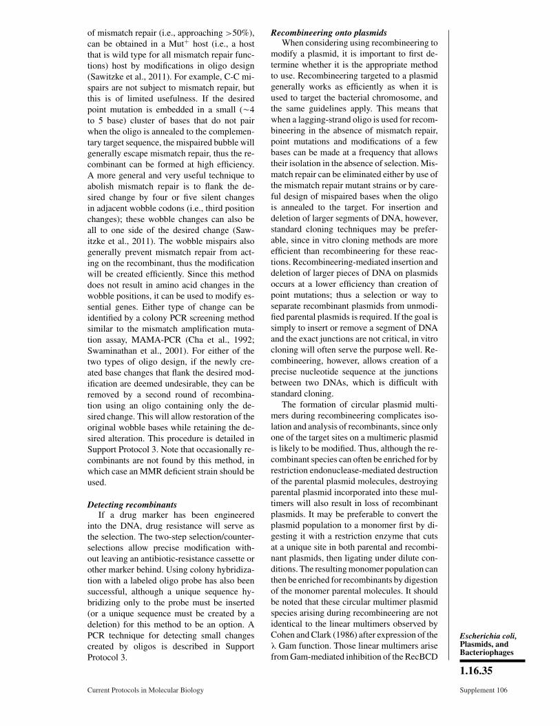
of mismatch repair (i.e., approaching >50%),can be obtained in a Mut+ host (i.e., a hostthat is wild type for all mismatch repair func-tions) host by modifications in oligo design(Sawitzke et al., 2011). For example, C-C mi-spairs are not subject to mismatch repair, butthis is of limited usefulness. If the desiredpoint mutation is embedded in a small (�4to 5 base) cluster of bases that do not pairwhen the oligo is annealed to the complemen-tary target sequence, the mispaired bubble willgenerally escape mismatch repair, thus the re-combinant can be formed at high efficiency.A more general and very useful technique toabolish mismatch repair is to flank the de-sired change by four or five silent changesin adjacent wobble codons (i.e., third positionchanges); these wobble changes can also beall to one side of the desired change (Saw-itzke et al., 2011). The wobble mispairs alsogenerally prevent mismatch repair from act-ing on the recombinant, thus the modificationwill be created efficiently. Since this methoddoes not result in amino acid changes in thewobble positions, it can be used to modify es-sential genes. Either type of change can beidentified by a colony PCR screening methodsimilar to the mismatch amplification muta-tion assay, MAMA-PCR (Cha et al., 1992;Swaminathan et al., 2001). For either of thetwo types of oligo design, if the newly cre-ated base changes that flank the desired mod-ification are deemed undesirable, they can beremoved by a second round of recombina-tion using an oligo containing only the de-sired change. This will allow restoration of theoriginal wobble bases while retaining the de-sired alteration. This procedure is detailed inSupport Protocol 3. Note that occasionally re-combinants are not found by this method, inwhich case an MMR deficient strain should beused.
Detecting recombinantsIf a drug marker has been engineered
into the DNA, drug resistance will serve asthe selection. The two-step selection/counter-selections allow precise modification with-out leaving an antibiotic-resistance cassette orother marker behind. Using colony hybridiza-tion with a labeled oligo probe has also beensuccessful, although a unique sequence hy-bridizing only to the probe must be inserted(or a unique sequence must be created by adeletion) for this method to be an option. APCR technique for detecting small changescreated by oligos is described in SupportProtocol 3.
Recombineering onto plasmidsWhen considering using recombineering to
modify a plasmid, it is important to first de-termine whether it is the appropriate methodto use. Recombineering targeted to a plasmidgenerally works as efficiently as when it isused to target the bacterial chromosome, andthe same guidelines apply. This means thatwhen a lagging-strand oligo is used for recom-bineering in the absence of mismatch repair,point mutations and modifications of a fewbases can be made at a frequency that allowstheir isolation in the absence of selection. Mis-match repair can be eliminated either by use ofthe mismatch repair mutant strains or by care-ful design of mispaired bases when the oligois annealed to the target. For insertion anddeletion of larger segments of DNA, however,standard cloning techniques may be prefer-able, since in vitro cloning methods are moreefficient than recombineering for these reac-tions. Recombineering-mediated insertion anddeletion of larger pieces of DNA on plasmidsoccurs at a lower efficiency than creation ofpoint mutations; thus a selection or way toseparate recombinant plasmids from unmodi-fied parental plasmids is required. If the goal issimply to insert or remove a segment of DNAand the exact junctions are not critical, in vitrocloning will often serve the purpose well. Re-combineering, however, allows creation of aprecise nucleotide sequence at the junctionsbetween two DNAs, which is difficult withstandard cloning.
The formation of circular plasmid multi-mers during recombineering complicates iso-lation and analysis of recombinants, since onlyone of the target sites on a multimeric plasmidis likely to be modified. Thus, although the re-combinant species can often be enriched for byrestriction endonuclease-mediated destructionof the parental plasmid molecules, destroyingparental plasmid incorporated into these mul-timers will also result in loss of recombinantplasmids. It may be preferable to convert theplasmid population to a monomer first by di-gesting it with a restriction enzyme that cutsat a unique site in both parental and recombi-nant plasmids, then ligating under dilute con-ditions. The resulting monomer population canthen be enriched for recombinants by digestionof the monomer parental molecules. It shouldbe noted that these circular multimer plasmidspecies arising during recombineering are notidentical to the linear multimers observed byCohen and Clark (1986) after expression of theλ Gam function. Those linear multimers arisefrom Gam-mediated inhibition of the RecBCD
Escherichia coli,Plasmids, andBacteriophages
1.16.35
Current Protocols in Molecular Biology Supplement 106

exonuclease, allowing the plasmid to repli-cate by the rolling circle mode of DNA repli-cation. Plasmid multimers that form duringrecombineering are circular and are foundin the recombinant plasmid population afteroligo recombination when only the λ Beta pro-tein (in the absence of Gam) has been used tocatalyze the reaction (Thomason et al., 2007).
The plasmid DNA can be introduced intothe cell at the same time as the linear substrateDNA (co-electroporation), or it can be resi-dent in the cell prior to electroporation. Eachoption has advantages and disadvantages. Co-electroporation allows more control over thenumber of plasmids introduced per cell, but isless convenient if the plasmid is large (>10kb). In the protocols described here, direc-tions are given for co-electroporation. To tar-get a resident plasmid, the episome should firstbe introduced into a recombineering-proficientrecA mutant host strain.
Recombineering onto BACsRed and RecET recombineering has also
been optimized for engineering bacterial ar-tificial chromosomes (BACs) (Muyrers et al.,1999; Copeland et al., 2001; Lee et al., 2001;Swaminathan and Sharan, 2004; Sharan et al.,2009), which are able to accommodate hun-dreds of kilobases of foreign DNA. DY380 isthe bacterial host of choice for use with BACs;it is a DH10B derivative containing the defec-tive prophage, as is SW102, a DY380 deriva-tive that has been modified to enable the GalKselection/counter-selection. The recombineer-ing plasmids listed in Table 1.16.3 may alsobe useful in manipulating BACs.
Undertaking in vivo assembly withoverlapping oligos
Complementary oligos will anneal in vivowhen introduced by electroporation into cellsexpressing Exo and Beta protein. Multipleoverlapping oligos can be used to build mod-erately sized DNA products (100 to 150 bp) inan in vivo reaction (Yu et al., 2003) similar toPCR assembly (Stemmer et al., 1995). Whenflanking homologies to chromosomal targetsare added, these overlapping oligos will as-semble into a linear dsDNA in vivo and re-combine into the target.
Mutating essential genesIt is possible to create point mutations in
essential genes using oligo recombination andidentifying them by a PCR screen as describedabove and detailed in Support Protocol 3.Deletion of an essential gene usually results
in cell death. Such a deletion can be recoveredin a diploid state; however, the cell will main-tain a wild-type copy of the gene as well asa mutant version. This diploid state can usu-ally be detected by PCR analysis (Bubuenenkoet al., 2007).
Moving the defective prophage used forrecombineering to a different background
If a different host is required for recombi-neering, the defective prophage can be movedby P1 transduction (UNIT 1.17; Miller, 1972;Yu et al., 2000). Grow a P1 lysate on DY329and transduce the strain of choice. Select fortetracycline resistance and screen for tem-perature sensitivity or an inability to growin the absence of biotin. Check the geno-type of the strain first and supplement theminimal plates to complement any additionalauxotrophies when performing the biotinscreen.
Troubleshooting
Troubleshooting dsDNA recombinationA number of factors can account for prob-
lems in obtaining adequate recombination fre-quencies. It has been observed that the final re-combination efficiency obtained results fromthe cumulative effect of a number of differentfactors, with each making an independent con-tribution. Thus, if one or more of these param-eters are not optimal, the recombination fre-quency can be dramatically reduced. These pa-rameters include the following. (1) Inductionof the recombination proteins: To obtain goodinduction using a temperature shift, it is es-sential to use a 42°C water bath with aeration.Air shakers will not provide adequate heat ex-change. Be sure to chill the cells thoroughly inan ice-water slurry after induction. (2) Too fewviable cells: If recombinants are not obtainedwhen inserting a drug marker, take the timeto determine the titer of viable cells followingoutgrowth. There should be �108 viable cellsper milliliter in the culture. If there are farfewer (i.e., <107 cells/ml), recombinants maynot be found. Take care when washing the cellswith water, as the cell pellet is very soft andcan easily be lost if the aqueous supernatantis not decanted promptly. Inadequate time forcell recovery after electroporation and beforeplating can also result in poor viability. (3)Inappropriate antibiotic concentration: Whenplating the cells on antibiotic plates, be sure touse the lower drug concentrations suggestedfor single-copy genes rather than the concen-trations appropriate for multi-copy plasmids.
Recombineering
1.16.36
Supplement 106 Current Protocols in Molecular Biology
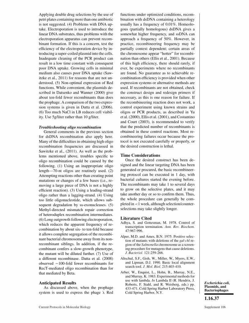
Applying double drug selections by the use ofpetri plates containing more than one antibioticis not suggested. (4) Problems with DNA up-take. Electroporation is used to introduce thelinear DNA substrates, thus problems with theelectroporation apparatus can prevent recom-binant formation. If this is a concern, test theefficiency of the electroporation device by in-troducing a super-coiled plasmid into the cells.Inadequate cleaning of the PCR product canresult in a low time constant with consequentpoor DNA uptake. Growing cells in minimalmedium also causes poor DNA uptake (Saw-itzke et al., 2011) for reasons that are not un-derstood. (5) Non-optimal expression of Redfunctions. While convenient, the plasmids de-scribed in Datsenko and Wanner (2000) giveabout ten-fold fewer recombinants than doesthe prophage. A comparison of the two expres-sion systems is given in Datta et al. (2006).(6) Too much NaCl in LB reduces cell viabil-ity. Use 5g/liter rather than 10 g/liter.
Troubleshooting oligo recombinationGeneral comments in the previous section
for dsDNA recombination also apply here.Many of the difficulties in obtaining high oligorecombination frequencies are discussed inSawitzke et al., (2011). As well as the prob-lems mentioned above, troubles specific tooligo recombination could be caused by thefollowing. (1) Using an inappropriate oligolength—70-nt oligos are routinely used. (2)Attempting reactions other than creating pointmutations or changes of a few bases (i.e., re-moving a large piece of DNA is not a highlyefficient reaction). (3) Using a leading-strandoligo rather than a lagging-strand. (4) Usingtoo little oligonucleotide, which allows sub-sequent degradation by ss-exonucleases. (5)Methyl-directed mismatch repair correctionof heteroduplex recombination intermediates.(6) Long outgrowth following electroporation,which reduces the apparent frequency of re-combination by about six- to ten-fold becauseit allows complete segregation of the recombi-nant bacterial chromosome away from its non-recombinant siblings. In addition, if the re-combinant confers a slow-growth phenotype,the mutant will be diluted further. (7) Use ofa different recombinase. Datta et al. (2008)observed �100-fold fewer recombinants forRecT-mediated oligo recombination than forthat mediated by Beta.
Anticipated ResultsAs discussed above, when the prophage
system is used to express the phage λ Red
functions under optimized conditions, recom-bination with dsDNA containing a heterologyusually has a frequency of 0.01%. Homeolo-gous (partially homologous) dsDNA gives asomewhat higher frequency, and ssDNA canapproach a frequency of 50%. However, inpractice, recombineering frequency may bepartially context dependent; certain areas ofthe chromosome appear “hotter” for recombi-nation than others (Ellis et al., 2001). Becauseof this high efficiency, there should rarely, ifever, be experiments where no recombinantsare found. No guarantee as to achievable re-combination efficiency is provided when otherexpression systems or alternative methods areused. If recombinants are not obtained, checkthe construct design and redesign primers ifnecessary, as this is one reason for failure. Ifthe recombineering reaction does not work, acontrol experiment using known strains andoligos or PCR products, as described in Yuet al. (2000), Ellis et al. (2001), and Costantinoand Court (2003), is recommended to verifythat the predicted number of recombinants isobtained in these control reactions. Most re-combineering failures occur because the pro-tocol is not executed carefully or properly, orthe desired construction is lethal.
Time ConsiderationsOnce the desired construct has been de-
signed and the linear targeting DNA has beengenerated or procured, the basic recombineer-ing protocol can be executed in 1 day, withbacterial cultures started the evening before.The recombinants may take 1 to several daysto grow on the selective plates, and it maytake another day or so to confirm them. Thus,the whole procedure can generally be com-pleted in <1 week, although selection/counter-selections may take slightly longer.
Literature CitedAdhya, S. and Gottesman, M. 1978. Control of
transcription termination. Ann. Rev. Biochem.47:967-996.
Alper, M.D. and Ames, B.N. 1975. Positive selec-tion of mutants with deletions of the gal-chl re-gion of the Salmonella chromosome as a screen-ing procedure for mutagens that cause deletions.J. Bacteriol. 121:259-266.
Altschul, S.F., Gish, W., Miller, W., Myers, E.W.,and Lipman, D.J. 1990. Basic local alignmentsearch tool. J. Mol. Biol. 215:403-410.
Arber, W., Enquist, L., Hohn, B., Murray, N.E.,and Murray, K. 1983. Experimental methods foruse with lambda. In Lambda II (R. Hendrix, J.Roberts, F. Stahl, and R. Weisberg, eds.) pp.433-471. Cold Spring Harbor Laboratory Press,Cold Spring Harbor, N.Y.
Escherichia coli,Plasmids, andBacteriophages
1.16.37
Current Protocols in Molecular Biology Supplement 106

Bird, A.W., Erler, A., Fu, J., Heriche, J-K., Maresca,M., Zhang, Y., Hyman, A.A., and Stewart.A.F. 2011. High-efficiency counterselection re-combineering for site-directed mutagenesis inbacterial artificial chromosomes. Nat. Methods9:103-109.
Blomfield, I.C., Vaughn, V., Rest, R.F., and Eisen-stein, B.I. 1991. Allelic exchange in Escherichiacoli using the Bacillus subtilis sacB gene anda temperature-sensitive pSC101 replicon. Mol.Microbiol. 5:1447-1457.
Bochner, B.R., Huang, H.C., Schieven, G.L., andAmes, B.N. 1980. Positive selection for lossof tetracycline resistance. J. Bacteriol. 143:926-933.
Bubuenenko, M., Baker, T., and Court, D.L. 2007.Essentiality of ribosomal and transcription an-titermination proteins analyzed by systematicgene replacement in Escherichia coli. J. Bac-teriol. 189:2844-2853.
Carter, D.M. and Radding, C.M. 1971. The role ofexonuclease and β protein of phage λ in geneticrecombination. II. Substrate specificity and themode of action of λ exonuclease. J. Biol. Chem.246:2502-2512.
Cha, R.S., Zarbl, H., Keohavong, P., and Thilly,W.G. 1992. Mismatch amplification mutationassay (MAMA): Application to the c-H-rasgene. PCR Methods Appl. 2:14-20.
Cohen, A. and Clark, A. J. 1986. Synthesis of linearplasmid multimers in Escherichia coli K-12. J.Bacteriol. 167:327-335.
Copeland, N.G., Jenkins, N.A., and Court, D.L.2001. Recombineering: A powerful new toolfor mouse functional genomics. Nat. Rev. Genet.2:769-779.
Costantino, N. and Court, D.L. 2003. Enhancedlevels of lambda Red-mediated recombinants inmismatch repair mutants. Proc. Natl. Acad. Sci.U.S.A. 100:15748-15753.
Court, D.L., Sawitzke, J.A., and Thomason,L.C. 2002. Genetic engineering using homol-ogous recombination. Ann. Rev. Genet. 36:361-388.
Court, D.L., Swaminathan, S., Yu, D., Wilson, H.,Baker, T., Bubunenko, M., Sawitzke, J., and Sha-ran, S.K. 2003. Mini-lambda: A tractable systemfor chromosome and BAC engineering. Gene315:63-69.
Datsenko, K.A. and Wanner, B.L. 2000. One-step inactivation of chromosomal genes in Es-cherichia coli K-12 using PCR products. Proc.Natl. Acad. Sci. U.S.A. 97:6640-6645.
Datta, S., Costantino, N., and Court, D.L. 2006.A set of recombineering plasmids for Gram-negative bacteria. Gene 379:109-115.
Datta, S., Costantino, N., Zhou, X., and Court,D.L. 2008. Identification and analysis of recom-bineering functions from Gram-negative andGram-positive bacteria and their phages. Proc.Natl. Acad. Sci. U.S.A. 105:1626-1631.
DeVito, J.A. 2008. Recombineering with tolC as aselectable/counter-selectable marker: Remodel-
ing the rRNA operons of Escherichia coli. Nu-cleic Acids Res. 36:e4.
Ellis, H.M., Yu, D., DiTizio, T., and Court, D.L.2001. High efficiency mutagenesis, repair, andengineering of chromosomal DNA using single-stranded oligonucleotides. Proc. Natl. Acad. Sci.U.S.A. 98:6742-6746.
Fu, J., Bian, X., Hu, S., Wang, H., Huang, F., Seib-ert, P., Plaza, A., Xia, L., Muller, R., Stewart,A.F., and Zhang, Y. 2012. Full-length RecE en-hances linear-linear homologous recombinationand facilitates direct cloning for bioprospecting.Nat. Biotechnol. 30:440-446.
Gay, P., Le Coq, D., Steinmetz, M., Berkelman, T.,and Kado, C.I. 1985. Positive selection proce-dure for entrapment of insertion sequence ele-ments in Gram-negative bacteria. J. Bacteriol.164:918-921.
Hall, S.D., Kane, M.F., and Kolodner, R. 1993.Identification and characterization of the Es-cherichia coli RecT protein, a protein encodedby the recE region that promotes renaturation ofhomologous single-stranded DNA. J. Bacteriol.175:277-287.
Jiang, W., Bikard, D., Cox, D., Zhang, F., and Mar-raffini, L.A. 2013. RNA-guided editing of bacte-rial genomes using CRISPR-Cas systems. Nat.Biotechnol. 31:233-239.
Joseph, J.W. and Kolodner, R. 1983. ExonucleaseVIII of Escherichia coli. II. Mechanism of ac-tion. J. Biol. Chem. 258:10418-10424.
Karu, A.E., Sakaki, Y., Echols, H., and Linn,S. 1975. The γ protein specified by bacterio-phage λ. Structure and inhibitory activity forthe RecBC enzyme of Escherichia coli. J. Biol.Chem. 250:7377-7387.
Kulkarni, S.K. and Stahl, F.W. 1989. Interactionbetween the sbcC gene of Escherichia coli andthe gam gene of phage λ. Genetics 123:249-253.
Lee, E.C., Yu, D., Martinez de Velasco, J., Tes-sarollo, L., Swing, D.A., Court, D.L., Jenkins,N.A., and Copeland, N.G. 2001. A highly effi-cient Escherichia coli–based chromosome engi-neering system adapted for recombinogenic tar-geting and subcloning of BAC DNA. Genomics73:56-65.
Li, X., Thomason, L.C., Sawitzke, J.A., Costantino,N., and Court, D.L. 2013. Positive and negativeselection using the tetA-sacB cassette: Recombi-neering and P1 transduction in Escherichia coli.Nucleic Acids Res. 41:e204.
Little, J.W. 1967. An exonuclease induced by bacte-riophage λ. II. Nature of the enzymatic reaction.J. Biol. Chem. 242:679-686.
Maloy, S.R. and Nunn, W.D. 1981. Selection forloss of tetracycline resistance by Escherichiacoli. J. Bacteriol. 145:1110-1111.
Maresca, M., Erler, A., Fu, J., Friedrich, A., Zhang,Y., and Stewart, A.F. 2010. Single-strand het-eroduplex intermediates in λ Red homologousrecombination. BMC Mol. Biol. 11:54.
Miller, J.M. 1972. Generalized transduction: Useof P1 in strain construction. In ExperimentsRecombineering
1.16.38
Supplement 106 Current Protocols in Molecular Biology
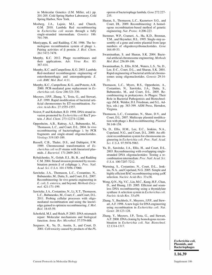
in Molecular Genetics (J.M. Miller, ed.) pp.201-205. Cold Spring Harbor Laboratory, ColdSpring Harbor, New York.
Mosberg, J.A., Lajoie, M.J., and Church,G.M. 2010. Lambda Red recombineeringin Escherichia coli occurs through a fullysingle-stranded intermediate. Genetics 186:791-799.
Muniyappa, K. and Radding, C.M. 1986. The ho-mologous recombination system of phage λ.Pairing activities of β protein. J. Biol. Chem.261:7472-7478.
Murphy, K.C. 2012. Phage recombinases andtheir applications. Adv. Virus Res. 83:367-414.
Murphy, K.C. and Campellone, K.G. 2003. LambdaRed-mediated recombinogenic engineering ofenterohemorrhagic and enteropathogenic E.coli. BMC Mol. Biol. 4:11.
Murphy, K.C., Campellone, K.G., and Poteete, A.R.2000. PCR-mediated gene replacement in Es-cherichia coli. Gene 246:321-330.
Muyrers, J.P.P., Zhang, Y., Testa, G., and Stewart,A.F. 1999. Rapid modification of bacterial arti-ficial chromosomes by ET-recombination. Nu-cleic Acids Res. 27:1555-1557.
Noirot, P. and Kolodner, R.D. 1998. DNA strand in-vasion promoted by Escherichia coli RecT pro-tein. J. Biol. Chem. 273:12274-12280.
Oppenheim, A.B., Rattray, A.J., Bubunenko, M.,Thomason, L.C., and Court, D.L. 2004. In vivorecombineering of bacteriophage λ by PCRfragments and single-strand oligonucleotides.Virology 319:185-189.
Russell, C.B., Thaler, D.S., and Dahlquist, F.W.1989. Chromosomal transformation of Es-cherichia coli recD strains with linearized plas-mids. J. Bacteriol. 171:2609-2613.
Rybalchenko, N., Golub, E.I., Bi, B., and RaddingC.M. 2004. Strand invasion promoted by recom-bination protein β of coliphage λ. Proc. Natl.Acad. Sci. U.S.A. 101:17056-17060.
Sawitzke, J.A., Thomason, L.C., Costantino, N.,Bubunenko, M., Datta, S., and Court, D.L. 2007.Recombineering: In vivo genetic engineering inE. coli, S. enterica, and beyond. Methods Enzy-mol. 421:171-199.
Sawitzke, J.A., Costantino, N., Li, X.T., Thomason,L.C., Bubunenko, M., Court, C., and Court, D.L.2011. Probing cellular processes with oligo-mediated recombination and using the knowl-edge gained to optimize recombineering. J. Mol.Biol. 18:45-59.
Schofield, M.J. and Hsieh, P. 2003. DNA mismatchrepair: Molecular mechanisms and biologicalfunction. Annu. Rev. Microbiol. 57:579-608.
Sergueev, K., Yu, D., Austin, S., and Court, D.2001. Cell toxicity caused by products of the PL
operon of bacteriophage lambda. Gene 272:227-235.
Sharan, S., Thomason, L.C., Kuznetsov S.G., andCourt, DL. 2009. Recombineering: A homol-ogous recombination-based method of geneticengineering. Nat. Protoc. 4:206-223.
Stemmer, W.P., Crameri, A., Ha, K.D., Brennan,T.M., and Heyneker, H.L. 1995. Single-step as-sembly of a gene and entire plasmid from largenumbers of oligodeoxyribonucleotides. Gene164:49-53.
Swaminathan, S. and Sharan, S.K. 2004. Bacte-rial artificial chromosome engineering. MethodsMol. Biol. 256:89-106.
Swaminathan, S., Ellis, H.M., Waters, L.S., Yu, D.,Lee, E-C., Court, D.L., and Sharan, S.K. 2001.Rapid engineering of bacterial artificial chromo-somes using oligonucleotides. Genesis 29:14-21.
Thomason, L.C., Myers, R.S., Oppenheim, A.,Costantino, N., Sawitzke, J.A., Datta, S.,Bubunenko, M., and Court, D.L. 2005. Re-combineering in prokaryotes. In Phages: TheirRole in Bacterial Pathogenisis and Biotechnol-ogy (M.K. Waldor, D.I. Friedman, and S.L. Ad-hya, eds.) pp. 383-399. ASM Press, Herndon,Virginia.
Thomason, L.C., Costantino, N., Shaw, D.V., andCourt, D.L. 2007. Multicopy plasmid modifica-tion with phage λ Red recombineering. Plasmid58:148-158.
Yu, D., Ellis, H.M., Lee, E.C., Jenkins, N.A.,Copeland, N.G., and Court, D.L. 2000. An effi-cient recombination system for chromosome en-gineering in Escherichia coli. Proc. Natl. Acad.Sci. U.S.A. 97:5978-5983.
Yu, D., Sawitzke, J.A., Ellis, H., and Court, D.L.2003. Recombineering with overlapping single-stranded DNA oligonucleotides: Testing a re-combination intermediate. Proc. Natl. Acad. Sci.U.S.A. 100:7207-7212.
Warming, S., Costantino, N., Court, D.L., Jenk-ins, N.A., and Copeland, N.G. 2005. Simple andhighly efficient BAC recombineering using galKselection. Nucleic Acids Res. 33:e36.
Wong, Q.N., Ng, V.C., Lin, M.C., Kung, H.F., Chan,D., and Huang, J.D. 2005. Efficient and seam-less DNA recombineering using a thymidylatesynthase A selection system in Escherichia coli.Nucleic Acids Res. 33:e59.
Zhang, Y., Buchholz, F., Muyrers, J.P.P., and Stew-art, A.F. 1998. A new logic for DNA engineeringusing recombination in Escherichia coli. Nat.Genet. 20:123-128.
Zhang, Y., Muyrers, J.P., Testa, G., and Stewart,A.F. 2000. DNA cloning by homologous recom-bination in Escherichia coli. Nat. Biotechnol.12:1314-1317.
Escherichia coli,Plasmids, andBacteriophages
1.16.39
Current Protocols in Molecular Biology Supplement 106
Recommended