23
DOI: 10.2478/bjmg-2014-002117 (1), 2014 l 23-30
ORIGINAL ARTICLE
* Pediatric Clinic, Medical Faculty, Skopje, Republic of Macedonia
PHENOTYPIC VARIATIONS IN WOLF-HIRSCHHORN SYNDROME
*Corresponding Author: Doz. Elena Sukarova-Angelovska, Pediatric Clinic, Medical Faculty, Vodnjanska 17, 1000 Skopje, Republic of Macedonia. Tel.: +389-70358582. Fax: +389-22439301. E-mail: [email protected]
Sukarova-Angelovska E*, Kocova M, Sabolich V, Palcevska S, Angelkova N
ABSTRACT
Wolf-Hirschhorn syndrome (WHS) is a rare chromosomal disorder caused by terminal deletion of the short arm of chromosome 4. The clinical pic-ture includes growth retardation, severe mental re-tardation, characteristic “Greek helmet” like face, seizures and midline defects in the brain, heart, palate and genitalia. Recently-used molecular techniques increase the number of diagnosed cases due to the detection of smaller deletions. The severity of the clinical presentation is variable depending on the haploinsufficiency of genes in a deleted region.
We present six children with WHS with vari-able clinical appearance. The assessment of several elements (facial dysmorphism, mental retardation, additional congenital anomalies) provided classifi-cation into minor, mild or severe forms. Three of the children had a visible cytogenetic deletion on chromosome 4p, two had microdeletions detected with fluorescent in situ hybridization (FISH), and one child with a less characteristic clinical picture had a mosaic type of the deletion. Correlation between the clinical presentation and the length of the deleted region was confirmed.
Keywords: Wolf-Hirschhorn syndrome (WHS); Phenotype; Microdeletion syndromes; Fluorescent in situ hybridization (FISH).
INTRODUCTION
Wolf-Hirschhorn syndrome (WHS) is a rare chromosomal disorder due to the deletion of the short arm of chromosome 4. The first description of the syndrome was published by Cooper and Hirschhorn [1] and Hirschhorn et al. [2]; it was afterwards con-firmed by Wolf et al. [3]. The syndrome occurs in ap-proximately 1:50,000 newborns [4]. Approximately 120 cases have been described thus far using con-ventional karyotyping [5]. Recently-used molecular techniques increase the number of diagnosed cases due to the detection of smaller deletions of the short arm of chromosome 4 [6,7].
Common features of the syndrome include mi-crocephaly, characteristic facial dysmorphism, fre-quently cited as a Greek helmet-like forehead, broad forehead with prominent glabella and shallow orbital arches; hyperthelorism, short and broad nose; short upturned philtrum and low-set dysplastic ears. Severe psychomotor delay is present in most cases. Seizures usually occur within the first 2 years of life and have variable presentation. Less frequent features are mid-line defects: congenital heart defect, cleft palate, hy-pospadia, as well as skeletal abnormalities, club foot, mesomelia, radioulnar synostosis, fused vertebrae and ribs, and hip dislocation. Growth is severely af-fected [8]. The infants are born small for gestational age. Receding of postnatal growth velocity continues in early childhood, the height of affected children is 3SDS (standard deviation score) below the mean. Some of the patients have a short life span, mainly

24
PHENOTYPE-GENOTYPE VARIATIONS
because of the lower respiratory infections and mul-tiple anomalies. However, the children with smaller deletions have a better survival rate [9].
Most cases are caused by a de novo deletion of chromosome 4p, while in 20.0% of cases, trans-location in the parental karyotype that includes chromosome 4, is responsible for the loss of the part of chromosome 4 in the offspring [4,10,11]. Other structural rearrangements including this re-gion, such as inversions, are a rare cause of WHS. Mosaic state of deletion of chromosome 4p(p15-pter) has been described in two cases so far, and has highly non specific clinical presentation [12,13]. Mosaicism in parental cells could be the reason for occurrence of overt WHS in the offspring [14]. Some investigators speculate that parental imprint-ing is a responsible mechanism for the phenotype, since the majority of deleted chromosomes 4 are of paternal origin [15].
The critical region for WHS is located on the terminal part of the chromosome 4p, with a length of approximately 1-5 Mbp. In rare cases the cytogeneti-cally visible deletion is interstitial. It has been esti-mated that the minimal critical region of the deleted region is 165 kb from the telomere and is up to 750 kb in length, most frequently between loci FGFR3 and D4S168 [7,16]. However, it is a gene-rich region and produces severe clinical presentation in most cases. In 80.0% of cases, the deletion is large, up to 4p14 [9]. The remaining 20.0% of patients have smaller deletions, mostly on the 4p16.3 band, a presentation called Pit-Roger-Danks syndrome [17]. This minor deletion leads to less severe and frequently unchar-acteristic clinical presentation.
Multiple genes are responsible for the clini-cal presentation. However, haploinsuficiency of the WHSC1 gene that encodes a DNA binding protein, is mostly believed to be a cause for the pleiotropic ef-fects. The WHSC1 gene is responsible for chromatin remodeling, and therefore causes insufficient regula-tion of many genes [18]. Zolino et al. [10] suggested that other genes outside the critical region contrib-uted to the phenotype. An additional gene within the WHS critical region, LETM1, involved in Ca binding signaling, is responsible for the seizures in these patients [4,10,]. The gene has been located at the proximal side of the Wolf-Hirschhorn critical region (WHCR) and has an influence on the mitochondrial ion homeostasis [19].
MATERIALS AND METHODS
Patient Analysis. In this study, six patients with classical features of WHS are presented. The dysmor-phic profile has been assessed in all of them, including major and minor anomalies that have been described in the literature, using a specific software program (London Dysmorphology Database; Oxford University Press, London, UK). Facial dysmorphism was evaluated by two independent examiners. The number and severity of each dysmorphic feature was classified as minor [+], mild [++] and severe [+++] (Table 1, Figure 1).
Assessment of postnatal adaptation was made according to the Apgar score and length of stay in the intensive care unit. Malformations of other or-gans and systems were described in all patients. Psy-chomotor evaluation was performed using standard neurological examination and developmental tests according to the Griffith scale.
Chromosome Aalysis and Fluorescent In Situ Hybridization (FISH). Standard chromosome analy-sis of blood lymphocytes was performed. Cultivation was made using phytohemaglutinine for 3 days, har-vesting was done using a standard procedure [20]. The slides were dyed by a G-banding technique. An Olympus BX51 microscope (Olympus Life Science Europa GmbH, Hamburg, Germany) and standard MetaSystems karyotyper (MetaSystems GmbH, Altussheim, Germany) was used for evaluating the chromosomes. At least 25 metaphases were analyzed for each patient (Figure 2).
In patients where the karyotype was normal, stan-dard protocol [21] for FISH analysis was performed us-ing a high-sensitive Vysis probe (4p16.3, Cat. #05J29-074; Abbott Molecular Inc., Des Plaines, IL, USA) with centromeric-control (green signal), and subtelomeric (red signal) including WHCR. Analysis was preformed both on chromosome preparations or interphase nuclei using fluorescent light microscope (Olympus BX51, Olympus Life Science Europa). At least 100 metaphase spreads or nuclei per case were analyzed. Adjacent fil-ters were used: DAPI (4’,6-diamidino-2-phenylindole) for counterstaining, FITC (fluorescein isothiocyanate) for green and TRITC (thetramethylrhodamine isothyo-cyanate) for visualizing the color red. The software for computer analysis, MetaSystems-FISH (MetaSystems GmbH) was used (Figure 3). The severity of the clinical presentation: facial dysmorphism, mental retardation, major organ anomalies were correlated with cytoge-
25
BALKAN JOURNAL OF MEDICAL GENETICSSukarova-Angelovska E, Kocova M, Sabolich V, Palcevska S, Angelkova N
netic findings (visible deletion of chromosome 4p or microdeletion) in all six patients.
RESULTS
Patient 1. An infant boy at the age of 24 days was admitted to the clinic because of asphyxia, hypotonia, poor sucking and failure to thrive. He was the first
child of young, unrelated parents. Intrauterine growth retardation was noticed during the fourth month of pregnancy. Delivery was at term, birth weight was 2200 gr, birth length 45 cm. The neonate was hypo-tonic, had frequent apneic episodes and poor sucking. Dysmorphic profile was estimated as severe [+++] according to the presence and severity of dysmorphic features (Table 1). Evaluation of the heart revealed
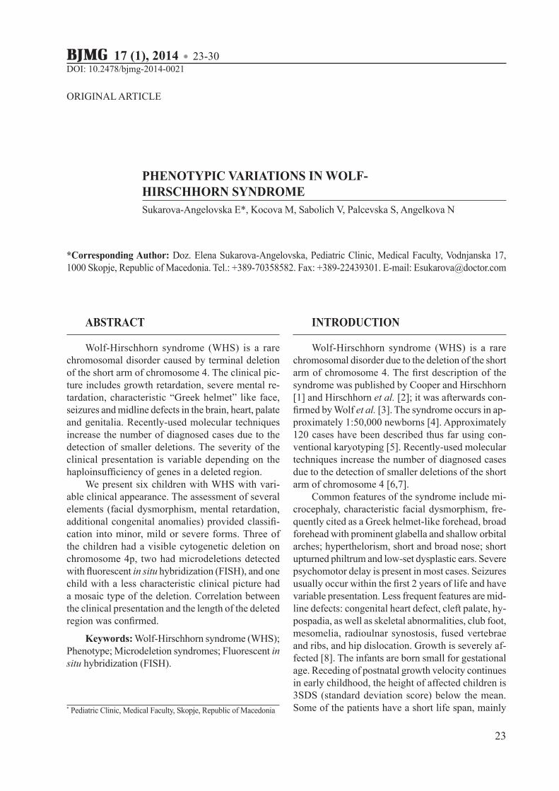
Table 1. Clinical data of our six patients.
Patient 1 (ML) Patient 2 (BI) Patient 3 (MK) Patient 4 (SS) Patient 5 (HJ) Patient 6 (TB)
Birth weight; birth length 2200 gr; 45 cm 2800 gr; 49 cm 2350 gr; 47 cm 2040 gr; 43 cm 1660 gr; 40 cm 2900 gr; 49 cmPoor postnatal adaptation [+++] [+++] [+] [++] [+++] [+]Growth retardation [+++] [+] [+++] [++] [++] [++]Developmental milestones [+++] [++] [+++] [++] [+] [++]Feeding difficulties [+++] [++] [+++] [+] [+] [+]Microcephaly –3 SDS –2.5 SDS –2.5 SDS –2 SDS –2 SDS –2 SDSPhenotype [+++] [+++] [+++] [++] [+] suggestiveGreek-helmet appearance [+++] [+++] [+++] [++] [++] [+]Ptosis of the palpebrae [+++] [++] [+] [+] [+] –Strabismus [++] [++] [+] iris coloboma – –Narrow palpebral fissures [+++] [+++] [+] – [+] [++]Hypertelorism [+++] [+++] [++] [++] [+] –Bat-like ears [++] [+] [+++] [++] [+] –Broad nose [+++] [+++] [++] [+] [+] –Short philtrum [+++] – [++] [+++] [+] [+]Down-turned mouth [+++] [++] [+] [++] [++] [+]Micrognathia [++] [+] [++] [+] [+] [+]
Cleft lip/palate – notch of the lower lip [++] [+]
Renal abnormalities absent left; hypoplastic right – – hypoplastic – –
Cardiac anomalies ASD ASD; dilated ascendant aorta ASD ASD FO, DAP; AP
ommunications –
CNS anomalies global atrophy minor hyrocephaly global atrophy large third
ventricleatrophy of
frontal cotex –
Seizures [+] ? [+] [+] abnormal EEG [+]Sacral dimple [++] [+] [+++] [+] [+] –
Skeletal abnormalities mesomelia club feet – short thorax club feet halux varus, sandal gap
Genital abnormalities hypospadia hypoplastic labia – hypoplastic labia hypospadia cryptorchidism cryptorchidism
Other intestinal malrotation
umbilical hernia; bifid tongue – sparse hair – –
Karyotype 46,XY,del(4)(p15.3)
46,XX,del(4)(P16.2)
46,XX,del(4)(P16.1)
46,XX,ish del(4)(p16.3)
46,XY,ish del(4)(p16.3)
46,XY,del(4)(p16.1)[30%]/ 46,XY[70%]
ASD: atrial septal defect; CNS: central nervous system.

26
PHENOTYPE-GENOTYPE VARIATIONS
a minor atrial septal defect (ASD). Ultrasonog-raphy of the kidneys showed agenesia of the left kidney and hypoplasia of the right kidney. Serum urea and creatinine were above the upper limit. At the age of 2 years, the child had an intestinal obstruction due to a malrotation and underwent surgery. Seizures started at the age of 1.5 months, and had tonic-clonic characteristics, with severe, long-lasting outbursts frequently leading to epi-leptic status; therefore, antiepileptic therapy was introduced. At the age of 4 years he was not able to walk without support and had autistic behavior. Chromosome analysis revealed a cytogenetically visible deletion of the short arm of chromosome 4: 46,XY,del(4)(p15.3).
Patient 2. A 3-month baby was referred to our clinic because of facial dysmorphism. She was the second child in a family of healthy and non consan-guinous parents. The pregnancy was uneventful; the delivery was after 41 weeks of gestation, birth weight was 2800 gr, birth length was 49 cm. The child had asphyxia and was resuscitated. At the time of the examination the baby had failed to thrive and did not achieve developmental milestones. She had facial dysmorphism ascertained as severe. Ad-ditional dysmorphic features included transversal crease on the left hand, club feet and joint stiffness of both legs; a small umbilical hernia was also noticed. Ultrasonography of the heart revealed a congeni-tal heart defect, an ASD with a dilated ascendant aorta. The lateral ventricles of the brain were slightly enlarged. The karyotype was 46,XX,del(4)(p16.1) (Figure 2). We have no data if this child had convul-sions afterwards as she was lost of follow-up.
Patient 3. The patient was a 6-month-old girl and was the first child born to unrelated parents. She was born after a normal pregnancy and deliv-ery, with birth weight of 2350 gr and length 47 cm. Motor delay was noticed from the fourth month of age. She had characteristic facial dysmorphism estimated as severe [+++]. Cleft of the palate was along the hard and soft palate. The sacral sinus was deep. At the age of 4 months she developed sei-zures (first febrile, afterwards afebrile and general-ized). The convulsions were frequent, prolonged and difficult to manage, requiring a combination of several anticonvulsant drugs. The karyotype was 46,XX,del(4)(p16.1).
Patient 4. A female patient, aged 16 months, was the second child of unrelated healthy parents.
Figure 1. a) Facial appearance in our patients. b) Deep sacral dimple in patient 1; sandal gap in patient 6.
a)
b)

27
BALKAN JOURNAL OF MEDICAL GENETICSSukarova-Angelovska E, Kocova M, Sabolich V, Palcevska S, Angelkova N
Their first child had been operated on because of an urethral obstruction but he was otherwise normal. The mother experienced vaginal bleeding during the first trimester of the pregnancy. Delivery was before term and the umbilical cord was wrapped around the baby’s neck; her birth weight was 2340 gr, birth length 44 cm. She had minor developmental delay, first thought to be caused by the neonatal asphyxia. She was estimated to have a moderate [++] phe-notype. She had a unilateral cleft lip, notched left nostril, small allae nasi, bat-like ears. Both kidneys were hypoplastic. The karyotype was normal. The microdeletion of 4p was detected by FISH. At the age of 3 years, she developed clonic-tonic seizures. Despite combined anticonvulsant therapy, her epi-lepsy remains resistant. After the start of the seizures, her motor and mental abilities started to deteriorate; she thus lost most of her motor abilities. Therefore, the estimation of her motor delay in this study was estimated to be before occurrence of the seizures.
Patient 5. This is the first child resulting from eigh in vitro fertilization (IVF) attempts by healthy parents. The fetus had prenatal growth delay. He was born pre term, at week 35 of gestation, and was small for gestational age; birth weight was 1660 gr and birth length 40 cm. Because of immaturity, feeding problems and respiratory distress syndrome, the baby remained in an incubator for 2 months. Dysmorphic profile was considered as minor [+] due to the pres-ence of only a few dysmorphic features, and smaller
central nervous system (CNS) changes. There was a cardiac defect showing foramen ovale and open ductus arteriosus. Left heart catheterization revealed multiple aortic-venous pulmonary communications, as well as truncus bicaroticus. During the follow-ing months, the baby achieved more developmental skills. However, some discharges of myoclonal char-acter on EEG (electroencephalography) were noticed without visible seizures. The karyotype was normal. Using FISH, chromosome 4p showed 46,XY,ish del(4)(p16.3) (Figure 3).
Figure 3. Microdeletion of chromosome 4p detected by FISH in patient 5.
Figure 2. Cytogenetically visible deletion of chromosome 4p in patient 2.

28
PHENOTYPE-GENOTYPE VARIATIONS
Patient 6. The proband was a 16-month-old boy referred because of developmental delay. The preg-nancy was uneventful; however, diminished fetal proportions were noticed on ultrasonography during the second trimester. The delivery was at term, birth weight was 2900 gr, birth length 49 cm. Psychomotor delay was evident after age 6 months. He started to walk at the age of 2 years, pronounced first words at 3 years; however, at the age of 6 years, he still had a poor vocabulary. His motor and mental disability was estimated as moderate according the Griffiths scale. The child had a broad forehead, prominent glabella and prominent orbital ridges that was sug-gestive of WHS. Examination of other organs and systems were normal. At the age of 3 years, the child was given anticonvulsant therapy because of con-vulsions. The karyotype was 46,XY,del(4)(p16.1)[30%]/46,XY[70%].
DISCUSSION
Most WHS cases described in the older litera-ture have sizeable terminal deletions with full blown clinical presentation of WHS. Advanced molecular techniques [FISH, comparative genomic hybridiztion (CGH)] provide the possibility of detecting smaller deletions with less evident phenotypes. Since it is a contiguous gene syndrome, variable numbers of genes contribute to the phenotype in each patient.
The spectrum of clinical manifestation of WHS is highly variable, including specific facial dysmorphism, growth and mental delay. In the cases described in the literature, severity of the clinical presentation has been associated with the size of the deleted region and the breakpoint site. However, there are some reports that patients with similar deletions still show clinical variability. Thus, other mechanisms are suspected, mu-tations in modifier genes outside the deleted region, postzygotic mutational events, gene silencing [4], and/or unmasked recessive mutations by a deletion [22]. Sometimes phenotypic variability and severity of the clinical manifestation do not correlate with the size of the deletion. Estabrooks et al. [23] pointed out that all his described cases shared the same phenotype, al-though the length of the deleted region was different. In his cases, the severity of the clinical presentation and greater number of dysmorphic features were not corre-lated with the amount of the deleted genes. However, all had deletion of the WHCR within the deleted segment.
Our study confirms the hypothesis in the litera-ture that the length of the deleted region is crucial to the severity of the phenotype [24,25]. Clinical as-sessment in our cohort of patients was based upon the number and severity of the separate facial features, as well as upon the presence of organ malformations. Three of the patients with pronounced phenotypes had a cytogenetically visible deletion of the short arm of chromosome 4. Patient 1 had the most severe clinical presentation and most extensive deletion beginning from band p15.3. Patient 3, except for characteristic features, had a cleft lip/palate and patient 4 had only a cleft palate, which indicates that the deleted region in both patients exceeds the WHCR. Cleft lip/palate has been described in the literature in cases with larger deletions centromeric to the WHCR [10]. Since the probe for FISH that we used does not include part of the chromosome 4p centromeric to WHCR, we could not confirm this result in patients 3 and 4.
In patients 4 and 5, the diagnosis was suspected due to the obvious phenotype of WHS, and the diag-nosis was established with a FISH probe for WHCR. However, the presence and severity of minor dysmor-phic features, as well as other malformations, was less pronounced than in the group with apparent cy-togenetically visible deletions. In some studies where microdeletions have been studied, cardiac anomalies were not found. In patients 4 and 5, however cardiac anomalies were present, which can be explained by other underlying mechanisms [10]. Although not severely affected, the cases with microdeletions in variable parts of the WHCR, can broaden genotype-phenotype studies. Some features described in our cases have not been reported previously such as in-testinal malrotation including both small and large intestines in patient 1, or unusual complex cardiac malformation in patient 5.
Patient 6 with a mosaic deletion had less evident clinical presentation with facial features only reminis-cent of a classical WHS phenotype accompanied with a moderate mental retardation. This is in accordance with the reported cases in the literature where differ-ent percent of mosaic cell lines were found together with severe mental retardation [12,13].
All our patients showed prenatal and postnatal growth delay and delayed developmental milestones. There was some discordance between cytogenetic findings and mental retardation (for example, patient 4 had microdeletions and pronounced motor and mental

29
BALKAN JOURNAL OF MEDICAL GENETICSSukarova-Angelovska E, Kocova M, Sabolich V, Palcevska S, Angelkova N
disability). However, it can be explained by the fre-quency and severity of the seizures and administration of potent antiepileptic drugs. Four of our patients had seizures, and the youngest one (patient 5) (now at the age of 5 months) had a specific EEG consistent with dysrhythmic action, but without apparent seizures so far. Seizures were of different pattern, general-ized, unilateral and myoclonic outbursts, as reported elsewhere [5]. The outbursts were difficult to control despite combined anticonvulsive therapy. Seizures are the most frequent cause of death in these patients.
CONCLUSIONS
If a cytogenetically visible deletion is present, a clinical diagnosis of WHS can easily be determined. If a microdeletion is present, the diagnosis is often difficult and can be suggested or made by an ex-perienced dysmorphologist. Therefore, if clinical suspicion of WHS is still present, other molecular techniques (FISH, CGH) are recommended. The col-laboration of clinical geneticist, cytogeneticist, and neurologists is essential for diagnosis, treatment, and follow-up of children with WHS.
Our cohort of six patients with a variable length of the deleted chromosome 4p, provides additional evidence that the amount of a deleted region is es-sential for the severity of the phenotype, i.e., a larger deletion correlates with a more severe clinical presen-tation. Additionally, several anomalies are described in this study that have not been described elsewhere, and should therefore be included in the phenotypic spectrum of the syndrome.
Declaration of Interest. The authors report no conflicts of interest. The authors alone are responsible for the content and writing of this article.
REFERENCES
1. Cooper H, Hirschhorn K. Apparent deletion of short arms of one chromosome (4 or 5) in a child with defects of midline fusion. Hum Chrom Newsl. 1961; 4(14): 14-16.
2. Hirschhorn K, Cooper HL, Firschein IL. Dele-tion of short arms of chromosome 4-5 in a child with defects of midline fusion. Humangenetik. 1965; 1(5): 479-482.
3. Wolf H, Porsch R, Schroeter R, Baitsch H. De-fizienz an den kurzen Arm eines Chromosoms nr. 4. Humangenetik. 1965; 1(5): 397-413.
4. Bergemann A, Cole F, Hircshhorn K. The etiol-ogy of Wolf-Hirschorn syndrome. Trends Genet. 2005; 21(3): 188-195.
5. Battaglia D, Zampino G, Zollino M, Mariotti P, Acquafondata C, Lettori D, et al. Electroclinical patterns and evolution of epilepsy in the 4p- syn-drome. Epilepsia. 2003; 44(9):1183-1190.
6. Fang YY, Bain S, Haan EA, Eyre HJ, MacDonald M, Wright TJ, et al. High resolution characteriza-tion of an interstitial deletion of less than 1.9 Mb at 4p16.3 associated with Wolf-Hirschhorn syn-drome. Am J Med Genet. 1997; 71(4): 453-457.
7. Wright TJ, Ricke DO, Denison K, Abmayr S, Cotter PD, Hirschhorn K, et al. A transcript map of the newly defined 165 kb Wolf-Hirschhorn syndrome critical region. Hum Mol Genet. 1997; 6(2): 317-324.
8. Antonius T, Draaisma J, Levtchenko E, Knoers N, Renier W, van Ravenswaaij C. Growth charts for Wolf-Hirschhorn syndrome (0-4 years of age). Eur J Pediatr. 2008; 167(7): 807-810.
9. Shannon NL, Maltby EL, Rigby AS, Quar-rell OWJ. An epidemiological study of Wolf-Hirschhorn syndrome: life expectancy and cause of mortality. J Med Genet. 2001; 38(10): 674-679.
10. Zollino M, Lecce R, Fischetto R, Murdolo M, Faravelli F, Selicorni A, et al. Mapping the Wolf-Hirschhorn syndrome phenotype outside the cur-rently accepted WHS critical region and defining a new critical region, WHSCR-2. Am J Hum Genet. 2003; 72(3): 590-597.
11. Zollino M, Lecce R, Selicorni A, Murdolo M, Mancuso I, Marang G, et al. A double cryptic chromosome imbalance is an important fac-tor to explain phenotypic variability in Wolf-Hirschhorn syndrome. Eur J Hum Genet. 2004; 12(10): 797-804.
12. Judge CG, Garson OM, Pitt DB, Sutherland GR. A girl with Wolf-Hirschorn syndrome and mo-saicism 46,XX-46,XX,4p-. J Ment Defic Res. 1974; 18(0):79-85.
13. Vockley J, Inserra JA, Breg WR, Yang-Feng TL. “Pseudomosaicism” for 4p- in amniotic fluid cell
30
PHENOTYPE-GENOTYPE VARIATIONS
culture proven to be true mosaicism after birth. Am J Med Genet. 1991; ;39(1): 81-83.
14. Fryns JP, Smeets E, Devriendt K, Petit P. Wolf-Hirschhorn syndrome with cryptic 4p16.3 dele-tion and balanced/unbalanced mosaicism in the mother. Ann Genet. 1998; 41(2): 73-76.
15. Tupler R, Bortotto L, Bühler EM, Alkan M, Malik NJ, Bösch-Al Jadooa N, et al. Paternal origin of the de novo deleted chromosome 4 in Wolf-Hirschhorn syndrome. J Med Genet. 1992; 29(1):53-55.
16. Altherr MR, Bengtsson U, Elder FF, Ledbetter DH, Wasmuth JJ, McDonald ME, et al. Molecu-lar confirmation of Wolf-Hirschhorn syndrome with a subtle translocation of chromosome 4. Am J Hum Genet. 1991; 49(6): 1235-42.
17. Kant S, van Haeringen A, Bakker E, Stec I, Donnai D, Mollevanger P, et al. Pitt-Rogers-Danks syn-drome and Wolf-Hirschhorn syndrome are caused by a deletion in the same region on chromosome 4p16.3. J Med Genet. 1997; 34(7): 569-572.
18. Stec I, Wright TJ, van Ommen GJ, de Boer PA, van Haeringen A, Moorman AF, et al. WHSC1, a 90 kb SET domain-containing gene, expressed in early development and homologous to a Drosophila dysmorphy gene maps in the Wolf-Hirschhorn syndrome critical region and is fused to IgH in t(4;14) multiple myeloma. Hum Mol Genet. 1998; 7(7): 1071-1082.
19. Dimmer KS, Navoni F, Casarin A, Trevisson E, Endele S, Winterpacht A, et al. LETM1, deleted in Wolf-Hirschhorn syndrome is required for normal mitochondrial morphology and cellular viability. Hum Mol Genet. 2008; 17(2): 201-214.
20. Lockwood DH, Johnston DA, Riccardi VM, Zimmerman SO. The use of subchromosome-length unique band sequences in the analysis of prophase chromosomes. Am J Hum Genet. 1988; 43(6): 934-947.
21. Liehr T, Claussen U. FISH on chromosome prep-arations of peripheral blood. In: Rautenstrauss BW, Liehr T, Eds. FISH Technology. Berlin, Germany: Springer. 2002: 73-81.
22. Flipsen-ten Berg K, van Hasselt PM, Eleveld MJ, van der Wijst SE, Hol FA, de Vroede MA, et al. Unmasking of a hemizygous WFS1 gene mutation by a chromosome 4p deletion of 8.3 Mb in a patient with Wolf-Hirschhorn syndrome. Eur J Hum Genet. 2007; 15(11): 1132-1138.
23. Estabrooks LL, Lamb AN, Aylsworth AS, Cal-lanan NP, Rao KW. Molecular characterisa-tion of chromosome 4p deletions resulting in Wolf-Hirschhorn syndrome. J Med Genet. 1994; 31(2): 103-107.
24. Maas NMC, van Buggenhout G, Hannes F, Thienpont B, Sanlaville D, Kok K, et al. Geno-type-phenotype correlation in 21 patients with Wolf-Hirschhorn syndrome using high resolu-tion array comparative genome hybridisation (CGH). J Med Genet. 2008; 45(2): 71-80.
25. Wieczorek D, Krause M, Majewski F, Albrecht B, Horn D, Riess O, et al. Effect of the size of the deletion and clinical manifestation in Wolf-Hirschhorn syndrome: analysis of 13 patients with a de novo deletion. Eur J Hum Genet. 2000; 8(7): 519-526.
Recommended