1
PATHOPHYSIOLOGY OF FUNCTIONAL
(PSYCHOGENIC) MOVEMENT
DISORDERS
Isabel Pareés Moreno
A thesis submitted to University College
London for the degree of PhD
Sobell Department of Motor
Neuroscience and Movement Disorders
UCL Institute of Neurology
Queen Square
London

2
DECLARATION THAT THE WORK PRESENTED IN THIS THESIS IS THE
CANDIDATE’S OWN
I, Isabel Pareés Moreno, confirm that the work presented in this thesis is my own.
Where information has been derived from other sources, I confirm that this has
been indicated in the thesis.
London, December 2014
Signature
Isabel Pareés Moreno
3
Esta tesis está dedicada a mis padres y a mi hermana por su apoyo y amor desde la
distancia, por ser el pilar inamovible de mi vida y estar siempre a mi lado. A Alejandro, por
ser mi cómplice, mi compañero de viaje y porque “en la calle codo a codo somos mucho más
que dos”. Finalmente a Guillermo, mi amor, por teñir cada uno de mis días con su sonrisa y
haberme hecho redescubrir la vida como madre.

4
“Be accustomed to the laws ruling the mind of the hysterics”
Pierre Janet
5
SUMMARY
This thesis describes a series of studies involving healthy subjects, carefully selected
patients with functional movement disorders and organic movement disorders, in
which different aspect of the mechanism underlying functional movement
disorders were explored:
1. The presence of physical precipitating factors at onset of functional
movement disorder by using semistructured interviews. I found that most
patients with functional movement disorder have a clear physical event
prior to the onset of functional symptoms.
2. The presence of a “jumping to conclusions” reasoning style that may
predispose patients with functional movement disorder to accept new
hypothesis on the basis of less evidence. They requested less evidence that
healthy controls to make a judgement, which is here suggested to influence
the manner in which they process novel sensory data occurring during
triggering events.
3. The role of attention in symptoms production by using different motor tasks
in which the predictability of movements as well as the effect of explicit and
implicit strategies in motor control were manipulated. Motor impairment in
patients with functional movement disorder was found to be related to the
employment of explicit strategies or when pre-planning movements is
possible.
4. The intensity and duration of tremor in patients with functional tremor in a
real life situation using accelerometers. They were found to fail to perceive
6
that tremor is not present most of the time compared with patients with
organic tremor.
5. Finally, I explored the phenomenon of the sensory attenuation using a force-
matching task as a measure of sense of agency for movement in these
patients. Patients with functional movement disorders have an abnormal
sensory attenuation for movement, which may help to explain the lack of
agency for the abnormal movement.
These results contribute to the understanding of the mechanisms underlying
functional movement disorders and by extension, other functional neurological
symptoms, and demonstrate that they are amenable to neuroscientific study.
7
STATEMENT OF PARTICIPATION IN STUDIES
DESCRIBED
The initial concept for the thesis was generated by Dr Mark Edwards. In what
follows, I make a statement of my contribution in each of the studies described
here.
Chapter 1 and 2. General introduction and historical view of functional movement
disorders. I performed the search of the literature, interpreted and wrote up the
findings.
Chapter 4. A study on the physical precipitating factors in functional movement
disorders. The conception of the study was generated by Dr Mark Edwards, Dr Jon
Stone, Dr Alan Carson and myself. I planned along with Dr Edwards the design of
the semi-structured interview and the selection of the questionnaires. I reviewed
previous literature on physical events preceding functional movement disorders.
Patient ascertainment was conducted by Dr Edwards and myself. Dr Maria Pires, Dr
Maja Kojovic and myself contributed to the collection of the data. I performed the
analysis of the data and writing up the results. The interpretation of the data was
performed by Dr Mark Edwards, Dr Jon Stone, Dr Alan Carson and myself.
Chapter 5. A study on the “jumping to conclusions” bias in functional movement
disorders. The conception of the study was generated by Dr Mark Edwards and
Katerina Fotopoulou. The experiments were planned by myself. I was involved in
the recruitment of participants and participated in all the studies. Dr Pedro Zapater
and Dr Horga de la Parte guided me in the statistical analysis and I wrote up the
8
results. The interpretation of the data was performed by Dr Mark Edwards, Katerina
Fotopoulou and myself.
Chapter 6. A study on the effect of explicit strategies and predictability on motor
control in functional movement disorders. The conception of the study was
generated by Dr Mark Edwards. Sven Bestman and Marco Davare were the mayor
contributors to the development of the explicit and implicit motor paradigms and
pre-cued task. I was involved in the recruitment of the participants and I run all the
experiments. The analysis was performed by Dr Mark Edwards and myself. I wrote
up the results. Dr Mark Edwards, Sven Bestman, Professor Rothwell and myself
contributed in the interpretation of the data.
Chapter 7. A study assessing functional motor symptoms in real life conditions
using a wrist-worn actigraph. The conception of the experiment was generated by
Dr Mark Edwards. I designed and planned the study. Dr Saifee and myself
conducted the ascertainment of the participants and the collection of the data. I
performed the analysis of the data. Dr Pedro Zapater and Dr Horga de la Parte gave
substantial input on it, mainly in the Bland-Altman analysis. I wrote up the results.
Dr Edwards and myself interpreted the results.
Chapter 8. A study on the lack of sense of agency for movement in functional
movement disorders. The concept of the study was generated by myself. Dr
Edwards, Atsuo Nuruki and myself planned the study but Atsuo Nuruki was the
major contributor programming the robots for the motor task. Marco Davare wrote
the script to analyse the results. I performed the analysis and wrote up the results.

9
Dr Harriet Brown, Dr Rick Adams and Professor Friston provided with substantial
input to our interpretation of the results.
Chapter 9. Discussion. I wrote the discussion and Dr Edwards guided me through it.
10
TABLE OF CONTENTS
SUMMARY .......................................................................................................................... 5
STATEMENT OF PARTICIPATION IN STUDIES DESCRIBED ....................................................... 7
ABBREVIATIONS ................................................................................................................ 13
LIST OF FIGURES ................................................................................................................ 15
LIST OF BOXES AND TABLES ............................................................................................... 17
ACKNOWLEDGEMENTS ...................................................................................................... 19
THESIS OVERVIEW ............................................................................................................. 21
Chapter 1: General introduction to functional movement disorders ................................... 27
1.1 Definition ..................................................................................................................... 27
1.2 Epidemiology ............................................................................................................... 27
1.3 Clinical Presentation.................................................................................................... 28
1.3.1 General clues .............................................................................................. 28
1.3.2 Functional tremor ....................................................................................... 29
1.3.3 Functional dystonia ..................................................................................... 31
1.3.4 Functional myoclonus ................................................................................. 32
1.3.5 Functional gait disorder ............................................................................... 33
1.3.6 Functional parkinsonism ............................................................................. 34
1.3.7 Other functional movement disorders: chorea, tics and paroxysmal
movement disorders .................................................................................................. 35
1.4 Diagnosis ..................................................................................................................... 36
1.5 Differential diagnosis .................................................................................................. 39
1.6 Treatment ................................................................................................................... 40
1.7 Prognosis ..................................................................................................................... 43
Chapter 2: The pathophysiology of functional movement disorders – the historical view .... 45
2.1 Ancient Egypt, Greece and Rome ................................................................................ 46
2.2 Middle Ages................................................................................................................. 47
2.3 Modern Age ................................................................................................................ 48
2.4 19th and 20th Centuries .............................................................................................. 48
2.4.1 Briquet ....................................................................................................... 48
2.4.2 Charcot ....................................................................................................... 49
2.4.3 Janet ........................................................................................................... 54
2.4.4 Freud .......................................................................................................... 59
Chapter 3: Aims and hypotheses ........................................................................................ 64
11
Chapter 4: A study on the physical precipitating factors in functional movement disorders . 67
4.1 Introduction ................................................................................................................. 67
4.2 Methods ...................................................................................................................... 71
4.2.1 Participants ................................................................................................. 71
4.2.2 Semi-structured interviews ......................................................................... 72
4.2.3 Questionnaires............................................................................................ 73
4.3 Results ......................................................................................................................... 75
4.3.1 Tempo of onset ........................................................................................... 77
4.3.2 Physical precipitating factors ....................................................................... 77
4.3.3 Case examples ............................................................................................ 79
4.3.4 Panic symptoms .......................................................................................... 81
4.3.5 HADS .......................................................................................................... 81
4.3.6 Life events .................................................................................................. 81
4.4 Discussion .................................................................................................................... 82
Chapter 5: A study on the “jumping to conclusions” bias in functional movement
disorders........................................................................................................................... 88
5.1 Introduction ................................................................................................................. 88
5.2 Methods ...................................................................................................................... 89
5.2.1 Participants ................................................................................................. 89
5.2.2 Design and measures .................................................................................. 90
5.2.3 Statistical analyses ...................................................................................... 94
5.3 Results ......................................................................................................................... 96
5.4 Discussion .................................................................................................................... 99
Chapter 6: A study on the effect of explicit strategies and predictability on motor control
in functional movement disorders ................................................................................... 103
6.1 Introduction ............................................................................................................... 103
6.2 Methods .................................................................................................................... 104
6.2.1 Participants ............................................................................................... 104
6.2.2 Experimental Procedure ............................................................................ 106
6.2.3 Statistical analysis ..................................................................................... 110
6.3 Results ....................................................................................................................... 111
6.3.1 Experiment 1: one-back reaching and rotation learning tasks ..................... 111
6.3.2 Experiment 2: pre-cued choice reaction time with variable cue validity ...... 114
6.4 Discussion .................................................................................................................. 115
12
Chapter 7: A study assessing functional motor symptoms in real life conditions using a
wrist-worn actigraph ....................................................................................................... 120
7.1 Introduction ............................................................................................................... 120
7.2 Methods .................................................................................................................... 121
7.2.1 Participants ............................................................................................... 121
7.2.2 Questionnaires and scales ......................................................................... 122
7.2.3 Tremor recording ...................................................................................... 124
7.2.4 Diaries ...................................................................................................... 125
7.2.5 Data analysis and statistics ........................................................................ 125
7.3 Results ....................................................................................................................... 126
7.3.1 Baseline characteristics ............................................................................. 126
7.3.2 Comparison between actigraphy and self-report of tremor ........................ 128
7.4 Discussion .................................................................................................................. 132
7.4.1 Implications for understanding the pathophysiology of functional tremor .. 133
7.4.2 Implications for clinical trials in functional tremor ...................................... 135
7.4.3 Limitations ................................................................................................ 136
7.5 Conclusions ................................................................................................................ 137
Chapter 8: A study assessing the lack of sense of agency for movement in functional
movement disorders. ...................................................................................................... 139
8.1 Introduction ............................................................................................................... 139
8.2 Methods .................................................................................................................... 141
8.2.1 Participants ............................................................................................... 141
8.2.2 Questionnaires.......................................................................................... 142
8.2.3 Materials .................................................................................................. 143
8.2.4 Experimental Design ................................................................................. 143
8.2.5 Procedure ................................................................................................. 143
8.2.6 Statistical analysis ..................................................................................... 145
8.3 Results ....................................................................................................................... 146
8.4 Discussion .................................................................................................................. 149
Chapter 9: General discussion and conclusions ................................................................. 154
9.1 Conclusions ................................................................................................................ 172
9.2 Implications for further research .............................................................................. 173
REFERENCES .................................................................................................................... 175
APPENDIX ....................................................................................................................... 194
13
ABBREVIATIONS
ANOVA Analysis of Variance
BOLD Blood-Oxygen-Level Dependent contrast imaging
BP Bereitschaftspotential
CBT Cognitive Behavioural Therapy
DaT SPECT Dopamine transporter imaging with single-photon
emission computed tomography
DR Displacement Ratio
DSM Diagnostic and Statistical Manual of Mental Disorders
EEG Electroencephalography
EMG Electromyography
FMD Functional Movement Disorder
FT Functional Tremor
FTM Fahn-Tolosa-Marin tremor scale
HADS Hospital Anxiety and Depression Scale
ION Institute of Neurology
IQ Intelligence Quotient
JTC Jumping to Conclusions
LEQ Life Events Questionnaire
MT Movement Time
NHNN National Hospital for Neurology and Neurosurgery
OB One-back Reaching
14
OrgT Organic Tremor
PD Parkinson's disease
PDI Peter's Delusions Inventory
ROT Rotation Learning Task
RT Reaction Time
SA Sensory attenuation
SAS Secondary Attentional System
SD Standard Deviation
SEP Somatosensory evoked potentials
SMA Supplementary Motor Area
TMS Transcranial Magnetic Stimulation
15
LIST OF FIGURES
Figure 2.1. Man with fixed dystonia after injury and application of a splint described
by Charcot in The Clinical Lectures on the Diseases of the Nervous System ............ 51
Figure 2.2. Device to demonstrate that patients with fixed dystonia are not feigning
described by Charcot at The Clinical Lectures on the Diseases of the Nervous System
.................................................................................................................................... 53
Figure 4.1. Mean negative events scores (in points) across the patients for each of
the categories recorded in the Life Event Questionnaire .......................................... 82
Figure 5.1. Schematic representation of the jars and the sequence used to drawn
the beads in condition 1. ............................................................................................ 91
Figure 5.2. Schematic representation of the jars and the sequence used to drawn
beads in condition 2. .................................................................................................. 92
Figure 5.3. Example of one of the items used in the short version of the Raven’s
Progressive Matrices…….. .......................................................................................... 93
Figure 5.4. Results of functional patients and healthy controls in condition 1 of the
bead task. ................................................................................................................... 97
Figure 6.1. Schematic representation of the three conditions of experiment 1
(baseline, one-back reaching task and rotation learning task). ............................... 107
16
Figure 6.2. Schematic representation of experiment 2 (pre-cued choice reaction
time with varying cue validity). ................................................................................ 109
Figure 6.3. Boxplot presenting ratios between the first ten trials and the last ten
trials of the temporal and spatial variables for patients with functional movement
disorders and controls (A: one-back reaching task and B: rotation learning task). 113
Figure 6.4. Results of experiment 2 (pre-cued choice reaction time with varying cue
validity). Ratios of the response time (ms) for valid/invalid cues in each validity
condition .................................................................................................................. 115
Figure 7.1. Tremor duration as percentage of the waking day as recorded in self-
report diaries and by actigraphy, in patients with organic tremor and functional
tremor ...................................................................................................................... 130
Figure 7.2. Bland-Altman plots for tremor duration as percentage of the intra-day
intervals, recorded in self-report diaries and by actigraphy, in patients with organic
tremor and functional tremor .................................................................................. 131
Figure 8.1. Experiment set-up of the force matching paradigm ............................. 144
Figure 8.2. Results of the force matching paradigm................................................ 148
17
LIST OF BOXES AND TABLES
Box 1. Fahn and Williams criteria for functional movement disorders ..................... 38
Box 2. DSM-IV criteria for panic attack Disorders ..................................................... 73
Table 4.1. Examples of studies where physical precipitating factors have been
reported in functional movement disorders ............................................................ 68
Table 4.2. Demographic characteristics of the patients recruited in the physical
precipitating factors study ......................................................................................... 76
Table 4.3. Number of patients with a particular physical precipitating factor for
each type of functional movement disorders ............................................................ 78
Table 5.1. Clinical and demographic characteristics of patients recruited in the bead
task ............................................................................................................................. 96
Table 5.2. Results in condition 1 and condition 2 of the bead task. ......................... 98
Table 6.1. Baseline characteristics of patients recruited in one-back reaching,
rotation learning and pre-cued choice reaction time with varying cue validity tasks
. ................................................................................................................................. 105
Table 6.2. One-back reaching and rotation learning tasks results .......................... 113
Table 7.1. Baseline characteristics of patients recruited in wrist-worn actigraph
study ......................................................................................................................... 127

18
Table 7.2. Results of the wrist-worn actigraph study .............................................. 128
Table 8.1. Demographic and clinical characteristics of the participants recruited in
the sensory attenuation study ................................................................................. 147
19
ACKNOWLEDGEMENTS
The patients and their families, who kindly took part in these studies and completed
long sessions in the lab.
Tabish Saifee, Panagiotis Kassavetis, Anna Sadnicka, Maja Kojovic and Ignacio Rubio
for their support and intellectual input on each of my results, for their friendship
and for the time we have spent together in and outside of the office during our
PhDs.
Atsuo Nuruki for his collaboration in the experiment assessing sensory attenuation.
Without him and his robots this would have never happened.
I would like also to thank Professor John Rothwell, Sven Bestman and Marco Davare
for his suggestions interpreting the results of the motor control tasks.
Harriet Brown, Rick Adams and Professor Karl Friston for kindly taking the time to
discuss my results and helpfully contextualise them within emerging theories that
attempt to provide a general model of brain function.
John Stone and Alan Carson for transmitting to me their enthusiasm for patients
with functional symptoms and for their input in the physical precipitating factors
study.
Pedro Zapater and Jose Francisco Horga for their help with the statistics employed
in this thesis.
Paul Hammond for his support in the lab, his patience and for making most devices
that I have used in this thesis.

20
Librarians from the Queen Square Library, who assisted me through the process of
searching the precious books from Charcot, Janet and Freud. The readings of those
helped me to contextualise functional symptoms and realise that important
concepts about them have not significantly changed after more than one century.
Dr Jorge Hernandez Vara from whom I discovered, during my first years of
residency, how fascinating movement disorders are.
My second supervisor Professor Kailash Bhatia who more than 5 years ago accepted
me as a fellow in Queen Square and whose clinical skills I will never forget.
Finally my supervisor, Dr Mark Edwards. It is extremely difficult to describe what
Mark represents to me in a language that is not my mother tongue. When I decided
to engage in the experience of doing clinical and research work abroad I would had
never thought to come across a person like him. Thank you for taking your time and
being so supportive and patient with me at the very beginning when English was
not my best skill. Thanks for your guidance and for teaching me to treat my patients
as a “whole” and not just as a neurological disease. Thanks for teaching me that
brain is more than white and grey matter. Thanks for showing me that one can see
interesting things where others see nothing and that simple paradigms in research
can provide exciting results. Thanks for your warmness, your kindness and for
looking after me when things have not been easy.
21
THESIS OVERVIEW
A personal view
Before coming to London, I completed my training in Neurology in one of the
biggest hospitals in Barcelona, Spain, between 2004 and 2009. During that time, I
dealt with the diagnosis and management of most acute and chronic neurological
diseases. However, my experience with patients suffering from functional
neurological symptoms was limited, not because they were not seen in the
emergency department or in the outpatient’s clinics, but because most patients
were lost to follow up. They usually were managed by explaining that they should
not be worried about the symptoms because all tests had come back normal, that
they must be anxious and depressed, and that this was the likely cause of their
symptoms. I could feel how disappointed most patients were with this explanation
and when they were told that they were discharged from the clinic to be referred to
the Psychiatry Department. Most patients said that they actually did not feel
depressed or anxious, that their symptoms were indeed very disabling and they
could not see how their symptoms were going to improve. I could also see how
uncomfortable the situation was for clinicians who were not confident in making a
diagnosis and explaining the problem to the patient in a positive/explicit way. Also,
I could perceive how the assumption of malingering was always (in an implicit
manner) present in the discussion.
The truth is that I was struck by how young and disabled many patients were and
how little we could offer. Having said that, I would have never expected to do my
PhD in functional neurological symptoms. I definitely preferred patients for whose
22
disorders there was no speculation or explanations that touched the barrier with
philosophy.
This impression has notably changed over the last 4 years. A more neuroscientific
approach to them has shown me how complicated and fascinating clinical problem
functional movement disorders are. The more I read, the more I realised that they
represent an enigmatic area of medicine that has always existed but has always
been considered the “ugly duckling” of neurology. I think the fear to face these
conditions by most clinicians has resulted in a lack of improvement in terms of
understanding of pathophysiology and treatment despite all the advances that have
occurred in Medicine over the past decades. This fear displayed by neurologists to
face functional neurological symptoms is in my opinion more than reasonable as no
one teaches you during your career how to manage these patients.
During these 4 years I have learnt the importance of making the diagnosis in a
positive fashion rather than as a diagnosis of exclusion. In this regards, patients
with functional motor symptoms are ideal because in contrast to patients with
symptoms such as sensory loss, pain, fatigue and memory disturbance, these
patients have objective signs on examination that are amenable to clinical and
experimental measurement. This provides a degree of certainty about the diagnosis
which may not be achievable in those whose symptoms are only measurable via
self-report.
I have also learnt that once one explains how the diagnosis has been reached, and it
is put in context, that patients are open to treatment and may improve with no
additional measures. Hostile patients turn out to be pleasant patients that look
23
forward to collaborating in research to better understand their condition. I have
learnt that the brain is more fascinating than I ever thought it was during my
neurology training.
I have been amazed how despite all the normal investigations, these patients can
inform you about different aspect of motor physiology and about other typical
neurological diseases.
From the academic point of view, there is still active debate about what to call this
group of patients. In this thesis I have used the term “functional” and this is not
something new. In the past, other authors including Yealland in Queen Square
(Linden et al., 2013) preferred this term when treating soldiers with functional
symptoms. “Hysteria” implies a pejorative meaning and it is not commonly used
nowadays. “Non-organic” denotes what one does not have and may infer that the
symptoms are imagined or not real. “Medically unexplained” is commonly used in
medical circles but when a patient receives this diagnosis they also receive a label
of uncertainty: “if my problem cannot be explained, is the doctor missing a rare
disease?” In the title of this thesis I have included the term “psychogenic” in
parenthesis because it is the most widespread term in the movement disorder
community. However, this term implies that the aetiology of the symptoms is
purely psychological. This one-dimensional approach is criticised in this thesis and
therefore I considered somehow inappropriate to hold this label through the text.
“Functional” does not imply aetiology and is not associated with negative
connotations (Stone et al., 2002). However, it may be considered to be vague as in
the past other conditions such as migraine or epilepsy were labelled as functional
24
because of the lack of structural abnormalities in the central nervous system.
Nevertheless, given the current level of understanding of the underlying
pathophysiology, I have considered that the term “functional” is the one that
comes closest to my understanding of these perplexing symptoms.
On the other hand, it may sound inconsistent that I have sometime used the term
“organic” to designate more typical neurological diseases but not by this I dismiss
the possibility that functional symptoms have a neurobiological basis.
Approval was obtained from the National Hospital for Neurology and
Neurosurgery/Institute of Neurology (NHNN/ION) Joint Ethics Committee for all the
studies included in this thesis and all participants provided written consent to
participate according to the Declaration of Helsinki. I have included the
methodology and the results of each of my studies in a different chapter, with the
hope of simplifying the reading of this thesis.
Chapter 1 is a general introduction to functional movement disorders. This chapter
covers the current state of knowledge regarding phenomenology, diagnosis and
management of this group of patients.
Chapter 2 is a review of previous literature on the pathophysiology of functional
symptoms in general. It covers information from ancient Egypt to the beginning of
the 20th century but with special focus on the 19th century and three of the most
important authors in this period: Charcot, Janet and Freud. Particular attention is
paid to functional movement disorders, when mentioned in their writings.
Chapter 3 describes the specific aims and hypothesis of this thesis.
25
Chapter 4 describes the methodology and results of a clinical study designed to
assess the presence of physical precipitating factors at the onset of functional
movement disorders. Psychological factors prior to the development of functional
symptoms have been classically highlighted in the past but potential physical
triggers to these symptoms have been mostly neglected. In this study, semi-
structured interviews were used to retrospectively identify physical events that
occurred closely related to the onset of functional movement disorders.
Chapter 5 describes the methodology and results of an experiment assessing the
presence of a cognitive bias known as “Jumping to Conclusions” in patients with
functional movement disorders. Patients who display this bias are more prone to
accept new hypothesis on the basis of limited evidence compare to healthy controls
and this might be hypothesised to favour the development of functional symptoms
along with other factors.
Chapter 6 describes the methodology and results of two experiments designed to
assess the effect of explicit strategies and predictability of events on motor control
in patients with functional movement disorders. Clinically attention is known to
play an important role in symptoms generation and these experiments are an
attempt to study this aspect in experimental conditions.
Chapter 7 describes the methodology and results of a study the assessing duration
and intensity of functional tremor in real life conditions compared to patients with
other types of tremor. For that, a wrist-watch actigraph that had been previously
demonstrated to optimally capture tremor was used for five consecutive days.
26
Results were compared with the patient’s subjective experience of tremor recorded
in a diary.
Chapter 8 describes the methodology and results of an experiment assessing the
phenomenon of sensory attenuation that has been previously proposed to be an
implicit measure of agency for movement. Patients with functional movement
disorders report their abnormal movement to be involuntary and it is believed that
most of them are not feigning. We sourced for an abnormality in the sensory
attenuation phenomena to help and explain the lack of agency for movement in
this group of patients.
Chapter 9 contains a description of more modern models about the
pathophysiology of functional symptoms (especially functional movement
disorders), a more unified discussion of the results presented in this thesis, and an
attempt to integrate them within a contemporary theory of brain function.
27
Chapter 1: General introduction to functional
movement disorders
Part of the information presented in this chapter was originally published in the form of a
book chapter: Pareés I, Edwards MJ. Psychogenic Movement Disorders. Ed Wolters &
Baumman. VU University Press, 2014. Page 675-690.
1.1 Definition
Functional movement disorders (FMD) are part of the broad spectrum of functional
neurological symptoms, which together account for 16% of new patients attending
neurology outpatients’ clinics (Stone et al., 2010). Patients can present with the
whole range of abnormal movements, which by definition are incongruous and
inconsistent with movement disorders that occur in typical neurological diseases.
The different terms used along the history to describe these patients (functional,
hysteria, psychogenic, psychosomatic, conversion disorder, somatisation disorder,
non-organic, medically unexplained) reflects the lack of understanding of the
mechanisms that contribute to FMD.
1.2 Epidemiology
The prevalence of FMD is uncertain due to the lack of consensus on diagnostic
criteria and different methodologies used to ascertain cases. It has been estimated
between 1% and 9% in general neurological clinics (Marsden, 1986, Lempert et al.,
1990, Factor et al., 1995). In adult movement disorders clinic this ranges between 2
and 20% (Hallett, 2006). The mean age at onset is between 37 and 50 years and
28
usually, women are more commonly affected (Hinson et al., 2005). In
approximately 70% of the cases, patients present with tremor or dystonia. Although
FMD usually occur as a single neurological diagnosis, they have been reported to be
associated with “organic” neurological disorders in 10-15% of the patients
(Ranawaya et al., 1990, Stone et al., 2012). This assciation has been called
“functional overlay” and has been the topic of recent studies (Onofrj et al., 2010,
Onofrj et al., 2011, Stone et al., 2012, Pareés et al., 2013). FMD are thought to be
uncommon in the elderly. However, one study has reported that 21% of a large
cohort of patients with FMD had an onset of the symptoms after the age of 60
years, which highlights the importance of symptoms recognition in this group of age
(Batla et al., 2013). Children can also develop FMD, with gait disorder and tremor
the most commonly seen (Schwingenschuh et al., 2008). In a series of children with
FMD reported by Schwingenschuh et al the average age at onset was 12.3 years,
with a clear predominance of females among the patients (80 %) (Schwingenschuh
et al., 2008).
1.3 Clinical Presentation
1.3.1 General clues
Different features from the clinical history and examination findings are commonly
noted in patients with FMD irrespective of the type of movement disorder that the
patient displays and, although none of these features is entirely specific for FMD
and diagnosis should not be based on these features alone, they can be helpful as
part of the diagnostic process.
29
FMD often have a sudden onset with rapid progression to maximum severity, they
can present spontaneous remissions, paroxysmal exacerbations, and relapses.
Patients may experience a shift in phenomenology over time (tremor turning to
abnormal posture for example), and may have a history of previous functional
medical symptoms.
General clues on clinical examination include the co-existence of other functional
signs such as “give-way” weakness, positive Hoover’s sign, non-physiological
patterns of sensory loss on clinical examination such as midline splitting of sensory
loss or altered vibration across frontal bone. It has recently highlighted the frequent
presence of convergence spasm during the examination of patients with FMD
(Fekete et al., 2012).
1.3.2 Functional tremor
Functional tremor (FT) is the most common form of FMD (Factor et al., 1995,
Hinson and Haren, 2006). A combination of rest, postural and intention tremor is
commonly seen, which is an unusual pattern for organic tremor. Arms are the most
common body part affected, usually sparing the fingers. Tremor of other body
parts, including legs, head or palate, can also be seen. The onset of the tremor is
abrupt in a large number of patients, often following a physical injury (Jankovic et
al., 2006).
Distracting the patient’s attention away from the tremor during examination
usually makes FT significantly change in frequency or even stop it. A range of
distraction tasks have been assessed including cognitive distracters (serial
subtraction), tapping with an unaffected limb at a different frequency to the tremor
30
and making a sudden ballistic movement with the other hand. With clinical
assessment alone (without supplementation with tremor recordings), tapping tasks
are most sensitive and specific for distinguishing essential tremor from FT. Self-
paced cognitive tasks are not very effective. Also, tapping with the hands may not
be a good distractor for tremors affecting legs, head or tongue. In these cases,
tapping with one foot or moving the tongue side to side respectively can be of help.
Using tremor recordings, tapping tasks have again been shown to be helpful in
distinguishing FT from organic tremors. Patients with FT may “entrain” to the
tapping frequency, may show a shift in tremor frequency towards the tapping
frequency, or may instead be inexplicably unable to perform the tapping task
correctly with their normal hand. This illustrates an important point with distractor
tasks which is that performance of the task must be adequate to draw attention
away from the tremoring limb. This is likely why self-paced tasks (whether cognitive
or motor) are not good at discriminating patients with FT from organic tremor
(Roper et al., 2013). Ballistic movements of the non-tremoring limb cause a small
pause in the tremor in patients with FT. Additional electrophysiological
characteristics include a paradoxical worsening of the tremor with loading (which
typically damps organic tremor), and co-contraction at the onset of tremor.
Recently these tests have been compared head-to-head in a group of patients with
FT and a mixed group of patients with organic tremors (Parkinson’s disease (PD),
dystonic tremor, essential tremor, neuropathic tremor)(Schwingenschuh et al.,
2011). No single test was found to be of sufficient sensitivity and specificity to
distinguish FT from organic tremor. However, a cut-off score was devised by
combining several of these measures and FT could be successfully distinguished
31
from “organic” tremor. Nevertheless, these preliminary results are awaiting
confirmation in a prospective study.
1.3.3 Functional dystonia
Functional dystonia is the second most common FMD after tremor (Factor et al.,
1995). Patients with functional dystonia are usually women who present with fixed
abnormal postures typically triggered by apparently minor injury, accompanied by
severe pain similar to that noted in chronic regional pain syndrome type 1. Nature
of fixed dystonia is under debate and concerns about whether it should be
classified as a FMD or as a form of “organic” movement disorder still exists.
Recently, it has been proposed that abnormalities in central body schema may be
present in these patients, which might contribute to pain and other unusual
features, such as the seeking of limb amputation seen in this condition (Edwards et
al., 2011).
Clinically, functional dystonia affects predominantly the limbs, and rarely the
neck/shoulder region or jaw (Schrag et al., 2004). Functional blepharospasm has
been recently reported, which displays different electrophysiological features
compared with typical blepharospasm (Schwingenschuh et al., 2011). An unusual
distribution of dystonia given the age of onset can be a further clue that points
toward functional dystonia. Primary dystonia has a very typical anatomical
distribution which depends on age of onset: generalized (with classic limb onset) in
individuals younger than 25 years, focal involving upper limb in individuals between
25-45 years and focal involving craniocervical area in individuals of more than 45-50
years. Importantly, unusual distribution given the age of onset age can also be a

32
clue for secondary/neurodegenerative dystonia and these should be ruled out. In
functional dystonia there is typically an absence of task/position specificity
commonly seen in “organic” dystonia and patients often do not have sensory tricks.
Some patients do develop limb contractures demonstrating maintenance of
postures even when unobserved. It can be difficult to demonstrate distractibility in
fixed dystonia. This difficulty may occur because fixed dystonia maintenance of
postures does not need a similar level of attention as maintenance of tremor.
However, a brief give way of muscle activity in the affected limb can be felt with
distraction in a number of patients.
It has been recently reported that some patients with fixed dystonia can respond
immediately (within minutes) to the botulinum toxin injections (Edwards et al.,
2011). This is in contrast to the known physiological effects of botulinum toxin
which usually take 36-72 hours to begin to become apparent. The dramatic
response seen in these patients is therefore likely to be due to placebo effect and
may help to confirm that such patients are different from those with typical
dystonia.
1.3.4 Functional myoclonus
Functional myoclonus is reported in about 20% of patients with FMD (Factor et al.,
1995). Functional myoclonus can be difficult to differentiate from typical myoclonus
as it is difficult to demonstrate distractibility in patients with intermittent
movements. Electrophysiological tests can be particularly helpful in supporting the
clinical diagnosis. Simple recording of the duration of the jerks can be of benefit,
particularly to demonstrate variability in duration and recruitment pattern of
33
electromyography (EMG) burst, suggestive of a functional cause. Bursts of less than
75ms in duration are unlikely to be functional. However, bursts of more than 75ms
do not prove myoclonus to be functional as some forms of “organic” myoclonus
(e.g. brainstem myoclonus, spinal segmental myoclonus) may have EMG burst
lengths longer than 75ms. The most definitive test to confirm the functional origin
of the myoclonus is detection of the readiness potential or Bereitschaftspotential
(BP). This electroencephalography (EEG) potential starts around 1.5s before
voluntary self-paced movement, and reflects activity in areas associated with
movement preparation (Shibasaki and Hallett, 2006). It can be found in patients
with functional myoclonus, but has never been reported in patients with typical
myoclonus. Three studies assessing the presence of BP have confirmed that most
patients carrying a diagnosis of idiopathic spinal myoclonus or propriospinal
myoclonus were in fact of functional origin (Esposito et al., 2009, van der Salm et
al., 2010, Erro et al., 2013).
1.3.5 Functional gait disorder
Abnormal gait can be an isolated phenomenon in patients with FMD or mixed with
other clinical manifestations. In the classical manifestation of functional gait
disturbance, patients veer from side to side when walking, often waving the arms at
the same time. They seem to be about to lose their balance, but tend not to. This
ability to shift their centre of gravity from one side to the other without losing
balance is actually a demonstration of good balance in direct opposition to the
patient’s subjective report of poor balance. This pattern has been termed the
“walking on ice” gait. Other features include: narrow base, hesitation, dramatic
34
response to Romberg’s test and tests of postural stability, “uneconomic” postures
or excessive slowness. Some patients with typical neurological conditions such as
Huntington’s disease or generalised dystonia can exhibit bizarre patterns of gait and
clinical experience in both functional and “organic” disorders can be required to
make a clear diagnosis.
1.3.6 Functional parkinsonism
Functional parkinsonism is relatively rare, accounting for the 10% of cases of FMD
(Hallett, 2011). The diagnosis is not always easy and a detailed clinical history and
physical examination looking for positive clinical signs of FMD is required. All
features of parkinsonism can be present. In a series of 9 patients, 7 had a
predominant tremor form and only two had akinetic-rigid form (Benaderette et al.,
2006). This indicates that in fact “true” functional parkinsonism, rather than people
who have functional rest tremor, is probably very rare. In such patients, rigidity may
be present but feels similar to voluntary oppositional resistance against passive
movements rather than true cogwheel rigidity. Movements may appear to be very
effortful and slow, but true bradykinesia with decrementing amplitude with rapid
repetitive movements is not seen. When patients are distracted, velocity of the
movements can normalize. Postural stability testing may lead to dramatic loss of
balance and falls. Speech often becomes stuttering, “baby-like” or develops a
foreign accent. The handwriting is laboured and irregular but without typical
micrographia (Jankovic, 2011). It is important to recognise that placebo response
can be quite sizeable in patients with PD where it is associated with dopamine
release (de la Fuente-Fernandez et al., 2001), so caution needs to be taken in
35
interpreting response to placebo in patients with suspected functional
parkinsonism. Dopamine transporter (DaT) single-photon emission computed
tomography (SPECT) scanning is a useful test to investigate the integrity of the
nigrostriatal system and discriminate functional parkinsonism from PD. This test will
be abnormal in patients with PD and normal in functional parkinsonism. It is
important to emphasize that normal DaT scans are seen in patients with “organic”
post-synaptic causes for parkinsonism such as drug-induced parkinsonism. Also, it is
worth to note that the diagnosis of PD does not exclude the presence of functional
symptoms or the reverse. Indeed, it has been suggested that patients with PD are
more prone to develop functional symptoms than other neurodegenerative
disorders (Onofrj et al., 2010, Onofrj et al., 2011).
1.3.7 Other functional movement disorders: chorea, tics and
paroxysmal movement disorders
Functional chorea is distinctly rare. So far, only two patients have been reported,
one of them with a family history of Huntington’s disease (Fekete and Jankovic,
2010). Here, clues for the diagnosis were normal saccadic eye movements, lack of
motor impersistence and marked decrease of chorea when patient was distracted
during performance of voluntary repetitive movements.
Functional tics are also rarely described. Surprisingly, in a series of patients with tics
reported by Mejia and Jankovic, 16 out of 155 patients with tics (10.3%) were
considered to have functional tics (Mejia and Jankovic, 2005). The rather high
frequency of functional tics this sample was subsequently questioned, arguing that
the criteria used to give the diagnosis of FMD in these patients was not specified
36
and that typical indicators of FMD such as abrupt onset, response to placebo or
suggestion or increase with attention and cessation with distraction may not help
differentiate “organic” from functional tics as “organic” tics may begin also
abruptly, be under some voluntary control and intensity can vary with attention.
This highlighted the need of well-defined criteria to characterize functional tics.
A ‘‘paroxysmal’’ component is one of the most important clinical presentations of
FMD. However, functional paroxysmal disorders have been rarely mentioned in the
literature. Because “organic” paroxysmal disorders such as paroxysmal kinesogenic
dyskinesia, paroxysmal non kinesogenic dyskinesia and paroxysmal exercise
induced dyskinesia or even focal seizures are by definition brief and reversible the
clinical diagnosis can be very difficult. “Organic” counterparts have, however,
typical precipitating factors and the length of the attacks is also well defined for
each type and incongruous features with them can therefore be suspicions of a
paroxysmal FMD (Ganos et al., 2014). Often, EEG and video-recording of the attack
are essential to reach the diagnosis.
1.4 Diagnosis
Over the past years, marked emphasis amongst movement disorder specialists has
been placed on using positive physical signs and investigation findings to support
the diagnosis of FMD, rather than making a diagnosis of exclusion or based on the
presence of psychological distress.
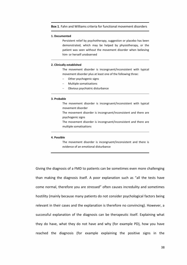
The most widely used criteria were developed by Fahn and Williams in 1989 (see
Box 1) (Fahn and Williams, 1988). FMD are divided into four categories of diagnostic
certainty: documented, clinically established, probable and possible. These criteria
37
were in fact first developed for functional dystonia alone, but were later expanded
to cover all FMD.
Gupta and Lang have suggested revisions to these criteria which delete the
“possible” category as being not sufficiently specific for FMD, and also seek to
introduce the concept of a laboratory supported level of certainty (Gupta and Lang,
2009). Shill and Gerber proposed alternative criteria, but these have been criticised
for relying too heavily on historical factors such as “disease modelling” without
reference to the movement disorder phenomenology (Shill and Gerber, 2006).
Recently, these criteria have been assessed with regard to inter-rater reliability, and
have been found to demonstrate moderate to poor reliability for the probable and
possible categories (Shill and Gerber, 2006). Therefore new criteria, which perhaps
include more specific direction as to the positive physical signs that predict FMD
rather than the unspecified “incongruency” with typical movement disorders, is
urgently needed to improve reliability.
38
Box 1. Fahn and Williams criteria for functional movement disorders
1. Documented
Persistent relief by psychotherapy, suggestion or placebo has been
demonstrated, which may be helped by physiotherapy, or the
patient was seen without the movement disorder when believing
him- or herself unobserved
2. Clinically established
The movement disorder is incongruent/inconsistent with typical
movement disorder plus at least one of the following three:
Other psychogenic signs
Multiple somatisations
Obvious psychiatric disturbance
3. Probable
The movement disorder is incongruent/inconsistent with typical
movement disorder
The movement disorder is incongruent/inconsistent and there are
psychogenic signs
The movement disorder is incongruent/inconsistent and there are
multiple somatisations
4. Possible
The movement disorder is incongruent/inconsistent and there is
evidence of an emotional disturbance
Giving the diagnosis of a FMD to patients can be sometimes even more challenging
than making the diagnosis itself. A poor explanation such as “all the tests have
come normal, therefore you are stressed” often causes incredulity and sometimes
hostility (mainly because many patients do not consider psychological factors being
relevant in their cases and the explanation is therefore no convincing). However, a
successful explanation of the diagnosis can be therapeutic itself. Explaining what
they do have, what they do not have and why (for example PD), how you have
reached the diagnosis (for example explaining the positive signs in the
39
examination), stating that the diagnosis is very common and that you believe them
(you do not think that it is all in their minds or they are putting the symptoms on)
are often of help (Stone et al., 2013). It is also important to explain that a potential
for reversibility does exist and that treating psychological issues (when relevant)
can also help to treat the condition.
1.5 Differential diagnosis
Many clinicians do not feel confident during the diagnostic process and the worry of
erroneously labelling a patient as functional is not uncommon. A systematic review
of studies of misdiagnosis however found that only 5% of patients had the wrong
diagnosis after an average of five years (Stone et al., 2005). This rate is similar to
those found for most neurological and psychiatric conditions. However, the
diagnosis is not always easy and it is usually prudent to ask a specialist to confirm
whether it is correct.
I have detailed general clues in the history and positive signs in the examination for
each type of FMD that may help to differentiate them from their “organic”
counterparts. Additionally, it have recently discussed general pitfalls in approaching
patients with functional symptoms (those that can lead erroneously to diagnosis
neurological disease as functional were called “mimics” and those that can lead to
diagnosis of functional symptoms in patients that have a typical neurological
disease were called “chamaleons”) (Stone et al., 2013). Putting too much emphasis
in the presence of psychiatric disorders and life events, failure to consider that
many patients may have an overlay of functional symptoms and typical neurological
diseases, normal imaging or the presence of “la belle indifference” can lead to
40
misdiagnose patients as functional. Among the chameleon, to rely on the opinion
that a patient is “nice, normal, or not stressed” to be functional as well as the fact
that symptoms can come after injury or a minor disease or that the patient is too
old can be misleading. Finally, assuming that normal neuroimaging exclude
neurological diseases or in contrast, assuming that all structural abnormalities are
relevant may also result in a misdiagnosis in both directions (Stone et al., 2013).
Because FMD resemble movement that are voluntarily produced, one can always
argue that patients are deliberately assuming symptoms in order to gain benefits. It
is generally acknowledged to be very difficult to distinguish malingering from “true”
FMD, but the consensus of opinion is that malingering is likely to be rare and is not
a satisfying explanation for the disorder in the majority of patients (van Beilen et
al., 2009, Hallett, 2010). Data arguing against the idea that malingering is the most
likely explanation for FMD comes from functional imaging studies in FT and fixed
dystonia (Voon et al., 2010, Schrag et al., 2013). Here, patterns of brain activation in
patients were different to those seen in subject feigning symptoms.
1.6 Treatment
There are no official guidelines for the treatment of FMD. However, an effective
communication of the diagnosis that allows patients to understand their symptoms
seems a good start. The benefit of simply explaining the diagnosis, at least in the
early stages, has been found to lead to long-term resolution of symptoms in overall
functional symptoms (Hall-Patch et al., 2010).
Referral of patients with FMD to physiotherapy services is common practice by
neurologists. However, in a recent survey most physiotherapists reported that
41
although they have interest for this group of patients, they have a low self-judged
knowledge about how to treat them. Preliminary evidence for regular low-medium
intensity walking exercise has been found in a single-blind study which assessed
patients with FMD after a 12 week program (Dallocchio et al., 2010). In a study of
60 patients from the Mayo clinic with functional motor symptoms, a 5 day inpatient
physical rehabilitation program produced benefits in over 60% of patients which
were sustained in most for over 2 years (Czarnecki et al., 2012). This encourages the
development of further studies to provide evidence for how physiotherapy services
could best be structured to design and deliver successful treatments to patients
with FMD.
Psychological intervention can be helpful in patients who consider psychological
factors as relevant in symptom development or maintenance. Indeed, a small study
provided preliminary evidence for a positive effect of antidepressant treatment in
those patients diagnosed with primary conversion disorder but not in those with
somatisation disorder (Voon and Lang, 2005). In patients with clear psychological
stressors but who are reluctant to try this strategy, explaining that cognitive
techniques are commonly used in medicine to help to control physical symptoms
(e.g. modern management of chronic pain) may encourage them to try this
approach. Recently, a community- based study of functional neurological symptoms
demonstrated that patients receiving cognitive behavioural therapy (CBT)-based
guided and usual care had more benefit than those who received usual care alone
(Sharpe et al., 2011).
42
The use of placebo as treatment strategy for FMD is still under debate. Because
prognosis and successful treatment of FMD may be highly dependent on the
patients’ belief that they will get better, some neurologists support the use of
placebo in this group of patients. Indeed, dramatic therapeutic benefits mediated
through placebo therapy have been described (Edwards et al., 2011). However, loss
of patient autonomy and the erosion of doctor-patient relationship are important
ethical concerns that should be taken into account. Recently, the need for clinical
trials to define optimal regimes for placebo therapy in these patients as well as for
health professional education in the use of placebos has been stressed
(Rommelfanger, 2013).
Additional treatments have been suggested to be effective in FMD but evidence is
poor. For example, intrathecal baclofen was reported to be effective in fixed
dystonia compare to placebo (van Hilten et al., 2000). However, placebo control
was only used for the initial test dose of intrathecal baclofen, and it is not known
whether there was systematic unblinding of the participant by systematic effects of
the baclofen. Low frequency repetitive Transcranial Magnetic Stimulation (TMS) has
been used as a therapeutical tool in FMD with some promising results (long-lasting
clinical improvement immediately after TMS session was seen in many patients)
(Dafotakis et al., 2011, Garcin et al., 2013). However, the unmasked nature of the
intervention in most of these studies makes placebo effect a likely explanation for
the results.
There is limited controlled trial data to guide treatment in FMD but the evidence
that is available suggests that a multidisciplinary approach gives the best chance of
43
benefit. A recent study that retrospectively evaluated a multidisciplinary inpatient
programme suggested this approach can provide long-lasting benefit for some
patients with treatment-refractory FMD, at least as measured by retrospective self-
report (Saifee et al., 2012). However, similar to other studies, most patients failed
to return to work and cessation of health-related financial benefits was
uncommonly seen despite of reporting clinical benefits.
1.7 Prognosis
Data on long-term prognosis are scarce, but most studies point to significant impact
in quality of life. For example, one study comparing patients with FMD and PD
patients on different measures of disability and quality of life showed that patients
with FMD reported levels of disability similar to those seen in PD (Anderson et al.,
2007). In a long-term follow up study, 90% of a group of 80 patients with a range of
FMD still had abnormal movements after a mean of 3.2 years since their initial
assessment (Feinstein et al., 2001). In other study, a third of patients were
employed at the time of follow up, while 11.5% were on disability and 1.3% were
involved in litigation (Thomas et al., 2006).
More optimistic studies of long-term outcome in FMD have showed that half of the
patients report an improvement in their symptoms at last follow up (3-5 years after
presentation)(Jankovic et al., 2006). Factors that predicted a favorable outcome
were a short duration of illness, patient's perception of effective treatment by the
physician and the presence of a comorbid psychiatric diagnosis of depression or
anxiety (which is therefore amenable to treatment) (Feinstein et al., 2001, Thomas
et al., 2006). Negative outcome at long-term follow-up is associated with long

44
standing symptoms (more than 6 months) (Factor et al., 1995), insidious onset of
movements and primary psychiatric disorder of hypochondriasis, factitious disorder
or malingering (Voon and Lang, 2005).
45
Chapter 2: The pathophysiology of functional
movement disorders – the historical view
An historical review of any illness is always important. It is a very useful instrument
to determine what happened in the past to an entity, and how previous
contributions made an impact on the modern concept of a particular illness. It also
often provides clues on future directions for research.
In this regard, functional symptoms are complex as there is a broad spectrum of
manifestation ranging from neurological symptoms such as sensory, motor,
memory or visual disturbances to non-neurological manifestation such as
gastrointestinal symptoms. Some authors have argued for a common theory
accounting for all the symptoms (Brown, 2004) whereas others have suggested a
specific mechanism for each one. The proneness of functional patients to develop
more than one type of symptoms over the time supports, in my opinion, the view
that there should be common underlying mechanisms.
In this chapter I will review different theories that have been proposed to explain
functional neurological symptoms. Here, I will use (in contrast with the rest of this
thesis), the word “hysteria” to be consistent with the nomenclature used in the
past. I have included information from ancient times (even though it is likely that
the term hysteria at that time was also employed to describe other entities
different from functional symptoms as defined in modern times). I have focused the
research mostly on the 19th century and the beginning of the 20th century, a period
in which hysteria was widely discussed and a theme of debate in the medical
literature. I have concentrated on reading some of the original work of three
46
important authors who showed a vivid scientific interest in hysteria: Jean-Martin
Charcot, Pierre Janet and Sigmund Freud. I present their main theories but I also
highlight the specific accounts of FMD when they reported them. By doing this, I do
not mean that these three authors are the only important ones. There are other
relevant thinkers, who are not mentioned in this thesis, whose contributions were
undoubtedly of value. My aim was, however, to read in depth and capture the most
illustrative thoughts from that time.
Janet once wrote that most theories have the inconvenience of being transitory, “of
disappearing soon after us, but it would be a singular illusion to seek to do
something eternal” (Janet, 1907). What follows demonstrates that in fact each
theory does have in itself something eternal, something that is still undoubtedly
influencing our current understanding of these common and disabling symptoms.
2.1 Ancient Egypt, Greece and Rome
It is thought that the first description of hysteria comes from the ancient Egyptians
(Kahun Papyrus, 1900 BC) (Tasca et al., 2012). They described them as being due to
spontaneous movements of the uterus in women’s body but symptoms had not yet
been given a specific term.
It was Hippocrates (5th century BC) who first used the term hysteria (Gilman, 1993).
He suggested that the causes of the symptoms were poisonous humours which,
due to an unsatisfactory sexual life, had never been expelled. He stated that
because a women’s body was naturally cold and wet, they were predisposed to
decomposition of the humours. As a prevention of the disease, the suggestion that
even widows and unmarried women should get married and live a satisfactory
47
sexual life was made. Once women had acquired the disease, they were advised to
treat themselves with acrid or fragrant fumigation of the face and genitals (Tasca et
al., 2012).
Although the theories on hysteria developed by one of the greatest physician of
ancient Rome, Claudius Galen (2nd century AD), were analogous to those of
Hippocrates, he was the first who emphasised the difficulties that just one single
organ such as the uterus, could cause several different symptoms. He wrote with
reference to Hippocrates: “Ancient physicians and philosophers have called this
disease hysteria from the name of the uterus, that organ given by nature to women
so that they might conceive. I have examined many hysterical women, some
stuporous, others with anxiety attacks [...]: the disease manifests itself with
different symptoms, but always refers to the uterus” (Tasca et al., 2012).
2.2 Middle Ages
The Roman Empire fell but Greco-Roman medical culture survived thanks to,
amongst others, the Persian Avicenna (980-1037) and the Andalusian Jew
Maimonides (1135-1204). The theories of Hippocrates and Galen were conserved
and hysterical symptoms were treated in a “scientific” way with the use of Melissa
as a natural remedy (Tasca et al., 2012).
From the 13th century onwards, the Inquisition played an important role in how
manifestations of illnesses, especially those due to psychiatric conditions, were
interpreted. If a physician was not able to identify the cause of a disease, likely to
occur with functional symptoms, it was thought to be due to the presence of a
48
demon. Therefore, “hysterical” women were commonly exorcised (Tasca et al.,
2012).
2.3 Modern Age
During the 16th and 17th century the basis of modern medical science were
established. The physician Thomas Willis (1621-1675) referred for the first time to
hysteria as being related to the brain and to the nervous system (Tasca et al., 2012).
While many of his contemporaries were looking for the causes of psychiatric
disorders in other organs, such as the uterus, lungs and spleen, he used to dissect
his own patients with the aim of relating the symptoms to brain pathology,
including patients with hysteria (Eadie, 2003, Molnar, 2004). It is during this period
that a door for a neurological explanation was opened and the suggestion that
perhaps hysteria was not a condition exclusive to females and instead, could affect
both sexes raised.
2.4 19th and 20th Centuries
In the 19th century, a burst of scientific interest to understand hysteria occurred and
France became the epicentre of this study. Prominent physicians developed
methods and treatments for hysterical symptoms which sometimes divided the
medical community.
2.4.1 Briquet
His Treatise on Hysteria, published in 1859, contains clinical and epidemiologic
details of 430 patients with hysteria seen over a decade. He described several
etiological factors such as “affective” temperament, family history, low social class,
49
sexual immorality or poor physical health (Mai and Merskey, 1981). Paul Briquet
regarded hysteria as a "Neurosis of the Brain" in which the causative agents can act
on the "affective part of the brain" in a susceptible and predisposed individual
(Hallett, 2006). In terms of treatment, Briquet emphasized the importance of an
improvement in social circumstances and the need to minimize environmental
problems. With Briquet the historic association of hysteria and disease of the uterus
was finally discredited (Mai and Merskey, 1981).
2.4.2 Charcot
Jean-Martin Charcot (1825-1893) was a pioneer of modern neurology during the 31
years of his working life. His contributions to medical knowledge were based on a
systematic use of physiology and pathology accompanied by a rigorous clinical
analysis. By the time of his death, the nosology of the main neurological diseases
had been carefully and methodologically classified. He tried to apply his
methodology to understand also hysteric symptoms and it was him the one who
treated, perhaps for the first time, hysteria as an issue worthy of serious study. The
Clinical Lectures on the Diseases of the Nervous System summarises all the lectures
given by Charcot between 1882 and 1885 in the lecture theatre of the Salpetriere
hospital in Paris (Charcot, 1889). Here, he focuses on the difficulties on diagnosing
and treating patients with hysteria among other neurological diseases. He gives a
detailed description of the phenomenology and care provided to patients admitted
on the ward suffering from several functional symptoms such as “hystero-epilepsy”,
“hysteric mutism”, “hysteric amyotrophic”, and “hysterical paralysis”. Several men
are described and this was used by Charcot to demonstrate to his students that
50
hysteria could occur in men, though he reported the proportion previously
suggested by Briquet of 1 man to 20 women was exaggerated (Charcot, 1889).
In these lectures, Charcot also presents cases of FMD. He describes hysterical tics
on the lower face in a 15 year-old girl and dedicates extensive work to what he
called “hysterical contractures of traumatic origin” highlighting the common
presence of physical injuries preceding the abnormal posture, a condition
nowadays called fixed dystonia.
In lecture III, he presented the case of a 34 year-old lady with a history of hystero-
epilepsy (she displayed in the past both epileptic seizures and non-epileptic
attacks). During a period in which non epileptic attacks had completely
disappeared, she had a work accident after tripping on the top of a staircase, falling
heavily on her left side. Charcot stressed the fact that the injury was mild but the
next morning she had a plantar flexion and inversion of the left foot, which in
Charcot’s words “attempts to passive movements were useless” (Charcot, 1889).
With this case, Charcot highlights several typical aspects of functional movement
disorders such as the sudden onset of the abnormal movements, that physical
triggers are common, that the abnormal movements reach their maximum “all in a
moment” and comments on the predisposition of functional patients to develop
new different symptoms over the time.
In lecture XXV, he also described a man, who after a physical injury and the
application of a splint in the arm developed a painful fixed posture with the arm in
flexion (Figure 2.1).
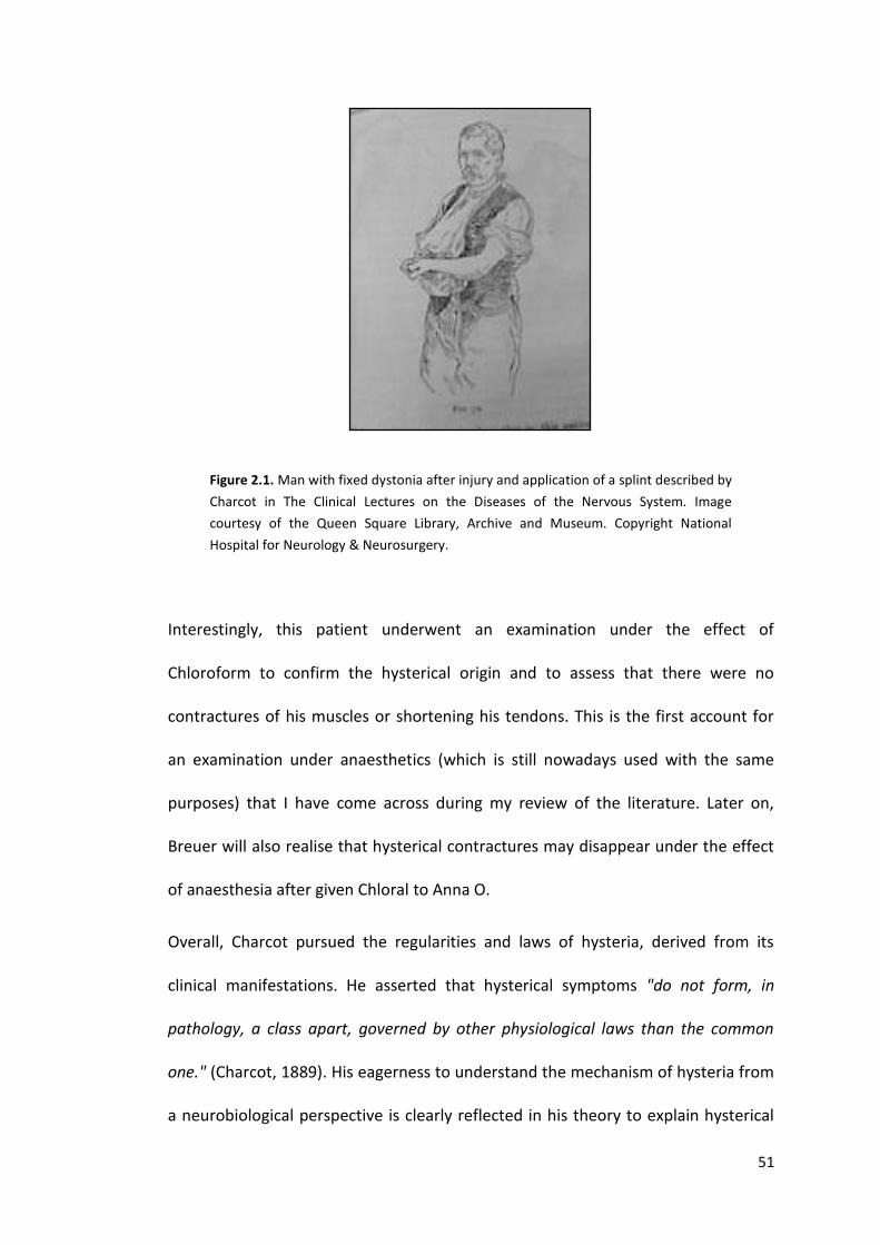
51
Figure 2.1. Man with fixed dystonia after injury and application of a splint described by
Charcot in The Clinical Lectures on the Diseases of the Nervous System. Image
courtesy of the Queen Square Library, Archive and Museum. Copyright National
Hospital for Neurology & Neurosurgery.
Interestingly, this patient underwent an examination under the effect of
Chloroform to confirm the hysterical origin and to assess that there were no
contractures of his muscles or shortening his tendons. This is the first account for
an examination under anaesthetics (which is still nowadays used with the same
purposes) that I have come across during my review of the literature. Later on,
Breuer will also realise that hysterical contractures may disappear under the effect
of anaesthesia after given Chloral to Anna O.
Overall, Charcot pursued the regularities and laws of hysteria, derived from its
clinical manifestations. He asserted that hysterical symptoms "do not form, in
pathology, a class apart, governed by other physiological laws than the common
one." (Charcot, 1889). His eagerness to understand the mechanism of hysteria from
a neurobiological perspective is clearly reflected in his theory to explain hysterical
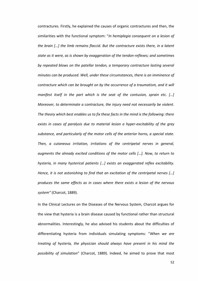
52
contractures. Firstly, he explained the causes of organic contractures and then, the
similarities with the functional symptom: “In hemiplegia consequent on a lesion of
the brain […] the limb remains flaccid. But the contracture exists there, in a latent
state as it were, as is shown by exaggeration of the tendon-reflexes; and sometimes
by repeated blows on the patellar tendon, a temporary contracture lasting several
minutes can be produced. Well, under these circumstances, there is an imminence of
contracture which can be brought on by the occurrence of a traumatism, and it will
manifest itself in the part which is the seat of the contusion, sprain etc. […]
Moreover, to determinate a contracture, the injury need not necessarily be violent.
The theory which best enables us to fix these facts in the mind is the following: there
exists in cases of paralysis due to material lesion a hyper-excitability of the grey
substance, and particularly of the motor cells of the anterior horns, a special state.
Then, a cutaneous irritation, irritations of the centripetal nerves in general,
augments the already excited conditions of the motor cells […]. Now, to return to
hysteria, in many hysterical patients […] exists an exaggerated reflex excitability.
Hence, it is not astonishing to find that an excitation of the centripetal nerves […]
produces the same effects as in cases where there exists a lesion of the nervous
system” (Charcot, 1889).
In the Clinical Lectures on the Diseases of the Nervous System, Charcot argues for
the view that hysteria is a brain disease caused by functional rather than structural
abnormalities. Interestingly, he also advised his students about the difficulties of
differentiating hysteria from individuals simulating symptoms: “When we are
treating of hysteria, the physician should always have present in his mind the
possibility of simulation” (Charcot, 1889). Indeed, he aimed to prove that most
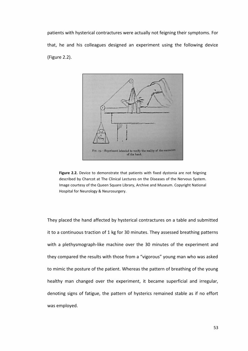
53
patients with hysterical contractures were actually not feigning their symptoms. For
that, he and his colleagues designed an experiment using the following device
(Figure 2.2).
Figure 2.2. Device to demonstrate that patients with fixed dystonia are not feigning
described by Charcot at The Clinical Lectures on the Diseases of the Nervous System.
Image courtesy of the Queen Square Library, Archive and Museum. Copyright National
Hospital for Neurology & Neurosurgery.
They placed the hand affected by hysterical contractures on a table and submitted
it to a continuous traction of 1 kg for 30 minutes. They assessed breathing patterns
with a plethysmograph-like machine over the 30 minutes of the experiment and
they compared the results with those from a “vigorous” young man who was asked
to mimic the posture of the patient. Whereas the pattern of breathing of the young
healthy man changed over the experiment, it became superficial and irregular,
denoting signs of fatigue, the pattern of hysterics remained stable as if no effort
was employed.
54
Nevertheless, the physiological view about mechanism of hysteria employed by
Charcot was not incompatible with more complicated psychological views about
the aetiology. In 1878 he introduced the technique of hypnotism into this research
and there was a shift towards acknowledging that psychological factors were also
relevant (Bogousslavsky, 2011).
2.4.3 Janet
Pierre Janet (1859-1947), perhaps due to the influence of his mentor Charcot, also
developed an avid interest for hysteria. In the Major Symptoms of Hysteria (1920)
(Janet, 1907), fifteen lectures given in the Medical School of Harvard University, he
summarises his thoughts about the underlying mechanism of functional symptoms.
Interestingly, his view about the personality of patients with hysteric symptoms
differed radically from the conception that most physicians have nowadays. He
described hysterics to be “easily managed, not dangerous, on whom we can
experiment without any great fear and who like to be observed” (Janet, 1907).
He was one of the first who emphasised the necessity of the early recognition of
the symptoms and the impact of communicating the diagnosis in the management.
In one of his lectures he advised the audience: “You must be able quickly to
recognise this disease, in order to foresee its evolution, to provide against its
dangers, and immediately to begin a rational treatment. This early diagnosis is
much more important still from another point of view: it will keep you from making
blunders. It is perhaps not very serious not to recognise a hysterical accident and not
to treat it, but what is always very serious is to mistake hysterical accident for
another one and to treat it for what is not” (Janet, 1907). These words can be
55
interpreted as an attempt to protect patients with functional symptoms from
unnecessary interventions, if the correct diagnosis is made.
Janet sought a balance between what he called the theories of the “clinical period”,
referring to Charcot and contemporaries from 19th century, who, in his opinion,
tried to give a medical character to hysteria; and what he called “psychological
period”, meaning by this his contemporaries, who described hysteria as a pure
mental phenomenon. He criticised Charcot: “[…] carried along by his habits as a
clinician, he has sought these general laws too much in the physiological domain,
which led him to a certain number of regrettable errors” (Janet, 1907) but he also
disagreed with a pure psychological view of hysteria: “A certain number of authors
have been seduced by the psychological explanation. It seemed to them that the
mere words “moral” and “thought” were enough to explain everything, and as
people generally like simple explanations, physicians are too disposed nowadays to
be content with a vaguely mental explanation. Hysteria, they say, is a psychic
disease, it is the disease of suggestion, taken in a vague sense […] There is some
truth in this view, for it brings into relief the psychic character of affection; but it is
quite insufficient. We should, in my opinion, retain something of the precise method
of Charcot, of the search after the determination and the laws of hysteria, and apply
it only to the psychological facts” (Janet, 1907).
In these lectures he meticulously described the most common FMD: tremor and
dystonia.
He stated that in FT tremor “the arm has regular little oscillations, of an average
rate of five to nine a second. These oscillations are nearly continual. There are some
56
subjects with whom they never stop, either when they rest or when they move;
there are some others with whom these tremors are intermittent, disappearing at
the time of voluntary activity and increasing at the time of diversion and rest. But it
is not possible to establish any rule, for you often observe the reverse in the form of
intentional trembling, analogous to that of disseminated sclerosis […] These tremors
occur under various conditions, sometimes gradually, after paralytic phenomena,
very often suddenly, after an emotion. One of the finest cases I have observed is that
of a workman, who, in consequence of the breaking of a scaffolding, remained
suspended at the height of a sixth floor. […] But in most cases, there is nothing
behind the tremor but a vague emotive state and a kind of transformation of the
motor function of the limb.” (Janet, 1907)
He described functional dystonia as “a state of moderate contraction of an
ensemble of muscles which maintains a limb in a determinate position and that in
an involuntary, unconscious, and indefinite manner. Such contractures can be
observed on absolutely all the muscles of the body, and in each region they raise
medical problems […] First we know that contractures are consequent, like all
hysteric phenomena, on thoughts and emotional phenomena. A shock has no action
in this direction except when it determines the great phenomena of imagination, I
will explain myself. An individual has his legs in a state of contraction because, he
says, a carriage ran over them. After verification, it is found that the carriage passed
besides him, and that he felt nothing at all. A real shock would do less than this
imaginary shock […] The contracture varies with certain psychological facts. If the
subject is very quiet, if nobody touches her contractured limb, and if she herself does
57
not try to make a voluntary movement, we may see than contracture decrease…”
(Janet, 1907).
Janet highlighted the genuine nature of the symptoms and differentiated them
from those that were feigned. He stated: “You will find that you are much more
awkward than a hysteric person, and that unless you have practiced specially to this
end, you cannot obtain the same regularity. Try to keep your arm in the position of a
hysteric contracture and describe the movement of the arm; you will remark that
you have not the same perseverance or courage as the patient. After a short time,
your arm trembles and is displaced, while the hysteric contracture has not changed.
If therefore we suppose there is a psychic action in these hysteric phenomena, it
must be acknowledged that this action is not identical with ours, but that is
performed in other conditions. Here is my hypothesis; […] “…Action by becoming
unconscious in hysterics, by separating from consciousness, loses something of its
dignity, retrograde in a manner and assumes an appearance that recalls the action
of visceral muscles, the action of the lower animals, and the movements of fatigued
muscles, as if the activity of the sarcoplasm prevailed over that of the fibrils. This is
what in my opinion gives to the subconscious action of the hysteric those abnormal
characteristics we saw in tremors and contractures” (Janet, 1907).
Overall, Janet summarized the peculiar mental state of hysterical by the words
“retraction of the field of consciousness”. Janet was one of the originators of the
idea of dissociation. He proposed that in the physiological state enormous amounts
of sensory information from all the points of our bodies constantly emerge.
However, a person never perceives them all. The number of elementary sensations
58
that can simultaneously reach consciousness depends on the extent of the field of
consciousness and this may differ very much within individuals and depends on
their state of mind (attention). The sensations which do not reach consciousness
must remain in the sub-consciousness, and he calls this “normal absent-
mindedness”. In the case of hysterics, the field of consciousness is so contracted
that the patient reserves the small share of perception for the sensation which is
considerate the most important one. For instance, if patients can perceive just two
stimuli at the same time and vision and auditory stimuli are prioritised, when
someone pinches the left arm of the patient, he cannot feel it consciously: it has
become anaesthetic. He considered that this restriction of the field of
consciousness was secondary to their fundamental mental state, which was
characterised by a “special moral weakness, consisting in the lack of power, on the
part of the feeble subject, to gather, to condense his physiological phenomena and
assimilate them to his personality” (Janet, 1907).
Janet’s theory accounted essentially for functional anaesthesia and he found it
more difficult to explain other functional symptoms.
Janet also emphasised the importance of physical events preceding the onset of
functional symptoms. He believed that the restriction of the field of consciousness
usually affected a function that for some reason or other had become weak. He
gave examples of cases such as that of a girl with functional paralysis in her right
leg, and he believed that the reason was that in her childhood the right leg was
affected with rachitis or the case of another girl with paralysis of the leg, which was
thought to be due to the fact that in her childhood the leg was affected by a tumour
59
and was kept wrapped in a bandage (Janet, 1907). He also brought to attention
what nowadays is called “functional overlay” by saying that “in certain cases,
hysteria makes conspicuous some light symptoms of organic disease of the nervous
system quite at the beginning by exaggerating them beyond all measures” (Janet,
1907).
2.4.4 Freud
In popular culture hysteria begins and ends with Sigmund Freud (1856-1939).
However, as previously reviewed, there is a tradition of study of hysterical
symptoms for more than three thousand year history before his work. Despite
being a pupil of Charcot in Paris, Freud rejected any account for hysteria but the
pure psychological one. This strong theoretical position is well reflected in Studies
on Hysteria (1893-1895) (Breuer and Freud, 1974), a book written with his mentor
in Vienna: Joseph Breuer. Here, they wrote “in what follows, little mention will be
made of the brain and none whatever of molecules. Physical processes will be dealt
with in the language of psychology; and indeed it cannot possibly be otherwise”.
The purpose of their work was to elucidate the precipitating cause of hysteria, “the
event which provided the first occurrence after many years earlier of the
phenomena in question”. They acknowledged the difficulties that go with it as “the
patient sometimes dislikes discussing, and mainly because they are genuinely
unable to recollect it and has no suspicion of the causal connection between the
precipitating event and the pathological phenomenon.”
It was clear to me on reading this text that they recognized physical precipitating
factors close to the onset of the functional symptoms in several clinical cases
60
through the book. For instance, in the famous case of Anna O they wrote “a girl,
watching beside a sick-bed in a torment of anxiety, fell into a twilight state and had
a terrifying hallucination, while her right arm, which was over the back of her chair
went to sleep; from this, she developed a paresis of the same arm accompanied by
contracture and anaesthesia” (Breuer and Freud, 1974). It is possible that in first
place, Anna had numbness and a degree of paresis secondary to nerve compression
caused by placing her arm on a hard surface (the back of a chair) for a number of
hours. Or the case of Miss Lucy R, a girl who suffered from suppurative rhinitis and
necrosis of the ethmoid bone and developed later hysterical symptoms in the form
of subjective olfactory sensations (Breuer and Freud, 1974). By observations like
these, they proposed that the concept of traumatic neurosis from Charcot should
be extended to traumatic hysteria. Here, it is not the physical injury but the affect
of fright that commonly accompanies the injury, the psychical trauma, which is
considered the cause of the illness. They neglected the importance of the physical
event itself by saying that their investigations reveals “for many, if not for most
hysterical symptoms, precipitating causes [can be identified] which can only
described as psychical traumas.” (Breuer and Freud, 1974)
When it was not possible to establish the point of origin of the illness, they
hypnotized the patient “As a rule it is necessary to hypnotize the patient and to
arouse his memories under hypnosis of the time at which the symptom made its first
appearance” (Breuer and Freud, 1974) and when patients denied any precipitant or
the one interpreted by them, they attempted to persuade the patients to believe
the opposite.
61
In the theoretical part of the Studies on hysteria, they summarize their views after
the observation of several hysterical patients. They use the analogy of an electrical
system through which there is a constant flow of current to explain what may
happen in the brain. In physiological conditions, they argued that the brain tries to
keep in a constant excitation “intracerebral tonic excitation” at that level which is
optimal to be “accessible to all external stimuli, the reflexes are facilitated, though
only to the extent of normal reflex activity, and the store of ideas is capable of being
aroused and open to association in the mutual relation between individual ideas
which corresponds to a clear and reasonable state of mind” (Breuer and Freud,
1974). Healthy people can get rid of any increased cerebral excitation associated to
emotional situations by using motor discharge or secretions (for instance shouting
and jumping for joy or sobs and tears when sadness). If these reactions are
suppressed, the affect associated with emotional situations is not “abreacted” and
may remain attached to the memory (Breuer and Freud, 1974).
Coming back to the analogy with an electrical system, they argue that there must
be resistances through the system, which prevent the general distribution of
excitation (mainly in organs which are of vital importance “the circulatory and
digestive organs are separated by strong resistances from the organs of ideation”).
The strength of these resistances varies from one person to another and they
suggested that this is the explanation why some people have a strong vegetative
reaction with low levels of stress and others do not (Breuer and Freud, 1974).
In the case of hysterics, Breuer and Freud argued that they have an innate
idiosyncrasy which is characterised by excess of energy. They understood hysterics
62
as lively people, full of intellectual interests before they fall ill. They thought that it
was uncommon “to find in them simply, dull intellectual inertia”. During puberty the
original excess is increased by additional excitation which arises with the
emergence of sexuality. After puberty, they have enormous quantity of free
nervous energy available for the production of pathological phenomena. But this
predisposition to hysteria does not lie down exclusively on excessive intracerebral
excitation. Hysterics were also thought to have abnormal weakness of the
resistances. These weak resistances were suggested to be determined by the
individual's initial constitution, by the long-term excitation itself or by weakening
factors, such as the presence of a previous illness of the organ concerned, which
may facilitate the paths to and from the brain (Breuer and Freud, 1974).
They suggested that in hysteria, there are difficulties to abreact traumatic
experiences and memories of these experiences are repressed from their
consciousness. These memories, which have a strong affect attached to them,
increase the excitation of the brain and this energy is “converted” into a somatic
phenomenon. This discharge “follows ‘the principle of least resistance’ and takes
place along those paths whose resistances have already been weakened by
concurrent circumstances” (Breuer and Freud, 1974). For instance, in the case of
Miss Lucy R, hysteric symptoms were subjective olfactory sensations and she had
recently suffered from suppurative rhinitis that might have acted as a weakening
factor.
Their therapeutic approach was the use of light hypnosis and suggestion to bring
back to consciousness the memory of the traumatic event and arousing its

63
accompanying affect. They assured readers that when patients had described the
event in the greatest possible detail and had put the affect into words, the
symptom immediately and permanently disappeared.
64
Chapter 3: Aims and hypotheses
The background above provides, I hope, a picture of how theories regarding the
pathophysiology of functional symptoms have largely focussed on causation (and
here have focussed on causation by a psychological trauma), and less on the
mechanism by which this event becomes translated into the type of physical
symptom experienced by the patient. In other words there has been a focus on
“why” rather than “how” symptoms develop. While I believe both questions are
very important, my sense at the beginning of the work was that the question of
“how” symptoms are produced might be more tractable and relevant. The other
key factor that helped in the formulation of the hypotheses I pursued was the
patient population that I had access to. These patients had disorders of movement,
providing a great opportunity to study symptoms where there are observable
clinical signs (in comparison to sensory loss or pain for example). The clinical
methods for making a diagnosis in such patients already provided key information
on the likely underlying mechanism, especially with regard to the importance of
attention. I therefore took as my starting point the manner in which the diagnosis
of FMD is made in neurological practice. The diagnosis can be made confidently on
the basis of positive physical signs, helped by certain historical features. This, to me,
was the most suitable starting point as it removed aetiological speculation,
particularly about the role of psychological trauma in causation of functional
symptoms, and instead focused on a more mechanistic level. This provided a better
fit with investigation of other illnesses where the main tractable areas of study (at
least initially) relate to how symptoms are produced, rather than why.
65
In addition to a clear role for attention in the generation of functional motor
symptoms being apparent from a clinical point of view, two other potentially
important mechanistic issues stood out for me. One was that some symptoms (for
example tubular visual fields) suggested a key role for beliefs about the way the
nervous system might malfunction due to illness which we know are contradicted
by basic anatomy and physiology. The other issue was that there must be an
abnormality in sense of agency for movement if movement that appears to be
voluntarily generated is not experienced as such. Finally, I was also interested in
understanding better the circumstances surrounding the onset of the functional
symptoms.
I looked for paradigms that might provide ways of exploring these three areas of
interest, and in this I was influenced in part by paradigms that had been previously
used in patients with delusional beliefs. It was against this background that I
developed the following hypotheses:
1. That physical precipitating factors will be common in patients with FMD, as
our clinical experience suggested.
2. That patients with FMD will display a reasoning style different to healthy
controls that may predispose them to accept new hypothesis on the basis of
less evidence.
3. That patients with FMD will have impaired motor control under
circumstances in which there is an opportunity for attention to be directed
towards movement production.

66
4. That patients with FMD will display abnormalities in paradigms designed to
assess the sense of agency for movement.
67
Chapter 4: A study on the physical precipitating factors
in functional movement disorders
The work presented in this chapter was originally published in the form of a research article:
Pareés I, Kojovic M, Pires I, Rubio-Agusti I, Saifee TA, Sadnicka A, Kassavetis P, Macerollo A,
Bhatia KP, Carson A, Stone J, Edwards MJ. Physical precipitating factors in functional
movement disorders. Journal of Neurological Sciences. 2014 Mar 15; 338(1-2):174-7.
4.1 Introduction
We have seen that typically, functional neurological symptoms, including FMD,
have been explained as resulting from psychological stressors which lead to
unconsciously produced physical symptoms. In keeping with this formulation,
several authors have found higher rates of childhood trauma in patients with
functional symptoms (Alper et al., 1993, Bowman and Markand, 1996, Roelofs et
al., 2002) or have highlighted the aetiological importance of emotional stress or
recent life events (Binzer et al., 1997, American Psychiatry Association, 2000, Irfan
and Badar, 2002). Indeed, it was not possible to make a diagnosis of conversion
disorder according to the Diagnostic and Statistical Manual of Mental Disorders
(DSM) IV criteria without the presence of a psychological stressor that precedes the
onset of physical symptoms (American Psychiatry Association, 2000).
My impression from my involvement in a specialised clinic for FMD was that many
patients did not report psychological stressors prior to the onset of symptoms.
Supporting this view, a recent study found few differences in self-reported recent
life events or past experience of sexual or physical abuse in patients with FMD,
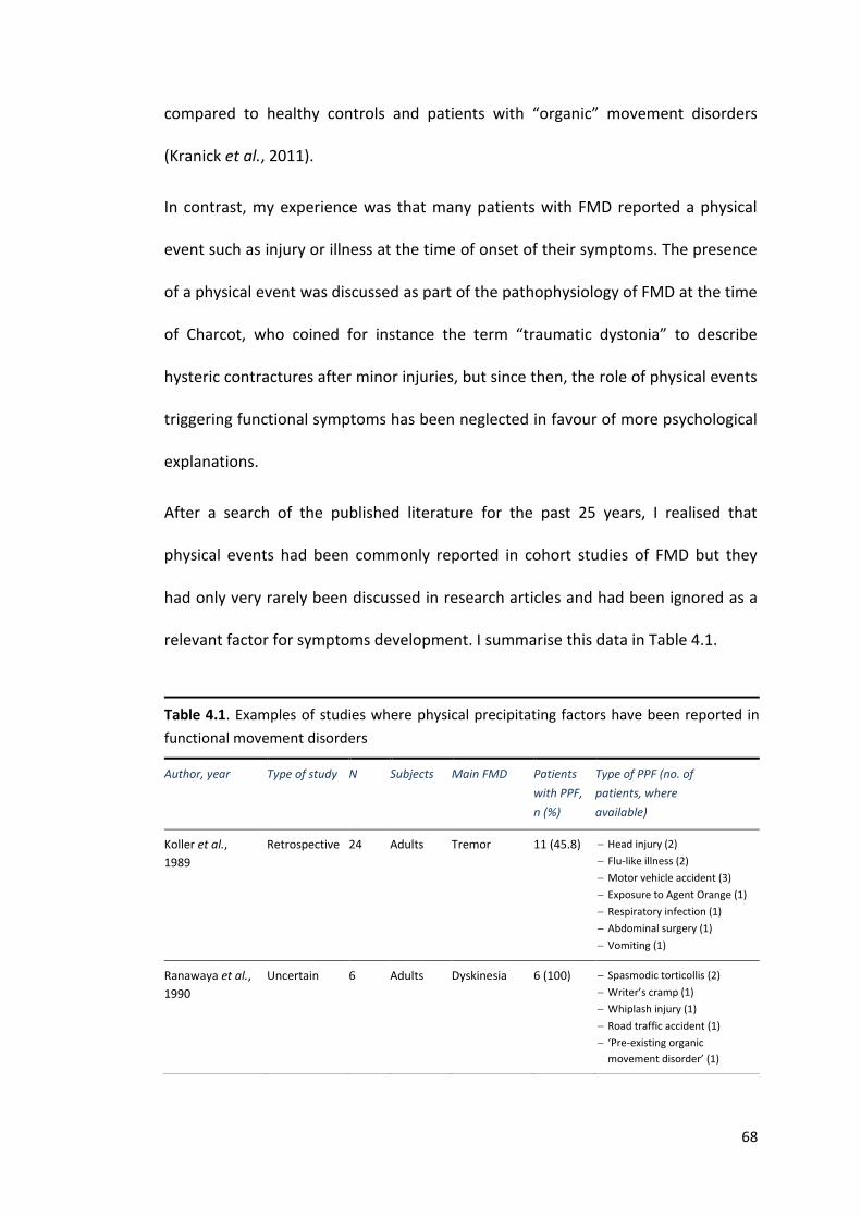
68
compared to healthy controls and patients with “organic” movement disorders
(Kranick et al., 2011).
In contrast, my experience was that many patients with FMD reported a physical
event such as injury or illness at the time of onset of their symptoms. The presence
of a physical event was discussed as part of the pathophysiology of FMD at the time
of Charcot, who coined for instance the term “traumatic dystonia” to describe
hysteric contractures after minor injuries, but since then, the role of physical events
triggering functional symptoms has been neglected in favour of more psychological
explanations.
After a search of the published literature for the past 25 years, I realised that
physical events had been commonly reported in cohort studies of FMD but they
had only very rarely been discussed in research articles and had been ignored as a
relevant factor for symptoms development. I summarise this data in Table 4.1.
Table 4.1. Examples of studies where physical precipitating factors have been reported in
functional movement disorders
Author, year Type of study N Subjects Main FMD Patients
with PPF,
n (%)
Type of PPF (no. of
patients, where
available)
Koller et al.,
1989
Retrospective 24 Adults Tremor 11 (45.8) Head injury (2)
Flu-like illness (2)
Motor vehicle accident (3)
Exposure to Agent Orange (1)
Respiratory infection (1)
Abdominal surgery (1)
Vomiting (1)
Ranawaya et al.,
1990
Uncertain 6 Adults Dyskinesia 6 (100) Spasmodic torticollis (2)
Writer’s cramp (1)
Whiplash injury (1)
Road traffic accident (1)
‘Pre-existing organic
movement disorder’ (1)
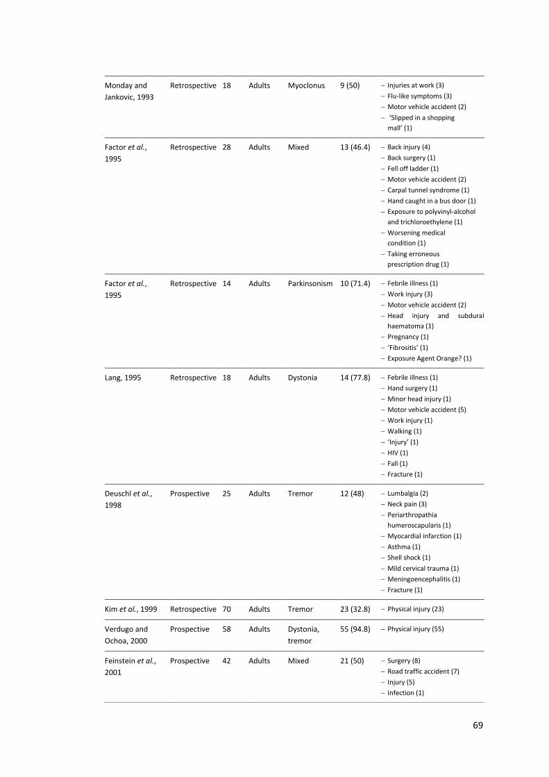
69
Monday and
Jankovic, 1993
Retrospective 18 Adults Myoclonus 9 (50) Injuries at work (3)
Flu-like symptoms (3)
Motor vehicle accident (2)
‘Slipped in a shopping
mall’ (1)
Factor et al.,
1995
Retrospective 28 Adults Mixed 13 (46.4) Back injury (4)
Back surgery (1)
Fell off ladder (1)
Motor vehicle accident (2)
Carpal tunnel syndrome (1)
Hand caught in a bus door (1)
Exposure to polyvinyl-alcohol
and trichloroethylene (1)
Worsening medical
condition (1)
Taking erroneous
prescription drug (1)
Factor et al.,
1995
Retrospective 14 Adults Parkinsonism 10 (71.4) Febrile illness (1)
Work injury (3)
Motor vehicle accident (2)
Head injury and subdural
haematoma (1)
Pregnancy (1)
‘Fibrositis’ (1)
Exposure Agent Orange? (1)
Lang, 1995 Retrospective 18 Adults Dystonia 14 (77.8) Febrile illness (1)
Hand surgery (1)
Minor head injury (1)
Motor vehicle accident (5)
Work injury (1)
Walking (1)
‘Injury’ (1)
HIV (1)
Fall (1)
Fracture (1)
Deuschl et al.,
1998
Prospective 25 Adults Tremor 12 (48) Lumbalgia (2)
Neck pain (3)
Periarthropathia
humeroscapularis (1)
Myocardial infarction (1)
Asthma (1)
Shell shock (1)
Mild cervical trauma (1)
Meningoencephalitis (1)
Fracture (1)
Kim et al., 1999 Retrospective 70 Adults Tremor 23 (32.8) Physical injury (23)
Verdugo and
Ochoa, 2000
Prospective 58 Adults Dystonia,
tremor
55 (94.8) Physical injury (55)
Feinstein et al.,
2001
Prospective 42 Adults Mixed 21 (50) Surgery (8)
Road traffic accident (7)
Injury (5)
Infection (1)
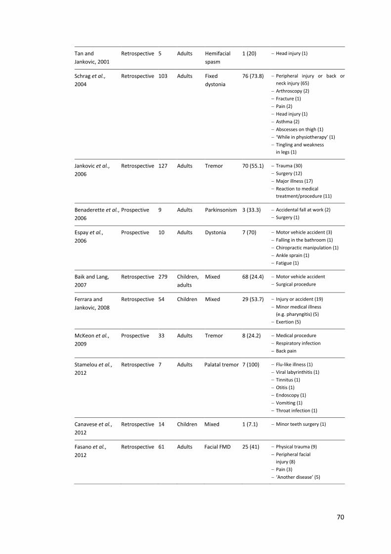
70
Tan and
Jankovic, 2001
Retrospective 5 Adults Hemifacial
spasm
1 (20) Head injury (1)
Schrag et al.,
2004
Retrospective 103 Adults Fixed
dystonia
76 (73.8) Peripheral injury or back or
neck injury (65)
Arthroscopy (2)
Fracture (1)
Pain (2)
Head injury (1)
Asthma (2)
Abscesses on thigh (1)
‘While in physiotherapy’ (1)
Tingling and weakness
in legs (1)
Jankovic et al.,
2006
Retrospective 127 Adults Tremor 70 (55.1) Trauma (30)
Surgery (12)
Major illness (17)
Reaction to medical
treatment/procedure (11)
Benaderette et al.,
2006
Prospective 9 Adults Parkinsonism 3 (33.3) Accidental fall at work (2)
Surgery (1)
Espay et al.,
2006
Prospective 10 Adults Dystonia 7 (70) Motor vehicle accident (3)
Falling in the bathroom (1)
Chiropractic manipulation (1)
Ankle sprain (1)
Fatigue (1)
Baik and Lang,
2007
Retrospective 279 Children,
adults
Mixed 68 (24.4) Motor vehicle accident
Surgical procedure
Ferrara and
Jankovic, 2008
Retrospective 54 Children Mixed 29 (53.7) Injury or accident (19)
Minor medical illness
(e.g. pharyngitis) (5)
Exertion (5)
McKeon et al.,
2009
Prospective 33 Adults Tremor 8 (24.2) Medical procedure
Respiratory infection
Back pain
Stamelou et al.,
2012
Retrospective 7 Adults Palatal tremor 7 (100) Flu-like illness (1)
Viral labyrinthitis (1)
Tinnitus (1)
Otitis (1)
Endoscopy (1)
Vomiting (1)
Throat infection (1)
Canavese et al.,
2012
Retrospective 14 Children Mixed 1 (7.1) Minor teeth surgery (1)
Fasano et al.,
2012
Retrospective 61 Adults Facial FMD 25 (41) Physical trauma (9)
Peripheral facial
injury (8)
Pain (3)
‘Another disease’ (5)
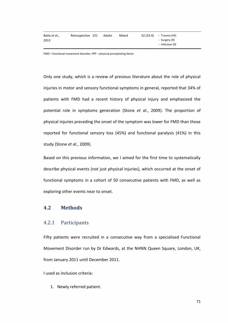
71
Only one study, which is a review of previous literature about the role of physical
injuries in motor and sensory functional symptoms in general, reported that 34% of
patients with FMD had a recent history of physical injury and emphasized the
potential role in symptoms generation (Stone et al., 2009). The proportion of
physical injuries preceding the onset of the symptom was lower for FMD than those
reported for functional sensory loss (45%) and functional paralysis (41%) in this
study (Stone et al., 2009).
Based on this previous information, we I aimed for the first time to systematically
describe physical events (not just physical injuries), which occurred at the onset of
functional symptoms in a cohort of 50 consecutive patients with FMD, as well as
exploring other events near to onset.
4.2 Methods
4.2.1 Participants
Fifty patients were recruited in a consecutive way from a specialised Functional
Movement Disorder run by Dr Edwards, at the NHNN Queen Square, London, UK,
from January 2011 until December 2011.
I used as inclusion criteria:
1. Newly referred patient.
Batla et al.,
2013
Retrospective 151 Adults Mixed 52 (33.4) Trauma (34)
Surgery (9)
Infection (9)
FMD = functional movement disorder; PPF = physical precipitating factor
72
2. Age over 18 years.
3. Diagnosis of clinically established or documented functional movement
disorders according to Fahn and Williams criteria (Fahn and Williams, 1988).
I used as exclusion criteria:
1. Age less than 18.
2. Unable to communicate with researcher (e.g. does not speak English).
4.2.2 Semi-structured interviews
We used face to face semi-structured interviews to characterise the circumstances
which surrounded the onset of FMD. We used this type of interview because,
although it was more time consuming than a structured interview and harder to
analyse, it is more flexible and allowed us to collect more detailed data. The
interviews were carried out by Dr Edwards and myself. I designed the data
collection sheet in which information on sex, age, marital status, presenting
symptoms, work status, presence of a disease model either at work or among
family and friends, receipt of financial benefits and the presence of litigation were
recorded. The interview also provided a retrospective account of the tempo of
onset, associated symptoms and circumstances prior to onset of the FMD, which
were also recorded.
There is no consensus about the timing between a precipitating event and the
onset of a FMD to be sure that there is a good chance that there is a causal
relationship. Some authors have considered one year between a physical injury
and the development for instance of functional dystonia. Others consider this gap
excessive and have reported that most triggers occur within the first few months.
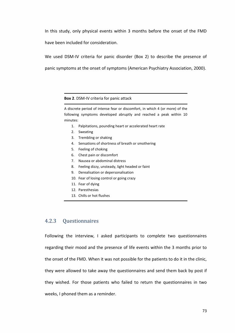
73
In this study, only physical events within 3 months before the onset of the FMD
have been included for consideration.
We used DSM-IV criteria for panic disorder (Box 2) to describe the presence of
panic symptoms at the onset of symptoms (American Psychiatry Association, 2000).
Box 2. DSM-IV criteria for panic attack
A discrete period of intense fear or discomfort, in which 4 (or more) of the
following symptoms developed abruptly and reached a peak within 10
minutes:
1. Palpitations, pounding heart or accelerated heart rate
2. Sweating
3. Trembling or shaking
4. Sensations of shortness of breath or smothering
5. Feeling of choking
6. Chest pain or discomfort
7. Nausea or abdominal distress
8. Feeling dizzy, unsteady, light headed or faint
9. Derealisation or depersonalisation
10. Fear of losing control or going crazy
11. Fear of dying
12. Paresthesias
13. Chills or hot flushes
4.2.3 Questionnaires
Following the interview, I asked participants to complete two questionnaires
regarding their mood and the presence of life events within the 3 months prior to
the onset of the FMD. When it was not possible for the patients to do it in the clinic,
they were allowed to take away the questionnaires and send them back by post if
they wished. For those patients who failed to return the questionnaires in two
weeks, I phoned them as a reminder.
74
4.2.3.1 Mood
We used The Hospital Anxiety and Depression rating scale (HADS) with reference to
their mood the week prior to testing (Zigmond and Snaith, 1983). There are 7 items
for anxiety and 7 for depression. Each item is scored on a 0-3 scale. Therefore, the
potential range of scores is 0-21 for both anxiety and depression scales, with higher
scores indicating greater emotional disturbance. A cut off level of 9 or more has
been found to have similar sensitivity and specificity in cancer patients. We used
HADS because it is brief and simple to administer and although it was originally
designed to be used with hospital populations, it has been found to perform well
with non-hospital groups.
4.2.3.2 Life events
We used the 82 items Life Events Questionnaire (LEQ) (Norbeck, 1984). This is a
self-report questionnaire addressing life events in the categories of health, work,
school, residence, love and marriage, family and close friends, parenting, personal
or social, financial and crime or legal matters. Patients are asked to indicate
whether each event is considered “good” or “bad”; and rate the impact of the
event on a 4-point scale (0-3). We used the negative events score (the sum of the
impact ratings for all items designated as "bad" by the patient: range from 0 (no
impact) to a maximum of 246). We chose this 82 items questionnaire because it
covers a broad range of life event and includes questions that increase the
relevance for adult female respondents of childbearing age, a group which was
predicted to be common in our cohort.

75
4.3 Results
We recruited 11 males and 39 females. Demographic and clinical characteristics are
shown in Table 4.2.
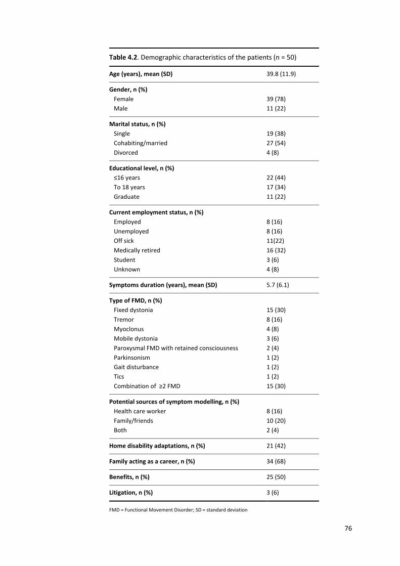
76
Table 4.2. Demographic characteristics of the patients (n = 50)
Age (years), mean (SD) 39.8 (11.9)
Gender, n (%)
Female
Male
39 (78)
11 (22)
Marital status, n (%)
Single
Cohabiting/married
Divorced
19 (38)
27 (54)
4 (8)
Educational level, n (%)
≤16 years
To 18 years
Graduate
22 (44)
17 (34)
11 (22)
Current employment status, n (%)
Employed
Unemployed
Off sick
Medically retired
Student
Unknown
8 (16)
8 (16)
11(22)
16 (32)
3 (6)
4 (8)
Symptoms duration (years), mean (SD) 5.7 (6.1)
Type of FMD, n (%)
Fixed dystonia
Tremor
Myoclonus
Mobile dystonia
Paroxysmal FMD with retained consciousness
Parkinsonism
Gait disturbance
Tics
Combination of ≥2 FMD
15 (30)
8 (16)
4 (8)
3 (6)
2 (4)
1 (2)
1 (2)
1 (2)
15 (30)
Potential sources of symptom modelling, n (%)
Health care worker
Family/friends
Both
8 (16)
10 (20)
2 (4)
Home disability adaptations, n (%) 21 (42)
Family acting as a career, n (%) 34 (68)
Benefits, n (%) 25 (50)
Litigation, n (%) 3 (6)
FMD = Functional Movement Disorder; SD = standard deviation
77
4.3.1 Tempo of onset
Twenty seven patients (54%) reported a sudden onset of symptoms (seconds-
minutes). Eighteen patients (36%) developed symptoms in hours - 1 day and only 5
patients reported a gradual onset (more than 1 day to maximal symptoms).
4.3.2 Physical precipitating factors
From a total of 50 patients, 40 (80%) patients reported a physical event within the
three months prior to the onset of the FMD. Three patients did report a physical
event which was related to the functional symptom (injuries in the same limb
where the FMD appeared) but these occurred before the 3 months period that we
set as inclusion criteria. One patient did not remember the exact timing between
the physical event and the onset of the symptoms.
Time from physical event to onset of FMD in those 41 patients was minutes in 8
(16%) patients, approximately one day in 6 (12%) patients, two days in 4 (8%)
patients, within the first week in 7 (14%) patients, one month in 9 (18%) patients
and within the 3 months prior to onset in 6 (13%) patients.
The FMD occurred after an injury in 11 (22%) patients. The injuries were mainly of
soft tissues, but some patients experienced more serious injury leading to fracture.
In 9 (18%) patients, FMD first started after an infection, most commonly a flu-like
illness. In another 8 (16%) patients functional symptoms appeared following a
neurological disorder (severe episode of migraine (n=3), brachial neuritis (n=1),
Bell’s palsy (n=1), carpal tunnel syndrome (n=1), restless legs syndrome (n=1) and
after a pituitary haemorrhage (n=1)). In 4 (8%) patients, pain appeared to be an
important factor at onset (either an episode of acute pain even though there was
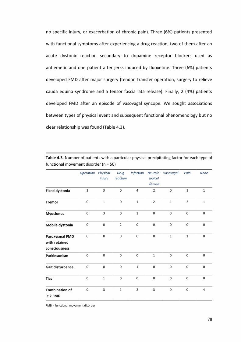
78
no specific injury, or exacerbation of chronic pain). Three (6%) patients presented
with functional symptoms after experiencing a drug reaction, two of them after an
acute dystonic reaction secondary to dopamine receptor blockers used as
antiemetic and one patient after jerks induced by fluoxetine. Three (6%) patients
developed FMD after major surgery (tendon transfer operation, surgery to relieve
cauda equina syndrome and a tensor fascia lata release). Finally, 2 (4%) patients
developed FMD after an episode of vasovagal syncope. We sought associations
between types of physical event and subsequent functional phenomenology but no
clear relationship was found (Table 4.3).
Table 4.3. Number of patients with a particular physical precipitating factor for each type of
functional movement disorder (n = 50)
Operation Physical
injury
Drug
reaction
Infection Neurolo-
logical
disease
Vasovagal Pain None
Fixed dystonia 3 3 0 4 2 0 1 1
Tremor 0 1 0 1 2 1 2 1
Myoclonus 0 3 0 1 0 0 0 0
Mobile dystonia 0 0 2 0 0 0 0 0
Paroxysmal FMD
with retained
consciousness
0 0 0 0 0 1 1 0
Parkinsonism 0 0 0 0 1 0 0 0
Gait disturbance 0 0 0 1 0 0 0 0
Tics 0 1 0 0 0 0 0 0
Combination of
≥ 2 FMD
0 3 1 2 3 0 0 4
FMD = functional movement disorder
79
4.3.3 Case examples
4.3.3.1 Case 1
This 34-year-old gentleman was fit and well until February 2010 when he was at
work and started to feel sick and light headed. He was diagnosed with a "sickness
bug" and was prescribed prochlorperazine to be taken four times a day. By the
following morning when he had taken four doses or so of the medication he noticed
the beginning of abnormal movements. This started with twitching and tightening
of the left arm so that it moved out at the shoulder. He continued to take the
tablets, but the movement problems worsened and progressed to his legs stiffening
and moving involuntarily as well as jerking of his head backwards. He remembers
his throat and face feeling tight and he developed involuntary tongue protrusion.
He went to the minor injury unit on the following morning but he was told to seek
advice from his GP again in a few days. No advice was given to stop medication. He
continued with the drugs and movement problems worsened. When he saw his GP,
he was told he had had a medication reaction and was advised to stop taking the
drug which he did right away.
His movements persisted and he again attended A &E where blood tests were
taken. He recalled being asked whether he had taken any illicit drugs and he was
given diazepam which decreased the severity of some of his movements.
About four weeks after the onset of symptoms he had the first episode of what
later became regular attacks of abnormal movements. He developed spasms
around the face and arms and severe gait disturbance. By this time, the original
movement disorder had almost completely resolved. Attacks could last from 10
80
minutes to 2 hours. He retained consciousness throughout these attacks. He was
admitted and various investigations were performed including imaging and an EEG
and no abnormality was found. He was discharged still on treatment with
Clonazepam. He continued to have attacks on most days. A few months later, he
had his worst ever attack which occurred at work. He became unable to speak and
also had a period of time lasting a couple of weeks where he was virtually unable to
walk. During this time, he received psychological input but he did not feel that the
emphasis on potential psychological triggering factors were of much relevance to
him. Our formulation was that he had experienced some extrapyramidal side
effects from prochlorperazine which had settled within days of the initial reaction
but which had triggered a functional movement disorder'.
4.3.3.2 Case 2
This 59 year old lady who previously worked as a child-minder attended our Clinic.
Ten years previously she had painful bunions on both feet and had an operation to
help with this. Post operatively both feet became infected and for unclear reasons
both legs below the knees were put in plaster for about 7 weeks. When the plaster
was taken off she had a problem with going over on her ankles when she was
walking. Because of this, she underwent a bilateral tendon transfer operation. The
right leg improved, but on the left immediately post-operatively she developed a
fixed abnormal posture of the foot which on examination had typical features of
functional fixed dystonia.
81
4.3.4 Panic symptoms
Most patients reported physical symptoms of panic at the onset of the FMD, which
were concurrent with the functional symptoms in the majority of them. Eighteen
(38%) fulfilled criteria for panic attack. Of those, 10 (55%) had an onset of the
functional symptoms in seconds/minutes and 7 (39%) had an onset in hours/day.
4.3.5 HADS
Twenty nine (58%) patients completed the HADS (Zigmond and Snaith, 1983). Mean
score for depression was 11.8 ±4.6 and mean score for anxiety was 6.7 ±3.8.
4.3.6 Life events
Twenty eight (56%) patients filled the in LEQ (Norbeck, 1984). The mean negative
score for life events within three months before symptom onset was 15.7±16.6
(maximum score 246). Figure 4.1 shows the characteristics of life events
experienced among patients. 14% of the patients that completed the questionnaire
did not report any life event, and 21% reported life events exclusively related to
health issues.
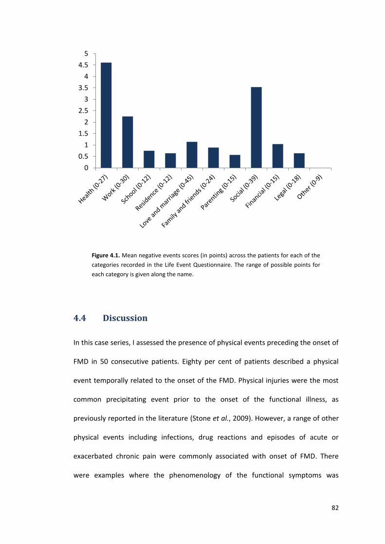
82
Figure 4.1. Mean negative events scores (in points) across the patients for each of the
categories recorded in the Life Event Questionnaire. The range of possible points for
each category is given along the name.
4.4 Discussion
In this case series, I assessed the presence of physical events preceding the onset of
FMD in 50 consecutive patients. Eighty per cent of patients described a physical
event temporally related to the onset of the FMD. Physical injuries were the most
common precipitating event prior to the onset of the functional illness, as
previously reported in the literature (Stone et al., 2009). However, a range of other
physical events including infections, drug reactions and episodes of acute or
exacerbated chronic pain were commonly associated with onset of FMD. There
were examples where the phenomenology of the functional symptoms was
0
0.5
1
1.5
2
2.5
3
3.5
4
4.5
5
83
plausibly related with the physical trigger. For example two patients who had a
clear acute dystonic reaction secondary to drugs developed a functional dystonia
which affected the same body parts as the original acute dystonic reaction. Also,
most patients with fixed dystonia developed symptoms in the limb that was injured,
where surgery had been performed or where neurological symptoms were present
(e.g. from brachial neuritis or carpal tunnel syndrome). This is in line with other
reports suggesting that the nature of the physical precipitating and the affected
body parts during the physical illness may influence the subsequent functional
symptom (Moss-Morris and Spence, 2006). For instance, a prospective study
showed that during follow up, patients who had suffered from Campylobacter
gastroenteritis had a greater risk for developing irritable bowel syndrome than
those who had an infectious mononucleosis, whereas infectious mononucleosis
patients had more significant risk to develop chronic fatigue (Moss-Morris and
Spence, 2006). We may remember that this potential association was also noted by
Breuer and Freud, for instance when they described the patient suffering from
hysteric olfactory symptoms, who previously had had severe infections of the nasal
cavity (Breuer and Freud, 1974).
Others have highlighted the occurrence and potential aetiological importance of
physical events preceding onset of functional neurological symptoms for example
physical injury to the limb which commonly precedes the onset of limb paralysis
(Stone et al., 2010).
As I have previously mentioned, in a review of the literature, it was suggested that
physical events were more common in functional paralysis (41%) than in FMD (34%)
84
but physical events in these studies were limited to physical injuries. Our results
suggest that a broader spectrum of physical events might be present at the onset of
FMD.
We found that 38% of patients with FMD fulfilled criteria for the diagnosis of a
panic attack during the physical triggering event, which is similar to that found in
patients with functional paralysis (34% of 107) (Stone et al., 2010). Similar to
patients with functional paralysis, most patients who fulfilled criteria of a panic
attack had an acute onset of the functional symptoms, suggesting a potential link.
Because of the uncontrolled design of our study we cannot conclude with
confidence that the physical events reported were causal in the development of
FMD. We only can conclude that most patients with FMD report a physical event
before the onset of the functional symptoms, and this is often accompanied by
physical symptoms of a panic attack. We hypothesise that physical events can be of
relevance themselves in the development of functional symptoms by providing new
sensory data which may be abnormally processed and could provide the substrate
for the development of an abnormal movement. However, most people do not
develop FMD after common physical events such as injuries or flu-like illness.
Therefore, there should be other predisposing factors, perhaps at a cognitive level,
which may influence the way initial sensory data is processed.
The presence of symptoms related to panic during the physical triggering event may
play also a role. It may provide an additional factor that could increase the salience
of the sensory information occurring during a coincident physical precipitating
event or could generate additional physical symptoms in itself (e.g. tremor).
85
There is clearly still a role for past and recent life events in generating vulnerability
in individual patients. In addition, no physical event can be experienced without a
concomitant psychological experience. Our impression is that in many patients it is
the physical triggering event itself which may be best thought of as the most
important “recent stressor” related to the symptom, rather than hunting in the
history for an emotional life event which reflects the general vulnerability of the
patient.
In this regard, we assessed the presence of life events three months prior to the
onset of the functional symptoms by using a self-report questionnaire (LEQ)
(Norbeck, 1984), which addressed several categories. We found that patients
scored low in the negative score for life events within three months before
symptom onset (15.7±16.6), which is similar to the score reported by others, for
instance in patients with colon polyps and normal controls (Ashktorab et al., 2013).
A sizable proportion of patients who reported life events mainly scored in the
health-related subscore, which linked with the high proportion reporting a physical
precipitating factor. This is in line with other studies of patients with FMD and other
functional neurological symptoms where patients do not commonly report adverse
life events (other than those associated with their physical health) close to onset of
FMD (Binzer et al., 1997, Kranick et al., 2011). However, it should be noted that the
retrospective assessment of life events is difficult especially with regard to recall
bias. This, along with the low response rate that we obtained for the LEQ
questionnaire, does not allow us to draw clear conclusions about the presence of
potential psychological stressors.
86
I acknowledge a number of other methodological limitations. First, we did not
include a control group and our result should be interpreted with caution in terms
of causality. The ideal control population would have been “organic” movement
disorder counterparts with a sudden onset. However, the vast majority of “organic”
movement disorders start gradually and this made any attempt at comparison
difficult. Second, there is the problem of recall bias. We cannot rule out that
patients have over reported physical events and/or under reported life events at
the onset of the FMD. In this regards, alexithymia has recently been found to be
common in patients with functional motor symptoms (Demartini et al., 2014). We
cannot rule out that potential difficulties to identify emotions were also influencing
the results of the LEQ. Also, the retrospective design of the study has difficulties
distinguishing a physical event and onset of functional symptoms. Third, because
patients were recruited in a specialist centre, one can argue that the data may not
be representative of all patients with FMD. However, I do not feel that the clinical
histories of the patients we included significantly differ from case series of patients
with FMD collected from less specialist services. In addition, recruitment was
consecutive in order to minimize a selection bias. Fourth, the response rate for
questionnaires was low, which makes it difficult to comment on more psychological
aspects. Fifth, we set an arbitrary limit of 3 months for both physical events and life
events to be included. A specific timing is not well established but we believe that
with this selection, we have assured a reasonable temporal relationship.
In summary, physical triggering events are common in FMD and an appreciation of
their role may help to move forward our understanding of the mechanism of FMD

87
beyond simplistic notions related to “psychological stressors” towards a more truly
biopsychosocial model of these common and disabling symptoms.
88
Chapter 5: A study on the “jumping to conclusions” bias
in functional movement disorders
The work presented in this chapter was originally published in the form of a research article:
Pareés I, Kassavetis P, Saifee TA, Sadnicka A, Bhatia KP, Fotopoulou A, Edwards MJ.
'Jumping to conclusions' bias in functional movement disorders. Journal Neurology
Neurosurgery and Psychiatry 2012; 83: 460-3. This is available online at
http://jnnp.bmj.com/content/83/4/460.long
5.1 Introduction
In the previous chapter, we have seen that most patients with FMD report a
physical event temporarily close to the onset of the functional symptoms which
often are phenomenologically related. We suggested that they may be of relevance
in symptom development by providing new sensory information which may be the
substrate for the functional symptom in question. However, the physical events
that we reported, particularly painful injury or flu-like illness, are almost universal
occurrences that do not trigger functional symptoms in most people who
experience them. Therefore, I wondered whether there should be differences in the
manner in which patients process novel information and use it to guide future
behaviour.
As a preliminary exploration of this question I assessed a well-known cognitive
reasoning model in psychiatric research, the “jumping-to-conclusions” (JTC) bias.
For that, I used the “bead task” (Garety et al., 1991). In this paradigm, participants
assess (within a Bayesian reasoning framework) the probabilities of events on the
89
basis of empirical evidence. Deluded patients have been found to exhibit a
tendency to the early acceptance of hypotheses formed in this task based on much
more limited evidence than controls. This style of reasoning has been suggested to
favour the formation of abnormal inferences, ultimately leading to the adoption of
abnormal beliefs (Fine et al., 2007). In this study, we hypothesised that similar
reasoning abnormalities might be present in patients with FMD which would favour
formation of abnormal inferences about the sensory data arising during the
physical event and could contribute to the development of functional symptoms.
5.2 Methods
5.2.1 Participants
Eighteen patients with FMD were recruited by two methods:
1. They were recruited from a pre-existing database of patients with
movement disorders who had previously given the consent for their medical
data to be stored in the ION Queen Square (Movement disorders Research
Database) and to be contacted regarding participation in research studies by
doctors authorised by Professor Bhatia or Dr Mark Edwards. These patients
were contacted by telephone and the specific study was discussed with
them. If the patient expressed interest in participating, a formal letter with a
full description of the study and stating the date and location of the
experiment was sent.
2. Patients were directly recruited from the General Movement Disorder clinic
of Dr Mark Edwards or Professor Kailash Bhatia as well as from the
specialised Functional Movement Disorder run by Dr Edwards, at the NHNN
90
Queen Square, London, UK. The specific study was personally explained in
detail and if the patient expressed interest in participating, a formal letter
with a full description of the study and stating the date and location of the
experiment was sent.
I used as inclusion criteria:
1. Age over 18 years.
2. Diagnosis of clinically established or documented functional movement
disorders according to Fahn and Williams criteria (Fahn and Williams, 1988).
I used as exclusion criteria:
1. Age less than 18.
2. Unable to communicate with researcher (e.g. does not speak English).
Eighteen healthy subjects who were matched by age and gender were also
recruited from a departmental register of volunteers and patient’s relatives.
5.2.2 Design and measures
5.2.2.1 Bead task
The reasoning task was similar to previous designs (Garety et al., 1991). Participants
were presented with two jars each containing 100 beads: Jar A with 85 red and 15
blue beads and Jar B with 85 blue and 15 red beads. Both jars were then hidden
from view. Participants were told that the experimenter would choose one jar and
draw beads one at a time from this jar. Beads would be replaced in the jar after
each draw.
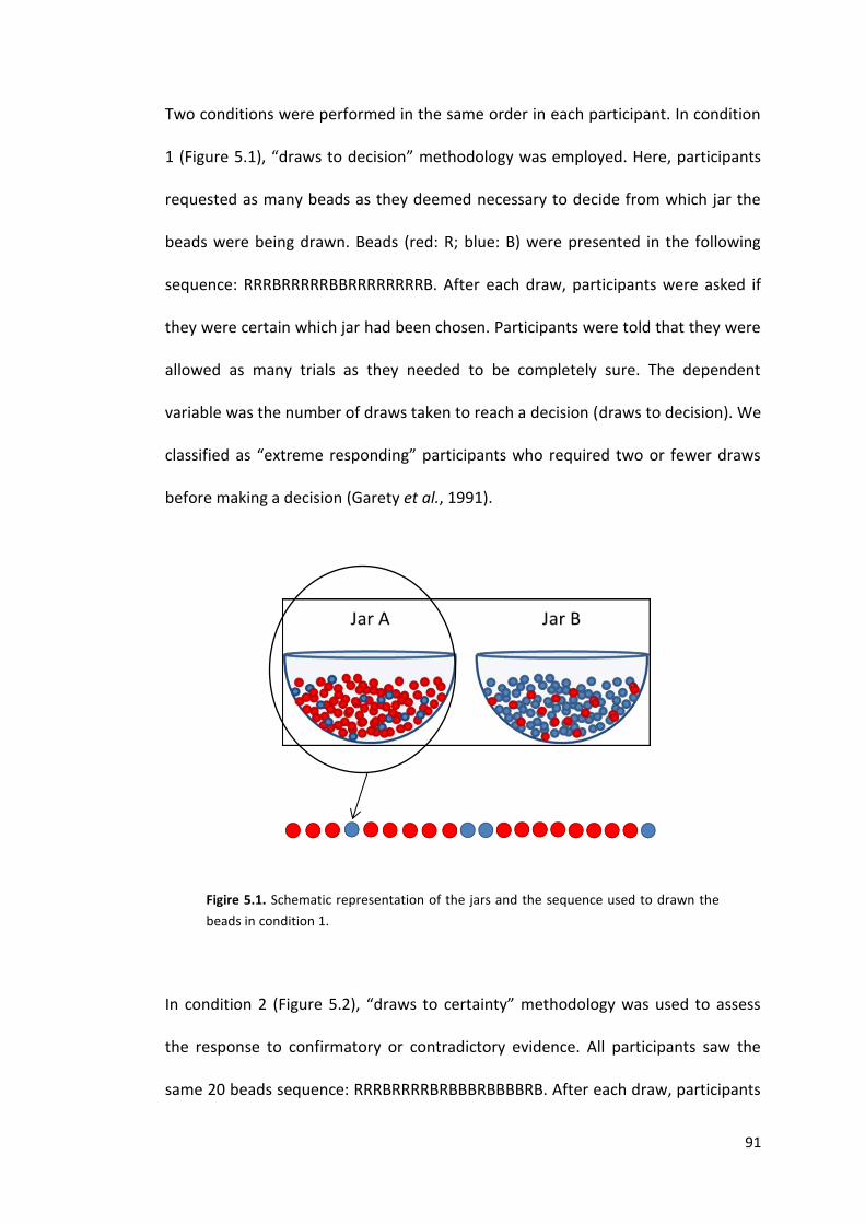
91
Two conditions were performed in the same order in each participant. In condition
1 (Figure 5.1), “draws to decision” methodology was employed. Here, participants
requested as many beads as they deemed necessary to decide from which jar the
beads were being drawn. Beads (red: R; blue: B) were presented in the following
sequence: RRRBRRRRRBBRRRRRRRRB. After each draw, participants were asked if
they were certain which jar had been chosen. Participants were told that they were
allowed as many trials as they needed to be completely sure. The dependent
variable was the number of draws taken to reach a decision (draws to decision). We
classified as “extreme responding” participants who required two or fewer draws
before making a decision (Garety et al., 1991).
Figire 5.1. Schematic representation of the jars and the sequence used to drawn the
beads in condition 1.
In condition 2 (Figure 5.2), “draws to certainty” methodology was used to assess
the response to confirmatory or contradictory evidence. All participants saw the
same 20 beads sequence: RRRBRRRRBRBBBRBBBBRB. After each draw, participants
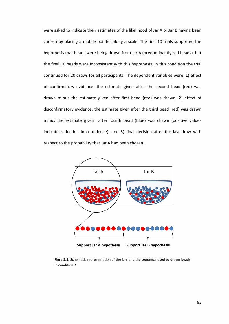
92
were asked to indicate their estimates of the likelihood of Jar A or Jar B having been
chosen by placing a mobile pointer along a scale. The first 10 trials supported the
hypothesis that beads were being drawn from Jar A (predominantly red beads), but
the final 10 beads were inconsistent with this hypothesis. In this condition the trial
continued for 20 draws for all participants. The dependent variables were: 1) effect
of confirmatory evidence: the estimate given after the second bead (red) was
drawn minus the estimate given after first bead (red) was drawn; 2) effect of
disconfirmatory evidence: the estimate given after the third bead (red) was drawn
minus the estimate given after fourth bead (blue) was drawn (positive values
indicate reduction in confidence); and 3) final decision after the last draw with
respect to the probability that Jar A had been chosen.
Figre 5.2. Schematic representation of the jars and the sequence used to drawn beads
in condition 2.
Support Jar A hypothesis Support Jar B hypothesis

93
5.2.2.2 Questionnaires
5.2.2.2.1 Intelligence level
To estimate the intelligence level of participants, we used the 12 items short form
of Raven's Progressive Matrices test (Raven JC, 1977). This is a classic test of
abstract or non-verbal reasoning in which a variety of figures, relationships and
transformations are presented and the person must select the best of multiple
choice alternatives to fill the empty corner (Figure 5.3).
Figure 5.3. Example of one of the items used in the short version of the Raven’s
Progressive Matrices.
I used this test because many of the participants of this study were non-native
English speakers and therefore we thought that a test avoiding verbal intelligence
would be more appropriate.
94
5.2.2.2.2 Delusional ideation
Delusional patients have been found to have the “jumping to conclusion” bias and
therefore delusional ideation was assessed in our participants. We used the Peters
et al. Delusions Inventory (PDI-21) which has been designed to assess delusional
ideation in general population (Peters et al., 2004). It includes 21 questions asking
whether or not the participant has a particular idea, incorporating measures of the
distress, preoccupation and conviction associated with delusional beliefs. For each
question participants answer yes/no and 1 point is assigned to each "yes" answer
and a 0 to each "no" answer. Therefore, the possible range of scores was 0 to 21.
The distress, preoccupation, and conviction ratings ranged from 0 to 5 for each
item. A "no" answer automatically scored 0 on each of the three dimensions. A
rating between 1 and 5 was obtained if the item had been answered "yes”. Total
score of the questionnaire range from 0 to 336. Higher scores indicate higher
delusional ideation.
5.2.2.2.3 Mood
Some authors have suggested that mood disturbances may influence results in this
cognitive task (Garety et al., 2005). Therefore participants completed the Hospital
Anxiety and Depression rating scale (HADS) (Zigmond and Snaith, 1983) with
reference to their mood the week prior to testing, which has been already
explained in the previous chapter.
5.2.3 Statistical analyses
I used PASW statistical package (version 18) for statistical analysis. P values
reported for categorical variables were calculated with the use of Fisher’s Exact
95
Test. Mann-Whitney U test was used to compare differences for numerical
variables between groups. In order to determine the independent contribution of
Raven’s matrices scores, delusional ideation, and anxiety and depression subscales
of the HADS to bead task performance, a simple linear regression analysis was first
performed. Those variables which were contributing factors to the results of the
task in this analysis were entered en bloc as independent variables in a subsequent
multiple linear regression analysis. A two-tailed α level of 0.05 was used as the
criterion for significance in all analyses.
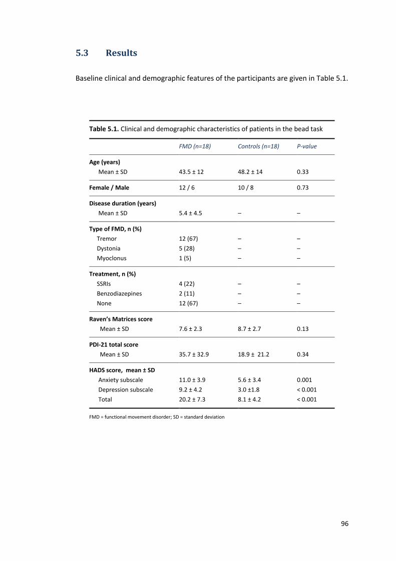
96
5.3 Results
Baseline clinical and demographic features of the participants are given in Table 5.1.
Table 5.1. Clinical and demographic characteristics of patients in the bead task
FMD (n=18) Controls (n=18) P-value
Age (years)
Mean ± SD
43.5 ± 12
48.2 ± 14
0.33
Female / Male 12 / 6 10 / 8 0.73
Disease duration (years)
Mean ± SD
5.4 ± 4.5
–
–
Type of FMD, n (%)
Tremor
Dystonia
Myoclonus
12 (67)
5 (28)
1 (5)
–
–
–
–
–
–
Treatment, n (%)
SSRIs
Benzodiazepines
None
4 (22)
2 (11)
12 (67)
–
–
–
–
–
–
Raven’s Matrices score
Mean ± SD
7.6 ± 2.3
8.7 ± 2.7
0.13
PDI-21 total score
Mean ± SD
35.7 ± 32.9
18.9 ± 21.2
0.34
HADS score, mean ± SD
Anxiety subscale
Depression subscale
Total
11.0 ± 3.9
9.2 ± 4.2
20.2 ± 7.3
5.6 ± 3.4
3.0 ±1.8
8.1 ± 4.2
0.001
< 0.001
< 0.001
FMD = functional movement disorder; SD = standard deviation
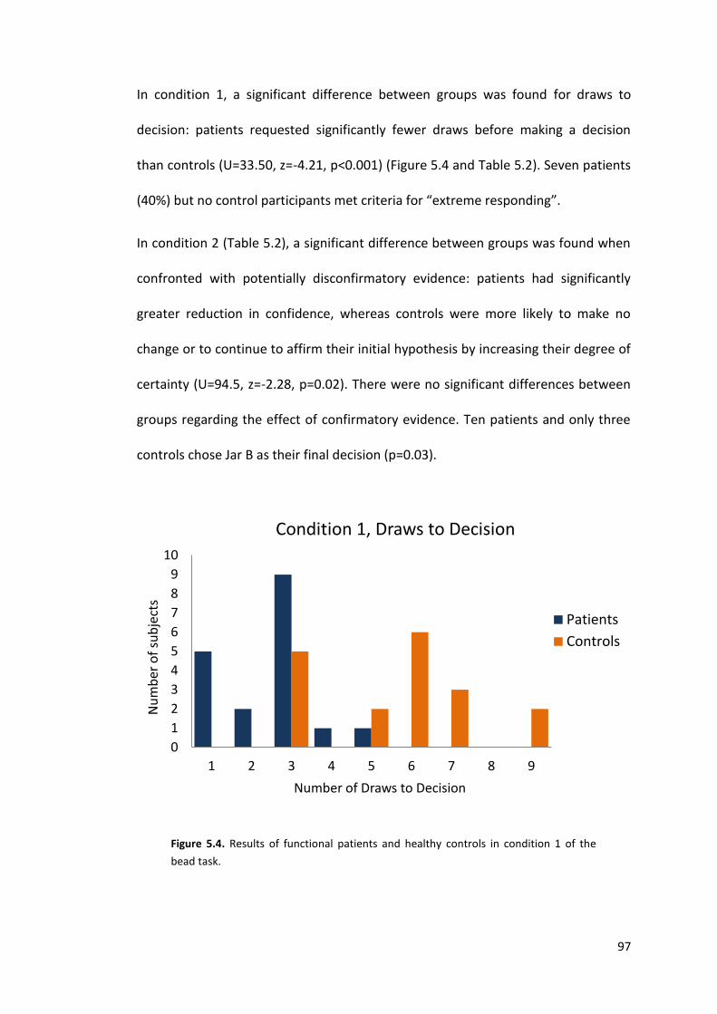
97
In condition 1, a significant difference between groups was found for draws to
decision: patients requested significantly fewer draws before making a decision
than controls (U=33.50, z=-4.21, p<0.001) (Figure 5.4 and Table 5.2). Seven patients
(40%) but no control participants met criteria for “extreme responding”.
In condition 2 (Table 5.2), a significant difference between groups was found when
confronted with potentially disconfirmatory evidence: patients had significantly
greater reduction in confidence, whereas controls were more likely to make no
change or to continue to affirm their initial hypothesis by increasing their degree of
certainty (U=94.5, z=-2.28, p=0.02). There were no significant differences between
groups regarding the effect of confirmatory evidence. Ten patients and only three
controls chose Jar B as their final decision (p=0.03).
Figure 5.4. Results of functional patients and healthy controls in condition 1 of the
bead task.
0
1
2
3
4
5
6
7
8
9
10
1 2 3 4 5 6 7 8 9
Nu
mb
er o
f su
bje
cts
Number of Draws to Decision
Condition 1, Draws to Decision
Patients
Controls
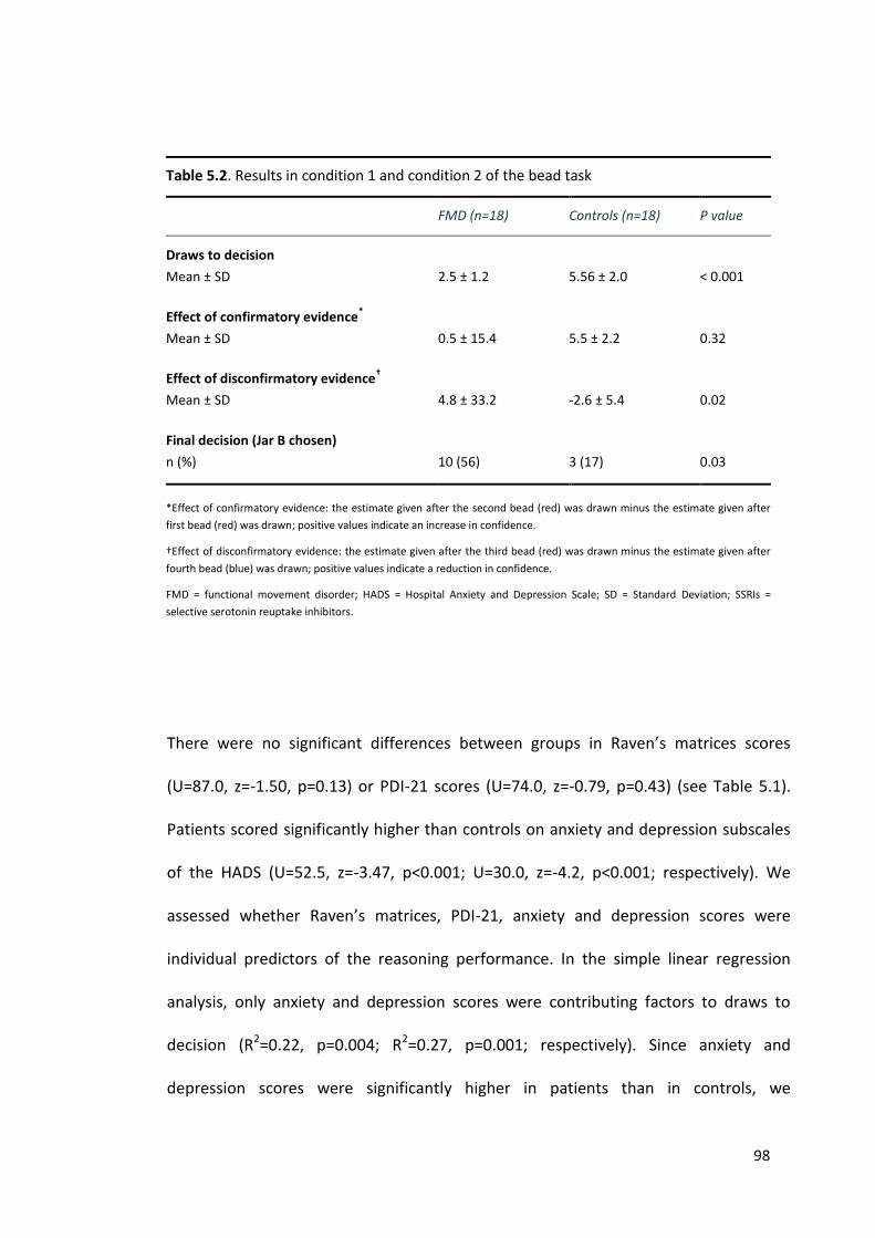
98
Table 5.2. Results in condition 1 and condition 2 of the bead task
FMD (n=18) Controls (n=18) P value
Draws to decision
Mean ± SD
2.5 ± 1.2
5.56 ± 2.0
< 0.001
Effect of confirmatory evidence*
Mean ± SD
0.5 ± 15.4
5.5 ± 2.2
0.32
Effect of disconfirmatory evidence†
Mean ± SD
4.8 ± 33.2
-2.6 ± 5.4
0.02
Final decision (Jar B chosen)
n (%)
10 (56)
3 (17)
0.03
*Effect of confirmatory evidence: the estimate given after the second bead (red) was drawn minus the estimate given after
first bead (red) was drawn; positive values indicate an increase in confidence.
†Effect of disconfirmatory evidence: the estimate given after the third bead (red) was drawn minus the estimate given after
fourth bead (blue) was drawn; positive values indicate a reduction in confidence.
FMD = functional movement disorder; HADS = Hospital Anxiety and Depression Scale; SD = Standard Deviation; SSRIs =
selective serotonin reuptake inhibitors.
There were no significant differences between groups in Raven’s matrices scores
(U=87.0, z=-1.50, p=0.13) or PDI-21 scores (U=74.0, z=-0.79, p=0.43) (see Table 5.1).
Patients scored significantly higher than controls on anxiety and depression subscales
of the HADS (U=52.5, z=-3.47, p<0.001; U=30.0, z=-4.2, p<0.001; respectively). We
assessed whether Raven’s matrices, PDI-21, anxiety and depression scores were
individual predictors of the reasoning performance. In the simple linear regression
analysis, only anxiety and depression scores were contributing factors to draws to
decision (R2=0.22, p=0.004; R2=0.27, p=0.001; respectively). Since anxiety and
depression scores were significantly higher in patients than in controls, we
99
investigated in more detail whether belonging to one group or the other was a
potential confounder factor. When both anxiety and depression scores were
considered in a multiple linear regression analysis and their contribution was adjusted
in a second step by group as a possible confounder neither anxiety nor depression
scores were predictive factors to draws to decision (β= −0.06, p=0.73; β= −0.036,
p=0.87; respectively).
5.4 Discussion
In this study we assessed probabilistic reasoning in patients with FMD using the
bead task, a classic paradigm in psychiatry research for the study of belief
formation under conditions of uncertainty (Garety et al., 1991, Fine et al., 2007).
We found differences in probabilistic reasoning between patients with FMD and
healthy participants. First, patients required less evidence before making a definite
decision on the task. Second, patients integrated new, potentially discomfirmatory
evidence into their decision making differently than controls. They were more likely
to make changes in their probability estimates in the direction suggested by the
new evidence, and to make a final decision consistent with this new evidence. Both
aspects of the bead task are conceptually related to frontal lobe functions such as
set shifting and impulsivity, and similar abnormalities have been found in delusional
patients. These data appear not to be influenced by the higher levels of depression
and anxiety found in the patient group.
How might these preliminary data help to inform our understanding of the
underlying mechanism of symptom production in FMD? This experiment was
prompted by the association between physical triggering events and the production
100
of phenomenologically related symptoms in patients with functional neurological
disorders. The process of making inferences on the causes of sensory data and
incorporating these into an internal model of the world is central to a modern
Bayesian approach to understanding the brain (Friston, 2010). Impulsivity in
decision-making may reflect an abnormal overweighting of the importance of
sensory data which causes an inappropriate updating of internal models relating to
the data. Extrapolating such a reasoning style to sensory data occurring during a
physical triggering event, this might produce inappropriate updating of
expectations regarding future sensory data, for example an expectation of pain,
abnormal movement or weakness, which might drive future physical symptoms.
This initial formulation, although speculative, could form the basis of a testable
biopsychosocial framework for FMD.
Patients were more likely to respond to potentially disconfirmatory evidence by
changing their probability decision in the direction of the new evidence, seemingly
at odds with the fixity with which illness beliefs are sustained by this patients group.
Indeed, a similar question has been raised by those studying delusions. Here the
suggestion has been, supported by experimental evidence, that over time beliefs
shift from a more flexible “goal-directed” ventral corticostriatal system to a more
inflexible “habit-related” dorsal striatal system (Corlett et al., 2010). If this is also
relevant to functional disorders, then the period of time following the initial
triggering event must be important in creating persistence and fixity of abnormal
beliefs. This would be consistent with the, negative correlation between time to
diagnosis and chance of recovery previously found in functional disorders (Wyllie et
al., 1990) and the fact that, at least in the early stages, simply explaining the
101
diagnosis can lead to long-term resolution of symptoms (Hall-Patch et al., 2010). In
contrast, fixity of illness belief is associated with poor long-term outcome
(Buchanan and Snars, 1993). Though we highlight the similarity of performance of
patients with FMD on this task and previous studies of patients with delusions, the
lack of difference between FMD patients and controls in the PDI-21 inventory
indicates that patients with FMD are not suffering generally from delusional beliefs.
Limitations
We acknowledge limitations to our study. First, the sample size is small and we
cannot exclude that in a larger cohort data may be different. However, we chose
patients with clinically typical FMD using standardized criteria and feel that they do
accurately represent patients with these diagnoses. Second, FMD patients tend to
have concomitant mood abnormalities as measured by HADS. However, multiple
regression analysis revealed that anxiety and depression scores were not
independent predictors of the draws to decision. This is in line with several reports
which have failed to find an, association between performance on the bead task
and mood disturbance. Future studies could nevertheless compare the cognitive
style of FMD patients with depressed and anxious patients without FMD. Third,
while we have controlled for the presence of delusions in general, we have not
measured other psychiatric comorbidities such as personality disorder that could be
confounding the results.
In conclusion, we present preliminary evidence of a “‘jumping to conclusions” style
of cognitive reasoning in patients with FMD. These data may have relevance to the
manner with which patients with functional symptoms process novel sensory data

102
occurring during physical triggering events commonly reported at onset of
symptoms.
103
Chapter 6: A study on the effect of explicit strategies
and predictability on motor control in functional
movement disorders
The work presented in this chapter was originally published in the form of a research article:
Pareés I, Kassavetis P, Saifee TA, Sadnicka A, Davare M, Bhatia KP, Rothwell JC, Bestmann S,
Edwards MJ. Failure of explicit movement control in patients with functional motor
symptoms. Movement Disorders 2013; 28: 517-23.
6.1 Introduction
I emphasized in Chapter 3 that one of the aims of this thesis is to better understand
mechanism of symptom production and that I have taken as the starting point the
manner in which the diagnosis of FMD is made in neurological practice.
In this regard, the clinical basis for making a positive diagnosis in this group of
patients is quite clear: movement becomes normal when attention is diverted away
from the movement, or when movement is triggered covertly. This typically occurs
during implicit motor control which is engaged in ‘automatic’ movements where
attentional processes have limited contribution. In contrast, motor impairments are
manifest only during periods of explicit attention to movement. Therefore, it would
seem a priori that an understanding of the mechanism underlying these robust
clinical phenomena would be essential to explain symptom generation in patients
with FMD. However, these apparently simple clinical tests are in fact complex tasks
when viewed from an experimental perspective, and at the present time it is not
104
clear which aspects of movement control might be important in generating
impairment.
Taking all these considerations into account, I decided to investigate the distinction
between implicit and explicit motor control in patients with FMD. Two different
experiments were designed in which voluntary movements were made either with
an opportunity for explicit awareness/control, or were performed in a largely
implicit fashion. We hypothesized that, even within the same limb, implicit
influences on movement would be preserved whereas explicit control would be
abnormal in patients with FMD.
6.2 Methods
6.2.1 Participants
Eleven patients were directly recruited from the General Movement Disorder clinic
of Dr Mark Edwards or Professor Kailash Bhatia as well as from the specialised
Functional Movement Disorder run by Dr Edwards, at the NHNN Queen Square,
London, UK. The specific study was personally explained in detail and if the patient
expressed interest in participating, a formal letter with a full description of the
study and stating the date and location of the experiment was sent.
I used as inclusion criteria:
1. Age over 18 years.
2. Diagnosis of clinically established or documented FMD according to Fahn
and Williams criteria (Fahn and Williams, 1988).
I used as exclusion criteria:
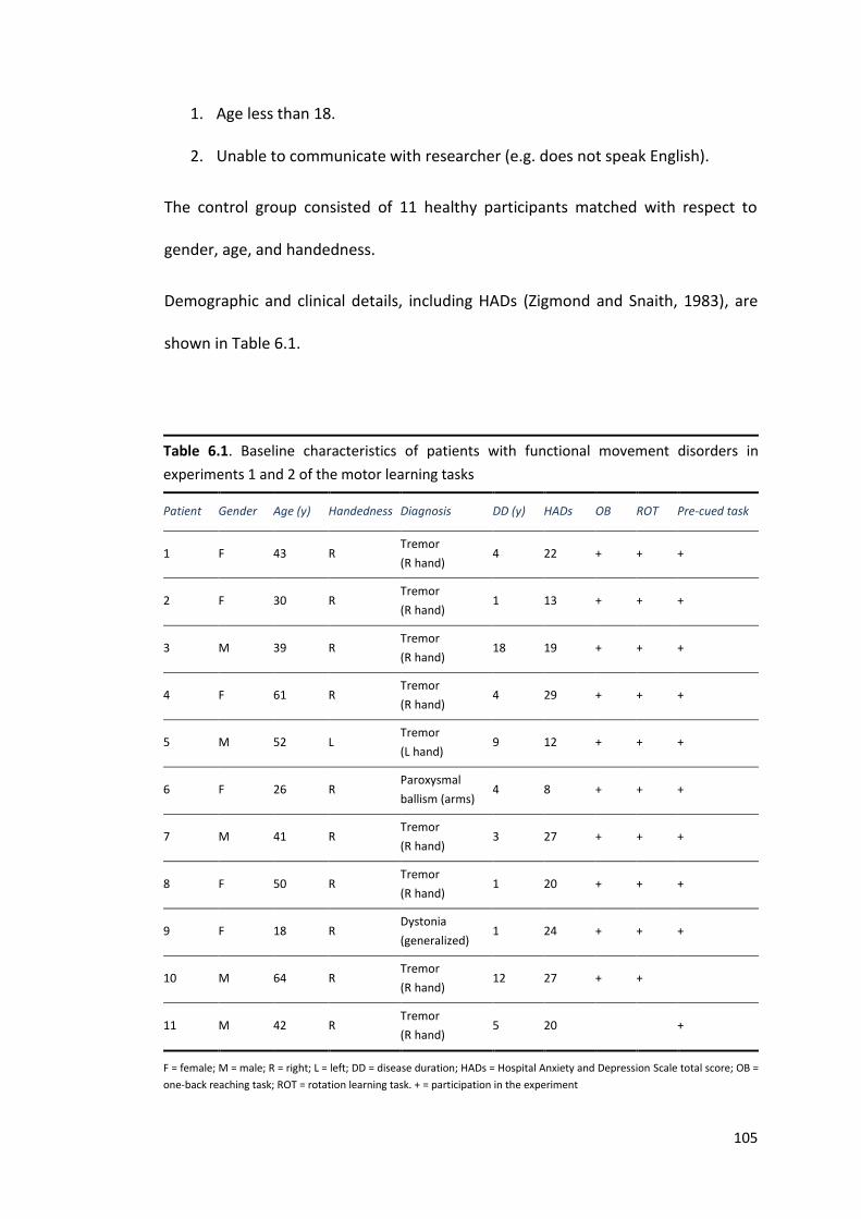
105
1. Age less than 18.
2. Unable to communicate with researcher (e.g. does not speak English).
The control group consisted of 11 healthy participants matched with respect to
gender, age, and handedness.
Demographic and clinical details, including HADs (Zigmond and Snaith, 1983), are
shown in Table 6.1.
Table 6.1. Baseline characteristics of patients with functional movement disorders in
experiments 1 and 2 of the motor learning tasks
Patient Gender Age (y) Handedness Diagnosis DD (y) HADs OB ROT Pre-cued task
1 F 43 R Tremor
(R hand) 4 22 + + +
2 F 30 R Tremor
(R hand) 1 13 + + +
3 M 39 R Tremor
(R hand) 18 19 + + +
4 F 61 R Tremor
(R hand) 4 29 + + +
5 M 52 L Tremor
(L hand) 9 12 + + +
6 F 26 R Paroxysmal
ballism (arms) 4 8 + + +
7 M 41 R Tremor
(R hand) 3 27 + + +
8 F 50 R Tremor
(R hand) 1 20 + + +
9 F 18 R Dystonia
(generalized) 1 24 + + +
10 M 64 R Tremor
(R hand) 12 27 + +
11 M 42 R Tremor
(R hand) 5 20 +
F = female; M = male; R = right; L = left; DD = disease duration; HADs = Hospital Anxiety and Depression Scale total score; OB =
one-back reaching task; ROT = rotation learning task. + = participation in the experiment
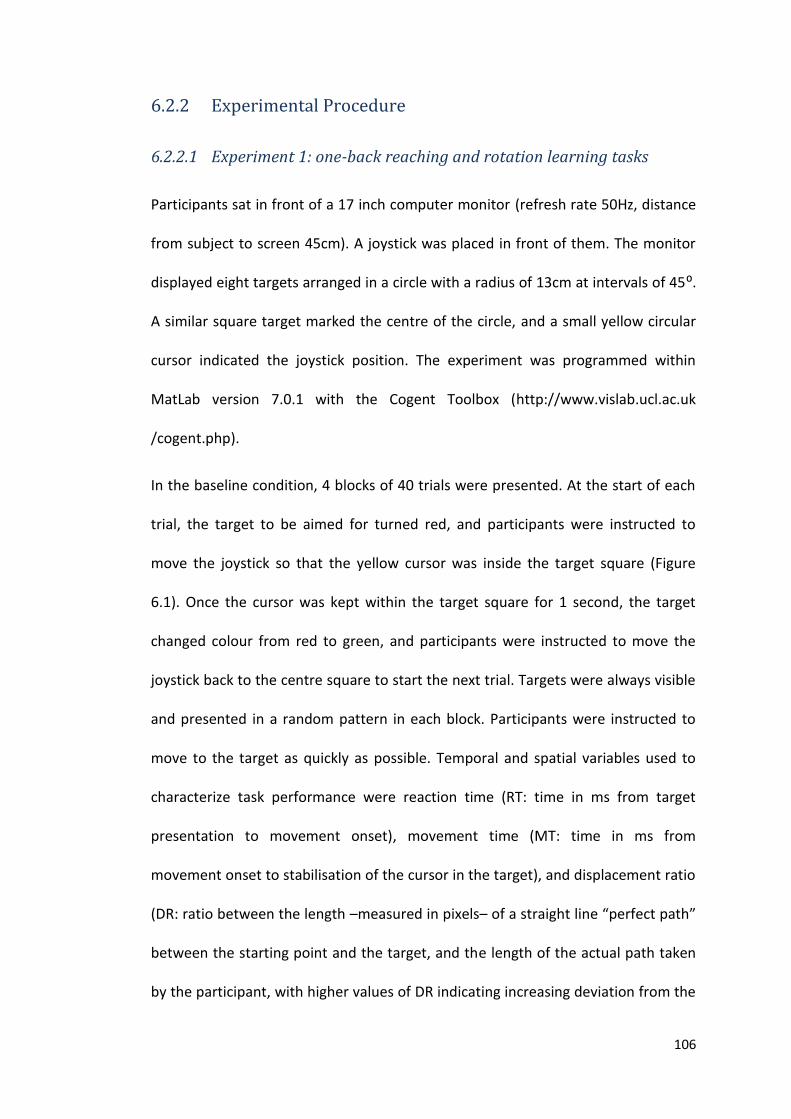
106
6.2.2 Experimental Procedure
6.2.2.1 Experiment 1: one-back reaching and rotation learning tasks
Participants sat in front of a 17 inch computer monitor (refresh rate 50Hz, distance
from subject to screen 45cm). A joystick was placed in front of them. The monitor
displayed eight targets arranged in a circle with a radius of 13cm at intervals of 45⁰.
A similar square target marked the centre of the circle, and a small yellow circular
cursor indicated the joystick position. The experiment was programmed within
MatLab version 7.0.1 with the Cogent Toolbox (http://www.vislab.ucl.ac.uk
/cogent.php).
In the baseline condition, 4 blocks of 40 trials were presented. At the start of each
trial, the target to be aimed for turned red, and participants were instructed to
move the joystick so that the yellow cursor was inside the target square (Figure
6.1). Once the cursor was kept within the target square for 1 second, the target
changed colour from red to green, and participants were instructed to move the
joystick back to the centre square to start the next trial. Targets were always visible
and presented in a random pattern in each block. Participants were instructed to
move to the target as quickly as possible. Temporal and spatial variables used to
characterize task performance were reaction time (RT: time in ms from target
presentation to movement onset), movement time (MT: time in ms from
movement onset to stabilisation of the cursor in the target), and displacement ratio
(DR: ratio between the length –measured in pixels– of a straight line “perfect path”
between the starting point and the target, and the length of the actual path taken
by the participant, with higher values of DR indicating increasing deviation from the
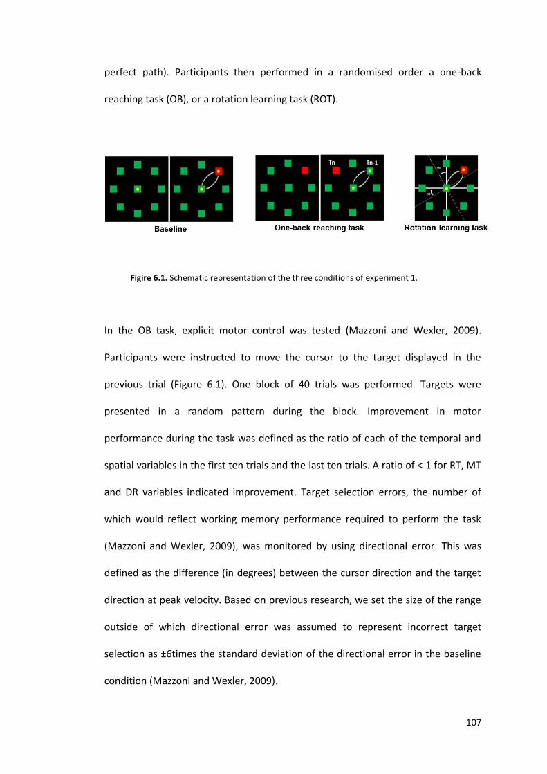
107
perfect path). Participants then performed in a randomised order a one-back
reaching task (OB), or a rotation learning task (ROT).
Figire 6.1. Schematic representation of the three conditions of experiment 1.
In the OB task, explicit motor control was tested (Mazzoni and Wexler, 2009).
Participants were instructed to move the cursor to the target displayed in the
previous trial (Figure 6.1). One block of 40 trials was performed. Targets were
presented in a random pattern during the block. Improvement in motor
performance during the task was defined as the ratio of each of the temporal and
spatial variables in the first ten trials and the last ten trials. A ratio of < 1 for RT, MT
and DR variables indicated improvement. Target selection errors, the number of
which would reflect working memory performance required to perform the task
(Mazzoni and Wexler, 2009), was monitored by using directional error. This was
defined as the difference (in degrees) between the cursor direction and the target
direction at peak velocity. Based on previous research, we set the size of the range
outside of which directional error was assumed to represent incorrect target
selection as ±6times the standard deviation of the directional error in the baseline
condition (Mazzoni and Wexler, 2009).
108
In the ROT task, implicit motor control was tested (Mazzoni and Wexler, 2009). This
task measured the ability of participants to adapt to a visuomotor perturbation. A
constant 30⁰ anticlockwise rotation was introduced into the path of the cursor
displayed on the screen (Figure 6.1). Targets were displayed in a randomised order,
and participants had to move the cursor to the highlighted target as quickly as
possible. Participants were not instructed how to compensate for the rotation.
Improvement would be indicated by a ratio in the first ten trials and the last ten
trials of < 1 for RT, MT and DR.
6.2.2.2 Experiment 2: pre-cued choice reaction time with varying cue
validity
In this experiment we manipulated the predictability of an upcoming movement
(Bestmann et al., 2008). Participants sat in front of a 17 inch computer monitor
(refresh rate 50Hz, distance from subject to screen 45cm). A standard QWERTY
computer keyboard was placed in front of them. Their left index finger was placed
over the "Z" key and the right index finger was placed over the "M" key.
In a training session, participants were required to respond with one or other key to
the presentation of two different symbols. They were told that “Z” was associated
with one symbol, and “M” with the other. Feedback was given on the accuracy of
their choice, and 40 trials were conducted. This ensured accurate response
mapping of keypress to symbol, with all subjects achieving a 100% correct response
level by the end of training.
There were three experimental conditions each performed twice in a randomised
order across participants. Each trial started with a fixation cross in the centre of the
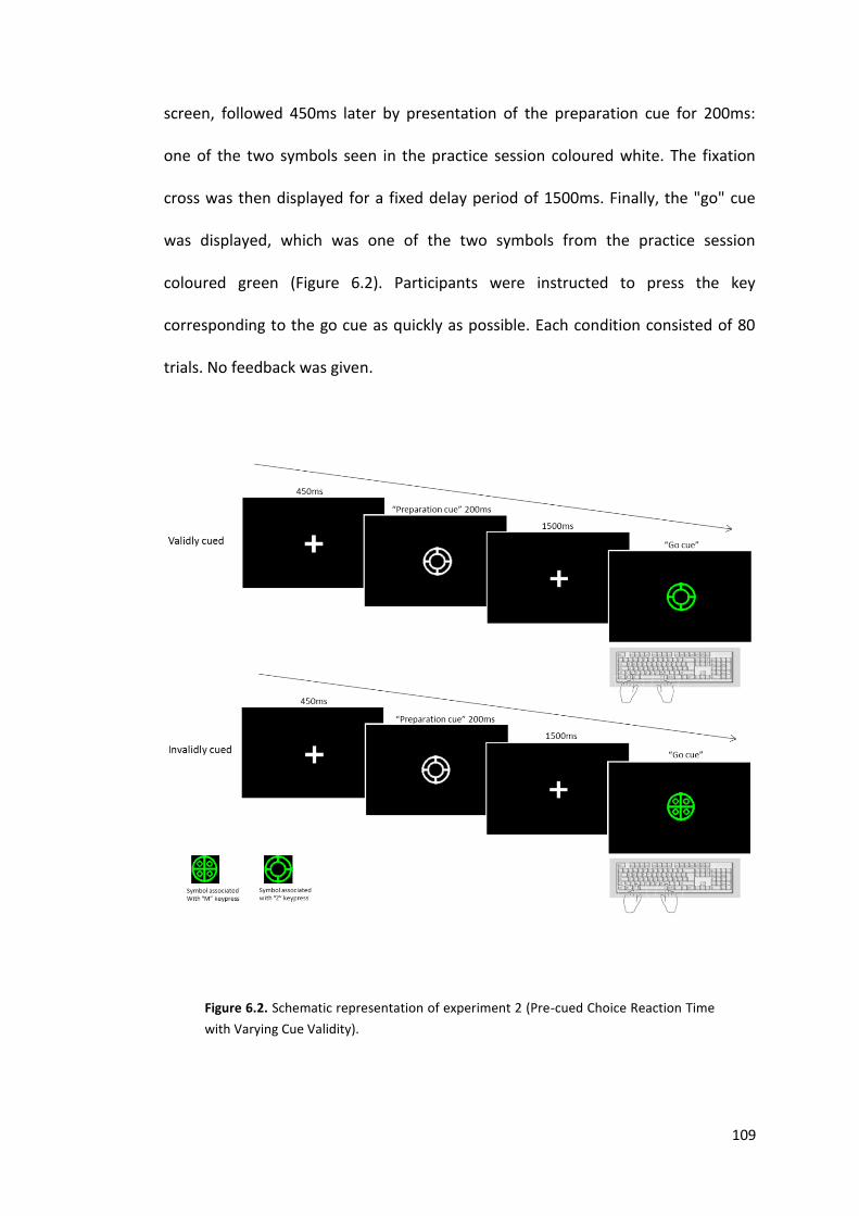
109
screen, followed 450ms later by presentation of the preparation cue for 200ms:
one of the two symbols seen in the practice session coloured white. The fixation
cross was then displayed for a fixed delay period of 1500ms. Finally, the "go" cue
was displayed, which was one of the two symbols from the practice session
coloured green (Figure 6.2). Participants were instructed to press the key
corresponding to the go cue as quickly as possible. Each condition consisted of 80
trials. No feedback was given.
Figure 6.2. Schematic representation of experiment 2 (Pre-cued Choice Reaction Time
with Varying Cue Validity).
110
In the 50% validity condition (50v), the preparation cue accurately predicted the
"go" cue in 50% of trials, i.e. it had no predictive value. In the 75% validity (75v) and
the 95% validity (95v) conditions, the preparation cue accurately predicted the "go"
cue in 75% and 95% of trials, respectively.
We calculated response time in ms (time from presentation of the "go" cue to key
press) for each trial. Trials where the preparation cue accurately predicted the "go"
cue (valid trials) were separated from those where the prediction was incorrect
(invalid trials). We then averaged response times for valid and invalid trials
separately across all trials performed for each of the three conditions. We expected
that response times for valid trials would become shorter for conditions with
increasing validity of the preparation cue.
6.2.3 Statistical analysis
We used the PASW statistical package (version 18). P values for categorical
variables were calculated with Fisher’s Exact T. Kolmogorov-Smirnov test was used
to assess the normal distribution of the data. Analysis of variance (ANOVA) was
used to compare differences in means for numerical data when parametric
assumptions were met. Post-hoc T-tests with Bonferroni corrections were used.
When data were not normally distributed, non-parametric tests (Mann-Whitney U
test, Friedman ANOVA, Wilcoxon test and Spearman correlation) were used.
Statistical significance of p<0.05 was assumed.
111
6.3 Results
6.3.1 Experiment 1: one-back reaching and rotation learning tasks
Ten patients and ten healthy controls participated. All patients performed the
experiments with the affected (dominant) hand except for one patient who was
unable to adequately control the joystick because of the severity of the tremor
affecting the dominant hand. The results of this patient did not differ systematically
from the others. All controls used their dominant hand. Exploration of the data
revealed them to be not normally distributed, and therefore non-parametric tests
were used.
6.3.1.1 Baseline Performance
Patients with FMD had a significant change over the four blocks of the baseline
condition in MT (χ2 (3)=17.16, p<0.001) and in DR (χ2 (3)=18.84, p<0.001). Post hoc
analysis using Wilcoxon tests showed improvement in performance from Block 1 to
Block 4 for MT (median Block 1=1695.5ms, median Block 4=1363.7ms; Z= -2.8,
p=0.002) and for DR (median Block 1=5.45, median Block 4=4.57; Z= -2.8, p=0.002).
There was no difference in these parameters between Block 3 and 4, indicating a
ceiling effect in baseline motor performance. In the control group, there was a
trend for improvement in performance across the 4 blocks. Comparison of temporal
and spatial variables in Block 4 of the baseline condition between patients and
control participants revealed that there was no difference in RT between groups
(323.9ms vs 286.5ms; U=24.0, Z= -1.07, p=0.32) but the patients were slower in MT
(1363.7ms vs 1036.3ms; U=24.0, Z= -2.34, p=0.02) and had poorer accuracy
measured as DR (4.57 vs 3.48; U=8.0, Z= -2.64, p=0.003).
112
6.3.1.1.1 One-Back Reaching
Patients and control participants differed in performance in the OB condition. Both
patients and controls had a similar improvement in RT and DR between the first and
the last ten trials (U=32.0, Z= -1.36, p=0.18 and U=39.0, Z= -0.83, p=0.42,
respectively). However, while MT also improved in control participants, it did not in
patients and a ratio of MT in first ten and last ten trials was greater than 1,
indicating a deterioration in performance over the course of the block (U=38.0, Z= -
2.19, p=0.03). Results of ratios are shown in Table 6.2 and illustrated in Figure 6.3.
There was no difference between number of target selection errors seen in patients
and control participants (U=29.5, Z= -1.64, p=0.12).
6.3.1.1.2 Rotation Learning
In the ROT condition both groups improved their performance across the block
when comparing the results of the temporal and spatial variables in the first 10 and
last 10 trials: RT (χ2 (1) =5.0, p=0.041); MT (χ2 (1)=7.2, p=0.012) and DR (χ2 (1)=9.8,
p=0.003). The amount of improvement in each variable, measured as the ratio of
the first 10 and last 10 trials, did not differ between groups. Results of ratios are
shown in Table 6.2 and illustrated in Figure 6.3.
Spearman correlation coefficient revealed no association between HADs scores and
target selection errors or temporal or spatial variables in patients or healthy
controls in either task.
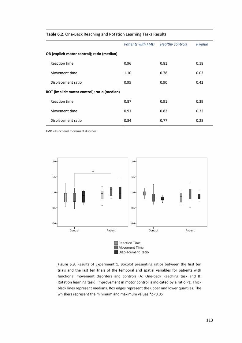
113
Table 6.2. One-Back Reaching and Rotation Learning Tasks Results
Patients with FMD Healthy controls P value
OB (explicit motor control); ratio (median)
Reaction time 0.96 0.81 0.18
Movement time 1.10 0.78 0.03
Displacement ratio 0.95 0.90 0.42
ROT (implicit motor control); ratio (median)
Reaction time 0.87 0.91 0.39
Movement time 0.91 0.82 0.32
Displacement ratio 0.84 0.77 0.28
FMD = Functional movement disorder
Figure 6.3. Results of Experiment 1. Boxplot presenting ratios between the first ten
trials and the last ten trials of the temporal and spatial variables for patients with
functional movement disorders and controls (A: One-back Reaching task and B:
Rotation learning task). Improvement in motor control is indicated by a ratio <1. Thick
black lines represent medians. Box edges represent the upper and lower quartiles. The
whiskers represent the minimum and maximum values.*p<0.05
114
6.3.2 Experiment 2: pre-cued choice reaction time with variable
cue validity
Ten patients and ten healthy controls participated in this experiment. We first
performed an ANOVA with PROBABILITY (0.5, 0.75, 0.95) and VALIDITY
(Valid/Invalid cue) as main factors, and with GROUP (Patients, Controls) as a
between subjects factor. This revealed a PROBABILITY x VALIDITY x GROUP
interaction (F(2,17)=12.2; p=0.001). We explored this interaction with separate
ANOVAs on the data in each of the three conditions (50v, 75v, 95v) with VALIDITY
(valid cue, invalid cue) as main factor and GROUP (patients, controls) as a between
subjects factor. In the 50v condition, there was no effect of validity (p=0.39), nor a
Group x Validity interaction (p=0.36). In the 75v condition there was an effect of
validity (F(1,18)=32.7; p<0.0001) due to faster response times to valid cues
compared with invalid cues. There was, however, no GROUP x VALIDITY interaction
(p=0.14). In the 95v condition, there was no effect of VALIDITY (p=0.51), but there
was a GROUP x VALIDITY interaction (F(1,18)=18.8; p<0.0001). Exploration of this
effect with post-hoc tests with Bonferroni corrections revealed this to be due to a
faster response time for valid cues compared with invalid cues in controls (t=-4.5;
p=0.001), but slower response time for valid compared with invalid cues in patients
(t=2.2; p=0.05) (response time ratios between valid and invalid cues for each
validity condition are shown in Figure 6.4). There were no differences in the
number of errors made (incorrect key presses) between patients and controls.
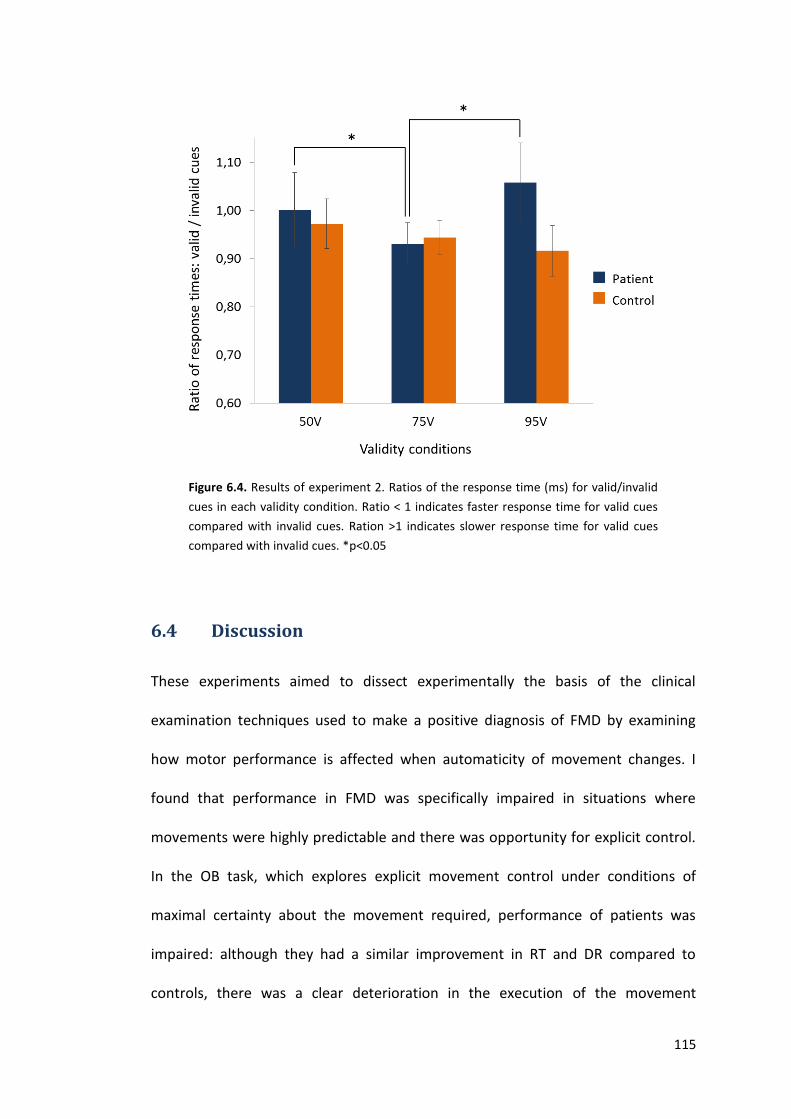
115
Figure 6.4. Results of experiment 2. Ratios of the response time (ms) for valid/invalid
cues in each validity condition. Ratio < 1 indicates faster response time for valid cues
compared with invalid cues. Ration >1 indicates slower response time for valid cues
compared with invalid cues. *p<0.05
6.4 Discussion
These experiments aimed to dissect experimentally the basis of the clinical
examination techniques used to make a positive diagnosis of FMD by examining
how motor performance is affected when automaticity of movement changes. I
found that performance in FMD was specifically impaired in situations where
movements were highly predictable and there was opportunity for explicit control.
In the OB task, which explores explicit movement control under conditions of
maximal certainty about the movement required, performance of patients was
impaired: although they had a similar improvement in RT and DR compared to
controls, there was a clear deterioration in the execution of the movement
116
measured as MT over the course of the block. Patients did not make more errors in
target selection compared with controls, suggesting poor performance was not due
to problems in the working memory requirement of the task. In contrast,
performance was similar to healthy subjects in the rotation learning task which
tests implicit motor performance.
Likewise, when I manipulated the predictability of an upcoming movement by
changing the validity of a pre-cue in a pre-cued reaction time task, patients had a
paradoxical slowing of response times to valid cues when they were highly
predictive of the movement required, despite normal performance in conditions
where cues were non-predictive or 75% predictive.
I have therefore demonstrated that under conditions of increasing certainty
regarding the movement to be performed, and crucially when the nature of the
task is one where pre-planning of movement can occur, impairment is seen in
patients. This is supported by previous work in functional paralysis where impaired
reaction time was seen after pre-cuing by a consciously perceived “endogenous”
cue, but a normal response to a non-consciously perceived “exogenous” cue
(Roelofs et al., 2003). The same group has reported increased N2 event-related
potential amplitude during an explicitly-cued movement task, interpreted as
reflecting enhanced “action monitoring” (Roelofs et al., 2006).
My results fit within a body of research which has explored the effect of explicit
strategies in motor control (Fourneret and Jeannerod, 1998). Healthy people do
not pay much attention to many aspects of their actions and normal movement is
associated with a remarkable lack of activity in brain areas that correspond to high-
117
level executive control (Jueptner et al., 1997). During motor learning, prefrontal
and anterior cingulate activity that is present early in the task disappears with
increasing movement automaticity. If over-trained subjects are then asked to
attend to their actions, prefrontal activity and anterior cingulate activity returns,
and there is deterioration in performance (Jueptner et al., 1997). Factors that have
previously been reported to favour a shift to attentive manner of movement
control include those associated with risk of development of functional symptoms,
such as injury, physical illness, anxiety, depression and childhood trauma (Woody,
1996, Orrell et al., 2009, Edwards and Rothwell, 2011).
I suggest that a shift from a normal procedural mode of movement to an attentive
self-focused action monitoring mode may occur in patients with FMD, which could
impair movement kinematics in a similar fashion to that reported in sportspeople
“choking” under pressure (Beilock and Carr, 2001). Such a shift would only be
possible during preparation for movement that was highly predictable and
accessible to pre-planning. This would explain my data showing no impairment
where movement parameters were likely governed by implicit processes or when
movement was not highly predictable. This explanation would be consistent with
resolution of functional motor symptoms when attention is distracted away
revealing an intact procedural memory for movement.
I acknowledge several limitations to these studies. We have studied a small cohort
of patients, and we cannot exclude that in a larger cohort data may be different. I
have interpreted the findings with reference to ideas of explicit versus automatic
(implicit) control of movement. These are well-researched topics within motor
118
control, but I also accept that they are not precisely defined. I have speculated that
increasing predictability of a required movement allows opportunity for explicit
control, but we are not able to measure it within this experimental framework, and
therefore this remains a speculative interpretation. I did not compare patients with
FMD and patients affected by “organic” movement disorders. It might be argued
that they can also develop an abnormal awareness of movement which could
specifically interfere with explicit motor control and with movements that are
predictable. However, previous studies in patients with well recognized “organic”
disorders such as Huntington’s disease and PD have reported abnormalities in both
explicit and implicit motor learning tasks (Ghilardi et al., 2003, Siegert et al., 2006,
Wilkinson and Jahanshahi, 2007, Ghilardi et al., 2008). Also, patients with PD have
been found to show similar improvements in RT as healthy controls in the context
of highly predictable events (Galea et al., 2012). Patients did not make more errors
in target selection compared with controls in the OB task, and I suggested that poor
performance here was not due to impairments in working memory. However,
formal assessment of working memory was not performed in these patients and we
acknowledge this would have been appropriate. Finally, I did not compare our
results with people feigning symptoms. However, previous work in volunteers
feigning found them to be poor at moving “slightly” slow: movements were often
performed with long delays (at least 500ms in duration) (Willison and Tombaugh,
2006, Reicker, 2008). In contrast, the impairments in movement and response
times in our patients were small (of the order of 50-100ms), and in our view not
likely to be consistent with malingered poor performance.
Conclusions

119
These data demonstrate that movement impairment in patients with FMD is
restricted to tasks where the predictability of movement is high and is therefore
accessible to pre-planning, and not where movement is unpredictable or where
movement occurs in an implicit fashion. This suggests that a shift to a conscious
attentive control of may play a relevant role in symptom generation.
120
Chapter 7: A study assessing functional motor
symptoms in real life conditions using a wrist-worn
actigraph
This is a pre-copyedited, author-produced PDF of an article accepted for publication in Brain
following peer review. The version of record [Pareés I, Saifee TA, Kassavetis P, Kojovic M,
Rubio-Agusti I, Rothwell JC, Bhatia KP, Edwards MJ.Believing is perceiving: mismatch
between self-report and actigraphy in psychogenic tremor. Brain. 2012 Jan; 135(Pt 1):117-
23] is available online at: http://brain.oxfordjournals.org/content/135/1/117.long.
7.1 Introduction
In the previous chapter, I explored (in an experimental setting) the role of explicit
strategies in motor control in FMD. I demonstrated that the motor impairment seen
in these patients seems to be restricted to tasks where the movement is accessible
to pre-planning, suggesting that a shift to a conscious attentive control of
movement may play a relevant role in symptoms generation. This fits with the
findings during the clinical examination that the symptom improves or even
disappears when attention is diverted away from the symptom.
However, the reality reported by most patients in the clinic is different: they
typically report abnormal movements to be present constantly often causing severe
disability and affecting their day to day life.
In this chapter, I describe the results of a study aimed to assess FMD, outside of the
clinic, in real life conditions. I decided to study patients with FT, as this is the most
common FMD, is relatively distractible and patients usually describe it as very
disabling. I took advantage of the ability to assess the duration and intensity of
121
tremor accurately, remotely and for long periods of time using a wrist-worn
actigraph device (Van Someren et al., 2006). In contrast to cumbersome devices
used in the past for ambulatory tremor monitoring (Spieker et al., 1997, Spieker et
al., 1998), this device is small and has been demonstrated to accurately
differentiate tremor from other movements. We used this device in a cohort of
patients with FT and patients with “organic” tremor (OrgT) in a natural setting over
5 days, and we compared these data with self-report of tremor duration over the
same period and a standardised face-to-face clinical assessment of tremor severity.
7.2 Methods
7.2.1 Participants
I recruited 10 patients with FT from the Movement Disorder outpatient clinics run
by Dr Edwards and Professor Bhatia at the NHNN, London, UK.
Inclusion criteria were:
1. Age over 18 years.
2. Diagnosis of clinically established or documented FT according to Fahn and
Williams criteria (Fahn and Williams, 1988).
3. Tremor in at least one arm at rest, on posture or both of a moderate/severe
level judged by a score of at least two on Part A of the Fahn-Tolosa-Marin
(FTM) scale (Fahn S, 1988).
Exclusion criteria were:
1. Patients with any major concurrent neurological disorder
122
I also recruited eight patients with OrgT who served as a control group.
Inclusion criteria were:
1. Age over 18 years.
2. Presence of clinically typical tremor and course of illness for their diagnosis.
3. Moderate/severe tremor (at rest, on posture or both) in at least one arm
judged by a score of at least two on Part A of the FTM scale (Fahn S, 1988).
Exclusion criteria:
1. Patients with marked clinical fluctuations in response to medication.
The purpose of the study was explicitly explained to the participants.
7.2.2 Questionnaires and scales
7.2.2.1 Fahn-Tolosa-Marin scale for tremor (FMT)
At the inclusion visit, tremor was rated using the FTM (Fahn S, 1988). This is a
widely used clinical rating scale in which severity of tremor is rated by body part
from 0 (none) to 4 (severe). It has three subscales: Part A assesses examiner-
reported tremor location and severity. In part B examiner reports the ability of the
patient to perform specific motor tasks (writing, drawing, and pouring with
dominant and non-dominant hands). Finally, in part C patient reports functional
disabilities due to tremor (speaking, eating, drinking, hygiene, dressing, writing,
working, and social activities). Participants were videotaped during clinical
assessment.
123
7.2.2.2 Handedness
Hand dominance was assessed by the Edinburgh Handedness Inventory (Oldfield,
1971). This is a ten-item questionnaire in which individuals have to self-report the
preferred hand for carrying out common activities such as writing and drawing,
throwing, and using utensils such as a toothbrush, knife, and spoon. Individuals
have to place 1 or 2 check marks under "left" or "right" to specify the strength of
preference for each activity (2 checks indicate the largest preference: the individual
"would never try to use the other hand unless absolutely forced to"). A laterality
measure can be calculated where a score of 100 indicates complete dextrality, and
a score of −100 indicates complete sinistrals.
7.2.2.3 Quality of life
EuroQol ED-5Q was used to assess quality of life (Brooks R, 2003). It assesses five
dimensions of functioning and quality of life (mobility, self-care, usual activities,
pain and anxiety and depression). All dimensions are divided into three levels
reflecting ‘no problem’, ‘some problem’ and extreme problem’, with the focus of
that dimension. EuroQoL ED-5Q also includes a 20 cm visual analogue scale as a
means of valuing the participant’s health state. The end-points of the scale are
labelled ‘best imaginable health state’ and ‘worse imaginable health state’
anchored at 100 and 0, respectively. Participants were asked to indicate how they
rate their own health state by drawing a line from an anchor box to that point on
the scale which best represents their own health.
124
7.2.2.4 Post-study questionnaire
I designed a post-study questionnaire to assure that participants had understood
the purpose of the study. Patients were retrospectively contacted by telephone
and were asked two questions: a) “What do you think was the purpose of wearing
the watch?” and b) “For how much of the time that you were wearing the watch do
you think it was turned on?”
7.2.3 Tremor recording
I used Actiwatch (Cambridge Technology, Cambridge UK) to objectively motorize
tremor. The Actiwatch contains a uniaxial accelerometer consisting of a small mass
fixed to a piezoceramic bar. When the piezoceramic bar is distorted by acceleration,
a current is induced that is proportional to acceleration. The device continuously
samples the output of this internal accelerometer at 64Hz, with an 8-bit resolution
covering -5 to +5G. The algorithm programmed in the Actiwatch has been validated
to discriminate tremor from other movements with high sensitivity and specificity
(Van Someren et al., 2006). Continuous recording of duration and intensity of
tremor for up to 22 days is possible. Optimal sensitivity is achieved for tremor of a
frequency equal to or above 3 Hz. Tremor duration is reported by the Actiwatch
software as seconds of tremor per minute of recording. Tremor intensity is
reported as the highest amplitude of tremulous movement in each minute
measured as counts (25 counts/second representing approximately an acceleration
of 1G) (The Actiwatch User Manual, version 7.2; http:www.camntech.com).
Participants were instructed to wear the actigraph on the wrist of the most
tremulous arm constantly for five consecutive days. They were instructed that the
125
actigraph should only be taken off when the hands were exposed to water (e.g.
showering or swimming).
7.2.4 Diaries
I designed a self-completed diary for the participants to subjectively rate their
tremor for the same days as the actigraph was worn. This had five sheets, each
sheet covering one day. On each sheet participants were asked to record the time
of waking up and going to bed, as well as any time that they spent without wearing
the actigraph. For three pre-defined intervals per day (time from waking to 1:00
pm, from 1:00 pm to 7 pm, and time from 7:00 pm to bedtime) they were asked to
mark the percentage of time they estimated themselves as having tremor on a
visual analogue scale marked from 0 (no tremor) to 100 (tremor 100% of the time
interval). I termed these ‘intra-day interval estimations’. I also asked participants to
estimate on average the proportion of the whole waking day that they had
experienced tremor by using the same type of visual scale. I termed this the ‘whole
day period estimation’. Finally, I asked subjects to record whether they felt that the
day had been typical for them in terms of the amount of tremor they experienced.
7.2.5 Data analysis and statistics
Duration of the waking day was defined as the time from waking up to bedtime
minus the amount of time that each participant took the actigraph off in minutes.
Duration of tremor was calculated as the total amount of seconds with tremor as
measured by the actigraph during the waking day. The results were expressed both
in minutes and as percentage of the duration of the waking day. To compare intra-
day interval estimations reported in the daily diary with the results of the actigraph,
126
I converted the amount of time with tremor measured by the actigraph to a
percentage of each interval of time.
We used the PASW statistical package (version 18) and MedCalc (version 11.6) for
statistical analysis. P values for categorical variables were calculated with the use of
Fisher’s Exact Test. T-Tests were used to compare differences in means for
numerical data when parametric assumptions were met and Mann-Whitney U tests
when data was not normally distributed. Statistical significance of p<0.05 was
assumed. Bland-Altman analysis corrected for repeated measurements was used to
assess the agreement between ‘intra-day interval estimations’ and tremor
measured by actigraphy (Bland and Altman, 2007). Results are expressed as the
mean bias (difference between tremor reported in diaries minus tremor recorded
by actigraphy: positive values indicating overestimation in diaries, negative values
indicating underestimation in diaries) and 95% confidence intervals. We plotted
these against the geometric mean of the two measures (calculated by multiplying
‘intra-day interval’ estimations and results from the actigraphy and taking the nth
root, where n was the number of values to average), giving me an opportunity to
assess bias when diary and actigraphy scores were at different levels. Intra-day
interval estimations not clearly reported were excluded from this analysis.
7.3 Results
7.3.1 Baseline characteristics
Eight patients with FT and 8 patients with OrgT completed the study. After initially
agreeing to take part, two patients with FT decided not to complete the study. One
reported severe pain in the affected arm meaning he could not wear the watch and

127
the other reported an allergic skin reaction due to the watch strap. In the OrgT
group, five (62.5%) patients had PD, two (25%) had dystonic tremor and one
(12.5%) had Wilson’s disease.
Table 7.1 summarises the clinical and demographic data.
Table 7.1. Baseline characteristics of patients
FT (n=8) OrgT (n=8) P value
Age (years) Mean (SD)
52.3 (12.1)
66.1 (14.1)
0.054
Sex, n (%) Male Female
4 (50) 4 (50)
6 (75) 2 (25)
0.61
Handedness, n (%) Right Left
6 (75) 2 (25)
7 (88) 1 (12)
1.0
Disease duration (years) Mean (SD)
10.7 (11.8)
7.6 (4.7)
0.92
Triggering event prior to onset of tremor Yes No
6 (75) 2 (25)
0 8 (100)
0.007
Fahn-Tolosa-Marin tremor scale, mean (SD) Subscale A Subscale B Subscale C Total
9.1 (3.7) 9.5 (6.0) 15.2 (4.4) 34.0 (11.3)
12.5 (7.8) 10.6 (4.8) 8.1 (5.4) 31.1 (13.8)
0.29 0.69 0.01 0.66
EQ-5D (dimensions), n (%)* Mobility Self-care Usual activities Pain Anxiety/depression
8 (100) 8 (100) 8 (100) 7 (88.5) 6 (75)
3 (37.5) 1 (12.5) 5 (62.5) 0 2 (25)
0.03 0.001 0.001 0.2 0.13
EQ-5D visual analogue scale Mean (SD)
42.6 (22.0)
75.4 (8.0)
0.002
Treatment for tremor, n (%) No Yes
6 (75) 2 (25)
2 (25) 6 (75)
0.13
Active compensation/litigation 0 0
SD = Standard deviation
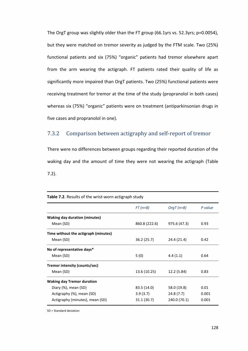
128
The OrgT group was slightly older than the FT group (66.1yrs vs. 52.3yrs; p=0.0054),
but they were matched on tremor severity as judged by the FTM scale. Two (25%)
functional patients and six (75%) “organic” patients had tremor elsewhere apart
from the arm wearing the actigraph. FT patients rated their quality of life as
significantly more impaired than OrgT patients. Two (25%) functional patients were
receiving treatment for tremor at the time of the study (propranolol in both cases)
whereas six (75%) “organic” patients were on treatment (antiparkinsonian drugs in
five cases and propranolol in one).
7.3.2 Comparison between actigraphy and self-report of tremor
There were no differences between groups regarding their reported duration of the
waking day and the amount of time they were not wearing the actigraph (Table
7.2).
Table 7.2. Results of the wrist-worn actigraph study
FT (n=8) OrgT (n=8) P value
Waking day duration (minutes)
Mean (SD)
860.8 (222.6)
975.6 (47.3)
0.93
Time without the actigraph (minutes)
Mean (SD)
36.2 (25.7)
24.4 (21.4)
0.42
No of representative days*
Mean (SD)
5 (0)
4.4 (1.1)
0.64
Tremor intensity (counts/sec)
Mean (SD)
13.6 (10.25)
12.2 (5.84)
0.83
Waking day Tremor duration
Diary (%), mean (SD)
Actigraphy (%), mean (SD)
Actigraphy (minutes), mean (SD)
83.5 (14.0)
3.9 (3.7)
31.1 (30.7)
58.0 (19.8)
24.8 (7.7)
240.0 (70.1)
0.01
0.001
0.001
SD = Standard deviation
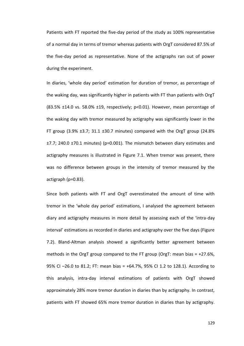
129
Patients with FT reported the five-day period of the study as 100% representative
of a normal day in terms of tremor whereas patients with OrgT considered 87.5% of
the five-day period as representative. None of the actigraphs ran out of power
during the experiment.
In diaries, ‘whole day period’ estimation for duration of tremor, as percentage of
the waking day, was significantly higher in patients with FT than patients with OrgT
(83.5% ±14.0 vs. 58.0% ±19, respectively; p<0.01). However, mean percentage of
the waking day with tremor measured by actigraphy was significantly lower in the
FT group (3.9% ±3.7; 31.1 ±30.7 minutes) compared with the OrgT group (24.8%
±7.7; 240.0 ±70.1 minutes) (p=0.001). The mismatch between diary estimates and
actigraphy measures is illustrated in Figure 7.1. When tremor was present, there
was no difference between groups in the intensity of tremor measured by the
actigraph (p=0.83).
Since both patients with FT and OrgT overestimated the amount of time with
tremor in the ‘whole day period’ estimations, I analysed the agreement between
diary and actigraphy measures in more detail by assessing each of the ‘intra-day
interval’ estimations as recorded in diaries and actigraphy over the five days (Figure
7.2). Bland-Altman analysis showed a significantly better agreement between
methods in the OrgT group compared to the FT group (OrgT: mean bias = +27.6%,
95% CI –26.0 to 81.2; FT: mean bias = +64.7%, 95% CI 1.2 to 128.1). According to
this analysis, intra-day interval estimations of patients with OrgT showed
approximately 28% more tremor duration in diaries than by actigraphy. In contrast,
patients with FT showed 65% more tremor duration in diaries than by actigraphy.
130
The fairly random spread of data points in Figure 7.2 with regard to the x axis
indicates that bias was not proportional to the mean of tremor duration estimates
and actigraphy, but rather subjects had an absolute systematic bias towards
overestimation of tremor.
Figure 7.1. Tremor duration as percentage of the waking day (mean ±standard
deviation), as recorded in self-report diaries and by actigraphy, in patients with
“organic” tremor (OrgT) and Functional tremor (FT).
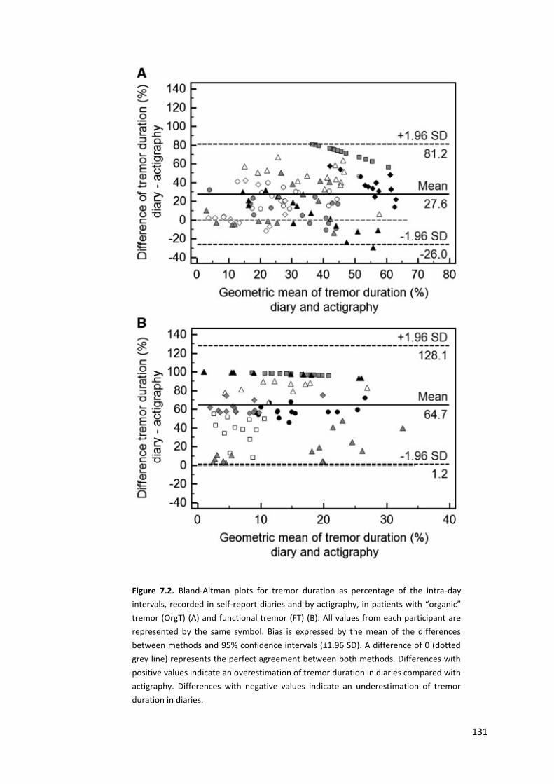
131
Figure 7.2. Bland-Altman plots for tremor duration as percentage of the intra-day
intervals, recorded in self-report diaries and by actigraphy, in patients with “organic”
tremor (OrgT) (A) and functional tremor (FT) (B). All values from each participant are
represented by the same symbol. Bias is expressed by the mean of the differences
between methods and 95% confidence intervals (±1.96 SD). A difference of 0 (dotted
grey line) represents the perfect agreement between both methods. Differences with
positive values indicate an overestimation of tremor duration in diaries compared with
actigraphy. Differences with negative values indicate an underestimation of tremor
duration in diaries.
132
I additionally assessed intensity and duration of tremor at the time when patients
were supposed to be filling in the diaries. Although I gave them instructions to fill in
the diary at particular times of the day, I cannot be completely sure that they
strictly followed the instructions. The most reliable moment that we believe we can
be certain that the patients were filling the questionnaire was at bedtime. I
therefore analysed the intensity and duration of tremor during the 30 minutes
before going to bed. I did not find any increase in the intensity of tremor during this
period in either group. However, for tremor duration, when I calculated the ratio
between the percentage time with tremor over the 30 minutes before going to bed
compared with the percentage time with tremor over the whole third interval (from
7:00pm to bedtime), I observed significant differences between groups (p=0.01).
The FT group had a ratio of 1.7 (indicating an increase of tremor during the 30
minutes before going to bed with respect to the rest of the interval) whereas the
OrgT group had a ratio of 0.7 (indicating a decrease of tremor in the 30 minutes
before going to bed).
In the post-study questionnaire, all patients answered that they believed that the
purpose of the study was to monitor tremor, and that the watch was recording
tremor 24 hours a day.
7.4 Discussion
In this study I have assessed duration and intensity of tremor in patients with FT
and OrgT by using a validated long-term actigraph during five days and
simultaneous self-rated measurements. I have demonstrated a remarkable absence
of tremor during most of the waking day in patients with FT. Despite this, patients
133
with FT reported tremor to be present the majority of the time. In comparison with
OrgT patients, they reported tremor to be present a significantly higher proportion
of the waking day, and compared with the results of actigraphy showed a
significantly greater bias towards over-estimation of tremor.
Patient groups were well matched in terms of objective clinical assessment of
tremor severity at baseline. In line with their estimates of tremor duration, but not
with the actigraphy data, patients with FT rated themselves as significantly more
disabled by tremor (Sub-scale C of the FTM scale), and as having a significantly
poorer quality of life as rated by the EQ-5D (Brooks R, 2003).
7.4.1 Implications for understanding the pathophysiology of
functional tremor
I suggest that these data provide supportive evidence that the majority of patients
with FT (and perhaps by inference other functional disorders) are not malingering. I
was explicit in my explanation of the study to the patients, specifically explaining
that we were using a “tremor watch” to record the actual duration of their tremor
and were comparing this with patients’ own estimates of tremor duration. I gave
patients the opportunity to abort the study at any point. Two of the patients with
FT we approached for the study initially accepted our invitation to take part, but
then returned the actigraph to us without wearing it or completing diaries. The
reasons given for this (arm pain, allergic reaction) may be genuine, but could also
be hypothesised to be excuses to avoid revealing that tremor was not present when
unobserved, compatible with the diagnosis of factitious disorder/malingering.
However, in the rest of the FT group patients completed the study in a similar
134
fashion to “organic” patients. This behaviour, given the explicit study design, seems
incompatible with malingering or factitious disorder, and instead suggests a
genuine perception that tremor was present to the extent reported in the diaries.
If malingering/factitious disorders are not explanations for the tremor in this group
of functional patients, how might these data inform our understanding of the
mechanism of symptom production? I believe that these data reflect the interaction
between perception (diary assessments) and real sensory data (actigraph
measurements). In an ideal neural model these would be identical (i.e. all sensory
data is correctly translated into perceived tremor), but it is clear from personal
experience and numerous experimental studies that perception of sensory data is
dramatically altered by expectation (Koyama et al., 2005, Colloca et al., 2008,
Bulsing et al., 2010). For example, perception of pain can be radically altered by
expectation of the intensity of the pain stimulus (Koyama et al., 2005, Colloca et al.,
2008).
Similarly, I suggest that my data reflect prior expectations about tremor in both
functional and “organic” patients and how this can influence the way they
estimated their tremor. Both patient groups overestimated their time with tremor,
and this could be conceptualised as an over-weighting of prior expectancy about
having tremor over actual sensory data. Such overvaluing of prior expectancy is
indeed the norm in most studies of perception in healthy populations, and may
reflect an evolutionarily beneficial tendency to value past experience over new
sensory information (Elze et al., 2011).
135
However, my data with regard to the significantly greater estimation/actigraph
mismatch in patients with FT fits with an abnormal weighting of prior expectancy in
this patient group such that it overwhelms sensory data that should inform the
patient that they do not have tremor. I hypothesise that whenever patients’
attention is turned towards the symptom (for example during clinical examination
or as demonstrated in the present study, around the time they had to fill in the
diaries) the expectation of the sensory consequences of the symptom is of
sufficient strength to drive the abnormal motor behaviour. On the contrary, when
attention is diverted away from the symptom (for example during most day-to-day
behaviour away from the clinic) tremor stops. However the patient’s perception,
which is moulded by the dominance of the abnormally strong prior expectation
over sensory data, remains that tremor is present most of the time.
7.4.2 Implications for clinical trials in functional tremor
This is the first study to assess FT objectively in a “real-life” ambulatory fashion. To
date, information regarding severity of symptoms in FT has been provided by
objective face-to-face clinical observation using standardised rating scales such as
the FTM or from patient self-report (Jankovic et al., 2006, McKeon et al., 2009). My
data demonstrate that although intensity and severity of FT, when present, is
similar to OrgT measured by actigraphy and rated using standardised scales,
patients’ self-reports do not capture the reality of tremor duration in day-to-day
life. This finding has implications for design of future clinical trials. In essence, my
data suggest that the disability reported by patients with FT is not due to the
tremor itself, but more to their abnormal perception that the tremor is continually
136
present. This would argue for the use of global disability and quality of life
measures along with specific tremor assessments, as outcomes in clinical trials in
this condition.
7.4.3 Limitations
I acknowledge some limitations to this study. Firstly, I have studied a small cohort
of functional and “organic” tremor patients, and I cannot exclude that in a larger
cohort data may be different. However, I chose patients with clinically typical
(albeit longstanding) functional and “organic” tremor diagnosed using standardised
criteria and feel that they do accurately represent patients with these diagnoses.
Secondly, I cannot rule out that the actigraph was underestimating tremor in both
groups as in a previous study 71% of 10 minutes of tremor observation were
classified as tremor by the actigraph (Van Someren et al., 2006). However, studies
using EMG, which is more sensitive to tremor than actigraphy (Spieker et al., 1997),
have found patients with PD to have tremor 28.9% of a 24 hour period and patients
with Essential Tremor to have 15.8%. These results are similar to my data in the
OrgT group. Thirdly, I cannot completely exclude the possibility that tremor was not
accurately recorded by actigraphy in patients with FT because of the known
variability in FT frequency. However, this seems to be unlikely since the filters for
tremor were set over a wide range (3 to 11Hz). Fourth, even though participants
were clearly instructed, we cannot exclude that they overestimated tremor
duration because tremor involving other body parts was also reported.
Nevertheless, the majority of patients with FT had tremor only in the arm wearing
the actigraph, and consequently, this explanation is unlikely to account for the
137
excessive overestimation of tremor in the FT group. Fifth, I set up arbitrarily only
three intra-day interval estimations. It is possible that with shorter intervals, results
would have been different as participants would have thought about the tremor
more often. However, I wanted to assure the correct completion of the study and I
felt three times a day was an achievable number for most of the participants. Six, I
studied patients with FT and no other FMD and one can argue that these results do
not apply for other FMD. Indeed, it seems less likely that patients with functional
fixed dystonia display their symptoms for short periods of time due to the presence
of muscle contractures and shortening of tendons commonly seen in some of these
patients (which would reflect somehow that the abnormal movement is present
most of the time). Seven, although on average patients with FT had 31 minutes of
tremor a day, there may well be patients with FT who have more tremor, if their
attention is turned towards it more often. Finally, I acknowledge that assessment of
the presence or absence of malingering is very difficult and my data cannot fully
exclude this possibility and therefore that there was some purposeful
embellishment in the way functional patients completed the diaries. However, a
post-study questionnaire indicated their understanding of the nature of the study
and I feel malingering is an unlikely explanation of my results.
7.5 Conclusions
In this study, I have demonstrated a dramatic overestimation of duration of tremor
in patients with FT compared with estimates of patients with OrgT and ambulatory
actigraphy. Our data do not support the hypothesis that these patients are
malingering. Instead, these data may reflect an abnormal perception of tremor in

138
patients with FT who might overweight their expectancies regarding tremor
duration.
139
Chapter 8: A study assessing the lack of sense of agency
for movement in functional movement disorders.
This is a pre-copyedited, author-produced PDF of an article accepted for publication in Brain
following peer review. The version of record [Pareés I, Brown H, Nuruki A, Adams RA,
Davare M, Bhatia KP, Friston K, Edwards MJ. Loss of sensory attenuation in patients with
functional (psychogenic) movement disorders. Brain. 2014 Nov;137(Pt 11):2916-21] is
available online at: http://brain.oxfordjournals.org/content/137/11/2916.long.
8.1 Introduction
So far, I have provided evidence for an abnormal attentional focus towards
movement in patients with FMD. However, this requirement of attending to the
movement for the dysfunction to manifest might be expected to be associated with
a strong sense of “voluntariness” or agency for the movement, which is in marked
contrast to what patients with FMD report: they experience the abnormal
movement as involuntary. This issue is at the heart of a centuries old debate on the
level conscious fabrication/manufacturing of functional symptoms. If feigning is not
an explanation for the vast majority of patients with FMD, then the logical
conclusion must be that these patients have an impairment of the mechanisms
implicated in the ability to recognize that one is initiating and controlling one's own
actions, i.e. the sense of agency for movement.
After reviewing the literature, I decided to assess the phenomenon of sensory
attenuation (SA), which is considered to be an implicit measure of the sense of
agency (Blakemore et al., 2002).
140
In brief, SA is the phenomenon whereby the intensity of sensation generated by
self-generated movement is reduced (Blakemore et al., 1998, Blakemore et al.,
2000, Shergill et al., 2003). A common example of this is the observation that while
one cannot tickle oneself, one can be tickled by others. The experience of SA is
important in labelling movements as self-generated and a loss of SA has been
proposed to lead to a loss of agency for movement (Blakemore et al., 2002). In the
experimental setting, SA has been most commonly assessed with a force matching
paradigm (Shergill et al., 2003, Shergill et al., 2005, Voss et al., 2007, Teufel et al.,
2010). Here, subjects are asked to match a force delivered to their finger, either by
pressing directly on their own finger with the other hand, or by operating a joystick
that, via a non-linear transform, causes a robot arm to press down on their finger.
Healthy subjects consistently generate more force than required when directly
pressing on their finger compared with using the joystick, where they are much
more accurate. It has been proposed that the excess force exerted in the first
condition reflects SA of the sensory consequences of self-generated movements,
something not present in the second condition, where the highly nonlinear
transform between movement and sensation disrupts the sense of agency.
I felt that exploring SA in patients with FMD would provide an opportunity to assess
a key psychophysical property of movement that is experienced as self-generated
or voluntary in this group of patients. I predicted that if patients with FMD had
impairment in the sensory attenuation mechanism, they would be more accurate
than controls matching forces when directly pressing on their finger.
141
8.2 Methods
8.2.1 Participants
Fourteen patients with FMD were selected from the General Movement Disorder
clinic of Dr Mark Edwards or Professor Kailash Bhatia as well as from the specialised
Functional Movement Disorder run by Dr Edwards, at the NHNN Queen Square,
London, UK. The specific study was personally explained in detail and if the patient
expressed interest in participating, a formal letter with a full description of the
study and stating the date and location of the experiment was sent.
Inclusion criteria were:
1. Age over 18 years.
2. Diagnosis of clinically established or documented FMD according to Fahn
and Williams criteria (Fahn and Williams, 1988).
3. The FMD was not affecting the upper limbs to ensure the correct
accomplishment of the motor task.
I used as exclusion criteria:
1. Age less than 18 years.
2. Unable to communicate with researcher (e.g. does not speak English).
3. Presence of sensory symptoms or sensory abnormalities (detected on
physical examination).
The control group consisted of 14 healthy participants who were recruited from a
Departmental pool of volunteers and from patients’ relatives who wished to

142
collaborate with the research. They were matched with respect to gender, age, and
handedness.
8.2.2 Questionnaires
8.2.2.1 Handedness
Hand dominance was assessed by the Edinburgh Handedness Inventory (Oldfield,
1971), which has been previously described in this thesis.
8.2.2.2 Mood
We administered the HADs to all participants to assess their mood the week prior
to testing, which has been also previously described in this thesis (Zigmond and
Snaith, 1983).
8.2.2.3 Intelligence level
To estimate the non-verbal intelligence level (IQ) of participants, the 12 items short
form of Raven's Progressive Matrices test was used, previously described in this
thesis (Raven JC, 1977).
8.2.2.4 Delusional ideation
Delusional ideation was assessed using the PDI-21, as previously described (Peters
et al., 2004). I decided to match both groups by delusional ideation because
healthy individuals with higher scores in delusional questionnaires have been
previously found to have a less amount of SA (Teufel et al., 2010).
143
8.2.3 Materials
A small, desk-mounted force-feedback robot arm (PHANTOM® Desktop™ Haptic
Device, Sensible Technologies, Cambridge, MA, USA) was programmed to apply
different forces over a custom-made force transducer, which was placed on the top
of the subject’s left index finger. The force output was recorded by a programmable
output system (Spike 2, version 6, Cambridge Electronic Design (CED), UK).
8.2.4 Experimental Design
I tested each participant in a single experimental session consisting of two main
conditions: matching a target force, either by 1) pressing on themselves with the
right index finger on the left index finger (self-condition) or 2) by manipulating a
robot which pressed down on the left index finger (external-condition). Five
different target forces (16 trials of each), increasing in increments of 0.5 Newton (N)
from 1N to 3N, were randomly presented in both conditions. All subjects completed
a total of four blocks of 20 trials each (80 trials in total) for each condition. The
order of conditions was counterbalanced across participants.
8.2.5 Procedure
Participants sat in front of a table and placed the tip of their left index finger under
the force transducer. The finger was taped to the table to avoid any movement. In
the self-condition, the robot exerted one of the five constant target forces in each
trial for 3s. After 2s of rest, an auditory “go” signal told the participants when to
start matching the target force – by directly pressing with their right index finger for
3s onto the force transducer resting on the left index finger (Figure 8.1). A “stop”
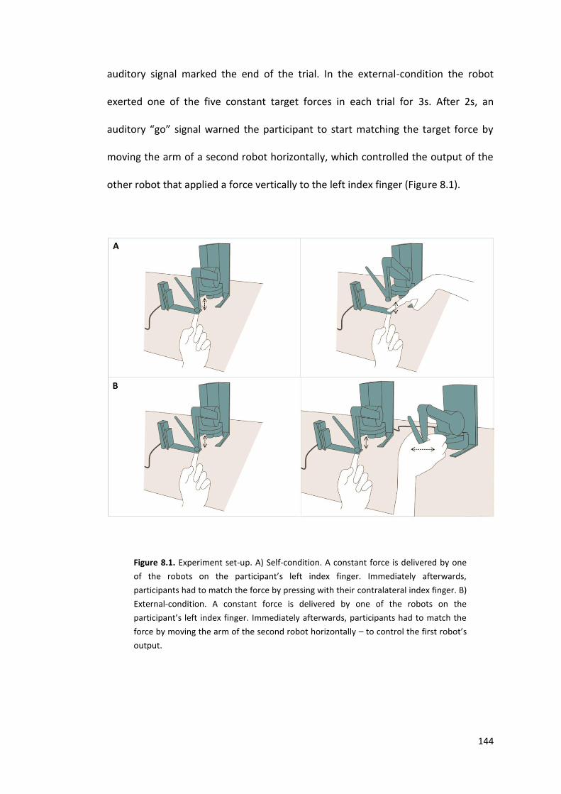
144
auditory signal marked the end of the trial. In the external-condition the robot
exerted one of the five constant target forces in each trial for 3s. After 2s, an
auditory “go” signal warned the participant to start matching the target force by
moving the arm of a second robot horizontally, which controlled the output of the
other robot that applied a force vertically to the left index finger (Figure 8.1).
Figure 8.1. Experiment set-up. A) Self-condition. A constant force is delivered by one
of the robots on the participant’s left index finger. Immediately afterwards,
participants had to match the force by pressing with their contralateral index finger. B)
External-condition. A constant force is delivered by one of the robots on the
participant’s left index finger. Immediately afterwards, participants had to match the
force by moving the arm of the second robot horizontally – to control the first robot’s
output.
145
The force level generated by the subject was calculated for each trial by taking the
mean force recorded by the force sensor between 2000 and 2500ms after the go-
signal. We calculated the ratio between the matched force and the target force for
both conditions (ratio > 1 indicating generation of excessive force) and this was our
measure of SA. This measure was averaged across trials to give the mean
attenuation for each force level and condition.
8.2.6 Statistical analysis
SPSS Statistics software (version 21.0.0) was used for the statistical analysis.
Normality of errors was assessed by using the Kolmogorov-Smirnoff test. When not
normally distributed, the data were subjected to a Log10 transformation. P-values
for categorical variables were calculated with the use of Fisher’s exact test. U-
Mann-Whitney test was used to compare differences in means for numerical data
in baseline characteristics.
A repeated-measures ANOVA was used to compare the results of the main
experiment with CONDITION (Self vs. External) and FORCE (1N, 1.5N, 2N, 2.5N, 3N)
as main factors and GROUP (patients vs. healthy participants) as a between-
subjects factor. Post-hoc tests were conducted with Bonferroni corrections for
multiple comparisons. We calculated potential associations between IQ, PDI-21 and
HADs and the amount of SA (calculated as the mean of the ratios for each force
level in the self-condition) by using Pearson’s correlation. Statistical significance of
p<0.05 was assumed.
146
8.3 Results
Baseline characteristics of the participants are shown in Table 8.1. Patients and
healthy participants were matched for age, gender, handedness, Raven’s and PDI
scores. Most patients had fixed dystonia of the lower limbs as FMD. Clinical
features included an acute onset and rapid escalation of the symptoms. Most
presented dramatic response to placebo or following examination under
anaesthesia and dystonic symptoms often disappeared for a period of time to recur
later on. Most patients were females which is consistent with the majority of them
having fixed dystonia, as there is female predominance in this group of patients.
None of the patients was treated with antipsychotic medication.
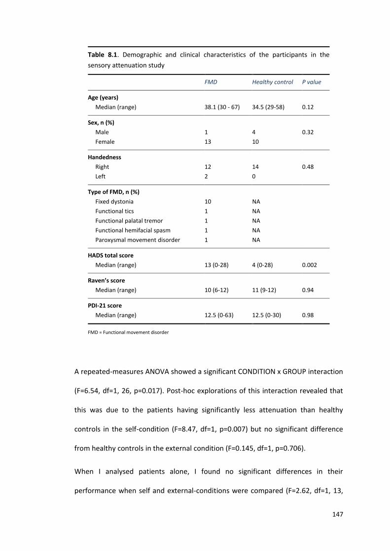
147
Table 8.1. Demographic and clinical characteristics of the participants in the
sensory attenuation study
FMD Healthy control P value
Age (years)
Median (range)
38.1 (30 - 67)
34.5 (29-58)
0.12
Sex, n (%)
Male
Female
1
13
4
10
0.32
Handedness
Right
Left
12
2
14
0
0.48
Type of FMD, n (%)
Fixed dystonia
Functional tics
Functional palatal tremor
Functional hemifacial spasm
Paroxysmal movement disorder
10
1
1
1
1
NA
NA
NA
NA
NA
HADS total score
Median (range)
13 (0-28)
4 (0-28)
0.002
Raven’s score
Median (range)
10 (6-12)
11 (9-12)
0.94
PDI-21 score
Median (range)
12.5 (0-63)
12.5 (0-30)
0.98
FMD = Functional movement disorder
A repeated-measures ANOVA showed a significant CONDITION x GROUP interaction
(F=6.54, df=1, 26, p=0.017). Post-hoc explorations of this interaction revealed that
this was due to the patients having significantly less attenuation than healthy
controls in the self-condition (F=8.47, df=1, p=0.007) but no significant difference
from healthy controls in the external condition (F=0.145, df=1, p=0.706).
When I analysed patients alone, I found no significant differences in their
performance when self and external-conditions were compared (F=2.62, df=1, 13,
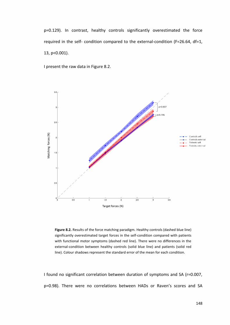
148
p=0.129). In contrast, healthy controls significantly overestimated the force
required in the self- condition compared to the external-condition (F=26.64, df=1,
13, p<0.001).
I present the raw data in Figure 8.2.
Figure 8.2. Results of the force matching paradigm. Healthy controls (dashed blue line)
significantly overestimated target forces in the self-condition compared with patients
with functional motor symptoms (dashed red line). There were no differences in the
external-condition between healthy controls (solid blue line) and patients (solid red
line). Colour shadows represent the standard error of the mean for each condition.
I found no significant correlation between duration of symptoms and SA (r=0.007,
p=0.98). There were no correlations between HADs or Raven’s scores and SA
149
(r=0.29, p=0.91; r=0.147, p=0.62 respectively). PDI-21 total score did not correlate
with the amount of SA (r=-0.67, p=0.82). We calculated a new score as the sum of
the score of just those items in the PDI-21 that imply somehow passivity experience
(i.e. questions 10, 18, 19, 20 and 21). No significant correlation was found between
this score and the amount of SA.
8.4 Discussion
In this study, I have assessed the phenomenon of SA as a measure of self-agency, in
patients with FMD. I have demonstrated that patients with FMD have a loss of SA in
a force-matching task compared with healthy control subjects. As expected, healthy
controls consistently overestimated the force required in the self-condition,
whereas patients did not, and were actually extremely accurate in their force
estimation performance when using their own contralateral index finger. In
contrast, both patients and control behaved exactly the same in the external or
control condition, when they manipulated a second robot to match the different
forces.
SA has been observed in auditory, visual and tactile domains (Martikainen et al.,
2005, Cardoso-Leite et al., 2010, Hughes and Waszak, 2011, Desantis et al., 2012)
and attenuation of Blood Oxygen Level-Dependent (BOLD) responses and
Somatosensor Evoked Potentials (SEP) related to self-generated sensations has also
been demonstrated (Cohen and Starr, 1987, Blakemore et al., 2000). Over the past
decade, the most prominent theoretical account of the phenomenon has been
based on motor control theory, where an efference copy of a motor command is
used to generate a “corollary discharge” – a prediction of the likely sensory
150
consequences of that movement (Blakemore et al., 1999). When movement occurs,
sensory feedback is compared with the sensory prediction and – after a
“subtraction” process – any mismatch can be used to update future commands. It is
proposed that in voluntary movements, predictions of sensory consequences are
very accurate and therefore there is little or no mismatch between predicted and
actual sensation. This lack of mismatch is assumed to attenuate the perceptual
consequences of self-produced movements and may be the cause of force
overestimation in the force matching task. Temporal and spatial offsetting between
the movement and its sensory consequences causes a gradual decline in SA
(Blakemore et al., 1999) Thus the degree of SA is proposed to index in some
manner the “voluntariness” of movement. Conversely, lack of SA has been
proposed to reflect a lack of agency for self-generated movement (Shergill et al.,
2005).
However, some difficulties with the classical model of SA have recently been
highlighted (Brown et al., 2013). For instance, different experiments have
demonstrated that attenuation of externally generated sensations is possible,
which, by definition, cannot be predicted by the forward model of the sensory
consequences of movement. Also, SA commences before the onset of a movement
(Bays et al., 2006), and is not related to the predictability of the stimulus (Bass et
al., 2008). A different approach based on a Bayesian model of the brain has been
recently developed to explain SA (Brown et al., 2013). Under this framework,
attention plays an important role modulating the suppression of the sensory
consequences of voluntary movement and inferring about internally and externally
generated sensations. This will be discussed in more detail in the next chapter.
151
Patients with schizophrenia and healthy individuals with high scores delusional on
questionnaires probing delusional beliefs have been also found to have reduced SA
using a similar paradigm (Shergill et al., 2005, Teufel et al., 2010). These results
have been explained on the basis of a dysfunction in generation of accurate internal
predictive models related to movement, which might be responsible for delusions
of control (or the abnormal belief that one’s own actions are controlled by an
external force). Despite this, psychotic symptoms are not a feature of patients with
FMD, and I did not find differences between delusional ideation measured by PDI-
21 between my patients and healthy controls. It is therefore likely that although
patients with schizophrenia and FMD share the same abnormalities in the SA, they
have different primary causes.
It is important to note that other studies have aimed to explore agency for
movement in patients with functional motor symptoms. However, the employed
methodology makes interpretation difficult. For instance, in a study using fMRI, a
relative reduction in activation of the right inferior parietal lobule was found in
patients with functional tremor comparing activation patterns while they were
tremoring and when they were voluntarily producing tremor (Voon et al., 2010).
Although right inferior parietal lobule is considered to be important in sense of
agency, these data only indirectly address the question of reduced sense of agency
in patients with functional motor symptoms. Two other studies have shown that
patients with FMD judged the feeling of intention to move significantly closer to the
action of moving compared to control participants and had a decreased action-
effect binding when making voluntary movements compared with healthy
volunteers (Edwards et al., 2011, Kranick et al., 2013). However, both studies rely
152
on subjective self-report and are clearly susceptible to important biases. Finally, a
recent study has shown that patients with functional paresis display distinct
electroencephalographic markers compared to feigners (Blakemore et al., 2013).
Among other results, they found that P3 event-related EEG potentials component
was enhanced when the symptomatic hand was moving in contrast to feigners, and
they suggested that this might be related to the lack of awareness that patients
have about the origin of their symptoms. Unlike the previous studies, I believe that
the paradigm employed in my study provides a more direct demonstration that a
key component of normal movement related to sense of agency, and which is
immune to feigned poor performance, differs from healthy controls. Patients fail to
attenuate the sensory consequences of self-produced movement and therefore
they were extremely accurate matching forces as compared to healthy controls.
Data such as these support that feigning is not a satisfactory explanation for the
majority of patients with functional symptoms and support that abnormalities in
the sense of agency for movement underlie this disorder.
I acknowledge a number of limitations of this study. First, the sample size is small
and I cannot exclude that in a larger cohort data may have greater statistical
efficiency. However, we chose patients with clinically typical FMD using
standardized criteria and feel that they do accurately represent patients with this
diagnosis. Furthermore, the fact that I was able to show a significant group by
condition interaction with a relatively small sample size suggests the effect sizes in
question are relatively large. Secondly, it is important to note that functional
tremor is considered the most common FMD and that in our sample, most patients
suffered from functional fixed dystonia, which is considered to be the second
153
commonest diagnosis. This is likely to be due to the exclusion criteria used in this
study. Patients with FMD involving upper limbs were not included and this is the
body part usually affected by functional tremor. Thirdly, while the self and external-
conditions were significantly different in controls we did not find the same amount
of SA in healthy controls compared with previous studies (Shergill et al., 2003,
Shergill et al., 2005). One possible explanation is that the experimental set-up
differed from that used in previous literature. Fourthly, this study does not resolve
the important question of why patients with FMD showed impaired SA in the force-
matching task using non-affected body parts. I believe that one possible
explanation is that lack of SA is a trait (perhaps related to the self-focussed
attention that these patients display), which predisposes them to develop
functional symptoms. It is of note that a proportion of patients with functional
symptoms develop progression and spread of symptoms over time, and while the
initial onset of symptoms is quite commonly associated with a physical or
psychological trigger, spread of symptoms is often apparently spontaneous. Further
studies in asymptomatic patients with a previous history of FMD would clarify this
important question.
In conclusion, patients with FMD display impairment in SA mechanism measured by
a force matching task. This might contribute to explaining the paradox of why
movements that superficially resemble voluntary movements are experienced as
involuntary in this group of patients.

154
Chapter 9: General discussion and conclusions
Part of the discussion described in this chapter has been also written as a review article:
Edwards MJ, Fotopoulou A, Pareés I. Neurobiology of functional (psychogenic) movement
disorders. Current Opinion in Neurology 2013; 26: 442-7.
In this thesis I have explored different aspects of the pathophysiology of FMD. As I
mentioned in the introduction, I took as my starting point the manner in which the
diagnosis of FMD is made in neurological practice and I focused on a mechanistic
rather than on an etiological level.
The findings of the different studies described here suggest that physical illnesses
preceding the onset of FMD, abnormal focus of attention to movement, abnormal
beliefs about the symptoms as well as an impairment in the sense of agency for
movement are elements that play an important role in the mechanism underlying
FMD and that it would be important to incorporate these concepts in any model
seeking to explain this perplexing condition.
In this final chapter I will discuss my results in the light of previous theories, I will
discuss in more detail the methodological limitations of each of my studies and
make suggestions for improvements. Finally, I will suggest directions for future
research.
I started saying that one of the main aims of this work was to try to better
understand the circumstances surrounding the onset of FMD. I have mentioned
how functional symptoms have been typically interpreted as a result of previous
psychological stressors, even if they preceded the onset of symptoms by decades.
155
This was the main argument of most of 19th/early 20th century theories, especially
the Freudian one and is still ingrained within much of modern medical practice.
Likewise, I have highlighted how physical events closely preceding the onset of
functional symptoms have been previously described in the literature although
their role in symptom generation has remained less defined. For instance, Janet
proposed in his dissociation theory that the restriction of the field of consciousness
usually affects a function that has previously become weak. He proposed that
previous physical illness involving the same body part affected by the functional
symptom was a potential causes for this weakening. In the case of Freud, he
proposed that it is actually the psychological stress that accompanies the physical
trauma that is the cause of the functional symptoms rather than the physical injury
itself. As part of the analogy with an electrical system that Freud used to explain
brain function, he considered that any physical illness affecting the body part
concerned could work as by potentially weakening the resistances that prevent the
distribution of the abnormally increased brain excitation in patients with function
al symptoms to other organs.
Based on these observations, in the first study of this thesis I aimed to investigate
how often patients with FMD report a physical event close to the onset of
functional symptoms in a systematic way. I recruited 50 consecutive patients and
studied them with a semi-structured interview. I found that most patients that
attended our clinic described a clear physical problem closely preceding the FMD
and that often, these were phenomenologically related to the functional symptom.
Rather than weakening factors, as proposed by 19th century’s theories, I speculated
156
on the potential role of physical events in generation of functional symptoms. I
suggested that they may provide initial sensory information that, along with other
cognitive and psychological factors, form the substrate for the development of a
specific FMD in vulnerable subjects.
However, it is important to acknowledge that although the causality between the
physical illness and the subsequent FMD sounds plausible, it cannot be established
with the results of a retrospective study and therefore my results should be
interpreted with caution. Important methodological limitations that need to be
underlined are: first, that I relied on the information obtained during a semi-
structured interview and I did not verify the biopsychosocial background of the
patients as well as their previous history including medical details of the physical
event and the onset of the functional symptoms by accessing patients’ medical
records from the two medical contact points initially used by most patients (GP
surgeries and A&E Services). Second, although it was not the main scope of my
study, I assessed life events occurring within the same time window used to assess
the presence of physical event by using a standard questionnaire. I have already
acknowledged how difficult the assessment of life events is, especially with regard
to recall bias. One way to decrease the recall bias would have been the use of a
more reliable method, for instance the Life Events and Difficulties Schedule (Brown
GW, Harris TO, 1979). Here, information about specific life events, timing and
relevant contextual information is collected by a panel of raters but the
participant’s report of his or her reaction to the event at that time is ignored. Based
on the contextual information, the threat for each event is rated.
157
However, the ultimate approach to avoid any recall bias and clarify the potential
causality between physical events and the generation of FMD would have been to
design a prospective multicentre study. Further studies could, for instance, recruit
patients presenting in different GP surgeries or A&E Services with some of the most
commonly physical events found in my study. Regular follow ups during the next
month and examination of suspected cases of FMD within that period by trained
neurologists would permit the identification of the cases and draw conclusions
about causality. However, the large cohort required in order to obtain a reasonable
number of cases as well as the likely costs of such a study make this approach less
feasible.
The second study described in this thesis aimed to explore the role of attention in
the generation of FMD. In one way, this was not a new area of study. We have seen
how the role of attention in functional symptoms dates back to the dissociation
theory of Janet. He proposed that in the case of patients with functional symptoms,
the amount of sensations that can be perceived consciously is limited. He proposed
that this is due to a spontaneous narrowing of their attention. The part of the mind
that become “unattended” is then “dissociated” from consciousness, and the
function of the resulting body part is impaired.
Contradicting somehow this theory, we have seen that it is clinically obvious that in
the case of FMD directing attention to the affected body part exacerbates
functional symptoms and when attention is distracted away they often improve or
disappear. In the study described in Chapter 6 I aimed to translate this clinical
158
observation to a more experimental setting. I used paradigms that had been
previously used in motor physiology literature. For instance, I explored the
capability of the brain to adapt to visuomotor distortions. Adaptation in this context
is the reduction in systematic errors introduced by a 30 degree counterclockwise
angle in the direction used to reach a target in order to return to the former level of
performance. Adaptation to visuomotor rotation is widely considered a form of
implicit motor learning. One reason is that subjects can adapt whilst being unaware
that they are making systematic directional errors. Also, the adaptation occurs even
when subjects are given explicit strategies to override it (Mazzoni P, Krakauer JW,
2006). Adaptation to visuomotor rotation has been found to be normal in patients
with other movement disorders such as patients with primary cervical dystonia
(Katschnig-Winter P, et al, 2014) and asymptomatic carriers with the genetic
mutation for Huntington’s disease (Mazzoni P, Krakauer JW, 2009). In contrast,
visuomotor adaptation has been found abnormal in patients with PD tested with
similar paradigms (Venkatakrishnan A et al, 2011). I found that patients with FMD
can adapt to visuomotor distortion to the same level than healthy subjects,
suggesting that implicit motor control is intact.
The paradigm used to explore explicit motor strategies in FMD was based in the n-
back task, previously used to assess working memory. Here, participants are fully
conscious of the underlying task structure and therefore movement pre-planning is
possible. This paradigm had been previously tested in healthy participants and
patients with the genetic mutation for Huntington’s disease who performed with no
difficulties (Mazzoni P, Krakauer JW, 2009). In contrast, I found that patients with
FMD were impaired in this task, and had slower movement times than controls.
159
Interestingly, RT and accuracy to the trajectory did not differ from controls,
suggesting that the execution of the movement was specifically affected.
Finally, I used a cued choice reaction time task to assess how predictability about
the movement may impact motor control en patients with FMD. I used a motor task
that had been previously designed in the Sobell Department as an adaptation of the
classical visual attention paradigm (Posner paradigm) for a motor setting. Briefly, in
the classical Posner’s paradigm two levels of spatial attention are explored. In the
first condition, voluntary/explicit orientating of attention to a visual target is
assessed. Here, the target is preceded by an arrow located in the centre of the
visual field (cue) which represents potential direction of the location where the
subsequent target may appear. The interpretation of this cue requires a
voluntary/explicit processing by the cognitive system. In the second condition, a
more reflexively/automatic way to orientate attention to a visual target is assessed.
The cue in this case is a peripheral cue displayed in the position where the target
may appear. It automatically attracts attention in a more reflexive way without a
clear voluntary/explicit processing by the cognitive system. In both conditions, cues
can predict or not correctly the position of the target. Valid cues result in a
decrease of the RT when participants respond about the position of the target
compared with invalid cues. In the paradigm used in my experiment, the subjects
were asked to make a fast movement as soon as they detect a target in their visual
field but instead of being presented with cues providing spatial information, the
cues were arbitrarily mapped onto required finger movement responses. The
advantage of this paradigm to study motor control was that attention is cued to the
aspects of movement itself and not only to spatial attention. I hypothesised that
160
increasing predictability of a required movement would allow the opportunity for
controlling the movement in a more explicit way. Therefore, I predicted that
patients would exhibit a paradoxical motor impairment for the valid cues only in
those conditions where the movement would be predictable. I found interesting
results in this paradigm that deserve further comments. First, I found that the
performance of patients with FMD was impaired only in the condition with higher
predictability for the movement (95V). However, patients’ performance did not
differ from healthy participants in the 75V condition, where the movement was also
predictable although to a lesser extent. One can argue that this must be due to the
fact that patients only employed explicit strategies when they were almost certain
about the movement to perform. However, the possibility that these results are
due to chance or other factors cannot be excluded. In this regard it would be
interesting to test additional levels of predictability (i.e. 50V, 65V, 75V, 85V, 95V) to
confirm these results. Also, it would be important to test other group of patients
with other movement disorders to explore how having an abnormal movement
itself may impact in this paradigm. Likewise, there is no data about how affective
disorders may influence the results of this paradigm. The patients included in my
study scored higher in HADs than healthy controls and it would be important for
further studies to include a control group matched by affective symptoms.
Taking into account all these limitations, the results suggest that movement
impairment is restricted to tasks where conscious movement control in the setting
of explicit movement production is possible, and not where movement occurs in a
more automatic, implicit fashion. This is in agreement with the clinical observation
161
of these patients but it would argue against the classical “dissociation” theory that
proposes that it is the inability to attend to the affected body part the likely cause
of functional symptoms. Instead, I propose that it is an abnormal focusing of
attention towards the movement (in case of FMD) which is essential for the
generation of the symptoms.
This view is in keeping with more recent theories about functional symptoms.
Richard J Brown formulated in 2004 a novel theoretical framework for functional
symptoms in which attention plays a central role (Brown, 2004). He suggested that
in the normal physiological state, an attentional system controls the selection of the
relevant sensory information from our body and the environment for further
processing and control of actions. He proposed the primary attentional system as
the one used to organize relevant sensory information into integrated multimodal
perceptual units (primary representations) that provide a working account of the
environment for the control of actions. When actions are well learned or become
into routine behaviours, they are controlled by a hierarchical system of procedural
representations (schemata) specifying the attentional, cognitive and motoric
processes involved in executing well-learned actions. This is a rapid system which
consumes few processing resources. Behaviours controlled in this way are
experienced as effortless. In situations where the system does not have the
appropriate schemata such as in novel actions, these are controlled by a secondary
attentional system (SAS) in a higher level of the system. Actions controlled by SAS
are perceived as mentally demanding and associated with a sense of conscious
volition and self-awareness (Brown, 2004). Brown explained the vulnerability of
162
some people to develop functional symptoms on the basis of self-focused
attention. In the proposed model, the recurrent redirection of attention onto
symptoms by the SAS is the primary pathogenic factor. Allocation of high level
attention to symptoms serves to increase the activation of their brain
representations and decrease the amount of activation required for it to be
selected in the future. In this context, anything that increases self-focused attention
can contribute to the development and maintenance of functional symptoms such
as misattribution of symptoms to physical illness, negative affectivity such as
anxiety and depression, illness worry and rumination, as well as previous traumatic
experiences (Brown, 2004).
My results are also in keeping with more recent studies. For instance, patients with
FT have been shown to spend significantly more time directly looking at their
affected limb during clinical examination compared to patients with “organic”
tremor, suggesting a role for self-directed visual attention in generation of motor
symptoms (van Poppelen et al., 2011). Also, a recent study that compared positron
emission tomography of regional cerebral blood flow in a small cohort of patients
with “fixed” functional dystonia and genetically-characterised primary dystonia as
well as healthy controls showed that fixed dystonia patients had reduced blood
flow in primary motor cortex and increased blood flow in basal ganglia and
cerebellum, which was contrary to that seen in patients with genetic primary
dystonia (Schrag et al., 2013). The authors suggested that the abnormal subcortical
activations in functional patients could reflect problems with self-directed

163
attention/monitoring, perhaps related to fronto-subcortical circuits mediating
motor attention.
Along with attention, there is another element that emerges in this thesis and can
play an important role in symptoms generation and have an important effect
altering the sensory experience of them: the presence of symptom-related
abnormal beliefs (understanding the concept of belief in this context as
expectations about the symptoms).
If we go back to the historical view about the pathophysiology of functional
symptoms, we may remember that it was Janet himself who first acknowledged
how expectations about symptoms may play a role in symptom generation. He
described a patient who developed fixed dystonia in both legs because he thought a
carriage had run over his legs although it was later demonstrated that no injury
actually occurred.
Interestingly, in this thesis I have found that patients with FMD required
significantly less evidence than controls to make a judgement in the JTC paradigm,
which could predispose them to appraise anomalous or ambiguous information
rapidly and produce (abnormal) beliefs on the basis of limited evidence, without a
thorough consideration of alternatives or a review of the evidence. One could
suggest that such a reasoning style, along with other factors, may predispose
patients with FMD to abnormally process sensory data arising from a triggering
event (which we have seen can be often a physical illness) and easily form the
abnormal belief that the sensation represents a symptom of a neurological disease.
164
One important limitation of this study is that patients with FMD scored higher than
healthy controls in the HADS. I acknowledged in chapter 5 that affective
disturbances were not a predictor of the results obtained in my sample (as assessed
by a regression analysis) and this would fit with other studies that also failed to find
a relationship between affective disturbances and the JTC bias. However, the
specific impact of affective symptoms in probabilistic reasoning bias is still to be
elucidated and I cannot exclude that they partially explain my results. Further
studies will therefor need to clarify whether the JTC bias is specific to the functional
condition itself or instead relates to the affective symptoms that commonly
accompany FMD. Either comparing patients with FMD with and without
depression/anxiety symptoms and a healthy control group or matching by affective
symptoms a group of patients with FMD and a group of depression/anxiety patients
with no FMD would help to answer this question. Also, I used only one
combination of beads in my study and it has been shown that task difficulty may
influence the results of in the probabilistic reasoning task (Young HF, Bentall RP,
1997). Participants are more certain in easier (85:15 ratio) than in difficult versions
of the bead task (60:40 ratio). Therefore, I cannot exclude that manipulating the
ratios of the beads in the containers the results in patients with FMD might be
different. Further studies could then assess the performance of these patients with
different ratios. One could argue that if these patients display a JTC bias because
they weight the initial information differently from normal subjects (as I have
proposed), they would express greater certainty in easier conditions (85:15)
compared with the most difficult one (60:40).
165
A reflection that abnormal beliefs about symptoms may be implicated in symptom
generation also comes from the study described in Chapter 7 assessing FT in real
life conditions. Here, I demonstrated that when patients with functional and
“organic” tremor were asked to wear an accelerometer in their most affected hand
in term of tremor, which constantly recorded and stored data over 5 days, both
patient groups subjectively overestimated the amount of time they had tremor, but
functional tremor patients did this to a much greater extent. I suggested that this
significant exaggeration of a natural bias in tremor patients to overestimate tremor
duration could reflect abnormal high level beliefs about there being constant
tremor present. An abnormally strong prediction or expectation about the
symptom may override the real sensory data from the affected limb tremor that
should alert the patient that tremor is not there most of the time. Therefore,
periods without tremor are simply not perceived. I have already acknowledged
limitations in to this study but I think it is important to consider two additional
aspects when interpreting my results. First, most patients with “organic” tremor
had idiopathic PD. Although I matched both group by tremor characteristics and
severity by using the FTM scale, it is likely that PD patients had predominantly rest
tremor. One could argue that because rest tremor causes almost no functional
disability it can be unnoticed and not reported in the diary. This could explain
partially why the “organic” group did not overestimate tremor duration at the same
level than functional patients. Second, there was a statistical trend for patients with
“organic” tremor to be older than patients with FT. Although our patients did not
have significant cognitive impairment, I cannot rule out that this factor was also
influencing the way patients were reporting subjectively their tremors. Therefore, if
166
similar studies are designed in the future to replicate my results, it would be
important, apart from increasing the sample size, to match groups by age and select
a more homogenised control group with predominately postural tremor, which is
clearly functionally disabling and are less likely to be unnoticed (i.e. patients with
essential or dystonic tremor).
The last element emerging from this thesis is the sense of agency relating to
movement (in other words that one is or is not the cause of the movement of one’s
body). This is perhaps the most intriguing aspect of patients with FMD, and the one
which perhaps has caused most difficulty in their interaction with healthcare. Their
movement abnormalities have features typical of voluntary movement and yet they
report them to be involuntary. The question of whether they are in fact malingering
symptoms is impossible to resolve in most ordinary clinical situations. Indeed, it
has been always the centre of debate. We have seen how Charcot and Janet
advised about the difficulties of differentiating patients with functional symptoms
from feigners. We have also seen how some of them, as in the case of Charcot,
argued that most patients with functional symptoms were actually not feigning and
aimed to prove it experimentally by designing specific devices (a plethysmograph-
like machine to assess breathing regularity and fatigue in patients with fixed
dystonia and healthy subjects maintaining voluntarily the same abnormal posture
against a continuous traction).
In this thesis, I have shown that the SA phenomenon (proposed to be a measure of
the sense of agency for movement) is impaired in patients with FMD. I used the
167
force matching paradigm which has been largely assessed in healthy volunteers and
has been demonstrated to be abnormal in patients with delusions or at high risk of
delusional state. I have shown that patients with FMD, similar to those with
delusions and in contrast with healthy controls, did not overestimate the amount of
force applied to match a target force in the self-condition suggesting a deficit in the
SA mechanisms and therefore in the sense of agency for movement. This suggests
that malingering is not an explanation for all these patients and instead, they
display abnormalities in the mechanisms implicated in the experience of oneself as
the agent controlling one's own movements.
However, when interpreting these results it is important to highlight three
important aspects regarding the methodology used:
First, although SA is a well-recognised phenomenon, the underlying mechanisms
and its link with the sense of agency is not well understood. Studies using functional
neuroimaging have shown differences in brain activity during self-generated
relative to externally generated tactile stimulation in healthy controls: an increase
in activity of the secondary somatosensory cortex and the anterior cingulate gyrus
when subjects experienced an externally produced tactile stimulus relative to a self-
produced tactile stimulus has been found. Also, the right anterior cerebellar cortex
was selectively deactivated by self-produced movement resulting in a tactile
stimulus and was activated by externally produced tactile stimulation. Patients with
schizophrenia, in contrast, do not demonstrate attenuation in somatosensory
cortical activation in association with self-generated movement. This may provide a
cerebral basis for the increasing body of behavioural evidence that suggests that
168
misjudgement of agency leads to a set of symptoms of schizophrenia such as
delusions of control, but the evidence is largely indirect.
Second, I have assessed for the first time with this paradigm a clinical population
different to patients with schizophrenia and delusions. Therefore data for
comparing with other patients, especially those with other involuntary movements,
and interpreting my results is lacking.
Third, patients with FMD commonly display emotional disturbances and so far little
is known about how different components of emotions may determine agency
processing at different stages.
Therefore, further studies comparing patients with FMD, patients with other
movement disorders and a group of patients with affective disorders are
encouraged. In this regard, additional work assessing SA with other non-
behavioural paradigms would be also important. They could study for instance the
physiological phenomenon of SEPs at the onset of self-generated movements as
this has been proposed to be the plausible electrophysiological correlate of the
psychophysiological reduction in intensity of self-generated stimuli probed by the
force matching and other behavioural paradigms of SA.
169
A new model for functional symptoms
Taking all the elements emerging from this thesis and summarised in this chapter, it
seems reasonable to suggest that any model seeking to explain FMD and by
extension, other functional neurological disorders, must be able to clarify how a
physical neurological symptom is adopted by an “a priori” normal neurological
system and also explain the paradox of a symptom that requires attention to
clinically manifest (which one can argue that it should be associated with a strong
sense of “voluntariness” ) and that, by contrast, it is felt as involuntary.
There is a contemporary theory, which is based on active inference and a
hierarchical Bayesian formulation of the brain, that could accommodate all findings
described in this thesis (Friston, 2010). There is a robust mathematical framework
that underpins this theory, which can explain the brain from a cellular to a more
behavioural perspective. However, in what follows, I will try to summarize the main
concepts of this theory in a more qualitative, non-mathematical manner.
Within this theory the brain is understood as an inference machine. Here,
perception arises from the interaction of the internal model of the world
(predictions/expectations/beliefs that the brain has about the world) and the
sensory data that the brain receives from the environment. Any mismatch between
the expectations/beliefs and the real sensory data from the environment is known
as prediction error. The aim of the brain is to minimise this mismatch through
interactions between multiple levels of the cortical hierarchy of the neuronal
system. The critical issue with this system is that it is possible to “weight” sensory
data and predictions about that data differently, so one may have more or less
170
influence on the other. Imagine navigating from your bed to the door to the
bedroom in the dark. If it is your own bedroom which you know well, you are likely
to “trust” (i.e. weight) your predictions about the structure of the room over any
sensory information, and so walk boldly (and hopefully correctly) towards the door.
If it is an unfamiliar room you are more likely to weight your sensory feedback, and
so feel carefully along the wall with your hand to try to find the door. Here
attention plays an important role. ‘Attended’ expectations/beliefs or sensations are
granted high weight or precision and perception (and movement control) is
adjusted accordingly.
Within this framework, it can be suggested that sensory data, for instance that
arising from physical events prior to the onset of FMD, combined with many other
factors, including panic or affective and cognitive biases, are afforded excessive
precision (weight) and may lead to the formation of abnormal expectations/beliefs
trying to explain or predict those sensations. This abnormal expectation/belief
whose content would be an abnormal movement or sensation may be rendered
resistant to extinction through the unusually high levels of precision enjoyed during
its formation.
When combined with self-directed attention, precision of these expectations are
high enough to overwhelm contrary sensory data from lower levels and
automatically produce the abnormal movement consistent with the content of the
expectation. This would fit with my results suggesting that the motor impairment
occurs when conscious attentive control of movement is possible or when attention
is directed to the symptom.
171
It is not clear at present exactly how one can place the phenomenon of SA within
this framework, and therefore how one can explain the way in which SA, sense of
agency and this Bayesian model of the brain all interact. One suggestion is that
abnormal attention towards the body in patients with FMD may itself disrupt SA
(Brown et al., 2013). If SA is part of the mechanism whereby a movement is
signalled as intended/willed or not, then its disruption by abnormal attention
towards the body in FMD may be in part a mechanism for why patients might be
more likely to lose a sense of agency for their actions (it is of note that a proportion
of patients with FMD develop progression and spread of symptoms over time with
no particular trigger).
This conclusion raises two crucial questions. First, is the loss of SA in FMD patients a
‘trait’: is it present before and after their motor symptoms? Second, what further
factors are required to transform a trait loss of SA into a state loss of agency for a
particular action? Hopefully, further work will help to provide answers.
172
9.1 Conclusions
FMD and functional neurological symptoms in general are common and disabling
and represent one of the most enigmatic disorders of the brain. Surprisingly, we are
still in the infancy of understanding the underlying pathophysiological mechanism.
The finding of the studies described in this thesis may go some way to enlightening
us on how these symptoms are generated and why they may feel involuntary.
Although these studies may be interpreted as snapshots of what may be occurring
in this group of patients, they do not, unfortunately, provide a clear explanation for
why these perplexing symptoms occur. It was Sigmund Freud himself who
recognized “if by doing this research we have taken a step forward along the path
first traced so successfully by Charcot with his explanation of hysteria, we cannot
conceal from ourselves that this has brought nearer to an understanding only of the
mechanism of hysterical symptoms and not of the internal causes of hysteria. We
have done no more than touch upon the aetiology of hysteria” (Breuer and Freud,
1974).
173
9.2 Implications for further research
As I mentioned at the very beginning of this thesis, I have assessed different aspects
of the pathophysiology of FMD in each of the studies included. In this last chapter I
have gone through each of them and I have suggested further work to improve
them from a methodological perspective. One step forward would be to look at
them before and after recovery from symptoms. This could help to identify whether
they are actually important in symptom generation or not.
For instance, if I was asked to start more studies on FMD, I would assess SA in
patients before and after recovery. In this way, I could better interpret whether
reduced SA is a trait which makes one vulnerable to developing FMD or it is a state
that is only present when patients are symptomatic. This work would be important
to be more confident about the underlying mechanisms of this condition and also
to develop biomarkers which could then be used as surrogate markers in clinical
trials or perhaps even as predictive markers of likely treatment response or poor
prognosis.
Finally, new areas to explore in the future would be to demonstrate whether other
functional neurological symptoms share common underlying mechanisms of
symptom production. I have only assessed patients with FMD in my work and
generalizability of my results to other functional patient samples is not possible. An
interesting aspect would be to demonstrate that abnormally focussed attention is
also present, for instance, in the generation of symptoms characterised by loss of
function such as weakness and hypoesthesia. From the clinical point of view it is
often possible to demonstrate that functional paresis may improve with distraction
174
but demonstration that functional anaesthesia may depend on the presence of
attention is more difficult. This is because attention is unavoidably directed to the
symptom when one assesses sensation. In this regard, further studies could
combine functional imaging and evoked potentials to explore different attentional
responses when manipulating attention towards/away the symptoms in patients
with functional sensory loss.
175
REFERENCES
Alper K, Devinsky O, Perrine K, Vazquez B, Luciano D. Nonepileptic seizures and
childhood sexual and physical abuse. Neurology. 1993;43(10):1950-3.
American Psychiatry Association. Diagnostic and statistical manual of mental
disorders (4th ed., text revision). Washington, DC: Author. 2000.
Anderson KE, Gruber-Baldini AL, Vaughan CG, Reich SG, Fishman PS, Weiner WJ, et
al. Impact of psychogenic movement disorders versus Parkinson's on disability,
quality of life, and psychopathology. Mov Disord. 2007;22(15):2204-9.
Ashktorab H, Hassanzadeh Namin H, Taylor T, Williams C, Brim H, Mellman T, et al.
Role of life events in the presence of colon polyps among African Americans. BMC
Gastroenterol. 2013;13(1):101.
Baik JS, Lang AE. Gait abnormalities in psychogenic movement disorders. Mov
Disord. 2007;22(3):395-9.
Bass P, Jacobsen T, Schroger E. Suppression of the auditory N1 event-related
potential component with unpredictable self-initiated tones: evidence for internal
forward models with dynamic stimulation. Int J Psychophysiol. 2008;70(2):137-43.
Batla A, Stamelou M, Edwards MJ, Pareés I, Saifee TA, Fox Z, et al. Functional
movement disorders are not uncommon in the elderly. Mov Disord.
2013;28(4):540-3.
Bays PM, Flanagan JR, Wolpert DM. Attenuation of self-generated tactile sensations
is predictive, not postdictive. PLoS Biol. 2006;4(2):e28.
176
Beilock SL, Carr TH. On the fragility of skilled performance: what governs choking
under pressure? J Exp Psychol Gen. 2001;130(4):701-25.
Benaderette S, Zanotti Fregonara P, Apartis E, Nguyen C, Trocello JM, Remy P, et al.
Psychogenic parkinsonism: a combination of clinical, electrophysiological, and
[(123)I]-FP-CIT SPECT scan explorations improves diagnostic accuracy. Mov Disord.
2006;21(3):310-7.
Bestmann S, Harrison LM, Blankenburg F, Mars RB, Haggard P, Friston KJ, et al.
Influence of uncertainty and surprise on human corticospinal excitability during
preparation for action. Curr Biol. 2008;18(10):775-80.
Binzer M, Andersen PM, Kullgren G. Clinical characteristics of patients with motor
disability due to conversion disorder: a prospective control group study. J Neurol
Neurosurg Psychiatry. 1997;63(1):83-8.
Blakemore RL, Hyland BI, Hammond-Tooke GD, Anson JG. Distinct modulation of
event-related potentials during motor preparation in patients with motor
conversion disorder. PLoS One. 2013;8(4):e62539.
Blakemore SJ, Frith CD, Wolpert DM. Spatio-temporal prediction modulates the
perception of self-produced stimuli. J Cogn Neurosci. 1999;11(5):551-9.
Blakemore SJ, Wolpert D, Frith C. Why can't you tickle yourself? Neuroreport.
2000;11(11):R11-6.
Blakemore SJ, Wolpert DM, Frith CD. Central cancellation of self-produced tickle
sensation. Nat Neurosci. 1998;1(7):635-40.
177
Blakemore SJ, Wolpert DM, Frith CD. Abnormalities in the awareness of action.
Trends Cogn Sci. 2002;6(6):237-42.
Bland JM, Altman DG. Agreement between methods of measurement with multiple
observations per individual. J Biopharm Stat. 2007;17(4):571-82.
Bogousslavsky J. Following Charcot: A Forgotten History of Neurology and
Psychiatry Basel: Karger; 2011.
Bowman ES, Markand ON. Psychodynamics and psychiatric diagnoses of
pseudoseizure subjects. Am J Psychiatry. 1996;153(1):57-63.
Breuer J, Freud S. Studies on hysteria. The Pelican Freud library. Penguin books.
1974.
Brooks R RR, Charro F. . The measurement and valuation of health status using EQ-
5D: A European perspective. Dordrecht, Netherlands: Kluwer Academic Publishers;
2003.
Brown GW, Harris TO. The Bedford College Life–Events and Difficulty Schedule:
Directory of severity for long–term difficulties. Bedford College, University of
London; London. 1979.
Brown H, Adams RA, Pareés I, Edwards M, Friston K. Active inference, sensory
attenuation and illusions. Cogn Process. 2013.
Brown RJ. Psychological mechanisms of medically unexplained symptoms: an
integrative conceptual model. Psychol Bull. 2004;130(5):793-812.
Buchanan N, Snars J. Pseudoseizures (non epileptic attack disorder)--clinical
management and outcome in 50 patients. Seizure. 1993;2(2):141-6.
178
Bulsing PJ, Smeets MA, Gemeinhardt C, Laverman M, Schuster B, Van den Hout MA,
et al. Irritancy expectancy alters odor perception: evidence from olfactory event-
related potential research. J Neurophysiol. 2010;104(5):2749-56.
Canavese C, Ciano C, Zibordi F, Zorzi G, Cavallera V, Nardocci N. Phenomenology of
psychogenic movement disorders in children. Mov Disord. 2012;27(9):1153-7.
Cardoso-Leite P, Mamassian P, Schutz-Bosbach S, Waszak F. A new look at sensory
attenuation. Action-effect anticipation affects sensitivity, not response bias. Psychol
Sci. 2010;21(12):1740-5.
Charcot JM. Clinical Lectures on Diseases of the Nervous System. London: The New
Sydenham Society; 1889.
Cohen LG, Starr A. Localization, timing and specificity of gating of somatosensory
evoked potentials during active movement in man. Brain. 1987;110 ( Pt 2):451-67.
Cojan Y, Waber L, Carruzzo A, Vuilleumier P. Motor inhibition in hysterical
conversion paralysis. Neuroimage. 2009;47(3):1026-37.
Colloca L, Sigaudo M, Benedetti F. The role of learning in nocebo and placebo
effects. Pain. 2008;136(1-2):211-8.
Corlett PR, Taylor JR, Wang XJ, Fletcher PC, Krystal JH. Toward a neurobiology of
delusions. Prog Neurobiol. 2010;92(3):345-69.
Czarnecki K, Thompson JM, Seime R, Geda YE, Duffy JR, Ahlskog JE. Functional
movement disorders: successful treatment with a physical therapy rehabilitation
protocol. Parkinsonism Relat Disord. 2012;18(3):247-51.
179
Dafotakis M, Ameli M, Vitinius F, Weber R, Albus C, Fink GR, et al. [Transcranial
magnetic stimulation for psychogenic tremor - a pilot study]. Fortschr Neurol
Psychiatr. 2011;79(4):226-33.
Dallocchio C, Arbasino C, Klersy C, Marchioni E. The effects of physical activity on
psychogenic movement disorders. Mov Disord. 2010;25(4):421-5.
de la Fuente-Fernandez R, Ruth TJ, Sossi V, Schulzer M, Calne DB, Stoessl AJ.
Expectation and dopamine release: mechanism of the placebo effect in Parkinson's
disease. Science. 2001;293(5532):1164-6.
de Lange FP, Toni I, Roelofs K. Altered connectivity between prefrontal and
sensorimotor cortex in conversion paralysis. Neuropsychologia. 2010;48(6):1782-8.
Demartini B, Petrochilos P, Ricciardi L, Price G, Edwards MJ, Joyce E. The role of
alexithymia in the development of functional motor symptoms (conversion
disorder). J Neurol Neurosurg Psychiatry. 2014. [Ahead of print]
Desantis A, Weiss C, Schutz-Bosbach S, Waszak F. Believing and perceiving:
authorship belief modulates sensory attenuation. PLoS One. 2012;7(5):e37959.
Deuschl G, Koster B, Lucking CH, Scheidt C. Diagnostic and pathophysiological
aspects of psychogenic tremors. Mov Disord. 1998;13(2):294-302.
Eadie MJ. A pathology of the animal spirits -- the clinical neurology of Thomas Willis
(1621-1675). Part II -- disorders of intrinsically abnormal animal spirits. J Clin
Neurosci. 2003;10(2):146-57.
180
Edwards MJ, Alonso-Canovas A, Schrag A, Bloem BR, Thompson PD, Bhatia K. Limb
amputations in fixed dystonia: A form of body integrity identity disorder? Mov
Disord. 2011.
Edwards MJ, Bhatia KP, Cordivari C. Immediate response to botulinum toxin
injections in patients with fixed dystonia. Mov Disord. 2011;26(5):917-8.
Edwards MJ, Moretto G, Schwingenschuh P, Katschnig P, Bhatia KP, Haggard P.
Abnormal sense of intention preceding voluntary movement in patients with
psychogenic tremor. Neuropsychologia. 2011;49(9):2791-3.
Edwards MJ, Rothwell JC. Losing focus: How paying attention can be bad for
movement. Mov Disord. 2011;26(11):1969-70.
Elze T, Song C, Stollhoff R, Jost J. Chinese characters reveal impacts of prior
experience on very early stages of perception. BMC Neurosci. 2011;12:14.
Erro R, Bhatia KP, Edwards MJ, Farmer SF, Cordivari C. Clinical diagnosis of
propriospinal myoclonus is unreliable: an electrophysiologic study. Mov Disord.
2013;28(13):1868-73.
Espay AJ, Morgante F, Purzner J, Gunraj CA, Lang AE, Chen R. Cortical and spinal
abnormalities in psychogenic dystonia. Ann Neurol. 2006;59(5):825-34.
Esposito M, Edwards MJ, Bhatia KP, Brown P, Cordivari C. Idiopathic spinal
myoclonus: a clinical and neurophysiological assessment of a movement disorder of
uncertain origin. Mov Disord. 2009;24(16):2344-9.
181
Factor SA, Podskalny GD, Molho ES. Psychogenic movement disorders: frequency,
clinical profile, and characteristics. J Neurol Neurosurg Psychiatry. 1995;59(4):406-
12.
Fahn S TE, Marin C. Parkinson's disease and movement disorders. Baltimore: Urban
and Schwarzenberg; 1988.
Fahn S, Williams DT. Psychogenic dystonia. Adv Neurol. 1988;50:431-55.
Fasano A, Valadas A, Bhatia KP, Prashanth LK, Lang AE, Munhoz RP, et al.
Psychogenic facial movement disorders: clinical features and associated conditions.
Mov Disord. 2012;27(12):1544-51.
Feinstein A, Stergiopoulos V, Fine J, Lang AE. Psychiatric outcome in patients with a
psychogenic movement disorder: a prospective study. Neuropsychiatry
Neuropsychol Behav Neurol. 2001;14(3):169-76.
Fekete R, Baizabal-Carvallo JF, Ha AD, Davidson A, Jankovic J. Convergence spasm in
conversion disorders: prevalence in psychogenic and other movement disorders
compared with controls. J Neurol Neurosurg Psychiatry. 2012;83(2):202-4.
Fekete R, Jankovic J. Psychogenic chorea associated with family history of
Huntington disease. Mov Disord. 2010;25(4):503-4.
Ferrara J, Jankovic J. Psychogenic movement disorders in children. Mov Disord.
2008;23(13):1875-81.
Fine C, Gardner M, Craigie J, Gold I. Hopping, skipping or jumping to conclusions?
Clarifying the role of the JTC bias in delusions. Cogn Neuropsychiatry.
2007;12(1):46-77.
182
Fourneret P, Jeannerod M. Limited conscious monitoring of motor performance in
normal subjects. Neuropsychologia. 1998;36(11):1133-40.
Friston K. The free-energy principle: a unified brain theory? Nat Rev Neurosci.
2010;11(2):127-38.
Galea JM, Bestmann S, Beigi M, Jahanshahi M, Rothwell JC. Action reprogramming
in Parkinson's disease: response to prediction error is modulated by levels of
dopamine. J Neurosci. 2012;32(2):542-50.
Ganos C, Aguirregomozcorta M, Batla A, Stamelou M, Schwingenschuh P, Munchau
A, et al. Psychogenic paroxysmal movement disorders - Clinical features and
diagnostic clues. Parkinsonism Relat Disord. 2014;20(1):41-6.
Garcin B, Roze E, Mesrati F, Cognat E, Fournier E, Vidailhet M, et al. Transcranial
magnetic stimulation as an efficient treatment for psychogenic movement
disorders. J Neurol Neurosurg Psychiatry. 2013.
Garety PA, Freeman D, Jolley S, Dunn G, Bebbington PE, Fowler DG, et al.
Reasoning, emotions, and delusional conviction in psychosis. J Abnorm Psychol.
2005;114(3):373-84.
Garety PA, Hemsley DR, Wessely S. Reasoning in deluded schizophrenic and
paranoid patients. Biases in performance on a probabilistic inference task. J Nerv
Ment Dis. 1991;179(4):194-201.
Ghaffar O, Staines WR, Feinstein A. Unexplained neurologic symptoms: an fMRI
study of sensory conversion disorder. Neurology. 2006;67(11):2036-8.
183
Ghilardi MF, Eidelberg D, Silvestri G, Ghez C. The differential effect of PD and
normal aging on early explicit sequence learning. Neurology. 2003;60(8):1313-9.
Ghilardi MF, Silvestri G, Feigin A, Mattis P, Zgaljardic D, Moisello C, et al. Implicit
and explicit aspects of sequence learning in pre-symptomatic Huntington's disease.
Parkinsonism Relat Disord. 2008;14(6):457-64.
Gilman SL. Hysteria Beyond Freud. Berkeley · Los Angeles · Oxford: University of
California Press; 1993.
Gupta A, Lang AE. Psychogenic movement disorders. Curr Opin Neurol.
2009;22(4):430-6.
Hall-Patch L, Brown R, House A, Howlett S, Kemp S, Lawton G, et al. Acceptability
and effectiveness of a strategy for the communication of the diagnosis of
psychogenic nonepileptic seizures. Epilepsia. 2010;51(1):70-8.
Hallett M. Psychogenic Movement Disorders. Neurology and Neuropsychiatry.
Philadelphia: Lippincott Williams & Wilkins; 2006.
Hallett M. Psychogenic movement disorders: a crisis for neurology. Curr Neurol
Neurosci Rep. 2006;6(4):269-71.
Hallett M. Physiology of psychogenic movement disorders. J Clin Neurosci.
2010;17(8):959-65.
Hallett M. Psychogenic parkinsonism. J Neurol Sci. 2011;310(1-2):163-5.
Hinson VK, Cubo E, Comella CL, Goetz CG, Leurgans S. Rating scale for psychogenic
movement disorders: scale development and clinimetric testing. Mov Disord.
2005;20(12):1592-7.
184
Hinson VK, Haren WB. Psychogenic movement disorders. Lancet Neurol.
2006;5(8):695-700.
Hughes G, Waszak F. ERP correlates of action effect prediction and visual sensory
attenuation in voluntary action. Neuroimage. 2011;56(3):1632-40.
Irfan N, Badar A. Top ten stressors in the hysterical subjects of Peshawar. J Ayub
Med Coll Abbottabad. 2002;14(4):38-41.
Janet P. The Major Symptoms of Hysteria. London: The Macmillan Company; 1907.
Jankovic J. Diagnosis and treatment of psychogenic parkinsonism. J Neurol
Neurosurg Psychiatry. 2011.
Jankovic J, Vuong KD, Thomas M. Psychogenic tremor: long-term outcome. CNS
Spectr. 2006;11(7):501-8.
Jueptner M, Stephan KM, Frith CD, Brooks DJ, Frackowiak RS, Passingham RE.
Anatomy of motor learning. I. Frontal cortex and attention to action. J
Neurophysiol. 1997;77(3):1313-24.
Kanaan RA, Craig TK, Wessely SC, David AS. Imaging repressed memories in motor
conversion disorder. Psychosom Med. 2007;69(2):202-5.
Katschnig-Winter P, Schwingenschuh P, Davare M, Sadnicka A, Schmidt R, Rothwell
JC, Bhatia KP, Edwards MJ. Motor sequence learning and motor adaptation in
primary cervical dystonia. J Clin Neurosci. 2014;21(6):934-8
Kim YJ, Pakiam AS, Lang AE. Historical and clinical features of psychogenic tremor: a
review of 70 cases. Can J Neurol Sci. 1999;26(3):190-5.
185
Koller W, Lang A, Vetere-Overfield B, Findley L, Cleeves L, Factor S, et al.
Psychogenic tremors. Neurology. 1989;39(8):1094-9.
Koyama T, McHaffie JG, Laurienti PJ, Coghill RC. The subjective experience of pain:
where expectations become reality. Proc Natl Acad Sci U S A. 2005;102(36):12950-
5.
Kranick S, Ekanayake V, Martinez V, Ameli R, Hallett M, Voon V. Psychopathology
and psychogenic movement disorders. Mov Disord. 2011.
Kranick SM, Moore JW, Yusuf N, Martinez VT, Lafaver K, Edwards MJ, et al. Action-
effect binding is decreased in motor conversion disorder: Implications for sense of
agency. Mov Disord. 2013.
Lang AE. Psychogenic dystonia: a review of 18 cases. Can J Neurol Sci.
1995;22(2):136-43.
Lempert T, Dieterich M, Huppert D, Brandt T. Psychogenic disorders in neurology:
frequency and clinical spectrum. Acta Neurol Scand. 1990;82(5):335-40.
Linden SC, Jones E, Lees AJ. Shell shock at Queen Square: Lewis Yealland 100 years
on. Brain. 2013;136(Pt 6):1976-88.
Mai FM, Merskey H. Briquet's concept of hysteria: an historical perspective. Can J
Psychiatry. 1981;26(1):57-63.
Mailis-Gagnon A, Giannoylis I, Downar J, Kwan CL, Mikulis DJ, Crawley AP, et al.
Altered central somatosensory processing in chronic pain patients with "hysterical"
anesthesia. Neurology. 2003;60(9):1501-7.
Marsden CD. Hysteria--a neurologist's view. Psychol Med. 1986;16(2):277-88.
186
Martikainen MH, Kaneko K, Hari R. Suppressed responses to self-triggered sounds
in the human auditory cortex. Cereb Cortex. 2005;15(3):299-302.
Mazzoni P, Krakauer JW. An implicit plan overrides an explicit strategy during
visuomotor adaptation. J Neurosci. 2006;26(14):3642-5.
Mazzoni P, Wexler NS. Parallel explicit and implicit control of reaching. PLoS One.
2009;4(10):e7557.
McKeon A, Ahlskog JE, Bower JH, Josephs KA, Matsumoto JY. Psychogenic tremor:
long-term prognosis in patients with electrophysiologically confirmed disease. Mov
Disord. 2009;24(1):72-6.
Mejia NI, Jankovic J. Secondary tics and tourettism. Rev Bras Psiquiatr.
2005;27(1):11-7.
Molnar Z. Thomas Willis (1621-1675), the founder of clinical neuroscience. Nat Rev
Neurosci. 2004;5(4):329-35.
Monday K, Jankovic J. Psychogenic myoclonus. Neurology. 1993;43(2):349-52.
Moss-Morris R, Spence M. To "lump" or to "split" the functional somatic
syndromes: can infectious and emotional risk factors differentiate between the
onset of chronic fatigue syndrome and irritable bowel syndrome? Psychosom Med.
2006;68(3):463-9.
Norbeck JS. Modification of life event questionnaires for use with female
respondents. Res Nurs Health. 1984;7(1):61-71.
Oldfield RC. The assessment and analysis of handedness: the Edinburgh inventory.
Neuropsychologia. 1971;9(1):97-113.
187
Onofrj M, Bonanni L, Manzoli L, Thomas A. Cohort study on somatoform disorders
in Parkinson disease and dementia with Lewy bodies. Neurology. 2010;74(20):1598-
606.
Onofrj M, Thomas A, Tiraboschi P, Wenning G, Gambi F, Sepede G, et al. Updates on
Somatoform Disorders (SFMD) in Parkinson's Disease and Dementia with Lewy
Bodies and discussion of phenomenology. J Neurol Sci. 2011;310(1-2):166-71.
Orrell AJ, Masters RS, Eves FF. Reinvestment and movement disruption following
stroke. Neurorehabil Neural Repair. 2009;23(2):177-83.
Pareés I, Saifee TA, Kojovic M, Kassavetis P, Rubio-Agusti I, Sadnicka A, et al.
Functional (psychogenic) symptoms in Parkinson's disease. Mov Disord. 2013.
Peters E, Joseph S, Day S, Garety P. Measuring delusional ideation: the 21-item
Peters et al. Delusions Inventory (PDI). Schizophr Bull. 2004;30(4):1005-22.
Ranawaya R, Riley D, Lang A. Psychogenic dyskinesias in patients with organic
movement disorders. Mov Disord. 1990;5(2):127-33.
Raven JC CJ, Raven J. Manual for Raven's progressive matrices and vocabulary
scales. Section 4, Advanced progressive matrices: sets I and II. London: H. K. Lewis;
1977.
Reicker LI. The ability of reaction time tests to detect simulation: an investigation of
contextual effects and criterion scores. Arch Clin Neuropsychol. 2008;23(4):419-31.
Roelofs K, de Bruijn ER, Van Galen GP. Hyperactive action monitoring during motor-
initiation in conversion paralysis: an event-related potential study. Biol Psychol.
2006;71(3):316-25.
188
Roelofs K, Keijsers GP, Hoogduin KA, Naring GW, Moene FC. Childhood abuse in
patients with conversion disorder. Am J Psychiatry. 2002;159(11):1908-13.
Roelofs K, van Galen GP, Eling P, Keijsers GP, Hoogduin CA. Endogenous and
exogenous attention in patients with conversion paresis. Cogn Neuropsychol.
2003;20(8):733-45.
Rommelfanger KS. Opinion: A role for placebo therapy in psychogenic movement
disorders. Nat Rev Neurol. 2013;9(6):351-6.
Roper LS, Saifee TA, Pareés I, Rickards H, Edwards MJ. How to use the entrainment
test in the diagnosis of functional tremor. Pract Neurol. 2013;13(6):396-8.
Saifee TA, Kassavetis P, Pareés I, Kojovic M, Fisher L, Morton L, et al. Inpatient
treatment of functional motor symptoms: a long-term follow-up study. J Neurol.
2012;259(9):1958-63.
Schrag A, Trimble M, Quinn N, Bhatia K. The syndrome of fixed dystonia: an
evaluation of 103 patients. Brain. 2004;127(Pt 10):2360-72.
Schrag AE, Mehta AR, Bhatia KP, Brown RJ, Frackowiak RS, Trimble MR, et al. The
functional neuroimaging correlates of psychogenic versus organic dystonia. Brain.
2013;136(Pt 3):770-81.
Schwingenschuh P, Katschnig P, Edwards MJ, Teo JT, Korlipara LV, Rothwell JC, et al.
The blink reflex recovery cycle differs between essential and presumed psychogenic
blepharospasm. Neurology. 2011;76(7):610-4.
189
Schwingenschuh P, Katschnig P, Seiler S, Saifee TA, Aguirregomozcorta M, Cordivari
C, et al. Moving toward "laboratory-supported" criteria for psychogenic tremor.
Mov Disord. 2011.
Schwingenschuh P, Pont-Sunyer C, Surtees R, Edwards MJ, Bhatia KP. Psychogenic
movement disorders in children: a report of 15 cases and a review of the literature.
Mov Disord. 2008;23(13):1882-8.
Sharpe M, Walker J, Williams C, Stone J, Cavanagh J, Murray G, et al. Guided self-
help for functional (psychogenic) symptoms: a randomized controlled efficacy trial.
Neurology. 2011;77(6):564-72.
Shergill SS, Bays PM, Frith CD, Wolpert DM. Two eyes for an eye: the neuroscience
of force escalation. Science. 2003;301(5630):187.
Shergill SS, Samson G, Bays PM, Frith CD, Wolpert DM. Evidence for sensory
prediction deficits in schizophrenia. Am J Psychiatry. 2005;162(12):2384-6.
Shibasaki H, Hallett M. What is the Bereitschaftspotential? Clin Neurophysiol.
2006;117(11):2341-56.
Shill H, Gerber P. Evaluation of clinical diagnostic criteria for psychogenic movement
disorders. Mov Disord. 2006;21(8):1163-8.
Siegert RJ, Taylor KD, Weatherall M, Abernethy DA. Is implicit sequence learning
impaired in Parkinson's disease? A meta-analysis. Neuropsychology.
2006;20(4):490-5.
Spieker S, Boose A, Breit S, Dichgans J. Long-term measurement of tremor. Mov
Disord. 1998;13 Suppl 3:81-4.
190
Spieker S, Strole V, Sailer A, Boose A, Dichgans J. Validity of long-term
electromyography in the quantification of tremor. Mov Disord. 1997;12(6):985-91.
Stamelou M, Saifee TA, Edwards MJ, Bhatia KP. Psychogenic palatal tremor may be
underrecognized: reappraisal of a large series of cases. Mov Disord.
2012;27(9):1164-8.
Stone J, Carson A, Aditya H, Prescott R, Zaubi M, Warlow C, et al. The role of
physical injury in motor and sensory conversion symptoms: a systematic and
narrative review. J Psychosom Res. 2009;66(5):383-90.
Stone J, Carson A, Duncan R, Roberts R, Coleman R, Warlow C, et al. Which
neurological diseases are most likely to be associated with "symptoms unexplained
by organic disease". J Neurol. 2012;259(1):33-8.
Stone J, Carson A, Duncan R, Roberts R, Warlow C, Hibberd C, et al. Who is referred
to neurology clinics?--the diagnoses made in 3781 new patients. Clin Neurol
Neurosurg. 2010;112(9):747-51.
Stone J, Reuber M, Carson A. Functional symptoms in neurology: mimics and
chameleons. Pract Neurol. 2013;13(2):104-13.
Stone J, Smyth R, Carson A, Lewis S, Prescott R, Warlow C, et al. Systematic review
of misdiagnosis of conversion symptoms and "hysteria". BMJ. 2005;331(7523):989.
Stone J, Warlow C, Sharpe M. The symptom of functional weakness: a controlled
study of 107 patients. Brain. 2010;133(Pt 5):1537-51.
191
Stone J, Wojcik W, Durrance D, Carson A, Lewis S, MacKenzie L, et al. What should
we say to patients with symptoms unexplained by disease? The "number needed to
offend". BMJ. 2002;325(7378):1449-50.
Stone J, Zeman A, Simonotto E, Meyer M, Azuma R, Flett S, et al. FMRI in patients
with motor conversion symptoms and controls with simulated weakness.
Psychosom Med. 2007;69(9):961-9.
Tan EK, Jankovic J. Psychogenic hemifacial spasm. J Neuropsychiatry Clin Neurosci.
2001;13(3):380-4.
Tasca C, Rapetti M, Carta MG, Fadda B. Women and hysteria in the history of
mental health. Clin Pract Epidemiol Ment Health. 2012;8:110-9.
Teufel C, Kingdon A, Ingram JN, Wolpert DM, Fletcher PC. Deficits in sensory
prediction are related to delusional ideation in healthy individuals.
Neuropsychologia. 2010;48(14):4169-72.
Thomas M, Vuong KD, Jankovic J. Long-term prognosis of patients with psychogenic
movement disorders. Parkinsonism Relat Disord. 2006;12(6):382-7.
van Beilen M, Griffioen BT, Gross A, Leenders KL. Psychological assessment of
malingering in psychogenic neurological disorders and non-psychogenic
neurological disorders: relationship to psychopathology levels. Eur J Neurol.
2009;16(10):1118-23.
van der Salm SM, Koelman JH, Henneke S, van Rootselaar AF, Tijssen MA. Axial
jerks: a clinical spectrum ranging from propriospinal to psychogenic myoclonus. J
Neurol. 2010;257(8):1349-55.
192
van Hilten BJ, van de Beek WJ, Hoff JI, Voormolen JH, Delhaas EM. Intrathecal
baclofen for the treatment of dystonia in patients with reflex sympathetic
dystrophy. N Engl J Med. 2000;343(9):625-30.
van Poppelen D, Saifee TA, Schwingenschuh P, Katschnig P, Bhatia KP, Tijssen MA,
et al. Attention to self in psychogenic tremor. Mov Disord. 2011;26(14):2575-6.
Van Someren EJ, Pticek MD, Speelman JD, Schuurman PR, Esselink R, Swaab DF.
New actigraph for long-term tremor recording. Mov Disord. 2006;21(8):1136-43.
Verdugo RJ, Ochoa JL. Abnormal movements in complex regional pain syndrome:
assessment of their nature. Muscle Nerve. 2000;23(2):198-205.
Parkinson’s disease differentially affects adaptation to gradual as compared to
sudden visuomotor distortions. Venkatakrishnan A, Banquet JP, Burnod Y,
Contreras-vidal JL. Hum Mov Sci. 2011;30(4):760-9.
Voon V, Brezing C, Gallea C, Ameli R, Roelofs K, LaFrance WC, Jr., et al. Emotional
stimuli and motor conversion disorder. Brain. 2010;133(Pt 5):1526-36.
Voon V, Brezing C, Gallea C, Hallett M. Aberrant supplementary motor complex and
limbic activity during motor preparation in motor conversion disorder. Mov Disord.
2011;26(13):2396-403.
Voon V, Gallea C, Hattori N, Bruno M, Ekanayake V, Hallett M. The involuntary
nature of conversion disorder. Neurology. 2010;74(3):223-8.
Voon V, Lang AE. Antidepressant treatment outcomes of psychogenic movement
disorder. J Clin Psychiatry. 2005;66(12):1529-34.
193
Voss M, Bays PM, Rothwell JC, Wolpert DM. An improvement in perception of self-
generated tactile stimuli following theta-burst stimulation of primary motor cortex.
Neuropsychologia. 2007;45(12):2712-7.
Wilkinson L, Jahanshahi M. The striatum and probabilistic implicit sequence
learning. Brain Res. 2007;1137(1):117-30.
Willison J, Tombaugh TN. Detecting simulation of attention deficits using reaction
time tests. Arch Clin Neuropsychol. 2006;21(1):41-52.
Woody SR. Effects of focus of attention on anxiety levels and social performance of
individuals with social phobia. J Abnorm Psychol. 1996;105(1):61-9.
Wyllie E, Friedman D, Rothner AD, Luders H, Dinner D, Morris H, 3rd, et al.
Psychogenic seizures in children and adolescents: outcome after diagnosis by ictal
video and electroencephalographic recording. Pediatrics. 1990;85(4):480-4.
Young HF1, Bentall RP. Probabilistic reasoning in deluded, depressed and normal
subjects: effects of task difficulty and meaningful versus non-meaningful material.
Psychol Med. 1997;27(2):455-65.
Zigmond AS, Snaith RP. The hospital anxiety and depression scale. Acta Psychiatr
Scand. 1983;67(6):361-70.
194
APPENDIX
The author’s publications in scientific journals in relation to functional movement
disorders (those not included in this thesis are also listed).
1. Pareés I, Brown H, Nuruki A, Adams RA, Davare M, Bhatia KP, Friston K, Edwards
MJ. Loss of sensory attenuation in patients with functional (psychogenic)
movement disorders. Brain 2014; 137:2916-21
2. Pareés I, Kojovic M, Pires C, Rubio-Agusti I, Saifee TA, Sadnicka A, Kassavetis P,
Macerollo A, Bhatia KP, Carson A, Stone J, Edwards MJ. Physical precipitating
factors in functional movement disorders. J Neurol Sci 2014; 338:174-7
3. Pareés I, Saifee TA, Edwards MJ. Functional motor or somatoform disorders in
PD? Mov Disord 2013. [Epub ahead of print].
4. Batla A, Panicker J, Edwards M, Stamelou M, Pareés I, Kasavetis P, Bhatia K.
Lower urinary tract symptoms in functional movement disorders. J Neurol
Neurosurg Psychiatry 2013. [Epub ahead of print].
5. Pareés I, Kassavetis P, Saifee TA, Sadnicka A, Davare M, Bhatia KP, Rothwell JC,
Bestmann S, Edwards MJ. Failure of explicit movement control in patients with
functional motor symptoms. Mov Disord 2013; 28:517-23.
6. Batla A, Stamelou M, Edwards MJ, Pareés I, Saifee TA, Fox Z, Bhatia KP.
Functional movement disorders are not uncommon in the elderly. Mov Disord
2013; 28:540-3.
7. Stamelou M, Cossu G, Edwards MJ, Murgia D, Pareés I, Melis M, Bhatia KP.
Familial psychogenic movement disorders. Mov Disord. 2013; 28:1295-8.
195
8. Pareés I, Saifee TA, Kojovic M, Kassavetis P, Rubio-Agusti I, Sadnicka A, Bhatia
KP, Edwards MJ. Functional (psychogenic) symptoms in Parkinson's disease.
Mov Disord 2013. [Epub ahead of print].
9. Brown H, Adams RA, Pareés I, Edwards M, Friston K. Active inference, sensory
attenuation and illusions. Cogn Process 2013 [Epub ahead of print].
10. Roper LS, Saifee TA, Pareés I, Rickards H, Edwards MJ. How to use the
entrainment test in the diagnosis of functional tremor. Pract Neurol 2013 [Epub
ahead of print].
11. Edwards MJ, Fotopoulou A, Pareés I. Neurobiology of functional (psychogenic)
movement disorders. Curr Opin Neurol 2013; 26:442-7.
12. Pareés I, Saifee TA, Kassavetis P, Kojovic M, Rubio-Agusti I, Rothwell JC, Bhatia
KP, Edwards MJ. Believing is perceiving: mismatch between self-report and
actigraphy in psychogenic tremor. Brain 2012; 135:117-23.
13. Pareés I, Kassavetis P, Saifee TA, Sadnicka A, Bhatia KP, Fotopoulou A, Edwards
MJ. 'Jumping to conclusions' bias in functional movement disorders. J Neurol
Neurosurg Psychiatry 2012; 83:460-3.
14. Kassavetis P, Batla A, Pareés I, Saifee TA, Schrag A, Cordivari C, Bhatia KP,
Edwards MJ. Joint hypermobility syndrome: a risk factor for fixed dystonia? Mov
Disord 2012; 27:1070
15. Saifee TA, Kassavetis P, Pareés I, Kojovic M, Fisher L, Morton L, Foong J, Price G,
Joyce EM, Edwards MJ. Inpatient treatment of functional motor symptoms: a
long-term follow-up study. J Neurol 2012; 259:1958-63.
16. Edwards MJ, Adams RA, Brown H, Pareés I, Friston KJ. A Bayesian account of
'hysteria'. Brain 2012; 135:3495-512.
196
The author’s contribution in books in relation to functional movement disorders:
1. Pareés and M.J. Edwards. Psychogenic Movement Disorders. In: Baumann and
Wolters (ed). Parkinson’s disease and other Movement Disorders. VU University
Press (2014).
2. Pareés and M.J. Edwards. Psychogenic Movement Disorders. In: David J Burn,
Christopher Kennard (ed). Oxford Textbook of Movement Disorders. Oxford
University Press (2012).

197
Recommended