Chapter 1
Introduction to Pathology
Zhao Guoqiang
Introduction to Pathology
• Definition of Pathology
• Evolution of Pathology
• Subdivision of Pathology
• Methods for the study of pathology
Definition of Pathology
The word “Pathology” is derived from two Greek word ----
pathos meaning suffering
logos meaning study
Definition of Pathology
Pathology is scientific study of structure and
function of the body in disease.
It deals with causes, effects, mechanisms and
nature of disease.
The knowledge and understanding of
pathology is essential for all would-be
doctors as well as general practitioners and
specialists since unless they know the causes
and mechanisms of disease and understand
the language spoken by the pathologist in
the form of laboratory reports, they would not
be able to institute appropriate treatment or
suggest preventive measures to the patient.
For the medical student, the discipline
of pathology forms a vital bridge
between initial learning phase of
preclinical science and the final phase
of clinical subjects.
Evolution of Pathology
• From religious beliefs to rational approach
(Antiquity to AD 1500)
• Era of gross pathology (AD 1500 to 1800)
• Era of technology development and cellular
pathology (AD 1800 to 1950s)
• Modern pathology (1950s to dawn of 21st
century)
From religious beliefs to rational approach (Antiquity to AD 1500)
• Hippocrates (Greece) 460-377 BCPermanently dissociated medicine from religious mysticism. Started study of patient’s symptoms as method of diagnosis.
• Cornelius Celsus (Rome) 53 BC-7 ADDescribed 4 cardinal signs of inflammation (redness, heat, swelling, pain)

Hippocrates (Greece) 460-377 BC
Era of gross pathology (AD 1500 to 1800)
• Giovanni B Morgagni (Italy) 1682-1771Introduced clinicopathologic correlation (CPC) in the study of disease
• John Hunter (Scotland) 1728-1793Introduced pathology museum in the study of disease.
• R.T.H. Laennec (France) 1781-1826Described several lung diseases such as various tuberculous lesions of lungs, bronchiectasis. Described cirrhosis of liver (later called Laennec’s cirrhosis).Invented stethoscope.

John Hunter (Scotland) 1728-1793
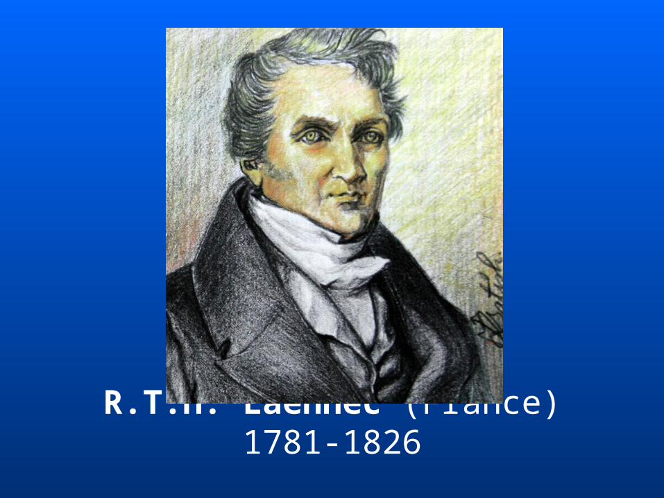
R.T.H. Laennec (France) 1781-1826
Era of technology development and cellular pathology (AD 1800 to 1950s)• Rudolf Virchow (Germany) 1821-1905
Father of cellular pathologyIntroduced histopathology as a diagnostic branch by his cellular theory
• George N. Papanicolaou (USA) 1883-1962Father of exfoliative cytology Developed Pap smear for detection of cervical cancer in 1930s
Rudolf Virchow (Germany) 1821-1905
George N. Papanicolaou (USA) 1883-1962
Modern pathology (1950s to dawn of 21st century)• Watson and Crick 1953
Described the structure of DNA
• Nowell and Hagerford 1960Philadelphia chromosome in CML i.e. t(9;22)
• Gall and Pardue 1969In Situ Hybridization
• Kary Mullis 1983Introduced polymerase chain reaction (PCR)
Prof. Liang Boqiang 1899-1968
Prof. Qin Guangyu 1902-1969
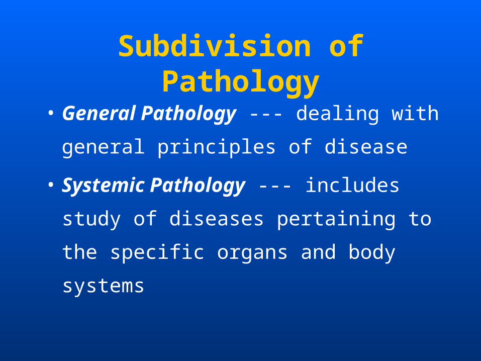
Subdivision of Pathology
• General Pathology --- dealing with general
principles of disease
• Systemic Pathology --- includes study of
diseases pertaining to the specific organs
and body systems
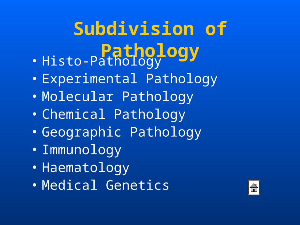
Subdivision of Pathology• Histo-Pathology• Experimental Pathology• Molecular Pathology • Chemical Pathology• Geographic Pathology • Immunology• Haematology• Medical Genetics
Methods for the study of Pathology
• Autopsy
• Biopsy
• Cytology
What is Autoposy?
Autopsy means "see for yourself". It is a
special surgical operation, performed by
specially-trained physicians, on a dead
body. Its purpose is to learn the truth about
the person's health during life, and how the
person really died.
What is Bioposy?
A biopsy is the removal of a sample of tissue
from the body for examination. The tissue
will be examined under a microscope to
assist in diagnosis. Therefore, only very small
samples are needed.
What is Cytology?
Cytology can also refer to cytopathology,
which analyzes cell structure to diagno
se disease .


肖 萍 (Xiao Ping) 老师办公室:新教学楼 4F
电话: 87330743
Chapter 2
Cellular Adaptations and Cell Injury
Cellular Adaptations and Cell Injury
• Cellular Responses to Stress and
Noxious Stimuli
• Cellular adaptations
• Causes of cell injury
• Mechanisms of cell injury
• Morphology of cell injury
Human body is quite complex and is
made of 70,000 billion cells.
In health, these cells remain in accord
with each other.
However, most forms of diseases begin
with cell injury and consequent loss of
cellular function .
Injury is defined as an alteration in cell
structure or function resulting from
some stress that exceeds the ability of
the cell to compensate through normal
physiologic adaptive mechanisms.
Cells typically respond to potentially injurious
stress in one of two ways:
Adaptation - Cells can alter their structure and/or biochemical processes in order to achieve a new "steady state" and maintain near-normal physiologic
functions (homeostasis).
Injury - If stressed cells cannot adequately adapt, critical cell functions may be impaired, and the cell
is said to be injured.
Reversible and irreversible injury
If injured cells recover their normal functions when the stress is removed, the injury is said to be reversible.
If the injury is severe enough, however, a “Point of no return” is reached and the cell suffers irreversible injury and dies.
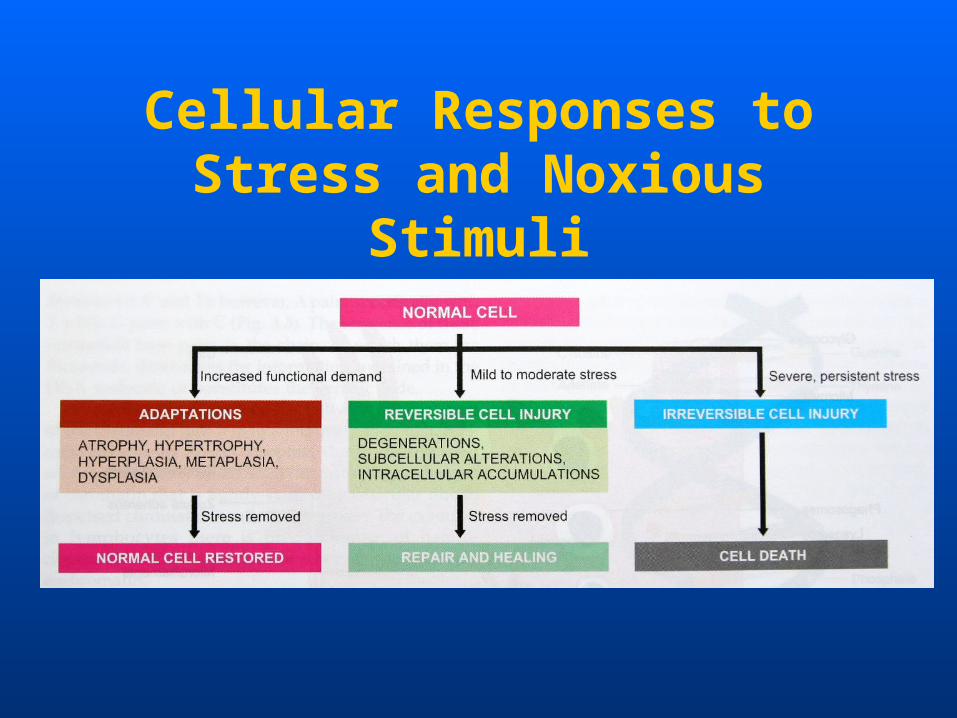
Cellular Responses to Stress and Noxious Stimuli
Adaptation,
Reversible injury,
Irreversible injury (Cell death)
may be considered as different stages of a
progressive impairment of the cell’s normal
function and structure.
Cellular adaptations
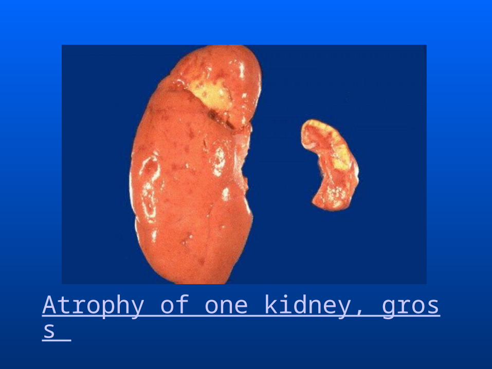
• Atrophy
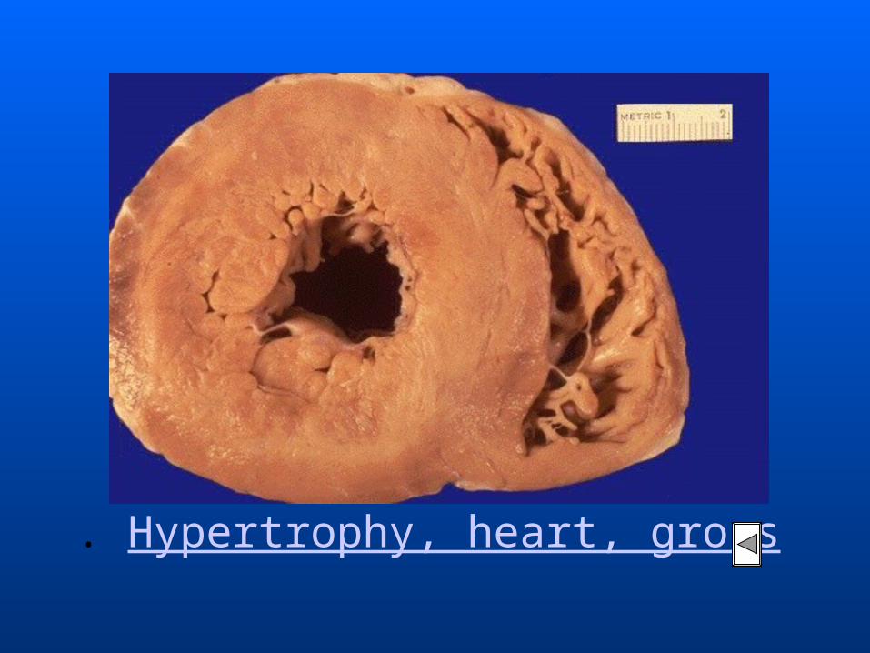
• Hypertrophy
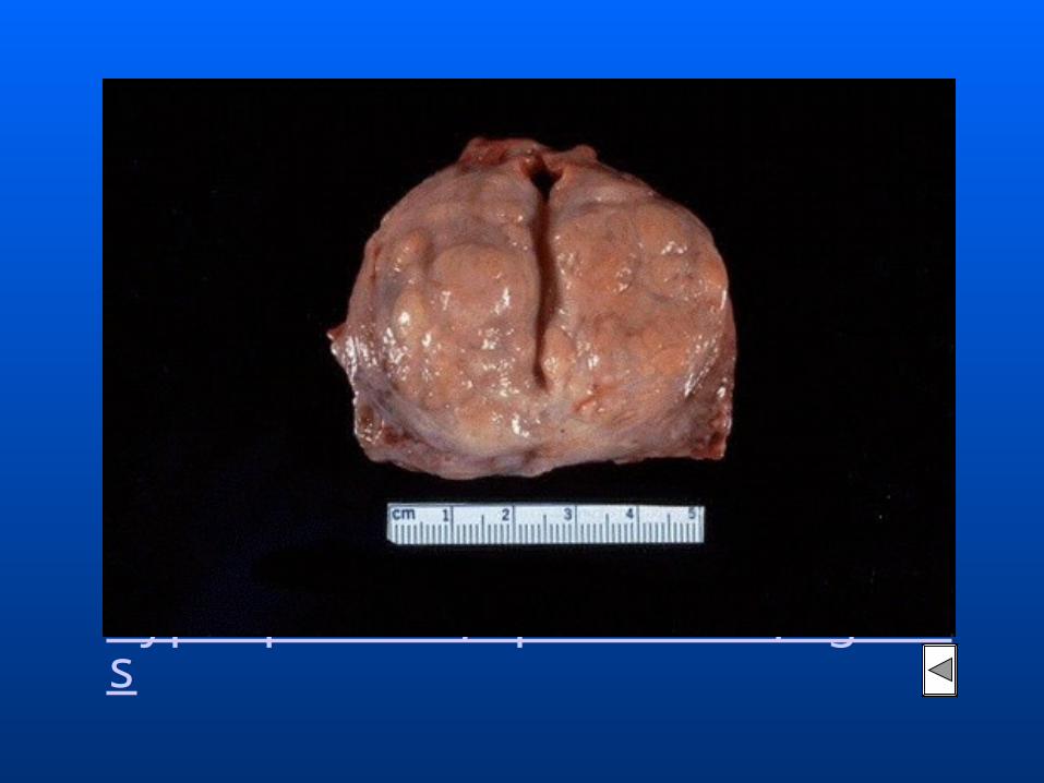
• Hyperplasia
• Metaplasia
Atrophy
Reduction of the number and size of pare
nchymal cells of an organ or its parts wh
ich was once normal is called atrophy.
It may occur from physiologic or pathol
ogic causes.
Physiologic Atrophy
• Atrophy of thymus after puberty
• Atrophy of gonads after menopause
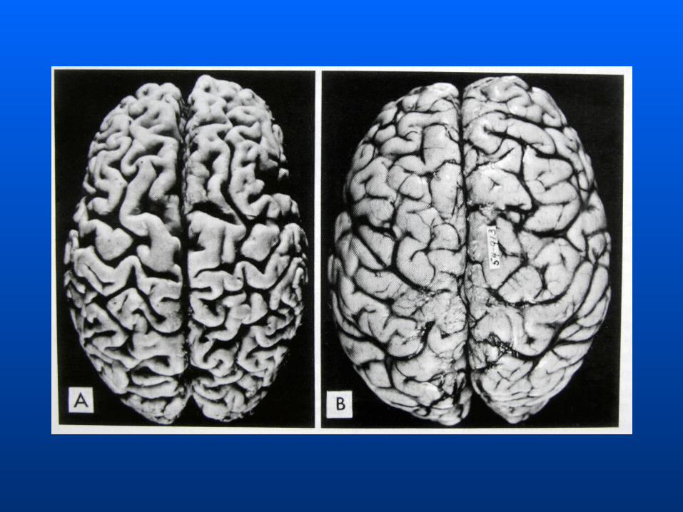
• Atrophy of brain with aging
Pathologic Atrophy
• Malnutrition atrophy
• Denervation atrophy
• Disuse atrophy
• Pressure atrophy
• Endocrine atrophy
• Ischaemic atrophy
Hypertrophy
Hypertrophy is an increase in the size of p
arenchymal cells resulting in enlargemen
t of the organ or tissue, without any cha
nge in the number of cells.
It may be physiologic or pathologic.
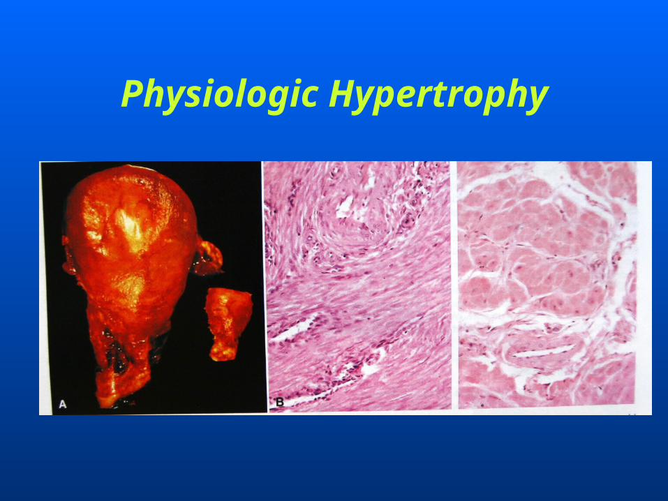
Physiologic Hypertrophy
Pathologic Hypertrophy
• Hypertrophy of cardiac muscle
• Hypertrophy of smooth muscle
• Hypertrophy of skeletal muscle
• Compensatory hypertrophy
Hyperplasia Hyperplasia is an increase in the number
of parenchymal cells resulting in enlarge
ment of the organ or tissue.
Hyperplasia occurs due to increased recr
uitment of cells from C0 (resting) phase
of the cell cycle to undergo mitosis,when
stimulated.
Metaplasia
Metaplasia is defined as a reversible chan
ge of one type of epithelial or mesenchyma
l adult cells to another type of adult epithel
ial or mesenchymal cells, usually in respo
nse to abnormal stimuli, and often revert
s back to normal on removal of stimulus.
Metaplasia
• Epithelial metaplasia
1. Squamous metaplasia
2. Columnar metaplasia
• Mesenchymal metaplasia
1. Osseous metaplasia
2. Cartilaginous metaplasia

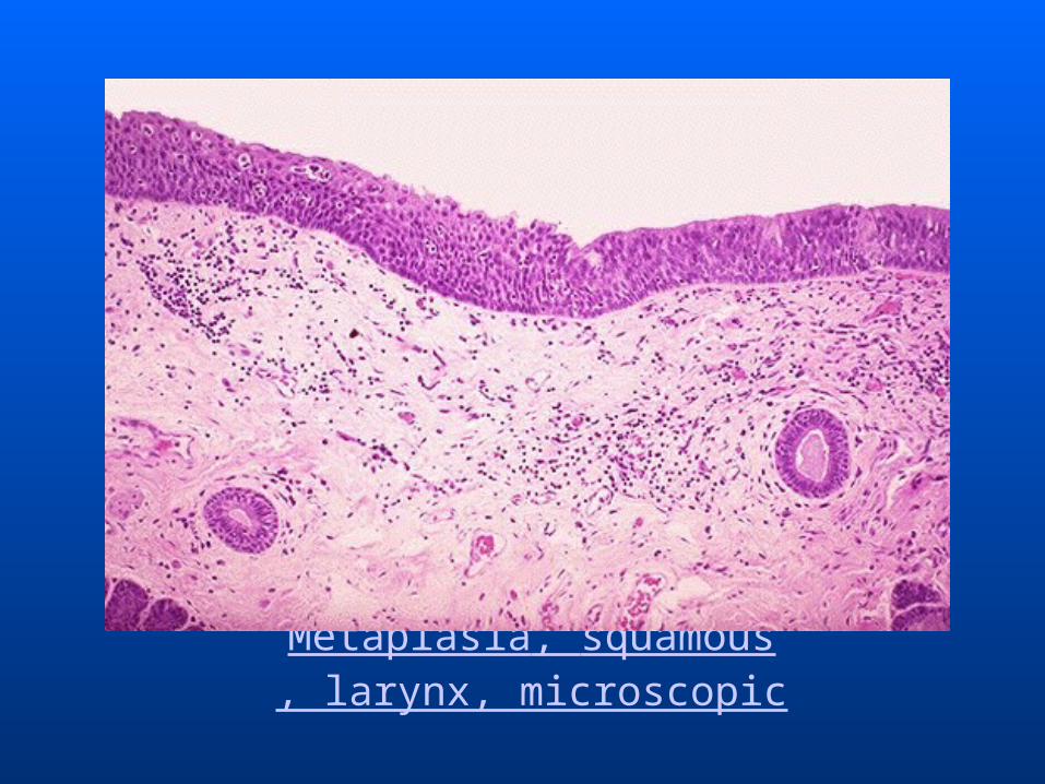
Metaplasia, squamous, larynx, microscopic
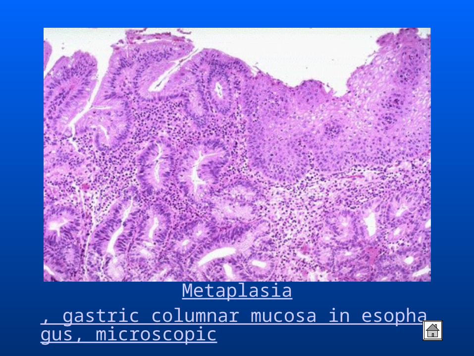
Metaplasia, gastric columnar mucosa in esophagus, microscopic
Causes of cell injury
• Hypoxia and ischaemia
• Physical agents
• Chemical agents and drugs
• Infection agents
• Immunologic reactions
• Genetic derangements
• Nutritional imbalances
Hypoxia and ischaemia
Hypoxia is the most common causes of cell injury. The causes of hypoxia are as under:
• The most common mechanism of hypoxic cell injury is by reduced supply of blood to cell i.e. ischaemia.
• Oxygen deprivation of tissues may result from other causes as well e.g. in anaemia, CO poisoning, cardiorespiratory insufficiency, and increased demand of tissues.
Physical agents
Physical agents in causation of disease are:
• Mechanical trauma (e.g. road accidents);• Thermal trauma (e.g. by heat and cold);• Electricity;• Radiation (e.g. ultraviolet and ionising);• Rapid changes in atmospheric pressure.
Chemicals and DrugesImportant example include:
• Chemical poisons such as cyanide, arsenic, mercury;
• Strong acids and alkalis;
• Environmental pollutants;
• Insecticides and pesticides;
• Oxygen at high concentration;
• Hypertonic glucose and salt;
• Social agents such as alcohol and narcotic drugs;
• Therapeutic administration of drugs.
Infection agentsInjuries by microbes include infections caused by :
• Bacteria;• Rickettsiae;• Viruses;• Fungi;• Protozoa;• Metazoa;• Other parasites.
Immunologic reactions
Immunity is a “double-edged sword”
--- it protects the host against various
injurious agents but it may also turn lethal
and cause cell injury e.g.
• Hypersensitivity reactions;
• Anaphylactic reactions;
• Autoimmune reactions.
Genetic derangementsGenetic defects as causes of cell injury are of major
interest to scientists and physicians today.
• The genetic injury may result in a defect caused by
a chromosomal abnormality (e.g. the congenital
malformations associated with Down syndrome).
• Variations in the genetic makeup can also influence
the susceptibility of cells to injury by chemicals and
other environmental insults.
Nutritional imbalancesA deficiency or an excess of nutrients may result in n
utritional imbalances.
• Nutritional deficiency diseases may be due to overall
deficiency of nutrients (e.g. starvation), of protein cal
orie (e.g. marasmus, kwashiorkor), of minerals (e.g.
anaemia), or of trace elements.
• Nutritional excess is a problem of affluent societies r
esulting in obesity, atherosclerosis, heart disease and
hypertension.

Mechanisms of cell injury
• Depletion of ATP
• Membrane damage
• Influx of intracellular calcium and loss
of calcium homeostasis
• Accumulation of oxygen-derived free
radicals (oxidative stress)
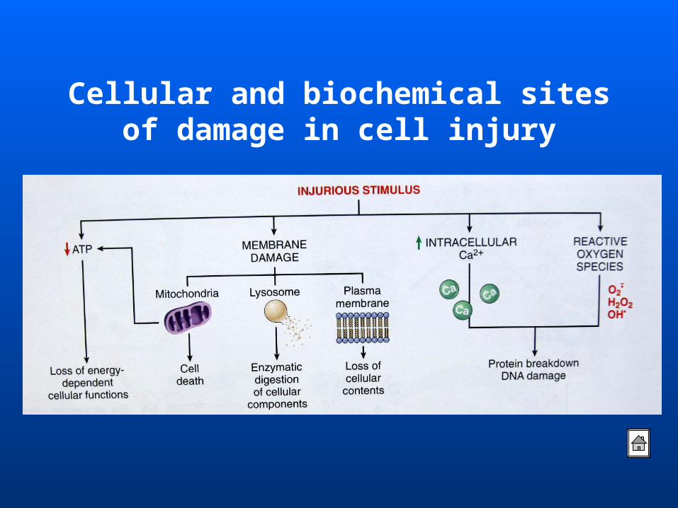
Cellular and biochemical sites of damage in cell injury
Morphology of cell injury
• Degeneration/Intracellular
Accumulations
• Cell death

Degeneration and Intracellular Accumulations
• In conventional description of morphologic
change, the term degeneration has been used to
denote morphology of reversible cell injury.
• Currently, more acceptable terms of reversible
cell injury are applied to non-lethal cell injury.
• One of the manifestations of metabolic
derangements in cells is the intracellular
accumulation of abnormal amounts of
various substances.
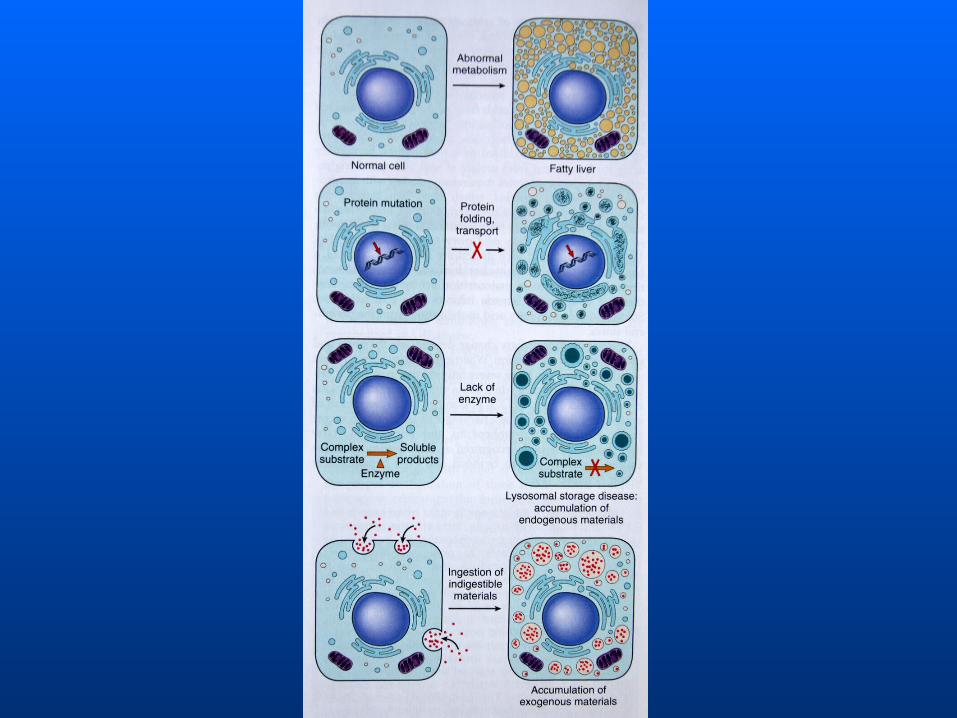
The stockpiled substances fall into three categories:
1. A normal cellular constituent accumulated in
excess, such as water, lipids, proteins, and
carbohydrates;
2. An abnormal substance, either exogenous, such
as a mineral or products of infectious agents, or
endogenous, such as a product of abnormal
synthesis or metabolism;
3. A pigment.
Degeneration and Intracellular Accumulations
• Cellular swelling
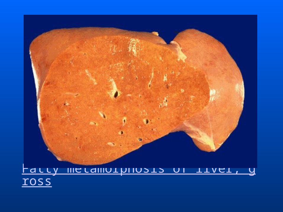
• Fatty change
• Hyaline change
• Amyloidosis
• Pigments
• Pathologic calcification
Cellular Swelling• Cellular swelling is the first manifestation of al
most all forms of injury to cells.• Other synonyms of cellular swelling used in the
past are: cloudy swelling (for gross appearance of the aff
ected organ) hydropic change (accumulation of water within
the cell) vacuolar degeneration (due to cytoplasmic vacu
olation)
Cellular Swelling
• Grossly, the affected organ such as
kidney, liver or heart muscle is enlarged
due to swelling. The cut surface bulges
outwards and is slightly opaque.
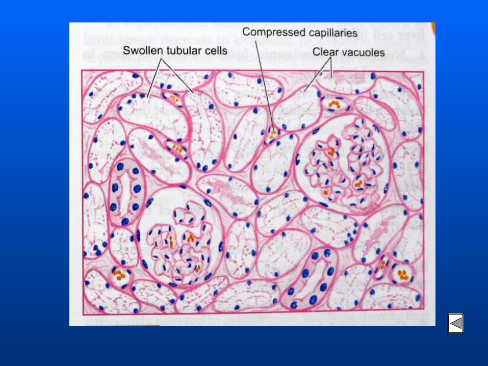
Cellular Swelling• Microscopically, it is characterised by th
e following features:
1. The cells are swollen and the microvas
culature compressed.
2. Small clear vacuoles are seen in the cel
ls and hence the term vacuolar degenera
tion.
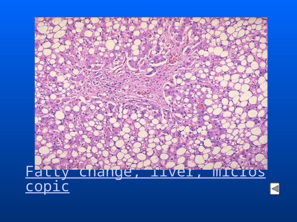
Fatty change (Steatosis)
• The terms fatty change and steatosis describe ab
normal accumulations of triglycerides within pa
renchymal cells.
• Fatty change is often seen in the liver because it
is the major organ involved in fat metabolism, b
ut it also occurs in heart, muscle, and kidney.
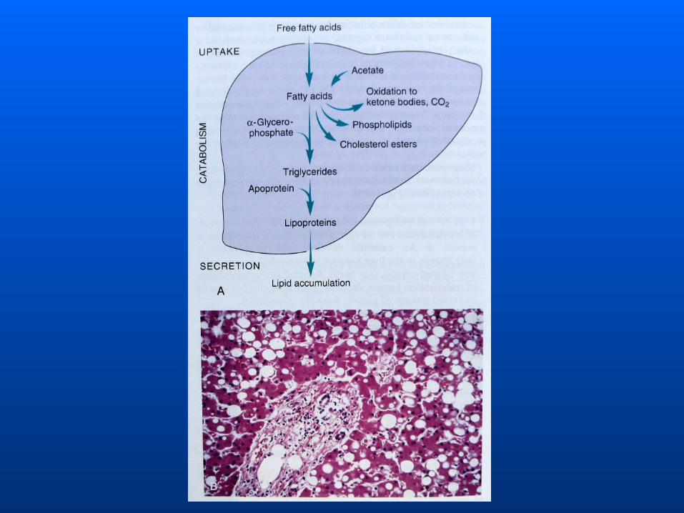

. Fatty metamorphosis of liver, microscopic
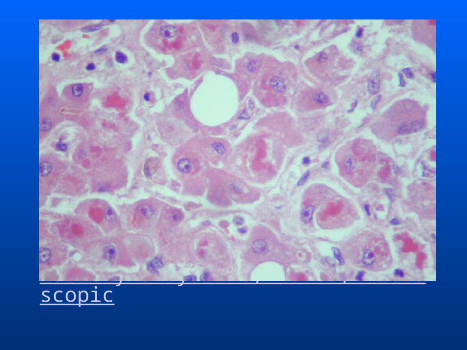
Hyaline Change• The word “Hyaline” means glassy (hyalos = glas
s).
• Hyaline is a descriptive histologic term for glass
y, homogeneous, eosinophilic appearance of mat
erial in H.E stained sections and does not refer to
any specific substance.
• Hyaline change is associated with heterogeneous
pathologic conditions and may be intracellular or
extracellular.
Intracellular Hyaline
• Intracellular hyaline is mainly seen in epithelial cells. For example:
1. Hyaline droplets in the proximal tubular epithelial cells in cases of excessive reabsorption of plasma.
2. Mallory’s hyaline represents aggregates of intermediate filaments in the hepatocytes in alcoholic liver cell injury.
Intracellular Hyaline
3. Nuclear or cytoplasmic hyaline inclusions seen in some viral infections.
4. Russel’s bodies representing excessive immunoglobulins in the RER of the plasma cells.
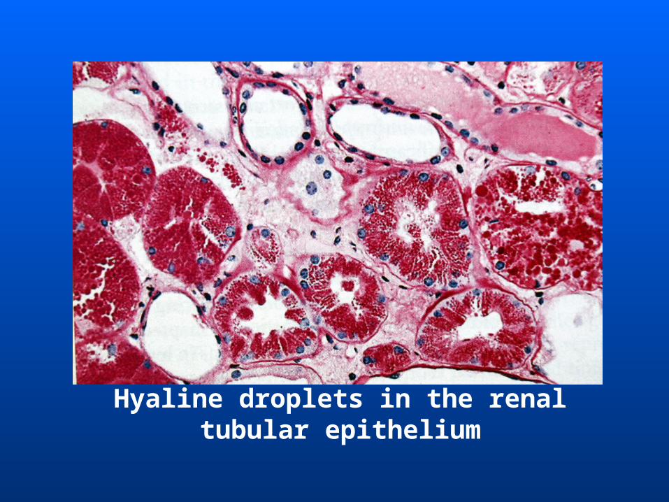
Hyaline droplets in the renal tubular epithelium
Extracellular Hyaline• Extracellular hyaline is seen in connective tissues.
A few examples of extracellular hyaline change are:1. Hyaline degeneration in leiomyomas of the uterus. 2. Hyalinised old scar of fibrocollagenous tissues. 3. Hyaline arteriosclerosis is renal vessels in hypertension and diabetes mellitus.4. Hyalinised glomeruli in chronic glomerulonephritis.
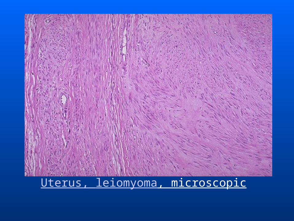
Uterus, leiomyoma, microscopic
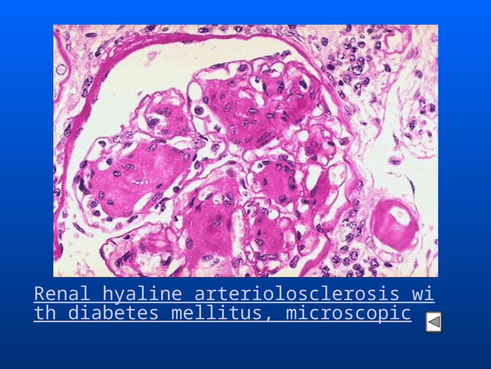
Renal hyaline arteriolosclerosis with diabetes mellitus, microscopic
Recommended