Orthopedics & Fractures

Orthopedics
“Orthopedics” is: that branch of surgery which is
specially concerned with the preservation and restoration of the function of the skeletal system, its joints, and associated structures like ligaments and tendons

Orthopedic Exam
• Meet Sam & Simon!!
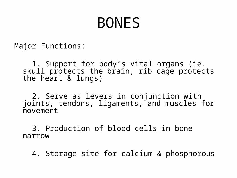
BONESMajor Functions:
1. Support for body’s vital organs (ie. skull protects the brain, rib cage protects the heart & lungs)
2. Serve as levers in conjunction with joints, tendons, ligaments, and muscles for movement
3. Production of blood cells in bone marrow
4. Storage site for calcium & phosphorous
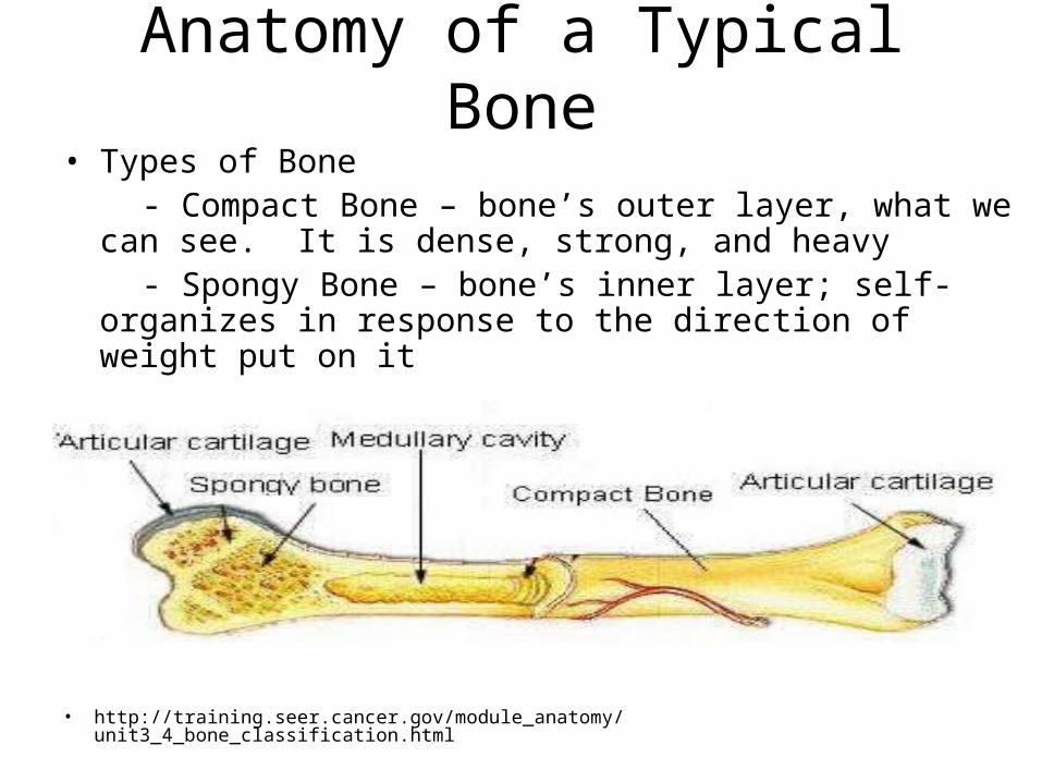
Anatomy of a Typical Bone• Types of Bone - Compact Bone – bone’s outer layer, what we can
see. It is dense, strong, and heavy - Spongy Bone – bone’s inner layer; self- organizes
in response to the direction of weight put on it
• http://training.seer.cancer.gov/module_anatomy/unit3_4_bone_classification.html

Associated Structures
• Joint – anytime 2 or more bones come together
• Articular Cartilage – cartilage covering the ends of bones that are in contact with adjacent bones to create smooth movement and shock absorption
• Tendon – connects muscle to bone• Ligament – connects bone to bone

Classification of Bones
• Long Bones – long! Bones of limbs• Short Bones – short! Small bones of hands
& feet • Flat Bones – flat! • Sesamoid Bones – small bones embedded
in tendon as it crosses a bony prominence. • Irregular Bones – jutting processes give
these bones an irregular shape.

Quiz
• Can you feel some of these bones in your own body?
- where would you feel flat bones? - where would you feel a sesmoid
bone (and its associated tendon)? - where would you feel short bones - where would you feel irregular
bones?
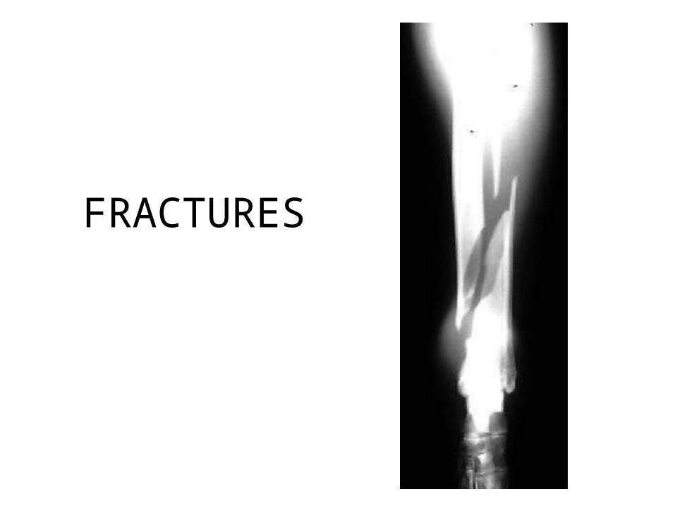
FRACTURES

Types of Fractures
• Open• Closed• Complete• Incomplete• Comminuted• Segmental
• Chip• Slab• Pathologic
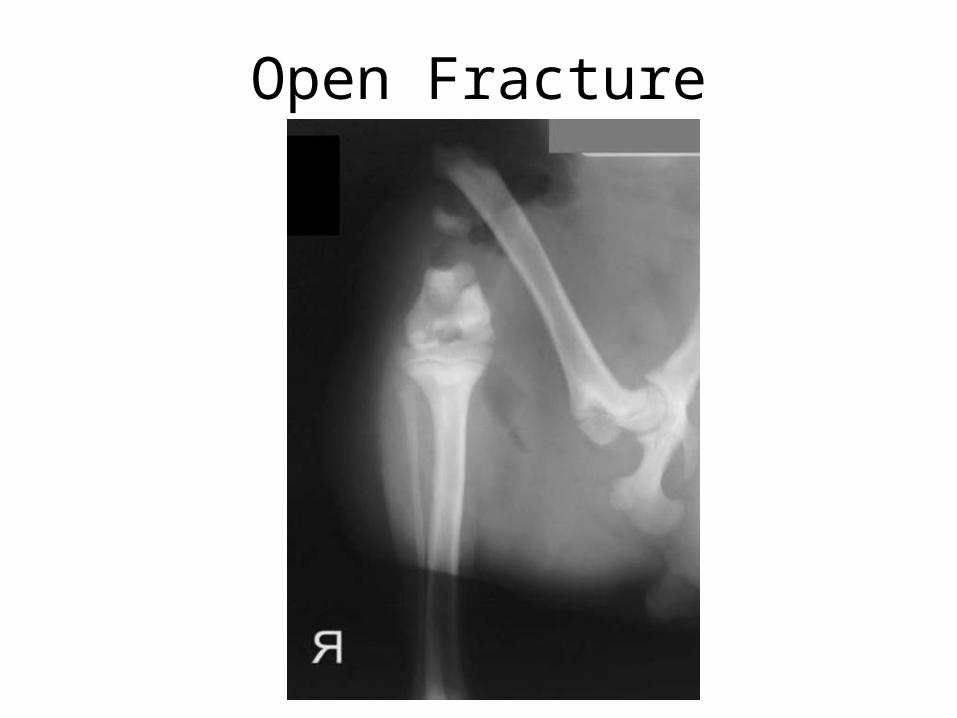
Open Fracture

Closed Fracture

Complete Fracture
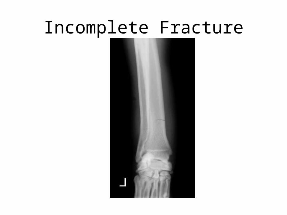
Incomplete Fracture

Comminuted Fracture

Segmental Fracture

Chip Fracture

Pathologic Fracture
• Fracture secondary to another disease process
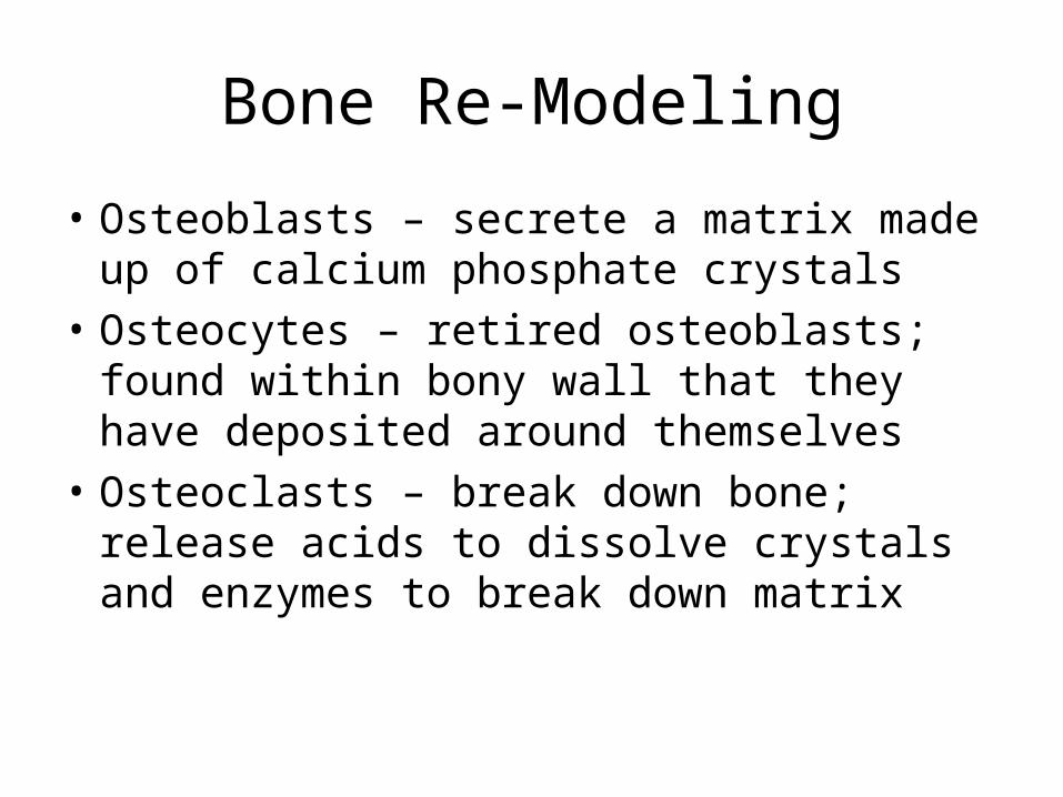
Bone Re-Modeling
• Osteoblasts – secrete a matrix made up of calcium phosphate crystals
• Osteocytes – retired osteoblasts; found within bony wall that they have deposited around themselves
• Osteoclasts – break down bone; release acids to dissolve crystals and enzymes to break down matrix

Bone Reacts to Stresses Put on It
• Greater physical stress placed on a bone at a particular site results in more bone deposition by osteoblasts at that site
• Another theory suggests electrical field change created by physical stress stimulates osteoblasts & matrix formation
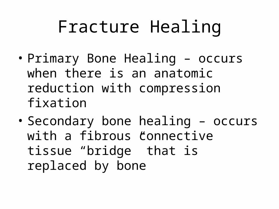
Fracture Healing
• Primary Bone Healing – occurs when there is an anatomic reduction with compression fixation
• Secondary bone healing – occurs with a fibrous connective tissue “bridge” that is replaced by bone

Normal Fracture Healing

What Is Necessary to Get Normal Healing?

Abnormal Fracture Healing
• Mal-union – a fracture that heals with abnormal alignment
• Non-union – fracture healing has STOPPED before completely healed
- elephant foot “hypertrophic non-union” - tapered “atrophic non-union”• Sequestrum – a dead bone fragment
separated from the rest of the bone• Osteomyelitis – infection of bone

Malunion
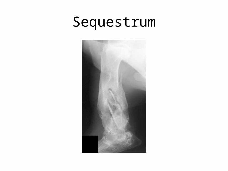
Sequestrum

Non-union

Osteomyelitis

Treatment Options
• Fracture Reduction - Closed * temporary (until surgery) * permanent (cast or splint) - Open (orthopedic surgery)• Intramedullary Fixation• Cerclage• External Fixation Devices

Fracture Reduction Goals
• Get bones close enough to heal• Proper alignment - avoid mal-union + loss of function• Avoid additional trauma - further fracture - infection
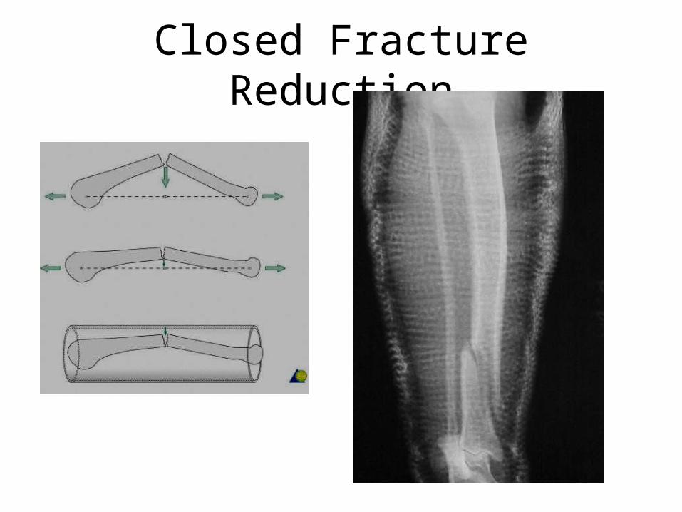
Closed Fracture Reduction
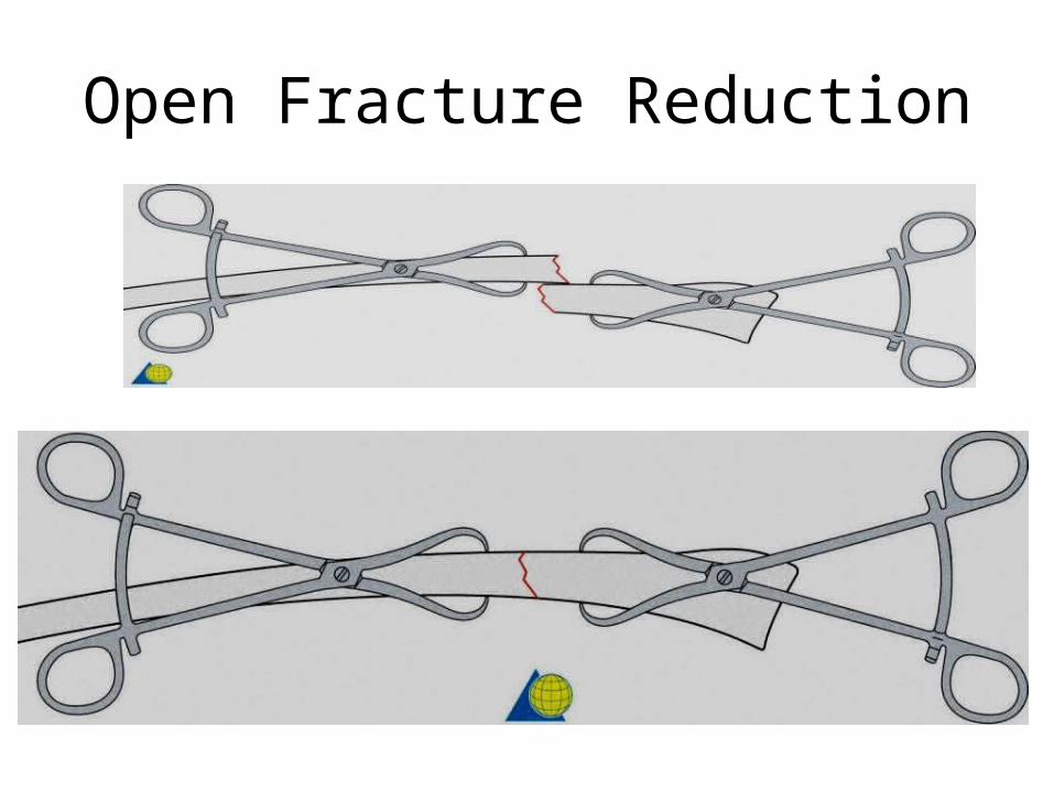
Open Fracture Reduction

Intramedullary Fixation

Pin Insertion
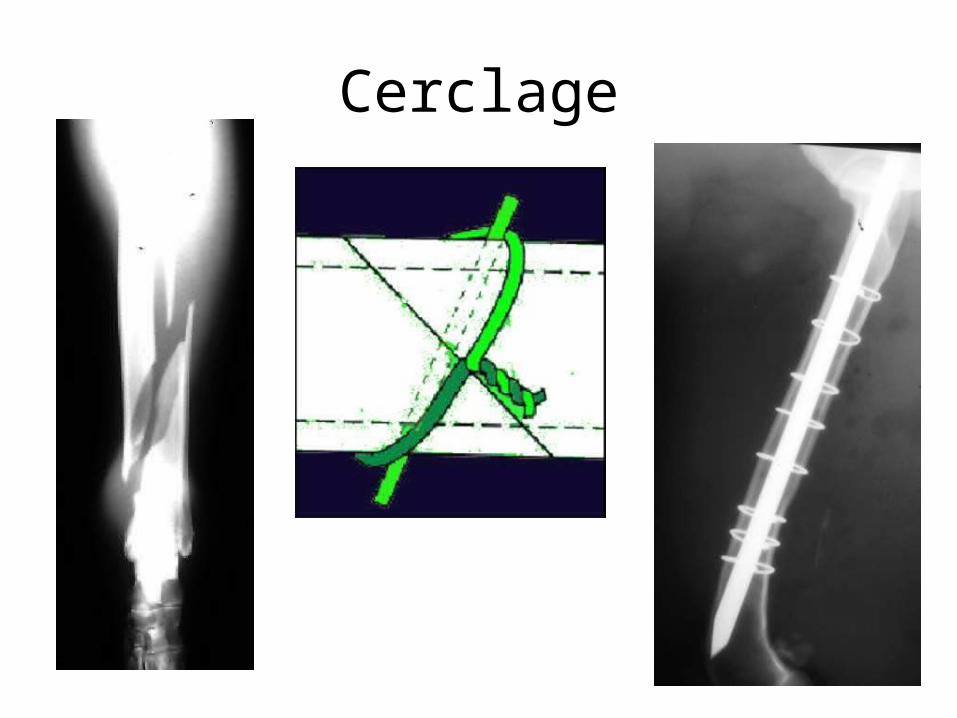
Cerclage

External Fixators

External Fixators

(view video)
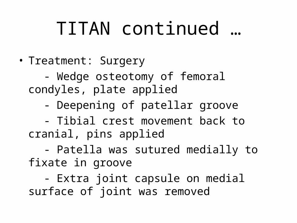
TITAN continued …
• Treatment: Surgery - Wedge osteotomy of femoral condyles,
plate applied - Deepening of patellar groove - Tibial crest movement back to cranial,
pins applied - Patella was sutured medially to fixate in
groove - Extra joint capsule on medial surface of
joint was removed

Follow-Up
• Re-Check in 2weeks for range of motion
• Re-check in 4weeks for progress of healing
• Recheck in 10weeks for further progress of healing
STRICT CAGE REST DURING THIS TIME!!

Surgery Tools

Animal Orthopedics as Human Model
• Animals are frequently used as models in clinical studies or experiments in the development of surgical procedures & drugs in veterinary medicine to be used in human medicine!
1st hip replacement surgery was developed in military dogs

Jeopardy!

Questions???
END
• All images used from government websites as indicated OR with permission from Dr. Sharon Kerwin & Dr. Ben Young, Texas A&M University College of Veterinary Medicine
Recommended


![[PPT]General principles of fractures III - Shanyar's Lecture …lectures.shanyar.com/5th_Stage/Orthopedics/Dr._'Ali/3... · Web viewThe available methods of holding reduction are:](https://img.dokumen.tips/doc/110x75/5afa34b77f8b9a19548dafb4/pptgeneral-principles-of-fractures-iii-shanyars-lecture-ali3web-viewthe.jpg)