Bulgarian Journal of Veterinary Medicine, 2017 ONLINE FIRST ISSN 1311-1477; DOI: 10.15547/bjvm.2066
Original article
MAST CELL DISTRIBUTION AROUND THE NEEDLE TRACT FOLLOWING ACUPUNCTURE IN ZUSANLI (ST36)
ACUPOINT IN RATS
N. D. DIMITROV1, D. Y. ATANASOVA1,3, N. S. TOMOV1, Y. A. STAYKOVA-PIROVSKA2, I. G. IVANOVA1 & D. P. SIVREV1
1Department of Anatomy; 2Department of General Medicine, Faculty of Medicine, Trakia University, Stara Zagora, Bulgaria;
3Institute of Neurobiology, Bulgarian Academy of Sciences, Sofia, Bulgaria
Summary
Dimitrov, N. D., D. Y. Atanasova, N. S. Tomov, Y. A. Staykova-Pirovska, I. G. Ivanova & D. P. Sivrev, 2017. Mast cell distribution around the needle tract following acupuncture in Zusanli (ST36) acupoint in rats. Bulg. J. Vet. Med. (online first). The aim of this study was to investigate mast cell (MCs) distribution in the vicinity of the needle tract formed after acupuncture in Zusanli (ST36) acupoint in rats. MCs were detected by histochemistry, immunohistochemistry and transmission electron microscopy, and evaluated quantitatively. It was established that after acupuncture in ST36 acupoint the integrity of the epithelium, dermis, subcutane-ous connective tissue, fascia, epimysium and striated muscles was disrupted and folded to the direc-tion of the needle tract. In the thickened connective tissue MCs were observed close to the needle tract, without visible differences in their number along the tract, but most of them were with signs of degranulation, possibly due to acupuncture. It could be presumed that acupuncture in ST36 caused recruitment and activation of MCs followed by degranulation which most probably influenced the local microenvironment.
Key words: acupuncture, degranulation, mast cells, needle tract, Zusanli (ST36)
INTRODUCTION
Acupuncture is a commonly used method of the Traditional Chinese Medicine. Zusanli (ST36) acupuncture point (acu-point) is one of the most important for treatment of both humans and animals. ST36 can be used in experimental acu-puncture by applying the method of stan-dard proportions of anatomical structures
under the control of an apparatus measu-ring skin resistance (White et al., 2008; Dimitrov et al., 2009). Mast cells (MCs) are resident mainly in the connective tis-sue, particularly in vicinity of small blood vessels and nerves. Their usual localisa-tion is in proximity to surfaces that inter-face the external environment. Biological
Mast cell distribution around the needle tract following acupuncture in Zusanli (ST36) acupoint in rats
BJVM, ××, No × 2
functions of MCs include a role in innate immunity, mechanisms against parasitic infestations, immunomodulation of the immune system, and tissue repair (Met-calfe et al., 1997). The MCs are also im-portant subject in experimental acupunc-ture. Many studies have been devoted to their role (Lin et al., 2007; Zhang et al., 2008).
Acupuncture point ST36 is one of fre-quently used points in experimental acu-puncture in rats, often in combination with electroacupuncture (Deng et al., 1996; Ming et al., 2000; Li et al., 2003). Some studies combine experimental acupuncture with moxibustion (He & Luo, 2007; Luo et al., 2007; He & Chen, 2010). There is an evidence for a link between the effects of laser acupuncture and the function of MCs (Cheng et al., 2009). Our previous studies have shown that the normal ana-tomic structures in ST36 acupoint are the epidermis, dermis, subcutis, deep fascia, epimysium, striated muscle, containing blood vessels and nerves (Dimitrov, 2012a). The impact of the acupuncture needle in ST36 acupoint comprised mor-phological changes in the tissues and MCs. Following acupuncture in ST36 acu-point significant degranulation of MCs occurred in the acupuncture area (Dimi-trov, 2012b).
The aim of this study was to study the distribution of MCs in the vicinity of the needle tract after acupuncture in ST36 acupoint in rats.
MATERIALS AND METHODS
Animals
The studies were carried out on ten adult male Wistar normotensive rats, weighing 220–350 g. The experimental design was approved by the Research Ethics Commit-tee at the Medical Faculty of Trakia Uni-
versity. All efforts were made to minimise the number of animals used and their suf-fering. The area around the acupoint ST36 was epilated, defined and marked using the method of standard proportions of anatomical structures under the control of the apparatus KWD-808 measuring skin resistance (Dimitrov, 2012b). A steel acu-puncture needles with size 0.25 × 13 mm were inserted in the ST36 of the ether nar-cotised rats just before perfusion of the experimental animals (from the left heart ventricle via aorta) with 0.05 M phosphate buffered saline (PBS) followed by 4% of paraformaldehyde (PFA, Sigma Aldrich Chemie, Switzerland) in 0.1 M phosphate buffer, pH 7.36.
Light microscopic histology for mast cells detection
The material (5×5×5 mm) from ST36 acu-point was taken immediately after the death of the animals together with the acupuncture needle. Some of the samples were dehydrated in ascending ethanol series, cleared (twice) in xylene and em-bedded in paraffin. From them sections of 5 µm thickness were prepared, rehydrated in descending ethanol series. Other sec-tions with 30 µm thickness were prepared on freezing microtome. Both sections were stained using toluidine blue (TB), Bismarck brown (BB) and Mallory's tri-chrome (MT) methods.
Light microscopic immunohistochemistry for mast cells detection
The paraffin sections with the same thick-ness were dewaxed twice in xylene and were rehydrated in descending ethanol series. Afterwards sections were washed in 0.1 M PBS, pH 7.4, incubated in 1.2% hydrogen peroxide in methanol for 30 min, followed by antigen retrieval in
N. D. Dimitrov, D. Y. Atanasova, N. S. Tomov, Y. A. Staykova-Pirovska, I. G. Ivanova & D. P. Sivrev
BJVM, ××, No × 3
10 mM citrate buffer (pH 9.0) for up to 10 min in pressure cooker.
Between the separate steps, the sec-tions were rinsed with cold PBS/Triton X-100. Subsequently, they were incubated with the primary antibody (Monoclonal Mast Cell Tryptase - clone 10D11, Leica Biosystems, Newcastle) diluted 1:100 in a humid chamber overnight at 4 oC. Follow-ing three washings with PBS, the slides were incubated with DAKO-REALTM En-VisionTM detection system (DAKO) for 60 min, then visualised with diaminoben-zidine and counterstained with Mayer’s haematoxylin. PBS instead of the primary antibody was used as a negative control.
All slides were observed by a light mi-croscope (Eclipse 80i, Nikon, Japan), analysed and photographed with a digital camera (Nikon DMX 1200).
Transmission electron microscopy for mast cells detection
The material (1 mm3) from the skin in the vicinity of the needle tract was fixed im-mediately in 2% glutaraldehyde and 2% paraformaldehyde in 0.1 M PBS for 24 h. After that the samples were dehydrated in ascending ethanol series, passed through
propylene oxide, immersed in propylene oxide and Durcupan, and embedded in Durcupan (Fluka AG, Buchs SG, Switzer-land). The resin block was cut into ultra-thin sections by a diamond knife in an ultramicrotome. Each section, 50-70 nm thick, was collected on metal mesh 'grids' and stained with electron dense stains before TEM observation.
Statistical analysis
The distribution of mast cells was quanti-tated and statistical analysis was per-formed using Student’s t-test for paramet-rical data (SigmaStat®11.0 software pack-age, Systat Software Inc). Differences were considered statistically significant at P<0.05.
RESULTS
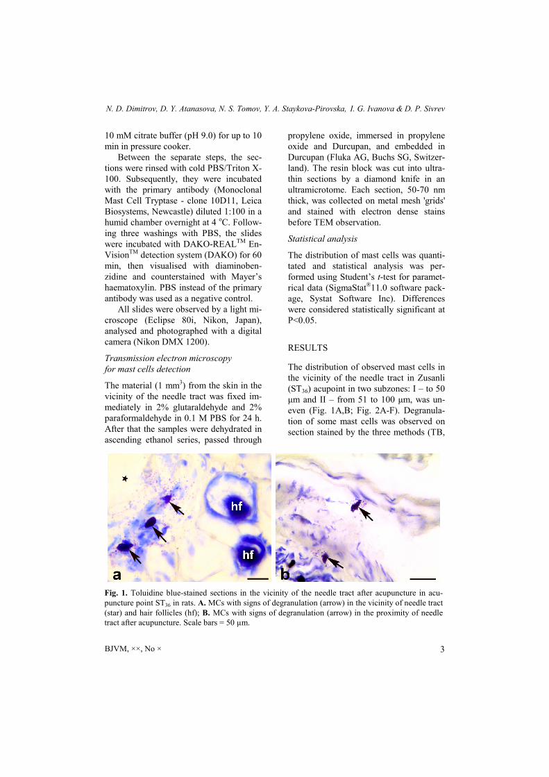
The distribution of observed mast cells in the vicinity of the needle tract in Zusanli (ST36) acupoint in two subzones: I – to 50 μm and II – from 51 to 100 μm, was un-even (Fig. 1A,B; Fig. 2A-F). Degranula-tion of some mast cells was observed on section stained by the three methods (TB,
Fig. 1. Toluidine blue-stained sections in the vicinity of the needle tract after acupuncture in acu-puncture point ST36 in rats. A. MCs with signs of degranulation (arrow) in the vicinity of needle tract (star) and hair follicles (hf); B. MCs with signs of degranulation (arrow) in the proximity of needle tract after acupuncture. Scale bars = 50 µm.
Mast cell distribution around the needle tract following acupuncture in Zusanli (ST36) acupoint in rats
BJVM, ××, No × 4
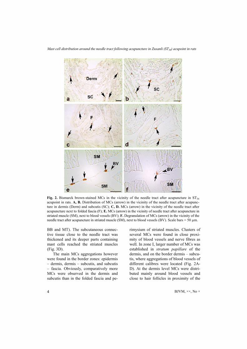
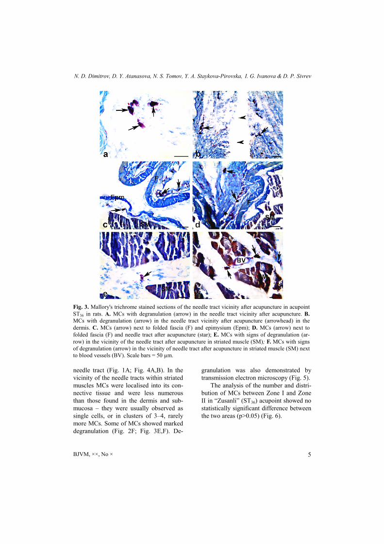
BB and MT). The subcutaneous connec-tive tissue close to the needle tract was thickened and its deeper parts containing mast cells reached the striated muscles (Fig. 3D).
The main MCs aggregations however were found in the border zones: epidermis – dermis, dermis – subcutis, and subcutis – fascia. Obviously, comparatively more MCs were observed in the dermis and subcutis than in the folded fascia and pe-
rimysium of striated muscles. Clusters of several MCs were found in close proxi-mity of blood vessels and nerve fibres as well. In zone I, larger number of MCs was established in stratum papillare of the dermis, and on the border dermis – subcu-tis, where aggregations of blood vessels of different calibres were located (Fig. 2A-D). At the dermis level MCs were distri-buted mainly around blood vessels and close to hair follicles in proximity of the
Fig. 2. Bismarck brown-stained MCs in the vicinity of the needle tract after acupuncture in ST36 acupoint in rats. A, B. Distribution of MCs (arrow) in the vicinity of the needle tract after acupunc-ture in dermis (Derm) and subcutis (SC); C, D. MCs (arrow) in the vicinity of the needle tract after acupuncture next to folded fascia (F); E. MCs (arrow) in the vicinity of needle tract after acupuncture in striated muscle (SM), next to blood vessels (BV); F. Degranulation of MCs (arrow) in the vicinity of the needle tract after acupuncture in striated muscle (SM), next to blood vessels (BV). Scale bars = 50 µm.
N. D. Dimitrov, D. Y. Atanasova, N. S. Tomov, Y. A. Staykova-Pirovska, I. G. Ivanova & D. P. Sivrev
BJVM, ××, No × 5
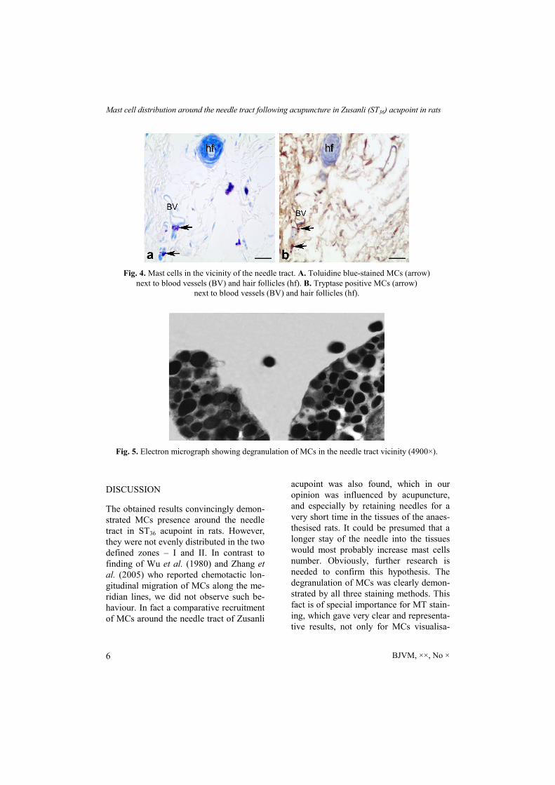
needle tract (Fig. 1A; Fig. 4A,B). In the vicinity of the needle tracts within striated muscles MCs were localised into its con-nective tissue and were less numerous than those found in the dermis and sub-mucosa – they were usually observed as single cells, or in clusters of 3–4, rarely more MCs. Some of MCs showed marked degranulation (Fig. 2F; Fig. 3E,F). De-
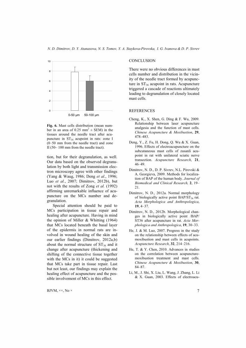
granulation was also demonstrated by transmission electron microscopy (Fig. 5).
The analysis of the number and distri-bution of MCs between Zone I and Zone II in “Zusanli” (ST36) acupoint showed no statistically significant difference between the two areas (p>0.05) (Fig. 6).
Fig. 3. Mallory's trichrome stained sections of the needle tract vicinity after acupuncture in acupoint ST36 in rats. A. MCs with degranulation (arrow) in the needle tract vicinity after acupuncture. B. MCs with degranulation (arrow) in the needle tract vicinity after acupuncture (arrowhead) in the dermis. C. MCs (arrow) next to folded fascia (F) and epimysium (Epm); D. MCs (arrow) next to folded fascia (F) and needle tract after acupuncture (star); E. MCs with signs of degranulation (ar-row) in the vicinity of the needle tract after acupuncture in striated muscle (SM); F. MCs with signs of degranulation (arrow) in the vicinity of needle tract after acupuncture in striated muscle (SM) next to blood vessels (BV). Scale bars = 50 µm.
Mast cell distribution around the needle tract following acupuncture in Zusanli (ST36) acupoint in rats
BJVM, ××, No × 6
DISCUSSION
The obtained results convincingly demon-strated MCs presence around the needle tract in ST36 acupoint in rats. However, they were not evenly distributed in the two defined zones – I and II. In contrast to finding of Wu et al. (1980) and Zhang et al. (2005) who reported chemotactic lon-gitudinal migration of MCs along the me-ridian lines, we did not observe such be-haviour. In fact a comparative recruitment of MCs around the needle tract of Zusanli
acupoint was also found, which in our opinion was influenced by acupuncture, and especially by retaining needles for a very short time in the tissues of the anaes-thesised rats. It could be presumed that a longer stay of the needle into the tissues would most probably increase mast cells number. Obviously, further research is needed to confirm this hypothesis. The degranulation of MCs was clearly demon-strated by all three staining methods. This fact is of special importance for MT stain-ing, which gave very clear and representa-tive results, not only for MCs visualisa-
Fig. 4. Mast cells in the vicinity of the needle tract. A. Toluidine blue-stained MCs (arrow) next to blood vessels (BV) and hair follicles (hf). B. Tryptase positive MCs (arrow)
next to blood vessels (BV) and hair follicles (hf).
Fig. 5. Electron micrograph showing degranulation of MCs in the needle tract vicinity (4900×).
N. D. Dimitrov, D. Y. Atanasova, N. S. Tomov, Y. A. Staykova-Pirovska, I. G. Ivanova & D. P. Sivrev
BJVM, ××, No × 7
tion, but for their degranulation, as well. Our data based on the observed degranu-lation by both light and transmission elec-tron microscopy agree with other findings (Yang & Waug, 1986; Deng et al., 1996; Luo et al., 2007; Dimitrov, 2012b), but not with the results of Zong et al. (1992) affirming unremarkable influence of acu-puncture on the MCs number and de-granulation.
Special attention should be paid to MCs participation in tissue repair and healing after acupuncture. Having in mind the opinion of Miller & Whitting (1964) that MCs located beneath the basal layer of the epidermis in normal rats are in-volved in wound healing of the skin and our earlier findings (Dimitrov, 2012a,b) about the normal structure of ST36 and it change after acupuncture (thickening and shifting of the connective tissue together with the MCs in it) it could be suggested that MCs take part in tissue repair. Last but not least, our findings may explain the healing effect of acupuncture and the pos-sible involvement of MCs in this effect.
CONCLUSION
There were no obvious differences in mast cells number and distribution in the vicin-ity of the needle tract formed by acupunc-ture in ST36 acupoint in rats. Acupuncture triggered a cascade of reactions ultimately leading to degranulation of closely located mast cells.
REFERENCES
Cheng, K., X. Shen, G. Ding & F. Wu, 2009. Relationship between laser acupuncture analgesia and the function of mast cells. Chinese Acupuncture & Moxibustion, 29, 478–483.
Deng, Y., Z. Fu, H. Dong, Q. Wu & X. Guan, 1996. Effects of electroacupuncture on the subcutaneous mast cells of zusanli acu-point in rat with unilateral sciatic nerve transection. Acupuncture Research, 21, 46–49.
Dimitrov, N. D., D. P. Sivrev, N.L. Pirovski &
A. Georgieva, 2009. Methods for localiza-tion of BAP of the human body. Journal of Biomedical and Clinical Research, 2, 19–21.
Dimitrov, N. D., 2012a. Normal morphology of biologically active point BAP/ST36 rat. Acta Morphologica and Anthropologica, 19, 4–37.
Dimitrov, N. D., 2012b. Morphological chan-ges in biologically active point /ВАР/ ST36 after acupuncture in rat. Acta Mor-phologica and Anthropologica, 19, 30–33.
He, J. & M. Luo, 2007. Progress in the study on the relationship between effects of acu-moxibustion and mast cells in acupoints. Acupuncture Research, 32, 214–216.
He, T. & Y. Chen, 2010. Advances in studies on the correlation between acupuncture-moxibustion treatment and mast cells. Chinese Acupuncture & Moxibustion, 30, 84–87.
Li, M., J. Shi, X. Liu, L. Wang, J. Zhang, L. Li & X. Guan, 2003. Effects of electroacu-
0
2
4
6
8
10
10-50 µm 50-100 µm
Fig. 6. Mast cells distribution (mean num-ber in an area of 0.25 mm2 ± SEM) in the tissues around the needle tract after acu-puncture in ST36 acupoint in rats: zone I (0–50 mm from the needle tract) and zone II (50– 100 mm from the needle tract).
Mast cell distribution around the needle tract following acupuncture in Zusanli (ST36) acupoint in rats
BJVM, ××, No × 8
puncture on the number of subcutaneous mast cells in and beside the acupoint and the inflammatory pain focus in the rat. Chinese Acupuncture & Moxibustion, 23, 597–601.
Lin, J., H. Huang, G. Ding & D. Zhang, 2007. Relationship between the function of mast cells and acupuncture analgesia in adju-vant arthritis rats. Acupuncture Research, 32, 16–19.
Luo, M., J. He, Y. Guo & C. Li, 2007. Effect of electroacupuncture and moxibustion of "Dazhui" (GV14) on the number and dis-tribution of degranulated mast cells in GV14 region. Acupuncture Research, 32, 327–329.
Metcalfe, D. D., D. Baram & Y. A. Mekori, 1997. Mast cells. Physiological Reviews, 77, 1033–1079.
Miller, L. & H. W. Whitting, 1964. Mast cells and wound healing of the skin in the rat. Zeitschrift fur Zellforschung und mikro-skopische Anatomie, 65, 597–606.
Ming, C., S. Cai, Y. Cai & T. Ma, 2000. Obser-vation of MC in the deep aponeurosis un-der the fluorescent microscope by electric needling in “Zusanli”. Acupuncture Re-search, 25, 51–53.
White, A., M. Cummings & J. Fishie, 2008. How to locate acupuncture points. In: An Introduction to Western Medical Acupunc-ture, ed. K. Morley, Churchill Livingstone Еlsevier, Edinburgh. pp. 185–189.
Wu, J. L., D. H. Chai, D. Cai & A. Zong, 1980. Observation on mast cells in subcutaneous connective tissue at points in albino rats. Acta Anatomica Sinica, 11, 308–312.
Yang Y. & P. Waug, 1986. Morphological observation on the effect of acupuncture on mast cells at the zusanli point. Acu-puncture Research, 11, 298–302.
Zhang D., G. Ding, X. Shen, W. Yao & J. Lin, 2005. Study on the correlation between meridian acupoints and mast cells. Acu-puncture Research, 3, 115–119.
Zhang D., G. H. Ding, X. Y. Shen, W. Yao, Z. Y. Zhang, Y. Q. Zhang, J. Y. Lin & Q. Gu, 2008. Role of mast cells in acupuncture ef-fect: A pilot study. Explore (NY), 4, 170–177.
Zong A., X. Shi & F. Zhang, 1992. Effects of electro-acupuncture on fascial mast cells in “Zusanli” acupoint area of rabbits. Journal of Zhengzhou University (Science Medi-cal), 27, 226–229.
Paper received 01.06.2017; accepted for publication 05.06.2017
Correspondence: Nikolay Dimitrov MD, PhD Department of Anatomy Faculty of Medicine Trakia University 11, Armejska Str. 6003 Stara Zagora, Bulgaria e-mail: [email protected]
Recommended