Int J Clin Exp Med 2017;10(9):13389-13396www.ijcem.com /ISSN:1940-5901/IJCEM0055734
Original ArticleGinsenoside Rg1 alleviates pulmonary inflammation caused by cigarette smoke-induced COPD
Yujing Tao1, Xinyan Liu1, Zhenhua Ni2, Xuejun Yu1
1Geriatrics Department, Putuo Hospital Affiliated to Shanghai University of Traditional Chinese Medicine, Shang-hai 200062, China; 2Central Laboratory, Putuo Hospital Affiliated to Shanghai University of Traditional Chinese Medicine, Shanghai 200062, China
Received April 18, 2017; Accepted August 17, 2017; Epub September 15, 2017; Published September 30, 2017
Abstract: Ginsenoside Rg1, the major effective component in ginseng, has been reported to have potent anti-inflammatory properties. However, the effect of Rg1 on pulmonary inflammation caused by exposure to cigarette smoke (CS) has not been investigated. In this study, we examined the molecular mechanisms underlying the effect of Rg1 on CS-induced inflammation using a mouse model. We found that inflammatory cell numbers (total cells, macrophages and neutrophils) in the bronchoalveolar lavage (BAL) fluid of the CS-exposed (CSE) mice group were remarkably increased compared with air-exposed mice, and these changes were significantly ameliorated in Rg1-treated mice. Rg1 treatment also reduced the CS-induced increase in TNF-α, IL-6 and TGF-β in BAL supernatant and lung tissues using ELISA and qRT-PCR analysis. Moreover, the administration of Rg1 significantly attenuated the phosphorylation of mitogen-activated protein kinase (MAPK) and downregulated the activation of nuclear factor-κB (NF-κB) in CSE mice. These results suggested that Rg1 may effectively attenuate inflammatory responses induced by CS through the MAPK and NF-κB pathways, and thus maybe an ideal agent for treating inflammatory pulmonary diseases.
Keywords: Ginsenoside Rg1, inflammatory responses, COPD, MAPK, NF-κB
Introduction
Chronic obstructive pulmonary disease (COPD) is defined by a progressive airflow limitation due to an abnormal inflammatory response of the lung to noxious particles and gases [1]. Cigarette smoking (CS) is by far the most com-mon risk factor for COPD, in addition to other risk factors such as genetic susceptibility and inhalation of occupational dusts, chemicals and pollutants [2]. CS exposure (CSE) induces inflammation in the lung and airway, charac- terized by marked recruitment of neutrophils to the site of inflammation [3]. The increas- ed numbers of neutrophils recruited following CSE aggravate airway inflammation resulting instructural damage via increased levels of pro-inflammatory mediators [4]. The prevalence of COPD has markedly increased worldwide and has become a significant health problem [4]. Despite significant progress, there are few effective disease-modifying drugs for COPD,
and there is an urgent need for more potent medicine for intervention in this disease.
Ginsenoside is a medicinal ingredient extracted from ginseng, and Rg1 is among the most important and active ingredients in various gin-senosides [5]. In the cardiomyocyte hypoxia-reoxygenation model, Rg1 has been shown to function as an antioxidant substance that can reduce the release of lactate dehydrogenase and intracellular ROS [6]. Rg1 has been report-ed to enhance immune responses induced by recombinant Toxoplasmagondii SAG1 anti-gen and may act as an adjuvant to promote both Thelper (Th) 1 and Th2 responses [7, 8]. Previous studies also reported that Rg1 res- trained inflammatory responses via downregu-lation of NF-κB activity [6, 9-11]. Furthermore, ginsenoside Rg1 improved lipopolysaccharide (LPS)-induced acute lung injury by inhibiting inflammatory responses and modulating infil-tration of M2 macrophages [12]. These evi-dences suggest the intriguing possibility that
Rg1 alleviates pulmonary inflammation of COPD
13390 Int J Clin Exp Med 2017;10(9):13389-13396
Rg1 maybe useful for treating COPD by inhibit-ing inflammatory responses.
In our study, we established a mouse model of COPD induced by CSE and found that inflam- matory cell numbers in the bronchoalveolar lavage (BAL) fluid of CSE mice were significantly enhanced compared with air-exposed mice, and these changes were ameliorated by Rg1 treatment. Furthermore, we revealed that Rg1 could attenuate the inflammatory cytokine lev-els in BAL fluid and lung tissues through inhibi-tion of NF-κB and MAPK pathways, and suggest that Rg1 maybe an ideal agent for treating COPD via inhibition of inflammatory respon- ses.
Materials and methods
Animals
C57BL/6J mice, 6-8 weeks old, were purchased from SLAC Lab Animal Co., Ltd (Shanghai, China), maintained in standard conditions under a 12 h light-dark cycle and provided a standard diet and chlorinated tap water ad libi-tum. All experimental procedures involving ani-mals were approved by the Institutional Animal Use and Care Committee of Shanghai University of Traditional Chinese Medicine.
Cigarette smoke exposure (CSE)
Mice were exposed whole body to CS, as described previously [13]. Mice were divided into three groups of six mice per group as fol-lows: air-exposed mice group (AE), CSE mice group and CSE+Rg1 group. Briefly, mice in the CSE group were exposed to the mainstream CS of 5 cigarettes, four times a day with 30 min-utes smoke-free intervals, 5 days a week for 24 weeks. Rg1 (20 mg/kg; Sigma-Aldrich Co- rporation, St. Louis, MO, USA) was adminis-tered tomic eby oral gavage 1 h before CSE. The control groups were exposed to room air.
Bronchoalveolar lavage (BAL)
Male mice were sacrificed 24 h after the last CSE with an overdose of pentobarbital (Sanofi, Libourne, France), and a tracheal cannula was inserted. Lungs were lavaged via the cannula using a total of 300 μl HBSS, free of Ca2+ and Mg2+ and supplemented with 1% BSA, and this process was repeated three times, followed three times with 1 ml HBSS supplemented with
0.6 mM EDTA. The BAL fluid was pooled and centrifuged at 400×g for 10 min. The superna-tant was stored at -70°C and used for determi-nation of cytokine concentrations by enzyme-linked immunosorbent assay (ELISA). The cell pellet was resuspended in 200 μl buffer (PBS supplemented with 1% BSA, 5 mM EDTA and 0.1% sodium azide). Total cell counts were obtained in a Bürker chamber and differential cell counts (on at least 400 cells) were per-formed on cytocentrifuged preparations (Auto smear CF-120; Sakura Finetek, Tokyo, Japan) after May-Grünwald-Giemsa staining [14]. Flow cytometric analysis of BAL cells was performed to determine macrophages, neutrophils and lymphocytes.
Measurement of cytokines and myeloperoxi-dase (MPO) activity
The levels of IL-6 and TNF-α in BAL supernatant were measured using commercially available ELISA kits (R&DSystems, Minneapolis, MN, USA) according to the manufacturer’s ins- tructions. MPO activity in lung tissues was assessed using an MPO assay kit (Nanjing Jiancheng Bioengineering Institute, Nanjing, China) according to the manufacturer’s ins- tructions.
Preparation of lung tissue
Lung tissue was either shock-frozen in liquid nitrogen to determine tissue mRNA expression or fixed in 10% (v/v) neutral buffered formalin for histological examination. Lung tissue was embedded inparaffin, cut into 4 μm-thick sec-tions and stained by hematoxylin and eosin (HE) for histopathological examination.
Quantitative reverse transcription PCR (qRT-PCR)
Following recovery of BAL fluid, lung tissue was rapidly excised en bloc and frozen in liquid nitrogen. Gene expression levels in samples of lung tissue were determined by qRT-PCR. Total RNA was extracted using an RNeasy kit (Qiagen, Hamburg, Germany) and cDNA was prepared from total mRNA using the ImPromII reverse transcription system (Promega, Madison, WI, USA). All procedures were performed accord- ing to the manufacturers’ instructions. Murine TNF-α, IL-6 and glyceraldehyde-3-phosphatede hydrogenase (GAPDH) were quantified by real-
Rg1 alleviates pulmonary inflammation of COPD
13391 Int J Clin Exp Med 2017;10(9):13389-13396
time PCR using are action mixture with SYBR Premix Ex Taq (Takara, Tokyo, Japan). The fol-lowing primer sequences were used for qRT-PCR: TNF-a Forward 5’-GCCTCTTCTCATTCCTG- CTC-3’, Reverse 5’-CCC ATTTGG GAACTTCTC- CT-3’; IL-6 Forward 5’-AGTCGGAGGCTTAATTA- CACATGTT-3’, Reverse 5’-AAGTGCATCATCGT- TGTTCATACA-3’; TGF-β Forward 5’-ATACGCCT- GAGTGGCTGTCT-3’, Reverse 5’-TCATGGATGGT- GCCCAGGTC-3’; GAPDH, Forward: 5’-TGA AGG GTG GAG CCA AAA GG-3’, Reverse: 5’-GAT GGC ATG GAC TGTGGT CA-3’. The mRNA expression level was normalized by GAPDH. The relative mRNA expression was calculated using the 2-ΔΔCT method.
Western blot analysis
Protein extracted from lung tissues using PIPA lysis buffer (Beyotime, Jiangsu, China) was measured with the bicinchoninic acid method (Pierce, Rockford, IL, USA). Equal amounts of protein were separated on 10% SDS acrylami- de gels and transferred to PVDF membranes. Membranes were blocked for 1 h at room tem-perature with 5% BSA in TBS-Tween and incu-
Effect of Rg1 on inflammatory cell recruitment in BAL fluid from CS-induced COPD mice
Lungs were lavaged 24 h post-last CSE, and dif-ferential cell counts were performed on BAL fluid to investigate the effect of Rg1 on inflam-matory cell influx. We found that CSE mice developed a progressive biphasic increase in the number of total cells, neutrophils, macro-phages and lymphocytes from BAL fluid com-pared with that in air-exposed mice, indicating that there was an accumulation of monocytes/macrophages, neutrophils and lymphocytes in BAL fluid (Figure 1A-D). Importantly, adminis-tration of Rg1 significantly ameliorated the CS-induced increase in inflammatory cells in BAL fluid. These results showed that Rg1 had potent anti-inflammatory properties on lung inflammation induced by CSE.
Effect of Rg1 on inflammatory cytokine levels in BAL fluid and lung tissues
In order to investigate the effects of Rg1 on lung inflammation during CSE in vivo, we first measured the inflammatory cytokine levels of
bated overnight at 4°C with the appropriate primary anti-bodies. After incubation with horseradish peroxidase-conju-gated secondary antibodies, the immune complexes were detected with Super Signal West Pico chemiluminescent substrate. Band intensities were quantified using com- puterized image analysis (Qu- antity One sofware, Bio-Rad, Hercules, CA, USA) [15].
Statistical analysis
The data were expressed as means ± standards deviation (SD) from at least three inde-pendent experiments. The st- atistical significance between groups was evaluated using Student’s t-test or one-way ANOVA analysis using Graph- pad Prism 5.0. P-values <0.05 were considered to be sig- nificant.
Results
Figure 1. Effect of Rg1 on inflammatory cell numbers in BAL fluid of mice in-duced by CSE. A. Numbers of total lavage cells. B. Numbers of macrophages. C. Numbers of neutrophils. D. Numbers of lymphocytes. AE: air-exposed mice; CSE: cigarette smoke-exposed mice; CSE+Rg1: cigarette smoke-ex-posed mice treated with Rg1. Animals (at week 24, n=6) were exposed to five cigarettes per group per exposure, 4 experiments day-1, 5 days week-1. *P<0.05.
Rg1 alleviates pulmonary inflammation of COPD
13392 Int J Clin Exp Med 2017;10(9):13389-13396
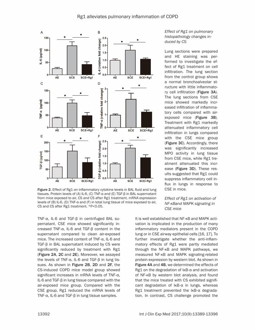
TNF-α, IL-6 and TGF-β in centrifuged BAL su- pernatant. CSE mice showed significantly in- creased TNF-α, IL-6 and TGF-β content in the supernatant compared to clean air-exposed mice. The increased content of TNF-α, IL-6 and TGF-β in BAL supernatant induced by CS were significantly reduced by treatment with Rg1 (Figure 2A, 2C and 2E). Moreover, we assayed the levels of TNF-α, IL-6 and TGF-β in lung tis-sues. As shown in Figure 2B, 2D and 2F, the CS-induced COPD mice model group showed significant increases in mRNA levels of TNF-α, IL-6 and TGF-β in lung tissue compared with the air-exposed mice group. Compared with the CSE group, Rg1 reduced the mRNA levels of TNF-α, IL-6 and TGF-β in lung tissue samples.
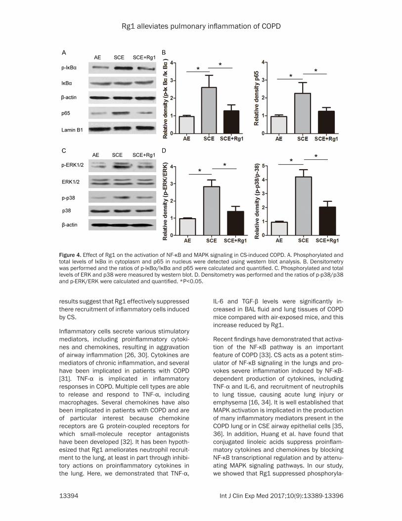
It is well established that NF-κB and MAPK acti-vation is implicated in the production of many inflammatory mediators present in the COPD lung or in CSE airway epithelial cells [16, 17]. To further investigate whether the anti-inflam-matory effects of Rg1 were partly mediated through the NF-κB and MAPK pathways, we measured NF-κB and MAPK signaling-related protein expression by western blot. As shown in Figure 4A and 4B, we determined the effects of Rg1 on the degradation of IκB-α and activation of NF-κB by western blot analysis, and found that the mice treated with CS exhibited signifi-cant degradation of IκB-α in lungs, whereas Rg1 treatment prevented the IκB-α degrada-tion. In contrast, CS challenge promoted the
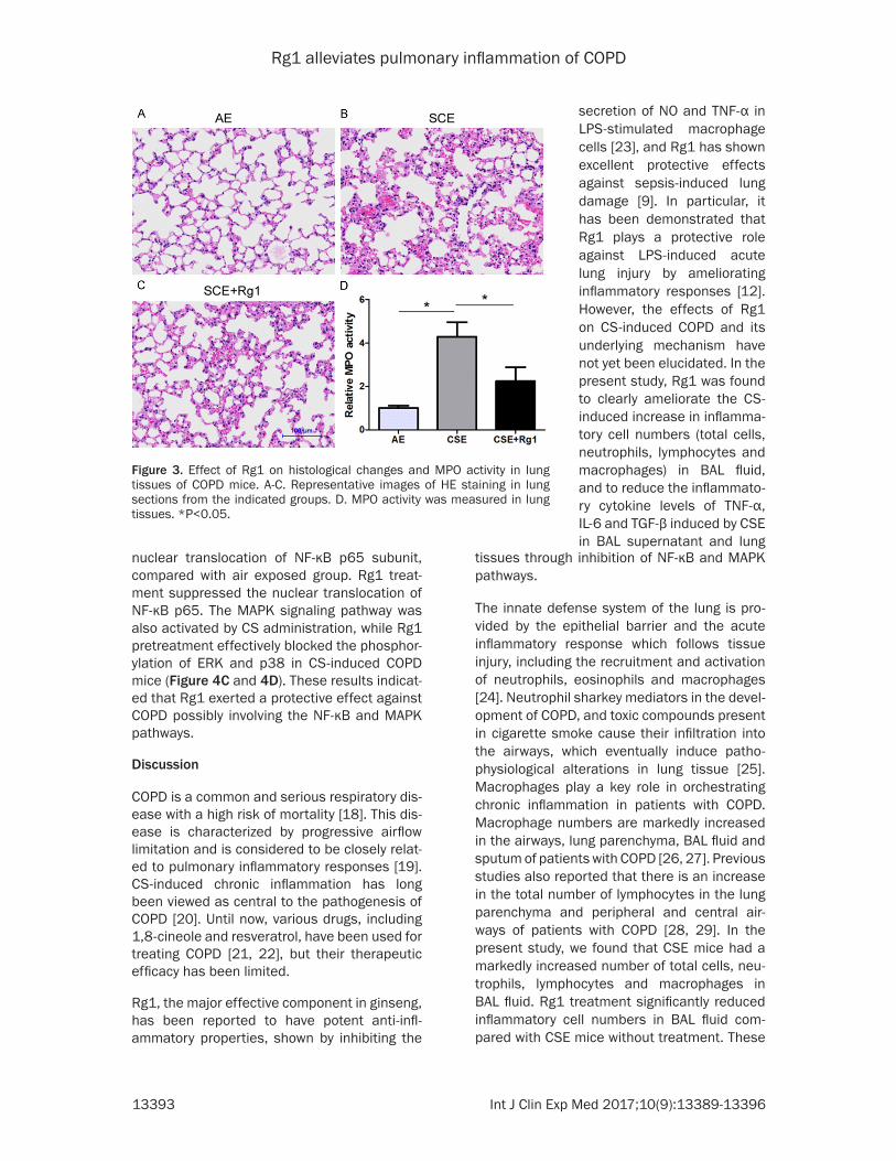
Effect of Rg1 on pulmonary histopathology changes in-duced by CS
Lung sections were prepared and HE staining was per-formed to investigate the ef- fect of Rg1 treatment on cell infiltration. The lung section from the control group shows a normal bronchoalveolar st- ructure with little inflammato-ry cell infiltration (Figure 3A). The lung sections from CSE mice showed markedly incr- eased infiltration of inflamma-tory cells compared with air-exposed mice (Figure 3B). Treatment with Rg1 markedly attenuated inflammatory cell infiltration in lungs compared with the CSE mice group (Figure 3C). Accordingly, there was significantly increased MPO activity in lung tissue from CSE mice, while Rg1 tre- atment attenuated this incr- ease (Figure 3D). These res- ults suggested that Rg1 could suppress inflammatory cell in- flux in lungs in response to CSE in mice.
Effect of Rg1 on activation of NF-κBand MAPK signaling in CSE mice
Figure 2. Effect of Rg1 on inflammatory cytokine levels in BAL fluid and lung tissues. Protein levels of (A) IL-6, (C) TNF-α and (E) TGF-β in BAL supernatant from mice exposed to air, CS and CS after Rg1 treatment. mRNA expression levels of (B) IL-6, (D) TNF-α and (F) in total lung tissue of mice exposed to air, CS and CS after Rg1 treatment. *P<0.05.
Rg1 alleviates pulmonary inflammation of COPD
13393 Int J Clin Exp Med 2017;10(9):13389-13396
nuclear translocation of NF-κB p65 subunit, compared with air exposed group. Rg1 treat-ment suppressed the nuclear translocation of NF-κB p65. The MAPK signaling pathway was also activated by CS administration, while Rg1 pretreatment effectively blocked the phosphor-ylation of ERK and p38 in CS-induced COPD mice (Figure 4C and 4D). These results indicat-ed that Rg1 exerted a protective effect against COPD possibly involving the NF-κB and MAPK pathways.
Discussion
COPD is a common and serious respiratory dis-ease with a high risk of mortality [18]. This dis-ease is characterized by progressive airflow limitation and is considered to be closely relat-ed to pulmonary inflammatory responses [19]. CS-induced chronic inflammation has long been viewed as central to the pathogenesis of COPD [20]. Until now, various drugs, including 1,8-cineole and resveratrol, have been used for treating COPD [21, 22], but their therapeutic efficacy has been limited.
Rg1, the major effective component in ginseng, has been reported to have potent anti-infl- ammatory properties, shown by inhibiting the
tissues through inhibition of NF-κB and MAPK pathways.
The innate defense system of the lung is pro-vided by the epithelial barrier and the acute inflammatory response which follows tissue injury, including the recruitment and activation of neutrophils, eosinophils and macrophages [24]. Neutrophil sharkey mediators in the devel-opment of COPD, and toxic compounds present in cigarette smoke cause their infiltration into the airways, which eventually induce patho-physiological alterations in lung tissue [25]. Macrophages play a key role in orchestrating chronic inflammation in patients with COPD. Macrophage numbers are markedly increased in the airways, lung parenchyma, BAL fluid and sputum of patients with COPD [26, 27]. Previous studies also reported that there is an increase in the total number of lymphocytes in the lung parenchyma and peripheral and central air-ways of patients with COPD [28, 29]. In the present study, we found that CSE mice had a markedly increased number of total cells, neu-trophils, lymphocytes and macrophages in BAL fluid. Rg1 treatment significantly reduced inflammatory cell numbers in BAL fluid com-pared with CSE mice without treatment. These
Figure 3. Effect of Rg1 on histological changes and MPO activity in lung tissues of COPD mice. A-C. Representative images of HE staining in lung sections from the indicated groups. D. MPO activity was measured in lung tissues. *P<0.05.
secretion of NO and TNF-α in LPS-stimulated macrophage cells [23], and Rg1 has shown excellent protective effects against sepsis-induced lung damage [9]. In particular, it has been demonstrated that Rg1 plays a protective role against LPS-induced acute lung injury by ameliorating inflammatory responses [12]. However, the effects of Rg1 on CS-induced COPD and its underlying mechanism have not yet been elucidated. In the present study, Rg1 was found to clearly ameliorate the CS- induced increase in inflamma-tory cell numbers (total cells, neutrophils, lymphocytes and macrophages) in BAL fluid, and to reduce the inflammato-ry cytokine levels of TNF-α, IL-6 and TGF-β induced by CSE in BAL supernatant and lung
Rg1 alleviates pulmonary inflammation of COPD
13394 Int J Clin Exp Med 2017;10(9):13389-13396
results suggest that Rg1 effectively suppressed there recruitment of inflammatory cells induced by CS.
Inflammatory cells secrete various stimulatory mediators, including proinflammatory cytoki- nes and chemokines, resulting in aggravation of airway inflammation [26, 30]. Cytokines are mediators of chronic inflammation, and several have been implicated in patients with COPD [31]. TNF-α is implicated in inflammatory responses in COPD. Multiple cell types are able to release and respond to TNF-α, including macrophages. Several chemokines have also been implicated in patients with COPD and are of particular interest because chemokine receptors are G protein-coupled receptors for which small-molecule receptor antagonists have been developed [32]. It has been hypoth-esized that Rg1 ameliorates neutrophil recruit-ment to the lung, at least in part through inhibi-tory actions on proinflammatory cytokines in the lung. Here, we demonstrated that TNF-α,
IL-6 and TGF-β levels were significantly in- creased in BAL fluid and lung tissues of COPD mice compared with air-exposed mice, and this increase reduced by Rg1.
Recent findings have demonstrated that activa-tion of the NF-κB pathway is an important feature of COPD [33]. CS acts as a potent stim-ulator of NF-κB signaling in the lungs and pro-vokes severe inflammation induced by NF-κB-dependent production of cytokines, including TNF-α and IL-6, and recruitment of neutrophils to lung tissue, causing acute lung injury or emphysema [16, 34]. It is well established that MAPK activation is implicated in the production of many inflammatory mediators present in the COPD lung or in CSE airway epithelial cells [35, 36]. In addition, Huang et al. have found that conjugated linoleic acids suppress proinflam-matory cytokines and chemokines by blocking NF-κB transcriptional regulation and by attenu-ating MAPK signaling pathways. In our study, we showed that Rg1 suppressed phosphoryla-
Figure 4. Effect of Rg1 on the activation of NF-κB and MAPK signaling in CS-induced COPD. A. Phosphorylated and total levels of IκBα in cytoplasm and p65 in nucleus were detected using western blot analysis. B. Densitometry was performed and the ratios of p-IκBα/IκBα and p65 were calculated and quantified. C. Phosphorylated and total levels of ERK and p38 were measured by western blot. D. Densitometry was performed and the ratios of p-p38/p38 and p-ERK/ERK were calculated and quantified. *P<0.05.
Rg1 alleviates pulmonary inflammation of COPD
13395 Int J Clin Exp Med 2017;10(9):13389-13396
tion of ERK and p38, inhibited the degradation of IkB-α and reduced nuclear translocation of NF-κB subunit p65 in CSE lung tissues.
In conclusion, our study demonstrated that Rg1 effectively inhibited the increased inflam-matory cell numbers (total cells, neutrophils, lymphocytes and macrophages) and activation of proinflammatory mediators in BAL fluid and lung tissues, and also ameliorated inflammato-ry cell infiltration into lung tissue of CS-induced COPD mice. The effects of Rg1 were likely caused by blocking NF-κB transcriptional re- gulation and by attenuating MAPK signaling pathways. These results suggest that Rg1 may have therapeutic potential for the suppression of inflammation, which is a crucial step in the development of COPD.
Disclosure of conflict of interest
None.
Abbreviations
Rg1, Ginsenoside Rg1; CS, cigarette smoke; BAL, bronchoalveolar lavage; CSE, CS-exposed; COPD, Chronic obstructive pulmonary disease; ELISA, enzyme-linked immunosorbent assay; MPO, myeloperoxidase; HE, hematoxylin and eosin; qRT-PCR, Quantitative reverse transcrip-tion PCR; GAPDH, glyceraldehyde-3-phosphat-ede hydrogenase.
Address correspondence to: Xinyan Liu, Geriatrics Department, Putuo Hospital Affiliated to Shanghai University of Traditional Chinese Medicine, No.164 lanxi Road, Putuo District, Shanghai 200062, China. E-mail: [email protected]
References
[1] Hogg JC and Timens W. The pathology of chronic obstructive pulmonary disease. Annu Rev Pathol 2009; 4: 435-459.
[2] Mannino DM and Buist AS. Global burden of COPD: risk factors, prevalence, and future trends. Lancet 2007; 370: 765-773.
[3] Chalmers GW, MacLeod KJ, Thomson L, Little SA, McSharry C and Thomson NC. Smoking and airway inflammation in patients with mild asthma. Chest 2001; 120: 1917-1922.
[4] Song HH, Shin IS, Woo SY, Lee SU, Sung MH, Ryu HW, Kim DY, Ahn KS, Lee HK, Lee D and Oh SR. Piscroside C, a novel iridoid glycoside isolated from Pseudolysimachion rotundum var. subinegrum suppresses airway inflamma-
tion induced by cigarette smoke. J Ethnophar-macol 2015; 170: 20-27.
[5] Huang Y, Wu D and Fan W. Protection of ginsenoside Rg1 on chondrocyte from IL-1be-ta-induced mitochondria-activated apoptosis through PI3K/Akt signaling. Mol Cell Biochem 2014; 392: 249-257.
[6] Zhang ZL, Fan Y and Liu ML. Ginsenoside Rg1 inhibits autophagy in H9c2 cardiomyocytes ex-posed to hypoxia/reoxygenation. Mol Cell Bio-chem 2012; 365: 243-250.
[7] Qu DF, Yu HJ, Liu Z, Zhang DF, Zhou QJ, Zhang HL and Du AF. Ginsenoside Rg1 enhances im-mune response induced by recombinant Toxo-plasma gondii SAG1 antigen. Vet Parasitol 2011; 179: 28-34.
[8] Sun J, Song X and Hu S. Ginsenoside Rg1 and aluminum hydroxide synergistically promote immune responses to ovalbumin in BALB/c mice. Clin Vaccine Immunol 2008; 15: 303-307.
[9] Zou Y, Tao T, Tian Y, Zhu J, Cao L, Deng X and Li J. Ginsenoside Rg1 improves survival in a mu-rine model of polymicrobial sepsis by sup-pressing the inflammatory response and apop-tosis of lymphocytes. J Surg Res 2013; 183: 760-766.
[10] Tao T, Chen F, Bo L, Xie Q, Yi W, Zou Y, Hu B, Li J and Deng X. Ginsenoside Rg1 protects mo- use liver against ischemia-reperfusion injury through anti-inflammatory and anti-apoptosis properties. J Surg Res 2014; 191: 231-238.
[11] Wang Y, Liu Y, Zhang XY, Xu LH, Ouyang DY, Liu KP, Pan H, He J and He XH. Ginsenoside Rg1 regulates innate immune responses in macro-phages through differentially modulating the NF-kappaB and PI3K/Akt/mTOR pathways. Int Immunopharmacol 2014; 23: 77-84.
[12] Bao S, Zou Y, Wang B, Li Y, Zhu J, Luo Y and Li J. Ginsenoside Rg1 improves lipopolysaccha-ride-induced acute lung injury by inhibiting in-flammatory responses and modulating infil- tration of M2 macrophages. Int Immunophar-macol 2015; 28: 429-434.
[13] D’Hulst A I, Vermaelen KY, Brusselle GG, Joos GF and Pauwels RA. Time course of cigarette smoke-induced pulmonary inflammation in mice. Eur Respir J 2005; 26: 204-213.
[14] Bracke KR, Verhamme FM, Seys LJ, Bantsim-ba-Malanda C, Cunoosamy DM, Herbst R, Hammad H, Lambrecht BN, Joos GF and Brus-selle GG. Role of CXCL13 in cigarette smoke-induced lymphoid follicle formation and chron-ic obstructive pulmonary disease. Am J Respir Crit Care Med 2013; 188: 343-355.
[15] Li D, Hu J, Wang T, Zhang X, Liu L, Wang H, Wu Y, Xu D and Wen F. Silymarin attenuates ci- garette smoke extract-induced inflammation via simultaneous inhibition of autophagy and
Rg1 alleviates pulmonary inflammation of COPD
13396 Int J Clin Exp Med 2017;10(9):13389-13396
ERK/p38 MAPK pathway in human bronchial epithelial cells. Sci Rep 2016; 6: 37751.
[16] Zhao Y, Xu Y, Li Y, Xu W, Luo F, Wang B, Pang Y, Xiang Q, Zhou J, Wang X and Liu Q. NF-kappaB-mediated inflammation leading to EMT via miR-200c is involved in cell transformation in-duced by cigarette smoke extract. Toxicol Sci 2013; 135: 265-276.
[17] Mehra D, Geraghty PM, Hardigan AA and Fo-ronjy R. A comparison of the inflammatory and proteolytic effects of dung biomass and ciga-rette smoke exposure in the lung. PLoS One 2012; 7: e52889.
[18] Sin DD, Anthonisen NR, Soriano JB and Agusti AG. Mortality in COPD: role of comorbidities. Eur Respir J 2006; 28: 1245-1257.
[19] Decramer M, Janssens W and Miravitlles M. Chronic obstructive pulmonary disease. Lan-cet 2012; 379: 1341-1351.
[20] Cigarette smoking and health. American Tho-racic Society. Am J Respir Crit Care Med 1996; 153: 861-865.
[21] Juergens UR. Anti-inflammatory properties of the monoterpene 1.8-cineole: current evide- nce for co-medication in inflammatory airway diseases. Drug Res (Stuttg) 2014; 64: 638-646.
[22] Knobloch J, Sibbing B, Jungck D, Lin Y, Urban K, Stoelben E, Strauch J and Koch A. Resvera-trol impairs the release of steroid-resistant inflammatory cytokines from human airway smooth muscle cells in chronic obstructive pul-monary disease. J Pharmacol Exp Ther 2010; 335: 788-798.
[23] Song Y, Zhao F, Zhang L, Du Y, Wang T and Fu F. Ginsenoside Rg1 exerts synergistic anti-in-flammatory effects with low doses of glucocor-ticoids in vitro. Fitoterapia 2013; 91: 173-179.
[24] Zhang P, Summer WR, Bagby GJ and Nelson S. Innate immunity and pulmonary host defense. Immunol Rev 2000; 173: 39-51.
[25] Stampfli MR and Anderson GP. How cigarette smoke skews immune responses to promote infection, lung disease and cancer. Nat Rev Im-munol 2009; 9: 377-384.
[26] Barnes PJ. Inflammatory mechanisms in pa-tients with chronic obstructive pulmonary dis-ease. J Allergy Clin Immunol 2016; 138: 16-27.
[27] Barnes PJ. Alveolar macrophages as orchestra-tors of COPD. COPD 2004; 1: 59-70.
[28] Hogg JC, Chu F, Utokaparch S, Woods R, Elliott WM, Buzatu L, Cherniack RM, Rogers RM, Sci-urba FC, Coxson HO and Pare PD. The nature of small-airway obstruction in chronic obstruc-tive pulmonary disease. N Engl J Med 2004; 350: 2645-2653.
[29] Grumelli S, Corry DB, Song LZ, Song L, Green L, Huh J, Hacken J, Espada R, Bag R, Lewis DE and Kheradmand F. An immune basis for lung parenchymal destruction in chronic obstruc-tive pulmonary disease and emphysema. PLoS Med 2004; 1: e8.
[30] Quint JK and Wedzicha JA. The neutrophil in chronic obstructive pulmonary disease. J Al-lergy Clin Immunol 2007; 119: 1065-1071.
[31] Barnes PJ. The cytokine network in chronic ob-structive pulmonary disease. Am J Respir Cell Mol Biol 2009; 41: 631-638.
[32] Donnelly LE and Barnes PJ. Chemokine recep-tors as therapeutic targets in chronic obstruc-tive pulmonary disease. Trends Pharmacol Sci 2006; 27: 546-553.
[33] Zaynagetdinov R, Sherrill TP, Gleaves LA, Hunt P, Han W, McLoed AG, Saxon JA, Tanjore H, Gul-leman PM, Young LR and Blackwell TS. Chronic NF-kappaB activation links COPD and lung cancer through generation of an immunosup-pressive microenvironment in the lungs. Onco-target 2016; 7: 5470-5482.
[34] Metcalfe HJ, Lea S, Hughes D, Khalaf R, Ab-bott-Banner K and Singh D. Effects of cigarette smoke on Toll-like receptor (TLR) activation of chronic obstructive pulmonary disease (COPD) macrophages. Clin Exp Immunol 2014; 176: 461-472.
[35] Renda T, Baraldo S, Pelaia G, Bazzan E, Turato G, Papi A, Maestrelli P, Maselli R, Vatrella A, Fabbri LM, Zuin R, Marsico SA and Saetta M. Increased activation of p38 MAPK in COPD. Eur Respir J 2008; 31: 62-69.
[36] Gaffey K, Reynolds S, Plumb J, Kaur M and Singh D. Increased phosphorylated p38 mito-gen-activated protein kinase in COPD lungs. Eur Respir J 2013; 42: 28-41.
Recommended