Bulgarian Journal of Veterinary Medicine, 2017 ONLINE FIRST ISSN 1311-1477; DOI: 10.15547/bjvm.1089
Original article
FLOW CYTOMETRIC ANALYSIS OF SOMATIC CELLS AND OXIDANT/ANTIOXIDANT PROFILE IN DAIRY COWS WITH
SUBCLINICAL MASTITIS
N-E. M. AREF1, A. S. SAYED1, A. M. ZAHRAN2, G. A. ABDELAAL3 & E. A. NASSER3
1Department of Animal Medicine, Faculty of Veterinary Medicine, Assiut University, Assiut, Egypt, 2Department of Clinical Pathology, South Egypt Cancer Institute, Assiut University, Assiut, Egypt, 3Animal Health Institute, Assiut, Egypt
Summary
Aref, N-E. M., A. S. Sayed, A. M. Zahran, G. A. Abdelaal & E. A. Nasser, 2017. Flow cy-tometric analysis of somatic cells and oxidant/antioxidant profile in dairy cows with sub-clinical mastitis. Bulg. J. Vet. Med. (online first). Flow cytometric analysis of somatic cells count (SCC) and oxidant/antioxidant status of cows with subclinical mastitis (SCM) were investigated in 75 lactating Holstein-Friesian cows (20 controls; 55 SCM cows). Milk from active quarters (n=280) and blood samples (n=75) were aseptically collected. Milk samples were subjected to California Mastitis Test (CMT), microbiologic examination and flow cytometric analysis. Blood and milk samples were analysed for malondialdehyde (MDA) concentra-tions and total antioxidant capacity (TAC). Microbiological examination revealed 3 major pathogens in a single or mixed infection: Streptococcus species (26.9%), Staphylococcus species (57.7%) and Escherichia coli (49.23%). Flow cytometric analysis showed significant increase (P<0.05) in inflam-matory cells in milk of cows with SCM compared to control cows. Differential SCC was characte-rised by significant increase (P<0.05) in polymorphonuclear leukocyte (PMNL) counts compared to normal milk. Additionally, Staphylococcus infection alone or mixed with other lactopathogens re-sulted in dramatic increase in PMNL. There was a significant decrease (P<0.01) of TAC in plasma and milk (0.49 ± 0.04, 0.27 ± 0.03 mM/L, respectively) and significantly increased (P<0.01) serum and milk MDA concentrations (13.72 ± 0.3, 7.72 ± 0.17 nmol/mL, respectively) in SCM cows. In conclusion, differential SCC is a reliable index for early detection of SCM. Milk PMNL population can be used as a useful indicator for evaluation of udder infection. Additionally, a combination of MDA and TAC can provide complementary information about the health status of udder in dairy farms.
Key words: antioxidant, flow cytometery, mastitis, somatic cell
INTRODUCTION
Mastitis is well known inflammatory dis-ease of the udder affecting dairy herds, causing devastating economic losses due
to reduction of milk production, treatment costs and early culling (Radostits et al., 2007). In dairy cattle, the prevalence of

Flow cytometric analysis of somatic cells and oxidant/antioxidant profile in dairy cows with ….
BJVM, ××, No × 2
subclinical mastitis (SCM) may reach up to 75% (Kahn & Line, 2010). Approxi-mately 70 to 80% of financial losses are caused by SCM (Colla et al., 2011).
The International Dairy Federation (IDF, 1981) has adopted the somatic cell count (SCC) and microbiological status of the milk samples for assessment of the udder health and diagnosis of SCM. How-ever, some logistic, financial and technical concerns associated with microbiological examination of all suspected milk sam-ples, have precluded this technique from being widely implemented (Sharma et al., 2010). While SCC is generally accepted for assessment of the health status of the mammary gland, the SCC results may va-ry with the status of lactation, age, stress of the animals, time and frequency of milking, and season. Additionally, SCC could not give information about the type of involved cells.
The ultimate identification of indivi-dual cell populations of milk leukocytes (lymphocytes, macrophages, and poly-morphonuclear leukocytes) using flow cytometry was recently introduced and proposed to give more accurate results than other tests of SCC (Rivas et al., 2001; Koess & Hamann, 2008).
The involvement of polymorphonu-clear leukocytes (PMNL) and macro-phages during inflammatory process trig-gered by pathogens is well documented. The activities of these cells during the early stages of mastitis play a vital role in defense mechanisms against intramam-mary infection (Abera et al., 2010). Reac-tive oxygen species (ROS) generated from resident and newly recruited leukocytes during microbial killing process may damage mammary epithelial cells with resultant oxidative stress (Barbano et al., 2006).
Therefore, the present study was car-ried out to differentiate somatic cells count in milk using flow cytometery for early detection of subclinical mastitis, to screen for the most commonly incrimi-nated lactopathogens, to define a relation-ship between milk differential somatic cells count and the type of lactopathogens and finally to evaluate the oxidant/an-tioxidant status of dairy cows with sub-clinical mastitis.
MATERIALS AND METHODS
Animals
Seventy five lactating Holstein/Friesian cows were included in the current study, housed in different governmental and pri-vate farms within the territory of Assiut Governorate – Egypt. They were appa-rently healthy after being subjected to general clinical examination according to Rosenberger (1990). Diseased cows, and those affected with clinical mastitis, in early lactation and late pregnancy were excluded from the study.
Samples
Thirty mL of milk samples (n=280) from each quarter were collected aseptically according to the National Mastitis Council (1990). Collected samples were tested by California Mastitis Test (CMT) and trans-ferred immediately to the laboratory for microbiologic, biochemical and flow cy-tometric analysis.
In addition, blood samples (5 mL each) were collected by jugular vein punc-ture using sterile disposable syringes. A whole blood sample was collected into vacutainer tubes coated with heparin to obtain non-haemolysed clear blood plasma for estimation of total antioxidant capacity. The second blood sample was
N-E. M. Aref, A. S. Sayed, A. M. Zahran, G. A. Abdelaal & E. A. Nasser
BJVM, ××, No × 3
collected into dry, clean centrifuge tube without anticoagulant to obtain non-haemolysed clear serum for estimation of MDA level.
Clinical, CMT and microbiological examination
All animals in the study were subjected to general clinical examination according to Rosenberger (1990). California Mastitis Test (CMT) results were read and evalu-ated with 5 scores: –, ±, +, ++, and +++, which are reported as homogenous fluid, very slight flocculation, slight floccula-tion, flocculation (flaks), and gel forma-tion, respectively (Schalm et al., 1971). Ten mL of milk samples were centrifuged in sterile screw-capped test tubes for 30 min at 3000 rpm. The cream and super-natant milk were discarded. Milk sediment was simultaneously cultured on selective media, isolated and identified for major lactopathogens including Staphylococcus, Streptococcus and E. coli. The resulting growth was identified on the basis of mor-phology, colony characteristics, Gram’s reaction and biochemical reactions. Ani-mals culturally positive for at least a sin-gle quarter were considered SCM posi-tive.
Flow cytometric analysis of somatic cells
Milk samples were centrifuged for 15 min at 200×g and 4 °C, cream layers and su-pernatants were discarded and pellets were suspended in phosphate buffered saline (PBS). Cell staining buffer (CSB) up to ~15 mL was added and centrifuged at 350×g for 5 minutes. Pellets were col-lected and 3 mL of lysing buffer was added to each tube, kept on ice for 5 min. Cell lysis was stopped by adding 10 mL CSB, centrifuged for 5 minutes at 1500×g for 3 min at 4 ºC, supernatants were dis-carded and cell pellet was agitated and
suspended in 3.5 mL PBS. Viable cells were counted and resuspended in CSB at 5–10 ×106 cells/mL and 100 μL/tube of cell suspension (5–10×105 cells/tube) was distributed into 12×75 mm plastic tubes. Surface monoclonal antibodies (mAb) against CD11a and CD18 were obtained from the Monoclonal Antibody Center at Washington State University (Cat. No. BOV2023 recognising CD11a and Cat.; No. BOV2030 recognising CD18) and dispensed into all appropriately labelled tubes. Tubes were agitated gently to mix and incubated in dark for 20 min at 4 ºC. Secondary coloured conjugated mono-clonal antibodies, Goat Anti-Mouse IgG1 Alexa Fluor 488, and Goat Anti-Mouse IgG2a, R-Phycoerythrin, (Cat. No. A21121 and Cat. No. P21139, Thermo Fisher Scientific, Rockford, IL, USA) were added, incubated for 30 min/4 oC. Samples were analysed by multicolour flow cytometry (FACS-Caliber, Beckton Dikinson, USA). The flow cytometry was adjusted to count up to 10,000 cells per sample. The percentage of specific cell population was calculated as its propor-tion to the total number of cells counted by flow cytometry.
Biochemical analysis
Total antioxidant capacity (TAC) was determined by the reaction of antioxidants in the sample with a defined amount of exogenously provided hydrogen peroxide (H2O2). The antioxidants in the sample eliminated a certain amount of the pro-vided H2O2. The residual H2O2 was de-termined colorimetrically by an enzymatic reaction, which involves the conversion of 3,5, dichloro-2-hydroxy benzensulphonate to a coloured product (Koracevic et al., 2001). The absorbance of the resultant product was measured at 505 nm wave-length.

Flow cytometric analysis of somatic cells and oxidant/antioxidant profile in dairy cows with ….
BJVM, ××, No × 4
Malondialdehyde (MDA) was deter-mined by measuring thiobarbituric reac-tive species using the method of Ruiz-Larrea et al. (1994). Thiobarbituric acid (TBA) reacts with MDA in acidic medium at 95 ºC for 30 min to form thiobarbituric acid reactive product. The absorbance of the resultant pink product was measured at 534 nm wavelength.
Spectrophotometry (Optizen 3220 UV, Mecasys Co. Ltd, Korea) and standard test kits for TAC and MDA (Biodiagnostica Co-Egypt) were used in both assays.
Statistical analysis
The SPSS package for Windows version 10.0 was used for statistical analysis (SPSS, 1999). Data were expressed as mean ± SEM. Differences between groups are determined by means of Student t-test or Duncan's new multiple range test whenever needed. Significance level was
set at P≤0.05 and P≤0.01.
RESULTS
Analysis of animals’ data revealed that the average age, parity and milking period of cows under investigation were 3.9 years, 2.25 parturitions and 3.7 months, respecti-vely. There were no significant differen-ces (P>0.05) in the age, parity and milk-ing period between control and subclinical mastitis groups. The majority of cows with subclinical mastitis were at the third (42%) and fourth (45%) months of lacta-tion.
A total number of 280 quarter milk samples were classified according to CMT into negative (n=80) from 20 healthy cows, one positive (n=85), two positives (n=50), and three positives (n=65).
Out of 200 CMT positive milk sam-
Table 1. Number and frequency of isolated pathogens from milk samples in cows with subclinical mastitis (n =130)
SCM milk samples Microorganism
Number Frequency, % Staphylococcus sp. 43 33.0 Streptococcus sp. 16 12.3 E. coli 32 24.7
Mixed infection
Staphylococcus + E. coli 20 15.4 Streptococcus + E. coli 7 5.4 Staphylococcus + Streptococcus 7 5.4 Staphylococcus + Streptococcus + E. coli 5 3.8
Total isolates 130 100.0
Table 2. Total antioxidant capacity and malondialdehyde in blood and milk in control cows and cows affected with subclinical mastitis. Data are presented as mean ±SEM
Control Subclinical mastitis
plasma (n=75) 0.88 ± 0.10 0.49 ± 0.04* Total antioxidants (mM/L)
milk (n=280) 0.40 ± 0.04 0.27 ± 0.03* serum (n= 75) 4.53 ± 0.38 13.72 ± 0.30*
Malondialdehyde (nmol/mL) milk (n=280) 5.27 ± 0.27 7.72 ± 0.17*
* statistically significant difference at P<0.01.

N-E. M. Aref, A. S. Sayed, A. M. Zahran, G. A. Abdelaal & E. A. Nasser
BJVM, ××, No × 5
ples, 130 samples were randomly selected and tested microbiologically. Three mi-crobial pathogens were isolated either alone or in association with other patho-gens: Streptococcus species (n=35, 26.9%), Staphylococcus species (n=75, 57.7%) and Escherichia coli (n=64, 49.23%). The frequency of isolation of each pathogen is illustrated in Table 1.
The levels of MDA and TAC in blood and milk of control and subclinical masti-tis cows are illustrated in Table 2. There was a significant (P≤0.01) decrease in the milk and plasma level of TAC in cows with subclinical mastitis compared to normal cows. Also, there was a marked (P≤0.01) increase in the milk and serum level of MDA in cows with subclinical mastitis compared to healthy cows.
R1
Fig. 1. Flow cytometric analysis of milk leukocytes. Forward (FSC) and side scatter (SSC) histogram
was used to define the total somatic cells in milk (R1). Events defined as somatic cells (R1) were then examined for their CD11a and CD18 binding, to detect total leukocyte
(the cells that co-express CD18 and CD11a) count.
A B
Fig. 2. Flow cytometric analysis of control (A) and SCM milk sample (B) with CD11a and CD18 mAb showing low and high number of inflammatory cells, respectively.
Flow cytometric analysis of somatic cells and oxidant/antioxidant profile in dairy cows with ….
BJVM, ××, No × 6
A B
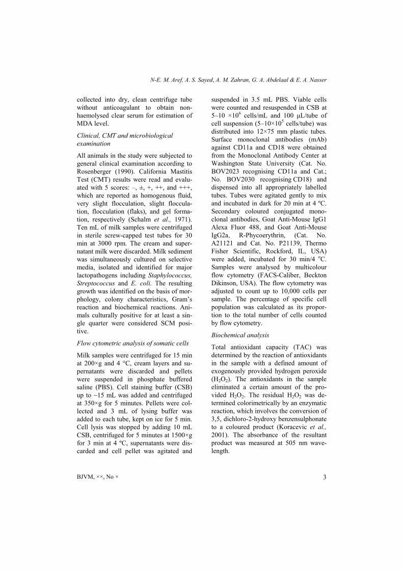
Fig. 3. Flow cytometric analysis of control and subclinical mastitis samples with CD11a and CD18 mAb showing predominant fields of lymphocytes (A) and neutrophils (B).
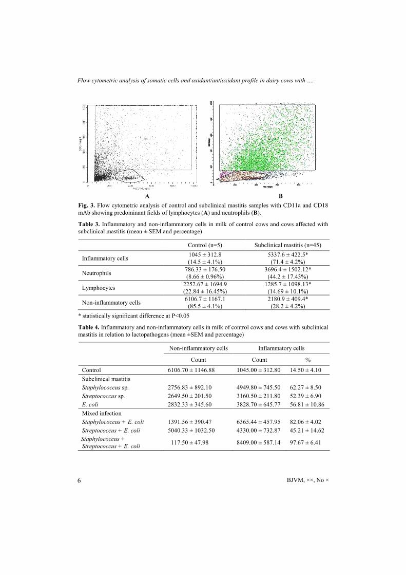
Table 3. Inflammatory and non-inflammatory cells in milk of control cows and cows affected with subclinical mastitis (mean ± SEM and percentage)
Control (n=5) Subclinical mastitis (n=45)
Inflammatory cells 1045 ± 312.8 (14.5 ± 4.1%)
5337.6 ± 422.5* (71.4 ± 4.2%)
Neutrophils 786.33 ± 176.50 (8.66 ± 0.96%)
3696.4 ± 1502.12* (44.2 ± 17.43%)
Lymphocytes 2252.67 ± 1694.9 (22.84 ± 16.45%)
1285.7 ± 1098.13* (14.69 ± 10.1%)
Non-inflammatory cells 6106.7 ± 1167.1
(85.5 ± 4.1%) 2180.9 ± 409.4* (28.2 ± 4.2%)
* statistically significant difference at P<0.05
Table 4. Inflammatory and non-inflammatory cells in milk of control cows and cows with subclinical mastitis in relation to lactopathogens (mean ±SEM and percentage)
Non-inflammatory cells Inflammatory cells
Count Count %
Control 6106.70 ± 1146.88 1045.00 ± 312.80 14.50 ± 4.10
Subclinical mastitis
Staphylococcus sp. 2756.83 ± 892.10 4949.80 ± 745.50 62.27 ± 8.50
Streptococcus sp. 2649.50 ± 201.50 3160.50 ± 211.80 52.39 ± 6.90
E. coli 2832.33 ± 345.60 3828.70 ± 645.77 56.81 ± 10.86
Mixed infection
Staphylococcus + E. coli 1391.56 ± 390.47 6365.44 ± 457.95 82.06 ± 4.02
Streptococcus + E. coli 5040.33 ± 1032.50 4330.00 ± 732.87 45.21 ± 14.62
Staphylococcus + Streptococcus + E. coli
117.50 ± 47.98 8409.00 ± 587.14 97.67 ± 6.41
N-E. M. Aref, A. S. Sayed, A. M. Zahran, G. A. Abdelaal & E. A. Nasser
BJVM, ××, No × 7
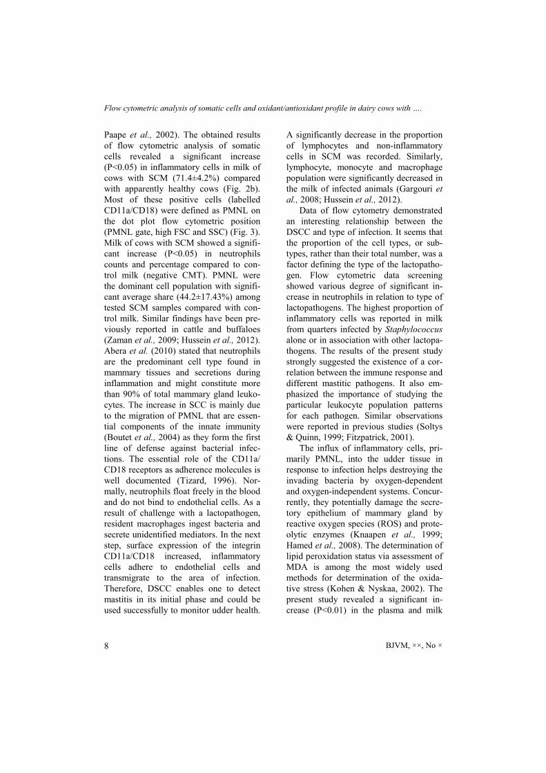
Leukocyte population were determined by flow cytometry and calculated as posi-tive cells (labelled) by the CD11a/ CD18 mAb. Epithelial cells were calculated as the negative cells (not labelled) by CD11a/ CD18 mAb. The location of posi-tive cells labelled with mAb used on the dot plot [Forward-angle light scatter (FSC) by logarithm side-light scatter (SSC)] map is presented on Fig. 1–3.
Quarters free of intramammary infec-tion had significantly increased CD11a/ CD18 negative cells and were reported as non-inflammatory cells. Additionally, healthy udder showed a higher population of lymphocyte than that reported in SCM based on the location of predominant cells on the flow cytometric scatter (low FSC and SSC) (Fig. 2A). On the other hand, flow cytometric analysis of somatic cells revealed significant (P≤0.05) increase in leukocytes (inflammatory) cells in milk of cows with SCM compared to control ones (Fig. 2B, Table 3). A dramatic increase in leukocyte population was reported in SCM cases due to Staphylococcus infec-tion either alone or in mixed infection (Table 4).
DISCUSSION
Somatic cells count in milk has been adopted in mastitis control programmes and milk quality control; however, some physiological factors such as status of lactation, age, stress, time and frequency of milking, and season may negatively affect the SCC. Moreover, SCC could not give information about the type of in-volved cells (inflammatory and non-inflammatory). Differential somatic cell count (DSCC) provides more detailed evaluation of the udder health status (Schwarz et al., 2011) and allows for ear-ly detection of mastitis.
The receptors of CD11a and CD18 are distributed on various immune cells in-cluding T, B and NK cells, macrophages and granulocytes (Larson & Springer, 1990). In this study, immune and non-immune cells (epithelial cells) were identi-fied by flow cytometry using specific monoclonal antibody against CD11a/ CD18 receptors. Epithelial cells were identified as the negative cells to the mAb anti-CD11a/CD18 receptor whereas posi-tive cells on dot plot of FC scatter were identified as lymphocytes and neutrophils. Fig. 1–2A showed forward (FSC) and side scatter (SSC) histogram that used to de-fine SCC and differentiate milk leukocytes (lymphocytes and granulocytes) according to their size and complexity. Lymphocytes were characterised by their small size (low forward scatter) and lower complexity and granulations (low side scatter) while granulocytes – by their large size (high forward scatter) and higher complexity and granulations (high side scatter) at the total somatic cells in milk.
Our results showed that milk samples from culture-negative udders revealed high epithelial cells and lymphocyte per-centages of 85.5±4.1% and 22.84±16.45%, respectively while PMNL were 8.66± 0.96%. Similar observations were re-ported in previous studies (Rivas et al., 2001; Schwarz et al., 2011).
On the other hand, inflammation is generally defined as increased leukocytes, especially PMNL in tissues or body fluids in response to pathogens. An increased transfer of PMNL from blood into the area of infection at the beginning of an inflammation is well documented and could be detected (Kehrli & Shuster, 1994; Paape et al., 2002). Therefore, a high PMNL percentage in milk is ac-cepted as an important indicator of in-flammatory reaction (Pillai et al., 2001;
Flow cytometric analysis of somatic cells and oxidant/antioxidant profile in dairy cows with ….
BJVM, ××, No × 8
Paape et al., 2002). The obtained results of flow cytometric analysis of somatic cells revealed a significant increase (P<0.05) in inflammatory cells in milk of cows with SCM (71.4±4.2%) compared with apparently healthy cows (Fig. 2b). Most of these positive cells (labelled CD11a/CD18) were defined as PMNL on the dot plot flow cytometric position (PMNL gate, high FSC and SSC) (Fig. 3). Milk of cows with SCM showed a signifi-cant increase (P<0.05) in neutrophils counts and percentage compared to con-trol milk (negative CMT). PMNL were the dominant cell population with signifi-cant average share (44.2±17.43%) among tested SCM samples compared with con-trol milk. Similar findings have been pre-viously reported in cattle and buffaloes (Zaman et al., 2009; Hussein et al., 2012). Abera et al. (2010) stated that neutrophils are the predominant cell type found in mammary tissues and secretions during inflammation and might constitute more than 90% of total mammary gland leuko-cytes. The increase in SCC is mainly due to the migration of PMNL that are essen-tial components of the innate immunity (Boutet et al., 2004) as they form the first line of defense against bacterial infec-tions. The essential role of the CD11a/ CD18 receptors as adherence molecules is well documented (Tizard, 1996). Nor-mally, neutrophils float freely in the blood and do not bind to endothelial cells. As a result of challenge with a lactopathogen, resident macrophages ingest bacteria and secrete unidentified mediators. In the next step, surface expression of the integrin CD11a/CD18 increased, inflammatory cells adhere to endothelial cells and transmigrate to the area of infection. Therefore, DSCC enables one to detect mastitis in its initial phase and could be used successfully to monitor udder health.
A significantly decrease in the proportion of lymphocytes and non-inflammatory cells in SCM was recorded. Similarly, lymphocyte, monocyte and macrophage population were significantly decreased in the milk of infected animals (Gargouri et al., 2008; Hussein et al., 2012).
Data of flow cytometry demonstrated an interesting relationship between the DSCC and type of infection. It seems that the proportion of the cell types, or sub-types, rather than their total number, was a factor defining the type of the lactopatho-gen. Flow cytometric data screening showed various degree of significant in-crease in neutrophils in relation to type of lactopathogens. The highest proportion of inflammatory cells was reported in milk from quarters infected by Staphylococcus alone or in association with other lactopa-thogens. The results of the present study strongly suggested the existence of a cor-relation between the immune response and different mastitic pathogens. It also em-phasized the importance of studying the particular leukocyte population patterns for each pathogen. Similar observations were reported in previous studies (Soltys & Quinn, 1999; Fitzpatrick, 2001).
The influx of inflammatory cells, pri-marily PMNL, into the udder tissue in response to infection helps destroying the invading bacteria by oxygen-dependent and oxygen-independent systems. Concur-rently, they potentially damage the secre-tory epithelium of mammary gland by reactive oxygen species (ROS) and prote-olytic enzymes (Knaapen et al., 1999; Hamed et al., 2008). The determination of lipid peroxidation status via assessment of MDA is among the most widely used methods for determination of the oxida-tive stress (Kohen & Nyskaa, 2002). The present study revealed a significant in-crease (P<0.01) in the plasma and milk

N-E. M. Aref, A. S. Sayed, A. M. Zahran, G. A. Abdelaal & E. A. Nasser
BJVM, ××, No × 9
concentration of MDA in cows with SCM compared to normal cows. Similar results have been reported in other studies on dairy cows (Suriyasathaporna et al., 2006; Andrei et al., 2010; Yang et al., 2011). In contrast, Ranjan et al. (2005) and Kizil et al. (2007) reported an enhanced erythro-cyte and plasma lipid peroxide content in clinical, but not in subclinical mastitis cases. On the other hand, concentration of TAC showed a significant decrease (P<0.01) in milk and blood samples of subclinically mastitic compared to normal cows. The high production of MDA and low levels of TAC in SCM milk reported in this study confirmed the occurrence of a process of auto-oxidation and oxidative stress associated with SCM in dairy cows. Similar results have been reported in pre-vious studies (Suriyasathaporna et al. 2006; Atakisi et al., 2010).
Total antioxidant capacity identifies a wide range of all activities of the measu-rable exogenous and endogenous antioxi-dants (Somogyi et al., 2007). TAC consum-ption by generated ROS leads to oxidative stress and resultant reduction in milk pro-duction. There is a significant negative cor-relation between the TAC and total oxi-dant capacity (TOC) values (Erel, 2005).
It can be concluded that differential somatic cell count was the most reliable index for early detection of SCM next to bacteriological examination. Neutrophil population in milk can be used a useful indicator in the evaluation of mammary gland infection. Additionally, a combina-tion of MDA and TAC can provide com-plementary information about the health status of udder in dairy farms.
REFERENCES
Abera, M., B. Demie, K. Aragaw, F. Regassa, & A. Regassa, 2010. Isolation and identi-
fication of Staphylococcus aureus from bovine mastitic milk and their drug resis-tance patterns in Adama Town, Ethiopia. Journal of Veterinary Medicine and Ani-mal Health, 2, 29–34.
Andrei, S., S. Matei, D. Zinveliu, A. Pintea, A. Bunea, S. Ciupe, & I. Groza, 2010. Corre-lations between antioxidant enzymes activ-ity and lipids peroxidation level in blood and milk from cows with subclinical mas-titis. Bulletin of University of Agricultural Sciences and Veterinary Medicine Cluj-Napoca, 67, 6–11.
Atakisi, O., O. H. Hasan, E. Atakisi, O. Mer-han, S. M. Pancarci, A. Ozcan, S. Marasli, B. Polat, A. Colak & S. Kaya, 2010. Sub-clinical mastitis causes alterations in nitric oxide, total oxidant and antioxidant capac-ity in cow milk. Research in Veterinary Science, 89, 10–13.
Barbano, D. M., Y. Ma & M. V. Santos, 2006. Influence of raw milk quality on fluid milk shelf life. Journal of Dairy Science, 89, E15–E19.
Boutet, P., D. Boulanger, L. Gillet, A. Vanderplasschen, R. Closset, F. Bureau & P. Lekeux, 2004. Delayed neutrophil apoptosis in bovine subclinical mastitis. Journal of Dairy Science, 87, 4104–4114.
Colla, M. F., S. F. Valle, P. Secchi, N. Duda, M. Scalon, J. W. Dürr & F. H. Diaz Gon-zález, 2011. Plasma haptoglobin values in cows with different somatic cell counting in milk samples. Acta Scientiae Veteri-nariae, 39, 944–949.
Erel, O., 2005. A new automated colorimetric method for measuring total oxidant status. Clinical Biochemistry, 38, 1103–1111.
Fitzpatrick, L. J., 2001. Milk somatic cells – what do they do? In: Proceedings of the British Mastitis Conference, Garstang, In-stitute for Animal Health/Milk Develop-ment Council, pp. 56–62.
Gargouri, A., H. Hamed & A. El Feki, 2008. Total and differential bulk cow milk so-matic cell counts and their relation with lipolysis. Livestock Science, 113, 274–279.

Flow cytometric analysis of somatic cells and oxidant/antioxidant profile in dairy cows with ….
BJVM, ××, No × 10
Hamed, H., A. El Feki & A. Gargouri, 2008. Total and differential bulk cow milk so-matic cell counts and their relation with antioxidant factors. Comptes Rendus Biologies, 331, 144–151.
Hussein, S. A., 2012. Prevalence and bacterial etiology of subclinical mastitis in dairy cows in Al Sulaimaniyah district, Kufa Journal of Veterinary Medical Sciences, 3, 190–203.
IDF (International Dairy Federation), 1981. Laboratory Methods for Use in Mastitis Work, Document 132, Brussels, Belgium.
Larson, R. S. & T. A. Springer, 1990. Struc-ture and function of leukocyte integrins. Immunological Reviews, 114, 181–217.
Kahn, C. M. & S. Line, 2010. The Merck Ve-terinary Manual, 10th edn, Merck and Co., USA. pp. 1248–1257.
Kehrli, M. E. & D. E. Shuster, 1994. Factors affecting milk somatic cells and their role in health of the bovine mammary gland. Journal of Dairy Sciences, 77, 619–627.
Kizil, O., Y. Akar, N. Saat, M. Kizil & M. Yuksel, 2007. The plasma lipid peroxida-tion intensity (MDA) and chain-breaking antioxidant concentrations in the cows with clinic or subclinic mastitis. Revue de Médecine Vétérinaire, 158, 529–533.
Knaapen, A. M., F. Seiler, P. A. Schilderman, P. Nehls, J. Bruch, R. P. Schins & P. J. Borm, 1999. Neutrophils cause oxidative DNA damage in alveolar epithelial cells. Free Radical Biology and Medicine, 27, 234–240.
Koess, C. & J. Hamann, 2008. Detection of mastitis in the bovine mammary gland by flow cytometry at early stages. Journal of Dairy Research, 75, 225–232.
Kohen, R. & A. Nyska, 2002. Oxidation of biological systems: Oxidative stress phe-nomena, antioxidants, redox reactions, and methods for their quantification. Journal of Toxicologic Pathology, 30, 620–650.
Koracevic, D., G. Koracevic, V. Djordjevic, S. Andrejevic & V. Cosic, 2001. Method for the measurement of antioxidant activity in
human fluids. Journal of Clinical Pathol-ogy, 54, 356–361.
National Mastitis Council (NMC), 1990. Microbiological Procedures for the Diag-nosis of Udder Infection. 3rd edn, Arling-ton V.A. National Mastitis Council Inc.
Paape, M. J., J. Mehrzad, X. Zhao, J. Detilleux & C. Burvenich, 2002. Defense of the bo-vine mammary gland by polymorphonu-clear neutrophil leukocytes. Journal of Mammary Gland Biology and Neoplasia, 7, 109–121.
Pillai, S. R., E. Kunze, L. M. Sordillo & B. M. Jayarao, 2001. Application of differential inflammatory cell count as a tool to moni-tor udder health. Journal of Dairy Science, 84, 1413–1420.
Radostits, O. M., C. C. Gay, K. W. Hinchcliff & P. D. Constable, 2007. Veterinary Medicine. A Textbook of the Diseases of Cattle, Horses, Sheep, Pigs and Goats, 10th edn, Saunders Elsevier, Edinburgh, pp. 673–759.
Ranjan, R., D. Swarup, R. Naresh & R. C. Patra, 2005. Enhanced erythrocytic lipid peroxides and reduced plasma ascorbic acid, and alteration in blood trace elements level in dairy cows with mastitis. Veteri-nary Research Communications, 29, 27–34.
Rivas, A. L., F. W. Quimby, J. Blue & O. Coksaygan, 2001. Longitudinal evaluation of bovine mammary gland health status by somatic cell counting, flow cytometry, and cytology. Journal of Veterinary Diagnos-tic Investigation, 13, 399–407.
Rosenberger, G., 1990. Clinical Examination of Cattle. Velage Paul Parey. Berlin and Hamburg, pp 120–138.
Ruiz-Larrea, M. B., A. M. Leal, M. Liza, M. Lacort & H. De Groot, 1994. Antioxidant effects of estradiol and 2-hydroxyestradiol on iron-induced lipid peroxidation of rat liver microsomes. Steroids, 59, 383–388.
Schalm, O. W., E. Carrol & N. C. Jain, 1971. Bovine Mastitis, 1st edn, Lea and Febiger, Philadelphia, USA.
N-E. M. Aref, A. S. Sayed, A. M. Zahran, G. A. Abdelaal & E. A. Nasser
BJVM, ××, No × 11
Schwarz, D., U. S. Diesterbeck, S. Koenig, K. Bruegemann, K. Schlez, M. Zschoeck, W. Wolter & C. P. Czerny, 2011. Flow cy-tometric differential cell counts in milk for the evaluation of inflammatory reactions in clinically healthy and subclinically in-fected bovine mammary glands. Journal of Dairy Science, 94, 5033–5044.
Sharma, N., V. Pandey & N. A. Sudan, 2010. Comparison of some indirect screening tests for detection of subclinical mastitis in dairy cows. Bulgarian Journal of Veteri-nary Medicine, 13, 98–103.
Soltys, J. & M. T. Quinn, 1999. Selective re-cruitment of T-cell subsets to the udder during staphylococcal and streptococcal mastitis: analysis of lymphocyte subsets and adhesion molecule expression. Infec-tion and Immunity, 67, 6293–6302.
Somogyi, A., K. Rosta, P. Pusztai, Z. Tulassay & G. Nagy, 2007. Antioxidant measure-ments. Physiological Measurement, 28, 41–55.
Suriyasathaporna, S., U. Vnitketkumnuenb, T. Chewonarinb, S. Boonyayatraa, K. Krea-usukona & Y. H. Schukken, 2006. Higher somatic cell counts resulted in higher malondialdehyde concentration in raw cow’s milk. International Dairy Journal, 16, 1088–1091.
Tizard, R. I., 1996. Veterinary Immunology, Chapter 5, W.B. Saunders, Philadelphia, pp. 43–54.
Yang, F. L., X. S. Li, B. X. He, X. L. Yang, G. H. Li, P. Liu, O. H. Huang, X. M. Pan & J. Li, 2011. Malondialdehyde level and some enzymatic activities in subclinical mastitis milk. African Journal of Biotechnology, 10, 5534–5538.
Zaman, M., S. Khan, M. Avais, M. Ijaz, J. A. Khan & A. Khan, 2009. Estimation of pro-tein, total leukocyte count and differential leukocyte count in the blood and milk of subclinically mastitic buffaloes. Pakistan Journal of Zoology, 9, 115–118.
Paper received 25.11.2016; accepted for publication 06.02.2017
Correspondence:
Nasr-Eldin M. Aref Department of Animal Medicine Faculty of Veterinary Medicine Assiut University Assiut 71526, Egypt tel. +201060024252 e-mail: [email protected]
Recommended