BioMed CentralMolecular Pain
ss
Open AcceResearchInvolvement of S-nitrosylation of actin in inhibition of neurotransmitter release by nitric oxideJingshan Lu1, Tayo Katano1, Emiko Okuda-Ashitaka1, Yo Oishi2, Yoshihiro Urade2 and Seiji Ito*1Address: 1Department of Medical Chemistry, Kansai Medical University, Moriguchi, Japan and 2Department of Molecular Behavioral Biology, Osaka Bioscience Institute, Osaka, Japan
Email: Jingshan Lu - [email protected]; Tayo Katano - [email protected]; Emiko Okuda-Ashitaka - [email protected]; Yo Oishi - [email protected]; Yoshihiro Urade - [email protected]; Seiji Ito* - [email protected]
* Corresponding author
AbstractBackground: The role of the diffusible messenger nitric oxide (NO) in the regulation of paintransmission is still a debate of matter, pro-nociceptive and/or anti-nociceptive. S-Nitrosylation, thereversible post-translational modification of selective cysteine residues in proteins, has emerged asan important mechanism by which NO acts as a signaling molecule. The occurrence of S-nitrosylation in the spinal cord and its targets that may modulate pain transmission remainunclarified. The "biotin-switch" method and matrix-assisted laser desorption/ionization time-of-flight mass spectrometry were employed for identifying S-nitrosylated proteins.
Results: Here we show that actin was a major protein S-nitrosylated in the spinal cord by the NOdonor, S-nitroso-N-acetyl-DL-penicillamine (SNAP). Interestingly, actin was S-nitrosylated, more inthe S2 fraction than in the P2 fraction of the spinal homogenate. Treatment of PC12 cells withSNAP caused rapid S-nitrosylation of actin and inhibited dopamine release from the cells. Just likecytochalasin B, which depolymerizes actin, SNAP decreased the amount of filamentous actincytoskeleton just beneath the membrane. The inhibition of dopamine release was not attenuatedby inhibitors of soluble guanylyl cyclase and cGMP-dependent protein kinase.
Conclusion: The present study demonstrates that actin is a major S-nitrosylated protein in thespinal cord and suggests that NO directly regulates neurotransmitter release by S-nitrosylation inaddition to the well-known phosphorylation by cGMP-dependent protein kinase.
BackgroundNitric oxide (NO) is produced from L-arginine by 3 iso-forms of NO synthase (NOS), i.e., neuronal NOS (NOS-1), inducible NOS (NOS-2), and endothelial NOS (NOS-3); and it plays important roles in a wide variety of physi-ological and pathophysiological processes such as neuro-transmission, regulation of vascular tone, and mediation
of immune responses [1,2]. The major intracellular recep-tor for NO is a soluble guanylyl cyclase that catalyzes thesynthesis of cGMP. This intracellular signaling moleculemodulates the activity of many targets in the cells includ-ing cGMP-dependent protein kinase (cGK), ion channels,and phosphodiesterases. In the central nervous system,NO is mainly produced by NOS-1 and has been impli-
Published: 29 September 2009
Molecular Pain 2009, 5:58 doi:10.1186/1744-8069-5-58
Received: 16 June 2009Accepted: 29 September 2009
This article is available from: http://www.molecularpain.com/content/5/1/58
© 2009 Lu et al; licensee BioMed Central Ltd. This is an Open Access article distributed under the terms of the Creative Commons Attribution License (http://creativecommons.org/licenses/by/2.0), which permits unrestricted use, distribution, and reproduction in any medium, provided the original work is properly cited.
Page 1 of 12(page number not for citation purposes)
Molecular Pain 2009, 5:58 http://www.molecularpain.com/content/5/1/58
cated in synaptic plasticity including long-term potentia-tion in the hippocampus and in pain transmission in thespinal cord [3-5]. Many behavioral studies including ourshave demonstrated that NO contributes to the develop-ment and maintenance of hyperalgesia and allodynia inmodels of acute and chronic pain, which are relieved bythe blockade of the NO/cGMP/cGK signaling pathway inthe spinal cord [6-9]. A rapid release of citrulline, a markerof NO synthesis, is observed in the spinal cord followinga subcutaneous injection of formalin and is associatedwith a biphasic flinching behavior of the injected paw[10]. On the other hand, spinally administered NOdonors cause a depression of ongoing impulse activity ofdorsal horn neurons [11]; and inhibition of spinal NOSleads to increased neuronal activity in the dorsal horn[12]. Furthermore, agents affecting NO and cGMP levelsshow no effect [13] or dual effects on nociception depend-ing on their concentrations [14,15]. Thus the involvementof NO in pain is not consistent and is still a matter ofdebate. Different from many conventional neurotrans-mitters that are stored in synaptic vesicles and released byexocytosis, the labile, free-radical mediator NO simplydiffuses from the nerve terminal into adjacent cells andacts as anterograde and retrograde messengers at nocicep-tive synapses in the spinal cord [3]. Therefore, the mecha-nisms through which NO mediates its nociception andpain transmission are not completely understood in thespinal cord [16].
In addition to the NO/cGMP/cGK signaling pathway, S-nitrosylation by NO, i.e., the covalent attachment of a -NO group to a cysteine thiol, has emerged as an importantfeature of NO signaling [17,18]. Through this reversiblepost-translational modification, NO is able to regulate thefunction of many target enzymes, ion channels, and struc-tural proteins including cyclooxygenase-2 [19], solubleguanylyl cyclase [20], NMDA receptors [21-23], actin[17,24], and other pathogenic proteins [25,26]. Amongmethods for studying protein S-nitrosylation, the biotin-switch method has rapidly gained popularity because ofthe ease with which it can detect individual S-nitrosylatedproteins in biological samples [17,27]; and the use of thismethod has revealed that S-nitrosylation is involved inthe physiology and pathophysiology of the central nerv-ous system [18]. Although the NO/cGMP/cGK signalingpathway has been extensively examined in terms of theinvolvement of NO in nociception and pain transmission,S-nitrosylation has not been studied in the spinal cord sofar. Here, by using the biotin-switch method, we demon-strate that actin is a major S-nitrosylated protein in thespinal cord by the NO donor S-nitroso-N-acetyl-DL-peni-cillamine (SNAP) and that interestingly, actin was S-nitrosylated, more in the S2 fraction than in the P2 frac-tion of the spinal homogenate.
MethodsMaterialsSNAP, nerve growth factor (NGF), 1H-[1,2,4]oxadiazolo-[4,3-a]quinoxalin-1-one (ODQ) and KT5823 wereobtained from Wako Pure Chemical (Osaka, Japan). Trif-luoroacetic acid (TFA), S-methyl methanethiosulfonate(MMTS), α-cyano-4-hydroxycinnamic acid (α-CHCA),bovine serum albumin (BSA), imipramine hydrochloride,dopamine, 8-bromoadenosine 3', 5'-cyclic monophos-phate (8-Br-cAMP), 8-Br-cGMP, and glibenclamide werepurchased from Sigma-Aldrich (St. Louis, MO, USA). Pitu-itary adenylate cyclase-activating polypeptide (PACAP)and N-[6-(biotinamido)hexyl]-3'-(2'-pyridyldithio)pro-pionamide (biotin-HPDP) were supplied by Peptide Insti-tute (Osaka, Japan) and Pierce Chemical (Rockford, IL,USA), respectively. Other chemicals were of reagent grade.
Preparation of S2 and P2 fractions from spinal cordsMale ddy mice (5 weeks old) were purchased from Shi-zuoka Laboratory Centre (Hamamatsu, Japan). The micewere housed under conditions of a 12-h light-12-h darkcycle, a constant temperature of 22 ± 2°C, and 60 ± 10%humidity. They received food and water ad libitum. All ani-mal experiments were carried out in accordance with theNational Institutes of Health guide for the care and use oflaboratory animals and were approved by the AnimalExperimentation Committee of Kansai Medical Univer-sity.
Under anesthesia with pentobarbital (50 mg/kg), mousespinal cords were quickly removed and homogenizedtwice for 30 s with a Polytron homogenizer containing 10volumes of HEN buffer consisting of 250 mM HEPES (pH7.7), 1 mM EDTA, and 0.1 mM neocuproine. Thehomogenate was centrifuged at 800 × g for 10 min, andthe supernatant was recovered and then centrifuged at10,000 × g for 20 min. After the resulting pellet had beendissolved in 10 volumes of HEN buffer, the resultingsupernatant and this dissolved pellet were employed as S2and P2 fractions, respectively.
S-Nitrosylation of proteins in vitro and biotin-switch methodS-Nitrosylated proteins were detected by the biotin-switchmethod as described by Jaffrey et al. [17]. Briefly, S2 andP2 fractions of the spinal cord were incubated at roomtemperature without or with various concentrations ofSNAP for 1 h in the dark. Then, SNAP was removed fromthe reaction mixture by cold acetone precipitation; andthe pellets were subsequently dissolved in HENS buffercontaining 25 mM HEPES, pH 7.7, 0.1 mM EDTA, 0.01mM neocuproine, and 1% sodium dodecyl sulfate (SDS).The SNAP-treated fractions were blocked with fresh 4 mMMMTS for 20 min at 50°C. After 2 steps of acetone precip-itation, the pellets were then resuspended in HENS buffer.
Page 2 of 12(page number not for citation purposes)
Molecular Pain 2009, 5:58 http://www.molecularpain.com/content/5/1/58
For labeling, the samples were subjected to the biotin-switch assay, in which the sample was mixed with 1 mMascorbic acid and 0.2-0.4 mM biotin-HPDP as final con-centrations and kept for 1 h in the dark. Biotinylated pro-teins were resolved by non-reducing SDS-polyacrylamidegel electrophoresis (PAGE), and transferred to a polyvi-nylidene difluoride membrane, followed by immunob-lotting with peroxidase-conjugated anti-biotin antibody(1:1000; Sigma-Aldrich). To confirm and quantify S-nitrosylated actin in the S2 fraction of the spinal cord orcell lysates, after the S-nitrosylated proteins had beenimmunoblotted with anti-biotin antibody, the samemembrane was stripped to detect total actin with anti-actin antibody (1:5000; BD Bioscience, San Jose, CA,USA) by using Enhanced Chemiluminescence (GEHealthcare, Piscataway, NJ, USA). The intensity of S-nitrosylated actin was quantified by using ImageJ softwareand normalized by that of total actin.
Furthermore, to detect individual S-nitrosylated proteinsincluding actin in S2 fractions of the spinal cord, weremoved free biotin-HPDP by using an NAP-5 column(GE Healthcare) after the biotin-switch assay and incu-bated the eluate overnight at 4°C with 50 μl of a strepta-vidin-agarose slurry in 2 volumes of a neutralizationbuffer (20 mM HEPES, pH 7.5, 100 mM NaCl, 1 mMETDA, and 0.5% Triton X-100). The beads were washed 4times with 1 ml of the neutralization buffer supplementedwith 500 mM NaCl. The adsorbed proteins were elutedwith SDS-sample buffer at room temperature for 20 min.The eluted sample was then analyzed by SDS-PAGE, fol-lowed by immunoblotting with anti-actin antibody(1:5000).
Identification of S-nitrosylated proteins by mass spectrometryMost of the sample eluted from streptavidin-agarose gelswas applied to a 10% gel for SDS-PAGE and used for iden-tification of S-nitrosylated proteins. After the gel had beenstained by the Vorum silver staining protocol [28], pro-teins of interest were excised from the gel, destained, andin-gel digested as described by Katano et al. [29]. The gelpiece was dehydrated with acetonitrile and dried by usinga Tomy CC-180 vacuum centrifuge concentrator (Tokyo,Japan). After reduction with 10 mM dithiothreitol andalkylation with 55 mM iodoacetamide, proteins in the gelwere digested overnight at 37°C with 10 μg/ml trypsin(Promega, Madison, WI, USA) in 25 mM NH4HCO3. Thedigested peptides were extracted with 50% acetonitrile in1% TFA. Peptides were desalted and concentrated byusing a Ziptip μC18 (Millipore, Billerica, MA, USA) andeluted with the matrix solution (1 mg/ml α-CHCA in 70%acetonitrile and 1% TFA) onto a target plate. Matrix-assisted laser desorption/ionization reflection time-of-flight mass spectrometry (MALDI-TOF MS) was per-formed by using a Voyager DE-PRO (Applied Biosystems).
All spectra were obtained in a positive reflector modeusing an accelerating voltage of 20 kV. Database searcheswere carried out by using the MASCOT search programhttp://www.matrixscience.com and NCBI protein data-bases. The specified taxonomy was Mus musculus (housemouse), and the specified initial mass tolerance was 50 or70 ppm.
Cell culture and S-nitrosylation in cellsPheochromocytoma cell line PC12 cells were maintainedin Dulbecco's modified Eagle medium supplementedwith 5% fetal calf serum, 10% horse serum, and 50 U/mlpenicillin and kept in a humidified environment of 95%air and 5% CO2 at 37°C. For the S-nitrosylation assay, PC12 cells were cultured on 6-cm dishes for 2 d; and themedium was replaced with serum-free medium 12 h priorto experiments. After 5-min treatment of the cells withPACAP (10 nM) and/or SNAP (100, 300, 500 μM), thecells were disrupted in HEN buffer by sonication; and thelysate was then subjected to the biotin-switch assay.
Measurement of dopamine release from PC12 cellsPC12 cells were seeded on 24-well plates. After 2 days inculture, the cells were preincubated for 15 min in 190 μlof HEPES buffer (140 mM NaCl, 5 mM KCl, 2 mM CaCl2,1.2 mM MgCl2, 10 mM glucose, and 10 mM HEPES, pH7.4); and then the appropriate agents (10 μl) were addedto the medium. Incubation was carried out 37°C for thedesired times in the absence or presence of 10 μM imi-pramine, an inhibitor of dopamine reuptake. After incu-bation, the culture medium in each well was harvested;and perchloric acid in HEPES buffer was then added toeach well for a final concentration of 3%. Culture mediaand cell lysates were adjusted to pH 4 by 1 M sodium ace-tate, and then the samples were centrifuged at 15,000 × gfor 5 min. The supernatants of culture media and celllysates were measured for dopamine released into themedium and cellular dopamine by using an HPLC col-umn equipped with an Eicom electrochemical detectormodel 700 (Kyoto, Japan). HPLC was performed by usinga reversed-phase C18 column (Eicom CA-50DS, 2.1 mm× 150 mm) with a phosphate-buffered mobile phase con-taining 20% methanol, 50 mg/L EDTA, and 0.5 mg/Lsodium 1-octanesulfonate. Cellular dopamine contentwas around 7.2 ± 0.8 ng/well; and basal and PACAP-evoked release of dopamine into the culture media were1.5-2 and 15-20% of cellular dopamine, respectively.
ImmunoblotCerebellum and dorsal root ganglia (DRG) of mice andPC12 cells were homogenized in 20 mM Tris-HCl (pH7.4) containing 150 mM NaCl, 4 mM EDTA, 0.5 mM phe-nylmethylsulfonyl fluoride, 1 μg/ml pepstatin A, 2 μg/mlaprotinin, and 2 μg/ml leupeptin; and the supernatants(100 μg) obtained after centrifugation at 10,000 × g for 30min were subjected to immunoblotting on a polyvinyli-
Page 3 of 12(page number not for citation purposes)
Molecular Pain 2009, 5:58 http://www.molecularpain.com/content/5/1/58
dene difluoride membrane. After blocking with 5% (w/v)BSA in TBS-T buffer at room temperature for 1 h, themembrane was incubated overnight at 4°C with rabbitanti-cGKI (type I cGK) antibody (1:200; Santa Cruz Bio-tech., Santa Cruz, CA, USA) and then at room temperaturefor 1 h with horseradish peroxidase-conjugated anti-rab-bit-IgG (1:20,000; GE Healthcare). The immunoreactivitywas detected by using Enhanced Chemiluminescence.
Fluorescence images for actin in PC12 cellsPC12 cells (3 × 10⁴ cells/well) were plated on poly-L-lysine-coated glass-bottomed dishes (35 mm) and causedto differentiate with NGF (50 ng/ml) in Dulbecco's mod-ified Eagle medium supplemented with 1% fetal calfserum and 2% horse serum for 4 days. After the cells hadbeen cultured overnight in serum-free medium, they wereincubated without or with SNAP (10, 30, 100 μM) or cyto-chalasin B (10 μM) for 5 min. After fixation in 4% para-formaldehyde in 0.12 M sodium phosphate buffer, pH7.4 for 10 min, incubation with 0.3% Triton X-100 inphosphate-buffered saline (PBS) for 15 min, 3 washingswith PBS, and blocking with 2% normal goat serum and1% BSA in PBS for 30 min, the PC12 cells were stained foractin. For this procedure, the cells were incubated for 2 hat room temperature with Alexafluora 488-phalloidin(1:500, Invitrogen, Eugeme, OR, USA) alone or with anti-actin monoclonal antibody (1:800), and then for 1 h withanti-mouse IgG-Alexafluora 546 antibody (1:500) in PBS.Digital images were captured by a Zeiss LSM510 laser-scanning confocal microscope (Oberkochen, Germany),and the fluorescence intensity was quantified by usingImageJ. More than 40 cells were quantified at each datumpoint, and 3 experiments were carried out for each analy-sis.
StatisticsData were presented as the mean ± SD or mean ± SEM.Data for dopamine release and S-nitrosylation of actinwere analyzed by paired Student's t-test and Mann-Whit-ney U-test, respectively. Data for F-actin level were ana-lyzed by one-way ANOVA and statistical significance wasfurther examined by Dunnett's test using a Statview soft-ware program. P < 0.05 was considered significant.
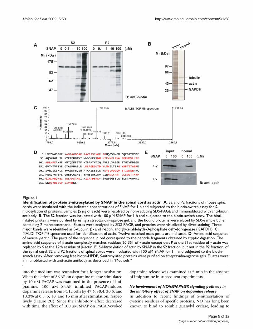
ResultsIdentification of S-nitrosylated proteins with SNAP in the spinal cordTo identify S-nitrosylated proteins in the spinal cord, weseparated homogenates of the spinal cord into superna-tant (S2) and pellet (P2) fractions after a 20-min centrifu-gation at 10,000 × g. They were then incubated for 1 hwith various concentrations of SNAP, an NO donor, andsubjected to the biotin-switch assay for S-nitrosylation. Asshown in Figure 1A, S-nitrosylated proteins were detectedwith anti-biotin antibody in both S2 and P2 fractions in a
concentration-dependent manner; and the extent of S-nitrosylation in the S2 fraction was much higher than thatin the P2 fraction at 10 and 100 μM SNAP. Next, bioti-nylated proteins in the S2 fraction treated with 100 μMSNAP were purified on a streptavidin-agarose gel andselectively eluted by using SDS-sample buffer containing2-mercaptoethanol. Three major bands appeared afterSDS-PAGE (Figure 1B) and were in-gel digested and thensubjected to MALDI-TOF MS. The mass data were proc-essed to assign candidate peptides in the NCBI databaseby using the MASCOT search program. These three majorbands were identified as β-tubulin, β- and γ-actin, andglyceraldehyde-3-phosphate dehydrogenase (Table 1).Figure 1C shows the mass spectrum corresponding totryptic digests of actin. The sequence coverage of γ-actinwas 39%, and the probability-based MOWSE score was130 with 12 matched peptides. Since the amino acidsequences are highly conserved between β- and γ-actin,these tryptic peptides did not discriminate them except forthe N-terminal portion (Figure 1D).
To confirm that S-nitrosylated protein identified byMALDI-TOF MS was indeed actin, we immunoblotted theeluate from the streptavidin-agarose gel with anti-actinantibody. S-nitrosylated actin was detected in the eluate ofthe S2 fraction treated with 100 μM SNAP (Figure 1E,upper panel). Interestingly, however, it was not found inthe P2 fraction (Figure 1E, lower panel), suggesting that S-nitrosylation was affected by the state of actin.
Effect of SNAP on dopamine release from PC12 cellsActing as a cell-permeable intercellular messenger, NO isknown to affect neurotransmitter release. PC12 cells are arat pheochromocytoma cell line that endogenously pro-duces dopamine and shares similar mechanisms of exocy-tosis with neurons [30]. Dopamine is the predominantcatecholamine in PC12 cells [31], and PACAP is known tostimulate the release of dopamine from PC12 cells. Tostudy whether SNAP affected the release of dopaminefrom PC12 cells, we first examined the release elicited byPACAP by using an HPLC column equipped with an elec-trochemical detector. The basal release of dopamine intothe medium represented about 1.5-2% of the total cellulardopamine in PC12 cells. Exposure to PACAP for 5 minstimulated the release of dopamine in a concentration-dependent manner, with an EC50 value of 2.42 nM; andthe release started to increase at 1 nM and reached themaximum at 10 nM, at which concentration 15-20% ofthe total cellular dopamine was released (Figure 2A).When PC12 cells were treated with 10 nM PACAP, therelease reached the maximum at 5-10 min and graduallydecreased for 60 min (Figure 2B). In the presence of 10μM imipramine, an inhibitor of dopamine reuptake, 10nM PACAP stimulated the release for 60 min in a time-dependent manner, suggesting that dopamine released
Page 4 of 12(page number not for citation purposes)
Molecular Pain 2009, 5:58 http://www.molecularpain.com/content/5/1/58
into the medium was reuptaken for a longer incubation.When the effect of SNAP on dopamine release stimulatedby 10 nM PACAP was examined in the presence of imi-pramine, 100 μM SNAP inhibited PACAP-induceddopamine release from PC12 cells by 47.6, 30.4, 30.5, and13.2% at 0.5, 5, 10, and 15 min after stimulation, respec-tively (Figure 2C). Since the inhibitory effect decreasedwith time, the effect of 100 μM SNAP on PACAP-evoked
dopamine release was examined at 5 min in the absenceof imipramine in subsequent experiments.
No involvement of NO/cGMP/cGK signaling pathway in the inhibitory effect of SNAP on dopamine releaseIn addition to recent findings of S-nitrosylation ofcysteine residues of specific proteins, NO has long beenknown to bind to soluble guanylyl cyclase, leading to
Identification of protein S-nitrosylated by SNAP in the spinal cord as actinFigure 1Identification of protein S-nitrosylated by SNAP in the spinal cord as actin. A. S2 and P2 fractions of mouse spinal cords were incubated with the indicated concentrations of SNAP for 1 h and subjected to the biotin-switch assay for S-nitrosylation of proteins. Samples (5 μg of each) were resolved by non-reducing SDS-PAGE and immunobloted with anti-biotin antibody. B. The S2 fraction was incubated with 100 μM SNAP for 1 h and subjected to the biotin-switch assay. The bioti-nylated proteins were purified by using a streptavidin-agarose gel, and the bound proteins were eluted by SDS-sample buffer containing 2-mercaptoethanol. Eluates were analyzed by SDS-PAGE, and proteins were visualized by silver staining. Three major bands were identified as β-tubulin, β- and γ-actin, and glyceraldehyde-3-phosphate dehydorogenase (GAPDH). C. MALDI-TOF MS spectrum used for identification of actin. Twelve matched mass peaks are indicated. D. Amino acid sequence of mouse γ-actin. The parts of the sequence in red correspond to the peptide fragments obtained by tryptic digestion. The amino acid sequence of β-actin completely matches residues 20-351 of γ-actin except that P at the 31st residue of γ-actin was replaced by S at the 12th residue of β-actin. E. S-Nitrosylation of actin by SNAP in the S2 fraction, but not in the P2 fraction, of the spinal cord. S2 and P2 fractions of spinal cords were incubated with 100 μM SNAP for 1 h and subjected to the biotin-switch assay. After removing free biotin-HPDP, S-nitrosylated proteins were purified on streptavidin-agarose gels. Eluates were immunoblotted with anti-actin antibody as described in "Methods."
Mr (kDa )
S2 P2
1000 10.1 10SNAP 1000 10.1 10
175
83
62
47
A B
D
C
97
66
45
30
actin
tubulin
GAPDH
Mr (kDa)
inputbound
IB: anti-biotin
0 0 100
input bound
SNAP
S2
P2IB: anti-actin
E
799.0 1439.4 2079.8 2720.2 3360.6Mass (m/z)//
6182.7
0
10
20
30
40
50
60
70
80
90
100
%In
ten
sit
y
1790.9584
1198.7416
1516.7665945.5837
2231.1481954.11
2343.0831548.801
923.58651132.55676
1515.58051639.8879
MALDI -TOF MS spectrum
(µM)100
(µM)
1 LVIDNGSGMC KAGFAGDDAP RAVFPSIVGR PRHQGVMVGM GQKDSYVGDE
51 AQSKRGILTL KYPIEHGIVT NWDDMEKIWH HTFYNELRVA PEEHPVLLTE
101 APLNPKANRE KMTQIMFETF NTPAMYVAIQ AVLSLYASGR TTGIVMDSGD
151 GVTHTVPIYE GYALPHAILR LDLAGRDLTD YLMKILTERG YSFTTTAERE
201 IVRDIKEKLC YVALDFEQEM ATAASSSSLE KSYELPDGQV ITIGNERFRC
251 PEALFQPSFL GMESCGIHET TFNSIMKCDV DIRKDLYANT VLSGGTTMYP
301 GIADRMQKEI TALAPSTMKI KIIAPPERKY SVWIGGSILA SLSTFQQMWI
351 SKQEYDESGP SIVHRKCF
Page 5 of 12(page number not for citation purposes)

Molecular Pain 2009, 5:58 http://www.molecularpain.com/content/5/1/58
Page 6 of 12(page number not for citation purposes)
Table 1: Proteins identified by MALDI-TOF MS
No Identified protein Mr (Da) % cover Pept matched Mowse score NCBI GI number
1 β-tubulin 50,281 27 12 96 45077292 γ-actin 41,340 39 12 130 809561
β-actin 39,451 39 12 131 498683 GAPDHa) 31,120 35 7 82 1.5E+08
a)GAPDH = glyceraldehyde-3-phosphate dehydorogenase.
Inhibition of PACAP-stimulated dopamine release from PC 12 cells by SNAPFigure 2Inhibition of PACAP-stimulated dopamine release from PC 12 cells by SNAP. A. Concentration dependency of PACAP for dopamine release from PC12 cells. PC12 cells (4 × 10⁵ cells/well) cultured on 24-well dishes were stimulated with various concentrations of PACAP for 5 min. B. Time courses of dopamine release from PC12 cells by PACAP. PC12 cells were stimulated for the indicated times with 10 nM PACAP in the absence (closed circles) or presence (open circles) of 10 μM imi-pramine, an inhibitor of catecholamine reuptake. C. Inhibition of PACAP-stimulated dopamine release by SNAP. PC12 cells were stimulated with 10 nM PACAP without (open columns) or with (closed columns) 100 μM SNAP in the presence of 10 μM imipramine for the indicated times. Dopamine released into the medium and cellular dopamine were measured by HPLC as described in "Methods." Dopamine (DA) release (mean ± SD, n = 3) was expressed as a percentage of total dopamine (7.2 ± 0.8 ng/well) in PC12 cells. *P < 0.05; **P < 0.01 vs. without SNAP.
A
0
DA
re
lease
(%
of
tota
l)
-log [PACAP (M)]
10 9 8 7
20
15
10
5
00 10
20
30 40 50 60
35
30
25
20
15
10
5
0
Time (min)
DA
re
lease
(%
of
tota
l) PACAPPACAP + imipramine
B
C
Time (min)
DA
re
lease
(%
of
tota
l)
0
5
15
20
25
0.5 5 10 15
PACAP + imipraminePACAP + imipramine + SNAP
**
**
*
*
10
Molecular Pain 2009, 5:58 http://www.molecularpain.com/content/5/1/58
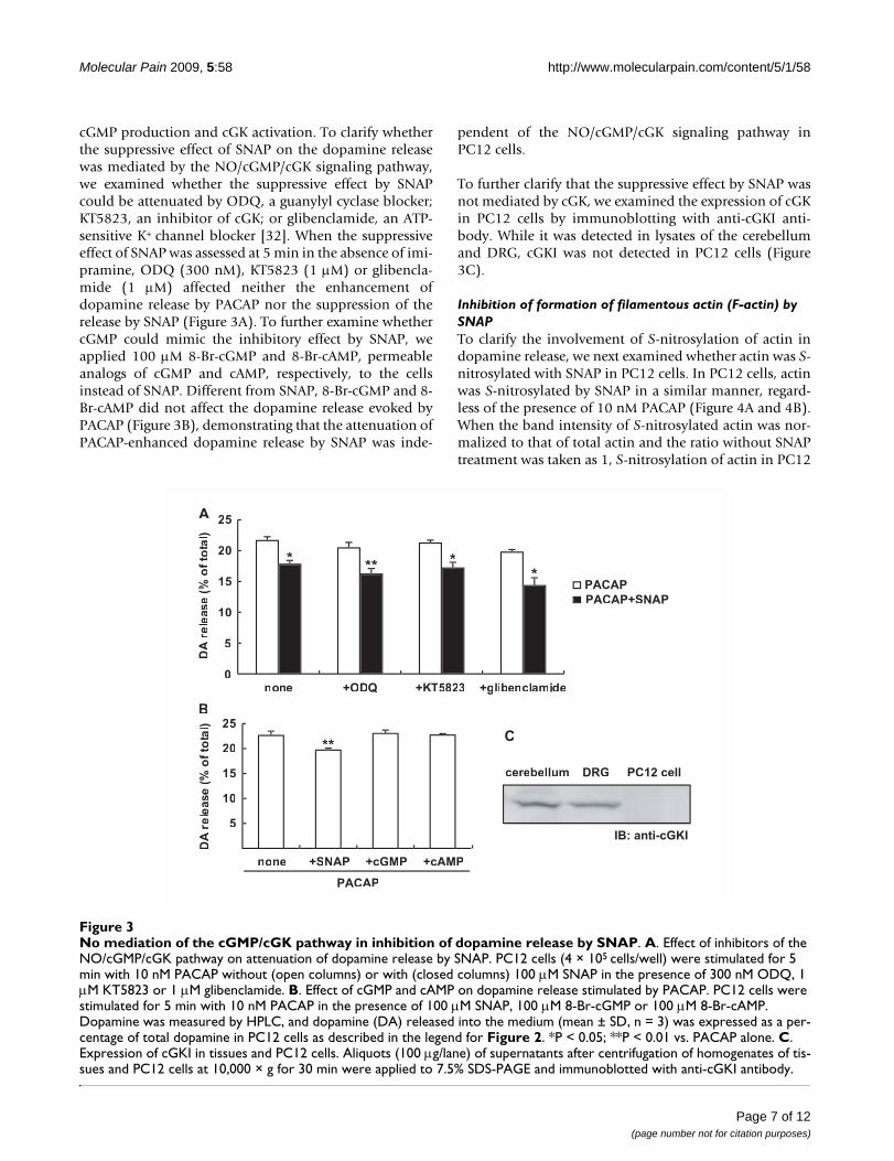
cGMP production and cGK activation. To clarify whetherthe suppressive effect of SNAP on the dopamine releasewas mediated by the NO/cGMP/cGK signaling pathway,we examined whether the suppressive effect by SNAPcould be attenuated by ODQ, a guanylyl cyclase blocker;KT5823, an inhibitor of cGK; or glibenclamide, an ATP-sensitive K+ channel blocker [32]. When the suppressiveeffect of SNAP was assessed at 5 min in the absence of imi-pramine, ODQ (300 nM), KT5823 (1 μM) or glibencla-mide (1 μM) affected neither the enhancement ofdopamine release by PACAP nor the suppression of therelease by SNAP (Figure 3A). To further examine whethercGMP could mimic the inhibitory effect by SNAP, weapplied 100 μM 8-Br-cGMP and 8-Br-cAMP, permeableanalogs of cGMP and cAMP, respectively, to the cellsinstead of SNAP. Different from SNAP, 8-Br-cGMP and 8-Br-cAMP did not affect the dopamine release evoked byPACAP (Figure 3B), demonstrating that the attenuation ofPACAP-enhanced dopamine release by SNAP was inde-
pendent of the NO/cGMP/cGK signaling pathway inPC12 cells.
To further clarify that the suppressive effect by SNAP wasnot mediated by cGK, we examined the expression of cGKin PC12 cells by immunoblotting with anti-cGKI anti-body. While it was detected in lysates of the cerebellumand DRG, cGKI was not detected in PC12 cells (Figure3C).
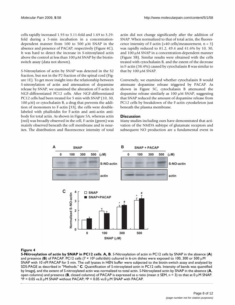
Inhibition of formation of filamentous actin (F-actin) by SNAPTo clarify the involvement of S-nitrosylation of actin indopamine release, we next examined whether actin was S-nitrosylated with SNAP in PC12 cells. In PC12 cells, actinwas S-nitrosylated by SNAP in a similar manner, regard-less of the presence of 10 nM PACAP (Figure 4A and 4B).When the band intensity of S-nitrosylated actin was nor-malized to that of total actin and the ratio without SNAPtreatment was taken as 1, S-nitrosylation of actin in PC12
No mediation of the cGMP/cGK pathway in inhibition of dopamine release by SNAPFigure 3No mediation of the cGMP/cGK pathway in inhibition of dopamine release by SNAP. A. Effect of inhibitors of the NO/cGMP/cGK pathway on attenuation of dopamine release by SNAP. PC12 cells (4 × 10⁵ cells/well) were stimulated for 5 min with 10 nM PACAP without (open columns) or with (closed columns) 100 μM SNAP in the presence of 300 nM ODQ, 1 μM KT5823 or 1 μM glibenclamide. B. Effect of cGMP and cAMP on dopamine release stimulated by PACAP. PC12 cells were stimulated for 5 min with 10 nM PACAP in the presence of 100 μM SNAP, 100 μM 8-Br-cGMP or 100 μM 8-Br-cAMP. Dopamine was measured by HPLC, and dopamine (DA) released into the medium (mean ± SD, n = 3) was expressed as a per-centage of total dopamine in PC12 cells as described in the legend for Figure 2. *P < 0.05; **P < 0.01 vs. PACAP alone. C. Expression of cGKI in tissues and PC12 cells. Aliquots (100 μg/lane) of supernatants after centrifugation of homogenates of tis-sues and PC12 cells at 10,000 × g for 30 min were applied to 7.5% SDS-PAGE and immunoblotted with anti-cGKI antibody.
**
DA
rele
ase
(%o
fto
tattl)
5
10
15
20
25
none +SNAP +cGMP +cAMP
PACAP
B
** **
*
none +ODQ +KTKK 5823 +glibenclamide0
5
10
15
20
25
DA
rele
ase
(%o
fto
tattl)
PACAP
PACAP+SNAP
A
cerebellum DRG
C
PC12 cell
IB: anti-cGKI
Page 7 of 12(page number not for citation purposes)
Molecular Pain 2009, 5:58 http://www.molecularpain.com/content/5/1/58
cells rapidly increased 1.93 to 3.11-fold and 1.69 to 3.29-fold during a 5-min incubation in a concentration-dependent manner from 100 to 500 μM SNAP in theabsence and presence of PACAP, respectively (Figure 4C).It was hard to detect the increase in S-nitrosylated actinabove the control at less than 100 μM SNAP by the biotin-switch assay (data not shown).
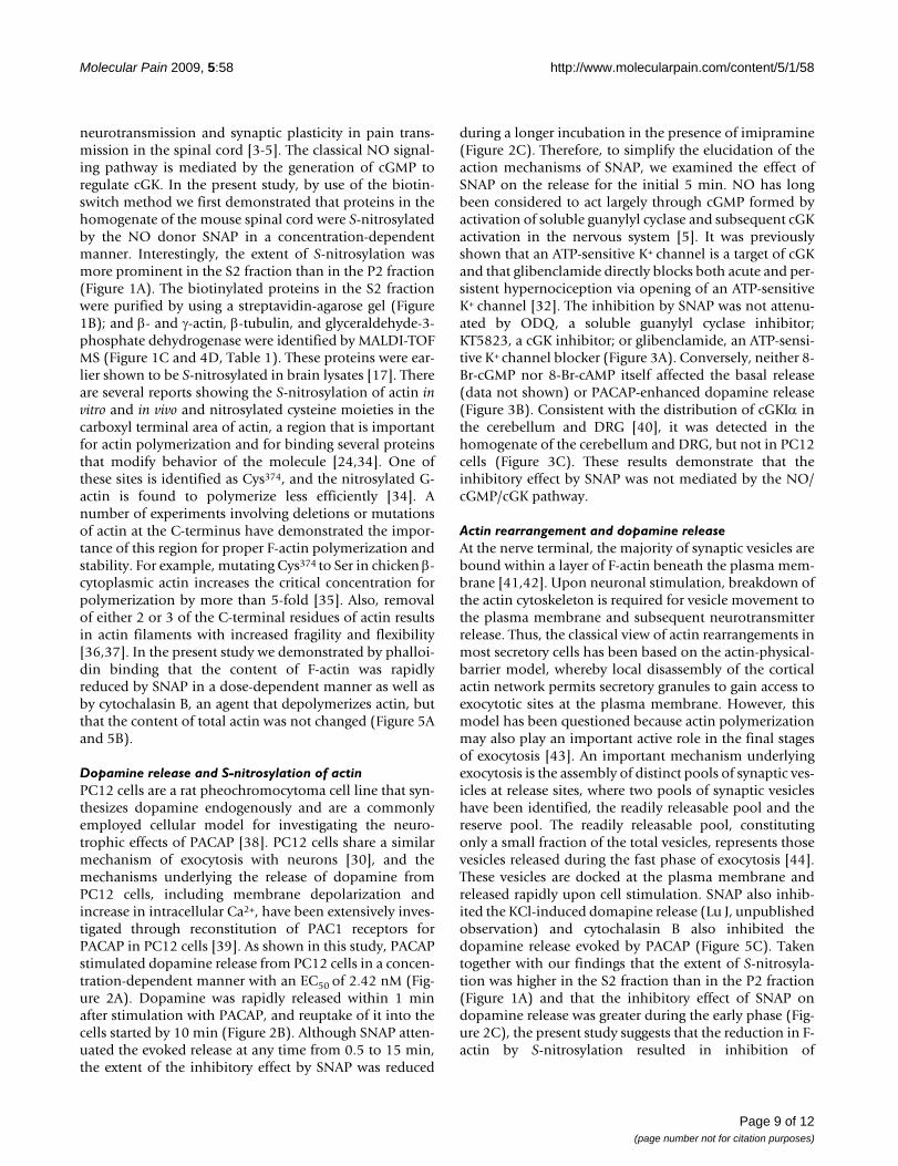
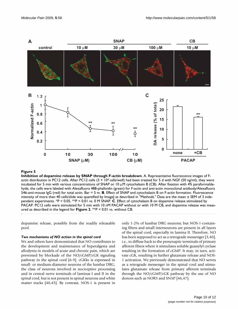
S-Nitrosylation of actin by SNAP was detected in the S2fraction, but not in the P2 fraction of the spinal cord (Fig-ure 1E). To get more insight into the relationship betweenS-nitrosylation of actin and attenuation of dopaminerelease by SNAP, we examined the alteration of F-actin inNGF-differentiated PC12 cells. After NGF-differentiatedPC12 cells had been treated for 5 min with SNAP (10, 30,100 μM) or cytochalasin B, a drug that prevents the addi-tion of monomers to F-actin [33], the cells were double-labeled with phalloidin for F-actin and anti-actin anti-body for total actin. As shown in Figure 5A, whereas actin(red) was broadly observed in the cell, F-actin (green) wasmainly observed beneath the cell membrane and in neur-ites. The distribution and fluorescence intensity of total
actin did not change significantly after the addition ofSNAP. When normalized to that of total actin, the fluores-cence intensity of F-actin (>40 cells/measurement, n = 3)was rapidly reduced to 81.2, 49.4 and 41.6% by 10, 30,and 100 μM SNAP in a concentration-dependent manner(Figure 5B). Similar results were obtained with the cellstreated with cytochalasin B, and the extent of the decreasein F-actin (38.4%) caused by cytochalasin B was similar tothat by 100 μM SNAP.
Conversely, we examined whether cytochalasin B wouldattenuate dopamine release triggered by PACAP. Asshown in Figure 5C, cytochalasin B attenuated thedopamine release similarly as 100 μM SNAP, suggestingthat SNAP reduced the amount of dopamine release fromPC12 cells by breakdown of the F-actin cytoskeleton justbeneath the plasma membrane.
DiscussionMany studies including ours have demonstrated that acti-vation of the NMDA subtype of glutamate receptors andsubsequent NO production are a fundamental event in
S-Nitrosylation of actin by SNAP in PC12 cellsFigure 4S-Nitrosylation of actin by SNAP in PC12 cells. A, B. S-Nitrosylation of actin in PC12 cells by SNAP in the absence (A) and presence (B) of PACAP. PC12 cells (7 × 10⁵ cells/dish) cultured in 6-cm dishes were exposed to 100, 300 or 500 μM SNAP with 10 nM PACAP for 5 min. The cell lysates in HEN buffer were subjected to the biotin-switch assay and analyzed by SDS-PAGE as described in "Methods." C. Quantification of S-nitrosylated actin in PC12 cells. Intensity of bands was quantified by ImageJ, and the extent of S-nitrosylated actin was normalized to total actin. S-Nitrosylated actin by SNAP in the absence (A, open columns) and presence (B, closed columns) of PACAP is expressed as a ratio (mean ± SEM, n = 3) to that at 0 μM SNAP. *P < 0.05 vs.0 μM SNAP without PACAP; #P < 0.05 vs.0 μM SNAP with PACAP.
B
1000 300 500
SNAP + PACAP
actin
1000 300 500
SNAP
actin
0
1
2
3
4
0 100 300 500
SNAP
SNAP+PACAP
*
*
#
#
A
(µM) (µM)
SNAP (µM)
S-NO-actin S-NO-actin
No
rma
lize
d S
-NO
-ac
tinC
Page 8 of 12(page number not for citation purposes)
Molecular Pain 2009, 5:58 http://www.molecularpain.com/content/5/1/58
neurotransmission and synaptic plasticity in pain trans-mission in the spinal cord [3-5]. The classical NO signal-ing pathway is mediated by the generation of cGMP toregulate cGK. In the present study, by use of the biotin-switch method we first demonstrated that proteins in thehomogenate of the mouse spinal cord were S-nitrosylatedby the NO donor SNAP in a concentration-dependentmanner. Interestingly, the extent of S-nitrosylation wasmore prominent in the S2 fraction than in the P2 fraction(Figure 1A). The biotinylated proteins in the S2 fractionwere purified by using a streptavidin-agarose gel (Figure1B); and β- and γ-actin, β-tubulin, and glyceraldehyde-3-phosphate dehydrogenase were identified by MALDI-TOFMS (Figure 1C and 4D, Table 1). These proteins were ear-lier shown to be S-nitrosylated in brain lysates [17]. Thereare several reports showing the S-nitrosylation of actin invitro and in vivo and nitrosylated cysteine moieties in thecarboxyl terminal area of actin, a region that is importantfor actin polymerization and for binding several proteinsthat modify behavior of the molecule [24,34]. One ofthese sites is identified as Cys374, and the nitrosylated G-actin is found to polymerize less efficiently [34]. Anumber of experiments involving deletions or mutationsof actin at the C-terminus have demonstrated the impor-tance of this region for proper F-actin polymerization andstability. For example, mutating Cys374 to Ser in chicken β-cytoplasmic actin increases the critical concentration forpolymerization by more than 5-fold [35]. Also, removalof either 2 or 3 of the C-terminal residues of actin resultsin actin filaments with increased fragility and flexibility[36,37]. In the present study we demonstrated by phalloi-din binding that the content of F-actin was rapidlyreduced by SNAP in a dose-dependent manner as well asby cytochalasin B, an agent that depolymerizes actin, butthat the content of total actin was not changed (Figure 5Aand 5B).
Dopamine release and S-nitrosylation of actinPC12 cells are a rat pheochromocytoma cell line that syn-thesizes dopamine endogenously and are a commonlyemployed cellular model for investigating the neuro-trophic effects of PACAP [38]. PC12 cells share a similarmechanism of exocytosis with neurons [30], and themechanisms underlying the release of dopamine fromPC12 cells, including membrane depolarization andincrease in intracellular Ca2+, have been extensively inves-tigated through reconstitution of PAC1 receptors forPACAP in PC12 cells [39]. As shown in this study, PACAPstimulated dopamine release from PC12 cells in a concen-tration-dependent manner with an EC50 of 2.42 nM (Fig-ure 2A). Dopamine was rapidly released within 1 minafter stimulation with PACAP, and reuptake of it into thecells started by 10 min (Figure 2B). Although SNAP atten-uated the evoked release at any time from 0.5 to 15 min,the extent of the inhibitory effect by SNAP was reduced
during a longer incubation in the presence of imipramine(Figure 2C). Therefore, to simplify the elucidation of theaction mechanisms of SNAP, we examined the effect ofSNAP on the release for the initial 5 min. NO has longbeen considered to act largely through cGMP formed byactivation of soluble guanylyl cyclase and subsequent cGKactivation in the nervous system [5]. It was previouslyshown that an ATP-sensitive K+ channel is a target of cGKand that glibenclamide directly blocks both acute and per-sistent hypernociception via opening of an ATP-sensitiveK+ channel [32]. The inhibition by SNAP was not attenu-ated by ODQ, a soluble guanylyl cyclase inhibitor;KT5823, a cGK inhibitor; or glibenclamide, an ATP-sensi-tive K+ channel blocker (Figure 3A). Conversely, neither 8-Br-cGMP nor 8-Br-cAMP itself affected the basal release(data not shown) or PACAP-enhanced dopamine release(Figure 3B). Consistent with the distribution of cGKIα inthe cerebellum and DRG [40], it was detected in thehomogenate of the cerebellum and DRG, but not in PC12cells (Figure 3C). These results demonstrate that theinhibitory effect by SNAP was not mediated by the NO/cGMP/cGK pathway.
Actin rearrangement and dopamine releaseAt the nerve terminal, the majority of synaptic vesicles arebound within a layer of F-actin beneath the plasma mem-brane [41,42]. Upon neuronal stimulation, breakdown ofthe actin cytoskeleton is required for vesicle movement tothe plasma membrane and subsequent neurotransmitterrelease. Thus, the classical view of actin rearrangements inmost secretory cells has been based on the actin-physical-barrier model, whereby local disassembly of the corticalactin network permits secretory granules to gain access toexocytotic sites at the plasma membrane. However, thismodel has been questioned because actin polymerizationmay also play an important active role in the final stagesof exocytosis [43]. An important mechanism underlyingexocytosis is the assembly of distinct pools of synaptic ves-icles at release sites, where two pools of synaptic vesicleshave been identified, the readily releasable pool and thereserve pool. The readily releasable pool, constitutingonly a small fraction of the total vesicles, represents thosevesicles released during the fast phase of exocytosis [44].These vesicles are docked at the plasma membrane andreleased rapidly upon cell stimulation. SNAP also inhib-ited the KCl-induced domapine release (Lu J, unpublishedobservation) and cytochalasin B also inhibited thedopamine release evoked by PACAP (Figure 5C). Takentogether with our findings that the extent of S-nitrosyla-tion was higher in the S2 fraction than in the P2 fraction(Figure 1A) and that the inhibitory effect of SNAP ondopamine release was greater during the early phase (Fig-ure 2C), the present study suggests that the reduction in F-actin by S-nitrosylation resulted in inhibition of
Page 9 of 12(page number not for citation purposes)
Molecular Pain 2009, 5:58 http://www.molecularpain.com/content/5/1/58
dopamine release, possibly from the readily releasablepool.
Two mechanisms of NO action in the spinal cordWe and others have demonstrated that NO contributes tothe development and maintenance of hyperalgesia andallodynia in models of acute and chronic pain, which areprevented by blockade of the NO/cGMP/cGK signalingpathway in the spinal cord [6-9]. cGKIα is expressed insmall- or medium-diameter neurons of the lumbar DRG,the class of neurons involved in nociceptive processingand in central nerve terminals of laminas I and II in thespinal cord, but is not present in spinal neurons and whitematter tracks [40,45]. By contrast, NOS-1 is present in
only 1-2% of lumbar DRG neurons; but NOS-1-contain-ing fibers and small interneurons are present in all layersof the spinal cord, especially in lamina II. Therefore, NOhas been supposed to act as a retrograde messenger [3,40],i.e., to diffuse back to the presynaptic terminals of primaryafferent fibers where it stimulates soluble guanylyl cyclaseresulting in the formation of cGMP. It may, in turn, acti-vate cGK, resulting in further glutamate release and NOS-1 activation. We previously demonstrated that NO servesas a retrograde messenger in the spinal cord and stimu-lates glutamate release from primary afferent terminalsthrough the NO/cGMP/cGK pathway by the use of NOdonors such as NOR3 and SNAP [46,47].
Inhibition of dopamine release by SNAP through F-actin breakdownFigure 5Inhibition of dopamine release by SNAP through F-actin breakdown. A. Representative fluorescence images of F-actin distribution in PC12 cells. After PC12 cells (3 × 10⁴ cells/well) had been treated for 5 d with NGF (50 ng/ml), they were incubated for 5 min with various concentrations of SNAP or 10 μM cytochalasin B (CB). After fixation with 4% paraformalde-hyde, the cells were labeled with Alexafluora 488-phalloidin (green) for F-actin and anti-actin monoclonal antibody/Alexafluora 546-anti-mouse IgG (red) for total actin. Bar = 5 m. B. Effect of SNAP and cytochalasin B on F-actin formation. Fluorescence intensity of more than 40 cells/slide was quantified by ImageJ as described in "Methods." Data are the mean ± SEM of 3 inde-pendent experiments. *P < 0.05, **P < 0.01 vs. 0 M SNAP. C. Effect of cytochalasin B on dopamine release stimulated by PACAP. PC12 cells were stimulated for 5 min with 10 nM PACAP without or with 10 M CB, and dopamine release was meas-ured as described in the legend for Figure 2. **P < 0.01 vs. without CB.
control 10 µM 30 µM 100 µM 10 µM
SNAP CB
** ****
0.2
0.4
0.6
0.8
1
1.2
0 10 30 100 10
No
rmali
ze
d F
-ac
tin
SNAP (µM)
**
DA
rele
as
e(%
of
tota
l)
CB (µM)
A
B C25
0
5
10
15
20
none +CB
PACAP
Page 10 of 12(page number not for citation purposes)
Molecular Pain 2009, 5:58 http://www.molecularpain.com/content/5/1/58
In the present study, we first demonstrated that actin inthe S2 fraction of the spinal cord was S-nitrosylated byexogenous NO; and our data suggest that S-nitrosylationattenuated dopamine release by decrease in F-actin con-tent in PC12 cells. It could be deduced that glutamaterelease can be affected by NO-induced S-nitrosylation ofactin in the spinal cord. In this connection, the NO donorsodium nitroprusside inhibited ongoing impulse activityin 49% of all spinal neurons in the laminas I and II andactivated only 28% of them [11]. Although the biphasiceffect of NO on neuronal activity was vaguely consideredto be the difference in experimental conditions underwhich neuronal background activity was examined, it maybe simply interpreted that NO affected glutamate releasein both inhibitory and stimulatory manners by the bal-ance of S-nitrosylation and cGMP/cGK pathway.
ConclusionUnlike the other second messengers, NO has the potentialto induce opposing effects and previous studies suggestthat the NO/cGMP signaling cascade showed both pro-and anti-nociceptive effects [16]. Although which types ofcells in the spinal cord are S-nitrosylated remains to beclarified, the present study suggests that 2 signaling path-ways of NO action exist and that the contribution of these2 pathways is determined to a considerable extent by thepresence of cGK in cells. Two signaling pathways mayexplain the discrepancy in pain behaviors and electro-physiological responses to NO donors in the spinal cord.
Abbreviationsbiotin-HPDP: N-[6-(biotinamido)hexyl]-3'-(2'-pyri-dyldithio)-propionamide; 8-Br-cAMP: 8-bromoadenos-ine 3', 5'-cyclic monophosphate; BSA: bovine serumalbumin; a-CHCA: a-cyano-4-hydroxycinnamic acid; cGK:cGMP-dependent protein kinase; DRG: dorsal root gan-glia; F-actin: filamentous actin; MALDI-TOF MS: matrix-assisted laser desorption/ionization reflection time-of-flight mass spectrometry; MMTS: S-methyl methanethio-sulfonate; NGF: nerve growth factor; NO: nitric oxide;NOS: NO synthase; ODQ: 1H-[1,2,4]oxadiazolo-[4,3-a]quinoxalin-1-one; PACAP: pituitary adenylate cyclase-activating polypeptide; PAGE: polyacrylamide gel electro-phoresis; PBS: phosphate-buffered saline; SDS: sodiumdodecyl sulfate; SNAP: S-nitroso-N-acetyl-DL-penicilla-mine; TFA: trifluoroacetic acid.
Competing interestsThe authors declare that they have no competing interests.
Authors' contributionsJL was involved in data acquisition of S-nitrosylation,identification of actin by MALDI-TOF MS, and cell exper-iments, TK and EO were involved in supervision of JL inall experiments. YO and YU participated experiments of
dopamine release. SI is a corresponding author and partic-ipated in the design of experiments and manuscript prep-aration. All authors read and approved the finalmanuscript.
AcknowledgementsThis work was supported in part by grants from the programs Grants-in-Aid for Scientific Research on Priority Areas from the Ministry of Educa-tion, Culture, Sports, Science, and Technology of Japan, and Grants-in-Aid for Scientific Research (S) and (C) from Japan Society for the Promotion of Science, the Science Research Promotion Fund of the Japan Private School Promotion Foundation, and Japan Foundation of Applied Enzymology.
References1. Alderton WK, Cooper CE, Knowles RG: Nitric oxide synthases:
structure, function and inhibition. Biochem J 2001, 357:593-615.2. Oess S, Icking A, Fulton D, Govers R, Müller-Esterl W: Subcellular
targeting and trafficking of nitric oxide synthases. Biochem J2006, 396:401-409.
3. Meller ST, Gebhart GF: Nitric oxide (NO) and nociceptiveprocessing in the spinal cord. Pain 1993, 52:127-136.
4. Ji RR, Kohno T, Moore KA, Woolf CJ: Central sensitization andLTP: do pain and memory share similar mechanisms? TrendsNeurosci 2003, 26:696-705.
5. Garthwaite J: Concepts of neural nitric oxide-mediated trans-mission. Eur J Neurosci 2008, 27:2783-2802.
6. Meller ST, Dykstra C, Gebhart GF: Production of endogenousnitric oxide and activation of soluble guanylate cyclase arerequired for N-methyl-D-aspartate-produced facilitation ofthe nociceptive tail-flick reflex. Eur J Pharmacol 1992, 214:93-96.
7. Minami T, Nishihara I, Ito S, Sakamoto K, Hyodo M, Hayaishi O:Nitric oxide mediates allodynia induced by intrathecaladministration of prostaglandin E2 or prostaglandin F2α inconscious mice. Pain 1995, 61:285-290.
8. Sluka KA, Willis WD: Increased spinal release of excitatoryamino acids following intradermal injection of capsaicin isreduced by a protein kinase G inhibitor. Brain Res 1998,798:281-286.
9. Tao YX, Johns RA: Activation of cGMP-dependent proteinkinase Iα is required for N-methyl-D-aspartate- or nitricoxide-produced spinal thermal hyperalgesia. Eur J Pharmacol2000, 392:141-145.
10. Malmberg AB, Yaksh TL: The effect of morphine on formalin-evoked behaviour and spinal release of excitatory aminoacids and prostaglandin E2 using microdialysis in consciousrats. Br J Pharmacol 1995, 114:1069-1075.
11. Pehl U, Schmid HA: Electrophysiological responses of neuronsin the rat spinal cord to nitric oxide. Neuroscience 1997,77:563-573.
12. Hoheisel U, Unger T, Mense S: A block of spinal nitric oxide syn-thesis leads to increased background activity predominantlyin nociceptive dorsal horn neurones in the rat. Pain 2000,88:249-257.
13. Hoheisel U, Unger T, Mense S: The possible role of the NO-cGMP pathway in nociception: Different spinal and suprasp-inal action of enzyme blockers on rat dorsal horn neurons.Pain 2005, 117:358-367.
14. Sousa AM, Prado WA: The dual effect of a nitric oxide donor innociception. Brain Res 2001, 897:9-19.
15. Tegeder I, Schmidtko A, Niederberger E, Ruth P, Geisslinger G: Dualeffects of spinally delivered 8-bromo-cyclic guanosine mono-phosphate (8-bromo-cGMP) in formalin-induced nocicep-tion in rats. Neurosci Lett 2002, 332:146-150.
16. Hucho T, Levine JD: Signaling pathways in sensitization:toward a nociceptors cell biology. Neuron 2007, 55:365-376.
17. Jaffrey SR, Erdjument-Bromage H, Ferris CD, Tempst P, Snyder SH:Protein S-nitrosylation: a physiological signal for neuronalnitric oxide. Nat Cell Biol 2001, 3:193-197.
18. Hess DT, Matsumoto A, Kim SO, Marshall HE, Stamler JS: ProteinS-nitrosylation: purview and parameters. Nat Rev Mol Cell Biol2005, 6:150-166.
Page 11 of 12(page number not for citation purposes)
Molecular Pain 2009, 5:58 http://www.molecularpain.com/content/5/1/58
Publish with BioMed Central and every scientist can read your work free of charge
"BioMed Central will be the most significant development for disseminating the results of biomedical research in our lifetime."
Sir Paul Nurse, Cancer Research UK
Your research papers will be:
available free of charge to the entire biomedical community
peer reviewed and published immediately upon acceptance
cited in PubMed and archived on PubMed Central
yours — you keep the copyright
Submit your manuscript here:http://www.biomedcentral.com/info/publishing_adv.asp
BioMedcentral
19. Tian J, Kim SF, Hester L, Snyder SH: S-nitrosylation/activation ofCOX-2 mediates NMDA neurotoxicity. Proc Natl Acad Sci USA2008, 105:10537-10540.
20. Sayed N, Baskaran P, Ma X, Akker F van den, Beuve A: Desensitiza-tion of soluble guanylyl cyclase, the NO receptor, by S-nitrosylation. Proc Natl Acad Sci USA 2007, 104:12312-12317.
21. Lipton SA, Choi YB, Pan ZH, Lei SZ, Chen HSV, Sucher NJ, LoscalzoJ, Singel DJ, Stamler JS: A redox-based mechanism for the neu-roprotective and neurodestructive effects of nitric oxide andrelated nitroso-compounds. Nature 1993, 364:626-632.
22. Choi YB, Tenneti L, Le DA, Ortiz J, Bai G, Chen HS, Lipton SA:Molecular basis of NMDA receptor-coupled ion channelmodulation by S-nitrosylation. Nat Neurosci 2000, 3:15-21.
23. Takahashi H, Shin Y, Cho SJ, Wagner M, Zago WM, Nakamura T, GuZ, Ma Y, Furukawa H, Liddington R, Zhang D, Tong G, Chen HSV, Lip-ton SA: Hypoxia enhances S-nitrosylation-mediated NMDAreceptor inhibition via a thiol oxygen sensor motif. Neuron2007, 53:53-64.
24. Thom SR, Bhopale VM, Mancini DJ, Milovanova TN: Actin S-nitrosylation inhibits neutrophil β2 integrin function. J BiolChem 2008, 282:10822-10834.
25. Chung KK, Thomas B, Li X, Pletnikova O, Troncoso JC, Marsh L,Dawson VL, Dawson TM: S-Nitrosylation of parkin regulatesubiquitination and compromises parkin's protective func-tion. Science 2004, 304:1328-1331.
26. Hara MR, Agrawal N, Kim SF, Cascio MB, Fujimuro M, Ozeki Y, Taka-hashi M, Cheah JH, Tankou SK, Hester LD, Ferris CD, Hayward SD,Snyder SH, Sawa A: S-nitrosylated GAPDH initiates apoptoticcell death by nuclear translocation following Siah1 binding.Nat Cell Biol 2004, 7:665-674.
27. Torta F, Usuelli V, Malgaroli A, Bachi A: Proteomic analysis of pro-tein S-nitrosylation. Proteomics 2008, 8:4484-4494.
28. Mortz E, Krogh TN, Vorum H, Görg A: Improved silver stainingprotocols for high sensitivity protein identification usingmatrix-assisted laser desorption/ionization-time of flightanalysis. Proteomics 2001, 1:1359-1363.
29. Katano T, Mabuchi T, Okuda-Ashitaka E, Inagaki N, Kinumi T, Ito S:Proteomic identification of a novel isoform of collapsinresponse mediator protein-2 in spinal nerves peripheral todorsal root ganglia. Proteomics 2006, 6:6085-6094.
30. Morgan A, Burgoyne RD: Common mechanisms for regulatedexocytosis in the chromaffin cell and the synapse. Semin CellDev Biol 1997, 8:141-149.
31. Kumar GK, Overholt JL, Bright GR, Hui KY, Lu H, Gratzl M, Prab-hakar NR: Release of dopamine and norepinephrine byhypoxia from PC-12 cells. Am J Physiol 1998, 274:C1592-C1600.
32. Sachs D, Cunha FQ, Ferreira SH: Peripheral analgesic blockadeof hypernociception: activation of arginine/NO/cGMP/pro-tein kinase G/ATP-sensitive K+ channel pathway. Proc NatlAcad Sci USA 2004, 101:3680-3685.
33. Cingolani LA, Goda Y: Actin in action: the interplay betweenthe actin cytoskeleton and synaptic efficacy. Nat Rev Neurosci2008, 9:344-356.
34. Dalle-Donne I, Milzani A, Giustarini D, di Simplicio P, Colombo R,Rossi R: S-NO-actin: S-nitrosylation kinetics and the effect onisolated vascular smooth muscle. J Muscle Res Cell Motil 2000,21:171-181.
35. Aspenström P, Schutt CE, Lindberg U, Karlsson R: Mutations in β-actin: influence on polymer formation and on interactionswith myosin and profilin. FEBS Lett 1993, 329:163-170.
36. O'Donoghue SI, Miki M, dos Remedios CG: Removing the two C-terminal residues of actin affects the filament structure. ArchBiochem Biophys 1992, 293:110-116.
37. Mossakowska M, Moraczewska J, Khaitlina S, Strzelecka-GolaszewskaH: Proteolytic removal of three C-terminal residues of actinalters the monomer-monomer interactions. Biochem J 1993,289:897-902.
38. Vaudry D, Stork PJ, Lazarovici P, Eiden LE: Signaling pathways forPC12 cell differentiation: making the right connections. Sci-ence 2002, 296:1648-1649.
39. Mustafa T, Grimaldi M, Eiden LE: The hop cassette of the PAC1receptor confers coupling to Ca2+ elevation required forpituitary adenylate cyclase-activating polypeptide-evokedneurosecretion. J Biol Chem 2007, 282:8079-8091.
40. Qian Y, Chao DS, Santillano DR, Cornwell TL, Nairn AC, GreengardP, Lincoln TM, Bredt DS: cGMP-dependent protein kinase in
dorsal root ganglion: relationship with nitric oxide synthaseand nociceptive neurons. J Neurosci 1996, 16:3130-3138.
41. Hirokawa N, Sobue K, Kanda K, Harada A, Yorifuji H: The cytoskel-etal architecture of the presynaptic terminal and molecularstructure of synapsin 1. J Cell Biol 1989, 108:111-126.
42. Phillips GR, Huang JK, Wang Y, Tanaka H, Shapiro L, Zhang W, ShanWS, Arndt K, Marcus Frank M, Gordon RE, Gawinowicz MA, Zhao Y,Colman DR: The presynaptic particle web: ultrastructure,composition, dissolution, and reconstitution. Neuron 2001,32:63-77.
43. Momboisse F, Ory S, Calco V, Malacombe M, Bader MF, Gasman S:Calcium-regulated exocytosis in neuroendocrine cells: Inter-sectin-1L stimulates actin polymerization and exocytosis byactivating Cdc42. Ann NY Acad Sci 2009, 1152:209-214.
44. Rosenmund C, Stevens CF: Definition of the readily releasablepool of vesicles at hippocampal synapses. Neuron 1996,16:1197-1207.
45. Schlossmann J, Hofmann F: cGMP-dependent protein kinases indrug discovery. Drug Discov Today 2005, 10:627-634.
46. Xu L, Matsumura S, Mabuchi T, Takagi K, Abe T, Ito S: In situ meas-urement of neuronal nitric oxide synthase activity in the spi-nal cord by NADPH-diaphorase histochemistry. J NeurosciMethods 2006, 150:174-184.
47. Xu L, Mabuchi T, Katano T, Matsumura S, Okuda-Ashitaka E,Sakimura K, Mishina M, Ito S: Nitric oxide (NO) serves as a ret-rograde messenger to activate neuronal NO synthase in thespinal cord via NMDA receptors. Nitric Oxide 2007, 17:18-24.
Page 12 of 12(page number not for citation purposes)
Recommended