M3 Muscarinic Acetylcholine Receptor-Mediated SignalingIs Regulated by Distinct Mechanisms
Jiansong Luo, John M. Busillo, and Jeffrey L. BenovicDepartment of Biochemistry and Molecular Biology, Thomas Jefferson University, Philadelphia, Pennsylvania
Received December 31, 2007; accepted April 2, 2008
ABSTRACTWe have used RNA interference previously to demonstrate thatG protein-coupled receptor kinase 2 (GRK2) regulates endog-enously expressed H1 histamine receptor in human embryonickidney 293 cells. In this report, we investigate the regulation ofendogenously expressed M3 muscarinic acetylcholine receptor(M3 mAChR). We show that knockdown of GRK2, GRK3, orGRK6, but not GRK5, significantly increased carbachol-mediatedcalcium mobilization. Stable expression of wild-type GRK2 or akinase-dead mutant (GRK2-K220R) reduced calcium mobiliza-tion after receptor activation, whereas GRK2 mutants defectivein G�q binding (GRK2-D110A, GRK2-R106A, and GRK2-R106A/K220R) had no effect on calcium signaling, suggestingthat GRK2 primarily regulates Gq after M3 mAChR activation.
The knockdown of arrestin-2 or arrestin-3 also significantlyincreased carbachol-mediated calcium mobilization. Knock-down of GRK2 and the arrestins also significantly enhancedcarbachol-mediated activation of extracellular signal-regulatedkinases 1 and 2 (ERK1/2), whereas prolonged ERK1/2 activa-tion was only observed with GRK2 or arrestin-3 knockdown.We also investigated the role of casein kinase-1� (CK1�) andfound that knockdown of CK1� increased calcium mobilizationbut not ERK activation. In summary, our data suggest thatmultiple proteins dynamically regulate M3 mAChR-mediatedcalcium signaling, whereas GRK2 and arrestin-3 play the pri-mary role in regulating ERK activation.
Activation of G protein-coupled receptors (GPCRs) by ago-nist occupancy leads to a conformational change in the re-ceptor that promotes the activation of heterotrimeric G pro-teins, which in turn activate a variety of effectors leading todownstream signaling events (Pierce et al., 2002). ActivatedGPCRs are regulated by three principal mechanisms: desen-sitization, internalization, and down-regulation. Receptor de-sensitization is initiated by the phosphorylation of serine/threonine residues by GPCR kinases (GRKs), which promotesthe high-affinity binding of arrestins, uncoupling the receptorfrom G protein and terminating signaling (Krupnick andBenovic, 1998).
There are seven members of the GRK family that aregrouped into three subfamilies based on sequence and func-tional similarity: GRK1 and GRK7; GRK2 and GRK3; and
GRK4, GRK5, and GRK6. GRK2, GRK3, GRK5, and GRK6are expressed ubiquitously, whereas GRK1, GRK4, andGRK7 have a restricted expression pattern. Much of theresearch determining specific GPCR-GRK interaction hasrelied on techniques such as heterologous overexpression,dominant-negative constructs, and more recently RNA inter-ference (Krupnick and Benovic, 1998; Iwata et al., 2005; Kimet al., 2005).
The nonvisual arrestins, arrestin-2 (�-arrestin1) and ar-restin-3 (�-arrestin2), bind to activated, phosphorylatedGPCRs, subsequently terminating G protein activation andtargeting the receptors to clathrin-coated pits for internal-ization (Moore et al., 2007). Arrestins have also been shownto act as scaffolding proteins to promote downstream signal-ing events, such as activation of mitogen-activated proteinkinases (Lefkowitz and Shenoy, 2005).
The muscarinic acetylcholine receptors (mAChRs) repre-sent a subfamily of GPCRs with five subtypes, M1 to M5. TheM3 mAChR couples to Gq, resulting in phospholipase C-�(PLC-�) activation, and production of inositol trisphosphate(IP3) and diacylglycerol (DAG), which leads to calcium re-
This work was supported by National Institutes of Health grants GM44944and GM47417 (to J.L.B). J.M.B. is supported by a predoctoral fellowship fromthe American Heart Association.
J.L. and J.M.B. contributed equally to this work.Article, publication date, and citation information can be found at
http://molpharm.aspetjournals.org.doi:10.1124/mol.107.044750.
ABBREVIATIONS: GPCR, G protein-coupled receptor; Bis I, bisindolymaleimide I; CK1�, casein kinase 1-�; DAG, diacylglycerol; ERK, extra-cellular signal-regulated kinase; GRK, G protein-coupled receptor kinase; IP3, inositol trisphosphate; M3 mAChR, muscarinic acetylcholinereceptor subtype 3; PKC, protein kinase C; PLC-�, phospholipase C-�; HEK, human embryonic kidney; siRNA, small interfering RNA; AM,acetoxymethyl ester; McN-A-343, 4-(m-chlorophenylcarbamoyloxy)-2-butynyltrimethylammonium; PMA, phorbol 12-myristate 13-acetate; PAGE,polyacrylamide gel electrophoresis; MEK, mitogen-activated protein kinase kinase.
0026-895X/08/7402-338–347$20.00MOLECULAR PHARMACOLOGY Vol. 74, No. 2Copyright © 2008 The American Society for Pharmacology and Experimental Therapeutics 44750/3349371Mol Pharmacol 74:338–347, 2008 Printed in U.S.A.
338
at ASPE
T Journals on M
ay 1, 2018m
olpharm.aspetjournals.org
Dow
nloaded from

lease from intracellular stores and protein kinase C (PKC)activation. In addition, the M3 mAChR can activate extracellu-lar signal-regulated kinase (ERK) in a calcium-independentand PKC-dependent manner (Kim et al., 1999; Wylie et al.,1999).
Upon activation, the M3 mAChR is rapidly phosphorylatedon serine/threonine residues within the third intracellularloop (Tobin et al., 1997) and C-terminal tail (Budd et al.,2000), although it is unclear which kinases mediate receptorphosphorylation and regulation. Wu et al. (2000) showed thatGRK2 phosphorylates the M3 mAChR in a G��-dependentmanner and mapped the phosphorylation sites to 331SSS333
and 348SASS351 in the third intracellular loop. GRK3 also hasthe ability to phosphorylate the receptor, but receptor regu-lation seems to occur primarily through modulation of PLC-�activity (Willets et al., 2001, 2002, 2003). Willets and cowork-ers (Willets et al., 2001, 2002, 2003) also showed that GRK6regulates the M3 mAChR by phosphorylation, whereas GRK2and GRK5 were found to have no effect on the receptor inSH-SY5Y cells. In addition to GRK-mediated phosphoryla-tion, casein kinase 1� (CK1�) has also been shown to phos-phorylate the M3 mAChR in an agonist-dependent manner,although this alone was insufficient to mediate receptor de-sensitization (Budd et al., 2000). Finally, arrestins do notseem to be required for M3 mAChR internalization (Lee et al.,1998; Mundell and Benovic, 2000) but are involved in recep-tor desensitization with no discernible specificity betweenarrestin-2 and arrestin-3 (Mundell and Benovic, 2000).
One major unanswered question regarding the physiolog-ical regulation of GPCRs is to understand which GRKs andarrestins regulate a given receptor subtype. To date, a lim-ited number of GRKs and arrestins have been identified,whereas more than 700 mammalian GPCRs have beencloned (Gainetdinov et al., 2004). Studies over the past de-cade have defined the ability of individual GRKs, secondmessenger-dependent kinases (e.g., protein kinase A orPKC), and arrestins to regulate GPCRs in model systems.However, the mechanisms by which GRKs target endogenousGPCRs are still unknown. Using either wild-type GRK2,kinase-dead GRK2, or mutants deficient in G�q binding, weshowed previously that the human H1 histamine receptorwas specifically regulated by GRK2, mainly through regula-tion of activated Gq (Iwata et al., 2005). In this report, weused RNA interference to target proteins specifically in-volved in the agonist-dependent regulation of the endogenousM3 mAChR in HEK-293 cells. We found that there was dif-ferential GRK-mediated regulation of this receptor as as-sessed by calcium signaling and ERK activation. In addition,knockdown of either arrestin-2 or arrestin-3 resulted in en-hanced signaling from the receptor with different temporaleffects. Furthermore, we show that in addition to GRKs,CK1� has a negative role in M3 mAChR-mediated calciummobilization. Taken together, our results show that multipleproteins mediate agonist-dependent regulation of M3 mAChRsignaling.
Materials and MethodsMaterials. HEK-293 cells were from Microbix Biosystems, Inc
(Toronto, Canada), whereas carbachol was from EMD Biosciences(San Diego, CA). Pirenzepine and p-fluorohexahydro-sila-difenolwere from Sigma-Aldrich (St. Louis, MO), and Lipofectamine 2000
and Opti-MEM were from Invitrogen (Carlsbad, CA). Phospho-spe-cific p44/p42 polyclonal antibody was from Cell Signaling Technolo-gies (Danvers, MA). Polyclonal ERK2, CK1�, and GRK3 antibodieswere from Santa Cruz Biotechnology (Santa Cruz, CA). Anti-�-ar-restin monoclonal antibody was from BD Biosciences Pharmingen(San Diego, CA). Anti-GRK4–6 monoclonal antibody was from Mil-lipore (Billerica, MA), whereas the GRK2 monoclonal antibody wasproduced in our laboratory; anti-�-tubulin monoclonal antibody wasfrom Sigma.
Synthesis of Small Interfering RNAs. All small interferingRNAs (siRNAs) were chemically synthesized by Dharmacon RNATechnologies (Lafayette, CO). The GRK2, GRK5, and CK1� siRNAswere reported previously (Liu et al., 2002; Iwata et al., 2005; Kim etal., 2005). The GRK3 siRNA sequence was 5�-GCAGAAGUCGA-CAAAUUUA-3�, whereas 5�-GCGCUUGGCCUACGCCUAU-3� wasused for GRK6. Arrestin-2 and -3 siRNAs were purchased as aSMARTpool. Nonspecific control siRNA VIII (5�-AAACUCUAUCUG-CACGCUGAC-3�) was used as the control for all siRNA experiments.
Cell Culture and siRNA Transfection. HEK-293 cells weremaintained in Dulbecco’s modified Eagle’s medium supplementedwith 10% fetal bovine serum, 25 mM HEPES, pH 7.2, and 0.1 mMnonessential amino acids in a 5% CO2 incubator at 37°C. For trans-fection of GRK and casein kinase siRNAs, HEK-293 cells grown to 85to 90% confluence in 100-mm dishes were transfected with 600 pmolof siRNA using Lipofectamine 2000 in Opti-MEM. After 6 h, cellswere split 1:2, and a second transfection of 600 pmol was performed24 h after the initial transfection. Forty-eight hours after the secondtransfection, cells were split for assay the following day. For arrestinSMARTpool siRNAs, cells �70% confluent were transfected with 600pmol of siRNA corresponding to either arrestin-2 or arrestin-3.Forty-eight hours later, cells were split for assay the following day.Control siRNA was transfected in a similar fashion as describedabove for each transfection condition.
Immunoblotting. To analyze siRNA target proteins, siRNAtransfected HEK-293 cells in a six-well plate were washed twice withice-cold phosphate-buffered saline and lysed with buffer [20 mMHEPES, pH 7.5, 10 mM EDTA, 150 mM NaCl, 1% Triton X-100, andone tablet of Complete protease inhibitor (Roche, Indianapolis, IN)per 50 ml] at 4°C on a rocker for 30 min. The lysates were centrifugedat 4°C at 30,000 rpm in a TLA45 rotor for 30 min. The supernatantswere electrophoresed on a 10% SDS polyacrylamide gel, transferredto nitrocellulose, and immunoblotted using monoclonal anti-GRK2(1:1000), polyclonal anti-GRK3 (1:200), monoclonal anti-GRK4–6(1:3000), monoclonal anti-�-arrestin-1 (1:1000), or polyclonal anti-CK1� (1:200), horseradish peroxidase-labeled secondary antibodies,and chemiluminescence. The blots were stripped and reprobed usingan antitubulin (1:7500) monoclonal antibody.
Measurement of Intracellular Calcium Mobilization. Cal-cium mobilization was performed as described previously with slightmodifications (Iwata et al., 2005). In brief, HEK-293 cells transfectedwith siRNAs were harvested with Cellstripper (Mediatech, Herndon,VA), washed twice with phosphate-buffered saline, and resuspendedat 5 � 106 cells/ml in Hanks’ balanced salt solution (140 mM NaCl,5 mM KCl, 10 mM HEPES, pH 7.4, 1 mM CaCl2, 1 mM MgCl2, and1 mg/ml glucose) (Invitrogen) containing 0.025% bovine serum albu-min. The cells were then loaded with 3 �M Fura-2 acetoxymethylester derivative (Fura-2/AM; Invitrogen, Carlsbad, CA) for 30 min at37°C. The cells were washed once in Hanks’ solution, resuspended inHanks’ solution containing 0.025% bovine serum albumin, incubatedat room temperature for 15 min, washed twice in Hanks’ solution,and then resuspended in Hanks’ at a concentration of 3 � 107
cells/ml. A typical experiment contained 1.5 � 106 cells/1.6 ml in aquartz cuvette and stimulation with different concentrations of car-bachol. Calcium mobilization was measured using excitation at 340and 380 nm and emission at 510 nm in a fluorescence spectrometer(LS55; PerkinElmer Life and Analytical Sciences, Waltham, MA).Calibration was performed using 0.1% Triton X-100 for total fluoro-phore release and 15 mM EGTA to chelate free calcium. When
M3 Muscarinic Receptor Signaling Is Differentially Regulated 339
at ASPE
T Journals on M
ay 1, 2018m
olpharm.aspetjournals.org
Dow
nloaded from

antagonists were used, cells were preincubated with the indicatedantagonist for 30 s before starting the fluorescent spectrometer andan additional 30 s before stimulation with carbachol. Intracellularcalcium concentrations were calculated using a fluorescence spec-trometer measurement program.
ERK Activation Assays. HEK-293 cells, �90% confluent in six-well plates, were serum-starved for at least 6 h. After serum starva-tion, cells were stimulated with 100 �M carbachol as indicated andwashed once with ice-cold phosphate-buffered saline. Lysis buffer(1% Triton X-100, 20 mM HEPES, pH 7.2, 150 mM NaCl, 10 mMEDTA, 1 �M sodium orthovanadate, 3 mM sodium pyrophosphate,10 mM sodium fluoride, and 1 Complete protease inhibitor tablet per50 ml) was added, and plates were stored at �80°C until harvesting.Cells were thawed and scraped into lysis buffer on ice, vortexedbriefly, and debris was cleared by centrifugation at 14,000 rpm for 15min. Equal amounts of whole-cell lysate were separated by electro-phoresis on a 10% SDS polyacrylamide gel, transferred to nitrocel-lulose, and proteins were detected by immunoblotting. Nitrocellulosemembranes were blocked for 1 h at room temperature in a 1:3dilution of ODYSSEY blocking buffer (LI-Cor Biosciences, Lincoln,NE). A mixture of primary antibodies directed at ERK2 (monoclonal;Santa Cruz) and phospho-ERK1/2 (polyclonal; Cell Signaling Tech-nologies) in 100% ODYSSEY blocking buffer were incubatedovernight at 4°C. Nitrocellulose membranes were washed with Tris-buffered saline containing 0.1% Tween 20 over 40 min. The mem-branes were then incubated for 1 h at room temperature with amixture of goat anti-rabbit Alexa Fluorophore 680 conjugated (In-vitrogen) and goat anti-mouse IRDye 800 conjugated (Rockland Im-munochemicals, Gilbertsville, PA) antibodies. After a 1-h incubation,the membranes were washed with Tris-buffered saline containing0.1% Tween 20 for 60 min. Fluorescence was detected simulta-neously using the ODYSSEY infrared imaging system (LI-Cor Bio-sciences). When antagonists were used, cells were incubated at 37°Cwith the indicated antagonist for 5 min before stimulation withcarbachol. Fluorescence intensity of phosphorylated ERK2 was nor-malized to total ERK2 fluorescence, and data are represented as thefold increase over basal (� S.E.M.).
Statistical Analysis. Results were analyzed using a paired, two-tailed, Student’s t test with significance at p � 0.05.
ResultsPharmacological Characterization of the Musca-
rinic Acetylcholine Receptor Subtype EndogenouslyExpressed in HEK-293 Cells. Using RNAi, we have shownpreviously that GRK2 regulates the endogenously expressedH1 histamine receptor in HEK-293 cells (Iwata et al., 2005).We wanted to expand this approach to determine the regu-lation of other endogenous GPCRs. Previous work has shownthat HEK-293 cells respond to stimulation with carbachol, anonspecific mAChR agonist, with robust IP3 production andcalcium mobilization that had been attributed to the M1
mAChR subtype (Mundell and Benovic, 2000). However, arecent microarray analysis of commonly used cell lines sug-gested that the mAChR endogenously expressed in thesecells is the M3 receptor subtype (Hakak et al., 2003). In lightof this, we sought to pharmacologically determine whichmAChR subtype is actually expressed in HEK-293 cells. Cellsloaded with the ratiometric calcium indicator Fura-2/AMdisplay a robust increase in calcium mobilization in responseto carbachol stimulation (Fig. 1A) with an EC50 value of 20�M (data not shown). Incubation with the antagonist p-FHHsiD, which has some selectivity for the M3 mAChR(pKi � 7.7) compared with the M1 mAChR (pKi � 7.1) (de laVega et al., 1997), completely inhibited calcium mobilization
in response to carbachol, whereas the selective M1 mAChRantagonist pirenzepine only slightly inhibited calcium mobi-lization (Fig. 1A). This result is in line with previous reportsdemonstrating that pirenzepine selectively inhibits the M1
mAChR (pKi � 8.0) but at higher concentrations is able toinhibit the M3 subtype (pKi � 6.7) (de la Vega et al., 1997). Inaddition, there was no calcium response when the cells were
Carbachol (100 μM)PMA (100 nM)
Bis I (2.5 μM)
Bis V (2.5 μM)Rottlerin (5 μM)
-- -++ +-+ --- ++- -
+ ++- --- -++ --- +-
WB: anti-phospho ERK1/2
A
B
C
(+)Vehicle
(+)Pirenzepine
(+)p-FHHsiD
0 5 15 30 60
WB: anti-phospho ERK1/2Time (min)
None
Fig. 1. Characterization of the muscarinic acetylcholine receptor subtypeendogenously expressed in HEK-293 cells. A, HEK-293 cells loaded withthe ratiometric calcium indicator Fura-2/AM were incubated with 100 nMpirenzepine (orange), 1 �M p-FHHsiD (green), vehicle (red), or not pre-treated (black) and stimulated with 100 �M carbachol. Changes in cal-cium mobilization were assayed by monitoring the change in Fura-2/AMfluorescence. Shown is a representative tracing from three independentexperiments. B, After a 6-h serum-starve, HEK-293 cells were incubatedwith 100 nM pirenzepine, 1 �M p-FHHsiD, vehicle, or not pretreated andstimulated with 100 �M carbachol for the indicated times. Cells from asix-well plate were harvested, and equal amounts of total cellular lysatewere separated by SDS-PAGE and probed for phospho-ERK1/2 as de-scribed under Materials and Methods. Shown is a representative immu-noblot of three independent experiments. C, cells were treated with Bis I(2.5 �M), bisindolymaleimide V (Bis V; 2.5 �M), or rottlerin (5 �M) for 30min before stimulation with carbachol (100 �M) for 5 min or PMA (100nM) for 15 min. pFHHsiD, p-fluorohexahydro-sila-difenol.
340 Luo et al.
at ASPE
T Journals on M
ay 1, 2018m
olpharm.aspetjournals.org
Dow
nloaded from

stimulated with the M1/M4 mAChR-selective agonistMcN-A-343 (data not shown).
To further investigate the subtype of mAChR expressed,we also analyzed the effects of the M1- and M3-selectiveantagonists on carbachol-stimulated ERK activation. GPCRsactivate ERK1/2 via a number of pathways (Werry et al.,2005), and both the M1 and M3 mAChRs have been shown toactivate ERK1/2 in a number of cell types (Budd et al., 1999;Guo et al., 2001). Carbachol-mediated ERK1/2 activation inHEK-293 cells is dose-dependent (EC50 �8 �M), peaking at 5min and returning to basal levels by 60 min (Fig. 1B, top).The addition of p-FHHsiD completely blocked ERK1/2 acti-vation in response to carbachol, whereas pirenzepine had noeffect (Fig. 1B). These results confirm that the primarymAChR subtype in HEK-293 cells is the M3.
We also wanted to determine whether PKC was responsi-ble for ERK activation after M3 mAChR stimulation. Previ-ous evidence suggests that the novel PKC isoforms are re-sponsible for M3 mAChR-mediated ERK activation, includingPKC� in SK-N-BE2(C) cells (Kim et al., 1999) and a calcium-independent PKC in Chinese hamster ovary cells (Wylie etal., 1999). Furthermore, it has been shown recently that theM3 mAChR regulates the Kir 3.1/3.2 potassium channelthrough activation of PKC-� in HEK-293 cells (Brown et al.,2005). To establish whether PKC-� is involved in M3 mAChR-mediated ERK activation, we used bisindolylmaleimide I (BisI), a general PKC inhibitor, and rottlerin, which selectivelyinhibits PKC-� (Gschwendt et al., 1994). Rottlerin signifi-cantly inhibited carbachol-mediated ERK activation,whereas Bis I only partially inhibited ERK activation (Fig.1C). The specificity of these inhibitors was confirmed by thedemonstration that rottlerin had minimal effects on PMA-induced ERK activation, whereas Bis I completely inhibitedPMA-promoted ERK activation (Fig. 1C). Taken together, weconclude that HEK-293 cells endogenously express the M3
mAChR and that carbachol-mediated activation of theERK1/2 cascade is dependent on PKC-�.
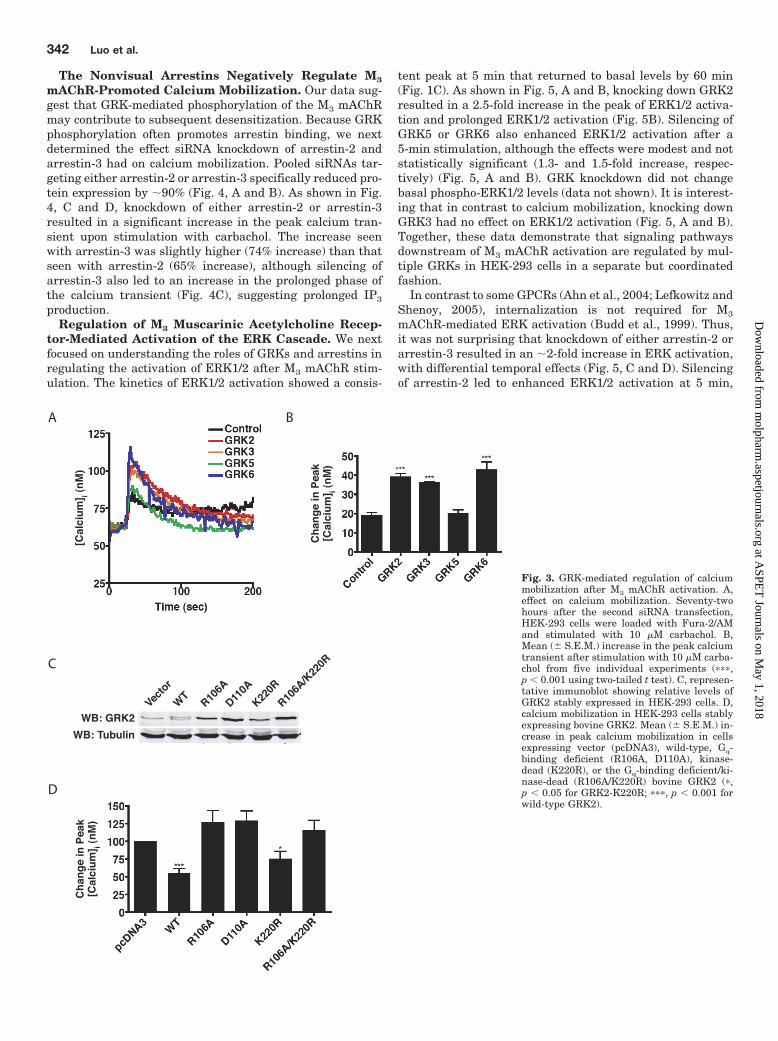
Regulation of M3 mAChR-Mediated Calcium Mobili-zation in HEK-293 Cells. We next evaluated the effect ofknocking down various regulatory proteins on M3 mAChRsignaling. Because the phosphorylation of activated GPCRsby GRKs is often an early step in signal termination, weinitially determined the effect that GRK knockdown wouldhave on calcium mobilization after carbachol treatment. Asshown in Fig. 2, A and B, we were able to selectively andspecifically knock down each of the four individual GRKsexpressed in HEK-293 cells. A modest increase in GRK3expression was observed when other GRKs, in particularGRK2, were knocked down (Fig. 2B). Knockdown of GRK2,GRK3, and GRK6 led to increases of 210% (p � 0.001), 190%(p � 0.001), and 230% (p � 0.001), respectively, in the peakcalcium transients, whereas knockdown of GRK5 had noeffect on calcium mobilization (Fig. 3, A and B). This effectwas also observed when methacholine was used to activatethe M3 mAChR (data not shown). These data suggest thatmultiple GRKs are involved in the desensitization of the M3
mAChR.GRK2 Interaction with Gq Is Primarily Responsible
for Increased Calcium Mobilization. The enhanced mo-bilization of calcium seen after silencing of GRK2 may arisefrom phosphorylation-dependent and/or phosphorylation-in-dependent mechanisms (Ribas et al., 2007). Therefore, we
next sought to further delineate the underlying mechanismobserved for calcium mobilization when GRK2 was knockeddown. Because we showed previously that GRK2 interactswith G�q through the RGS homology domain of GRK2 (Car-man et al., 1999), the increase in peak calcium mobilizationcould be a result of a loss of receptor phosphorylation, a lossof the ability of GRK2 to inhibit activated Gq, or both. Toaddress this, we generated cell lines that stably expresseither wild-type bovine GRK2, kinase dead GRK2 (K220R),GRK2 point mutants defective in binding G�q (R106A,D110A), or a GRK2 mutant that was both kinase-dead andGq-deficient (R106A/K220R). Cloned cell lines expressingwild-type or mutant bovine GRK2 at levels close to endoge-nous GRK2 levels (1- to 5-fold overexpression) were selectedfor study (Fig. 3C). SDS-PAGE revealed that bovine GRK2ran slightly slower than endogenous human GRK2 whenexpressed in HEK-293 cells (Fig. 3C). Stable expression ofeither wild-type or the kinase-dead mutant reduced carba-chol-stimulated calcium mobilization by �50% (Fig. 3D). Instriking contrast, stable expression of the G�q binding-defi-cient mutants (R106A and D110A) or the double mutant(R106A/K220R) had no effect on calcium mobilization (Fig.3D). This suggests that GRK2 primarily regulates the activ-ity of the M3 mAChR through its ability to interact with theactivated pool of G�q.
A
B
WB: anti-GRK2
WB: anti-GRK3
WB: anti-GRK4-6
WB: anti-Tubulin
Contro
l
GRK2
GRK3
GRK5
GRK6
Fig. 2. Knockdown of endogenous GRK isoforms in HEK-293 cells. A,HEK-293 cells were transfected twice within a 24-h interval with GRK-specific or nonspecific control siRNA. Seventy-two hours after the secondtransfection, cells were harvested, and equal amounts of total cellularlysate were separated by 10% SDS-PAGE, transferred to nitrocellulose,and incubated with the indicated antibodies. Blots were stripped andreprobed for �-tubulin to control for loading. Shown is a representativeimmunoblot. B, mean relative level of GRK expression after siRNA quan-tified by densitometry from five separate experiments.
M3 Muscarinic Receptor Signaling Is Differentially Regulated 341
at ASPE
T Journals on M
ay 1, 2018m
olpharm.aspetjournals.org
Dow
nloaded from
The Nonvisual Arrestins Negatively Regulate M3
mAChR-Promoted Calcium Mobilization. Our data sug-gest that GRK-mediated phosphorylation of the M3 mAChRmay contribute to subsequent desensitization. Because GRKphosphorylation often promotes arrestin binding, we nextdetermined the effect siRNA knockdown of arrestin-2 andarrestin-3 had on calcium mobilization. Pooled siRNAs tar-geting either arrestin-2 or arrestin-3 specifically reduced pro-tein expression by �90% (Fig. 4, A and B). As shown in Fig.4, C and D, knockdown of either arrestin-2 or arrestin-3resulted in a significant increase in the peak calcium tran-sient upon stimulation with carbachol. The increase seenwith arrestin-3 was slightly higher (74% increase) than thatseen with arrestin-2 (65% increase), although silencing ofarrestin-3 also led to an increase in the prolonged phase ofthe calcium transient (Fig. 4C), suggesting prolonged IP3
production.Regulation of M3 Muscarinic Acetylcholine Recep-
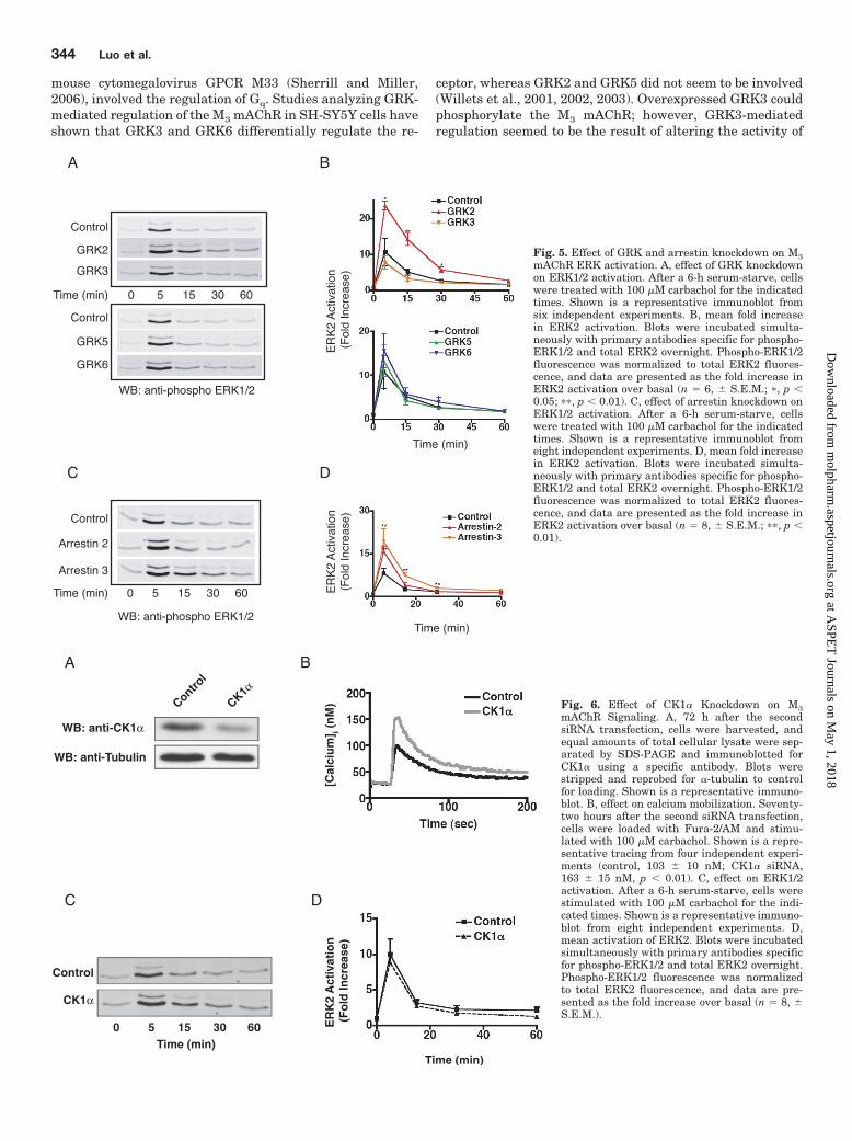
tor-Mediated Activation of the ERK Cascade. We nextfocused on understanding the roles of GRKs and arrestins inregulating the activation of ERK1/2 after M3 mAChR stim-ulation. The kinetics of ERK1/2 activation showed a consis-
tent peak at 5 min that returned to basal levels by 60 min(Fig. 1C). As shown in Fig. 5, A and B, knocking down GRK2resulted in a 2.5-fold increase in the peak of ERK1/2 activa-tion and prolonged ERK1/2 activation (Fig. 5B). Silencing ofGRK5 or GRK6 also enhanced ERK1/2 activation after a5-min stimulation, although the effects were modest and notstatistically significant (1.3- and 1.5-fold increase, respec-tively) (Fig. 5, A and B). GRK knockdown did not changebasal phospho-ERK1/2 levels (data not shown). It is interest-ing that in contrast to calcium mobilization, knocking downGRK3 had no effect on ERK1/2 activation (Fig. 5, A and B).Together, these data demonstrate that signaling pathwaysdownstream of M3 mAChR activation are regulated by mul-tiple GRKs in HEK-293 cells in a separate but coordinatedfashion.
In contrast to some GPCRs (Ahn et al., 2004; Lefkowitz andShenoy, 2005), internalization is not required for M3
mAChR-mediated ERK activation (Budd et al., 1999). Thus,it was not surprising that knockdown of either arrestin-2 orarrestin-3 resulted in an �2-fold increase in ERK activation,with differential temporal effects (Fig. 5, C and D). Silencingof arrestin-2 led to enhanced ERK1/2 activation at 5 min,
BA
C
Vecto
r
WT
K220R
R106A
D110A
R106A
/K22
0R
WB: GRK2
WB: Tubulin
D
Ch
ang
e in
Pea
k[C
alci
um
] i (n
M)
Ch
ang
e in
Pea
k[C
alci
um
] i (n
M)
[Cal
ciu
m] i (
nM
)
Fig. 3. GRK-mediated regulation of calciummobilization after M3 mAChR activation. A,effect on calcium mobilization. Seventy-twohours after the second siRNA transfection,HEK-293 cells were loaded with Fura-2/AMand stimulated with 10 �M carbachol. B,Mean (� S.E.M.) increase in the peak calciumtransient after stimulation with 10 �M carba-chol from five individual experiments (���,p � 0.001 using two-tailed t test). C, represen-tative immunoblot showing relative levels ofGRK2 stably expressed in HEK-293 cells. D,calcium mobilization in HEK-293 cells stablyexpressing bovine GRK2. Mean (� S.E.M.) in-crease in peak calcium mobilization in cellsexpressing vector (pcDNA3), wild-type, Gq-binding deficient (R106A, D110A), kinase-dead (K220R), or the Gq-binding deficient/ki-nase-dead (R106A/K220R) bovine GRK2 (�,p � 0.05 for GRK2-K220R; ���, p � 0.001 forwild-type GRK2).
342 Luo et al.
at ASPE
T Journals on M
ay 1, 2018m
olpharm.aspetjournals.org
Dow
nloaded from

whereas silencing of arrestin-3 led to both enhanced andprolonged activation (Fig. 5D). These data suggest that undernormal physiological conditions, either arrestin-2 or arres-tin-3 is sufficient to negatively regulate acute signalingevents upon M3 mAChR activation, although arrestin-3seems to play a larger role in terminating signaling in re-sponse to prolonged agonist exposure.
Regulation of the M3 Muscarinic Acetylcholine Re-ceptor by Casein Kinase 1�. CK1� also phosphorylates theM3 receptor in an agonist-dependent manner, although itdoes not seem to be required for desensitization of the recep-tor (Tobin et al., 1997; Budd et al., 2000, 2001). CK1� hasalso been shown to phosphorylate the M1 mAChR and rho-dopsin in vitro (Tobin et al., 1997; Waugh et al., 1999). Todetermine whether CK1� has a role in regulating the endog-enous M3 mAChR, HEK-293 cells were transfected withCK1� siRNA that specifically reduced CK1� protein levels to�40% of that seen in control cells (Fig. 6A). Knockdown ofCK1� resulted in a significant increase (62%, p � 0.01, n � 4)in the peak calcium transient compared with cells treatedwith control siRNA (Fig. 6B). To determine whether thiseffect was specific to CK1�-mediated regulation of the M3
mAChR and not to some other aspect of the Gq signalingpathway, we also tested the ability of CK1� to regulate thehistamine H1 receptor, which is regulated by GRK2 in HEK-293 cells (Iwata et al., 2005). Knockdown of CK1� had noeffect on calcium mobilization upon stimulation with 100 �Mhistamine (data not shown), suggesting that the effect ofCK1� knockdown was specific for M3 mAChR signaling. It isinteresting that knockdown of CK1� had no effect on carba-chol-mediated activation of ERK1/2 (Fig. 6, C and D). These
data demonstrate that, in addition to the GRK family, theagonist-activated M3 mAChR is also regulated by CK1�.
DiscussionGPCRs transduce extracellular stimuli into specific intra-
cellular signals that regulate a variety of cellular functions.GPCR desensitization is typically mediated by members ofthe GRK family, which specifically phosphorylate the ago-nist-occupied receptor, promoting the subsequent high-affin-ity binding of arrestins. For most GPCRs, the specificity ofGRKs and arrestins in cells remains poorly defined. In thisreport, we used an siRNA-based approach in HEK-293 cellsto characterize the role of these proteins in M3 mAChR sig-naling. We found that the M3 mAChR displays a complexpattern of regulation, such that GRK2, GRK3, GRK6, arres-tin-2, arrestin-3, and CK1� all participate to negatively reg-ulate calcium signaling upon receptor activation.
It was shown previously that GRK2 can be recruited to andphosphorylate the M3 mAChR at two separate serine clusterswithin the third intracellular loop (Wu et al., 2000). In addi-tion to receptor phosphorylation, GRK2 is able to bind bothGTP-bound G�q (Carman et al., 1999) and free G�� (Pitcheret al., 1992). The crystal structure of GRK2 (Tesmer et al.,2005) suggests that it may simultaneously sequester bothactive G�q and free G��, which, in addition to receptor phos-phorylation, may increase the strength and effectiveness ofGRK2-mediated receptor regulation. We and others havedemonstrated that GRK2-regulated GPCRs, such as the H1histamine (Iwata et al., 2005), M1 mAChR (Willets et al.,2005), metabotropic glutamate (Dhami et al., 2005), and
A
DC
B
WB: anti-arrestin
WB: anti-Tubulin
Contro
l
Arresti
n 2
Arresti
n 3
Ch
ang
e in
Pea
k[C
alci
um
] i (n
M)
[Cal
ciu
m] i (
nM
)
Fig. 4. Effect of arrestin knockdown on cal-cium mobilization after M3 mAChR activation.A, cells were transfected with SMARTpoolsiRNA and harvested 72 h later. Blots wereincubated with a monoclonal antibody for ar-restin-2 that cross-reacts with arrestin-3.Blots were stripped and reprobed for �-tubulinto control for loading. Shown is a representa-tive immunoblot. B, mean relative level of ar-restin expression after siRNA quantified bydensitometry from five separate experiments.C, effect on calcium mobilization. Cells wereharvested 72 h after transfection and pro-cessed as described previously. Shown is a rep-resentative calcium trace from five indepen-dent experiments. D, mean (� S.E.M.)increase in the peak calcium transient afterstimulation with 100 �M carbachol from fiveindividual experiments (���, p � 0.001 usingtwo-tailed t test).
M3 Muscarinic Receptor Signaling Is Differentially Regulated 343
at ASPE
T Journals on M
ay 1, 2018m
olpharm.aspetjournals.org
Dow
nloaded from
mouse cytomegalovirus GPCR M33 (Sherrill and Miller,2006), involved the regulation of Gq. Studies analyzing GRK-mediated regulation of the M3 mAChR in SH-SY5Y cells haveshown that GRK3 and GRK6 differentially regulate the re-
ceptor, whereas GRK2 and GRK5 did not seem to be involved(Willets et al., 2001, 2002, 2003). Overexpressed GRK3 couldphosphorylate the M3 mAChR; however, GRK3-mediatedregulation seemed to be the result of altering the activity of
Control
GRK2
GRK3
Control
GRK5
GRK6
0 6030155
WB: anti-phospho ERK1/2
Control
Arrestin 2
Arrestin 3
0 6030155
WB: anti-phospho ERK1/2
Time (min)
Time (min)
A
DC
B
Time (min)
ER
K2
Act
ivat
ion
(Fol
d In
crea
se)
ER
K2
Act
ivat
ion
(Fol
d In
crea
se)
Time (min)
Fig. 5. Effect of GRK and arrestin knockdown on M3mAChR ERK activation. A, effect of GRK knockdownon ERK1/2 activation. After a 6-h serum-starve, cellswere treated with 100 �M carbachol for the indicatedtimes. Shown is a representative immunoblot fromsix independent experiments. B, mean fold increasein ERK2 activation. Blots were incubated simulta-neously with primary antibodies specific for phospho-ERK1/2 and total ERK2 overnight. Phospho-ERK1/2fluorescence was normalized to total ERK2 fluores-cence, and data are presented as the fold increase inERK2 activation over basal (n � 6, � S.E.M.; �, p �0.05; ��, p � 0.01). C, effect of arrestin knockdown onERK1/2 activation. After a 6-h serum-starve, cellswere treated with 100 �M carbachol for the indicatedtimes. Shown is a representative immunoblot fromeight independent experiments. D, mean fold increasein ERK2 activation. Blots were incubated simulta-neously with primary antibodies specific for phospho-ERK1/2 and total ERK2 overnight. Phospho-ERK1/2fluorescence was normalized to total ERK2 fluores-cence, and data are presented as the fold increase inERK2 activation over basal (n � 8, � S.E.M.; ��, p �0.01).
ER
K2
Act
ivat
ion
(F
old
Incr
ease
)
Time (min)
Control
CK1α
0 5 15 30 60Time (min)
A B
C D
WB: anti-CK1α
WB: anti-Tubulin
Control
CK1α
[Cal
ciu
m] i (
nM
) Fig. 6. Effect of CK1� Knockdown on M3mAChR Signaling. A, 72 h after the secondsiRNA transfection, cells were harvested, andequal amounts of total cellular lysate were sep-arated by SDS-PAGE and immunoblotted forCK1� using a specific antibody. Blots werestripped and reprobed for �-tubulin to controlfor loading. Shown is a representative immuno-blot. B, effect on calcium mobilization. Seventy-two hours after the second siRNA transfection,cells were loaded with Fura-2/AM and stimu-lated with 100 �M carbachol. Shown is a repre-sentative tracing from four independent experi-ments (control, 103 � 10 nM; CK1� siRNA,163 � 15 nM, p � 0.01). C, effect on ERK1/2activation. After a 6-h serum-starve, cells werestimulated with 100 �M carbachol for the indi-cated times. Shown is a representative immuno-blot from eight independent experiments. D,mean activation of ERK2. Blots were incubatedsimultaneously with primary antibodies specificfor phospho-ERK1/2 and total ERK2 overnight.Phospho-ERK1/2 fluorescence was normalizedto total ERK2 fluorescence, and data are pre-sented as the fold increase over basal (n � 8, �S.E.M.).
344 Luo et al.
at ASPE
T Journals on M
ay 1, 2018m
olpharm.aspetjournals.org
Dow
nloaded from
PLC-� and not via receptor phosphorylation (Willets et al.,2001, 2002, 2003). In contrast, overexpressed GRK6 couldphosphorylate the M3 mAChR, leading to a decrease in sig-naling. This effect was reversed upon expression of a kinase-dead GRK6 (Willets et al., 2003).
Using siRNA coupled with stable expression of low levels ofvarious GRK2 mutants, we found that the enhanced calciummobilization observed upon GRK2 knockdown is primarilydue to a loss in regulation of activated Gq after M3 mAChRstimulation (Fig. 3). Furthermore, we showed that loss ofGRK2 leads to enhanced and prolonged activation of theERK1/2 cascade (Fig. 5). The observed effects of GRK2 knock-down are 2-fold: the enhanced calcium mobilization seems tobe primarily due to the loss of inhibition of activated Gq,whereas the enhanced and prolonged activation of ERK1/2probably reflects enhanced DAG production/PKC-� activa-tion and a relief of inhibition of mitogen-activated proteinkinase kinase 1 (MEK1) (Jimenez-Sainz et al., 2006). How-ever, we cannot completely rule out the possibility that GRK2also mediates receptor phosphorylation because endogenousM3 mAChR levels are too low to evaluate phosphorylation(Tovey and Willars, 2004).
We have also found that GRK3 and GRK6 negatively reg-ulate calcium mobilization after M3 mAChR stimulation. Al-though knockdown of either kinase led to significant in-creases in calcium mobilization (Fig. 3, A and B), silencing ofGRK3 had no effect on activation of ERK1/2, whereas loss ofGRK6 had only a minor effect (Fig. 5, A and B). The possi-bility exists that there is overlap between these kinases andthat regulation might involve a competition for receptor bind-ing, as has been suggested for the angiotensin receptor (Kimet al., 2005). These previous studies suggested that GRK2and GRK3 negatively regulate whereas GRK5 and GRK6positively regulate ERK1/2 activation and that differences inthe phosphorylation pattern mediated by GRK2/3 or GRK5/6could alternatively promote the binding of arrestin-2 or ar-restin-3, respectively (Kim et al., 2005). However, our resultssuggest that the M3 mAChR is not subject to this type ofoverlapping regulation. Furthermore, the GRKs do not play apositive role in M3 mAChR signaling. There is a growingnumber of nonreceptor substrates that have been identifiedfor the GRKs (Ribas et al., 2007), and in line with previousfindings, GRK3 could be primarily regulating PLC-� activityvia binding to G�� or G�q (Willets et al., 2001). This mightallow for a very rapid and robust production of IP3 andsubsequent calcium release that is not evident at later timepoints because other kinases (e.g., GRK6) may phosphorylatethe receptor resulting in desensitization. In addition, mech-anisms regulating downstream signaling events (e.g., IP3
hydrolysis, calcium reuptake, etc.) also shape both calciummobilization and ERK1/2 activation responses after carba-chol stimulation. Because we have identified three GRKsthat are involved in M3 mAChR regulation, multiple proteinsmay need to be knocked down simultaneously to producemore prolonged signaling.
We reported previously that an �50% reduction in arrestinlevels using antisense strategies had no effect on calciummobilization in HEK-293 cells (Mundell and Benovic, 2000).In the present study, we were able to reduce protein levels by�90% and show that the loss of either arrestin-2 or arres-tin-3 enhanced the peak calcium transient seen upon activa-tion of the M3 mAChR (Fig. 4, C and D). Taking into consid-
eration previous reports demonstrating that the M3 mAChRinternalizes in an arrestin-independent manner (Lee et al.,1998), our results suggest that arrestins primarily mediatedesensitization of the M3 mAChR after agonist activation.Consistent with this and with previous reports (Budd et al.,1999), knockdown of either arrestin-2 or arrestin-3 also en-hanced ERK1/2 activation (Fig. 5, C and D). This is in con-trast to the emerging paradigm that has been proposed for anumber of other GPCRs, in which arrestins promote G pro-tein-independent signaling pathways (Lefkowitz and She-noy, 2005) or even have opposing effects to one another, ashas been shown for the angiotensin II receptor (Ahn et al.,2004). In light of the fact that HEK-293 cells express similarlevels of endogenous arrestin-2 and arrestin 3 (J.L.B., unpub-lished results), our data suggest an inherent specificity forthe M3 mAChR by arrestin-3 because both calcium mobiliza-tion and ERK activation were enhanced and prolonged witharrestin-3 knockdown. This also suggests that the PLC-�/PKC arm of signaling is responsible for ERK activation,consistent with previous reports (Budd et al., 1999; Kim etal., 1999; Wylie et al., 1999). It is interesting that arrestinscan also terminate muscarinic receptor signaling by recruit-ing diacylglycerol kinases and enhancing the degradation ofthe second-messenger DAG, thereby coordinately terminat-ing GPCR/G protein interaction and promoting second-mes-senger degradation (Nelson et al., 2007). Taken together, theprolonged ERK activation observed after GRK2 and arres-tin-3 knockdown can be attributed to enhanced Gq activity,sustained DAG production, and subsequent PKC-� activation(Fig. 7).
CK1� has a variety of functions within the cell (Knipps-child et al., 2005) and has been shown recently to regulateheterologously expressed M3 mAChR in HEK-293 and COS7cells (Tobin et al., 1997; Budd et al., 2000). Likewise, wedemonstrate that CK1� knockdown results in enhanced cal-cium mobilization upon M3 receptor activation, suggestingthat CK1� is also involved in desensitization of endogenousM3 mAChR in HEK-293 cells. Knockdown of CK1� had noeffect on calcium mobilization upon H1 histamine receptoractivation, demonstrating that this effect was specific to theM3 mAChR. Previous studies have also shown that expres-sion of a peptide corresponding to the CK1� binding region oroverexpression of a mutated receptor lacking a portion of thethird intracellular loop led to a decrease in ERK1/2 activationupon receptor stimulation, suggesting that CK1�-mediatedphosphorylation was necessary for ERK activation (Budd etal., 2001). Although we show that knockdown of CK1� has noeffect on ERK1/2 activation (Fig. 6, C and D), indicating thatCK1� only plays a partial role in the regulation of M3
mAChR similar to GRK3 and GRK6, this may be due to thefact that we only achieved �60% knockdown of CK1�. Thethird intracellular loop of the M3 mAChR contains 12 puta-tive CK1� phosphorylation motifs (Tobin, 2002), two of whichoverlap with the proposed GRK2 phosphorylation sites (Wuet al., 2000). Thus, under physiological conditions, therecould be competition between these kinases for receptor bind-ing and phosphorylation.
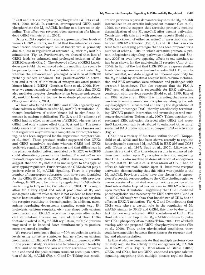
In this study, we demonstrate that multiple proteins coor-dinately regulate the activity of the endogenous M3 mAChRin HEK-293 cells (Fig. 7). Knockdown of GRK2, GRK3,GRK6, and CK1�, but not GRK5, enhanced receptor calciumsignaling, suggesting that multiple kinases regulate down-
M3 Muscarinic Receptor Signaling Is Differentially Regulated 345
at ASPE
T Journals on M
ay 1, 2018m
olpharm.aspetjournals.org
Dow
nloaded from

Arrestin 2/3
GRK6
Gq
Carbachol
PLCβ PKCδPIP2 DAG
IP3
Ca++
ERK1/2
Ras
Raf
MEK
GRK2/3CK1α
Gq
Carbachol
PLCβPIP2
IP3
Ca++
PKCδDAG
ERK1/2
Ras
Raf
MEK
GRK2
B
A
DGK
Fig. 7. Regulation of the endogenous M3 mAChR in HEK-293 cells. A, carbachol binding to the M3 mAChR results in activation of the Gq family ofheterotrimeric G proteins, leading to the dissociation of G�q and G��. Activated G�q activates PLC-�, resulting in the hydrolysis of phosphatidyl-inositol bisphosphate (PIP2) to form the second messengers IP3 and DAG. IP3 interacts with the IP3 receptor located at the endoplasmic reticulum,resulting in a robust but transient increase in cytosolic calcium. The formation of DAG recruits and activates the novel PKC isoform PKC-�. Onceactivated, PKC-� leads to the activation of a Ras-Raf-MEK-ERK1/2 cascade. B, phosphorylation of the M3 mAChR by GRK6 and possibly CK1� recruitsarrestin-2 and arrestin-3 to the receptor, preventing further G protein activation and terminating signaling. In addition, arrestins are able to recruitdiacylglycerol kinases (DGK) to the membrane and terminate the PKC-dependent arm of the signaling cascade. GRK2 and GRK3, through a conservedRGS domain, are able to interact with and sequester free G�q and prevent activation of PLC-�. This results in the inhibition of both calciummobilization and activation of the ERK1/2 cascade. GRK2 is also able to regulate the activation of the ERK1/2 cascade by interacting with andnegatively regulating the activity of MEK1.
346 Luo et al.
at ASPE
T Journals on M
ay 1, 2018m
olpharm.aspetjournals.org
Dow
nloaded from

stream signaling after M3 mAChR activation. An effect ofGRK2 on calcium flux could be observed with both wild-typeand a kinase-dead mutant but not with G�q-binding defectivemutants, demonstrating that GRK2 primarily regulates ac-tivated Gq. It is interesting that only silencing of GRK2 led toboth an enhanced and prolonged ERK activation. Consistentwith our findings that GRK2 primarily regulated Gq activity,this is probably a result of enhanced activation of the Gq/PLC-�/PKC-� signaling pathway (Fig. 7). Finally, both arres-tin-2 and arrestin-3 are involved in negatively regulating theM3 mAChR as knockdown of either protein enhanced calciummobilization and ERK activation. Overall, our data suggestthat multiple proteins dynamically regulate M3 mAChR-me-diated signal transduction.
ReferencesAhn S, Shenoy SK, Wei H, and Lefkowitz RJ (2004) Differential kinetic and spatial
patterns of �-arrestin and G protein-mediated ERK activation by the angiotensinII receptor. J Biol Chem 79:35518–35525.
Brown SG, Thomas A, Dekker LV, Tinker A, and Leaney JL (2005) PKC-deltasensitizes Kir3.1/3.2 channels to changes in membrane phospholipid levels afterM3 receptor activation in HEK-293 cells. Am J Physiol Cell Physiol 289:C543–C556.
Budd DC, McDonald JE, and Tobin AB (2000) Phosphorylation and regulation of aGq/11-coupled receptor by casein kinase 1�. J Biol Chem 275:19667–19675.
Budd DC, Rae A, and Tobin AB (1999) Activation of the mitogen-activated proteinkinase pathway by a Gq/11-coupled muscarinic receptor is independent of receptorinternalization. J Biol Chem 274:12355–12360.
Budd DC, Willars GB, McDonald JE, and Tobin AB (2001) Phosphorylation of theGq/11-coupled m3-muscarinic receptor is involved in receptor activation of theERK-1/2 mitogen-activated protein kinase pathway. J Biol Chem 276:4581–4587.
Carman CV, Parent JL, Day PW, Pronin AN, Sternweis PM, Wedegaertner PB,Gilman AG, Benovic JL, and Kozasa T (1999) Selective regulation of G�(q/11) byan RGS domain in the G protein-coupled receptor kinase, GRK2. J Biol Chem274:34483–34492.
de la Vega MT, Nunez A, and Arias-Montano JA (1997) Muscarinic M1 and M3receptors in rat striatum: a binding study. Arch Med Res 28:493–497.
Dhami GK, Babwah AV, Sterne-Marr R, and Ferguson SS (2005) Phosphorylation-independent regulation of metabotropic glutamate receptor 1 signaling requires gprotein-coupled receptor kinase 2 binding to the second intracellular loop. J BiolChem 280:24420–24427.
Gainetdinov RR, Premont RT, Bohn LM, Lefkowitz RJ, and Caron MG (2004)Desensitization of G protein-coupled receptors and neuronal functions. Annu RevNeurosci 27:107–144.
Gschwendt M, Kittstein W, and Marks F (1994) Elongation factor-2 kinase: effectiveinhibition by the novel protein kinase inhibitor rottlerin and relative insensitivitytowards staurosporine. FEBS Lett 338:85–88.
Guo FF, Kumahara E, and Saffen D (2001) A CalDAG-GEFI/Rap1/B-Raf cassettecouples M1 muscarinic acetylcholine receptors to the activation of ERK1/2. J BiolChem 276:25568–25581.
Hakak Y, Shrestha D, Goegel MC, Behan DP, and Chalmers DT (2003) Globalanalysis of G-protein-coupled receptor signaling in human tissues. FEBS Lett550:11–17.
Iwata K, Luo J, Penn RB, and Benovic JL (2005) Bimodal regulation of the humanH1 histamine receptor by G protein-coupled receptor kinase 2. J Biol Chem280:2197–2204.
Jimenez-Sainz MC, Murga C, Kavelaars A, Jurado-Pueyo M, Krakstad BF, HeijnenCJ, Mayor F Jr, Aragay AM (2006) G protein-coupled receptor kinase 2 negativelyregulates chemokine signaling at a level downstream from G protein subunits. MolBiol Cell 17:25–31.
Kim J, Ahn S, Ren XR, Whalen EJ, Reiter E, Wei H, and Lefkowitz RJ (2005)Functional antagonism of different G protein-coupled receptor kinases for �-ar-restin-mediated angiotensin II receptor signaling. Proc Natl Acad Sci U S A 102:1442–1447.
Kim JY, Yang MS, Oh CD, Kim KT, Ha MJ, Kang SS, and Chun JS (1999) Signallingpathway leading to an activation of mitogen-activated protein kinase by stimulat-ing M3 muscarinic receptor. Biochem J 337:275–280.
Knippschild U, Wolff S, Giamas G, Brockschmidt C, Wittau M, Wurl PU, Eismann T,
and Stoter M (2005) The role of the casein kinase 1 (CK1) family in differentsignaling pathways linked to cancer development. Onkologie 28:508–514.
Krupnick JG and Benovic JL (1998) The role of receptor kinases and arrestins in Gprotein-coupled receptor regulation. Annu Rev Pharmacol Toxicol 38:289–319.
Lee KB, Pals-Rylaarsdam R, Benovic JL, and Hosey MM (1998) Arrestin-independent internalization of the m1, m3, and m4 subtypes of muscarinic cho-linergic receptors. J Biol Chem 273:12967–12972.
Lefkowitz RJ and Shenoy SK (2005) Transduction of receptor signals by beta-arrestins. Science 308:512–517.
Liu C, Li Y, Semenov M, Han C, Baeg GH, Tan Y, Zhang Z, Lin X, and He X (2002)Control of beta-catenin phosphorylation/degradation by a dual-kinase mechanism.Cell 108:837–847.
Moore CA, Milano SK, and Benovic JL (2007) Regulation of receptor trafficking byGRKs and arrestins. Annu Rev Physiol 69:451–482.
Mundell SJ and Benovic JL (2000) Selective regulation of endogenous G protein-coupled receptors by arrestins in HEK293 cells. J Biol Chem 275:12900–12908.
Nelson CD, Perry SJ, Regier DS, Prescott SM, Topham MK, and Lefkowitz RJ (2007)Targeting of diacylglycerol degradation to M1 muscarinic receptors by beta-arrestins. Science 315:663–666.
Pierce KL, Premont RT, and Lefkowitz RJ (2002) Seven-transmembrane receptors.Nat Rev Mol Cell Biol 3:639–650.
Pitcher JA, Inglese J, Higgins JB, Arriza JL, Casey PJ, Kim C, Benovic JL, KwatraMM, Caron MG, and Lefkowitz RJ (1992) Novel role for the �� subunits ofheterotrimeric G proteins: targeting of beta-adrenergic receptor kinase to mem-brane bound receptors. Science 257:1264–1267.
Ribas C, Penela P, Murga C, Salcedo A, Garcia-Hoz C, Jurado-Pueyo M, Aymerich I,and Mayor F Jr (2007) The G protein-coupled receptor kinase (GRK) interactome:role of GRKs in GPCR regulation and signaling. Biochim Biophys Acta 1768:913–922.
Sherrill JD and Miller WE (2006) G protein-coupled receptor (GPCR) kinase 2regulates agonist-independent Gq/11 signaling from the mouse cytomegalovirusGPCR M33. J Biol Chem 281:39796–39805.
Tesmer VM, Kawano T, Shankaranarayanan A, Kozasa T, and Tesmer JJ (2005)Snapshot of activated G proteins at the membrane: the Galphaq-GRK2-Gbetagamma complex. Science 310:1686–1690.
Tobin AB (2002) Are we beta-ARKing up the wrong tree? Casein kinase 1 alphaprovides an additional pathway for GPCR phosphorylation. Trends Pharmacol Sci23:337–343.
Tobin AB, Totty NF, Sterlin AE, and Nahorski SR (1997) Stimulus-dependentphosphorylation of G-protein-coupled receptors by casein kinase 1�. J Biol Chem272:20844–20849.
Tovey SC and Willars GB (2004) Single cell imaging of intracellular Ca2� andphospholipase activity reveals that RGS 2, 3, and 4 differentially regulate signal-ing via the G�q/11-linked muscarinic M3 receptor. Mol Pharmacol 66:1453–1464.
Waugh MG, Challiss RA, Berstein G, Nahorski SR, and Tobin AB (1999) Agonist-induced desensitization and phosphorylation of m1-muscarinic receptors. BiochemJ 338:175–183.
Werry TD, Sexton PM, and Christopoulos A (2005) “Ins and outs” of seven-transmembrane receptor signalling to ERK. Trends Endocrinol Metab 16:26–33.
Willets JM, Challiss RA, Kelly E, and Nahorski SR (2001) G protein-coupled receptorkinases 3 and 6 use different pathways to desensitize the endogenous M3 musca-rinic acetylcholine receptor in human SH-SY5Y cells. Mol Pharmacol 60:321–330.
Willets JM, Challiss RA, and Nahorski SR (2002) Endogenous G protein-coupledreceptor kinase 6 Regulates M3 muscarinic acetylcholine receptor phosphorylationand desensitization in human SH-SY5Y neuroblastoma cells. J Biol Chem 277:15523–15529.
Willets JM, Mistry R, Nahorski SR, and Challiss RA (2003) Specificity of G protein-coupled receptor kinase 6-mediated phosphorylation and regulation of single-cellm3 muscarinic acetylcholine receptor signaling. Mol Pharmacol 64:1059–1068.
Willets JM, Nahorski SR, and Challiss RA (2005) Roles of phosphorylation-dependent and -independent mechanisms in the regulation of M1 muscarinicacetylcholine receptors by G protein-coupled receptor kinase 2 in hippocampalneurons. J Biol Chem 280:18950–18958.
Wu G, Bogatkevich GS, Mukhin YV, Benovic JL, Hildebrandt JD, and Lanier SM(2000) Identification of G�� binding sites in the third intracellular loop of theM3-muscarinic receptor and their role in receptor regulation. J Biol Chem 275:9026–9034.
Wylie PG, Challiss RA, and Blank JL (1999) Regulation of extracellular-signalregulated kinase and c-Jun N-terminal kinase by G-protein-linked muscarinicacetylcholine receptors. Biochem J 338:619–628.
Address correspondence to: Dr. Jeffrey L. Benovic, Department of Bio-chemistry and Molecular Biology, Thomas Jefferson University, BLSB 350,Philadelphia, PA 19107. E-mail: [email protected]
M3 Muscarinic Receptor Signaling Is Differentially Regulated 347
at ASPE
T Journals on M
ay 1, 2018m
olpharm.aspetjournals.org
Dow
nloaded from
Recommended