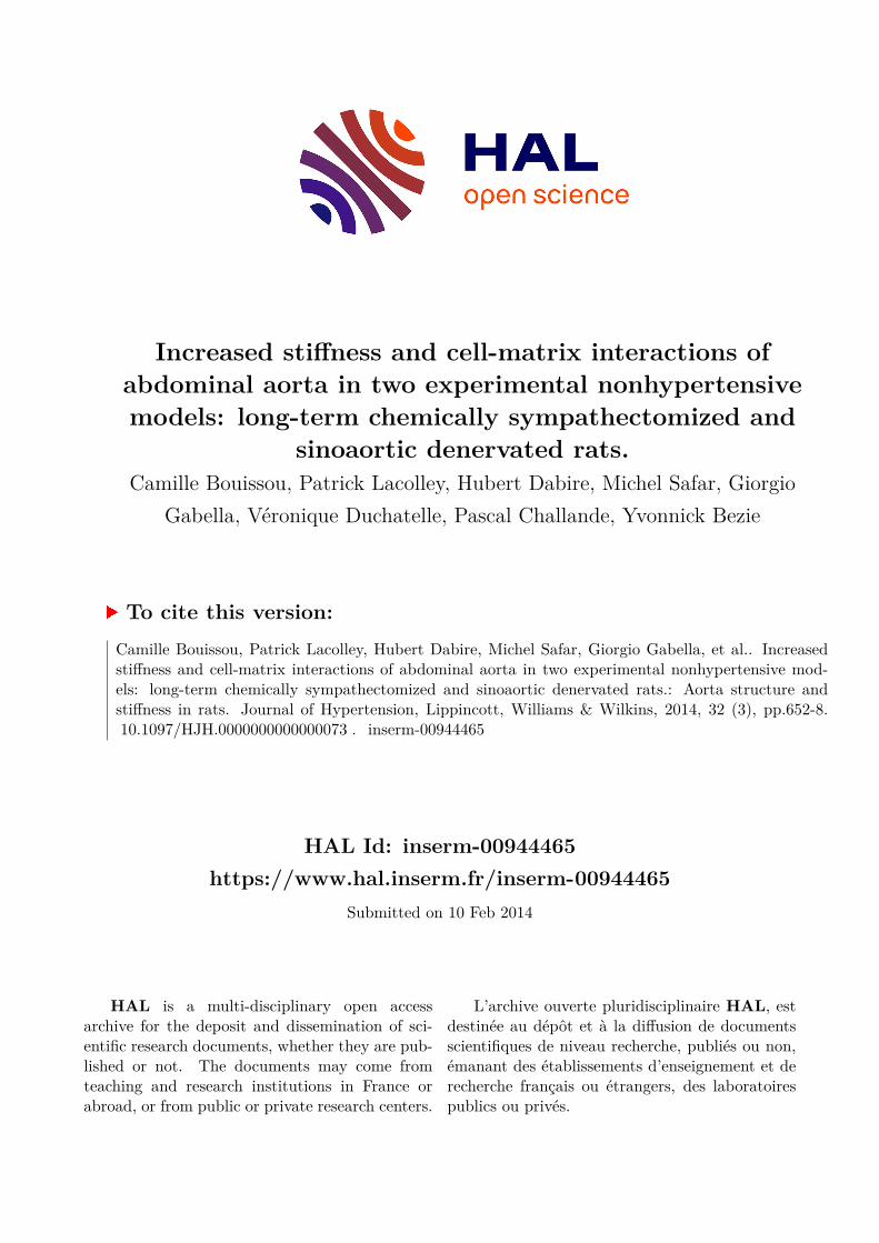
HAL Id: inserm-00944465https://www.hal.inserm.fr/inserm-00944465
Submitted on 10 Feb 2014
HAL is a multi-disciplinary open accessarchive for the deposit and dissemination of sci-entific research documents, whether they are pub-lished or not. The documents may come fromteaching and research institutions in France orabroad, or from public or private research centers.
L’archive ouverte pluridisciplinaire HAL, estdestinée au dépôt et à la diffusion de documentsscientifiques de niveau recherche, publiés ou non,émanant des établissements d’enseignement et derecherche français ou étrangers, des laboratoirespublics ou privés.
Increased stiffness and cell-matrix interactions ofabdominal aorta in two experimental nonhypertensivemodels: long-term chemically sympathectomized and
sinoaortic denervated rats.Camille Bouissou, Patrick Lacolley, Hubert Dabire, Michel Safar, Giorgio
Gabella, Véronique Duchatelle, Pascal Challande, Yvonnick Bezie
To cite this version:Camille Bouissou, Patrick Lacolley, Hubert Dabire, Michel Safar, Giorgio Gabella, et al.. Increasedstiffness and cell-matrix interactions of abdominal aorta in two experimental nonhypertensive mod-els: long-term chemically sympathectomized and sinoaortic denervated rats.: Aorta structure andstiffness in rats. Journal of Hypertension, Lippincott, Williams & Wilkins, 2014, 32 (3), pp.652-8.�10.1097/HJH.0000000000000073�. �inserm-00944465�

Increased stiffness and cell-matrix interactions of abdominal aorta in
two experimental non-hypertensive models: long-term chemical
sympathectomized and sinoaortic-denervated rats
Camille BOUISSOUa, Patrick LACOLLEYb , Hubert DABIREa,
Michel E. SAFARc, Giorgio GABELLAd, Véronique DUCHATELLEe,
Pascal CHALLANDEf$, Yvonnick BEZIEg$
Short title: Aorta structure and stiffness in rats
aINSERM, U955, Equipe 03, Créteil, France ; bINSERM, U1116, Nancy, France;
cHôtel-Dieu Hospital, Diagnosis center and Université René Descartes, UFR Médecine,
Paris, France; dDepartment of Anatomy and Developmental Biology, University
College London, London, United Kingdom; eDepartment of Pathology, Groupe
hospitalier Paris Saint-Joseph, Paris, France ; fUPMC Univ Paris 06 ; CNRS UMR
7190, Paris, France ; gDepartment of Pharmacy, Groupe hospitalier Paris Saint-Joseph,
Paris, France.
$equal contribution
Disclosure: No disclosure
Conflict of interest: None
Corresponding Authors:

2
Dr. Yvonnick BEZIE, Groupe Hospitalier Paris Saint-Joseph,185 Rue Raymond Losserand
F-75674 Paris Cedex 14, France.
Tel : +33 1 44 12 36 16
Fax : +33 1 44 12 34 61
E-mail address: [email protected]
Dr. Patrick Lacolley, INSERM U1116, 9 avenue de la Forêt de Haye. BP 184, 54500
Vandoeuvre-lés-Nancy Cedex. France
Tel : +33 3 83 68 36 23
Fax : +33 3 83 68 36 39
E-mail address: [email protected]
Word count:
Total (including references, but not tables and legends): 4210
Abstract: 243
Number of tables: 1
Number of figures: 4

ABSTRACT
RATIONALE: Sinoaortic denervated (SAD) and chemically sympathectomized (SNX)
rats are characterized by a decrease in arterial distensibility without hypertension and
would thus be relevant for analyzing arterial wall stiffening independently of blood
pressure level. The fibronectin network, which plays a pivotal role in cell matrix
interactions, is a major determinant of arterial stiffness. We hypothesized that in SAD
and SNX rats, arterial stiffness is increased, due to alterations of cell-matrix anchoring
leading to spatial reorganization of the extracellular matrix.
METHODS: The intrinsic elastic properties of the arterial wall were evaluated in vivo
by the relationship between incremental elastic modulus determined by echotracking
and circumferential wall stress. The changes of cell-extracellular matrix links in the
abdominal aorta were evaluated by studying fibronectin, vascular integrins receptors
and ultrastructural features of the aorta by immunochemistry.
RESULTS: In both experimental conditions wall stiffness increased, associated with
different modifications of cell-extracellular matrix adhesion. In SAD rats, increased
media-cross sectional area was coupled with an increase of muscle cell attachments to
its extracellular matrix via fibronectin and its α5-β1 integrin. In SNX rats, reduced
media-cross sectional area was associated with up-regulation of αv-β3 integrin and
more extensive connections between dense bands and elastic fibers despite the
disruption of the elastic lamellae.
CONCLUSION: In aorta of SNX and SAD rats, a similar arterial stiffness is
associated to different structural alterations. An increase in αvβ3 or α5β1 integrins
together with the already reported increase in the proportion of less distensible
(collagen) to more distensible (elastin) components in both models, contribute to

4
remodeling and stiffening of the abdominal aorta.

CONDENSED ABSTRACT
Sinoaortic denervated (SAD) and chemically sympathectomized (SNX) rats are models
of decreased arterial distensibility without hypertension allowing analyzing arterial
stiffening and structure independently of blood pressure level. Increase in arterial wall
stiffness in both models was associated with structural alterations. In SAD, increased
media cross sectional area was coupled with increased muscle cell attachments via
fibronectin and α5-β1 integrin. In SNX, reduced media cross sectional area was
associated with up-regulation of αv-β3 integrin and alteration of elastin fibers. In these
rats, similar arterial stiffness in absence of hypertension is associated to differential
structural alterations.
Key words: Sino-aortic denervation; Sympathectomy; fibronectin; Arterial Stiffness,
Integrins

INTRODUCTION
Increased stiffness of large arteries is a significant and independent predictor of
cardiovascular (CV) diseases [1]. Arterial stiffness is evaluated by the elastic properties
of the artery as a whole measured by arterial distensibility and by the elastic properties
of the arterial wall material measured by the incremental elastic modulus (Einc) [2].
Arterial distensibility and Einc are then 2 complementary parameters used to describe
arterial stiffness. Until now, it remains difficult to separate the causal effects of blood
pressure elevation from that of the mechanical and functional properties of the arterial
wall that lead to alterations of distensibility and stiffness. We have previously shown
that the spontaneously hypertensive rat (SHR) is characterized by a decreased
distensibility at its operational pressure compared to its normentensive control [3].
Nevertheless evaluation of the arterial wall stiffness, assessed by the elastic modulus
measurement, shows that for a given level of stress, SHR and Wistar rats have similar
mechanical properties. These results indicate that the decrease of distensibility observed
in SHR is related to hypertension, rather than to increased stiffness of the arterial wall.
Therefore, other experimental models must be used to analyze the intrinsic stiffness of
the aortic tissue. Sinoaortic denervated (SAD) rats and chemically sympathectomized
(SNX) rats should help to investigate this issue. Both models are characterized by a
decrease in arterial distensibility without hypertension compared to their respective
control [4, 5], suggesting that they would be relevant for analyzing arterial wall
stiffening independently of blood pressure level.
The fibronectin (Fn) network, which plays a pivotal role in cell matrix interactions, is a
major determinant of arterial stiffness [3, 6]. Fibronectin controls deposition and
organization of extra cellular matrix and modulates both cell proliferation and vascular

7
smooth muscle cell (SMC) phenotype. Thus, by increasing cell-matrix anchoring
through a α5-ß1-integrin, aortic fibronectin accumulation may contribute to protect the
arterial wall components from the increased mechanical loads associated with
hypertension in young and old SHRs [3]. The accumulation of other integrins, such as
αv-β3 integrin, has also been observed in the mesenteric artery of SHR [7]. These
results suggest that cell-matrix interactions, which play a major role in SMC function,
are also involved in the mechanical properties of the vascular wall. To our knowledge,
cell-matrix interactions have never been described in non-hypertensive models related
to arterial stiffness.
We hypothesized that in SAD and SNX rats, arterial stiffness is increased, and this may
be due to alterations of cell-matrix anchoring leading to spatial reorganization of the
extracellular matrix. We aim to determine in SAD and SNX rats (i) the intrinsic elastic
properties of the arterial wall by evaluating in vivo the relationship between Einc and
circumferential wall stress, and (ii) the changes of cell-extracellular matrix links in the
abdominal aorta, by studying fibronectin, vascular integrins receptors and ultrastructural
features of the aorta.
MATERIALS AND METHODS
Animals
Male Wistar rats (Iffa-Credo, Fresnes, France) were used. All procedures were in
accordance with institutional guidelines for animal experimentation and conformed to the
Guide for the Care and Use of Laboratory Animals, published by the National Institutes
of Health.

8
Sino-aortic denervation and SNX were performed as previously described [4, 5]. In
brief, SAD was performed at 10 weeks of age on anesthetized rats. Sham-operated rats
were used as control of SAD rats. All the rats of these groups were examined and killed
at 16 weeks of age. SNX rats were sympathectomized by subcutaneous injections of 50
mg/kg guanethidine sulfate for 12 weeks, 5 times a week, from day 5 after birth. The
control (CO) rats received saline injections according to the same schedule, and both
sets of rats were investigated during their 13th week of age. A total of 27 rats were used
for the in vivo experiments and 38 rats for immunohistochemistry and electron
microscopy.
Hemodynamic investigations
Mechanical properties of the abdominal aorta were assessed by circumferential wall
stress (σ) and Einc. At the end of the treatment, under pentobarbital anesthesia (60
mg/kg ip), a catheter was introduced in the lower abdominal aorta via the femoral artery
for blood pressure recording. A midline laparotomy was then performed and the probe
of the ultrasonic device (NIUS-01, Asulab SA) positioned 1 cm above the aortic
bifurcation for recording of internal arterial diameter. Blood pressure and internal
arterial diameter were then simultaneously recorded. The relationship between pressure
and lumen cross-sectional area was calculated by means of an arctangent
function. σ and Einc were calculated with the above-mentioned parameters and medial
cross-sectional area of the aorta determined by histomorphometry as previously
described [3, 6, 8].
Antibodies

9
The antibodies used were monoclonal mouse antibodies (mAbs) reactive with an
alternatively spliced form of fibronectin, EIIIA-Fibronectin (clone IST-9, Valbiotech,
France) and all FN isoforms (Total-Fn, Valbiotech, France) [3], a mouse anti-vimentin
monoclonal antibody (1/400, clone V9, Dako, France), a mouse anti-actin monoclonal
antibody (1/500, clone 1A4, Dako, France), a rabbit anti-integrin αv polyclonal
antibody (1/250, chemicon) a rabbit anti-integrin α5 subunit polyclonal antibody
(Valbiotech, France) [3].
Immunohistochemical investigation
Immunohistochemical staining was performed on fresh unfixed freeze-dried suprarenal
abdominal aorta [3]. We used the indirect immunoperoxidase technique as previously
described for the determination of fibronectin, the EIIIA-Fibronectin isoform and the α5
integrin [3, 9]. The determination of actin, vimentin, αv integrin were performed on a
Dako automate as described elsewhere [10]. No specific staining was observed when
primary antibody was omitted from the protocol (negative control). The distribution and
quantification of staining were determined by computer-directed color analysis
performed with the Quant’Image software (Quancoul, Talence, France) [3].
Electron microscopy
The thoracic aortas (2 SAD, 2 SNX and 5 controls) were fixed in situ by transcardiac
perfusion of fixative (4% glutaraldehyde and 1% formaldehyde in 0.1M Na-cacodylate).
Thin (0.5-3.0 µm) and ultrathin (100 nm) sections were cut on a plane transverse to the
thickness of the media and parallel to the length of the vessel, an approach that
produced approximately transverse sections of the muscle cells. The sections were

10
stained with uranyl acetate and lead citrate and viewed in a Philips 400 microscope.
Photographic montages were made, covering the thickness of the media over a length of
150-400 µm, at a magnification of 8000x. On these montages the size of nucleated
muscle cell profiles was measured as well as the percentage of the cell membrane
displaying dense bands and the percentage of cell membrane in contact with lamellae of
elastin [8]. Ultrastructure characterization and quantification were evaluated by
counting tissue points of randomly selected photographic fields and a minimum of 6
nucleated cells per rat were quantified as previously reported [8]. Quantification of
elastin and collagen have been presented elsewhere [4, 5].
Statistical Analysis
All values were averaged and expressed as mean ± SEM. Unpaired Student’s t tests
were performed to compare SNX and SAD rats with their respective controls for arterial
and immunohistochemical parameters [3]. For statistical comparison of Einc-σ curves
between groups, Einc was log transformed to generate linear relationships. Calculating
the r2 of the linear regression obtained with the new parameters for each individual
checked the quality of the transformation. After this transformation, we calculated the
mean slopes of the curves. If the slopes were not significantly different, we compared
the curves by calculating the mean wall stress at 800 kPa of Einc (σ800), a value
common to all groups [11]. Differences were considered significant at values of P<0.05.
RESULTS
Hemodynamic and aortic mechanics

11
Body weight of SNX rats was significantly lower than that of control rats (303 ± 10 g
vs. 353 ± 3 g, p<0.05). Likewise, SAD rats were significantly lighter than sham-
operated rats (428 ± 13 g vs. 464 ± 7 g, p<0.05). Compared to their respective controls,
heart rate (data not shown) and mean arterial pressure (MAP) were significantly
reduced in SNX rats but remained unchanged in SAD rats. Media cross sectional area
was significantly reduced in SNX rats compared to their controls; but was significantly
increased in SAD rats compared to sham-operated rats. Therefore, SAD rats had
significantly higher MCSA than SNX rats. Einc at MAP remained unchanged in all
groups of rats. The circumferential wall stress was similar in SNX and SAD rats both at
MAP and at 800 kPa of Einc but was significantly reduced when compared to their
respective controls (Table 1). The Einc-wall stress curves of SNX and SAD rats were
similarly shifted leftwards compared with their controls, indicating increased stiffness
of the wall in both models (Figure 1).
Immunohistochemistry
Total-fibronectin and α5 integrin subunit staining were diffuse in the media of all
control rats (Figures 2 and 3). Staining for both components was markedly increased in
SAD rats compared to Sham-operated rats (Figure 2) whereas no accumulation of
fibronectin and its α5 integrin were found in SNX rats compared to their controls
(Figure 3).
In Sham-operated and CO rats, immunoreactivity for cellular fibronectin (EIIIA-
fibronectin), was observed in the inner part of the media. In SAD rats the EIIIA-
fibronectin staining was equally intense but it involved the entire thickness of the media

12
(Figure 2). A slight increased of EIII-A fibronectin was found in SNX rats compared to
CO (Figure 3).
The endothelium highly expressed αv staining and the media showed relatively low but
extensive expression in all control rats. In contrast with α5 integrin, αv and vimentin
staining were increased in SNX rats (Figure 3) but unchanged in SAD rats (Figure 2)
compared to their respective controls. Immunostaining for actin was equally intense in
all conditions.
Electron Microscopy
The intima of the rats’ aortic wall is composed of a thin endothelium, sub-endothelium
connective tissue and an inner elastic lamina. The endothelium of the thoracic aorta
appeared similar in all groups.
The media, composed of elastic lamellae, with interposed layers of muscle cell and
interconnected by elastic fibers and bundles of collagen fibrils, showed some alterations
in the experimental conditions; however, the amount of elastic material in the wall
appeared unchanged. In SNX rats some disruption of the elastic lamellae was observed,
including the breaking up of some lamellae into large elastic bundles (Figure 4A).
The muscle cells profiles, observed in transverse section, had an irregular contour, with
large processes and invaginations, in all preparations. Numerous dense bands associated
with actin bundles on the cytoplasmic side and with collagen and elastic fibers
extracellularly, occupied the muscle cell membrane.
Despite the restricted number of animals used per groups, results observed were quite
reproducible as already validated [8] and allow pooling data from sham-operated rats
and control of SNX rats. In control rats, dense bands occupied 42 ± 2 % of the cell

13
perimeter and half of it (20% ± 2 %) was connected to elastic fibers. The number of
dense bands appeared obviously increased in SAD rats (57% ± 3 %; p<0.05) compared
with controls but not in SNX rats (45% ± 2 %). In the latter there were more extensive
connections between dense bands and elastic fibers than in controls (30 ± 2 %; p<0.05).
The percentage of dense bands connected to elastic lamellae remained unchanged in
SAD rats (Figure 4B).
DISCUSSION
In the present study, structural changes of the abdominal aorta were evaluated in SNX
and SAD rats, two models of decreased arterial distensibility without hypertension. We
observed an increase in wall stiffness in both experimental conditions, but different
structural changes in the vessel wall. In SAD rats, aortic hypertrophy was coupled with
an increase of muscle cell attachments to its extracellular matrix via fibronectin and its
α5-β1 integrin. In SNX rats, aortic hypotrophy was associated with αv-β3 integrin up-
regulation and alteration of elastin fibers.
In contrast to acute treatment, chronic treatment with guanethine significantly reduced
blood pressure as already reported [5, 12]. This effect may be due to the succession of
many hypotensive episodes, previously reported in this model [13]. A weight loss was
also observed as previously reported in both models [12].
We have previously shown a similar reduction of carotid distensibility in SAD and SNX
rats [4, 5]. While arterial distensibility is an indicator of the elastic properties of the
artery as a hollow structure, the Einc expresses the elastic properties of the wall material
that is independent of wall intima-media thickness [6]. Enhanced aortic stiffness is a
significant and independent risk factor for all-cause and cardiovascular mortality [1],

14
primarily coronary heart disease [14] and stroke [15] in human. Thus, elaboration of
Einc/stress curves addresses arterial wall stiffness, independently of the wall thickness
and of the pressure level. To our knowledge, stiffness of the arterial wall material had
never been evaluated and compared in SNX and SAD rats. Therefore, the first new
finding of the present experiments is that SNX and SAD rats are characterized by a
similar increase in arterial wall stiffness (leftwards shift of the Einc-stress curves).
We have previously shown that in SHRs, the Einc of the aortic wall material,
determined for a given level of circumferential wall stress, was not significantly
different from that of Wistar rats. This indicates that arterial wall materials in SHR and
its control strain have similar mechanical behavior [3]. In contrast, the increased
stiffness of the arterial observed in the present study suggest that SNX and SAD rats
seem pretty relevant models for analyzing arterial remodeling associated with stiffness.
The second new finding of the present study derived from the characterization of
extracellular matrix changes in both models. Extracellular matrix proteins determine the
passive biomechanical properties, collagen providing tensile strength and elastin
enabling vascular elasticity [16]. Indeed we and others have shown a strong relationship
between decreased elastin/collagen ratio and arterial stiffness in both models indicating
an alteration in the organization of the ECM [4, 5, 17]. However, because SNX is
characterized by reduced MCSA (present results) with a predominant reduction in
elastin [5], and SAD by an increased MCSA (present results) and a predominant
increase in collagen [4], a different structure-function relationship is present in the two
experimental conditions.
The dense bands of muscle cells provide a link between contractile apparatus and
extracellular matrix, mediated by integrin receptors on the cell membrane [8, 18, 19]. In

15
rat aorta, the major integrins ligands are fibronectin, a glycoprotein that plays an
important role in the organization and assembly of the extracellular matrix, collagen and
laminin. Accumulation of collagen in the aorta of SAD rats is associated with
accumulation of total-fibronectin and its α5ß1-integrin receptor, indicating an increased
mechanical linking/coupling between muscle cells and extracellular matrix [19-21].
Alteration of cell-matrix attachments might thus contribute to increase arterial stiffness,
as already reported in SHRs [3]. This result is strengthened by the obvious
ultrastructural changes of the aorta shown in the present study where the number of
dense bands per muscle cell profile is enhanced. In SAD rats, extracellular matrix
composition is also characterized by an accumulation of EIIIA-fibronectin, up regulated
during hypertension and aging [3, 21, 22], and closely associated with arterial stiffness
[3, 6, 8, 23].
Despite the disruption of the elastic lamellae, we also observed an increase of cell-
elastin connections and accumulation of αvβ3 integrin and vimentin in SNX rats. It is
now well established that many αvβ3 integrin-rich focal are associated with vimentin
intermediate filament cytoskeletons in parallel [24, 25]. Therefore, the accumulation of
vimentin observed in SNX rats is in good agreement with αvβ3 integrin up-regulation.
We and others have already observed enhancement of ultrastructural connections of
smooth muscle with elastin in rat vessels, as reported in the present study [8, 26]. We
suggest that αvβ3 integrin accumulation is mirrored by increase in the spatial density of
dense bands observed. It should contribute to add strength to the structure of the
vascular wall through focal attachments of vascular SMC with extracellular matrix.
Aside from acting as a physical joint, αvβ3 integrins may also promote vascular
remodeling. The isolated increase of EIIIA-fibronectin associated with αvβ3 and

16
vimentin accumulation, already reported in hypertensive rats, is associated with
eutrophic inward remodeling of small arteries [27]. It is well established that arterial
total-fibronectin content increases with increased arterial pressure. Nevertheless, the
small increase of EIII-A fibronectin observed is independent of the blood pressure level,
as guanethine significantly reduced blood pressure in SNX rats compared to control.
Our data support the concept that sympathectomy favors the expression of the immature
phenotype of smooth muscle [28-30].
Beside hypertension and vascular disease such as atherosclerosis, increase blood
pressure variability might be a possible mechanism of increase arterial stiffness as
recently reported in human [31-33] and rats [15]. Indeed, we and other have shown that
both models are characterized by an increase in blood pressure variability [4, 5, 12, 34].
Blood pressure variability leads to the mechanical process of fatigue, which might be
buffered by modification in cell to matrix interactions. This contributes to the
maintenance of aortic structure through morphological changes that take place in the
vessel wall. The activation of the renin-angiotensin system and the central
noradrenergic neurons described after long-term sino-aortic denervation [35], lead to
vascular hypertrophy through fibronectin-α5 integrin complex. In the opposite, SNX
rats are characterized by aortic catecholamine depletion after chemical sympathectomy
[36]. Arterial wall hypotrophy is associated with serious alterations of the vessel
integrity and elastin alteration as widely observed with aging [37], despite the up-
regulation of αvβ3 integrin [38].
The presented data show the interplay between structure and mechanics of abdominal
aorta in SNX and SAD rats. In the 2 models, increase in αvβ3 or α5β1 integrins
together with the already reported increase in the proportion of less distensible

17
(collagen) to more distensible (elastin) components plays a key role in remodeling and
stiffening of the abdominal aorta.

18
Acknowledgments
We thank Carlos Labat, Huguette Louis and Véronique Reygnault for fruitful discussions. We
also want to thank Marie Paul Boscher for her help and support for immunochemistry analysis.

References
[1] Laurent S, Boutouyrie P, Asmar R, Gautier I, Laloux B, Guize L, et al. Aortic
stiffness is an independent predictor of all-cause and cardiovascular mortality in
hypertensive patients. Hypertension. 2001 May;37(5):1236-41.
[2] Pannier BM, Avolio AP, Hoeks A, Mancia G, Takazawa K. Methods and
devices for measuring arterial compliance in humans. Am J Hypertens. 2002
Aug;15(8):743-53.
[3] Bezie Y, Lamaziere JM, Laurent S, Challande P, Cunha RS, Bonnet J, et al.
Fibronectin expression and aortic wall elastic modulus in spontaneously hypertensive
rats. Arteriosclerosis, thrombosis, and vascular biology. 1998 Jul;18(7):1027-34.
[4] Lacolley P, Bezie Y, Girerd X, Challande P, Benetos A, Boutouyrie P, et al.
Aortic distensibility and structural changes in sinoaortic-denervated rats. Hypertension.
1995 Aug;26(2):337-40.
[5] Lacolley P, Glaser E, Challande P, Boutouyrie P, Mignot JP, Duriez M, et al.
Structural changes and in situ aortic pressure-diameter relationship in long-term
chemical-sympathectomized rats. The American journal of physiology. 1995 Aug;269(2
Pt 2):H407-16.
[6] Lacolley P, Labat C, Pujol A, Delcayre C, Benetos A, Safar M. Increased carotid
wall elastic modulus and fibronectin in aldosterone-salt-treated rats: effects of
eplerenone. Circulation. 2002 Nov 26;106(22):2848-53.
[7] Intengan HD, Thibault G, Li JS, Schiffrin EL. Resistance artery mechanics,
structure, and extracellular components in spontaneously hypertensive rats : effects of
20
angiotensin receptor antagonism and converting enzyme inhibition. Circulation. 1999
Nov 30;100(22):2267-75.
[8] Bezie Y, Lacolley P, Laurent S, Gabella G. Connection of smooth muscle cells
to elastic lamellae in aorta of spontaneously hypertensive rats. Hypertension. 1998
Jul;32(1):166-9.
[9] Louis H, Kakou A, Regnault V, Labat C, Bressenot A, Gao-Li J, et al. Role of
alpha1beta1-integrin in arterial stiffness and angiotensin-induced arterial wall
hypertrophy in mice. American journal of physiology. 2007 Oct;293(4):H2597-604.
[10] Molinie V, Balaton A, Rotman S, Mansouri D, De Pinieux I, Homsi T, et al.
Alpha-methyl CoA racemase expression in renal cell carcinomas. Human pathology.
2006 Jun;37(6):698-703.
[11] Mercier N, Osborne-Pellegrin M, El Hadri K, Kakou A, Labat C, Loufrani L, et
al. Carotid arterial stiffness, elastic fibre network and vasoreactivity in semicarbazide-
sensitive amine-oxidase null mouse. Cardiovascular research. 2006 Nov 1;72(2):349-57.
[12] Johnson EM, Jr., O'Brien F. Evaluation of the permanent sympathectomy
produced by the administration of guanethidine to adult rats. The Journal of
pharmacology and experimental therapeutics. 1976;196(1):53-61.
[13] Julien C, Kandza P, Barres C, Lo M, Cerutti C, Sassard J. Effects of
sympathectomy on blood pressure and its variability in conscious rats. The American
journal of physiology. 1990 Nov;259(5 Pt 2):H1337-42.
[14] Alarhabi AY, Mohamed MS, Ibrahim S, Hun TM, Musa KI, Yusof Z. Pulse
wave velocity as a marker of severity of coronary artery disease. Journal of clinical
hypertension (Greenwich, Conn. 2009 Jan;11(1):17-21.

21
[15] Laurent S, Katsahian S, Fassot C, Tropeano AI, Gautier I, Laloux B, et al. Aortic
stiffness is an independent predictor of fatal stroke in essential hypertension. Stroke; a
journal of cerebral circulation. 2003 May;34(5):1203-6.
[16] Wagenseil JE, Mecham RP. Vascular extracellular matrix and arterial
mechanics. Physiological reviews. 2009 Jul;89(3):957-89.
[17] Li ZY, Xu TY, Zhang SL, Zhou XM, Xu XW, Guan YF, et al. Telemetric
Ambulatory Arterial Stiffness Index, a Predictor of Cardio-Cerebro-Vascular Mortality,
is Associated with Aortic Stiffness-Determining Factors. CNS neuroscience &
therapeutics. 2013 May 22.
[18] Lacolley P, Safar ME, Regnault V, Frohlich ED. Angiotensin II,
mechanotransduction, and pulsatile arterial hemodynamics in hypertension. American
journal of physiology. 2009 Nov;297(5):H1567-75.
[19] Moiseeva EP. Adhesion receptors of vascular smooth muscle cells and their
functions. Cardiovascular research. 2001 Dec;52(3):372-86.
[20] Martinez-Lemus LA, Hill MA, Meininger GA. The plastic nature of the vascular
wall: a continuum of remodeling events contributing to control of arteriolar diameter
and structure. Physiology (Bethesda, Md. 2009 Feb;24:45-57.
[21] Astrof S, Hynes RO. Fibronectins in vascular morphogenesis. Angiogenesis.
2009;12(2):165-75.
[22] Takasaki I, Chobanian AV, Mamuya WS, Brecher P. Hypertension induces
alternatively spliced forms of fibronectin in rat aorta. Hypertension. 1992 Jul;20(1):20-
5.

22
[23] Kakou A, Bezie Y, Mercier N, Louis H, Labat C, Challande P, et al. Selective
reduction of central pulse pressure under angiotensin blockage in SHR: role of the
fibronectin-alpha5beta1 integrin complex. Am J Hypertens. 2009 Jul;22(7):711-7.
[24] Bhattacharya R, Gonzalez AM, Debiase PJ, Trejo HE, Goldman RD, Flitney
FW, et al. Recruitment of vimentin to the cell surface by beta3 integrin and plectin
mediates adhesion strength. Journal of cell science. 2009 May 1;122(Pt 9):1390-400.
[25] Tsuruta D, Jones JC. The vimentin cytoskeleton regulates focal contact size and
adhesion of endothelial cells subjected to shear stress. Journal of cell science. 2003 Dec
15;116(Pt 24):4977-84.
[26] Clark JM, Glagov S. Structural integration of the arterial wall. I. Relationships
and attachments of medial smooth muscle cells in normally distended and
hyperdistended aortas. Laboratory investigation; a journal of technical methods and
pathology. 1979 May;40(5):587-602.
[27] Heerkens EH, Shaw L, Ryding A, Brooker G, Mullins JJ, Austin C, et al. alphaV
integrins are necessary for eutrophic inward remodeling of small arteries in
hypertension. Hypertension. 2006 Feb;47(2):281-7.
[28] Hachani R, Dab H, Sakly M, Vicaut E, Callebert J, Sercombe R, et al. Influence
of antagonist sensory and sympathetic nerves on smooth muscle cell differentiation in
hypercholesterolemic rat. Auton Neurosci. 2010 Jun 24;155(1-2):82-90.
[29] Kacem K, Seylaz J, Issertial O, Aubineau P. Chemical sympathectomy favours
vimentin expression in arterial smooth muscle cells of young rats. Journal of the
autonomic nervous system. 1995 May 17;53(1):57-68.
[30] Damon DH. Sympathetic innervation promotes vascular smooth muscle
differentiation. American journal of physiology. 2005 Jun;288(6):H2785-91.

23
[31] Schillaci G, Bilo G, Pucci G, Laurent S, Macquin-Mavier I, Boutouyrie P, et al.
Relationship between short-term blood pressure variability and large-artery stiffness in
human hypertension: findings from 2 large databases. Hypertension. 2012
Aug;60(2):369-77.
[32] Garcia-Garcia A, Garcia-Ortiz L, Recio-Rodriguez JI, Patino-Alonso MC,
Agudo-Conde C, Rodriguez-Sanchez E, et al. Relationship of 24-h blood pressure
variability with vascular structure and function in hypertensive patients. Blood pressure
monitoring. 2013 Apr;18(2):101-6.
[33] Giannattasio C, Failla M, Hennig M, Hollweck R, Laurent S, Mallion JM, et al.
Different relation between 24-h blood pressure and distensibility at different peripheral
arteries. Data from the European Lacidipine Study on Atherosclerosis (ELSA). Journal
of hypertension. 2005 Mar;23(3):557-62.
[34] Krieger. Neurogenic hypertension in the rat. Handbook of Hypertension:
Experimental and Genetic Models of Hypertension, edited by J D New York: Elsevier.
1984:350-63.
[35] Shan ZZ, Dai SM, Fang F, Su DF. Changes of central norepinephrine, beta-
endorphin, LEU-enkephalin, peripheral arginine-vasopressin, and angiotensin II levels
in acute and chronic phases of sino-aortic denervation in rats. Journal of cardiovascular
pharmacology. 2004 Feb;43(2):234-41.
[36] Zhang H, Facemire CS, Banes AJ, Faber JE. Different alpha-adrenoceptors
mediate migration of vascular smooth muscle cells and adventitial fibroblasts in vitro.
American journal of physiology. 2002 Jun;282(6):H2364-70.
[37] Hodis S, Zamir M. Mechanical events within the arterial wall: The dynamic
context for elastin fatigue. Journal of biomechanics. 2009 May 29;42(8):1010-6.
24
[38] Yang JT, Hynes RO. Fibronectin receptor functions in embryonic cells deficient
in alpha 5 beta 1 integrin can be replaced by alpha V integrins. Molecular biology of the
cell. 1996 Nov;7(11):1737-48.

Table 1: Arterial properties of abdominal aorta in sinoaortic denervated (SAD) and in
chemical sympathectomized (SNX) rats.
Sham SAD CO SNX
Number 8 8 6 5
MAP, mmHg 121 ± 2 116 ± 8 113 ± 3 70 ± 2*†
Media thickness, µm 81 ± 1 79 ± 1 52 ± 2 $ 44 ± 2*†
MCSA, mm2 0.31 ± 0.01 0.40 ± 0.03* 0.22 ± 0.01 $ 0.16 ± 0.01*†
Einc at MAP, kPa 720 ± 80 760 ± 180 660 ± 40 570 ± 90
σ at MAP, kPa 208 ± 13 152 ± 20* 203 ± 10 120 ± 12*
σ at Einc=800, kPa 222 ± 8 172 ± 19* 224 ± 8 152 ± 12*
Einc /σ 204 ± 9 176 ± 32 265±32 228 ± 37
Values are mean ± SEM. CO, control of SNX rats; MAP, mean arterial pressure;
MCSA, medial cross sectional area; Einc, incremental elastic modulus; σ, circumferential
wall stress. *, P<0.05 compared to Sham-operated or CO rats; $, P<0.05 between Sham-
operated rats and CO; †, P<0.05 between SAD and SNX.

Figure Legends
Figure 1
Mean aortic Einc-wall stress curves in chronic sinoaortic denervated (SAD) and chronic
sympathectomized (SNX) rats and their respective control (Sham and CO). Each point
is the mean ± SEM.
Figure 2
Aortic immunostaining of total fibronectin, EIIIA-fibronectin, α5 and α v integrins,
smooth muscle alpha actin and vimentin of sinoaortic denervated (SAD) rats and their
controls (Sham). Bottom panel presents the quantification of the immunostaning
expressed in percent changes over Sham-operated rats. Each bar is the mean±SEM of 5-
9 rats. * P<0.05 vs. SHAM.
Figure 3
Aortic immunostaining of total fibronectin, EIIIA-fibronectin, α5 and α v integrins,
smooth muscle alpha actin and vimentin of sympathectomized (SNX) rats and their
controls. Bottom panel presents the quantification of the immunostaning expressed in
percent changes from controls. Each bar is the mean±SEM of 5-9 rats. * P<0.05 vs.
controls.
Figure 4
Electronic microscopy of elastic lamellae (A) and smooth muscle cell (B) of the aorta of
sinoaortic denervated (SAD), control (data pooled from sham-operated and control of
SNX rats) and sympathectomized (SNX) rats.
A- The elastic lamellae are bridged by elastic fibers and are separated by muscle cells and
bundles of collagen fibrils. The interlamellar space, defined as the space between

27
consecutive lamellae, is increased on average in SAD rats. The elastic lamellae are thinner
and altered in SNX rats, in which some lamellae appear broken into large elastic bundles.
B- Dense bands are a prominent feature in the media of the rat aorta. In SAD rats, the
percentage of cell surface occupied by dense bands is increased compared with sham-
operated rats. Dense bands connected to elastic lamellae remain unchanged. In SNX rats,
cell surface occupied by dense bands is well conserved. Nevertheless, the percentage of cell
surface connected to the elastic lamellae is twice as high in SNX rats compared with their
controls.
C- Characterization of Dense bands
Representative images of dense bands associated with collagen (full arrows) and elastic
fibers (dashed arrows)

0
200
400
600
800
1000
1200
1400
100 150 200 250 300
Ein
c (k
Pa)
σ (kPa)
SAD
SHAM
SNX
CO
Bouissou et al., Figure 1

SAD
SHAM
EIIIA-fibronectin Total fibronectin α5-integrin αv-integrin Actin Vimentin
Bouissou et al., Figure 2
-100
-50
0
50
100
150
200
250
300
350 *
*
*
Per
cent
cha
nges
from
sha
m
EIIIA-fibronectin Total fibronectin α5-integrin αv-integrin Actin Vimentin

SNX
Control
EIIIA-fibronectin Total fibronectin α5-integrin αv-integrin Actin Vimentin
Bouissou et al., Figure 3
-100
-50
0
50
100
150
200
250
300
350
* *
*
EIIIA-fibronectin Total fibronectin α5-integrin αv-integrin Actin Vimentin Per
cent
cha
nges
from
Co-
SN
X

7.3 µm
B
SAD Control SNX
C
A
Recommended