Idiopathic Pulmonary Fibrosis Idiopathic Pulmonary Fibrosis Idiopathic Pulmonary Fibrosis Idiopathic Pulmonary Fibrosis Detailed History of Presenting Illness (HPI)
• Several years history of increasing exertional breathlessness, limiting activities • Severity: cannot climb one flight of stairs without stopping.
• 2 years history mainly non-productive cough occasionally productive of small amounts of greyish-white sputum in the mornings.
• Vague occasional left posterior chest pains of only mild severity with no clear precipitants.
• Other symptoms include • tiredness,
• lethargy,
• weight loss, over 2 years, with a normal appetite. • SMOKING and ALCOHOL HISTORY
Pertinent Findings on History (Hx) Past medical: - History of pleurisy (inflammation of pleura, sharp pain on breathing) - History of pleural effusion - bleomycin chemotherapy. (may result in pleural fibrosis) - Chronic bronchitis (episodes of fever and cough productive of yellow-green sputum)
- diagnostic criteria for C.B. = productive cough for 3 months in 2 years
Past personal: - OCCUPATIONAL HISTORY IS CRUCIAL:
- Occupations at risk include: - Navy Engineering - Mining (Coal, Beryllium, Asbestos, Silicon) - Building - Rail road maintenance
Must discover - Duration of exposure - Length of shifts - Attempts to reduce exposure - Time elapsed since exposure - The EXACT functions performed in the workplace (.e a mere job title is not enough) - Any workmates with similar illness or filing compensation claims
- Family History: - Relatives affected by a pneumconiosis or mesothelioma
- History of cancer in family suggesting familial susceptibility
Pertinent findings on Examination (Ex) - Cyanosis
- Finger clubbing
- Wasting due to chronically increased respiratory effort
- Use of accessory muscles
- Increased respiratory rate
- NIL CVS ABNORMALITIES
- Chest expansion decreased symmetrically at both lung bases
- CHANGES ARE USUALLY BILATERAL (bibasal)
AUSCULTATION:
- Breath sounds are vescicular
- End-inspiratory crackles in both lung bases (PATHOGNOMIC OF FIBROSIS)
(silicosis usually has end-inspiratory crackles at the APEX of lung)
put together by Alex Yartsev: Sorry if i used your imagesor data and forgot to reference you. Tell me who you are.
Differential Diagnoses (DDx) - Pneumoconiosis - Hypersensitivity pneumonitis - Asthma - Emphysema - Neoplasm of lung - Infectious lung disease
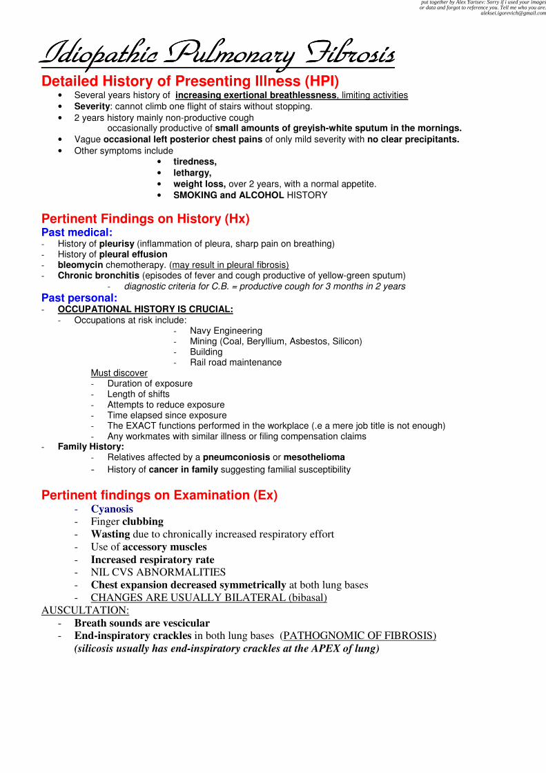
Tests and Investigations Chest X-ray - Example below: Anterior view
- bilateral interstitial shadowing, more pronounced in the bases;
- blunting of costophrenic angles, may be more marked on one side (shown here: Rt side)
chest radiograph may show irregular or linear opacities, usually first found in the lower lung fields.
When the disease is extensive, opacities may be seen in the middle and upper lung fields
Also seen, but not expected:
indistinct heart border
ground glass appearance
***Thickening or calcification along the lower lung fields, the diaphragm, and the cardiac
border characterizes pleural plaques.
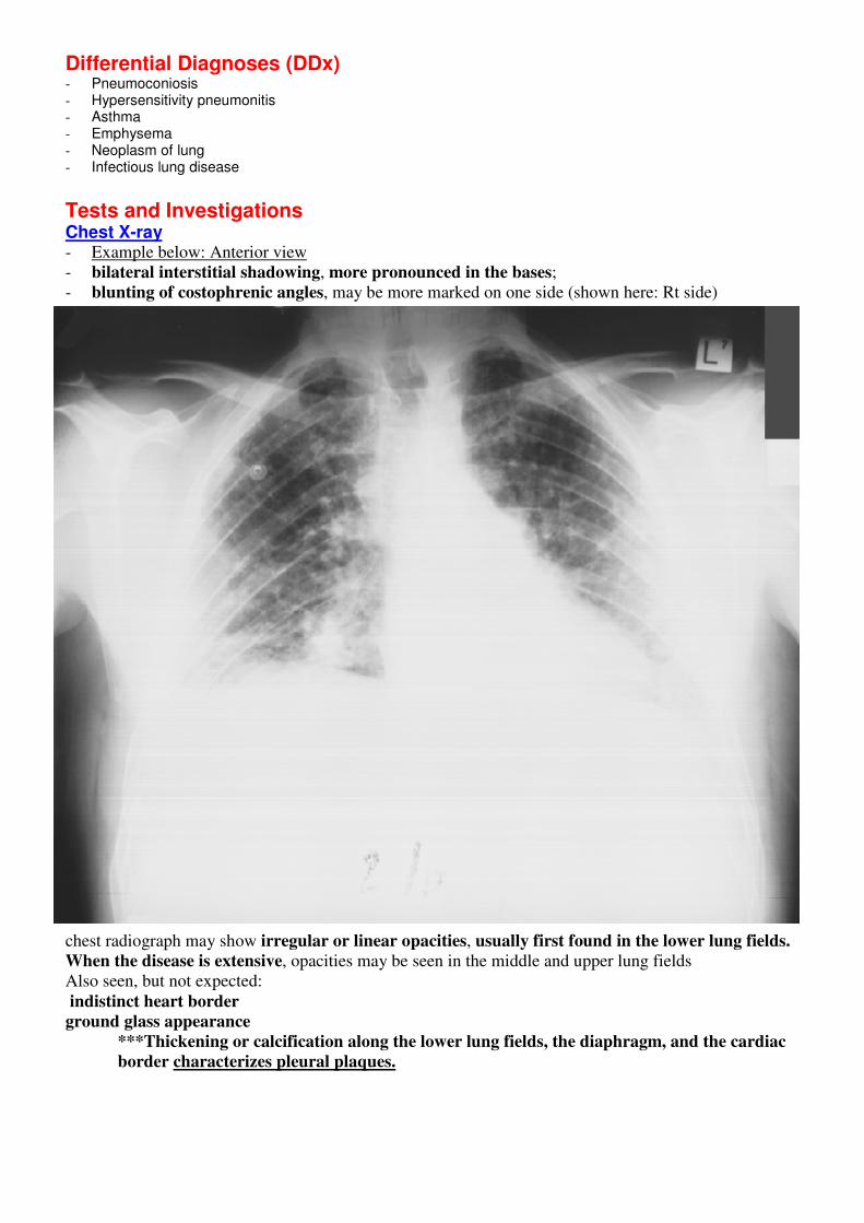
Lateral X-ray of he chest:
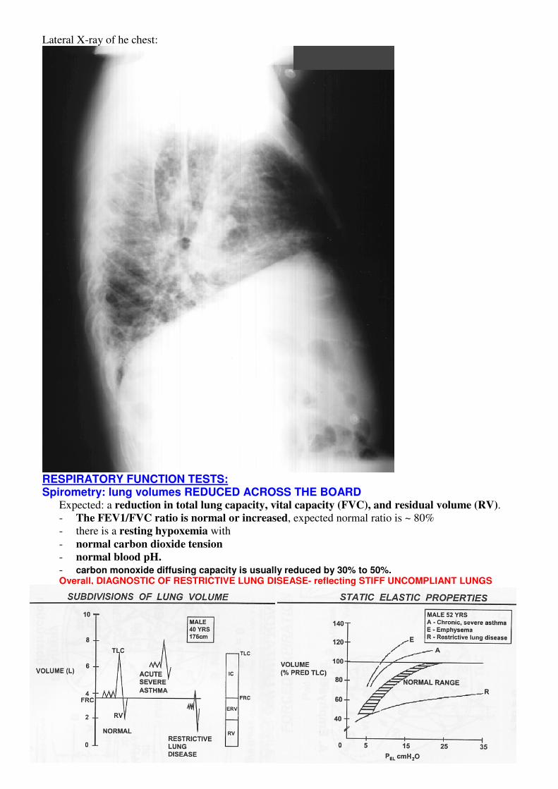
RESPIRATORY FUNCTION TESTS: Spirometry: lung volumes REDUCED ACROSS THE BOARD
Expected: a reduction in total lung capacity, vital capacity (FVC), and residual volume (RV).
- The FEV1/FVC ratio is normal or increased, expected normal ratio is ~ 80%
- there is a resting hypoxemia with
- normal carbon dioxide tension
- normal blood pH.
- carbon monoxide diffusing capacity is usually reduced by 30% to 50%. Overall, DIAGNOSTIC OF RESTRICTIVE LUNG DISEASE- reflecting STIFF UNCOMPLIANT LUNGS
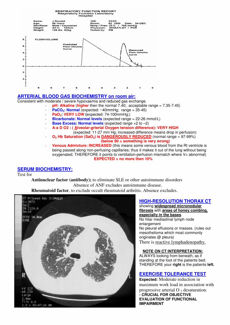
ARTERIAL BLOOD GAS BIOCHEMISTRY on room air: Consistent with moderate / severe hypoxaemia and reduced gas exchange.
- pH: Alkaline (higher then the normal 7.40; acceptable range = 7.35-7.45) - PaCO2: Normal (expected: ~40mmHg; range = 35-45) - PaO2: VERY LOW (expected: 74-100mmHg;) - Bicarbonate: Normal levels (expected range = 22-26 mmol/L) - Base Excess: Normal levels (expected range +2 to –2) - A-a D O2 : ( Alveolar-arterial Oxygen tension difference): VERY HIGH
(expected: 11-27 mm Hg; increased difference means drop in perfusion) - O2 Hb Saturation (SaO2) is DANGEROUSLY REDUCED (normal range = 97-99%)
(below 90 = something is very wrong) - Venous Admixture: INCREASED (this means some venous blood from the Rt ventricle is
being passed along non-perfusing capillaries; thus it makes it out of the lung without being oxygenated; THEREFORE it points to ventilation-perfusion mismatch where V= abnormal)
EXPECTED = no more then 10%
SERUM BIOCHEMISTRY: Test for
Antinuclear factor (antibody); to eliminate SLE or other autoimmune disorders
Absence of ANF excludes autoimmune disease.
Rheumatoid factor, to exclude occult rheumatoid arthritis. Absence excludes.
HIGH-RESOLUTION THORAX CT showing widespread micronodular fibrosis with areas of honey combing, especially in the bases. No hilar mediastinal lymph node enlargement No pleural effusions or masses. (rules out mesothelioma which most commonly originates @ pleura) There is reactive lymphadenopathy.
NOTE ON CT INTERPRETATION:
ALWAYS looking from beneath, as if standing at the foot of the patients bed. THEREFORE your right is the patients left.
EXERCISE TOLERANCE TEST Expected: Moderate reduction in
maximum work load in association with
progressive arterial O 2 desaturation. - CRUCIAL FOR OBJECTIVE EVALUATION OF FUNCTIONAL IMPAIRMENT
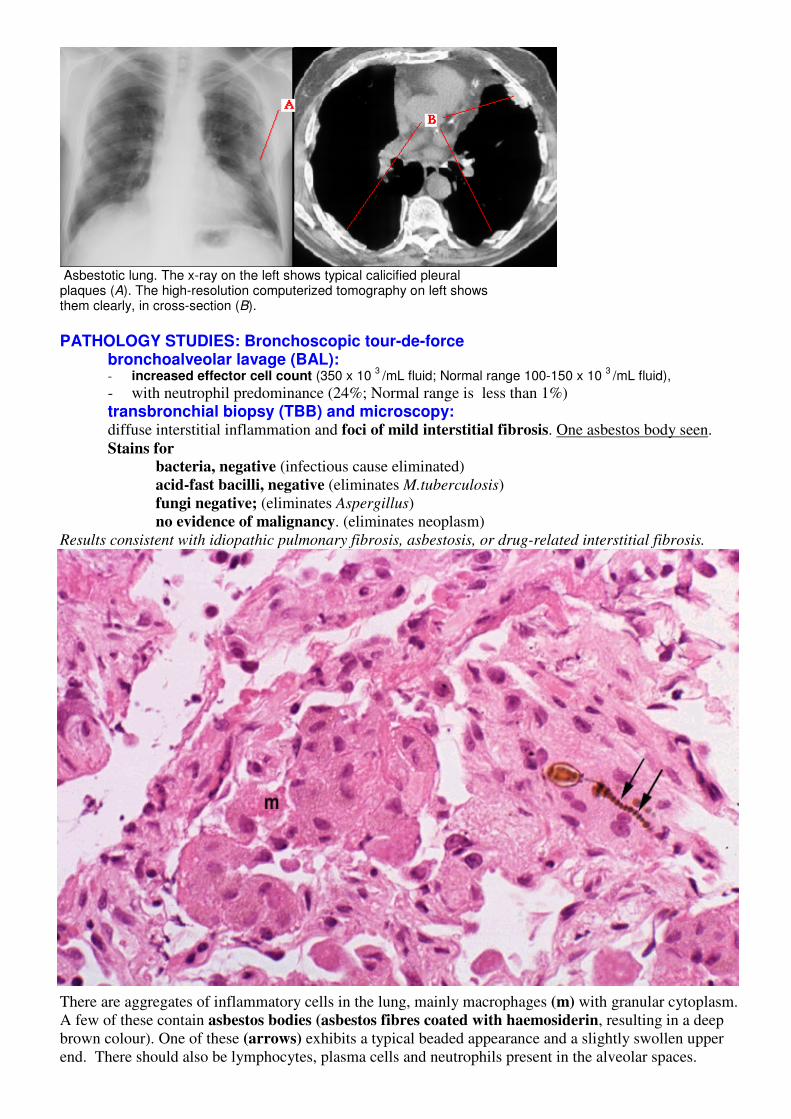
Asbestotic lung. The x-ray on the left shows typical calicified pleural plaques (A). The high-resolution computerized tomography on left shows them clearly, in cross-section (B). PATHOLOGY STUDIES: Bronchoscopic tour-de-force
bronchoalveolar lavage (BAL): - increased effector cell count (350 x 10
3 /mL fluid; Normal range 100-150 x 10
3 /mL fluid),
- with neutrophil predominance (24%; Normal range is less than 1%)
transbronchial biopsy (TBB) and microscopy: diffuse interstitial inflammation and foci of mild interstitial fibrosis. One asbestos body seen.
Stains for bacteria, negative (infectious cause eliminated)
acid-fast bacilli, negative (eliminates M.tuberculosis)
fungi negative; (eliminates Aspergillus)
no evidence of malignancy. (eliminates neoplasm)
Results consistent with idiopathic pulmonary fibrosis, asbestosis, or drug-related interstitial fibrosis.
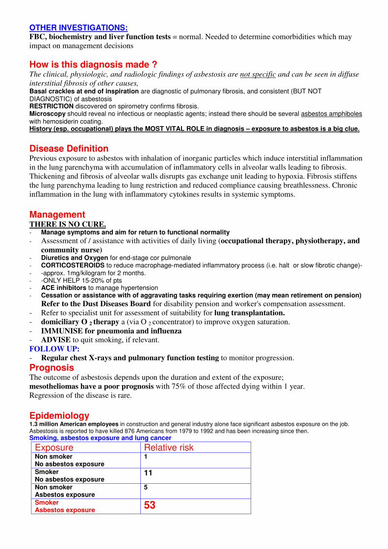
There are aggregates of inflammatory cells in the lung, mainly macrophages (m) with granular cytoplasm.
A few of these contain asbestos bodies (asbestos fibres coated with haemosiderin, resulting in a deep
brown colour). One of these (arrows) exhibits a typical beaded appearance and a slightly swollen upper
end. There should also be lymphocytes, plasma cells and neutrophils present in the alveolar spaces.
OTHER INVESTIGATIONS: FBC, biochemistry and liver function tests = normal. Needed to determine comorbidities which may
impact on management decisions
How is this diagnosis made ? The clinical, physiologic, and radiologic findings of asbestosis are not specific and can be seen in diffuse
interstitial fibrosis of other causes, Basal crackles at end of inspiration are diagnostic of pulmonary fibrosis, and consistent (BUT NOT DIAGNOSTIC) of asbestosis RESTRICTION discovered on spirometry confirms fibrosis. Microscopy should reveal no infectious or neoplastic agents; instead there should be several asbestos amphiboles with hemosiderin coating. History (esp. occupational) plays the MOST VITAL ROLE in diagnosis – exposure to asbestos is a big clue.
Disease Definition Previous exposure to asbestos with inhalation of inorganic particles which induce interstitial inflammation
in the lung parenchyma with accumulation of inflammatory cells in alveolar walls leading to fibrosis.
Thickening and fibrosis of alveolar walls disrupts gas exchange unit leading to hypoxia. Fibrosis stiffens
the lung parenchyma leading to lung restriction and reduced compliance causing breathlessness. Chronic
inflammation in the lung with inflammatory cytokines results in systemic symptoms.
Management THERE IS NO CURE. - Manage symptoms and aim for return to functional normality
- Assessment of / assistance with activities of daily living (occupational therapy, physiotherapy, and
community nurse) - Diuretics and Oxygen for end-stage cor pulmonale - CORTICOSTEROIDS to reduce macrophage-mediated inflammatory process (i.e. halt or slow fibrotic change)- - -approx. 1mg/kilogram for 2 months. - -ONLY HELP 15-20% of pts - ACE inhibitors to manage hypertension - Cessation or assistance with of aggravating tasks requiring exertion (may mean retirement on pension)
Refer to the Dust Diseases Board for disability pension and worker's compensation assessment.
- Refer to specialist unit for assessment of suitability for lung transplantation.
- domiciliary O 2 therapy a (via O 2 concentrator) to improve oxygen saturation.
- IMMUNISE for pneumonia and influenza
- ADVISE to quit smoking, if relevant.
FOLLOW UP:
- Regular chest X-rays and pulmonary function testing to monitor progression.
Prognosis The outcome of asbestosis depends upon the duration and extent of the exposure;
mesotheliomas have a poor prognosis with 75% of those affected dying within 1 year.
Regression of the disease is rare.
Epidemiology 1.3 million American employees in construction and general industry alone face significant asbestos exposure on the job. Asbestosis is reported to have killed 876 Americans from 1979 to 1992 and has been increasing since then. Smoking, asbestos exposure and lung cancer
Exposure Relative risk Non smoker No asbestos exposure
1
Smoker No asbestos exposure
11
Non smoker Asbestos exposure
5
Smoker Asbestos exposure 53
Prevalence of mesothelioma in workers who have had heavy exposure over extended periods is about 2 to 3 % and has been reported to approach 10 %.
More than 80 % of mesotheliomas may be associated with asbestos exposure. Pathophysiology, Aetiology and Pathology INFLAMMATION IN THE LUNG: Most often due to inhaled particles: OBSTACLES to inhalation are
- Nasal conchi (trap particles) - Mucous secretions - Ciliary escalator - Alveolar macrophages
(may either digest the particle, be moved to a bronchiole for clearance by the mucociliary raft or the macrophage may enter the interstitial space, from where it may enter the lymphatics.)
Inflammatory and immune effector cells normally account for <7% of the total lung population They consist of - macrophages (93%), lymphocytes (7%),neutrophils and eosinophils (<1%). - Neutrophil infiltration is usually the result of macrophage activation
(macrophages produce IL-8 which is chemotactic to neutrophils) Size of particles:
- >10 micrometers are deposited in the upper airways, - 3-10 micrometers lodge in the trachea and bronchi, - 1-5 micrometers particles (eg bacteria) can make their way to the alveoli, - smaller particles may remain suspended in air and can be exhaled.
Properties of particles relevant to pathogenesis: - Solubility - Surface Area
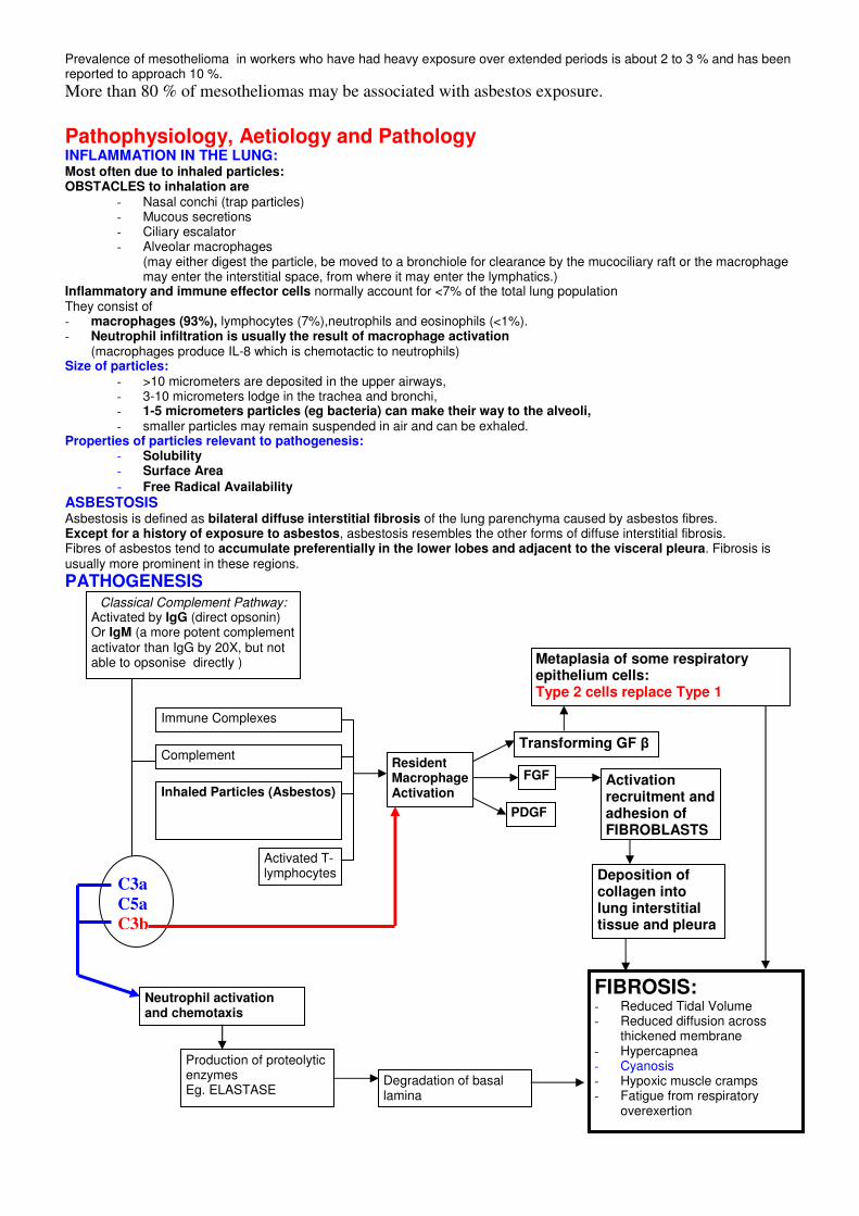
- Free Radical Availability ASBESTOSIS Asbestosis is defined as bilateral diffuse interstitial fibrosis of the lung parenchyma caused by asbestos fibres. Except for a history of exposure to asbestos, asbestosis resembles the other forms of diffuse interstitial fibrosis. Fibres of asbestos tend to accumulate preferentially in the lower lobes and adjacent to the visceral pleura. Fibrosis is usually more prominent in these regions. PATHOGENESIS
Classical Complement Pathway: Activated by IgG (direct opsonin) Or IgM (a more potent complement activator than IgG by 20X, but not able to opsonise directly )
Immune Complexes
Complement
Inhaled Particles (Asbestos)
Activated T-lymphocytes
C3a
C5a
C3b
Resident Macrophage Activation
PDGF
FGF
Transforming GF β
Activation recruitment and adhesion of FIBROBLASTS
FIBROSIS: - Reduced Tidal Volume - Reduced diffusion across
thickened membrane - Hypercapnea - Cyanosis - Hypoxic muscle cramps - Fatigue from respiratory
overexertion
Neutrophil activation and chemotaxis
Production of proteolytic enzymes Eg. ELASTASE
Degradation of basal lamina
Deposition of collagen into lung interstitial tissue and pleura
Metaplasia of some respiratory epithelium cells: Type 2 cells replace Type 1
Physiology
Compliance of the Chest Wall and the Lungs Lung Compliance: how easily the lungs expand under distending pressure.
Lung Elastance is the resistance to this pressure, i.e the tendency of the lungs to collapse like an empty balloon elastic recoil pressure = the distending pressure that must be applied to produce any particular lung and chest wall volume Distending Pressure = transmural pressure = DIFFERENCE BETWEEN ALVEOLAR AND PLEURAL
elastic effects of fibrosis and emphysema. Stiffness Compliance Elastance Restrictive
Fibrosis Increased Decreased Increased Yes
Emphysema Decreased Increased Decreased No
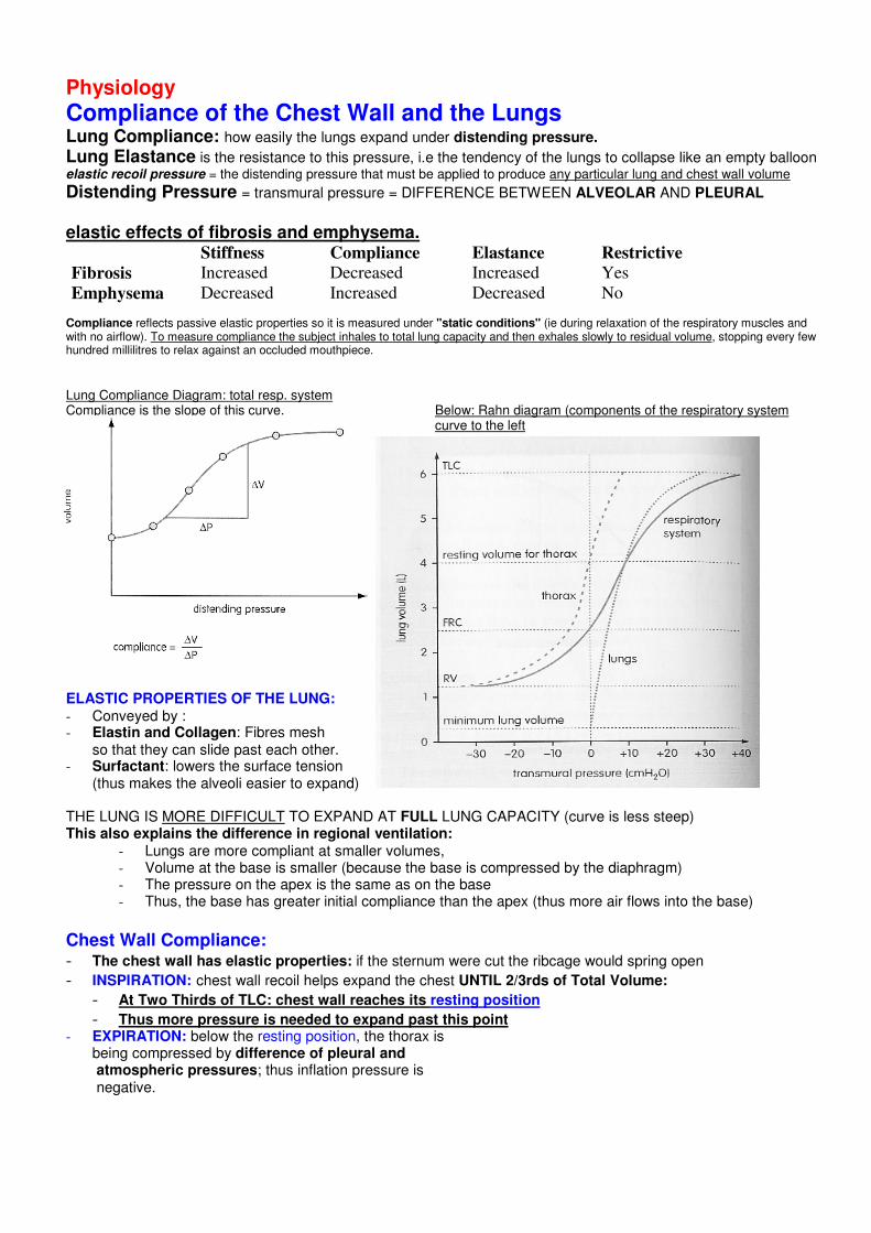
Compliance reflects passive elastic properties so it is measured under "static conditions" (ie during relaxation of the respiratory muscles and with no airflow). To measure compliance the subject inhales to total lung capacity and then exhales slowly to residual volume, stopping every few hundred millilitres to relax against an occluded mouthpiece.
Lung Compliance Diagram: total resp. system Compliance is the slope of this curve. Below: Rahn diagram (components of the respiratory system
curve to the left ELASTIC PROPERTIES OF THE LUNG: - Conveyed by : - Elastin and Collagen: Fibres mesh
so that they can slide past each other. - Surfactant: lowers the surface tension
(thus makes the alveoli easier to expand) THE LUNG IS MORE DIFFICULT TO EXPAND AT FULL LUNG CAPACITY (curve is less steep) This also explains the difference in regional ventilation:
- Lungs are more compliant at smaller volumes, - Volume at the base is smaller (because the base is compressed by the diaphragm) - The pressure on the apex is the same as on the base - Thus, the base has greater initial compliance than the apex (thus more air flows into the base)
Chest Wall Compliance: - The chest wall has elastic properties: if the sternum were cut the ribcage would spring open - INSPIRATION: chest wall recoil helps expand the chest UNTIL 2/3rds of Total Volume:
- At Two Thirds of TLC: chest wall reaches its resting position - Thus more pressure is needed to expand past this point
- EXPIRATION: below the resting position, the thorax is being compressed by difference of pleural and atmospheric pressures; thus inflation pressure is negative.
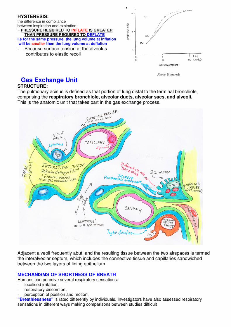
HYSTERESIS: the difference in compliance between inspiration and expiration; = PRESSURE REQUIRED TO INFLATE IS GREATER
THAN PRESSURE REQUIRED TO DEFLATE I.e for the same pressure, the lung volume at inflation will be smaller then the lung volume at deflation
- Because surface tension at the alveolus contributes to elastic recoil
Above: Hysteresis
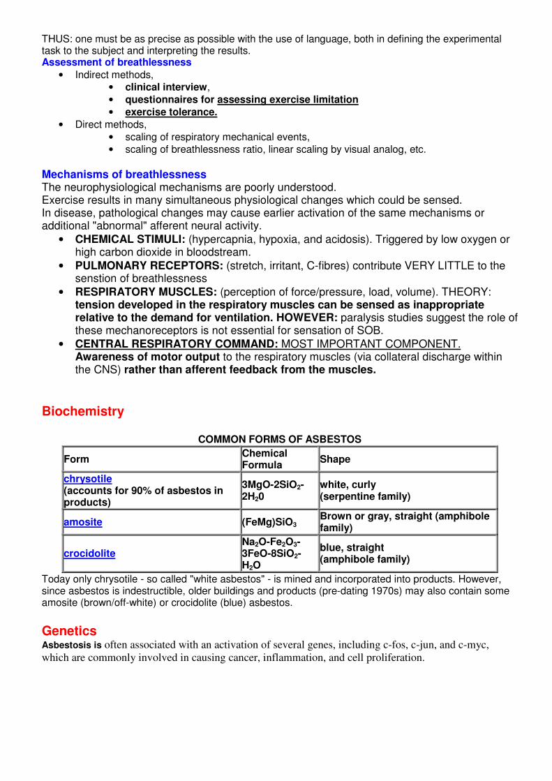
Gas Exchange Unit STRUCTURE: The pulmonary acinus is defined as that portion of lung distal to the terminal bronchiole, comprising the respiratory bronchiole, alveolar ducts, alveolar sacs, and alveoli. This is the anatomic unit that takes part in the gas exchange process.
Adjacent alveoli frequently abut, and the resulting tissue between the two airspaces is termed the interalveolar septum, which includes the connective tissue and capillaries sandwiched between the two layers of lining epithelium. MECHANISMS OF SHORTNESS OF BREATH Humans can perceive several respiratory sensations: - localised irritation, - respiratory discomfort, - perception of position and motion. “Breathlessness” is rated differently by individuals. Investigators have also assessed respiratory sensations in different ways making comparisons between studies difficult
THUS: one must be as precise as possible with the use of language, both in defining the experimental task to the subject and interpreting the results. Assessment of breathlessness
• Indirect methods, • clinical interview, • questionnaires for assessing exercise limitation • exercise tolerance.
• Direct methods, • scaling of respiratory mechanical events, • scaling of breathlessness ratio, linear scaling by visual analog, etc.
Mechanisms of breathlessness The neurophysiological mechanisms are poorly understood. Exercise results in many simultaneous physiological changes which could be sensed. In disease, pathological changes may cause earlier activation of the same mechanisms or additional "abnormal" afferent neural activity.
• CHEMICAL STIMULI: (hypercapnia, hypoxia, and acidosis). Triggered by low oxygen or high carbon dioxide in bloodstream.
• PULMONARY RECEPTORS: (stretch, irritant, C-fibres) contribute VERY LITTLE to the senstion of breathlessness
• RESPIRATORY MUSCLES: (perception of force/pressure, load, volume). THEORY: tension developed in the respiratory muscles can be sensed as inappropriate relative to the demand for ventilation. HOWEVER: paralysis studies suggest the role of these mechanoreceptors is not essential for sensation of SOB.
• CENTRAL RESPIRATORY COMMAND: MOST IMPORTANT COMPONENT. Awareness of motor output to the respiratory muscles (via collateral discharge within the CNS) rather than afferent feedback from the muscles.
Biochemistry
COMMON FORMS OF ASBESTOS
Form Chemical Formula
Shape
chrysotile (accounts for 90% of asbestos in products)
3MgO-2SiO2-2H20
white, curly (serpentine family)
amosite (FeMg)SiO3 Brown or gray, straight (amphibole family)
crocidolite Na2O-Fe2O3-3FeO-8SiO2-H2O
blue, straight (amphibole family)
Today only chrysotile - so called "white asbestos" - is mined and incorporated into products. However, since asbestos is indestructible, older buildings and products (pre-dating 1970s) may also contain some amosite (brown/off-white) or crocidolite (blue) asbestos. Genetics Asbestosis is often associated with an activation of several genes, including c-fos, c-jun, and c-myc,
which are commonly involved in causing cancer, inflammation, and cell proliferation.
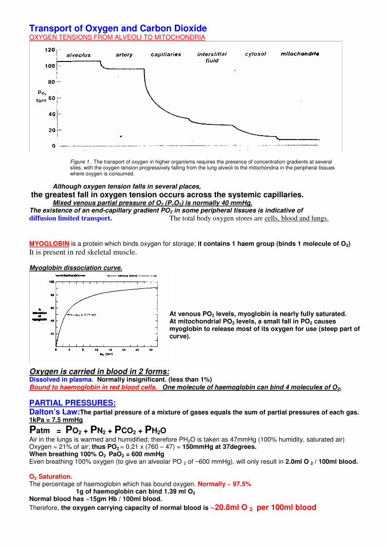
Transport of Oxygen and Carbon Dioxide OXYGEN TENSIONS FROM ALVEOLI TO MITOCHONDRIA
Figure 1. The transport of oxygen in higher organisms requires the presence of concentration gradients at several sites, with the oxygen tension progressively falling from the lung alveoli to the mitochondria in the peripheral tissues where oxygen is consumed.
Although oxygen tension falls in several places,
the greatest fall in oxygen tension occurs across the systemic capillaries. Mixed venous partial pressure of O2 (PvO2) is normally 40 mmHg.
The existence of an end-capillary gradient PO2 in some peripheral tissues is indicative of
diffusion limited transport. The total body oxygen stores are cells, blood and lungs.
MYOGLOBIN is a protein which binds oxygen for storage; it contains 1 haem group (binds 1 molecule of O2)
It is present in red skeletal muscle. Myoglobin dissociation curve.
At venous PO2 levels, myoglobin is nearly fully saturated. At mitochondrial PO2 levels, a small fall in PO2 causes myoglobin to release most of its oxygen for use (steep part of curve).
Oxygen is carried in blood in 2 forms: Dissolved in plasma. Normally insignificant. (less than 1%) Bound to haemoglobin in red blood cells. One molecule of haemoglobin can bind 4 molecules of O2.
PARTIAL PRESSURES: Dalton’s Law:The partial pressure of a mixture of gases equals the sum of partial pressures of each gas. 1kPa = 7.5 mmHg
Patm = PO2 + PN2 + PCO2 + PH2O Air in the lungs is warmed and humidified; therefore PH2O is taken as 47mmHg (100% humidity, saturated air) Oxygen = 21% of air; thus PO2 = 0.21 x (760 – 47) = 150mmHg at 37degrees. When breathing 100% O2 PaO2 = 600 mmHg Even breathing 100% oxygen (to give an alveolar PO 2 of ~600 mmHg), will only result in 2.0ml O 2 / 100ml blood. O2 Saturation. The percentage of haemoglobin which has bound oxygen. Normally ~ 97.5%
1g of haemoglobin can bind 1.39 ml O2
Normal blood has ~15gm Hb / 100ml blood.
Therefore, the oxygen carrying capacity of normal blood is ~20.8ml O 2 per 100ml blood
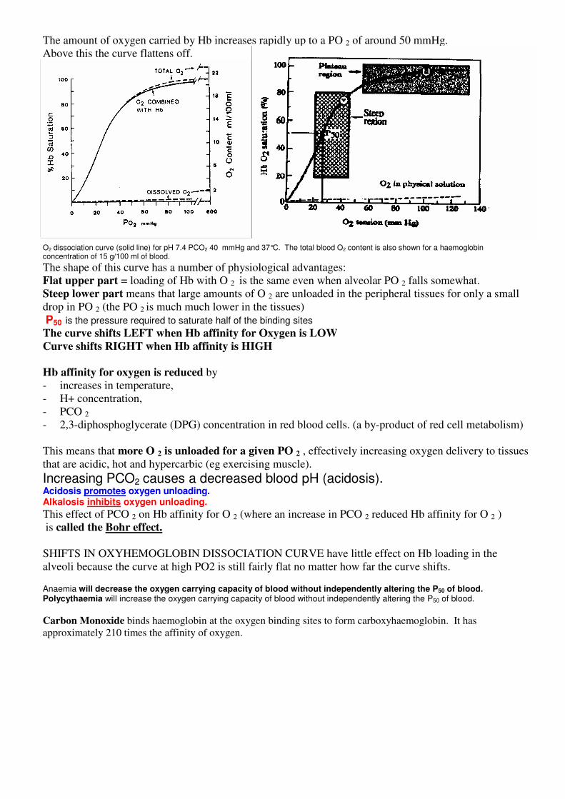
The amount of oxygen carried by Hb increases rapidly up to a PO 2 of around 50 mmHg.
Above this the curve flattens off.
O2 dissociation curve (solid line) for pH 7.4 PCO2 40 mmHg and 37°C. The total blood O2 content is also shown for a haemoglobin concentration of 15 g/100 ml of blood.
The shape of this curve has a number of physiological advantages:
Flat upper part = loading of Hb with O 2 is the same even when alveolar PO 2 falls somewhat.
Steep lower part means that large amounts of O 2 are unloaded in the peripheral tissues for only a small
drop in PO 2 (the PO 2 is much much lower in the tissues)
P50 is the pressure required to saturate half of the binding sites
The curve shifts LEFT when Hb affinity for Oxygen is LOW
Curve shifts RIGHT when Hb affinity is HIGH
Hb affinity for oxygen is reduced by
- increases in temperature,
- H+ concentration,
- PCO 2
- 2,3-diphosphoglycerate (DPG) concentration in red blood cells. (a by-product of red cell metabolism)
This means that more O 2 is unloaded for a given PO 2 , effectively increasing oxygen delivery to tissues
that are acidic, hot and hypercarbic (eg exercising muscle).
Increasing PCO2 causes a decreased blood pH (acidosis). Acidosis promotes oxygen unloading. Alkalosis inhibits oxygen unloading. This effect of PCO 2 on Hb affinity for O 2 (where an increase in PCO 2 reduced Hb affinity for O 2 )
is called the Bohr effect.
SHIFTS IN OXYHEMOGLOBIN DISSOCIATION CURVE have little effect on Hb loading in the
alveoli because the curve at high PO2 is still fairly flat no matter how far the curve shifts. Anaemia will decrease the oxygen carrying capacity of blood without independently altering the P50 of blood. Polycythaemia will increase the oxygen carrying capacity of blood without independently altering the P50 of blood.
Carbon Monoxide binds haemoglobin at the oxygen binding sites to form carboxyhaemoglobin. It has
approximately 210 times the affinity of oxygen.
CARBON DIOXIDE STORES
Carbon dioxide is carried in the plasma in two forms.
Dissolved CO2. At PvCO2 = 45 mmHg the dissolved CO2 concentration is 3.4 mL/dL. Carbamino compounds. Plasma protein concentration is about 7%. CO2 binds the amine groups of plasma proteins to form carbamino compounds. The hydrogen ions formed are buffered by plasma proteins.
R - NH CO R - NH - COO H2 2
- ++ ⇔ +
Plasma has little carbonic anhydrase so CO2 forms little carbonic acid in plasma. Carbon dioxide is carried by the red blood cell in three forms.
Dissolved CO2. CO2 can cross the red cell membrane and dissolve in RBC water. Carbamino compounds. Approximately 30% of RBC contents is haemoglobin. CO2 can form carbamino haemoglobin on amine groups. The H
+ released by this reaction is buffered by histidine
residues (imidazole group) on the haemoglobin itself. Bicarbonate. Carbonic anhydrase is present in RBCs and catalyze the formation of carbonic acid which dissociated to hydrogen ion and bicarbonate. The H
+ is buffered by haemoglobin.
CO2
H2
O Carbonic Anhydrase
H2
CO3
3-HCO H+ ← → ← → +
+
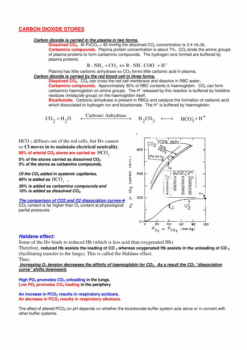
HCO 3 diffuses out of the red cells, but H+ cannot
so Cl moves in to maintain electrical neutrality.
90% of arterial CO2 stores are carried as HCO3−
5% of the stores carried as dissolved CO2
5% of the stores as carbamino compounds. Of the CO2 added in systemic capillaries,
60% is added as HCO3−
,
30% is added as carbamino compounds and 10% is added as dissolved CO2. The comparison of CO2 and O2 dissociation curves���� CO2 content is far higher than O2 content at physiological partial pressures.
Haldane effect: Some of the H+ binds to reduced Hb (which is less acid than oxygenated Hb).
Therefore, reduced Hb assists the loading of CO 2 whereas oxygenated Hb assists in the unloading of CO 2
(facilitating transfer to the lungs). This is called the Haldane effect.
Thus: Increasing O2 tension decreases the affinity of haemoglobin for CO2. As a result the CO2 “dissociation curve” shifts downward.
High PO2 promotes CO2 unloading in the lungs. Low PO2 promotes CO2 loading in the periphery An increase in PCO2 results in respiratory acidosis. An decrease in PCO2 results in respiratory alkalosis.
The effect of altered PCO2 on pH depends on whether the bicarbonate buffer system acts alone or in concert with other buffer systems.
IN BODY FLUIDS: CSF. The bicarbonate buffer system works alone and PCO2 has a larger effect on pH. Blood. Haemoglobin buffers H
+ changes in addition to HCO3
-. Thus, changes in PCO2 have somewhat blunted effect on pH.
The lungs excrete 100 times more acid than the kidneys each day
pH and the Arterial Blood Gases ABG analysis measures the partial pressures of O 2 and CO 2 in arterial blood. (not quantity- pressure!)
Optimally a sample volume of 2.5 to 3 mls is required in the adult.
Usually taken from a peripheral artery (eg radial, brachial, femoral),
Taken when the patient
- is relaxed at the time of the procedure
- is in a steady state (usually at rest)
- has been inspiring a constant, known level of oxygen for at least fifteen to twenty minutes.
Use local anaesthetic to prevent hyperventilation
The blood is collected into a pre-heparinised syringe that is sealed air-tight after sampling. The sample is
transported (on ice) immediately for laboratory analysis.
The concentration or fraction of oxygen in the inspired air (FiO 2 ) should be noted when the ABG
sample is collected (eg room air = 0.21), remembering that the FiO 2 delivered by nasal prongs or a mask
apparatus is unreliable and non-constant.
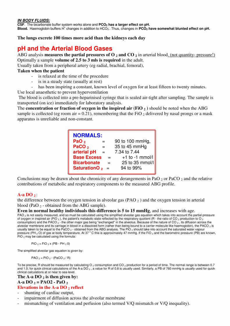
NORMALS: PaO 2 = 90 to 100 mmHg, PaCO 2 = 35 to 45 mmHg arterial pH = 7.34 to 7.44 Base Excess = +1 to -1 mmol/l Bicarbonate = 25 to 35 mmol/l SaturationO 2 = 94 to 99%
Conclusions may be drawn about the chronicity of any derangements in PaO 2 or PaCO 2 and the relative
contributions of metabolic and respiratory components to the measured ABG profile.
A-a DO 2 :
the difference between the oxygen tension in alveolar gas (PAO 2 ) and the oxygen tension in arterial
blood (PaO 2 - obtained from the ABG sample).
Even in normal healthy individuals this difference is 5 to 15 mmHg, and increases with age. PAO 2 is not easily measured, and so must be calculated using the simplified alveolar gas equation which takes into account the partial pressure of oxygen in inspired air (PiO 2 ), the patient's metabolic state reflected by the respiratory quotient (R - the ratio of CO 2 production to O 2 consumption) and the PACO 2 - the other major gas being "exchanged" in the alveolus. Because of the nature of CO 2 , its diffusion across the alveolar membrane and its carriage in blood in a dissolved form (rather than being bound to a carrier molecule like haemoglobin), the PACO 2 is usually taken to be equal to the PaCO 2 - obtained from the ABG analysis. The PiO 2 should take into account the saturated water vapour pressure (PH 2 O) of gas at body temperature. At 37 o C this is approximately 47 mmHg. If the FiO 2 and the barometric pressure (PB) are known, PiO 2 may be calculated using the formula: PiO 2 = FiO 2 x (PB - PH 2 0) The simplified alveolar gas equation is given by:
PAO 2 = PiO 2 - (PaCO 2 / R) To be precise, R should be measured by calculating O 2 consumption and CO 2 production for a period of time. The normal range is between 0.7 and 1.0; for quick clinical calculations of the A-a DO 2 , a value for R of 0.8 is usually used. Similarly, a PB of 760 mmHg is usually used for quick clinical calculations at or near to sea-level. The A-a DO 2 is then given by:
A-a DO 2 = PAO2 - PaO 2
Elevations in the A-a DO 2 reflect
- shunting of cardiac output,
- impairment of diffusion across the alveolar membrane
- mismatching of ventilation and perfusion (also termed V/Q mismatch or V/Q inequality).
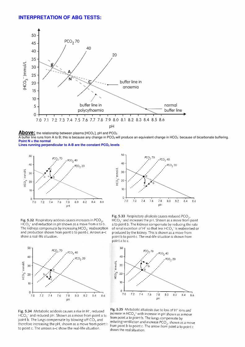
INTERPRETATION OF ABG TESTS:
Above: the relationship between plasma [HCO3-], pH and PCO2
A buffer line runs from A to B; this is because any change in PCO2 will produce an equivalent change in HCO3- because of bicarbonate buffering.
Point N = the normal Lines running perpendicular to A-B are the constant PCO2 levels
Pathology Causes of Fibrotic Lung Disease this is a group of diseases of lung parenchyma which may result in pulmonary fibrosis clinicians recognise ILD as a syndrome with the following clinical features: - exertional dyspnoea - cough - rapid shallow breathing pattern - bilateral coarse crackles on auscultation. - bilateral interstitial infiltrates on chest x-ray and high resolution CT scan (reticulo-nodular or ground glass
patterns) - RESTRICTION defect (reduction in all lung volumes, normal or high FEV1/VC ratio) - impaired gas exchange (hypoxaemia, V/Q mismatching at rest) - clubbing and development of right heart failure (late signs) - reduced DLCO (diffusing capacity
- histopathologic features of inflammation and fibrosis of the pulmonary parenchyma on biopsy. FIBROTIC LUNG DISEASE IS BROADLY CLASSIFIED INTO SIX GROUPS:
1. Idiopathic pulmonary fibrosis (cryptogenic fibrosing alveolitis) 2. Granulomatous diseases
• Unknown cause • Sarcoid • Histiocytosis-X • Known causes • Hypersensitivity pneumonitis (extrinsic allergic alveolitis)
3. Collagen-vascular/connective tissue disease
• Scleroderma, rheumatoid arthritis 4. Inhalational Causes
• Occupational • Asbestosis • Silicosis • Coal-workers pneumoconiosis • Environmental gases and fumes
5. Inherited Causes
• Tuberous Sclerosis, Neurofibromatosis 6. Other Specific Entities
• Drug Induced eg chemotherapeutic agents (including Bleomycin), nitrofurantoin, methysergide • Alveolar proteinosis • Lymphangitic carcinomatosis • Idiopathic pulmonary haemosiderosis • Eosinophilic lung diseases.
These diseases are ‘restrictive’ lung diseases – ie no airway obstruction (and no wheeze) - total surface area of gas exchange is reduced hence symptoms
- dyspnea - tachypnea - shallow breathing - eventual cyanosis and cardiac sequelae of chronic hypoxia
(notably secondary pulmonary hypertension and right-sided heart failure (cor pulmonale)
Diffuse interstitial lung disease is most commonly of environmental aetiology (25%), sarcoidosis (20%), idiopathic pulmonary fibrosis (15%) collagen vascular diseases (10%).
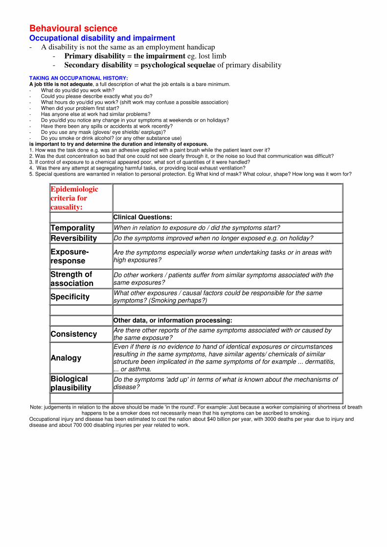
Behavioural science Occupational disability and impairment - A disability is not the same as an employment handicap
- Primary disability = the impairment eg. lost limb
- Secondary disability = psychological sequelae of primary disability TAKING AN OCCUPATIONAL HISTORY: A job title is not adequate, a full description of what the job entails is a bare minimum. - What do you/did you work with? - Could you please describe exactly what you do? - What hours do you/did you work? (shift work may confuse a possible association) - When did your problem first start? - Has anyone else at work had similar problems? - Do you/did you notice any change in your symptoms at weekends or on holidays? - Have there been any spills or accidents at work recently? - Do you use any mask (gloves/ eye shields/ earplugs)? - Do you smoke or drink alcohol? (or any other substance use) is important to try and determine the duration and intensity of exposure. 1. How was the task done e.g. was an adhesive applied with a paint brush while the patient leant over it? 2. Was the dust concentration so bad that one could not see clearly through it, or the noise so loud that communication was difficult? 3. If control of exposure to a chemical appeared poor, what sort of quantities of it were handled? 4. Was there any attempt at segregating harmful tasks, or providing local exhaust ventilation? 5. Special questions are warranted in relation to personal protection. Eg What kind of mask? What colour, shape? How long was it worn for?
Epidemiologic
criteria for
causality:
Clinical Questions:
Temporality When in relation to exposure do / did the symptoms start?
Reversibility Do the symptoms improved when no longer exposed e.g. on holiday?
Exposure-response
Are the symptoms especially worse when undertaking tasks or in areas with high exposures?
Strength of association
Do other workers / patients suffer from similar symptoms associated with the same exposures?
Specificity What other exposures / causal factors could be responsible for the same symptoms? (Smoking perhaps?)
Other data, or information processing:
Consistency Are there other reports of the same symptoms associated with or caused by the same exposure?
Analogy
Even if there is no evidence to hand of identical exposures or circumstances resulting in the same symptoms, have similar agents/ chemicals of similar structure been implicated in the same symptoms of for example ... dermatitis, ... or asthma.
Biological plausibility
Do the symptoms 'add up' in terms of what is known about the mechanisms of disease?
Note: judgements in relation to the above should be made 'in the round'. For example: Just because a worker complaining of shortness of breath happens to be a smoker does not necessarily mean that his symptoms can be ascribed to smoking.
Occupational injury and disease has been estimated to cost the nation about $40 billion per year, with 3000 deaths per year due to injury and disease and about 700 000 disabling injuries per year related to work.
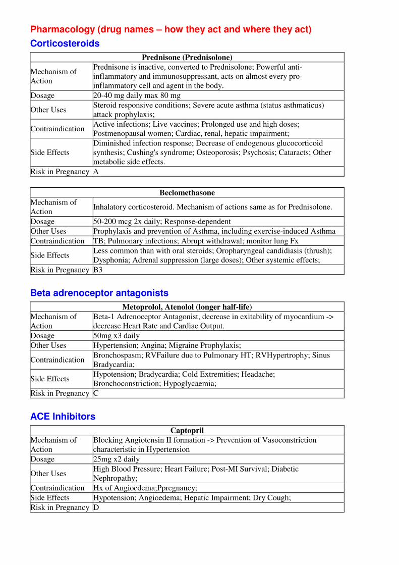
Pharmacology (drug names – how they act and where they act)
Corticosteroids
Prednisone (Prednisolone)
Mechanism of
Action
Prednisone is inactive, converted to Prednisolone; Powerful anti-
inflammatory and immunosuppressant, acts on almost every pro-
inflammatory cell and agent in the body.
Dosage 20-40 mg daily max 80 mg
Other Uses Steroid responsive conditions; Severe acute asthma (status asthmaticus)
attack prophylaxis;
Contraindication Active infections; Live vaccines; Prolonged use and high doses;
Postmenopausal women; Cardiac, renal, hepatic impairment;
Side Effects
Diminished infection response; Decrease of endogenous glucocorticoid
synthesis; Cushing's syndrome; Osteoporosis; Psychosis; Cataracts; Other
metabolic side effects.
Risk in Pregnancy A
Beclomethasone
Mechanism of
Action Inhalatory corticosteroid. Mechanism of actions same as for Prednisolone.
Dosage 50-200 mcg 2x daily; Response-dependent
Other Uses Prophylaxis and prevention of Asthma, including exercise-induced Asthma
Contraindication TB; Pulmonary infections; Abrupt withdrawal; monitor lung Fx
Side Effects Less common than with oral steroids; Oropharyngeal candidiasis (thrush);
Dysphonia; Adrenal suppression (large doses); Other systemic effects;
Risk in Pregnancy B3
Beta adrenoceptor antagonists
Metoprolol, Atenolol (longer half-life)
Mechanism of
Action
Beta-1 Adrenoceptor Antagonist, decrease in exitability of myocardium ->
decrease Heart Rate and Cardiac Output.
Dosage 50mg x3 daily
Other Uses Hypertension; Angina; Migraine Prophylaxis;
Contraindication Bronchospasm; RVFailure due to Pulmonary HT; RVHypertrophy; Sinus
Bradycardia;
Side Effects Hypotension; Bradycardia; Cold Extremities; Headache;
Bronchoconstriction; Hypoglycaemia;
Risk in Pregnancy C
ACE Inhibitors
Captopril
Mechanism of
Action
Blocking Angiotensin II formation -> Prevention of Vasoconstriction
characteristic in Hypertension
Dosage 25mg x2 daily
Other Uses High Blood Pressure; Heart Failure; Post-MI Survival; Diabetic
Nephropathy;
Contraindication Hx of Angioedema;Ppregnancy;
Side Effects Hypotension; Angioedema; Hepatic Impairment; Dry Cough;
Risk in Pregnancy D
Recommended