Hemodynamic Rounds
The Science Behind Percutaneous HemodynamicSupport: A Review and Comparison of Support
Strategies
Daniel Burkhoff,1* MD, PhD, and Srihari S. Naidu,2 MD
Patients in a variety of cardiovascular disease states may benefit from temporary percu-taneous cardiac support, including those in acute decompensated heart failure, fulmi-nant myocarditis, acute myocardial infarction with or without cardiogenic shock andthose undergoing high-risk percutaneous coronary intervention. The ideal percutaneouscardiac support device is safe, easy to use and versatile enough to meet the needs of var-ious clinical situations and patient cohorts. In addition, it should provide maximal hemo-dynamic support and protection against myocardial ischemia. With these goals in mind,the scientific principles that govern hemodynamic effectiveness and myocardial protec-tion as they pertain to acute support devices are reviewed. VC 2012 Wiley Periodicals, Inc.
Key words: hemodynamics; support; ventricular assist device; ischemia
CLINICAL INDICATIONS FOR PERCUTANEOUSCARDIAC SUPPORT
The practice of interventional cardiology is movingtoward aggressive therapy for higher-risk and complexpatient subsets to improve cardiac function, quality oflife and overall survival. As a result, the field of percu-taneous cardiac support devices has evolved rapidly.Three populations appear most likely to benefit fromsuch devices: (1) those in need of high-risk percutane-ous coronary intervention (PCI), (2) those with acutemyocardial infarction with or without cardiogenicshock, to reduce infarct size and support end-organ per-fusion, and (3) those with acute decompensated heartfailure, be it due to acute coronary syndrome, myocar-ditis or exacerbation of a chronic heart failure state.Clinical indications for percutaneous cardiac assist de-vice placement may therefore be grouped according towhether device placement occurs electively (high-riskPCI), urgently (acute myocardial infarction or mild tomoderate decompensated heart failure), or emergently(cardiogenic shock due to acute myocardial infarctionor severe decompensated heart failure).
Recently completed clinical trials such as the syn-ergy between PCI with Taxus and cardiac surgery(SYNTAX) trial, as well as the upcoming evaluationof Xience prime versus coronary artery bypass surgeryfor effectiveness of left main revascularization
(EXCEL), suggest that PCI may become more fre-quently utilized in the setting of high-risk multivesseland/or unprotected left main disease [1,2]. Updatedguidelines by the American College of Cardiology andthe American Heart Association support left main PCIin patients in whom the risks and benefits of a percuta-neous approach appear as good, if not better, than cor-onary bypass surgery [3]. With the aging population,many patients with coronary artery disease will havehemodynamic instability, severe reductions in left ven-tricular function, or other comorbidities that elevate
1Division of Cardiology, Columbia University School of Medicine,New York, New York2Division of Cardiology, Winthrop University Hospital, Mineola,New York
Conflict of interest: Dr. Burkhoff has an educational grant
(Abiomed), and Circulite employee. Dr. Naidu is a consultant for
Abiomed.
*Correspondence to: Daniel Burkhoff, MD, PhD, Division of Cardi-
ology, Columbia University, New York, NY 10032.
E-mail: [email protected]
Received 2 September 2011; Revision accepted 10 March 2012
DOI 10.1002/ccd.24421
Published online in Wiley Online Library (wiley
onlinelibrary.com)
VC 2012 Wiley Periodicals, Inc.
Catheterization and Cardiovascular Interventions 00:000–000 (2012)
risk of both PCI and open-heart surgery. In suchpatients, PCI is often the most appropriate option, de-spite associated procedural and in-hospital risk. Cardiacassist devices have been increasingly called upon tominimize procedural risk, facilitate single- or multives-sel PCI and improve short- and long-term outcome.
Patients with acute myocardial infarction with orwithout cardiogenic shock, a high-risk subset forin-hospital mortality, form another group in whom per-cutaneous cardiac assist devices may be increasinglyutilized. Such patients are increasingly offered primaryPCI, as supported by current guideline recommenda-tions and clinical trials, regardless of age or comorbid-ity [3,4]. While associated in-hospital mortality hasimproved in these patients, few treatments other thanearly revascularization have shown potential to reduceinfarct size. In addition, those with cardiogenic shockremain at elevated risk for both 30-day and 6-monthmortality despite early revascularization and are often-times left with severe ventricular and end-organ dys-function [5]. By facilitating emergent revascularization,maintaining end-organ perfusion and favorably impact-ing myocardial oxygen consumption, cardiac assistdevices may both preserve cardiac function andimprove survival.
Finally, patients with acute decompensated heartfailure, either so-called ‘‘acute-on-chronic failure’’ inthose with previous cardiac dysfunction, or acute ful-minant decompensation, as in acute myocarditis,remain without good medical options to prevent wor-sening heart failure (WHF), progression to cardiogenicshock, or death [6]. Inotrope therapy, while beneficialin supporting the hemodynamic state acutely [7], isassociated with worse morbidity and mortality, makinginvasive options such as cardiac assist devices poten-tially more palatable [8,9]. Indeed, the ability to restthe failing heart while maintaining peripheral perfusionwithout initiation or escalation of inotropic therapy hasthe potential to improve mortality in these patientswhile preserving cardiac function.
In each of these settings, the goals of cardiac supportare distinctly different. For the elective setting, thegoal is primarily to bridge a stable hemodynamic statethrough a complex interventional procedure; in essenceto allow the cardiovascular system to weather transientderangements and resume normal function immediatelypostprocedure or shortly thereafter. In addition, theideal device should alter myocardial ischemic thresholdto allow time for complex PCI and any associated pro-cedural complications to resolve, such as distal emboli-zation or coronary dissection. In contrast, in the urgentand emergent settings, the goal is often to take overthe work, partly or wholly, of a struggling heart, mini-mize ongoing ischemic damage (especially in acute
myocardial infarction), and promote a stable hemody-namic state of systemic pressure and perfusion withoutthe need for deleterious vasopressors and inotropes [8–12]. By resting the heart and simultaneously ensuringend-organ perfusion, the patient returns to an autono-mous cardiovascular state with minimal decline in car-diac or end-organ function and, potentially, improvedsurvival.
The goal of this review is to summarize the basichemodynamic principles that form a basis for under-standing and comparing the mechanisms by which cur-rently available percutaneous support devices providecirculatory support and their impact on myocardialenergetics. Issues related to ease-of-use and safety willalso be discussed as substantiated by data in the litera-ture. The primary devices to be reviewed include theintraaortic balloon pump (IABP), extracorporeal cardio-pulmonary support (CPS or ECMO), left atrial-to-arte-rial pumping (e.g., TandemHeart, Cardiac Assist) andintracorporeal transvalvular ventricular-to-aortic pump-ing (e.g., Impella, Abiomed).
GOALS OF PERCUTANEOUS CARDIAC SUPPORT
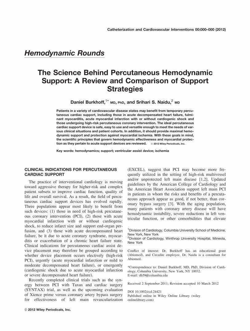
From a clinical perspective, the ideal percutaneouscardiac support strikes an optimal balance betweensafety and efficacy, while maintaining adaptability tovarious clinical situations and patient cohorts. Indeed,the ideal device could be initiated rapidly using basicinvasive cardiology techniques and would cause novascular or other complications. In addition, it wouldprovide normal or supra-normal levels of cardiac out-put and blood pressure (hemodynamic) support whilereducing pulmonary capillary wedge pressure to nor-mal, thus providing optimal conditions for maintainingperfusion of all vital organs (Fig. 1).
Principles of Hemodynamic Support
The basic goal of hemodynamic support is to pro-duce a stable and physiologically acceptable bloodpressure, cardiac output, and pulmonary venous pres-sure. When these goals are attained, end-organ perfu-sion is maintained, blood can be adequately oxygen-ated by the lungs and diuresis is promoted in states ofvolume overload. While also relevant to high-risk PCI,the ability to achieve and maintain adequate hemody-namic support has traditionally been most appreciatedfor patients presenting with low cardiac output acuteheart failure, acute myocardial infarction, and cardio-genic shock.
Vasopressor and inotropic therapies have tradition-ally been utilized as first line treatment for hemody-namic support [13,14]. However, while shown to
2 Burkhoff and Naidu
Catheterization and Cardiovascular Interventions DOI 10.1002/ccd.Published on behalf of The Society for Cardiovascular Angiography and Interventions (SCAI).
increase blood pressure and cardiac output, the impacton end organ perfusion can be variable (depending onthe degree of peripheral vasoconstriction) and there arewell-established adverse effects on the heart itself [8–12,15,16]. Indeed, the combination of multiple agentsappears to be associated with worse outcome [16]. Partof the problem may be a lack of understanding whenthe goals of hemodynamic support have been reached,as under-reaching or overstepping such goals may bepartly responsible for the higher morbidity and mortal-ity associated with these agents. To this end, heavyreliance has typically been placed on the pulmonary ar-tery catheter (PAC) to titrate medication dosage to var-ious hemodynamic parameters, including cardiac output(CO), pulmonary capillary wedge pressure and sys-temic vascular resistance. Yet, despite this, multiplestudies have shown little to no benefit in PAC-guidedtherapy in such patients [17–19].
Most recently, prognostic information of cardiacpower output (CPO) has been explored. Defined as thecardiac output multiplied by the mean arterial pressure(divided by 451 to convert to units of Watts), the pa-rameter takes into account the ability of the heart togenerate systemic flow and blood pressure, thus provid-ing a target for optimizing hemodynamic support [20].Such a parameter is congruent with collective experi-ence, which had suggested that cardiac output is neces-sary but not sufficient for end-organ perfusion;adequate mean arterial pressure is also required.
Multiple studies have now corroborated the inde-pendent association and predictive power of CPO onmortality in various severe cardiac dysfunctional states,including myocardial infarction-related cardiogenicshock, ischemic and nonischemic cardiomyopathy andacute myocarditis [20–22]. Further, CPO was able topredict worsening heart failure (WHF) in patients pre-senting with milder degrees of acute heart failure aswell as those nearing cardiogenic shock [6]. In con-trast, CO and the other more traditional hemodynamicparameters did not show independent associations withmortality [22]. Studies have also now established acut-point CPO associated with a reduced incidence ofworsening heart failure and mortality in high-riskpatients. In those admitted with acute heart failure, forexample, a CPO < 0.6W maximized sensitivity andspecificity in predicting worsening heart failure at 30days, while a CPO cut-point of 0.53W proved predic-tive of mortality in cardiogenic shock [21,22].
Taken together, although there currently exists noprospective data on the clinical utility of CPO targets,the ideal cardiac assist device would provide augmen-tation of both cardiac output and mean arterial pressure(and thereby augment CPO) to ensure systemic perfu-sion and end-organ function while also ensuring thatpulmonary venous pressure is below a level that causespulmonary edema. For patients undergoing high-riskPCI, stabilization of mean arterial pressure during PCIis the primary goal, with maintenance of CPO impor-tant only if significant drops in native cardiac outputoccur, as with PCI-related complication or global is-chemia. In contrast, those with heart failure and cardio-genic shock, including those presenting with acutemyocardial infarction, are more dependent on device-mediated increases in CPO, ideally to maintain CPO>x0.6W without use of deleterious vasopressors andinotropes. Cardiac assist devices may be judged ontheir ability to achieve and significantly surpass thesethresholds of CO, mean arterial pressure and CPO, toachieve hemodynamics that are more favorably associ-ated with improved survival in high-risk patients.
Principles of Myocardial Protection
While most obvious in the setting of acute myocar-dial infarction, ischemia also occurs in other severecardiac disease states, including acute heart failure,high-risk PCI, and cardiogenic shock and contributesto clinical deterioration. Therefore, targeting a hemody-namic state that maximizes energy supply to the heartwhile minimizing energy demand of the myocardiumas a goal for the ideal cardiac-assist device is likely tobenefit multiple patient populations. Such a goalachieves maximization of myocardial performance at
Fig. 1. Indications and goals of the ideal cardiac assist de-vice. The ideal cardiac assist device should be safe, easy-to-use and provide adequate hemodynamic support and protectagainst myocardial ischemia to enhance outcomes inhigh-risk cohorts, including those with heart failure, acutemyocardial infarction, cardiogenic shock, and those in needof high-risk percutaneous coronary intervention. AMI 5 acutemyocardial infarction, HR PCI 5 high-risk percutaneous coro-nary intervention.
Science of Hemodynamic Support 3
Catheterization and Cardiovascular Interventions DOI 10.1002/ccd.Published on behalf of The Society for Cardiovascular Angiography and Interventions (SCAI).
the moment and, in the long term, optimal preservationof myocardial tissue. Along with arguments related tofavorable energy supply and demand balance, otherfactors impacted by percutaneous circulatory assistdevices will undoubtedly have an impact on myocar-dial preservation. For example, LV unloading has alsobeen shown to produce important reductions in endo-thelin release, calcium overload and the rate of apopto-sis which may well contribute to reductions and infarctsize in the setting of myocardial ischemia [23]. It islikely that many other mechanisms are also involved.
Myocardial oxygen demand (left ventricularunloading). Factors that contribute to myocardial oxy-gen consumption have been carefully elucidated overthe past 50 years and include primarily heart rate, con-tractility, preload, afterload, and muscle mass [24,25].There are different means of indexing contractility(ejection fraction, dP/dtmax, Emax), preload (end-dia-stolic pressure or end-diastolic volume) and afterload(arterial impedance, effective arterial elastance, arterialpressure, wall stress) for purposes of understanding thedeterminants of oxygen consumption. From a physio-logical perspective, however, these relatively complexinterrelations are most conveniently unified throughpressure-volume analysis [26].
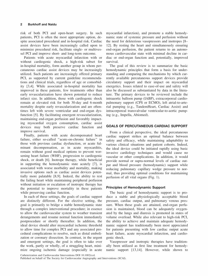
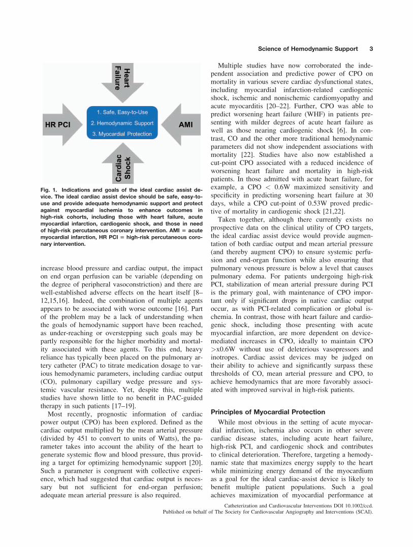
By way of review (Fig. 2, in the absence of aortic ormitral regurgitation), point A on the pressure-volumediagram represents end-diastole, the time when theheart begins contraction. As the myocardium contracts,the mitral valve closes and pressure builds rapidlywithout any change in volume (isovolumic contrac-tion). At point B, the aortic valve opens as ventricularpressure surpasses aortic diastolic pressure and a vol-ume of blood begins to be ejected. The ventricularpressure continues to rise to a maximum after which itreaches point C (end systole), when the aortic valvecloses. Pressure then falls rapidly with a constant vol-ume within the ventricle (isovolumic relaxation). AtPoint D, the mitral valve opens and the ventriclebegins filling with a new volume of blood enteringfrom the atrium for the next cycle. The PV loop isbounded inferiorly by the end-diastolic pressure-volumerelationship (EDPVR) and superiorly by the end-systolicpressure-volume relationship (ESPVR) (Fig. 3). TheEDPVR uniquely defines the passive properties of theLV and the slope of the ESPVR (Emax, also frequentlyreferred to as end-systolic elastance, or Ees), along withthe volume axis intercept (Vo), provides a load-inde-pendent index of ventricular contractility.
Research performed over the past three decades hasshown that left ventricular pressure–volume area(PVA) provides the strongest index of oxygen con-sumption per beat (Fig. 4) [27–29]. PVA is the area onthe pressure–volume diagram bounded by the end-
Fig. 2. Pressure-volume (PV) loop. In the absence of mitral oraortic valve pathologies, point A denotes time of mitral valveclosing, point B denotes time of aortic valve opening, point Cdenotes time of aortic valve closure and point D denotes timeof mitral valve opening. The four phases of the cardiac cycle(isovolumic contraction, ejection, isovolumic relaxation, anddiastolic filling) are identified on the diagram. Green dotshows the point of aortic valve opening, which signifies aorticdiastolic pressure. Blue dot shows the point of peak ventricu-lar pressure which coincides with arterial systolic pressure.
Fig. 3. Parameters associated with the pressure–volumeloop. ESPVR 5 end-systolic pressure–volume relationship,EDPVR 5 end-diastolic pressure–volume relationship, Emax 5slope of ESPVR, V0 5 volume at which end-systolic pressureis zero.
4 Burkhoff and Naidu
Catheterization and Cardiovascular Interventions DOI 10.1002/ccd.Published on behalf of The Society for Cardiovascular Angiography and Interventions (SCAI).
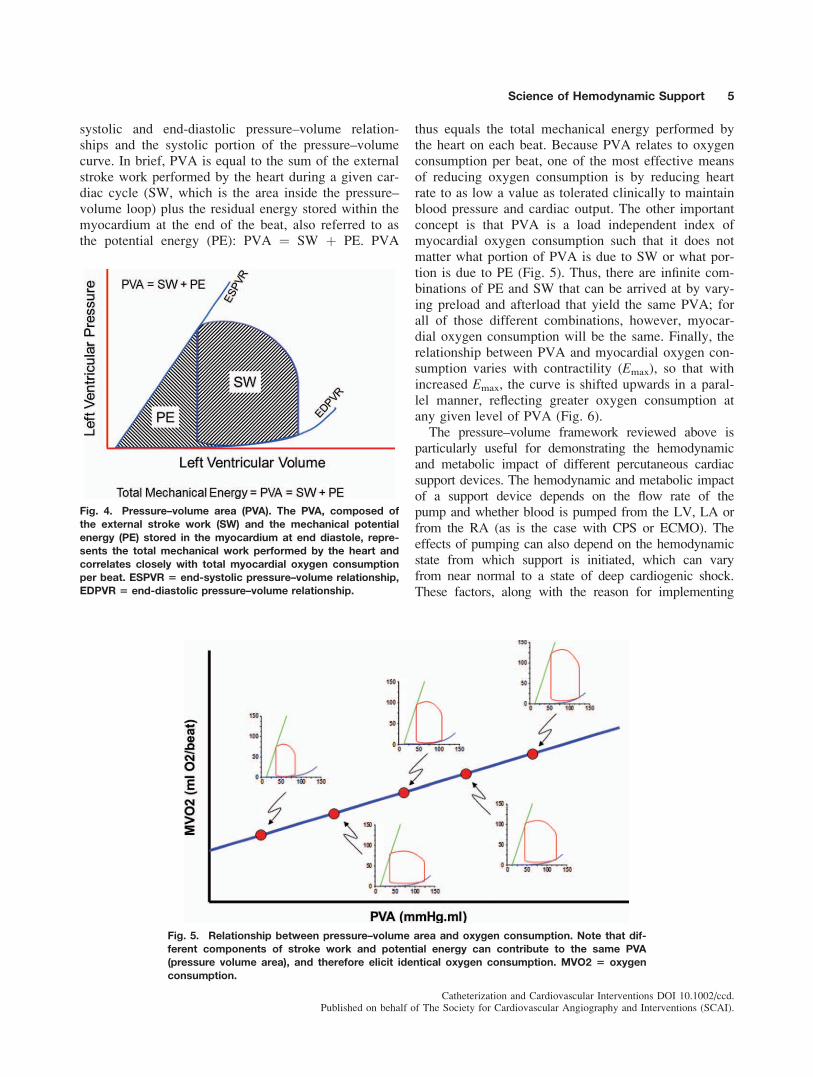
systolic and end-diastolic pressure–volume relation-ships and the systolic portion of the pressure–volumecurve. In brief, PVA is equal to the sum of the externalstroke work performed by the heart during a given car-diac cycle (SW, which is the area inside the pressure–volume loop) plus the residual energy stored within themyocardium at the end of the beat, also referred to asthe potential energy (PE): PVA ¼ SW þ PE. PVA
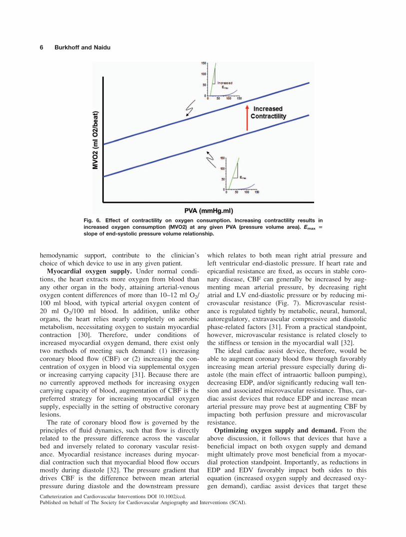
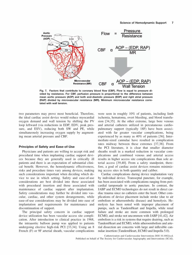
thus equals the total mechanical energy performed bythe heart on each beat. Because PVA relates to oxygenconsumption per beat, one of the most effective meansof reducing oxygen consumption is by reducing heartrate to as low a value as tolerated clinically to maintainblood pressure and cardiac output. The other importantconcept is that PVA is a load independent index ofmyocardial oxygen consumption such that it does notmatter what portion of PVA is due to SW or what por-tion is due to PE (Fig. 5). Thus, there are infinite com-binations of PE and SW that can be arrived at by vary-ing preload and afterload that yield the same PVA; forall of those different combinations, however, myocar-dial oxygen consumption will be the same. Finally, therelationship between PVA and myocardial oxygen con-sumption varies with contractility (Emax), so that withincreased Emax, the curve is shifted upwards in a paral-lel manner, reflecting greater oxygen consumption atany given level of PVA (Fig. 6).
The pressure–volume framework reviewed above isparticularly useful for demonstrating the hemodynamicand metabolic impact of different percutaneous cardiacsupport devices. The hemodynamic and metabolic impactof a support device depends on the flow rate of thepump and whether blood is pumped from the LV, LA orfrom the RA (as is the case with CPS or ECMO). Theeffects of pumping can also depend on the hemodynamicstate from which support is initiated, which can varyfrom near normal to a state of deep cardiogenic shock.These factors, along with the reason for implementing
Fig. 4. Pressure–volume area (PVA). The PVA, composed ofthe external stroke work (SW) and the mechanical potentialenergy (PE) stored in the myocardium at end diastole, repre-sents the total mechanical work performed by the heart andcorrelates closely with total myocardial oxygen consumptionper beat. ESPVR 5 end-systolic pressure–volume relationship,EDPVR 5 end-diastolic pressure–volume relationship.
Fig. 5. Relationship between pressure–volume area and oxygen consumption. Note that dif-ferent components of stroke work and potential energy can contribute to the same PVA(pressure volume area), and therefore elicit identical oxygen consumption. MVO2 5 oxygenconsumption.
Science of Hemodynamic Support 5
Catheterization and Cardiovascular Interventions DOI 10.1002/ccd.Published on behalf of The Society for Cardiovascular Angiography and Interventions (SCAI).
hemodynamic support, contribute to the clinician’schoice of which device to use in any given patient.
Myocardial oxygen supply. Under normal condi-tions, the heart extracts more oxygen from blood thanany other organ in the body, attaining arterial-venousoxygen content differences of more than 10–12 ml O2/100 ml blood, with typical arterial oxygen content of20 ml O2/100 ml blood. In addition, unlike otherorgans, the heart relies nearly completely on aerobicmetabolism, necessitating oxygen to sustain myocardialcontraction [30]. Therefore, under conditions ofincreased myocardial oxygen demand, there exist onlytwo methods of meeting such demand: (1) increasingcoronary blood flow (CBF) or (2) increasing the con-centration of oxygen in blood via supplemental oxygenor increasing carrying capacity [31]. Because there areno currently approved methods for increasing oxygencarrying capacity of blood, augmentation of CBF is thepreferred strategy for increasing myocardial oxygensupply, especially in the setting of obstructive coronarylesions.
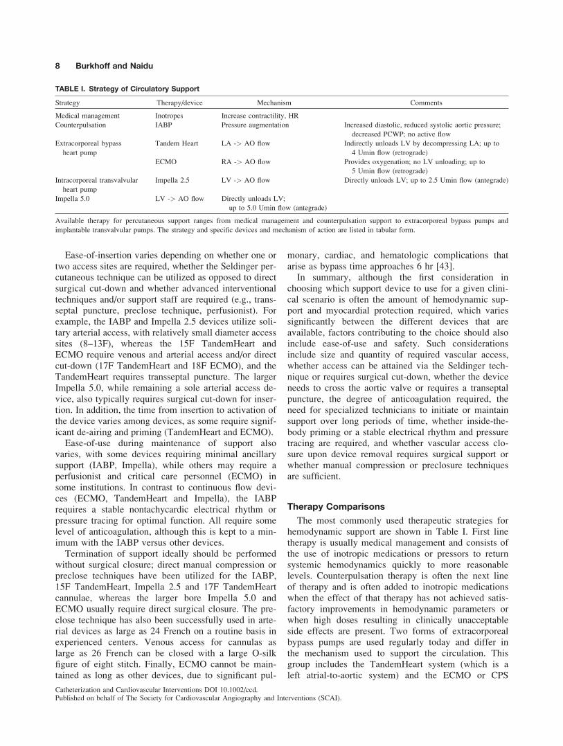
The rate of coronary blood flow is governed by theprinciples of fluid dynamics, such that flow is directlyrelated to the pressure difference across the vascularbed and inversely related to coronary vascular resist-ance. Myocardial resistance increases during myocar-dial contraction such that myocardial blood flow occursmostly during diastole [32]. The pressure gradient thatdrives CBF is the difference between mean arterialpressure during diastole and the downstream pressure
which relates to both mean right atrial pressure andleft ventricular end-diastolic pressure. If heart rate andepicardial resistance are fixed, as occurs in stable coro-nary disease, CBF can generally be increased by aug-menting mean arterial pressure, by decreasing rightatrial and LV end-diastolic pressure or by reducing mi-crovascular resistance (Fig. 7). Microvascular resist-ance is regulated tightly by metabolic, neural, humoral,autoregulatory, extravascular compressive and diastolicphase-related factors [31]. From a practical standpoint,however, microvascular resistance is related closely tothe stiffness or tension in the myocardial wall [32].
The ideal cardiac assist device, therefore, would beable to augment coronary blood flow through favorablyincreasing mean arterial pressure especially during di-astole (the main effect of intraaortic balloon pumping),decreasing EDP, and/or significantly reducing wall ten-sion and associated microvascular resistance. Thus, car-diac assist devices that reduce EDP and increase meanarterial pressure may prove best at augmenting CBF byimpacting both perfusion pressure and microvascularresistance.
Optimizing oxygen supply and demand. From theabove discussion, it follows that devices that have abeneficial impact on both oxygen supply and demandmight ultimately prove most beneficial from a myocar-dial protection standpoint. Importantly, as reductions inEDP and EDV favorably impact both sides to thisequation (increased oxygen supply and decreased oxy-gen demand), cardiac assist devices that target these
Fig. 6. Effect of contractility on oxygen consumption. Increasing contractility results inincreased oxygen consumption (MVO2) at any given PVA (pressure volume area). Emax 5
slope of end-systolic pressure volume relationship.
6 Burkhoff and Naidu
Catheterization and Cardiovascular Interventions DOI 10.1002/ccd.Published on behalf of The Society for Cardiovascular Angiography and Interventions (SCAI).
two parameters may prove most beneficial. Therefore,the ideal cardiac assist device would reduce myocardialoxygen demand and wall tension by shifting the PVloop leftward (via reductions in EDP, EDV, peak pres-sure, and ESV), reducing both SW and PE, whilesimultaneously increasing oxygen supply by augment-ing mean arterial pressure and CBF.
Principles of Safety and Ease-of-Use
Physicians and patients are willing to accept risk andprocedural time when implanting cardiac support devi-ces because they are generally used in critically illpatients and there is an expectation of substantial clini-cal benefit. However, the hemodynamic effectiveness,risks and procedure times vary among devices, makingsuch considerations important when deciding which de-vice to use in which setting. Safety and ease-of-useconsiderations are best divided into those associatedwith procedural insertion and those associated withmaintenance of cardiac support after implantation.Safety considerations may further be divided into vas-cular, cardiac, and other system derangements, whileease-of-use considerations may be divided into ease ofimplantation and requirements for maintenance anddiscontinuation of support.
The principal safety concern in cardiac supportdevice utilization has been vascular access site compli-cation. After introduction to clinical practice in 1968,the intraaortic balloon pump was applied to patientsundergoing elective high-risk PCI [33,34]. Using an 8French (F) or 9F arterial sheath, vascular complications
were seen in roughly 10% of patients, including limbischemia, hematoma, overt bleeding, and blood transfu-sion [34,35]. At the other extreme, large bore venousand arterial catheters utilized in percutaneous cardio-pulmonary support (typically 18F) have been associ-ated with far greater vascular complications, beingexperienced by as many as 40% of patients [36]. Inter-mediate-sized cannulae have resulted in complicationrates midway between these extremes [37,38]. Fromthe PCI literature, it is clear that smaller diametersheaths result in a marked reduction in vascular com-plications and combined venous and arterial accessresults in higher access site complications than sole ar-terial access [39,40]. From a safety standpoint, there-fore, a goal of cardiac assist devices remains minimiz-ing access sites in both quantity and caliber.
Cardiac complications during device implantation varyby individual device. Transseptal puncture, for example,has been associated with complications ranging from peri-cardial tamponade to aortic puncture. In contrast, theIABP and ECMO technologies do not result in direct car-diac trauma since no catheters enter the heart. Other com-plications of device placement include stroke (due to airembolism or atheroembolic disease) and hemolysis. He-molysis has been noted with improper placement ofpumps, such as TandemHeart and Impella, while renalfailure and stroke are more common with prolongedECMO, and stroke not uncommon with IABP [41,42]. Airembolism is a risk in systems that require deairing, such asTandemHeart and ECMO, while atheroembolism and arte-rial dissection are concerns with large and inflexible can-nulae insertion (TandemHeart, ECMO and Impella 5.0).
Fig. 7. Factors that contribute to coronary blood flow (CBF). Flow is equal to pressure di-vided by resistance. For CBF, perfusion pressure is proportional to the difference betweenmean aortic pressure (AOP) and both end-diastolic pressure (EDP) and right atrial pressure(RAP) divided by microvascular resistance (MR). Minimum microvascular resistance corre-lated with wall tension.
Science of Hemodynamic Support 7
Catheterization and Cardiovascular Interventions DOI 10.1002/ccd.Published on behalf of The Society for Cardiovascular Angiography and Interventions (SCAI).
Ease-of-insertion varies depending on whether one ortwo access sites are required, whether the Seldinger per-cutaneous technique can be utilized as opposed to directsurgical cut-down and whether advanced interventionaltechniques and/or support staff are required (e.g., trans-septal puncture, preclose technique, perfusionist). Forexample, the IABP and Impella 2.5 devices utilize soli-tary arterial access, with relatively small diameter accesssites (8–13F), whereas the 15F TandemHeart andECMO require venous and arterial access and/or directcut-down (17F TandemHeart and 18F ECMO), and theTandemHeart requires transseptal puncture. The largerImpella 5.0, while remaining a sole arterial access de-vice, also typically requires surgical cut-down for inser-tion. In addition, the time from insertion to activation ofthe device varies among devices, as some require signif-icant de-airing and priming (TandemHeart and ECMO).
Ease-of-use during maintenance of support alsovaries, with some devices requiring minimal ancillarysupport (IABP, Impella), while others may require aperfusionist and critical care personnel (ECMO) insome institutions. In contrast to continuous flow devi-ces (ECMO, TandemHeart and Impella), the IABPrequires a stable nontachycardic electrical rhythm orpressure tracing for optimal function. All require somelevel of anticoagulation, although this is kept to a min-imum with the IABP versus other devices.
Termination of support ideally should be performedwithout surgical closure; direct manual compression orpreclose techniques have been utilized for the IABP,15F TandemHeart, Impella 2.5 and 17F TandemHeartcannulae, whereas the larger bore Impella 5.0 andECMO usually require direct surgical closure. The pre-close technique has also been successfully used in arte-rial devices as large as 24 French on a routine basis inexperienced centers. Venous access for cannulas aslarge as 26 French can be closed with a large O-silkfigure of eight stitch. Finally, ECMO cannot be main-tained as long as other devices, due to significant pul-
monary, cardiac, and hematologic complications thatarise as bypass time approaches 6 hr [43].
In summary, although the first consideration inchoosing which support device to use for a given clini-cal scenario is often the amount of hemodynamic sup-port and myocardial protection required, which variessignificantly between the different devices that areavailable, factors contributing to the choice should alsoinclude ease-of-use and safety. Such considerationsinclude size and quantity of required vascular access,whether access can be attained via the Seldinger tech-nique or requires surgical cut-down, whether the deviceneeds to cross the aortic valve or requires a transeptalpuncture, the degree of anticoagulation required, theneed for specialized technicians to initiate or maintainsupport over long periods of time, whether inside-the-body priming or a stable electrical rhythm and pressuretracing are required, and whether vascular access clo-sure upon device removal requires surgical support orwhether manual compression or preclosure techniquesare sufficient.
Therapy Comparisons
The most commonly used therapeutic strategies forhemodynamic support are shown in Table I. First linetherapy is usually medical management and consists ofthe use of inotropic medications or pressors to returnsystemic hemodynamics quickly to more reasonablelevels. Counterpulsation therapy is often the next lineof therapy and is often added to inotropic medicationswhen the effect of that therapy has not achieved satis-factory improvements in hemodynamic parameters orwhen high doses resulting in clinically unacceptableside effects are present. Two forms of extracorporealbypass pumps are used regularly today and differ inthe mechanism used to support the circulation. Thisgroup includes the TandemHeart system (which is aleft atrial-to-aortic system) and the ECMO or CPS
TABLE I. Strategy of Circulatory Support
Strategy Therapy/device Mechanism Comments
Medical management Inotropes Increase contractility, HR
Counterpulsation IABP Pressure augmentation Increased diastolic, reduced systolic aortic pressure;
decreased PCWP; no active flow
Extracorporeal bypass
heart pump
Tandem Heart LA -> AO flow Indirectly unloads LV by decompressing LA; up to
4 Umin flow (retrograde)
ECMO RA -> AO flow Provides oxygenation; no LV unloading; up to
5 Umin flow (retrograde)
Intracorporeal transvalvular
heart pump
Impella 2.5 LV -> AO flow Directly unloads LV; up to 2.5 Umin flow (antegrade)
Impella 5.0 LV -> AO flow Directly unloads LV;
up to 5.0 Umin flow (antegrade)
Available therapy for percutaneous support ranges from medical management and counterpulsation support to extracorporeal bypass pumps and
implantable transvalvular pumps. The strategy and specific devices and mechanism of action are listed in tabular form.
8 Burkhoff and Naidu
Catheterization and Cardiovascular Interventions DOI 10.1002/ccd.Published on behalf of The Society for Cardiovascular Angiography and Interventions (SCAI).
system which, for the purposes of cardiac support, isessentially a right atrial-to-aortic system. In this com-parison, we will use the term ECMO (extracorporealmembrane oxygenation) rather than CPS (cardiopulmo-nary support) since percutaneous ECMO systems areused for support today both in the cath lab and formore prolonged support in the ICU. These ECMO sys-tems are generally veno-arterial systems meaning thatthe blood is generally drained from a groin vein (femo-ral vein) and returned to a systemic artery (femoral ar-tery). This discussion will not address the veno–venosystems used primarily for patients who need only oxy-genation support. Finally, the Impella system is cur-rently the only example of the intracorporeal transvalv-ular technology in use today and includes both theImpella 2.5 and the Impella 5.0 platform (Impella 5.0and Impella LD). The Impella systems use a transvalv-ular LV-to-aorta methodology.
The impact of these various interventions in cardio-genic shock can be compared using PV loop analysisto illustrate the hemodynamics effects and implicationsfor myocardial oxygen consumption. Reducing myocar-dial oxygen demand is achieved primarily by reducingor ‘‘unloading’’ peak left ventricular pressure and vol-
ume and is best characterized by the ventricular pres-sure–volume (PV) loop as discussed earlier (Fig. 2).
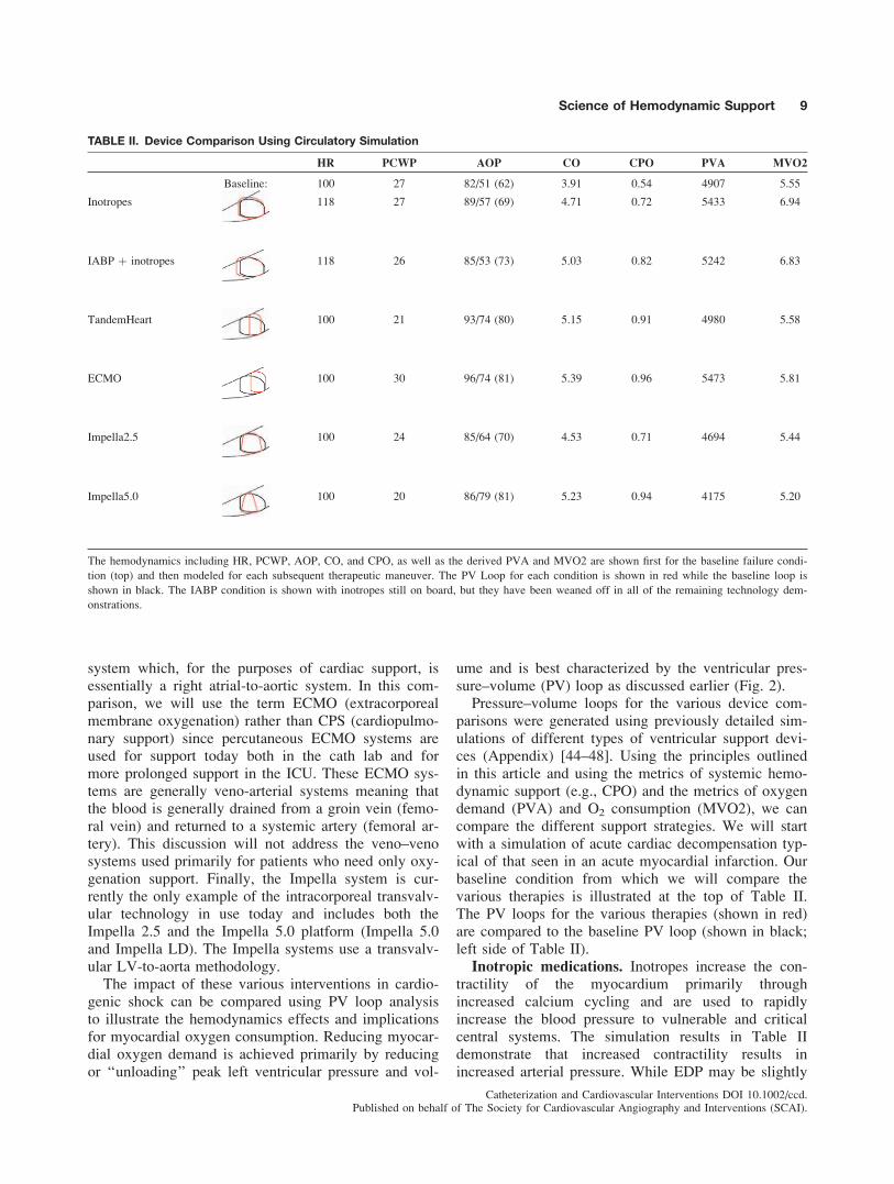
Pressure–volume loops for the various device com-parisons were generated using previously detailed sim-ulations of different types of ventricular support devi-ces (Appendix) [44–48]. Using the principles outlinedin this article and using the metrics of systemic hemo-dynamic support (e.g., CPO) and the metrics of oxygendemand (PVA) and O2 consumption (MVO2), we cancompare the different support strategies. We will startwith a simulation of acute cardiac decompensation typ-ical of that seen in an acute myocardial infarction. Ourbaseline condition from which we will compare thevarious therapies is illustrated at the top of Table II.The PV loops for the various therapies (shown in red)are compared to the baseline PV loop (shown in black;left side of Table II).
Inotropic medications. Inotropes increase the con-tractility of the myocardium primarily throughincreased calcium cycling and are used to rapidlyincrease the blood pressure to vulnerable and criticalcentral systems. The simulation results in Table IIdemonstrate that increased contractility results inincreased arterial pressure. While EDP may be slightly
TABLE II. Device Comparison Using Circulatory Simulation
HR PCWP AOP CO CPO PVA MVO2
Baseline: 100 27 82/51 (62) 3.91 0.54 4907 5.55
Inotropes 118 27 89/57 (69) 4.71 0.72 5433 6.94
IABP þ inotropes 118 26 85/53 (73) 5.03 0.82 5242 6.83
TandemHeart 100 21 93/74 (80) 5.15 0.91 4980 5.58
ECMO 100 30 96/74 (81) 5.39 0.96 5473 5.81
Impella2.5 100 24 85/64 (70) 4.53 0.71 4694 5.44
Impella5.0 100 20 86/79 (81) 5.23 0.94 4175 5.20
The hemodynamics including HR, PCWP, AOP, CO, and CPO, as well as the derived PVA and MVO2 are shown first for the baseline failure condi-
tion (top) and then modeled for each subsequent therapeutic maneuver. The PV Loop for each condition is shown in red while the baseline loop is
shown in black. The IABP condition is shown with inotropes still on board, but they have been weaned off in all of the remaining technology dem-
onstrations.
Science of Hemodynamic Support 9
Catheterization and Cardiovascular Interventions DOI 10.1002/ccd.Published on behalf of The Society for Cardiovascular Angiography and Interventions (SCAI).
reduced, this change is typically clinically insignificant.The overall effect is an increase in systemic bloodpressure, a chronotropic response of increased heartrate, an elevation of the CO and CPO and an elevationof the PVA and MVO2. The change in the PV loopcompared to the baseline failure state is seen in TableII with the new loop representation shown in red. Notethat the new ESPVR line illustrated by the simulationcorresponds to the increased contractility with inotropeadministration.
Counterpulsation. Counterpulsation is often addedto patients who remain compromised after initialattempts at correction with inotropes and optimizationof intravascular volume status. Counterpulsation ther-apy is typically used in addition to inotropic drug ther-apy as a direct clinical manifestation of the fact thatIABP counterpulsation alone does not directly pumpblood but relies on the native heart to provide forwardflow. Balloon deflation during ejection serves to reducethe effective afterload against which the heart beats,but does not result in substantial improvements in car-diac output. Diastolic aortic pressure augmentation pro-vided by the IABP itself achieves an increased level ofpressure and flow support to increase coronary bloodflow. This strategy of combined IABP and inotropicsupport, however, is often counter-productive since thesystemic flow augmentation is still accomplished at theexpense of increasing the native cardiac work and oxy-gen consumption despite the afterload-reducing effectsof the IABP. Particularly in the setting of cardiogenicshock, this can exacerbate myocardial ischemia pro-moting the downward spiral of shock. A number ofinvestigators have reported minimal, if any, direct he-modynamic impact of IABP counterpulsation [49–52].
Progressing from baseline acute decompensationthrough inotropes and then through to the addition ofIABP, (IABPþInotropes; Table II) we see a slightreduction of PCWP, a slight increase in mean aorticpressure, an increase in CO and CPO with a slightreduction of the PVA and MVO2. Note however, thatthe PVA and MVO2 can remain above the baselinecondition of the acute decompensation. Therefore, the
systemic hemodynamic condition is improved at theexpense of a higher heart rate and more oxygendemand. The increased oxygen demand and increasedwork are at odds with the ideal goal of decreased workand diminished demand when dealing with an injuredand ischemic myocardium. Although not addressed bythis simulation, coronary blood flow (CBF) is aug-mented during counterpulsation therapy primarily byincreasing the driving (diastolic) pressure.
Extracorporeal bypass heart pumps. Two technol-ogies used today employ extracorporeal pumps. Tan-demHeart (Cardiac Assist, Pittsburg, PA) employs a leftatrial-to-aorta (LV-to-Ao) support strategy while theCPS or ECMO systems (using a variety of extracorporealpumps and circuits) use a systemic venous pickup (RA-to-Ao) with the addition of an interposed oxygenator.
As seen in Table II, the LA-to-Ao strategy (Tandem-Heart) reduces EDP while providing significantincreases in both CO and MAP. Because of this levelof systemic support, inotropes can be removed in thesimulation, while still maintaining adequate hemody-namic levels. Despite the reduction in heart rate associ-ated with the discontinuation of inotropes, the PVAremains at or slightly increased over the baseline con-dition due to the increase in arterial pressure that iscommensurate with the LA-to-Ao support strategy. Thenet effect is an increase in oxygen delivery due toincreased aortic pressure but there may be little impacton oxygen consumption.
ECMO as a strategy for circulatory support resultsin even better systemic hemodynamic support but isdisadvantaged by moving further away from the ventri-cle we are seeking to unload. Despite higher arterialpressure, more CO and higher CPO, we see a signifi-cantly larger PVA and MVO2 primarily due to anincrease in both left ventricular preload and afterloadpressure (ECMO; Table II). The increased preloadarises from left atrial filling through residual pulmo-nary venous return thereby increasing EDV and EDPand thus increasing the PVA and myocardial oxygenconsumption. Confirmation of this effect was recentlyreported by Kawashima who noted consistent elevation
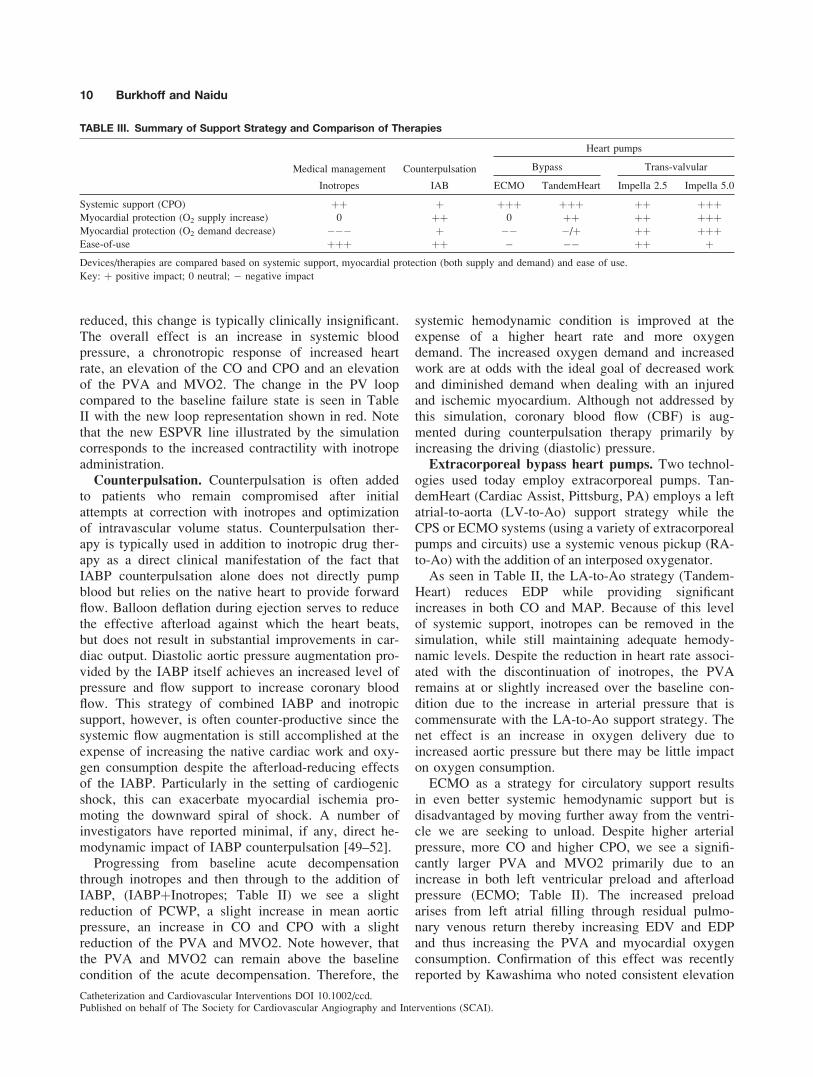
TABLE III. Summary of Support Strategy and Comparison of Therapies
Medical management Counterpulsation
Heart pumps
Bypass Trans-valvular
Inotropes IAB ECMO TandemHeart Impella 2.5 Impella 5.0
Systemic support (CPO) þþ þ þþþ þþþ þþ þþþMyocardial protection (O2 supply increase) 0 þþ 0 þþ þþ þþþMyocardial protection (O2 demand decrease) ��� þ �� �/þ þþ þþþEase-of-use þþþ þþ � �� þþ þDevices/therapies are compared based on systemic support, myocardial protection (both supply and demand) and ease of use.
Key: þ positive impact; 0 neutral; � negative impact
10 Burkhoff and Naidu
Catheterization and Cardiovascular Interventions DOI 10.1002/ccd.Published on behalf of The Society for Cardiovascular Angiography and Interventions (SCAI).
of PVA across a spectrum of failure severity modelssupported by ECMO [52].
Intracorporeal transvalvular heart pumps. TheImpella platform consists of three commercially avail-able pumps. The Impella 2.5 is placed thru a 13Fsheath and is a purely percutaneous application of theintracorporeal transvalvular heart pump. The largerImpella 5.0 requires a surgical cutdown, while theImpella LD technology is designed to be placeddirectly in the ascending aorta if the chest is alreadyopened. From the perspective of systemic hemody-namic support and myocardial protection, since allthree of these technologies employ an LV-to-Ao sup-port strategy, the 5.0 and LD abide by the same princi-ples as the Impella 2.5 and will not be considered sep-arately.
Like the extracorporeal heart pumps, the Impella 2.5maintains systemic hemodynamics above critical levelswithout inotropes, although with less absolute elevationin CPO compared to TandemHeart and ECMO(Table II). A transvalvular support strategy like Impellatakes advantage of the fact that the ability of anypump to produce forward flow is dependent on thepressure difference between the inlet and the outlet ofthe device. A larger positive pressure difference frominlet to outlet yields a lower flow rate. For the extrac-orporeal bypass strategies, this pressure difference(between LA and Ao, or RA and Ao) is always netpositive. For a transvalvular strategy the pressure dif-ference is markedly reduced during systole and caneven drop to zero if the ventricle is able to generateenough pressure to open the aortic valve. Thus, theability of the device to augment forward flow is maxi-mized during systole. This principle allows an LV-to-Ao pump to augment systemic hemodynamics moreprofoundly than a pump with an equivalent powerapplied in an LA- or RA-to-Ao strategy. Conversely,the relatively less powerful Impella 2.5 pump, applyingits support from LV-to-Ao is able to maintain criticalhemodynamic levels comparable to the more powerfulextracorporeal bypass pumps by taking advantage ofthis systolic boost.
From the perspective of myocardial protection, theincrease in MAP (in particular due to increased dia-stolic aortic pressure) combined with the reduction inEDP with Impella will promote increased coronaryflow as has been demonstrated in patients [53]. TheLV-to-Ao support strategy is also the first demon-strated in this simulation to reduce the PVA andMVO2 below the baseline condition, indicating areduction in myocardial oxygen consumption. Thiseffect is more pronounced in the Impella 5.0 simula-tion. The reduction of PVA stems from a reduction inEDV and EDP (preload) similar to the TandemHeart,
but without the significant increase in systolic aorticpressures that was seen with both of the extracorporealstrategies. Additionally, with the LV-to-Ao approach,the isovolumic periods of ejection and relaxation nolonger exist since the pump is constantly deliveringvolume from the LV to the ascending aorta independ-ent of the phase of the cardiac cycle. This too contrib-utes to the overall reduction of PVA.
CONCLUSIONS
Despite best efforts, patients admitted with acutemyocardial infarction, decompensated heart failure orcardiogenic shock, as well as those undergoing high-risk PCI continue to have significant morbidity andmortality rates with standard guideline-based therapy,including revascularization, vasopressor, inotrope, anddiuretic therapy, and other forms of support (e.g., me-chanical ventilation). For the sickest patients, progres-sive cardiac and other end-organ dysfunction is theinevitable short- or long-term consequence of failing tomeet metabolic demands (heart failure, cardiogenicshock) or achieve successful revascularization (acutemyocardial infarction, high-risk PCI). Clearly, there-fore, there is a distinct need to push our understandingof the principles guiding safety, ease-of-use, hemody-namic support and myocardial protection of cardiacsupport devices that add value to current treatmentstrategies.
Although the current manuscript discusses the princi-ples that identify the ideal cardiac support device, intruth the ideal device for one indication may differfrom that for another indication, based the immediateneeds of the patient and the level of risk that will betolerated. For example, a safe and rapidly instituted de-vice to provide myocardial protection in the setting ofacute myocardial infarction may not necessarily needto provide maximal hemodynamic support. Conversely,a device that provides particularly potent hemodynamicsupport may be required for those in cardiogenicshock, whether or not the device can be instituted rap-idly and easily. Table III summarizes the differences inthe various support technologies and contrasts thesedevices according to systemic hemodynamic support,myocardial protection (O2 supply increase and O2
demand decrease) and ease of use.
REFERENCES
1. Serruys PW, Morice MC, Kappetein AP, Colombo A, Holmes
DR, Mack MJ, Stahle E, Feldman TE, van den Brand M, Bass
EJ, Van Dyck N, Leadley K, Dawkins KD, Mohr FW; Syntax
Investigators. Percutaneous coronary intervention versus coro-
nary-artery bypass grafting for severe coronary artery disease. N
Engl J Med 2009;360:961–972.
Science of Hemodynamic Support 11
Catheterization and Cardiovascular Interventions DOI 10.1002/ccd.Published on behalf of The Society for Cardiovascular Angiography and Interventions (SCAI).
2. Stone GW. Evaluation of Xience prime versus coronary artery
bypass surgery for effectiveness of left main revascularization
(The EXCEL Trial). Verbal Communication, Transcatheter
Cardiovascular Therapeutics Conference, San Francisco, CA,
2009.
3. Kushner FG, Hand M, Smith SC, King SB III, Anderson JL,
Antman EM, Bailey SR, Bates ER, Blankenship JC, Casey DE
Jr, Green LA, Hochman JS, Jacobs AK, Krumholz HM, Morri-
son DA, Ornato JP, Pearle DL, Peterson ED, Sloan MA, Whit-
low PL, Williams DO. 2009. Focused updates: ACC/AHA
guidelines on the management of patients with ST-elevation
myocardial infarction (Updating the 2004 guideline and 2007
focused update) and ACC/AHA/SCAI guidelines on percutane-
ous coronary intervention (updating the 2005 guideline and
2007 focused update): A report of the American College of Car-
diology Foundation/American Heart Association Task Force on
Practice Guidelines. J Am Coll Cardiol 2009;54:2205–2241.
4. Dzavik V, Sleeper LA, Cocke TP, Moscucci M, Saucedo J,
Hosat S, Jiang X, Slater J, LeJemtel T, Hochman JS; SHOCK
Investigators. Early revascularization is associated with
improved survival in elderly patients with acute myocardial in-
farction complicated by cardiogenic shock: A report from the
SHOCK Trial Registry. Eur Heart J 2003;24:828–837.
5. Hochman JS, Sleeper LA, Webb JG, Sanborn TA, White HD,
Talley JD, Buller CE, Jacobs AK, Slater JN, Col J, McKinlay
SM, LeJemtel TH. Early revascularization in acute myocardial
infarction complicated by cardiogenic shock. SHOCK Investiga-
tors. N Engl J Med 1999;341:625–634.
6. Torre-Amione G, Milo-Cotter O, Kaluski E, Perchenet L,
Kobrin I, Frey A, Rund MM, Weatherley BD, Cottee G. Early
worsening heart failure in patients admitted for acute heart fail-
ure: Time course, hemodynamic predictors, and outcome. J Car-
diac Failure 2009;15:639–644.
7. Shoemaker WC, Appel PL, Kram HB, Duarte D, Harrier HD,
Ocampo HA. Comparison of hemodynamic and oxygen trans-
port effects of dopamine and dobutamine in critically ill surgi-
cal patients. Chest 1989;96:120–126.
8. Cuffe MS, Califf RM, Adams KF Jr, Benza R, Bourge R,
Colucci WS, Massie BM, O’Connor CM, Pina I, Quigg R, Sil-
ver MA, Gheorghiade M. Outcomes of a prospective trial of in-
travenous milrinone for exacerbations of chronic heart failure
(OPTIME-CHF) investigators. Short-term intravenous milrinone
for acute exacerbation of chronic heart failure: A randomized
controlled trial. JAMA 2002;287:1541–1547.
9. Abraham WT, Adams KF, Fonarow GC, Costanzo MR, Ber-
kowitz RL, LeJemtel TH, Cheng ML, Wynne J, ADHERE
Study Group. In-hospital mortality in patients with acute
decompensated heart failure requiring intravenous vasoactive
medications: An analysis from the acute decompensated heart
failure national Registry (ADHERE). J Am Coll Cardiol 2005;
46:57–64.
10. Dunser MW, Hasibeder WR. Sympathetic overstimulation dur-
ing critical illness: Adverse effects of adrenergic stress. J Inten-
sive Care Med 2009;24:293–316.
11. Goldspink DF, Burniston JG, Ellison GM, Clark WA, Tan LB.
Catecholamine-induced apoptosis and necrosis in cardiac and
skeletal myocytes of the rat in vivo: The same or separate death
pathways? Exp Physiol 2004;89:407–416.
12. Culling W, Penny WJ. Arrhythmogenic and electrophysiologi-
cal effects of alpha adrenoreceptor stimulation during myocar-
dial ischemia and reperfusion. J Mol Cell Cardiol 1987;19:
251–258.
13. Petersen JW, Felker GM. Inotropes in the management of acute
heart failure. Crit Care Med 2008;36(1 Suppl):S106–S111.
14. El Mokhtari NE, Arit A, Meissner A, Lins M. Inotropic therapy
for cardiac low output syndrome: Comparison of hemodynamic
effects of dopamine/dobutamine versus dopamine/dopexamine.
Eur J Med Res 2008;13:459–463.
15. DeBacker D, Biston P, Devriendt J, et al. Comparison of dopa-
mine and norepinephrine in the treatment of shock. N Engl J
Med 2010;362:779–789.
16. Samuels LE, Kaufman MS, Thomas MP, Holmes EC, Brockman
SK, Wechsler AS. Pharmacological criteria for ventricular assist
device insertion following postcardiotomy shock: Experience
with the Abiomed BVS system. J Card Surg 1999;14:288–293.
17. Connors AF Jr, Speroff T, Dawson NV, Thomas C, Harrell FE
Jr, Wagner D, Desbiens N, Goldman L, Wu AW, Califf RM,
Fulkerson WJ Jr, Vidaillet H, Broste S, Bellamy P, Lynn J,
Knaus WA. The effectiveness of right heart catheterization in
the initial care of critically ill patients. SUPPORT Investigators.
JAMA 1996;276:889–897.
18. Dalen JE, Bone RC. Is it time to pull the pulmonary artery
catheter? JAMA 1996;276:916–918.
19. Binanay C, Califf RM, Hasselblad V, O’Connor CM, Shah MR,
Sopko G, Stevenson LW, Francis GS, Leier CV, Miller LW.
Evaluation study of congestive heart failure and pulmonary ar-
tery catheterization effectiveness: The ESCAPE trial. JAMA
2005;294:1625–1633.
20. Fincke R, Hochman JS, Lowe AM, Menon V, Slater JN, Webb
JG, LeJemtel TH, Cotter G; SHOCK Investigators. Cardiac
power is the strongest hemodynamic correlate of mortality in
cardiogenic shock: A report from the SHOCK trial registry. J
Am Coll Cardiol 2004;44:340–348.
21. Torgerson C, Schmittinger CA, Wagner S, Ulmer H, Takala J,
Jakob SM, Dunser MW. Hemodynamic variables and mortality
in cardiogenic shock: A retrospective cohort study. Crit Care
2009;13:R157.
22. Mendoza DD, Cooper HA, Panza JA. Cardiac power output pre-
dicts mortality across a broad spectrum of patients with acute
cardiac disease. Am Heart J 2007;153:366–370.
23. Tamareille S, Achour H, Amirian J, Felli P, Bick RJ, Poindexter
B, Geng YJ, Barry WH, Smalling RW. Left ventricular unload-
ing before reperfusion reduces endothelin-1 release and calcium
overload in porcine myocardial infarction. J Thoracic Cardio-
vasc Surg 2008;136:343–351.
24. Chatterjee K. Coronary hemodynamics in heart failure and
effects of therapeutic interventions. J Cardiac Fail 2009;15:116–
123.
25. Rodbard S, Williams CB, Rodbard D, Berglund E. Myocardial
tension and oxygen uptake. Circ Res 1964;14:139–149.
26. Burkhoff D, Mirsky I, Suga H. Assessment of systolic and dia-
stolic ventricular properties via pressure-volume analysis: A
guide for clinical, translational, and basic researchers. Am J
Physiol Heart Circ Physiol 2005;289:H501–H512.
27. Suga H. Total mechanical energy of a ventricle model and car-
diac oxygen consumption. Am J Physiol 1979;236:H498–H505.
28. Takaoka H, Takeuchi M, Odake M, Hayashi Y, Hata K, Mori
M, Yokoyama M. Comparison of hemodynamic determinants
for myocardial oxygen consumption under different contractile
states in human ventricle. Circulation 1993;87:59–69.
29. Schipke JD, Burkhoff D, Kass DA, Alexander J Jr, Schaefer J,
Sagawa K. Hemodynamic dependence of myocardial oxygen
consumption indices. Am J Physiol 1990;258:H1281–H1291.
30. Braunwald E. Myocardial oxygen consumption: The quest for
its determinants and some clinical fallout. J Am Coll Cardiol
1999;34:1365–1368.
31. Ardehali A, Ports TA. Myocardial oxygen supply and demand.
Chest 1990;98:699–705.
12 Burkhoff and Naidu
Catheterization and Cardiovascular Interventions DOI 10.1002/ccd.Published on behalf of The Society for Cardiovascular Angiography and Interventions (SCAI).
32. Krams R, Sipkema P, Westerhof N. Varying elastance concept
may explain coronary systolic flow impediment. Am J Physiol
1989;257:H1471–1479.
33. Kantrowitz A, Tjonneland S, Freed PS, Phillips SJ, Butner AN,
Sherman JL Jr. Initial clinical experience with intraaortic bal-
loon pumping in cardiogenic shock. JAMA 1968;203:113–118.
34. Kreidieh I, Davies DW, Lim R, Nathan AW, Dymond DS,
Banim SO. High-risk coronary angioplasty with elective intra-
aortic balloon pump support. Int J Cardiol 1992;35:147–152.
35. Osterne EC, Alexim GA, da Motta VP, Lins RM, Carvalho EF,
Carneiro MV, de Morais ER, Brick AV. Intraaortic balloon
pump support during coronary angioplasty. Initial experience.
Arq Bras Cardiol 1999;73:191–200.
36. Teirstein PS, Vogel RA, Dorros G, Stertzer SH, Vandormael
MG, Smith SC Jr, Overlie PA, O’Neill WW. Prophylactic ver-
sus standby cardiopulmonary support for high-risk percutaneous
transluminal coronary angioplasty. J Am Coll Cardiol
1993;21:590–596.
37. Dixon SR, Henriques JP, Mauri L, Sjauw K, Civitello A, Kar
B, Loyalka P, Resnic FS, Teirstein P, Makkar R, Palacios IF,
Collins M, Moses J, Benali K, O’Neill WW. A prospective fea-
sibility trial investigating the use of the Impella 2.5 system in
patients undergoing high-risk percutaneous coronary interven-
tion (PROTECT 1 Trial): Initial US experience.
38. Vranckx P, Meliga E, De Jaegere PP, Van den Ent M, Regar
ES, Serruys PW. The TandemHeart, percutaneous transseptal
left ventricular assist device: A safeguard in high-risk percuta-
neous coronary interventions. The six-year Rotterdam experi-
ence. EuroIntervention 2008;4:331–337.
39. Applegate RJ, Sacrinty MT, Kutcher MA, Kahl FR, Gandhi SK,
Santos RM, Little WC. Trends in vascular complications after
diagnostic cardiac catheterization and percutaneous coronary
intervention via the femoral artery: 1998 to 2007. JACC Cardio-
vasc Interv 2008;1:317–326.
40. Dumont CJ, Keeling AW, Bourguignon C, Sarembock IJ,
Turner M. Predictors of vascular complications post diagnostic
cardiac catheterization and percutaneous coronary interventions.
Dimens Crit Care Nurs 2006;25:137–142.
41. Dens J, Meyns B, Hilgers RD, Maessend J, van Ommen V,
Gerckens U, Grube E. First experience with the Impella
Recover LP 2.5 micro axial pump in patients with cardiogenic
shock or undergoing high-risk revascularization. EuroInterven-
tion 2006;2:84–90.
42. Kolobow T, Rossi F, Borelli M, Foti G. Long-term closed chest
partial and total cardiopulmonary bypass by peripheral cannula-
tion for severe right and/or left ventricular failure, including
ventricular fibrillation. ASAIO Trans 1988;34:485.
43. Mulukutla SR, Pacella JJ, Cohen HA. Percutaneous mechanical
assist devices for severe left ventricular dysfunction. In: Hasdai
E, Berger PB, Battler A, Holmes DR Jr, editors. Contemporary
Cardiology: Cardiogenic Shock: Diagnosis and Treatment.
Totowa NJ: Humana Press; 2002, pp 303–324.
44. Burkhoff D, Tyberg JV. Why does pulmonary venous pressure
rise following the onset of left ventricular dysfunction: A theo-
retical analysis. Am J Physiol 1993;265:H1819–H1828.
45. Santamore WP, Burkhoff D. Hemodynamic consequences of
ventricular interaction as assessed by model analysis. Am J
Physiol 1991;260:H146–H157.
46. Dickstein ML, Burkhoff D. A theoretical analysis of the effect
of pulmonary vasodilation on pulmonary venous pressure:
Implications for inhaled nitric oxide therapy. J Heart Lung
Transplant 1996;15:715–721.
47. Hay I, Rich J, Ferber P, Burkhoff D, Maurer MS. Role of
impaired myocardial relaxation in the production of elevated
left ventricular filling pressure. Am J Physiol Heart Circ Physiol
2005;288:H1203–H1208.
48. Morley D, Litwak K, Ferber P, Spence P, Dowling R, Meyns B,
Griffith B, Burkhoff D. Hemodynamic effects of partial ventricular
support in chronic heart failure: Results of simulation validated
with in vivo data. J Thorac Cardiovasc Surg 2007;133:21–28.
49. Sauren LD, Accord RE, Hamzeh K, et al. Combined Impella
and intra-aortic balloon pump support to improve both ventricu-
lar unloading and coronary blood flow for myocardial recovery:
An experimental study. Artif Organs 2007;31:839–842.
50. Reesink KD, Dekker AL, van Ommen V, et al. Miniature intra-
cardiac assist device provides more effective cardiac unloading
and circulatory support during severe left heart failure than
intraaortic balloon pumping. Chest 2004;126:896–902.
51. Seyfarth M, Sibbing D, Bauer I, et al. A randomized clinical
trial to evaluate the safety and efficacy of a percutaneous left
ventricular assist device versus intra-aortic balloon pumping for
treatment of cardiogenic shock caused by myocardial infarction.
J Am Coll Cardiol 2008;52:1584–1588.
52. Kawashima D, Gojo S, Nishimura T, Itoda Y, Kitahori K,
Motomura N, Morota T, Murakami A, Takamoto S, Kyo S,
Ono M. Left ventricular mechanical support with Impella pro-
vides more ventricular unloading in heart failure than extracor-
poreal membrane oxygenation. ASIO 2011;57:169–176.
53. Remmelink M, Sjauw KD, Henriques JP, et al. Effects of left
ventricular unloading by Impella Recover LP2.5 on coronary
hemodynamics. Catheter Cardiovasc Interv 2007;70:532–537.
54. Dickstein ML, Spotnitz HM, Rose EA, Burkhoff D. Heart
reduction surgery: An analysis of the impact on cardiac func-
tion. J Thorac Cardiovasc Surg 1997;113:1032–1040.
55. Artrip JH, Oz M, Burkhoff D. Left ventricular volume reduction
surgery for heart failure: A physiologic perspective. J Thoracic
Cardiovasc Surgery 2001;122:775–782.
56. Burkhoff D, Wechsler AS. Surgical ventricular remodeling: A
balancing act on systolic and diastolic properties. J Thorac Car-
diovasc Surg 2006;132:459–463.
57. Kelsey R, Botello M, Millard B, Zimmerman J. An online heart
simulator for augmenting first-year medical and dental educa-
tion. Proc AMIA Symp 2002:370–374.
APPENDIX
The simulation used in this article is based on amodel has been described in detail previously [44–48,54–57] and integrates time-varying elastance modelsof the cardiac chambers with Windkessel representa-tions of the systemic and pulmonary vascular systems.There are several lines of evidence that support theutility and accuracy of the integrated model. First, theindividual components of the model used to simulatethe heart chambers (time varying elastances) and thevasculatures (resistance-capacitance networks) havebeen studied and validated for over 40 years. In addi-tion, there have been at least nine publications inwhich the integrated model has been used successfullyto illustrate important hemodynamic principles and topredict hemodynamic effects of several clinical thera-pies, including the Batista procedure, the Dor proce-dure and the impact of inhaled pulmonary vasodilators[44–48,54–57]. Most relevant, however, the integrated
Science of Hemodynamic Support 13
Catheterization and Cardiovascular Interventions DOI 10.1002/ccd.Published on behalf of The Society for Cardiovascular Angiography and Interventions (SCAI).

model was validated quantitatively against measure-ments in an animal model of acute heart failure inwhich an LA-to-Aorta device was used [48]. Havingsaid this, the main goal of using the simulation is toillustrate the important principles of how the differentdevices impact on ventricular hemodynamics. Although
the simulation provides quantitative numerical resultsand we present quantitative results (Table II), it is thequalitative effects (i.e., the way the loops are impacted)and the trends in the numeric values that are of pri-mary interest. It cannot be claimed that the results arequantitatively accurate.
14 Burkhoff and Naidu
Catheterization and Cardiovascular Interventions DOI 10.1002/ccd.Published on behalf of The Society for Cardiovascular Angiography and Interventions (SCAI).
Recommended


