Genome-wide analysis of thylakoid-bound ribosomesin maize reveals principles of cotranslational targetingto the thylakoid membraneReimo Zoschke1 and Alice Barkan2
Institute of Molecular Biology, University of Oregon, Eugene, OR 97403
Edited by Natasha V. Raikhel, Center for Plant Cell Biology, Riverside, CA, and approved February 24, 2015 (received for review December 23, 2014)
Chloroplast genomes encode ∼37 proteins that integrate into thethylakoid membrane. The mechanisms that target these proteinsto the membrane are largely unexplored. We used ribosome pro-filing to provide a comprehensive, high-resolution map of ribo-some positions on chloroplast mRNAs in separated membraneand soluble fractions in maize seedlings. The results show thattranslation invariably initiates off the thylakoid membrane andthat ribosomes synthesizing a subset of membrane proteinssubsequently become attached to the membrane in a nuclease-resistant fashion. The transition from soluble to membrane-attached ribosomes occurs shortly after the first transmembranesegment in the nascent peptide has emerged from the ribosome.Membrane proteins whose translation terminates before emer-gence of a transmembrane segment are translated in the stromaand targeted to the membrane posttranslationally. These resultsindicate that the first transmembrane segment generally comprisesthe signal that links ribosomes to thylakoid membranes for cotrans-lational integration. The sole exception is cytochrome f, whosecleavable N-terminal cpSecA-dependent signal sequence engagesthe thylakoid membrane cotranslationally. The distinct behaviorof ribosomes synthesizing the inner envelope protein CemAindicates that sorting signals for the thylakoid and envelope mem-branes are distinguished cotranslationally. In addition, the fraction-ation behavior of ribosomes in polycistronic transcription unitsencoding both membrane and soluble proteins adds to the evi-dence that the removal of upstream ORFs by RNA processing isnot typically required for the translation of internal genes in poly-cistronic chloroplast mRNAs.
ribosome profiling | protein targeting | plastid | chloroplast | SecA
The chloroplast thylakoid membrane is a highly organized,protein-rich, and dynamic membrane system that is the site of
the light reactions of photosynthesis (1). The majority of proteinsin the thylakoid membrane are subunits of photosynthetic en-zyme complexes: photosystem II (PSII), the cytochrome b6 fcomplex, photosystem I (PSI), the ATP synthase, and the NADHdehydrogenase-like complex (NDH) (2). In land plants andgreen algae, roughly half of the subunits of these complexes areencoded by the plastid genome and half by the nuclear genome(3, 4). This genetic arrangement necessitates a coordination ofprotein synthesis and assembly among cooperating proteins thatoriginate in two compartments.Intensive study of the mechanisms underlying the thylakoid
localization of nucleus-encoded proteins revealed the participa-tion of four machineries of cyanobacterial ancestry: the cpSec,cpTAT, cpSRP, and ALB3 systems (reviewed in ref. 5). Whereasthe cpTAT pathway operates independently to mediate thetranslocation of folded proteins across the membrane, thecpSRP, cpSec, and ALB3 machineries cooperate in the targetingand integration of certain substrates. The bacterial orthologs ofcpSRP and ALB3, known as SRP and YidC, respectively, in-tegrate proteins into the cytoplasmic membrane in a cotransla-tional manner (6, 7). However, the targeting of nucleus-encodedproteins to the thylakoid membrane is posttranslational, as they
are synthesized in the cytosol and then imported into the chlo-roplast stroma before their membrane localization.In contrast to the sophisticated understanding of mechanisms
that localize nucleus-encoded proteins to the thylakoid mem-brane, little information is available about the analogous issuesfor plastid-encoded proteins. Pioneering studies demonstratedthat some chloroplast ribosomes are attached to the thylakoidmembrane by the nascent peptide, implying a cotranslational in-tegration mechanism (8, 9). Several specific plastid-encoded pro-teins have been shown to integrate cotranslationally: the PSIIsubunits PsbA (also known as D1), PsbB, PsbC, and PsbD; the PSIsubunits PsaA and PsaB; and the cytochrome b6f subunit PetA (alsoknown as cytochrome f) (10–14). The insertion of PetA into themembrane requires cpSecA (12, 15, 16), whereas PsbA integratesindependent of both the cpSecA and cpTAT systems (17). In vitrocross-linking experiments showed further that nascent PsbA is inproximity to both cpSRP54 (18) and cpSecY (19). However, it isnot known whether the majority of chloroplast-encoded thylakoidproteins are co- or posttranslationally integrated, nor is it knownwhich, if any, of the known thylakoid targeting machineries areinvolved in their targeting and integration.In this work, we revisited these long-standing questions by
taking advantage of technical advances that allow the precisemapping of ribosomes on mRNAs. A method termed ribosomeprofiling generates a genome-wide, quantitative map of ribo-some positions in vivo by sequencing the ribonuclease-resistant“footprints” left by ribosomes (20). We adapted this method forthe rapid analysis of chloroplast translation by substituting high-resolution tiling microarrays for the deep-sequencing step (21).
Significance
Proteins in the chloroplast thylakoid membrane system arederived from both the nuclear and plastid genomes. Mecha-nisms that localize nucleus-encoded proteins to the thylakoidmembrane have been studied intensively, but little is knownabout the analogous issues for plastid-encoded proteins. Thisgenome-wide, high-resolution analysis of the partitioning ofchloroplast ribosomes between membrane and soluble frac-tions revealed that approximately half of the chloroplast-encoded thylakoid proteins integrate cotranslationally and halfintegrate posttranslationally. Features in the nascent peptidethat underlie these distinct behaviors were revealed by anal-ysis of the position on each mRNA at which elongating ribo-somes first become attached to the membrane.
Author contributions: R.Z. and A.B. designed research; R.Z. performed research; R.Z. andA.B. analyzed data; and R.Z. and A.B. wrote the paper.
The authors declare no conflict of interest.
This article is a PNAS Direct Submission.1Present address: Max Planck Institute of Molecular Plant Physiology, 14476 Potsdam-Golm, Germany.
2To whom correspondence should be addressed. Email: [email protected].
This article contains supporting information online at www.pnas.org/lookup/suppl/doi:10.1073/pnas.1424655112/-/DCSupplemental.
E1678–E1687 | PNAS | Published online March 16, 2015 www.pnas.org/cgi/doi/10.1073/pnas.1424655112
Dow
nloa
ded
by g
uest
on
Janu
ary
11, 2
020

In this work, we modified the microarray approach by profilingchloroplast ribosomes in separated membrane and soluble frac-tions of leaf tissue. The results provide a genome-wide and high-resolution view of the partitioning of chloroplast ribosomes be-tween the soluble and membrane phase and provide insight intothe signals that target proteins for cotranslational integrationinto the thylakoid membrane.
ResultsSpatially Resolved Ribosome Profiling Distinguishes Plastid-EncodedProteins That Are Co- and Posttranslationally Targeted to the ThylakoidMembrane. The method we used to map membrane-bound andsoluble chloroplast ribosomes is shown in Fig. 1. Leaf homoge-nates were initially treated with micrococcal nuclease to releaseribosomes from membranes that were tethered only by mRNA;this treatment will release ribosomes that are bound to mem-branes due to their presence on an mRNA that is membrane-tethered via a different ribosome or via an RNA binding protein.Subsequently, membrane and soluble fractions were separated bycentrifugation. Ribosome footprints were purified from eachfraction, labeled with fluorescent dyes, and hybridized to a high-resolution tiling microarray covering all chloroplast ORFs, usingthe methods described previously (21). Due to the 20-nucleotideoverlap of the 50-mers on the array, this procedure maps ribosomefootprints with a resolution of ∼30 nucleotides.Normalized signals from thylakoid-attached and soluble ribo-
some footprints were plotted according to position on the chlo-roplast genome (Fig. 2B) either as a ratio (upper plot) or sep-arately (lower plot). All ORFs encoding proteins that wereshown previously to integrate cotranslationally into the thylakoidmembrane (PsbA, PsbB, PsbC, PsbD, PsaA PsaB, and PetA)(10–13, 22) are represented by prominent membrane-associatedpeaks, validating the method. Additional peaks revealed thethylakoid-associated translation of 12 proteins that had not beenassayed in prior studies: AtpF, AtpI, PetB, NdhA, NdhB, NdhC,NdhD, NdhE, NdhF, NdhG, Ycf4, and CcsA (Fig. 2B). Each ofthese proteins has at least one transmembrane segment (TMS).By contrast, the ratios of membrane to soluble ribosome foot-prints for all chloroplast proteins lacking a TMS (e.g., ribosomalproteins, RbcL, AtpB) were very low; in fact, many such ORFslacked any detectable signal in the membrane fraction (markedby red diamonds in the Upper panel of Fig. 2B). When lysateswere not treated with nuclease before membrane pelleting (Fig.2C), similar trends were observed; differences, however, high-light regions in which ribosomes were tethered to the membranesolely by RNA (see arrows in Fig. 2 B and C).These results show that ribosomes transiting many chloroplast
ORFs encoding integral thylakoid proteins are bound to the thy-lakoid membrane in a nuclease-resistant manner, implying acotranslational targeting mode. However, ribosomes synthesizingmany other transmembrane (TM) proteins are not bound to themembrane under these conditions. Proteins in the latter groupinclude the multispanning proteins PetD and AtpH and proteinswith a single TMS, such as PetL and PsbH (Fig. 2B and SI Ap-pendix, Table S1). These proteins must integrate into the mem-brane posttranslationally. The basis for the distinct behaviorsof these two sets of membrane proteins was clarified in the sub-sequent analyses.
Synthesis of Cotranslationally Targeted Proteins Initiates on StromalRibosomes and Transitions to Thylakoid-Bound Ribosomes. The highresolution of the data revealed the spatial dynamics of proteinsynthesis as chloroplast ribosomes move along each mRNA.Experiments involving nuclease treatment before membranepelleting were most informative in this regard. In these assays,ribosome footprints near the start of all chloroplast ORFs werefound predominantly in the soluble fraction (Figs. 2B and 3 andSI Appendix, Fig. S1), indicating that initiating ribosomes are not
bound to the membrane. However, elongating ribosomes re-locate to the membrane at a particular point along each ORFthat encodes a cotranslationally targeted protein (Figs. 2B and 3and SI Appendix, Fig. S1). Consider, for example, the psaA andpsaB ORFs, which are separated by only 25 nucleotides on thesame polycistronic mRNA. Ribosomes at the end of the psaAORF remain bound to the membrane after ribonuclease treat-ment, whereas ribosomes at the start of the psaB ORF do not(Fig. 3A). Similar phenomena are apparent for the cotranscribedpsbD and psbC genes (Fig. 3B), psbB and petB genes (Fig. 3C),
Membrane Soluble
footprints (RF)
5. Direct RNA-labeling:membrane RF:
Array design:ORF
soluble RF:
Nucleus
Chloroplast
Mitochondrion
Ribosome footprints:
probes
solublemembrane-bound
nuclease treatment,
Fig. 1. Method for profiling chloroplast ribosome positions in separatedmembrane and soluble fractions. The method is similar to that used previously(21), except that separated membrane and soluble fractions are used as thesource of ribosome footprints. Leaves are flash frozen in liquid nitrogen, andhomogenates are prepared in the absence of detergents and presence ofchloramphenicol to stall translation. Lysates are treated with micrococcal nu-clease to release ribosomes that are tethered to membranes solely by mRNA.Membrane and soluble fractions are then separated by centrifugation. Ribo-some footprints purified from the two fractions are differentially labeled withfluorescent dyes, combined, and hybridized to a tiling microarray spanning allchloroplast ORFs. Array probes are 50 nt in length and overlap by 20 nt,providing a resolution of ∼30 nucleotides. In some experiments, the lysateswere not pretreated with nuclease before pelleting membranes; these aredenoted “– nuclease pretreatment” in subsequent figures.
Zoschke and Barkan PNAS | Published online March 16, 2015 | E1679
PLANTBIOLO
GY
PNASPL
US
Dow
nloa
ded
by g
uest
on
Janu
ary
11, 2
020

and atpI and atpF genes (Fig. 3D). Other examples are presentedin SI Appendix, Fig. S1. This position-dependent relocation ofribosomes from the soluble to membrane fraction was confirmedby slot-blot hybridization analysis of ribosome footprints, usingprobes specific for the 5′ and 3′ regions of selected ORFs (SIAppendix, Fig. S2).These results suggest that ribosomes become attached to the
membrane in a nuclease-resistant fashion after a particular fea-ture in the nascent peptide has emerged from the ribosome’s exitchannel. According to this view, ribosomes that have not yetpassed the point at which this signal is exposed are released tothe soluble fraction when membranes are treated with ribonu-clease (see cartoon in Fig. 3E). As predicted by this model,omission of the ribonuclease treatment before pelleting themembranes reduced the recovery of 5′-proximal ribosome foot-prints in the soluble fraction and increased their recovery in the
membrane fraction (Figs. 2 and 3, compare + and – nucleasepretreatment). Hence, ribosomes at the start of each of the 19membrane-translated ORFs are tethered to the thylakoid mem-brane by mRNA, whereas the elongating ribosomes downstreamare attached via the nascent peptide. These results imply thata first round of translation anchors the mRNA to the thylakoidmembrane via the nascent peptide and that subsequent roundsinitiate in the vicinity of the membrane due to mRNA tethering.
Emergence of Either a TMS or cpSecA-Dependent Signal Sequencefrom the Ribosome Correlates with Cotranslational MembraneAnchoring. Next, we sought to understand (i) the features thatdetermine which chloroplast-encoded TM proteins are cotrans-lationally targeted to the thylakoid membrane and (ii) the pointat which elongating ribosomes acquire a nuclease-resistant at-tachment to the membrane. All 19 membrane-translated ORFs
10 1108030 1009070605040200
Ribo
foot
prin
ts 7
0
65
321
4
Ribosome footprints (-nuclease pre-treatment) soluble membrane
Sign
al in
tens
ity
8
10 1108030 1009070605040200
Ribo
foot
prin
tsSi
gnal
inte
nsity
Ribosome footprints (+nuclease pre-treatment) soluble membrane7
0
65
321
4
8
Ribo
foot
prin
ts[m
em/s
ol]
10 1108030 1009070605040200
256
1/41/16
1/4096
4
64
1
16
mem/sol (+nuclease pre-treatment)
1/2561/64
1/1024
rps16
psbA
matK
petN
rps14
psaB
psaA
ycf3
rps4
ndhJ
ndhK
ndhC
atpE
atpB
10 kbp
psbE
psbF
psbL
psbJ
clpP
rps12
rpl20
psbN
rpoA
rpl23
rpl2
rps19
rpl22
rps3
rpl16
rpl14
rps8
rpl36
rps11
ndhB
rps7
rps12
ndhF
ndhH
ndhA
ndhI
ndhG
ndhE
psaC
ndhD
Zea mays chloroplast genome (1-117,700 bp)
psbK
psbI
psbD
psbC
psbZ
psbM
rpoB
rpoC
1
rpoC
2rps2
atpI
atpH
atpF
atpA
petA
ycf4
psaI
rbcL
petL
petG
psaJ
rpl33
rps18
psbH
psbT
psbB
petB
petD
rps15
rpl32
ccsA
cemA
infA
* * * * * * * * * * * * * ***
**********************
B
C
A
soluble signal only
10 1108030 1009070605040200
Ribo
foot
prin
ts 7
0
65
321
4
Ribosome footprints (-nuclease pre-treatment) soluble membrane
Sign
al in
tens
ity
8
10 1108030 1009070605040200
Ribo
foot
prin
tsSi
gnal
inte
nsity
Ribosome footprip nts (+nuclease p( pre-treatment)) soluble membrane7
0
65
321
4
8
Ribo
foot
prin
ts[m
em/s
ol]
10 1108030 1009070605040200
256
1/41/16
/
1/4096
4
64
1
16
mem/sol (+nuclease pre-treatment)
1/2561/64
//
/1/1024
//
/
rprrs16
psbAbb
matK
petN
rpprrs14
psaB
psaAaa
ycf3
rprrs4
ndhJ
ndhK
ndhC
atpE
atpBpp
10 kbp
psbE
psbF
psbL
psbJbb
clpP
rp llrrs12
rpl20
psbppN
rpoA
rpl23
prpl2 l2
rprrs19
rpl22 19
rprrs3
rpl16 3
rpl14
l16
rprrs8
rpl36
rprrs11
ndhB
p rprrs7
rprrs12
ndhF
ndhH
ndhA
ndhI
ndhGdhndhE
ppsaC
ndhD
Zea maysZZ chloroplast genome (1-117,700 bp)
psbK
psbI
psbD
ppsbC
psbZ
psbM
rpoB
rpoC
1
rpoC
2rprrs2
atpI
atpIppp atpHppp
atpFpp
atpAAppp
peAtA
yycf4
ppsaI
rbcL
petLtL
petG
psaJaa
rpl33
p rpprrs18
ppsbH
psbT
ppsbbB
ppetB
petD
rprrs15
rpl32
p ccsApcemAA
yf
yf
infA 8
pp
* * * * * * * * * * * * * ***
**********************
B
C
A
soluble signal only
Fig. 2. Overview of spatially resolved profiling of chloroplast ribosomes. The plotted signal intensities are the normalized values × 10−4 and are mediansfrom two biological replicates, each with three replicate spots per array element. The data are provided in Dataset S1. Arrows mark ribosome footprintswhose association with the membrane was reduced markedly when lysates were treated with nuclease before membrane pelleting (compare panels B and C).(A) Map of the maize chloroplast genome showing only protein coding regions and only one of the two large inverted repeats. Asterisks mark genes codingfor proteins that contain TM segments. Genes highlighted in green are represented by abundant membrane-bound ribosome footprints in the + nucleaseexperiments (B). Genome position refers to the reference maize chloroplast genome (51). The map was created with OGDraw (52). (B) Ribosome footprints inmembrane and soluble fractions from assays that included nuclease treatment before membrane pelleting. With this protocol, ribosomes that are tethered tomembranes solely by mRNA are recovered in the soluble fraction. The ratio of signal in the membrane relative to soluble fraction is shown in the Upper panelusing a log scale; green shaded regions represent ORFs whose transiting ribosomes are attached to the membrane even after nuclease treatment. Arrayelements with no detectable signal in the membrane fraction are marked by red diamonds at the bottom of the plot. The individual signals for the membraneand soluble fractions are plotted below (green and red lines, respectively). (C) Ribosome footprints in membrane and soluble fractions from assays that didnot include nuclease pretreatment. This protocol recovers ribosomes in the membrane fraction when they are tethered either by mRNA or by protein. Theindividual signals for the membrane and soluble fractions are represented with green and red lines, respectively.
E1680 | www.pnas.org/cgi/doi/10.1073/pnas.1424655112 Zoschke and Barkan
Dow
nloa
ded
by g
uest
on
Janu
ary
11, 2
020

psbBTMSC
petB petDpsbT psbHnortninortni
psbN
34
21
Sign
al in
tens
ity 5
0
soluble membraneribosome footprints (+nuclease pre-treatment)
72.070.6 74.0 76.073.071.0 75.074.573.572.571.5
34
21
Sign
al in
tens
ity 5
0
soluble membraneribosome footprints (-nuclease pre-treatment)
72.070.6 74.0 76.073.071.0 75.0 75.574.573.572.571.5
psaATMSA psaB rps14
34
21
Sign
al in
tens
ity 5
0
soluble membraneribosome footprints (+nuclease pre-treatment)
39.042.043.8 40.041.043.0
34
21
Sign
al in
tens
ity 5
0
soluble membraneribosome footprints (-nuclease pre-treatment)
39.042.043.8 40.041.043.0
psbDTMSB psbC
34
21
Sign
al in
tens
ity 5
0
soluble membraneribosome footprints (-nuclease pre-treatment)
10.09.0 11.0 11.510.59.5
34
21
Sign
al in
tens
ity 5
0
soluble membraneribosome footprints (+nuclease pre-treatment)
10.09.0 11.0 11.510.59.5
atpITMSD
atpH atpF atpAintron
53421
Sign
al in
tens
ity 8
0
6
36.0 37.032.6 35.034.0 38.0
7 soluble membraneribosome footprints (+nuclease pre-treatment)
53421
Sign
al in
tens
ity 8
0
6
36.0 37.032.6 35.034.0 38.0
7 soluble membraneribosome footprints (-nuclease pre-treatment)
E
Thylakoid membrane
’3’5
Stroma
Lumen
GUA
potS
Soluble Membrane-boundRibosomefootprints:
Sign
al in
tens
ity soluble membrane
75.5
Fig. 3. Zoom-in images of data for several polycistronic transcription units. Data and annotations are as in Fig. 2. Each TMS is represented by a gray rectangle withinan ORF. Gray vertical lines mark the boundaries of ORFs and introns, and dashed lines mark each TMS. Gaps in the data correspond to large intergenic regionsand introns, which were not represented on the array. Analogous images for additional genes are shown in SI Appendix, Fig. S1 and Fig. 5. TMS positions are basedeither on experimental data or prediction, as summarized in SI Appendix, Table S1. (A) The psaA transcription unit. The psaA and psaB ORFs are always representedon the same polycistronic mRNA, whereas rps14 is also found in a monocistronic RNA isoform (53–55). (B) The psbD transcription unit. The psbD and psbC ORFs arefound together on polycistronic mRNAs and also on separate processed transcripts (53). (C) The psbB transcription unit. A primary transcript spanning psbB–psbT–psbH–petB–petD is processed to yield numerous processed RNAs with intercistronic termini (31). The psbN gene is encoded by the opposite strand. (D) The atpItranscription unit. A primary transcript spanning atpI–atpH–atpF–atpA is processed to yield numerous processed RNAs with intercistronic termini (56). (E) Model forthe relocation of ribosomes from the soluble to membrane fraction. Ribosomes become attached to the membrane in a nuclease-resistant fashion after the nascentpeptide stably engages the thylakoidmembrane, either directly or via a thylakoid-bound protein. Ribosomes whose nascent peptide is not sufficiently long to exposethe signal for membrane attachment are released to the soluble fraction by the nuclease pretreatment. Scissors represent nuclease cleavage sites. The associationof the nascent peptide with a hypothetical channel in the membrane is shown for illustration only and is not intended to imply a particular mechanism.
Zoschke and Barkan PNAS | Published online March 16, 2015 | E1681
PLANTBIOLO
GY
PNASPL
US
Dow
nloa
ded
by g
uest
on
Janu
ary
11, 2
020

contain at least one TMS (SI Appendix, Table S1), whereas thoseproteins lacking a TMS were invariably translated off themembrane (Fig. 2B). Additionally, the transition from soluble tomembrane-attached translation occurred downstream of se-
quences encoding the first TMS (Fig. 3 and SI Appendix, Fig. S1and Table S1), with the sole exception of PetA (discussed be-low). These results suggested that the emergence of a TMS in thenascent peptide triggers nuclease-resistant attachment to themembrane, as occurs for SRP-mediated targeting of signal-anchorproteins to the endoplasmic reticulum (ER) and to the bacterialcytoplasmic membrane (23, 24).To evaluate this possibility, we calculated the distance between
the start of the first TMS and the point at which ribosomessynthesizing each protein relocate from the soluble to membranefraction (Fig. 4A). If emergence of the first TMS is a requirementfor cotranslational membrane integration, a minimum distance of∼60 amino acids is expected: This corresponds to the length ofa TMS (∼20 amino acids) added to the length of the nascent chainthat is obscured by the exit tunnel of the ribosome (∼40 aminoacids) (25). The actual distances observed here vary between ∼66and 155 amino acids. Thus, the first TMS in each nascent peptidewill have emerged fully from the ribosome before the relocation ofelongating ribosomes to the membrane. That being said, there wasconsiderable variation in the positioning of this relocation event: Insome cases, it occurred soon after the predicted emergence of thefirst TMS (e.g., PsbD), whereas in others considerably more nascentpeptide was synthesized before the relocation (e.g., PsaB). Thisdifference did not correlate with protein topology (N terminus inthe Stroma or Lumen marked in Fig. 4A) or hydrophobicity of thefirst TMS (SI Appendix, Table S1). The variation in the positioningof membrane engagement following emergence of the first TMSmight be influenced by the kinetics of ribosome movement orpeptide-specific association with chaperones. A recent similar analysisof cotranslational targeting to the ER (26) revealed a bimodaldistribution of the position at which ribosomes engage themembrane, centered at ∼60 and 120 amino acids after the startof the first TMS. This correlated with distinct requirements forcomponents of the ER targeting machineries and was suggestedto reflect either “head first” or looped insertion mechanisms.Our results hint at a similar bimodal distribution (Fig. 4A).The results above suggested that the exposure of the first TMS
from the ribosome is required to attach ribosomes to thylakoidmembranes in a nuclease-resistant fashion. This hypothesispredicts that any protein whose first TMS is so close to the stopcodon that it would remain hidden in the ribosome untiltranslation terminates would be translated off the membrane.The placement of the first TMS in those TM proteins that areposttranslationally targeted support this prediction. To illustratethis point, the distance between the first TMS and the stop codonwas plotted for each plastid-encoded TM protein (Fig. 4B). All
psbL
psaB
psaA
ndhD
psbB
ndhB
psbC
ndhA
psbD
psbA
ccsA
cemA
atpI
petB
ndhGycf4
atpF
petD
ndhC
ndhE
atpH
psbE
psbZ
psbNpsaJ
petA
psbH
petG
psbJ
psbT
psbMpsbI
petL
psaI
petN
psbF
ndhF
psbK
A
0
160
604020
80100120140
psbD
ndhC ycf4
ccsA
ndhB
atpF
petB
ndhA
ndhE
ndhF
ndhG
psbA
ndhD atpI
psaB
psaA
psbB
psbC
Distance from start of first TMS to stop codon [codons]B
0
800
600
400
200
500
300
100
700
*#
> 80 codons< 80 codons
AUG
STO
P
AUG
STO
PTMS
C
ORF
Thylakoid membrane
20 AA40 AA
60 AA~~ ~Expected
minimum distance
~ ~
S
SSS
S
S
S
LLL
Fig. 4. Full exposure of the first TMS in the nascent peptide correlates withnuclease-resistant attachment of ribosomes to the thylakoid membrane.(A) Distance from start of first TMS to the positionwhere ribosomes remain boundto the membrane after nuclease treatment. All ORFs encoding proteins that arecotranslationally targeted to the membrane are shown, except petA, whoseN-terminal signal peptide mediates membrane contact before appearance of thefirst TMS (Results). Proteins with experimentally validated topologies are anno-tated with S (N terminus in stroma) or L (N terminus in lumen). The diagramillustrates the minimum distance between the start of a TMS and its full emer-gence from the ribosome. The data and criteria used to define the point of re-location to the membrane are provided in SI Appendix, Table S1 and Materialsand Methods. Note that the resolution of our assay is ∼10 amino acids. (B) Dis-tance between the start of the first TMS and the stop codon for plastid-encodedTM proteins. Red and green bars denote soluble and membrane-attachedtranslation, respectively, as shown in this study (Fig. 2). The two exceptional ORFsdiscussed in Results (petA and cemA) are marked with an asterisk and hashmark,respectively. The shaded region marks the range of distances following the firstTMS in which nascent peptides engage themembrane (from panel A). (C) Modelfor targeting of plastid-encoded proteins to the thylakoid membrane. ORFswhose first TMS is fully exposed before translation termination (greater than∼80amino acids upstream of the stop codon) are cotranslationally targeted via en-gagement of the first TMS by a thylakoid-bound component of the targetingmachinery. ORFs whose first TMS is not fully exposed before termination aretranslated on ribosomes that are not attached to the membrane and are post-translationally targeted. The 80-amino-acid demarcation is an estimate that isbased on the data summarized in Fig. 4B and SI Appendix, Table S1. This is notintended to imply a strict rule, as proteins whose first TMS maps between 79 and125 amino acids upstream of the stop codon exhibit variable behavior.
61.0 0.265.06 61.5
TMSSignal
petA
321
Sign
al in
tens
ity 4
0
ribosome footprints (+nuclease pre-treatment)soluble membrane
62.5
cemA
Fig. 5. Zoom-in image of data for the cotranscribed cemA and petA genes.Data representations and annotations are as described in Fig. 3. Ribosomestransiting petA are recovered in the membrane fraction shortly after thecpSecA-dependent signal sequence emerges. Ribosomes transiting the cemAORF are poorly recovered in the membrane fraction, even though two TMSsare predicted to be exposed before translation termination. CemA is the onlychloroplast-encoded protein that localizes to the inner envelope in maize.
E1682 | www.pnas.org/cgi/doi/10.1073/pnas.1424655112 Zoschke and Barkan
Dow
nloa
ded
by g
uest
on
Janu
ary
11, 2
020
proteins for which this distance is less than the minimal 60 aminoacids required to expose the first TMS before termination aretranslated off the membrane (with the exception of PetA, thespecial case discussed below). As noted above, there is a range ofdistances past the first TMS within which ribosomes engage themembrane (between ∼66 and 155 amino acids) (Fig. 4A). Withinthis range of distances with respect to the stop codon, some pro-teins are cotranslationally bound to the membrane (NdhE andNdhC), whereas others are not (AtpH and PetD). This differencemight reflect distinct kinetics of ribosome movement or “headfirst” versus looped insertion mechanisms, as discussed above.Two ORFs, petA and cemA, were exceptions to these trends.
Each is a special and informative case. CemA is the sole plastid-encoded protein in maize that localizes to the inner envelopemembrane and is discussed below. PetA is the sole plastid-encoded protein harboring a cleavable N-terminal cpSecA-dependent signal sequence, which mediates its targeting to thethylakoid membrane (12, 15, 16). The petA ORF engages themembrane after synthesis of ∼100 amino acids (Fig. 5). This iswell before the single TMS in PetA has been synthesized, but it is65 amino acids after the signal peptide cleavage site (27). Thus,the PetA signal peptide likely anchors the translating ribosometo the thylakoid membrane cotranslationally in vivo.Taken together, the results described above provide strong evi-
dence that exposure of either a full TMS or cpSecA-dependentsignal sequence is necessary for the cotranslational targeting ofchloroplast-encoded proteins to the thylakoid membrane. Itseems likely that these features in the nascent peptide anchor theribosome to the membrane by engaging a translocon. Thosechloroplast-encoded proteins whose translation terminates beforethe emergence of a full TMS from the ribosome (roughly half ofthe plastid-encoded TMS proteins) are posttranslationally tar-geted to the membrane (Fig. 4C).
Spatial Dynamics of Plastid Ribosomes Synthesizing Envelope Proteins.CemA’s first and second predicted TMS map far upstream ofthe stop codon (SI Appendix, Table S1) and are expected to beaccessible for cotranslational membrane attachment. Nonetheless,ribosomes transiting the cemA ORF were recovered predominantlyin the soluble fraction (Fig. 5). This unusual behavior correlates
with the fact that CemA localizes to the inner envelope membrane(28) and is the only plastid-encoded protein in maize to do so.To determine whether plastid ORFs encoding other inner
envelope proteins behave similarly, we took advantage of the factthat the tobacco chloroplast genome encodes a second integralinner envelope protein, Ycf1/TIC214 (29). Ribosome footprintswere prepared from membrane and soluble fractions of tobaccoleaves as shown in Fig. 1. The partitioning of ribosome footprintsbetween the two fractions was assessed by slot-blot hybridizationusing probes for specific ORFs (Fig. 6). Ribosomes transitingtobacco rbcL and psaA behaved as they did in maize (see SIAppendix, Fig. S2 for the maize data): Those in rbcL were largelyin the soluble fraction, whereas those in psaA started in thesoluble fraction but relocated to membranes within the ORF.Ribosomes synthesizing the integral inner envelope proteinsCemA and Ycf1 did not fall into either of these categories: They
membrane footprints
ribosomefootprints: 5’ 3’
cemA5’ 3’psaA
3’5’ycf1
soluble footprints
0
80
60
40
20
100
5’ 3’cemA
5’ 3’psaA
3’5’ycf1
Probes
Ribo
som
e fo
otpr
ints
[%]
membrane
soluble
3’5’rbcL
5’ 3’rbcL
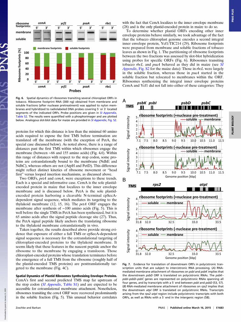
Fig. 6. Spatial dynamics of ribosomes transiting several chloroplast ORFs intobacco. Ribosome footprint RNA (300 ng) obtained from membrane andsoluble fractions (after nuclease pretreatment) was applied to nylon mem-branes and hybridized to radiolabeled DNA probes covering 5′ or 3′ locatedsegments of the indicated ORFs. Probe positions are given in SI Appendix,Table S2. The results were quantified with a phosphorimager and are plottedbelow. Analogous slot-blot data for maize are provided in SI Appendix, Fig. S2.
10.07.1 11.0 11.510.59.59.08.58.07.5
psbDTMS
A psbC
34
21
Sign
al in
tens
ity 5
0
ribosome footprints (-nuclease pre-treatment)
34
21
Sign
al in
tens
ity 5
0
soluble membrane
soluble membraneribosome footprints (+nuclease pre-treatment)
10.07.1 11.0 11.510.59.59.08.58.07.5
psbK psbI
atpITMSB
321
Sign
al in
tens
ity 4
0
ribosome footprints (+nuclease pre-treatment)
321
Sign
al in
tens
ity 4
0
soluble membrane
soluble membraneribosome footprints (-nuclease pre-treatment)
0.338.13 32.5 33.532.0
0.338.13 32.5 33.532.0
rps2
Fig. 7. Evidence for translation of downstream ORFs in polycistronic tran-scription units that are subject to intercistronic RNA processing. (A) RNA-mediated membrane attachment of ribosomes on psbI and psbK implies thatthe downstream psbD ORF is translated on polycistronic RNAs. The psbK–psbI–psbD–psbC genes are represented on polycistronic RNAs spanning allfour genes, and by transcripts with a 5′ end between psbI and psbD (53, 57).(B) RNA-mediated membrane attachment of ribosomes on rps2 implies thatthe downstream atpI ORF is translated on polycistronic RNAs. Transcriptsarising from the rps2–atpI region include polycistronic transcripts with bothORFs, as well as RNAs with a 5′ end in the intergenic region (58).
Zoschke and Barkan PNAS | Published online March 16, 2015 | E1683
PLANTBIOLO
GY
PNASPL
US
Dow
nloa
ded
by g
uest
on
Janu
ary
11, 2
020

were equally distributed between the soluble and membranefractions, even at the 3′ ends of the ORFs where multiple TMSsare expected to have emerged. The analogous slot-blot assay formaize cemA gave similar results (SI Appendix, Fig. S2). Duringour fractionation protocol, a marker for the inner envelope wasrecovered primarily in the “soluble” fraction (SI Appendix, Fig.S2), presumably due to the low density of envelope membranes.Thus, our data cannot distinguish between the possibilities thatCemA and Ycf1 integrate co- or posttranslationally into the innerenvelope. Nonetheless, these results indicate that the TMSs inCemA and Ycf1 either lack a signal needed to engage the thylakoidmembrane or have a feature that prevents them from doing so.
Ribosome Dynamics in Polycistronic Transcription Units ProvideEvidence That Intercistronic RNA Processing Is Not GenerallyRequired for Translation. Chloroplast genes in land plants aretypically organized in polycistronic transcription units that giverise to processed RNAs with termini between ORFs. It has beensuggested that intercistronic RNA processing is a mechanism toimprove translational efficiency, but there is conflicting evidencein this regard (reviewed in 30). The results presented here pro-vide insight into this issue. Consider, for example, the petD genein the psbB transcription unit (Fig. 3C). The petD ORF is foundon processed mRNAs with a proximal 5′ end as well as ontranscripts that include the petB ORF upstream (31). Ribosomestransiting petD were similarly abundant in membrane and solublefractions when lysates were not pretreated with nuclease (Fig.3C). However, nuclease pretreatment removed the petD ribo-some footprints from the membrane, but not those from petB.These results imply that ribosomes transiting petD are tetheredto the membrane via the nascent peptide on the upstream petBORF. In other words, translation initiates on petD in the contextof polycistronic mRNAs even though monocistronic transcriptsare available. These results are consistent with those in a priorstudy, in which mRNA isoforms engaged in synthesizing PetBand PetD were identified by immunoprecipitation with anti-bodies to the nascent peptides (31).Other particularly informative gene sets are psbK–psbI–psbD
and rps2–atpI (Fig. 7). In each case, the downstream ORF isrepresented on processed transcripts with a proximal 5′ end aswell as on polycistronic RNAs that include the upstream ORF.Ribosomes transiting the rps2 and psbK/I ORFs are releasedfrom the membrane by nuclease pretreatment, but remain at-tached to the membrane in the absence of nuclease pretreatment.These results imply that ribosomes transiting each upstream ORFare tethered to the membrane by the nascent peptides arising fromthe downstream ORF. In other words, the downstream ORF is, ineach case, translated in the context of polycistronic mRNAs de-spite the fact that each is also encoded by processed mRNAs witha proximal 5′ end. After extending this logic to all polycistronictranscription units, we saw no evidence that removal of upstreamORFs is a prerequisite for translation. However, this assay cannoteliminate the possibility that RNA processing can, in someinstances, enhance translational efficiency.
DiscussionResults presented here provide a comprehensive description ofwhich plastid-encoded proteins are cotranslationally targeted tothe thylakoid membrane, and they elucidate the signals thattrigger cotranslational targeting. Nineteen of the 37 plastid-encoded integral thylakoid proteins in maize are cotranslation-ally targeted; ribosomes synthesizing these proteins initiatetranslation off the membrane and then become attached to themembrane in a nuclease-resistant manner shortly after a com-plete TMS or cpSecA-dependent signal sequence emerges fromthe exit channel. Those membrane proteins whose translationterminates before exposure of one of these signals are translatedoff the membrane and must be posttranslationally targeted.
Early studies in Chlamydomonas and pea concluded thatchloroplast translation always initiates in the stroma and thatsome ribosomes subsequently become coupled to the thylakoidmembrane via the nascent peptide (32, 33). Later reports iden-tified several chloroplast mRNAs that are translated in asso-ciation with the thylakoid membrane, but the results weresometimes conflicting (22, 34, 35). The most thorough study ofthis type (22) concluded that the psaA, psbB, psbC, psbD, andpetA gene products integrate into the membrane cotranslation-ally, whereas the psbE, petD, and atpH gene products do not. Thegenome-wide analysis presented here corroborates and extendsthose findings by identifying all ORFs that attach to the mem-brane via the nascent peptide, by mapping the position at whichthis transition occurs, and by revealing principles that dictatewhich path is taken by each protein. Conflicting reports aboutmembrane-localized translation of several ORFs in prior studies(34, 35) are likely due to their presence on polycistronic RNAsthat encode both co- and posttranslationally targeted proteins.The ability of ribosome profiling to resolve different ORFswithin polycistronic transcripts is a great advantage for studies oftranslation in chloroplasts and in bacteria.
Cotranslational Targeting to the Thylakoid Membrane. Results pre-sented here provide strong evidence that the first TMS is bothnecessary and sufficient to trigger the cotranslational targeting ofthe vast majority of chloroplast proteins to the thylakoid membrane.This view is supported by two reciprocal correlations: (i) Ribosomesinvariably become attached to the membrane in a nuclease-resistant fashion shortly after the first TMS is fully emerged fromthe exit channel, and (ii) proteins that terminate translationbefore that point are translated off the membrane (with theexception of PetA, discussed below). It seems likely that the firstTMS is recognized by the same machineries that promote theposttranslational targeting of nucleus-encoded proteins (5). Forexample, cpSRP54 might bind the first TMS cotranslationally, asdo its orthologs in bacteria and the ER. In vitro crosslinking datasupport that possibility for the PsbA protein (18), but the mildphenotype of Arabidopsis mutants lacking cpSRP54 (36) indicatesthat this interaction is not essential for the targeting of mostplastid-encoded proteins. The establishment of a stable interactionbetween the nascent peptide and the membrane might be medi-ated by membrane extrinsic proteins such as cpFtsY or cpSecA orcould require the TMS to enter the cpSecY/E, ALB3, or TATtranslocon. The close proximity of cpSecY to the PsbA nascentpeptide in isolated chloroplasts (19) implicates cpSecY in theintegration of PsbA, but these issues have not been addressedfor other proteins. Extension of the approach described here tomutants lacking specific components of the thylakoid targetingmachineries should clarify the early events in thylakoid targetingfor the 19 cotranslationally targeted thyakoid proteins.PetA (cytochrome f) presents a special case, as it is the
only plastid-encoded protein to harbor a cleavable, cpSecA-dependent signal sequence at its N terminus (12, 15, 16). SecA-mediated targeting to the bacterial cytoplasmic membrane isconsidered to be a posttranslational process (6, 37), but ourresults show unambiguously that the PetA signal sequenceengages the thylakoid membrane shortly after it emerges from theribosome. Bacterial SecA is bound to the ribosome (38) andcould potentially bind signal sequences cotranslationally. Assayssimilar to those used here could be used to address whether thisin fact occurs.Recent analyses of ribosome profiling data have revealed
mRNA-programmed ribosome pauses that enhance SRP bindingand faithful membrane integration (39, 40). In yeast, these eventsare mediated by rare codons, whereas in Escherichia coli they aremediated by ORF-internal Shine–Dalgarno elements. Classicexperiments showed that chloroplast ribosomes in barley pauseat discrete sites in the psbA ORF, and it was suggested that these
E1684 | www.pnas.org/cgi/doi/10.1073/pnas.1424655112 Zoschke and Barkan
Dow
nloa
ded
by g
uest
on
Janu
ary
11, 2
020

pauses facilitate membrane integration (41). It is intriguing thatthe major pause detected in that study maps ∼50 nucleotidesdownstream of the point that we see the nascent peptide attachto the membrane. We did not, however, detect unambiguousribosome pauses correlating with membrane attachment in ourdata. However, our microarray-based assay is not ideal for thispurpose due to its inability to map ribosome positions to greaterthan 30-nucleotide resolution and to the fact that short ribosomefootprints that reflect certain stages of the elongation cycle maynot be detected (42). Use of deep sequencing to analyze spatiallyresolved ribosome footprints should allow the interplay betweenribosome kinetics and thylakoid targeting/integration to be thor-oughly addressed.
Posttranslational Targeting of Plastid-Encoded Proteins to theThyakoid Membrane. Our data revealed that half of the integralthylakoid membrane proteins encoded by the plastid genomeare targeted to the membrane after their synthesis is complete.The posttranslationally integrating proteins include the multi-spanning proteins PetD, AtpH, PsbK, and PsbZ and 14 proteinsconsisting of little more than a single TMS. These may integrateinto the thylakoid membrane without the aid of a proteinaceousmachinery, as has been shown for several single-spanning nu-cleus-encoded proteins (reviewed in ref. 43). On the other hand,YidC is required to integrate the AtpH ortholog in bacteria, Foc(44), and it has been suggested that many short membrane pro-teins with C-terminal signal anchor sequences are posttranslationallyintegrated by the YidC translocon (45). The degree to whichALB3 (the chloroplast YidC ortholog) participates in the post-translational integration of plastid-encoded proteins remains tobe determined.
Targeting of Plastid-Encoded Proteins to the Inner Envelope. Mostchloroplast genomes encode one or two proteins that integrateinto the inner envelope (CemA and Ycf1). How these proteinsfind their way to the inner envelope is not known. Interestingly,ribosomes transiting the cemA ORF in maize behaved differentlyfrom those synthesizing thylakoid proteins: Despite the fact thattwo TMSs are predicted to emerge from the ribosome beforetermination, cemA ribosome footprints were recovered pre-dominantly in the soluble fraction. Tobacco cemA and ycf1behaved similarly, albeit with roughly equal representation ofribosome footprints in the membrane and soluble fraction.These observations raise an interesting question: What prevents
the TMSs in CemA and Ycf1 from engaging the thylakoid mem-brane cotranslationally? Some nucleus-encoded envelope proteinsare imported into the stroma and subsequently “exported” to theinner envelope (46). TIC40, a well-studied example, has a serine/proline-rich domain that is crucial for inner envelope targeting(47). When TIC40 was expressed from a chloroplast transgene, itwas targeted to the inner envelope (48), suggesting that similarmechanisms can target nucleus- and plastid-encoded proteins tothe inner envelope. CemA, however, lacks a serine/proline-richregion. Interestingly, the CemA N terminus does resemble a bac-terial signal sequence: a lysine-rich segment followed by a predictedTMS (MKKKKALPSFLYLVFIVLLPWGVSFSF. . .). An appeal-ing possibility is that the novel Sec translocase discovered recentlyin the inner envelope (49) mediates CemA and Ycf1 targeting.Regardless of the machineries responsible, the distinct behavior ofribosomes synthesizing inner envelope and thylakoid proteinsindicates that the sorting signals are distinguished cotranslationally.Lysine-rich stretches do not precede the first TMS in any of the 19cotranslationally targeted thylakoid membrane proteins. A testablehypothesis is that the lysine-rich stretch at the CemA N terminuseither interferes with the engagement of thylakoid translocons or isquickly bound by a protein that masks the TMS from the thylakoidtargeting machineries.
In situ assays in Chlamydomonas chloroplasts revealed thatpsbA mRNA is bound to thylakoid membranes via the nascentpeptide during PSII repair, but is bound independent of trans-lation to distinct “biogenic membranes” shortly after a dark-to-light shift (14, 50). Our assays were performed 1 h into the lightcycle on seedling leaf tissue at a stage with a young but assem-bled photosynthetic apparatus. It is difficult to compare theresults of these two studies due to the very different organismsand assays used. We saw no apparent nuclease-resistant mem-brane association of ribosome footprints at the start of anychloroplast ORF, but this does not eliminate the possibility thatRNA binding proteins might tether some RNAs to membranesfor localized translation. Future studies that combine more re-fined membrane fractionation approaches with ribosome pro-filing should be useful for dissecting the interplay betweenmembrane biogenesis and localized translation in the chloro-plasts of plants and algae.
Materials and MethodsPlant Material. Zea mays (inbred line B73) was grown in soil in cycles of 16 hlight (∼300 μmol·m−2·s−1) at 28 °C and 8 h dark at 26 °C. The second and thirdleaf were harvested 1 h into the light cycle on the eighth day after sowing.Tobacco (Nicotiana tabacum cultivar Petit Havana) was grown in soil incycles of 16 h light (∼350 μmol·m−2·s−1) at 23 °C and 8 h dark at 22 °C. Leaveswere harvested 1 h into the light cycle on the 25th day after sowing. Tissuewas snap-frozen in liquid nitrogen and stored at –80 °C.
Preparation of Ribosome Footprints from Membrane and Soluble Fractions. Allsteps were performed at 4 °C unless otherwise noted. Leaf tissue (∼1 g freshweight) was ground in liquid nitrogen with a mortar and pestle and thawedin 5 mL ribosome extraction buffer (0.2 M Sucrose, 0.2 M KCl, 40 mM Tris-Acetate pH 8.0, 10 mM MgCl2, 10 mM 2-mercaptoethanol, 100 μg/mLchloramphenicol, and 100 μg/mL cycloheximide). For + nuclease pre-treatment experiments, 750 U micrococcal nuclease (Roche, 10107921001)was added and the homogenate was incubated on a rotator at 23 °C for15 min. The suspension was centrifuged for 20 min at 15,000 × g in a JA-20rotor (Beckman). The supernatant (soluble fraction) was transferred toa new tube, and the pellet was washed by resuspension in 5 mL extractionbuffer and recentrifugation. The supernatant was discarded, and the pellet(the membrane fraction) was solubilized in 5 mL extraction buffer withadded detergents [2% (vol/vol) Polyoxyethylene tridecyl ether, 1% TritonX-100]. After pelleting insoluble material (20 min at 15,000 × g), the super-natant contained virtually all of the chlorophyll and was transferred toa new tube. Monosomes were generated from each fraction by the additionof 25 μL·1 M CaCl2 and 750 U micrococcal nuclease and incubation on a ro-tator at 23 °C for 1 h. Monosomes were purified by ultracentrifugationthrough a sucrose cushion, and RNA fragments of ∼22–38 nt (“ribosomefootprints”) were purified by polyacrylamide gel electrophoresis as de-scribed previously (21).
Microarray Hybridization and Data Analysis. Ribosome footprints derived frommembrane and soluble fractions (∼3 μg of each gel purified sample) werelabeled with Cy5 and Cy3, respectively, using the ULS aRNA labeling kit(Kreatech Diagnostics). The labeled footprints were combined and hybrid-ized to custom microarrays (Mycroarray) consisting of overlapping 50 mersrepresenting all chloroplast ORFs (in triplicate), as previously described (21).Ribosome footprint signal intensity is the intensity of fluorescence resultingfrom hybridization of Cy-labeled ribosome footprints to each spot on thearray. The presented analyses combine data from two biological replicates.Probe spots with background subtracted signals <0 were assigned valuesof 0. Note that the colors used to plot the data (green for membrane and redfor soluble) were chosen to make the figures more intuitive and do notreflect the actual fluorescence wavelengths of the dyes.
The single channel data from each dataset were normalized based on themedian values of the 233 probes in each dataset with the highest signal(median of the top 10% of probes in each of the two channels in each of thetwo biological replicates): The median value of each single channel datasetwas set to this value by a multiplication factor. The ratio of membrane tosoluble signal within each dataset was then adjusted to mimic that in vivo asfollows: (i) For experiments that included a nuclease pretreatment, fivepublished datasets from unfractionated wild-type seedling leaf tissue (21)were used to calculate the median ratio of ribosome footprint signals in thefirst 200 nt relative to the last 200 nt of the psbD, psbC, psaA, psaB, and psbB
Zoschke and Barkan PNAS | Published online March 16, 2015 | E1685
PLANTBIOLO
GY
PNASPL
US
Dow
nloa
ded
by g
uest
on
Janu
ary
11, 2
020

ORFs (excluding the psbC/D overlap). The observed ratio was 1.24. Theanalogous ratio was calculated for the normalized + nuclease pretreatmentdataset, using the soluble signal for the 5′ region and the membrane signalfor the 3′ region; this method is appropriate, as virtually all of the signals inthe 5′ and 3′ regions of the selected ORFs came from the soluble andmembrane fractions, respectively. This ratio was then adjusted to 1.24: thesoluble values were multiplied and the membrane values were divided bythe same factor, to bring their ratio to 1.24. (ii) For experiments that ex-cluded the nuclease pretreatment, the overall ratio of membrane to solublesignal was adjusted based on ribosome footprint signal in the last 500 nt ofthe membrane-translated psbD, psbC, psaA, psaB, and psbB ORFs relative tothat in the last 500 nt of the soluble atpB and rbcL ORFs. In the publisheddata for unfractionated wild-type leaf (21), this ratio was ∼0.58. This ratio inthe – nuclease dataset was adjusted to 0.58 by multiplying all soluble valuesand dividing all membrane values as described above. This normalizationmode is appropriate because of the virtual absence of soluble and mem-brane signals at the 3′ and 5′ ends of membrane-translated ORFs (psbD,psbC, psaA, psaB, and psbB) and soluble-translated ORFs (atpB and rbcL),respectively. We chose different normalization approaches for experimentsthat did or did not include nuclease pretreatment due to the different na-ture of the data; although these gave slightly different overall ratios ofsoluble to membrane signal, this does not impact any of the conclusionsmade in this study.
The position at which ribosomes relocate to the membrane fraction wasestimated with two methods, both of which are tabulated in SI Appendix,Table S1. In one method, the relocation point was defined as the midpoint
of the position at which two consecutive array elements have values that arehalf the maximal membrane-associated value for that ORF. In the secondmethod, the relocation was defined as the point at which the normalizedmembrane signal first exceeded the normalized soluble signal. The twomethods generally gave similar results and corresponded well with a quali-tative evaluation of the point at which the membrane signal had un-ambiguously increased from the background level. However, in severalinstances, there was considerable discrepancy, due either to noisiness in thesoluble signals or to defective array elements (i.e., no signal at all) at criticalpositions. The method used for each ORF to obtain the data plotted in Fig. 4is highlighted in bold in SI Appendix, Table S1.
Slot-Blot Hybridizations. Slot-blot hybridizations were performed as describedpreviously (21) using the PCR-generated probes described in SI Appendix,Table S2.
ACKNOWLEDGMENTS. We thank Tiffany Kroeger, Susan Belcher, and RosalindWilliams-Carrier for excellent technical support; Kenneth Watkins for theWestern blot analysis of IM35 partitioning; and Ralph Bock for his generoussupport while we completed a final experiment at the Max Planck Instituteof Molecular Plant Physiology. We gratefully acknowledge Danny Schnell forproviding the antibody to IM35; Kenneth Watkins and Kevin McNaught forhelpful discussions; and Kenneth Watkins and Non Chotewutmontri for com-ments on the manuscript. This work was supported by a postdoctoral fellowshipfrom the German Research Foundation (Grants Zo 302/1-1 and Zo 302/2-1; toR.Z.) and by National Science Foundation Grant IOS-1339130 (to A.B.).
1. Pribil M, Labs M, Leister D (2014) Structure and dynamics of thylakoids in land plants.J Exp Bot 65(8):1955–1972.
2. Nelson N, Ben-Shem A (2004) The complex architecture of oxygenic photosynthesis.Nat Rev Mol Cell Biol 5(12):971–982.
3. Lyska D, Meierhoff K, Westhoff P (2013) How to build functional thylakoid mem-branes: From plastid transcription to protein complex assembly. Planta 237(2):413–428.
4. Allen JF, de Paula WB, Puthiyaveetil S, Nield J (2011) A structural phylogenetic mapfor chloroplast photosynthesis. Trends Plant Sci 16(12):645–655.
5. Celedon JM, Cline K (2013) Intra-plastid protein trafficking: How plant cells adaptedprokaryotic mechanisms to the eukaryotic condition. Biochim Biophys Acta 1833(2):341–351.
6. Xie K, Dalbey RE (2008) Inserting proteins into the bacterial cytoplasmic membraneusing the Sec and YidC translocases. Nat Rev Microbiol 6(3):234–244.
7. Driessen AJ, Nouwen N (2008) Protein translocation across the bacterial cytoplasmicmembrane. Annu Rev Biochem 77:643–667.
8. Chua NH, Blobel G, Siekevitz P, Palade GE (1973) Attachment of chloroplast polysomesto thylakoid membranes in Chlamydomonas reinhardtii. Proc Natl Acad Sci USA 70(5):1554–1558.
9. Yamamoto T, Burke J, Autz G, Jagendorf AT (1981) Bound ribosomes of pea chloro-plast thylakoid membranes: Location and release in vitro by high salt, puromycin, andRNase. Plant Physiol 67(5):940–949.
10. Herrin D, Michaels A (1985) The chloroplast 32 kDa protein is synthesized on thyla-koid-bound ribosomes in Chlamydomonas reinhardtii. FEBS Lett 184(1):90–95.
11. Kim J, Eichacker LA, Rudiger W, Mullet JE (1994) Chlorophyll regulates accumulationof the plastid-encoded chlorophyll proteins P700 and D1 by increasing apoproteinstability. Plant Physiol 104(3):907–916.
12. Röhl T, van Wijk KJ (2001) In vitro reconstitution of insertion and processing of cy-tochrome f in a homologous chloroplast translation system. J Biol Chem 276(38):35465–35472.
13. van Wijk KJ, Bingsmark S, Aro EM, Andersson B (1995) In vitro synthesis andassembly of photosystem II core proteins. The D1 protein can be incorporatedinto photosystem II in isolated chloroplasts and thylakoids. J Biol Chem 270(43):25685–25695.
14. Uniacke J, Zerges W (2009) Chloroplast protein targeting involves localized trans-lation in Chlamydomonas. Proc Natl Acad Sci USA 106(5):1439–1444.
15. Voelker R, Barkan A (1995) Two nuclear mutations disrupt distinct pathways fortargeting proteins to the chloroplast thylakoid. EMBO J 14(16):3905–3914.
16. Voelker R, Mendel-Hartvig J, Barkan A (1997) Transposon-disruption of a maize nu-clear gene, tha1, encoding a chloroplast SecA homologue: In vivo role of cp-SecA inthylakoid protein targeting. Genetics 145(2):467–478.
17. van Wijk KJ, Knott TG, Robinson C (1995) Evidence for SecA- and delta pH-independent insertion of D1 into thylakoids. FEBS Lett 368(2):263–266.
18. Nilsson R, Brunner J, Hoffman NE, vanWijk KJ (1999) Interactions of ribosome nascentchain complexes of the chloroplast-encoded D1 thylakoid membrane protein withcpSRP54. EMBO J 18(3):733–742.
19. Zhang L, Paakkarinen V, Suorsa M, Aro EM (2001) A SecY homologue is involved inchloroplast-encoded D1 protein biogenesis. J Biol Chem 276(41):37809–37814.
20. Ingolia NT, Ghaemmaghami S, Newman JR, Weissman JS (2009) Genome-wide anal-ysis in vivo of translation with nucleotide resolution using ribosome profiling. Science324(5924):218–223.
21. Zoschke R, Watkins KP, Barkan A (2013) A rapid ribosome profiling method elucidateschloroplast ribosome behavior in vivo. Plant Cell 25(6):2265–2275.
22. Friemann A, Hachtel W (1988) Chloroplast messenger RNAs of free and thylakoid-bound polysomes from Vicia faba L. Planta 175(1):50–59.
23. Bibi E (2011) Early targeting events during membrane protein biogenesis in Escher-ichia coli. Biochim Biophys Acta 1808(3):841–850.
24. Denks K, et al. (2014) The Sec translocon mediated protein transport in prokaryotesand eukaryotes. Mol Membr Biol 31(2-3):58–84.
25. Matlack KE, Walter P (1995) The 70 carboxyl-terminal amino acids of nascent secre-tory proteins are protected from proteolysis by the ribosome and the protein trans-location apparatus of the endoplasmic reticulum membrane. J Biol Chem 270(11):6170–6180.
26. Jan CH, Williams CC, Weissman JS (2014) Principles of ER cotranslational trans-location revealed by proximity-specific ribosome profiling. Science 346(6210):1257521.
27. Anderson CM, Gray J (1991) Cleavage of the precursor of pea chloroplast cytochromef by leader peptidase from Escherichia coli. FEBS Lett 280(2):383–386.
28. Sasaki Y, Sekiguchi K, Nagano Y, Matsuno R (1993) Chloroplast envelope proteinencoded by chloroplast genome. FEBS Lett 316(1):93–98.
29. Kikuchi S, et al. (2013) Uncovering the protein translocon at the chloroplast innerenvelope membrane. Science 339(6119):571–574.
30. Barkan A (2011) Expression of plastid genes: Organelle-specific elaborations ona prokaryotic scaffold. Plant Physiol 155(4):1520–1532.
31. Barkan A (1988) Proteins encoded by a complex chloroplast transcription unit areeach translated from both monocistronic and polycistronic mRNAs. EMBO J 7(9):2637–2644.
32. Chua NH, Blobel G, Siekevitz P, Palade GE (1976) Periodic variations in the ratio offree to thylakoid-bound chloroplast ribosomes during the cell cycle of Chlamydo-monas reinhardtii. J Cell Biol 71(2):497–514.
33. Hurewitz J, Jagendorf AT (1987) Further characterization of ribosome binding tothylakoid membranes. Plant Physiol 84(1):31–34.
34. Ibhaya D, Jagendorf AT (1984) Synthesis of subunit III of CF0 by thylakoid-boundpolysomes from pea chloroplasts. Plant Mol Biol 3(5):277–280.
35. Shinohara K, Minami E, Watanabe A (1988) Synthesis and assembly of H+-ATPasecomplex by isolated “rough” thylakoids. Arch Biochem Biophys 260(1):452–460.
36. Tzvetkova-Chevolleau T, et al. (2007) Canonical signal recognition particle compo-nents can be bypassed for posttranslational protein targeting in chloroplasts. PlantCell 19(5):1635–1648.
37. Chatzi KE, Sardis MF, Economou A, Karamanou S (2014) SecA-mediated targeting andtranslocation of secretory proteins. Biochim Biophys Acta 1843(8):1466–1474.
38. Huber D, et al. (2011) SecA interacts with ribosomes in order to facilitate post-translational translocation in bacteria. Mol Cell 41(3):343–353.
39. Fluman N, Navon S, Bibi E, Pilpel Y (2014) mRNA-programmed translation pauses inthe targeting of E. coli membrane proteins. eLife 3:e03440.
40. Pechmann S, Chartron JW, Frydman J (2014) Local slowdown of translation by non-optimal codons promotes nascent-chain recognition by SRP in vivo. Nat Struct MolBiol 21(12):1100–1105.
41. Kim J, Klein PG, Mullet JE (1991) Ribosomes pause at specific sites during synthesisof membrane-bound chloroplast reaction center protein D1. J Biol Chem 266(23):14931–14938.
42. Lareau LF, Hite DH, Hogan GJ, Brown PO (2014) Distinct stages of the translationelongation cycle revealed by sequencing ribosome-protected mRNA fragments. eLife3:e01257.
43. Aldridge C, Cain P, Robinson C (2009) Protein transport in organelles: Protein trans-port into and across the thylakoid membrane. FEBS J 276(5):1177–1186.
E1686 | www.pnas.org/cgi/doi/10.1073/pnas.1424655112 Zoschke and Barkan
Dow
nloa
ded
by g
uest
on
Janu
ary
11, 2
020

44. van der Laan M, Bechtluft P, Kol S, Nouwen N, Driessen AJ (2004) F1F0 ATP synthasesubunit c is a substrate of the novel YidC pathway for membrane protein biogenesis.J Cell Biol 165(2):213–222.
45. Robinson PJ, Woolhead CA (2013) Post-translational membrane insertion of an en-dogenous YidC substrate. Biochim Biophys Acta 1833(12):2781–2788.
46. Li M, Schnell DJ (2006) Reconstitution of protein targeting to the inner envelopemembrane of chloroplasts. J Cell Biol 175(2):249–259.
47. Tripp J, Inoue K, Keegstra K, Froehlich JE (2007) A novel serine/proline-rich domain incombination with a transmembrane domain is required for the insertion of AtTic40into the inner envelope membrane of chloroplasts. Plant J 52(5):824–838.
48. Singh ND, Li M, Lee SB, Schnell D, Daniell H (2008) Arabidopsis Tic40 expression intobacco chloroplasts results in massive proliferation of the inner envelope membraneand upregulation of associated proteins. Plant Cell 20(12):3405–3417.
49. Skalitzky CA, et al. (2011) Plastids contain a second sec translocase system with es-sential functions. Plant Physiol 155(1):354–369.
50. Schottkowski M, et al. (2012) Biogenic membranes of the chloroplast in Chlamydo-monas reinhardtii. Proc Natl Acad Sci USA 109(47):19286–19291.
51. Maier RM, Neckermann K, Igloi GL, Kössel H (1995) Complete sequence of the maizechloroplast genome: Gene content, hotspots of divergence and fine tuning of geneticinformation by transcript editing. J Mol Biol 251(5):614–628.
52. Lohse M, Drechsel O, Kahlau S, Bock R (2013) OrganellarGenomeDRAW—A suite oftools for generating physical maps of plastid and mitochondrial genomes and visu-alizing expression data sets. Nucleic Acids Res 41(Web Server issue):W575–W581.
53. Berends T, Gamble PE, Mullet JE (1987) Characterization of the barley chloroplasttranscription units containing psaA-psaB and psbD-psbC. Nucleic Acids Res 15(13):5217–5240.
54. Zhelyazkova P, et al. (2012) Protein-mediated protection as the predominant mech-anism for defining processed mRNA termini in land plant chloroplasts. Nucleic AcidsRes 40(7):3092–3105.
55. Lezhneva L, Meurer J (2004) The nuclear factor HCF145 affects chloroplast psaA-psaB-rps14 transcript abundance in Arabidopsis thaliana. Plant J 38(5):740–753.
56. Pfalz J, Bayraktar OA, Prikryl J, Barkan A (2009) Site-specific binding of a PPR proteindefines and stabilizes 5′ and 3′ mRNA termini in chloroplasts. EMBO J 28(14):2042–2052.
57. Berends Sexton T, Jones JT, Mullet JE (1990) Sequence and transcriptional analysisof the barley ctDNA region upstream of psbD-psbC encoding trnK(UUU), rps16,trnQ(UUG), psbK, psbI, and trnS(GCU). Curr Genet 17(5):445–454.
58. Stahl DJ, Rodermel SR, Bogorad L, Subramanian AR (1993) Co-transcription pattern ofan introgressed operon in the maize chloroplast genome comprising four ATP syn-thase subunit genes and the ribosomal rps2. Plant Mol Biol 21(6):1069–1076.
Zoschke and Barkan PNAS | Published online March 16, 2015 | E1687
PLANTBIOLO
GY
PNASPL
US
Dow
nloa
ded
by g
uest
on
Janu
ary
11, 2
020
Recommended