\6ò5
FUNCTIONAL GROUPS IN RHODOPSIN AND
THE ROD PHOTORECEPTOR MEMBRANE
W. J. DE GRIP


FUNCTIONAL GROUPS
IN RHODOPSIN ANO
THE ROD PHOTORECEPTOR MEMBRANE

Promotorr : Prof.Dr. S.L. Bonting
Co-Referent : Dr. P.J.M. Daeaien
These investigations were carried out in the depart
ment of Biochemistry, University of Nijmegen, The Nether
lands. Additional financial support was received from the
Netherlands Organization for the Advancement of Basic
Research (Z.W.O.), through the Foundation for Chemical
Research in the Netherlands (S.O.N.)

FUNCTIONAL GROUPS IN RHODOPSIN AND
THE ROD PHOTORECEPTOR MEMBRANE
PROEFSCHRIFT
TER VERKRIJGING VAN БЕ GRAAD VAN DOCTOR IN DE
WISKUNDE EN NATUURWETENSCHAPPEN
AAN DE KATHOLIEKE UNIVERSITEIT TE NIJMEGEN, OP GEZAG VAN
DE RECTOR MAGNIFICUS PROF.MR. F.J.F.M. DUYNSTEE
VOLGENS HET BESLUIT VAN HET COLLEGE VAN DECANEN
IN HET OPENBAAR TE VERDEDIGEN
OP DONDERDAG 28 FEBRUARI 1974
DES NAMIDDAGS TE 4 UUR
DOOR
WILLEM JOHAN DE GRIP
GEBOREN TE APELDOORN
druk : Kripa Repro М рреі


Aan Riky

Aan het tot atand komen van dit proefschrift
hebben velen meegewerkt,ieder van wie ik bij deze gaarne
wil bedanken,
In eerate instantie gaat mijn dank uit naar
mevr. A.Valenteijn-Temmink, de heer G.van Gogh en,in
het bizonder, de heer G.L.M.van de Laar voor hun tech
nische assistentie, en tevens naar mej. B.Renckens voor
haar bijdrage aan de jodaat experimenten (sectie 5.3.3.).
De aminozuur analyses werden uitstekend ver
zorgd door mej. M.Versteeg, en de heer G.Groenewoud en,
met name, de heer M.G.J.Buys.
Ik ben voorts erkentelijk voor de plezierige
samenwerking met de medewerkers van de afdeling Submi
croscopische Morfologie, de Instrumentele Dienst en de
afdelingen Medische Illustratie en Fotografie.
Tenslotte dank ik mevr. J.Jansen-Ctoosterman
en met name mevr. C.Hafkenscheid-Albers voor hun
bijdrage aan het typewerk.


ABBREVIATIONS 12
STRUCTURAL ANO FUNCTIONAL ORGANIZATION OF THE ROD 13
1.1. General aspects 13
1.2. The rod photoreceptor membrane 16
1. Isolation 16
2. Composition 17
3. Structure 19
4. Biosynthesis 24
1.3. The rod visual pigment rhodopsin 25
1. Extraction and absorption spectrum 25
2. Purification 28
3. Chromophoric group 30
4. Photolysis 33
5. Regeneration 36
1.4. Excitation and adaptation 38
1. Introduction 38
2. Current status 39
1.5. Aims of this investigation 43
ISOLATION OF BOVINE PHOTORECEPTOR MEMBRANES WITH
MAXIMAL RHODOPSIN CONTENT 46
2.1. Introduction 46
2.2. Materials and methods 48
1. Preparation of 11-cis retinaldehyde 48
2. Isolation of bovine rod outer segment
membranes 49
3. Analytical methods 52
2.3. Results 55
1. Comparison of isolation techniques 55
2. Justification of the isolation procedure 56
3. Purity of the photoreceptor membrane
preparations 63
2.4. Discussion 64

CHEMICAL CHARACTERIZATION OP BOVINE ROO PHOTORECEPTOR MEMBRANES 70 3.1. Introduction 70 3.2. Materials and methods 71
1. Materials 71 2. Protein analysis 71
3. Sugar determinations 74 4. Lipid extraction and analysis 76 5. Determination of primary amino groups 78
3.3. Results 81 1. Chemical composition of lyophilized rod
photoreceptor membranes 81 2. Primary amino groups 84 3. Sulfhydryl groups 93 4. Rhodopsin as photoreceptor membrane protein 94
3.4. Discussion 96
FUNCTIONAL ANALYSIS OP AMINO GROUPS IN THE ROD PHOTORECEPTOR MEMBRANE 100 4.1. Introduction 100 4.2. Materials and methods 104
1. Materials 104 2. Amidination 105 3. Trinitrophenylation 106
4. Modification with fluorobenzene derivatives 106
5. Succinylation 107 6. Analytical techniques 107 7. Assay of retinol:NADP oxidoreductase
activity 107 8. Photolysis 108
4.3. Results 109 1. Amidination 109 2. Trinitrophenylation 118
3. Other reagents 120 4. Photolysis 121

4.4. Discussion 124
1. Chromophoric binding site 124
2. Migration of the chromophore following
illumination 125
3. Retinol:NADP oxidoreductaae 129
4. Structural aspects 131
SULFHYDRYL GROUPS IN THE ROD PHOTORECEPTOR
MEMBRANE 134
5.1. Introduction 134
5.2. Materials and methods 136
1. Materials 136
2. Assay of sulfhydryl groups 136
3. Modification of sulfhydryl groups 136
4. Determination of mercury 138
5. Treatment with iodate 138
5.3. Results 139
1. Exposed sulfhydryl groups 139
2. Modification of sulfhydryl groups 139
3. Iodate effects 145
5.4. Discussion 146
1. Sulfhydryl groups and photolysis of
rhodopsin I46
2. Modification with p-chloromercuribenzoate I48
3. Iodate effect 149
4. Conclusion I52
GENERAL DISCUSSION 154
6.1. Introduction 154
6.2. Action of detergents on the rod photoreceptor
membrane 155
1. Accessibility of amino and sulfhydryl
groups 155
2. Light-induced detergent effects 155
3. Other detergent effects 156
4. Mechanism of action of detergents 157

5. Concluaion 161
6.3. Structure-function relationships in the rod
photoreceptor тешЪгапе 161
1. Introduction 161
2. Chromophoric site 162
3. Binding site of retinaldehyde in
photolytic intermediates 163
4. Recombination 163
5. Enzymatic activities 164
6. Structural aspects of the rod
photoreceptor membrane 164
7. Perspective 165
SUMMARY 167
SAMENVATTING 172
REFERENCES 178
CURRICULUM VITAE 198

ABBREVIATIONS
A absorbance
ATP adenoaine 5,-triphosphate
BDH British Drug Houses Ltd, Poole, England
СТАВ cetyltrimethylammonium bromide
ББАО dodecyldimethylaminoxyde (Ammonyx LO)
ΌΝΡΒ dinitrofluorobenzene
БТАВ dodecyltrimethylammonium bromide
БТЕ dithioerythritol
BTNB 5,5l-dithiobis(2-nitrobenzoic acid) (Ellman's reagent)
ε molar absorbance coefficient
hr hour
Λ wavelength
MW molecular weight
NADP nicotinamide-adenine dinucleotide phosphate
PCMB p-chloromercuribenzoic acid
PCMBS p-chloromercuribenzene sulphonic acid
SBS sodium dodecylsulfate (sodium laurylsulfate)
TNBS 2,4,6,trinitrobenzene-1-sulphonic acid
TRIS tris(hydroxymethyl)aminomethane
12

CHAPTER 1
STRUCTURAL ANO FUNCTIONAL ORGANIZATION ΟΓ THE ROD
1.1. General aspects
Light perception in the vertebrates is mediated by
the retina, a thin (0.1-0.5 mm) film of tissue, lining
the posterior half of the eye-cup. It consists of three
layers, containing five different cell types, four of
which are nerve cells involved in the processing of the
electrical impulse, generated upon light absorption by
the fifth type, the photoreceptor cells. The latter lie
at the back aide of the retina, bordering the pigment
epithelium. The front side of the retina, bordering the
vitreous body, is covered by nerve fibers concentrating
into the nervus opticus.
Most vertebrates possess two types of photoreceptor
cells, rods and cones, of which the former generally
greatly outnumber the latter. In their functioning as
light receptors the rods and cones are complementary to
each other. The rod has a very high light sensitivity and
functions primarily in twilight (scotopic vision). It can
already be excited by absorption of a single photon
(Hecht et al, 1942; Bouman and van der Velden, 1947). In
further contrast to the cone, which functions under day
light conditions (photopic vision) and is able to discri
minate between light of different wavelengths (color vi
sion), the rod only "perceives" intensity differences
(black-white vision).
Viewed ander an electronmicroscope, the rod cell ap
pears as an elongated tubular structure (Pig. 1), compo
sed of two distinct parts, called the inner segment and
the outer segment, which are connected by a non-motile
cilium.
13

The inner segment contains the common cell organelles
like nucleus, Golgi apparatus, endoplasmatic reticulum and
mitochondria. At the base of the rod, gap junctions may
be formed with neighbouring photoreceptor cells (Raviola
and Gilula, 1973) and synaptic connections of the inva-
ginating type are made with terminals of a horizontal and
a bipolar cell, the first two in the series of nerve cells
(Bowling, 1970; Lasansky, 1972; Boycott and Kolb, 1973a,b),
In contrast, the outer segment has a very unusual
appearance. It contains a pile of hundreds of flat sacs
or discs, which is enclosed by the cell membrane (Pig. 1).
The sacs are formed by invagination of the cell membrane
near the base of the outer segment and are subsequently
pinched off. The sac membranes or photoreceptor membranes
must be the main site of light absorption, since they con-
pigment epithelium
rod
outer segment
inner segment
disksn
-plasma membrane
connecting cilium mitochondria
endoplasmatic reticulum
nucleus
synaps Pig. 1. Schematic diagram of the rod photoreceptor cell.
The arrow shows the direction of the incident light. Modified after Young (1971b).
14

tain the bulk if not all of the rod visual pigment rhodop-
sin. Although thia is generally assumed on the basis of
antibody binding experiments (Dewey et al, 1969), no fur
ther evidence has ever been presented that the outer mem
brane also contains visual pigment- Even if this would be
the ca3e(Hagins and Riippel, 1971), the outer membrane can
not play a crucial role in light absorption, since it con
tributes less than 3$ of the total membrane content of the
outer segment (Daemen, 1973) and only 10$ of the outer mem
brane is in the optimal position for light absorption, i.e.
perpendicular to the direction of the incident light. It
is still a matter of discussion, whether the interior of
the sacs consists of a fluid medium or a rigid matrix, or
whether they are only composed of a double layer of two
membranes sticking together (Heller et al, 1969; Cohen,
1971; Etingof, 1972).
The outer segment has no energy supply system or bio-
synthetic apparatus of its own and is, therefore, lar
gely dependent in this respect on the inner segment. The
products of these two systems must be transported to the
outer segment by means of diffusion through the cilium
(Young, 1967). This explains the location of the metaboli-
cally active cell organelles (mitochondria, Golgi complex,
endoplasmic reticulum) in the upper part of the inner
segment close to the connecting cilium (Pig. 1).
This means that the rod cell is a unique and high
ly specialized type of cell. This is undoubtedly a conse
quence of the special demands made by its function: the
generation of a nerve impulse upon light absorption, in
volving both a transduction of electromagnetic into (elec
tro) chemical energy and an amplification of the order
of 1000-fold.
15

1.2. The rod photoreceptor membrane
1.2.1. Isolation
Рог biochemical atucliea it is relatively eaay to
isolate intact outer segments, since two characteristics
greatly facilitate their isolation. Firstly, the fragile
connection between the inner and outer segment is easily
broken during mild homogenization, while secondly, the
density of the resulting free outer segments (1,08-1,10)
is low in comparison to cell organelles, like plasma mem
branes (1,12-1,16) and mitochondria (1,14-1,18). This per
mits separation of the outer segments from other retinal
material by centrifugation in density gradients of sucrose
(Collins et al, 1952; McGonnell, 1965), Picoll (Lolley
and Hess, 1969) or albumin (Falk and Fatt, 1973b) or by
flotation on a sucrose cushion (Saito, 1938; Wald and
Brown, 1952; Heller, 1968a). Electronmicrographs reveal
that after such treatments the structure of the outer seg
ments has remained largely intact, although they may have
become bent or broken into smaller segments.
Free photoreceptor membranes can be obtained by
treatment of the isolated outer segments with hypotonic
solutions. In order to avoid illumination of the visual
pigment, the entire isolation and all further manipula
tions are performed in darkness or under red light
( > 620 nm). As an explanation for the remarkable rigidity
of the outer segment structure under these and other con
ditions, a rather rigid structural matrix in the inter-
disc space as well as connections of a non-protein charac
ter between the disc edges and the outer membrane have
been postulated (Falk and Fatt, 1969; Cohen, 1973; Lieb-
man, 1973; Borovjagin et al, 1973).
16

1.2.2. Composition
As a consequence of its unique properties and its
relatively easy isolation the rod photoreceptor membrane
belongs to the beat analyzed biological membranes. For
practical reasons attention has been primarily focused on
cattle and frog. The results from various investigators
yield a fairly consistent picture, showing close similari
ty between the two species. On a dry weight basis, 40-50$
of the membrane consists of lipids, about 80$ of which
are phospholipids and only 3$ is accounted for by choles
terol (cf. Daemen, 1973). Phosphatidylethanolamine (39-
43$), phosphatidylcholine (36-45$) and phosphatidylse-
rine (11-14$) comprise the bulk of the phospholipids.
Such high amounts of PE have to date been found only in
membranes of nervous tissue and in the inner mitochondri
al membrane. The distribution of the fatty acids in the
lipids is rather unique: 52-58$ of the fatty acids is
unsaturated, and the majority (about 80$) of these are
highly unsaturated species (20:4, 22:6; Borggreven et al,
1970; Anderson and Maude, 1970; Nielsen et al, 1970;
Poincelot and Abrahamson, 1970). The unsaturated fatty
acids are fairly evenly distributed over the various
phospholipids and are, as usual, almost exclusively lo
cated at the 2-position of the glycerol moiety (Anderson and
Sperling, 1971). The importance of these highly unsa
turated fatty acids is suggested by observations that
they are retained during induced deficiency of essential
fatty acids, in the course of which most other tissues
become seriously depleted of poly-unsaturated fatty acids
(Putterman et al, 1971; Anderson and Maude, 1972).
The protein distribution in the photoreceptor mem
brane has so far been less thoroughly investigated, main
ly due to their intrinsic insolubility in aqueous solu
tion. They can only be solubilized by means of detergents,
17

i.e. by incorporating them into micelües, which still does
not make them readily accessible to conventional tech
niques of protein analysis. In addition, their solubili
zation often results in partial to complete inactivation
or denaturation. Our current knowledge of the protein com
position of rod photoreceptor membranes is discussed in
more detail in Chapter 3. Noteworthy in this context is a-
nother unique feature of the photoreceptor membrane, na
mely that the visual pigment comprises the bulk {80-30%)
of the total membrane protein of the frog and bovine rod
photoreceptor membranes (Hall et al, 1969; Bownda et al,
1971; Daemen et al, 1972; Heitzmann, 1972; Robinson et al,
1972). This is another example of the high degree of spe
cialization of the outer segment. The remaining membrane
protein comprises a number of enzymic activities, presu
mably involved in the excitation or regeneration proces
ses. To date the presence has been reported of a NADPH-
dependent retinol dehydrogenase, which we will call
retinol: NADP oxidoreductase (Bridges, 1962; Futterman,
1963; de Pont et al, 1970a; Kissun et al, 1972), Na-K
ATPase (Bonting et al, 1964; Frank et al, 1973), Mg -
ATPase (Ostwald and Heller, 1972), adenylate cyclase (Bi-
tensky et al, 1971; Hendriks et al, 1973), guanylate cy
clase (Goridis et al, 1973) and a protein-kinase (Pann-
backer and Schoch, 1973). Whether proteins with a mere
structural function are present at all, remains to be
settled.
The sugars in the membrane have received little at
tention, but they are only present in small amounts (Chap
ter 3). Rhodopsin contains a small oligosaccharide, pre
sumably composed of three mannose residues and an equal
number of glucosamine residues, attached to a aspartic
acid residue of the polypeptide chain (Heller and Law
rence, 1970). This oligosaccharide accounts for about 4$
of the weight of rhodopsin. The sugar content of the other
18

membrane constituenta is presumably less than 1# (Chapter
3).
1.2.3. Structure
Most information concerning the overall molecular
structure of the rod photoreceptor membrane has to date
been obtained from electronmicroscopic, spectroscopic and
low angle X-ray diffraction studies. Lately, additional
and more detailed information has been sought from spin-
label and chemical modification experiments. Another new
and powerful tool, Fourier-Transform nuclear magnetic
resonance has recently entered the field (Millettet al,
1973).
Early electronmicroscopic studies yielded a similar
triple layer picture for the photoreceptor membrane as ob
tained for most other biological membranes (cf. Daemen,
1973), indicating the presence of a lipid bilayer covered
on either side by proteins, which may penetrate to a grea
ter or lesser extent into the lipid phase. This interpre
tation is generally accepted now (Stoeckenius and Engel
man, 1969; Hendler, 1971). Estimations of the photorecep
tor membrane thickness vary between 55-75 Ä, but the more
recent data are close to 70 Ä. Preeze-etch studies have
not yielded much additional information (Clark and Bran-
ton, 1968; Leeson, 1971; Korenbrot et al, 1973), although
curious interpretations have been presented (Rosenkranz,
1970). A subunit structure, especially attractive in view
of the high concentration of one protein species, has
been proposed (Nillson, 1964; Blaeie et al, 1965), but
definitive evidence is lacking to date. Oetailed studies
on the biosynthetic mechanism of the membrane or structu
ral studies employing NMR might provide an answer.
The regular arrangement of the photoreceptor membranes
in the outer segment offers special advantages for X-ray
diffraction studies. Elegant application of this tech-
19

nique has revealed interesting features. The calculated
electron density profile shows a core of low density, cor
responding to lipid fatty acid chains, bordered by two re
gions of higher density, representing proteins and phos-
pholipid-phosphate groups (Blasie et al, 1965 and 1969;
Blasie and Worthington, 1969; Gras and Worthington, 1969;
Worthington, 1971; Blasie, 1972a, b; Blaurock, 1972; Blau-
rock and Wilkins, 1972). In agreement with some electron-
microscopic observations and gel filtration data in deter
gent solution (Heller, 1968b), the data are consistent
with the presence of globular substructures, 40-50 S in
diameter, presumably representing rhodopsin molecules,
which partly intrude into the lipid layer (Pig. 2* Blasie,
1972a). Upon illumination these structures appear to sink
deeper into the lipid layer. These results need further
confirmation, since all calculations assume rhodopsin to
be spherical. Recent energy transfer data employing fluo
rescent probes, however, are interpreted to indicate that
the rhodopsin molecule has an elongated structure with a
rod disk
rhodopsin hydrocarbon core
I polar head groups 10 Angstrom
Pig. 2. Schematic model of the frog disc membrane. After Daemen (1975).
20

long axis of about 75 S and thus would theoretically be
able to traverse the membrane (Wu and Stryer, 1972).
The electron density profile is consistent with an
asymmetrical protein distribution on either side of the
lipid layer, presumably brought about by an asymmetrical
distribution of rhodopsin. Calculations of the rhodopsin
density in the photoreceptor membrane from various analy
tical data are compatible with a location of rhodopsin at
one side of the membrane only (Daemen, 1973). However,
various authors come to different conclusions as to the
side at which rhodopsin is located (Blaurock and Wilkins,
1969; Worthington, 1971; Blasie, 1972a). Fig. 2 depicts
a schematical drawing of the more generally accepted view,
which has rhodopsin facing the interdiscal space. Recently,
attempts have been mode to resolve this discrepancy by
means of chemical modification techniques (cf. last sec
tion). Using reagents with different membrane permeabi
lity, results indicative of an outside location have been
reported (Bratz and Schwartz, 1973). However, these should
be considered with caution since the actual membrane per- ·
meabilities of these reagents have not been determined so
far (Schmidt-Ullrich et al, 1973; Zwaai et al, 1973).
Finally, the sugar moiety of rhodopsin appears to be ex
posed into the aqueous intradiscal environment, since it
is easily accessible to concanavalin A, a water soluble
agglutinin with a molecular weight (monomer) of 26.000,
which is specific for mannose and glucose residues (Steine-
mann and Stryer, 1973). It has been proposed that the
sugar group functions as a marker site during the biosyn
thesis of the photoreceptor membrane (Heller and Lawrence,
1970).
Interactions between the various membrane constituents
have been investigated by spectroscopic and spin label
techniques. Studies on individual rods revealed that the
outer segment shows no linear dichroism with respect to
21

light propagated along its longitudinal axis (end-on illu
mination) , but does so with respect to light propagated
perpendicular to this axis (side-on illumination). In the
latter case light polarised parallel to the plane of the
disc membranes is absorbed more, yielding a dichroic ratio
of 4 to 5 (Schmidt, 1938; Denton, 1959; Hagins and Jennings,
I960; Liebman, 1962; Wald et al, 1963). This observation
indicates an orientation of the polyene chromophoric group
of rhodopsin, 11-сі retinaldehyde, predominantly parallel
to the plane of the disc membrane. More recently a tran
sient dichroism (t '^ ~ 1 0 " sec) has been observed upon
end-on illumination of intact rods (Cone, 1972), which be
comes permanent upon fixation with glutaraldehyde (Brown,
1972). These findings indicate that in intact rods the
rhodopsin molecules must have a high degree of rotational
freedom within the plane of the membrane. A rotational re
laxation time of about 20 us has been calculated (Cone,
1972), which implies a relatively low viscosity of the
lipid phase surrounding the rhodopsin molecule, about
equal to that of olive oil. This is in good agreement with
the highly unsaturated character of the membrane lipids,
since the presence of unsaturated fatty acids lowers for
instance the transition point of artificial phospholipid
bilayers in a concentration-dependent way (Hubbell and
McConnell, 1971; Bangham, 1972; Lee et al, 1973). Actually,
it is generally being realized that most biological mem
branes are not the more or leas rigid structures they have
always been regarded to be, but that they are rather fluid
(Singer and Nicolson, 1972). Thus, the lateral diffusion
coefficient for phospholipids in egg lecithin bilayers
with a low unsaturated fatty acid content is about —θ 2
10" cm /sec (Bevaux and McConnell, 1972). Similar values
have been found for the lipids in the rather unsaturated
sarcoplsmic reticulum membrane (Lee et al, 1972). Values
for lateral diffusion constants of antigenic sites in
22

plasma membranes have been eatimated at about 5.10"
cm /sec (Frye and Edidin, 1970). In comparison, the dif
fusion coefficient for hemoglobin in aqueous solution has —7 2
a value of 7.10 cm /sec (Singer and Nicolson, 1972),
while the diffusion constant of ions and small molecules —5 2 in aqueous solutions is of the order of 10 cm /sec.
Recently, evidence has been presented that rhodopsin is
also able to move laterally through the disk membrane
(Cone and Poo, 1974).
Thus, the rhodopsin molecules may be considered to be
floating, spinning and drifting in and through the photo
receptor membrane, awaiting their chance to catch a pho
ton. However, in order to maintain the proper orientation
of the chromophore for optimal absorption of light, the
molecule should be prevented from tumbling. This may be
effected by its partial immersion in the hydrophobic li
pid phase of the membrane. The part of the protein immer
sed in the lipid phase should therefore be predominantly
composed of apolar amino acids. The other more polar part
should have a relatively high concentration of polar ami
no acid residues,resulting in a high charge density, thus
preventing it from intrusion in the lipid phase. A simi
lar situation has recently been proposed for the major
glycoprotein of the erythrocyte membrane (Segrest et al,
1973; Marchesi et al, 1973). Such a polarity separation
would help rhodopsin to maintain its orientation with
regard to the plane of the membrane. X-ray diffraction
studies at different pH values of the medium in which the
rods are suspended are indeed consistent with the presen
ce of a net negative charge upon the water-exposed part
of rhodopsin which upon illumination seems to decrease
somewhat, enabling the molecule to sink deeper into the
lipid phase (Blasie, 1972a). The negative charge further
prevents aggregation of the rhodopsin molecules and may
keep them fairly regularly dispersed.
23
These findings tend to suggest that rhodopsin molecu
les are not specifically associated with particular mem
brane lipids. This suggestion receives added confirmation
from the finding that complete delipidation of photorecep
tor membranes has little effect on the spectral properties
of rhodopsin (Heller, 1968a; Hall and Bacharach, 1970).
However, in enzymatic delipidation one or two phosphati-
dylserine residues are rather strongly retained (Borggre-
ven et al, 1971). Hence it cannot be excluded that each
rhodopsin molecule is associated with a few phospholipid
molecules, and as such floats and spins in the lipid pha
se of the photoreceptor membrane.
From the data presented so far, a fairly detailed
picture of the rod photoreceptor membrane emerges: a
rather fluid structure, with a high density of oriented
photoreceptive entities, which upon illumination undergo
a small change in structure and position.
1.2.4. Biosynthesis
A series of elegant pulse-labeling studies, combining
autoradiography and electronmicroscopy, have revealed
that new discs are continuously being synthesized at the
base of the outer segment, at a rate of about one every
40 min for the cold-blooded frog to about one every 7 min
for warm-blooded animals like the monkey (Droz, 1963;
Young, 1967; Young and Бгог, 1968; Young, 1971a). At the
same time, groups of 5-30 discs are detached from the top
of the outer segment and degraded by lysosomes of the
neighbouring pigment epithelium (Young and Bok, 1969;
Ishikawa and Yamada, 1970; Young, 1971c). Thus, the rod
photoreceptor membranes have a relatively high turnover
rate, the content of one outer segment being completely
renewed in 10 days (warm-blooded species) to 8 weeks
(cold-blooded species) under normal day-night conditions.
The process is presumably not influenced by the light-
24

dark hiatory of the animal (Baainger and Hall, ІЭТЗ).
The various membrane constituenta (proteina and lipids)
are synthesized in the inner segment apex (Young and Droz,
1968; Young, 1971Ъ). Attachment of the oligosaccharide to
the opsin moiety takes presumably place in the Golgi com
plex (O'Brien, 1972). From there the compounds are trans
ported through the cilium to the outer segment. Special
marker points might be present on the plasma membranes
(Hall et al, 1969). on which continuously new photorecep
tor membrane is synthesized until a new disc has been com-
pleted,which is then detached to join the pile of sacs mo
ving slowly upwards in the outer segment. No detectable
turnover of the membrane proteins then takes place until
they are scavenged by the pigment epithelium (Young and
Bok, 1969; Hall et al, 1969). Apparently, no protein ex
change system is present in the rod outer segment, in
contrast to what seems to be the case for the membrane
lipids. Exchange of lipids between pigment epithelium,
outer segment and inner segment appears to take place
(Young, 1974; Hall et al, 1973; Young and Bibb, 1973).
It has not yet been settled where the chromophoric
group is inserted (Hall et al, 1973). Perhaps 11-cis re-
tinaldehyde is not inserted until the chromophore-less
pigment (opsin) is aaaembled into the growing disc mem
brane. This would avoid transportation of the chromopho-
re containing pigment in an inactive pre-rhodopsin form.
1.3. The rod visual pigment: rhodopsin
1.3.1. Extraction and absorption spectrum
The photoreceptor membrane constituents are insolub
le in aqueous media of any ionic strength. This greatly
hampers accurate spectroscopic measurements and the appli
cation of conventional protein separation and purifica-
25

tion techniques. Therefore, methods have been sought to
solubilize them. For this purpose organic solvents like
formic acid, acetic acid and chloroethanol can be used,
but under these conditions the proteins are completely
denatured. This can be avoided by employing aqueous solu
tions of appropriate detergents. Commonly used are the
cationic detergents cetyltrimethylammoniumbromide (СТАВ;
Bridges, 1957) and dodecyltrimethylammoniumbromide (DTAB;
Hong and Hubbell, 1972), the zwitterionic detergent dode-
cyldimethylaminoxyde (ББАО; Ebrey,1971) and the nonionic
detergents Emulphogene BC-720 (Shichi et al, 1969), Triton
X-100 (Crescitelli, 1967) and digitonin. The latter, a
cholestane-glycoside from Digitalis, first introduced for
this purpose by Tansley (1931) is fairly selective for
rhodopsin, but has the unwelcome tendency to flocculate
unexpectedly from aqueous solutions. Rhodopsin appears to
be rather unstable in anionic detergents like sodium cho-
late and sodium desoxycholate. In sodiumdodecylaulphate
complete denaturation is observed, as is the case for
most known proteins.
The characteristic absorption spectrum of rod photo
receptor membranes solubilized in digitonin solution in
darkness is depicted in Pig. 3. It shows the three main
absorption bands with peaks at 500 nm ( α-band), 340 nm
( γ -band) and 278 nm ( /5-band). The β-band is the typical
protein absorption band, originating from the aromatic
residues tryptophan and tyrosine. The α-band and /5-band
are characteristic for rhodopsin. They disappear upon il
lumination, making way for a new absorption band around
380 nm, arising from the liberated chromophoric group.
Since the latter band barely absorbs at 500 nm, the dif
ference in absórbanos at 500 nm before and after illumi
nation is proportional to the amount of rhodopsin origi
nally present. Except by illumination , the α-band
also disappears under strongly denaturing conditions, like
extreme pH values, heat and addition of polar organic
26

1 о
0 8
g 0 6
•«->
υ С
•ZJ
S 04
02
250 300 350 400 450 500 550 600 650 w a v e l e n g t h ( n m )
Pig. 3. Absorption spectrum of rod photoreceptor membranes solubilized in 1$ digitonin solution, before (solid line) and after (dashed line) illumination in the presence of hydroxylamine.
solvents. It can therefore be used as a parameter for the
structural integrity of rhodopsin.
Direct evidence for the involvement of rhodopsin in
light perception was presented by several investigators,
who showed that the spectral sensitivity of the human rod
system exactly matches the 500 nm absorption band of rho
dopsin (Lythgoe, 1937; Chase and Haig, 1938; Wald, 1945;
Crescitelli and Dartnall, 1953; Wald and Brown, 1958).
This agreement appears to hold even for the y-band (Tan,
1971). The rhodopsin süectrum in situ, determined by micro-
spectrophotometry, appears to be the same as that in de
tergent micelles (Dartnall, 1961). Recent careful reinves
tigations indicate a small difference, which, however,
does not invalidate the earlier conclusions, namely , the
in situ spectrum continues a few nm further into the red
region (Bowmaker, 1973). This discrepancy has tentatively
27

been assigned to possible changes in the micro-environment
of rhodopsin during isolation of the outer segments. Ano
malous dispersion in the typical elongated rod structure
could however also be responsible for this phenomenon
(Snyder and Richmond, 1972).
1.3-2. Purification attempts
The isolated photoreceptor membrane may Ь^ considered
as a fairly pure rhodopsin preparation, lipids being the
main other species present. Рог analytical purposes, how
ever, further purification is desirable. This requires
the separation of rhodopsin from the other membrane con
stituents. It implies désintégration of the membrane
structure, which necessitates the use of detergent solu
tions. The presence of detergents hampers, however, re
liable estimations of specific properties. A major problem
arising in this respect is how to define pure rhodopsin.
Two spectral parameters have commonly been used: the ratio
APQQ/ACQO a n^ ^*16 ra'fcio ΑΑ00//Α500·
Tlrie ^
о г т е г і a measure
for the amount of protein present per amount of rhodopsin,
the latter is a purely empirical parameter (Collins et al,
1952). In both cases a lower ratio is taken as indicative
of a purer rhodopsin solution. Although these relation
ships are qualitatively useful, they should not be employed
for quantitative calculations of purity. The lowest values
reported so far are 1.7 for Α2θΟ^
Α[500
a n a 0·
1^ ^
о г ^дпс/
Α500·
Conventional column chromatography techniques, adapted
for the presence of detergents, have been used in attempts
to purify rhodopsin: agarose gel permeation employing СТАВ
(Heller, 1968a; Hall et al, 1969; Hall and Bacharach, 1970)
or БТАВ (Hong and Hubbell, 1972 and 1973) and calcium phos
phate adsorption or ECTEOLA-anion exchange chromatography
employing Б М О (ЕЪгеу, 1971), Emulphogene BC-720 (Shichi
et al, 1969) or digitonin (Bownesa, 1959). Only the first
28

method appears to yield reasonably pure rhodopsin prepa
rations. The latter two procedures yield preparations,
partly contaminated either with lipids or with other
proteins.
Recently, a new technique, affinity chromatography,
has been introduced into this field, employing agarose
gels containing covalently bound concanavalin A, to which
rhodopsin binds by virtue of its mannose residues (Stei-
nemann and Stryer, 1973). The conditions necessary for a
successful application of this technique, a low carbohy
drate content in the rest of the membrane and a high affi
nity for rhodopsin, are both fulfilled (Steinemann and
Stryer, 1973; chapter 3). In view of the high selectivity
and rapidity of this procedure, it is presumably the best
approach presented to date. However, the major disadvan
tage, shared by all purifications performed so far, arises
from the use of detergents. The replacement of membrane
lipids by detergent molecules, i.e. the incorporation of
rhodopsin into detergent micelles, induces conformational
changes in the protein, resulting in a substantial decrease
in thermal stability (Hubbard, 1958; Johnson and Williams,
1970; P. van Breugel, unpublished), an increased photoly-
tic rate (Snodderly, 1967; von Sengbusch and Stieve, 1971a
and 1971b; Baker et al, 1972) and a complete loss of re
combination capacity for all detergents except digitonin
(Wald and Brown, 1956) and Tween 80 (Zorn and Putterman,
1973). The latter parameter represents the capacity of
illuminated rhodopsin to combine in vitro with extrane-
ously added 11-сів retinaldehyde under regeneration of
rhodopsin.
Therefore, purification of rhodopsin through the use
of detergents only seems suitable for analytical purposes.
A recent report, however, claims that the destabilizing
effect of the detergent may be reversed by an exchange
for added phospholipids under formation of rhodopsin-
29

containing liposomes (Hong and НиЪЪ ІІ, 1972 and 1973).
If such systems would be structured like photoreceptor
membranes, they might be ideally suited for the study of
the light reaction.
1.3.3. Chromophoric group
Rhodopsin may be considered as a lipoglycoprotein,
since it owes its visible properties to the presence of
a very lipophilic chromophoric group. Around 1950 evidence
began to accumulate concerning the chemical nature of this
chromophore, which was finally proven to be the aldehyde
derivative of vitamin A, currently named retinaldehyde
(Pig. 4; Ball et al, 1948).
Such polyenic molecules may exist in various cis-
trans stereo-isomers. The isomeric form present in rhodop
sin was identified as the 11-cis isomer (Hubbard and Wald,
1952a,b). Recently, this has been confirmed by direct ex
traction of the chromophore with organic solvents under
denaturation of rhodopsin (Rotmans et al, 1972). The 11-
cis isomer is, thermodynamically, the least stable of the
Pig. 4. Structure of the visual chromophores, 11-cis (upper figure) and all-trans (lower figure) retinaldehyde, as determined by X-ray analysis (From Gilardi et al, 1971)
30

various known stereo-isomers (9-cis, 11-cis, 13-cis, 11,
13-dicia, all-trans). In order to relieve the steric hin
drance between the 10-C proton and the 13-C methyl group,
the molecule adopts the 11-cis, 12-s-cis position in the
crystalline form (Fig. 4; Guardi et al, 1971; Hamanaka
et al, 1972) and, presumably, in solution as well (Honig
and Karplus, 1971), indicating that rotation around the
the single 12-13 bond is also restricted.
The chromophore is linked to an amino group in the
photoreceptor membrane under formation of a protonated
Schiff base, also called azomethine or aldimine group
(Collins, 1953; Morton and Pitt, 1957; Rimai et al, 1970).
The term "aldimine" will be used throughout this thesis.
The aldimine bond is very labile in model compounds, in
asmuch as hydrolysis, exchange between various amino
groups and reduction are easily accomplished (Pig. 5).
However, in rhodopsin this bond appears to be remarkably
inert and is presumably shielded in a hydrophobic region
of the protein (Wald and Hubbard, 1960; Bownds and Wald,
1965). It becomes only chemically reactive after illumi
nation (Bownds and Wald, 1965; Akhtar et al, 1965 and
1967; Bownds, 1967) or denaturation (Poincelot et al,
1970; Баетеп et al, 1971) of the visual pigment. The ac
tual binding site in rhodopsin could therefore only recent
ly be identified as the e-amino group of a lysine residue
of opsin (Баетеп et al, 1971; Pager et al, 1972; de Grip
et al, 1973; cf. chapter 4).
In detergent solution the main absorption bands of
free retinaldehyde, its unprotonated and its protonated
aldimine derivatives have peaks at 3Θ0, 365 and 440 nm,
respectively· The rhodopsin absorption maxima for various
species, covering the region between 430 to 530 nm (lyth-
goe, 1972) generally show a remarkable bathochromic shift
relative to those of retinylidene-imines. Two possible
explanations have been offered for this shift. On the one
31

REACTIONS OF RETINYLIDENE-IMINES
pnotonation
l?e-C=N-A H
reduction
(?e-C = N-A M
transimmizat·
+ H+
on
l ?e -C=N-A H H
¿ R e - C - N - A H2 H
product»
AMO-450nm
330 nm
Re-C=N-A + H ,N-B - — • R e - C = N - B + H2N-A "365 nm" H H
Pig. 5. Reactions of retinylidene-ітіп
hand, the absorption maxima of protonated retinylidene-
imines is shifted to the red (about 520 nm) in highly
polarizable dipolar solventa (methylenechloride, o-dichlo-
robenzene; Irving et al, 1970). Aromatic amino acid side
chains of the opsin protein might exert a similar effect
in rhodopsin. On the other hand, quantum mechanical cal
culations show that a negative charge, placed at various
distances and positions relative to the retinaldehyde
group may shift the absorption maximum of protonated re-
tinylidene-imines to longer wavelenghts up to 560 nm
(Wiesenfeld and Abrahamson, 1968; Waleh and Ingrahem, 1973).
Both phenomena might occur together in rhodopsin. These
findings emphasize the crucial influence of the opsin
moiety on the spectral properties of rhodopsin, which may
explain the variety in absorption characteristics of rho-
dopsins from different species.
In addition, the conformation of the part of the opsin
molecule surrounding the chromophore appears to be
fairly rigid, presenting a close fit for the chromophoric
group and allowing only limited freedom of motion for the
32

participating amino acid side chains. Sizeable conforma
tional changes in the area around the chromophore should
result in a change in the spectral properties. In confor
mation of this, only minor structural changes in the
chromophoric group are allowed: The all-trans, 13-cis and
11,13-dici3 isomers of retinaldehyde do not yield a visual
pigment analogue. Only the 9-cis isomer combines with
opsin under formation of isorhodopsin (л : 485 nm). The max
elegant studies of Kropf and coworkers (of. Kropf 1974)
have shown, that some minor structural modifications in
11-cis retinaldehyde do not prevent reaction with opsin,
while others do (cf. Bridges, 1967). The principles under
lying this selectivity are not very well understood at
present. The absorption maxima of resulting rhodopsin ana
logues vary between 460 and 500 nm. In all cases, these
analogues, for example 9- and 13-desmethylrhodopsin, are
less stable and less photosensitive than rhodopsin itself.
1.5.4. Photolysis
The sole action of light on rhodopsin is a catalytic
one: excitation of the chromophoric group, 11-cis retinal
dehyde, which thereupon isomerizes to the all-trans isomer
(Hubbard and Wald, 1952a,b). This isomerization step in
duces a protein-conformational change, which initiates a
chain reaction, various intermediates of which have been
identified by virtue of their spectral properties (Pig. 6).
This may explain nature's choice for 11-cis retinaldehyde
as the chromophoric group, triggering photolysis. First,
the least stable isomer will yield, upon excitation, the
highest degree of conversion to the all-trans form, thus
conferring maximal quantum photosensitivity upon rhodop
sin. Secondly, upon isomerization of 11-сіэ to all-trans
the aldimine group will be displaced over a distance of
about 8 Ä, assuming the ionone ring system to be fixed in
the hydrophobic cleft.
33

( 1 1 - c i s ) RhodopaiOt-Qg
(а11-т,гапз) Prelumirhodopsin (Batnorhodopsin^^g)
I > - H 0 0 C
Lumirhodopain.Q-
I >-400C
Metarhodopsin I¿7g
I H+,>-15O0
Metarhodopsin H ^ Q Q
Metarhodopsin III 465
N-retinylidene ораіп4 4 0 > p H
^^
н2о
365, pH 7.7
all-trans retinaldehyde^g- + opsin
Pig. 6. Intermediate sequence in the photolysis of vertebrate (bovine) rhodopsin in vitro. Arrow with dotted lines denote the photörëaction, while those with solid lines denote thermal, dark reactions. Subscripts give the absorption maximum (nm) of the corresponding intermediate. The rhodopsin absorption has been measured at -2680G. Adapted from Abrahamson (1973).
The thermal reactions following the isomerization step may be considered as relaxation steps, during which, as long as illumination proceeds, photoisomerization to rhodopsin may occur. The first four steps proceed very fast, metarhodopsin II being formed in about 1 ms at room temperature. The following reactions are rather slow, taking minutes at this temperature.
The transition metarhodopsin I — • metarhodopsin II
34

is generally considered to be the impulse initiating step,
Its time course about coincides with the appearance of the
first light evoked electrical responses in the retina,
which presumably originate in the outer segment (early
receptor potential), and may be due to changes in dipole
momentum (cf. section 1.4.2.)· It is moreover the first
process, in which the environment participates. Water is
required, since in dry rhodopsin preparations the reaction
does not proceed beyond the metarhodopsin I stage, a pro
ton is taken up during formation of metarhodopsin II, and
the largest shift in absorption maximum is observed (Pig. -M- *
6). Finally, the thermodynamical parameters AS and ΔΕ
for the various photolytic steps indicate that the largest
conformational changes occur during the transition meta
rhodopsin I • metarhodopsin II (cf. Abrahamson, 1973;
Abrahamson and Fager, 1973).
The binding place of the chromophoric group in rhodop
sin and in the various photolytic intermediates has been
a matter of confusion for some time. It was first demon
strated, that in the chemically more accessible metarho
dopsin II stage, the all-trans retinaldehyde is linked to
an «-amino lysine group, presumably via a non-protonated
aldimine bond (Bownds and Wald, 1965; Bownds, 1967; Akhtar
et al, 1965, 1967, 1968 and 1969). The amino acid compo
sition around this lysine residue appears to consist pre
dominantly of hydrophobic residues: Phe,, Ala,, lie. Pro
and Thr (Bownds, 1967). Recently, evidence has been ob
tained in our laboratory, that in rhodopsin retinaldehyde
is bound to the same lysine residue as in metarhodopsin II,
while upon decay of metarhodopsin II the chromophore mi
grates to other sites, among them the active site of a
NADP dependent retinol oxidoreductase present in the
photoreceptor membrane (cf. chapter 4). This enzyme re
duces the liberated chromophore in the presence of NADPH
to retinol, which in vivo then appears to diffuse to the
35

pigment epithelium, where it іэ stored as the palmitate
or stéarate ester (Wald, 1935; Krinsky, 1958; Hubbard and
Colman, 1959; Eowling, I960; Hubbard and Bowling, 1962;
Futterman and Andrews, 1964; Zimmerman, 1973). The retinol
returns to the outer and inner segment in an as yet un
identified form, since the pigment epithelium appears to
supply the bulk of the chromophore for rhodopsin regene
ration and biosynthesis.
The photolytic sequence appears to proceed along the
same paths in vivo as in membrane suspensions or detergent
solution, except that in the latter no reduction to reti
nol takes place in the absence of МБРН or through loss
of enzyme activity. In detergent solution the reactions
are considerably accelerated, while the conversion meta-
rhodopsin I » II behaves as an equilibrium, with higher
temperature and acidity favoring metarhodopsin II forma
tion (cf. Donner and Reuter, 1969; Abrahamson and Fager,
1973). The precise role of metarhodopsin III in this
context is not at all clear.
1.3.5. Regeneration
In order to retain visual sensitivity, regeneration
of the photolyzed pigment must take place. In vitro this
can be accomplished by mere addition of the chromophoric
group, 11-cis retinaldehyde, to opsin, assuming that the
structural integrity of the protein has been preserved
sufficiently (Wald and Brown, 1956). In vivo the last
step in the regeneration process is presumably the same:
recombination of opsin with 11-сіз retinaldehyde. Since
a small store of 11-cis retinol has been demonstrated in
the pigment epithelium (Krinsky, 1958; Hubbard and Bowling,
1962), the presence of a retinaldehyde- or retinol-isome-
rase in the outer segment or pigment epithelium is expec
ted. Claims in this direction have been made, but could
so far not be substantiated (Krinsky, 1958; Hubbard, 1956a;
36

Amer and Akhtar, 1972a,b and 1973). Possibly, the in vitro
demonstration of this activity is hampered by the lability
of the enzyme or the loss of cofactors through oxidation
or diffusion. Recent data indicate that redox-coenzymes
like riboflavin may be involved (Putterman and Rollins,
1973). The retinol liberated upon illumination appears to
recycle between rod and pigment epithelium, the so-called
long regeneration cycle. However, a short regeneration
cycle might be functioning in the outer segment as well,
since isolated retina's are reported to regenerate rhodop-
sin upon moderate illumination, if excess all-trans reti-
naldehyde is added (Amer and Akhtar, 1973). This could
indicate, that the choice between long and short regenera
tion cycle is regulated by light, high light intensities
producing high amounts of retinol, which in view of its
lytic properties at higher concentrations is stored in the
pigment epithelium in an inactive form.
In vivo the regeneration proceeds equally fast in the
light as in darkness, showing half tines of 4 to 30 min
depending on the species (Rushton et al, 1955; Lewis, 1957;
Rushton, 1961; Bowling, 1963; Reuter, 1964 and 1966; Ripps
and Weale, 1969; Alpern, 1971). Illumination therefore
results in an equilibrium between photolysis and regenera
tion, the amount of remaining visual pigment being depen
dent on the light intensity. An increase in light inten
sity causes a decrease in visual sensitivity (light adap
tation) until a level is reached, where the rod system
does not respond to further increments (saturation level).
Under these conditions more than 4$ of rhodopsin is con
stantly photolyzing and the cone system takes over comple
tely. The saturation level of the latter lies at least 6
log units of light intensity above that of the rods, which
in turn saturate at about 4 to 5 log units above their
threshold level (cf. Gouras, 1972; Whitten and Brown, 1972).
At normal day-light intensities of 0.2 to 0.3 cd/cm
37

(Marshall et al, 1972) presumably no more than 30 to 40$
of the rod visual pigment is present as rhodopsin (cf.
Alpern, 1971). Upon a subsequent stay in darkness, the
increase in rod sensitivity (dark adaptation) proceeds
parallel to rhodopsin regeneration (cf. Gouras, 1972).
This correlation is however not linear. Part of the dark
adaptation seems to be of another, presumably neural,
origin, since in the excised retina the rod sensitivity
shows a partial recovery, although no rhodopsin resynthe-
sis is observed (Wald and Clark, 1937; bowling, 1963;
Weinstein et al, 1967; Baumann and Scheiber, I968; Hood
and Mansfield, 1972; Sillman et al, 1973). The latter
phenomenon also suggests the involvement of the pigment
epithelium in the process of rhodopsin regeneration.
1.4. Excitation and adaptation
1.4.1. Introduction
If light absorption by a rhodopsin molecule is the
trigger for ultimate light perception, the cis-trans
isomerization of the chromophore should somehow be trans
lated into a neural response. Leaving the processing of
the signal by the neural cells entirely out of considera
tion, there are three major problems to be considered:
1. The isomerization-induced conformational change of the
visual protein should be transformed into a response,
which can stimulate the synapse. This also implies a
large amplification factor, since the rod can respond
to the absorption of a single photon.
2. The entire process should take place within 50 to 150
msec, since after that time interval the first rod res
ponse to illumination is observed (a-wave of the ERG),
the time lag being inversely related to the light in
tensity.
3. The processes of light and dark adaptation are not
38

only governed by the rhodopsin concentration. Decaying
photoproducts, non-linearity in the electrical response
and neural factors may also be involved.
In the next section some recent developments and hy
potheses will be discussed, which may shed some light on
these points. For more detailed information, the reader
is referred to a number of excellent review articles pu
blished recently (Arden, 1969; Bonting, 1969; Bowling,
1970;, Tornita, 1970; Gouras, 1972; Hagins, 1972).
1.4.2. Current status
Upon illumination of the retina, in vivo as well аз
in vitro, two electrical responses are observed, differing
in time course and in origin: the early receptor potential
(ERP) and the late receptor potential or electroretinogram
(ERG). The ERP, which is only detectable after strong light
flashes, has a very short latency (< 1 msec) and probably
arises from charge dislocation in photoreceptor membranes
connected with the extracellular apace (cf. Bonting, 1969;
Tornita, 1970; Gedney et al, 1971; Falk and Fatt, 1972;
Rüppel and Hagins, 1973; Petersen and Cone, 1973). The
ERG has a latency of 50 to 150 msec, depending on the light
intensity, and represents a potential wave beginning with
a sharp negative deflection (the a-wave), followed by two
slower positive components, the b- and c-wave. Electro
physiological measurements at various depths in the retina
demonstrated that at least four processes, differing in
time course and origin, cause these waves. The fastest
and next-fastest one, which both make up the edge of the
negative component, the a-wave, arise from the photore
ceptors and the horizontal cells respectively. Horizontal,
bipolar and Müller cells may contribute to the b-wave,
while the c-wave appears to represent a trans-retinal
potential (cf. bowling, 1970; Tomita, 1970). The contri
bution of the rods to the a-wave appears to arise from a
39

(a) DARK
(b) LIGHT
•\ Я - / * іо-»м со-~ψa' dar* curren!)
Fig. 7. A ргорозесі mechanistr for excitation in vertebrate roda and cones. Values for calcium ion activities in cytoplasm and discs are estimates derived from studies of nerve and muscle and do not represent actual measurements, (a) Rod/cone in darkness, (b) Rod/cone in the light. Prom Hagins (1972).
light-induced decrease in rod dark current (Fig. 7; Hagins
et al, 1970). This current represent a steady flow of so
dium ions through and along the entire rod, passing through
the plasma membrane of the inner segment in the outward
direction, and entering via the plasma membrane in the
outer segment. Illumination decreases the relatively high
sodium permeability of the outer segment plasma membrane,
which leads to a decrease in dark current (Tornita, 1970;
Hagins et al, 1970; ÎCorenbrot and Cone, 1972; Chabre and
Cavaggioni, 1973). In darkness this ion current seems to
keep the synapse activated. The latter presumably responds
by continuously releasing a transmitter substance (cf.
40

Lam, 1972; Bowling and Rippa, 1973), which keeps the con
nected horizontal and dipolar cells depolarized (Bowling
and Ripps, 1973). The light-induced decrease in dark cur
rent then leads to a decrease in transmitter release and
consequently to a hyperpolarization of horizontal and di
polar cells.
Since the photolysis of rhodopsin in the disc membrane
can thus express itself in the plasma membrane, a trans-2+ mitter substance may here also be involved. High calcium
concentrations in the extracellular medium mimic the ef
fect of light, while removal of calcium ions eventually
desensitizes the rod (Yoshikami and Hagins, 1973). It is, 2+
moreover, known that Ca can block membrane sodium channels (cf. Brown et al, 1970). These findings led Hagins
2 + to the hypothesis, that Ca is involved as a transmitter
within the rod outer segment (Hagins, 1972; Yoshikami and
Hagins, 1973). The light-induced conformational changes 2 + in the visual pigment should lead to a release of Ca
ions, either bound to the photoreceptor membrane or sto
red within the discs, which thereupon diffuse to the plas
ma membrane and decrease its sodium permeability. Calcu
lations show that this model may satisfy the experimental
observations, if at least 100 transmitter molecules are
released per excited rhodopsin molecule (Cone, 1973). A l
though the relative calcium content of the outer segments 2+
is rather low (5-10 Ca per rhodopsin molecule), the ab
solute amount appears to be sufficient (10-20 mM; Liebman,
1973; Cone, 1974; Hendriks et al, unpublished). Further
more, recent observations from various laboratories show
a loss of calcium ions from the rod photoreceptor membra
nes upon illumination (Mason et al, 1973a; Liebman, 1973;
Cone, 1974; Hendriks et al, unpublished). Finally, light-
induced changes in electrical impedance of rod outer seg
ments are interpreted to arise from a permeability in
crease in the disc membrane (Falk and Fatt, 1973a,b),
41

A condition, required for the postulated mechanism,
is the presence of a highly active sodium pump in the
plasma membrane surrounding the inner segment. In this
connection it is noteworthy, that a retinal membrane
fraction, containing mitochondria and plasma membranes
not derived from the outer segment, shows an exceptional
ly high Na+, K+- activated ATPase activity (cf. chapter 2).
During dark adaptation the original membrane proper
ties should be restored. This implies readsorption of the
released Ca . The mechanism by which this could be accom
plished is unclear, but perhaps phosphorylation of photo
receptor membrane proteins, which has recently been obser
ved to follow illumination (Bownds et al, 1972 and 1973;
Kühn and Dreyer, 1972; Prank et al, 1973; Kühn et al, 1973)
is related to this process.
Light adaptation, the decrease in visual sensitivity
induced by illumination, is not linearly correlated to
the rhodopsin concentration. The same is the case for the
reverse process, dark adaptation. First, rod saturation
is already observed after photolysis of less than 4$ of
the rhodopsin present. Secondly, rapid partial recovery
of visual sensitivity is observed m the absence of rho
dopsin regeneration. Additional involvement of retinol
(Baumann, 19Ь7) as well as of decaying photoproducts have
been proposed in this situation, since the recovery of
light sensitivity appears to coincide with the decay of
metarhodopsin II (Donner and Reuter, 1965, 196? and 1968;
Mainster and White, 1972; Rushton and Powell, 1972; Don
ner, 1973), as well as of metarhodopsin III (Ernst and
Kemp, 1972; Weale, 1973). The underlying mechanism ia un
clear, but the decay might be correlated with the regene
ration of the original membrane properties. The presence
of a neural component in the adaptation mechanisms (Dow-
ling, 1963; Hood and Mansfield, 1972) complicates the
situation.
42

1.5. Aims of this investigation
The preceding introduction shows, that as a result of
the application of techniques from various disciplines the
mechanism of light perception in the retina is becoming
more understandable. However, on the molecular level the
information is least complete, as is the case for most
membrane-located processes. E.g. the link between the
trigger reaction, the light-induced chromophore isomeri-
zation in rhodopsin, and the proposed transmitter release
from the photoreceptor membrane can only be speculated on.
The same is true for the molecular mechanisms underlying
the light and dark adaptation processes. One of the seve
ral approaches, that could shed more light on these events,
is chemical modification combined with spectroscopic tech
niques and enzymatic analysis. This technique has been
introduced more than a decade ago into the biochemical
research of soluble proteins and has since found wide
spread application (cf. Vallee and Hiordan, 1969; Shaw,
1970; Means and Peeney, 1971). However, in membrane bio
chemistry this approach has so far been employed mainly
for permeability studies, and is only lately facing a
development comparable to the one encountered for soluble
macromolecules.
Chemical modification involves the use of specially
designed organic reagents for the specific modification
of functional groups in macromolecules, like amino, sulf-
hydryl and carboxyl groups, and the aromatic residues of
histidine, tyrosine and tryptophan. This permits deter
mining which side chains are involved in e.g. binding of
substrate, coenzymes or prosthetic groups and in the ac
tive site of enzymes. It also permits the discrimination
between buried and exposed residues. The easy adaptation
to specific problems, such as the insertion of fluorescent,
NMR and ESR labels or the design of highly specific active
site directed reagents, has made this technique highly
43

popular.
Aa a starting point for the application of chemical
modification studies on the rod photoreceptormembrane,
we decided to investigate the effect of a number of amino
and sulfhydryl reagents on various properties of the mem
brane, such as the light absorption, photolysis and re
combination capacity of rhodopsin, and enzymatic activi
ties, especially the retinol:NADP oxidoreductase.
Por this purpose a reproducible membrane preparation
with constant rhodopsin content was needed. Isolation of
cattle photoreceptor membranes by sucrose gradient centri-
fugation under rigorously controlled conditions gave con
siderable seasonal variation in the rhodopsin content.
This turned out to be due to the presence of part of the
pigment as opsin. Since this would seriously .hamper our
approach, we worked out a method for converting the opsin
in vitro to rhodopsin by treatment with 11-cis retinalde-
hyde. Simultaneously, a number of commonly used isolation
techniques were evaluated in order to arrive at the opti
mal isolation conditions. The development of this proce
dure is described in chapter 2.
Since this procedure permitted for the first time the
isolation of pure bovine rod photoreceptor membranes with
maximal and reproducible rhodopsin content, we performed
a chemical analysis, particularly of the protein part. In
addition, preparatory to the chemical modification studies,
we determined the total number of amino and sulfhydryl
groups present in the membrane (chapter 3).
Chapter 4 describes the results obtained with the
modification studies employing amino group reagents. These
studies led to the determination of the chromophoric bin
ding site in native rhodopsin, supplied information about
the migration of the chromophore after illumination, about
substrate binding on the retinol:NADP oxidoreductase and
about some structural properties of the photoreceptor
44

membrane.
Similar studies were performed with sulfhydryl group
reagents, which led to the conclusion that illumination
of rhodopsin in membrane suspensions does not unmask
additional sulfhydryl groups, in contrast to what has
been observed in detergent solubllized rhodopsin.
In addition, the effects of iodate ions on sulfhydryl
groups were determined in an effort to explain the toxic
effects of iodate on photoreceptor cell properties in
vivo (chapter 5).
The final chapter (chapter 6) presents a general
discussion and evaluation of the results described in the
preceding chapters. First, it is shown that the use of
detergents may lead to definite changes in photoreceptor
membrane properties. Secondly, our arguments are summari
zed, that the use of photoreceptor membrane suspensions
presents a fertile basis for further research involving
chemical modification as well as other techniques.
45

CHAPTER 2
ISOLATION OP PHOTORECEPTOR MEMBRANES WITH MAXIMAL
ННОБОРЗШ CONTENT
2.1. Introduction
A prerequisite for biochemical studies of the rod
photoreceptor membrane is the availability of pure and
reproducible preparations. This implies not only the ab
sence of other membranes or organelles, but also the ab
sence of photolyzed pigment molecules (opsin). The pre
sence of opsin would cause errors in stoechiometric cal
culations, interfere with photolytic and regeneration
studies and might have a destabilizing effect on the mem
brane structure, since opsin ia thermally less stable than
rhodopsin. Under normal daylight conditions the larger
part of the rod visual pigment is present as opsin (Alpern,
ISTI). In the absence of light the regeneration process
brings the rhodopsin content back to the maximal level in
1 to 2 hours, depending on the species (cf. section 1.3.
5.). Laboratory animals (frog, rat, mouse) are, therefore,
dark adapted for at least 2 hours, but usually overnight,
before killing the animal and isolating the rod outer
segments.
However, in order to obtain large quantities of mem
brane material (50-100 mg on a dry weight basis) the use
of cattle eyes, obtainable from local slaughterhouses, is
indicated. Bark adaptation of the animals prior to slaugh
tering is obviously not feasible in this case, and, as an
alternative, the excised eyes are usually dark adapted for
two hours at room temperature (Reuter, 1964; Tronche et
al, 1965). It has, however, never been convincingly demon
strated, that all or most of the opsin is converted to
rhodopsin in this way. The retina has a high metabolic
activity and the observation, that irreversible damage
46

occurs after deprivation of oxygen for more than an hour
(Tazawa and Seaman, 1972) already casts doubt on this as
sumption. Indeed, we have found in our laboratory a strong
seasonal variation in the rhodopsin content of bovine rod
outer segment membranes isolated from bovine eyes, "dark
adapted" after excision. This variation is due to the
presence of variable amounts of opsin, since incubation
of the isolated rod outer segments with 11-cis retinalde-
hyde increases the rhodopsin levels to a constant maximal
value, which does not vary with the season. This finding
has been exploited in developing a procedure for the pre
paration of photoreceptor membranes with maximal and re
producible rhodopsin content.
The various methods used for the separation of rod
outer segments from other retinal material invariably con
tain the following three consecutive steps: (1) homogeni-
zation of the excised retina, (2) filtration of the homo-
genate to remove coarse debris, (3) density centrifugation
to isolate the outer segments. In our laboratory the stan
dard isolation procedure employed so far was adapted from
McConnell (1965)» and consists of mild homogenization in
a Potter-Elvehjem tube, filtration through a steel wire
screen, and centrifugation on a sucrose density gradient.
This procedure has now been reevaluated by assessing the
effect of the alum treatment introduced by Collins et al
(I952) on the purity of the resulting preparation, com
paring the efficiency of the sucrose gradient with that
of the more commonly employed sucrose flotation method
for the separation of outer segments from other cell or
ganelles and membranes, and determining the optimal con
ditions for the gradient centrifugation. The following
parameters have been used in this evaluation: (1) rhodop
sin content on a dry weight basis, (2) the spectral ratio's
A 2 7 8/A 5 0 0 and A 4 0 0/A 5 0 0, (3) recombination capacity, (4)
structural features of the outer segments by means of
47

light- and electronmicroscopy, (5) activities of the mar
ker enzymes Na tK - ATPase (plasma membranes) and suoci-
nate dehydrogenase (mitochondrial membranes).
On the basis of our findings a procedure has been
developped, which includes gradient centrifugation and
enrichment of rhodopsin by incubation with 11-cis retinal-
dehyde and which yields photoreceptor membrane preparations
of high purity and constant quality.
2.2. Materials and methods
2.2.1. Preparation of 11-cis retinaldehyde
11-cis retinaldehyde is prepared by photoisomerization
of all-trans retinaldehyde (Eastman Kodak, Hochester, N.J.,
USA) according to the procedure of Brown and Wald (1956),
as modified by Rotmans (1973). During all manipulations
nitrogen is bubbled through the solutions. All-trans re
tinaldehyde (0.5 g) is dissolved in 1 1 ethanol. The solu
tion is cooled to 00C and illuminated from four sides with
75 W bulbs, placed at a distance of about 15 cm. After 2
hours an equilibrium mixture of isomers is obtained, in
which the 11-cis isomer represents about 20$ of the re
tinaldehyde. The solution is evaporated in vacuo in dark
ness and the residue is taken up in 5 ml benzene-hexane
(1:9 by volume). Insoluble material is sedimented. The
bulk of the all-trans isomer is removed by crystallization
at -'\50C overnight. The remaining isomers are separated
on an aluminium oxyde column (60 χ 1.6 cm; packed with
A120, containing 10$ H
20).
Upon elution with the same benzene-hexane mixture
(rate: 6 ml/min, 10 ml fractions collected) degradation
products (л „„: 330 nm) are eluted first, followed by max
11-cis, 13-cis, 9-cis and all-trans retinaldehyde, res
pectively. The fractions are checked for their spectral
properties and iodine isomerization factor (ratio of A,/-n
48

after and before illumination in the presence of 5.10 M
iodine; Hubbard, 1956b). Fractions containing pure 11-cis
retinaldehyde (-^Бс/^бО = ®·^4 a n d
iodine isomerization
factor > 1.60)'are pooled, evaporated and stored as a con
centrated hexane solution at -70oC in darkness under nitro
gen. In this way the solution can be stored for months
without detectable breakdown or isomerization of 11-cis
retinaldehyde.
All other materials are of the highest purity available.
2.2.2. Isolation of bovine rod outer segment membranes
The procedure developed here is based on the original
standard method of our laboratory described previously
(Borggreven et al, 1970). All manipulations are performed
in dim red light ( > 620 nm).
1. 50-60 bovine eyes, placed,immediately after death of
the animal, at room temperature in a light-tight con
tainer, are dissected within 2 hrs. The retina's are
mildly homogenized in 30 ml ice-cold TRIS-HCl buffer
(0.16 M, pH 7.1, containing 4 mM CaClp) by means of
a loosely fitting Potter-Elvehjem homogenizer.
2. The homogenate is filtered through 120-mesh stainless
wire screen under cautious stirring. The residue is
rehomogenized with the same TRIS-HCl buffer and the
combined filtrates are made up to 82 ml and are mixed
with 25 ml 66.7$ (w/w; 2.52 M) aqueous sucrose to a
final concentration of 0.58 M sucrose and a final volume
of 105 ml.
3. With this suspension and an equal volume of 40$ (w/w;
1.38 M) aqueous sucrose solution containing 4 mM CaClp»
four continuous gradients with a density range of 1.08
- 1.18 (0.58-1.38 M) are prepared. After centrifugation
in a swing-out rotor (1 hr, 27,000 xg, 150C) a sediment
and two layers are obtained. The upper purple layer at
d**1.10 contains the rod outer segments.
49

4. The purple layers are collected and added, while sha
king, to a fresh solution of 11-сіэ retinaldehyde (25
nMol per retina) in 100 μΐ acetone. The suspension is
incubated for 1.5 hour under nitrogen at room tempera
ture with occasional shaking. Thereupon ΝΑΌΡΗ is added
(0.2 mg = 180 nMol per retina) and the incubation is
continued for another half hour.
5. After dilution of the suspension with one volume of the
same TRIS-HCl buffer, but containing 1 mM dithioery-
thritol (БТЕ), a second sucrose density gradient is
prepared as described under 3. Centrifugation (1 hr,
27,000xg, 15Ο0) yields a single layer, which contains
the outer segments.
6. This purple layer, after isolation, is diluted with
one volume of the same TRIS-HCl buffer and centrifugea
at 27,000 χ g and 10oC for 15 min.
7. The sediment is washed twice with distilled water if
lyophilization follows, precipitated after each washing
(45,000xg, 40C, 30 min) and lyophilized. The lyophi-
lized product is stored at -70 С in a light-proof con
tainer. If intact outer segment structures are desired,
the sediment resulting from step 6, is washed three
times with 0.16 M TRIS-HCl buffer (pH 7.1) and either
stored at 4 С as a sediment, or, preferably, used at
once.
The entire procedure can be carried out in 8 hr and the
yield is 2 to 2.5 rag lyophilized material per retina. The
procedure may be interrupted either after filtration (step
2) by freezing the homogenate very rapidly and storing it
at -70oC, or after sedimentation (step 6) by storing the
sediment at 40C.
Frozen retina's as starting material gave less satis
factory results than obtained with fresh retina's. Appa
rently, freezing and thawing cause aspecific désintégra
tion of the retina, since the amount of retinal debris
50

remaining on the filter ia much smaller than after homo-
genization of fresh retina's. TRIS-HCl buffer gives a
better separation in the sucrose gradient than isotonic
phosphate buffer or aqueous NaCl, and has the additional
advantage of not interfering with the phosphate assay.
Calcium chloride is added for its stabilizing effect on
the membrane structure, while DTE is added in order to
prevent oxidation of membrane sulfhydryl groups, which
may effect the recombination capacity of rhodopsin (cf.
chapter 5). DTE appeared, however, to interfere with the
enrichment procedure, apparently by increasing the rate
of iaomerization of 11-cis to all-trans retinaldehyde,
and is therefore only employed after the enrichment
procedure.
A number of experiments have been performed in which
the outer segments are isolated by repeated flotation on
a sucrose cushion, employing the technique described by
Heller (1968a). The first steps are similar to the gra
dient method, except that the addition of sucrose is o-
mitted. Thereupon 10 ml of homogenate is carefully layered
upon 40 ml of 33$ (w/w; 1.1 M) aqueous sucrose solution.
After centrifugation (27,000 χ g, 1 hr, 150C) the pasta of
membrane material, floating on the sucrose layer, is col
lected and diluted with the concentrated TRIS-HCl buffer.
Then the flotation is either repeated, or the gradient
procedure is followed, starting with step 6. The incuba
tion with 11-cis retinaldehyde has been omitted in these
experiments.
The effect of alum treatment (Collins et al, 1952) on
the purity of the isolated rod outer segment membranes
has also been investigated. Part of the outer segment
suspension, obtained after the first sucrose gradient and
subsequent sedimentation, is incubated in 30 ml of a 4$
alum solution (pH 6.5) for varying lengths of time. After
centrifugation (10,000xg, 10 min, 40C) the gradient pro-
51
cedure іэ followed again from step 6 onwards.
2.2.3. Analytical methods
The absorption spectrum is measured by solubilization
of 0.5 to 1.0 mg of lyophilized material or an equivalent
amount of suspended membranes in 1 ml aqueous detergent
solution (1$ w/w digitonin, Triton X-100 or cetyltri-
methylammoniumbromide (СТАВ) in 0.067 M phosphate buffer,
pH 6.5) in small potter tubes. The lyophilizates are
soaked for 15 min and homogenized. The tubes are then
shaken regularly for 30 min (Triton X-100, СТАВ) to 2 hrs
(digitonin), and finally 50 «1 1 M hydroxylamine solution
(pH 6.5) is added in order to trap retinaldehyde libera
ted during illumination by formation of its oxime (л :
365 nm). The resulting solution is clarified by centrifu-
gation (20,000 χg, 5 min, 20oC). Spectra are measured
directly on a Zeiss PMQ II spectrophotometer in micro-
cuvettes with a light path of 1 cm, as well as after
subsequent illumination (300 W bulb at 30 cm distance,
filtered through two 3 mm thick Schott-Jena filters: KG..
(cut off wavelength 900 nm) and O G ^ Q (cut off wavelength
550 nm). The rhodopsin content of the preparations is
expressed by converting ¿AJ-QQ, the difference in absor-
bance at 500 nm before and after illumination in a cuvet
te with 1 cm light path, to the value ¿AC-QQ, which is the
¿At-flo for a solution containing 1 mg of lyophilized mem
branes per ml. The purity of the membrane preparations is
also assessed by means of the ^-oin^^dù a n^ A4.00//'AciOO ratio's.
The recombination capacity of the rhodopsin prepara
tions is determined by incubation with 11-cis retinalde
hyde in the dark, both with and without prior illumination.
A weighed amount of 1.5 mg membrane material or its sus
pended equivalent is homogenized in 0.5 ml 0.067 M phos
phate buffer (pH 6.5). One aliquot of 200 ul is directly
52

added to a fresh solution of 20 nMol 11-cis retinaldehyde
in 10 μΐ methanol, thoroughly mixed and incubated for 1.5
hr under nitrogen at room temperature. The rest of the
suspension is illuminated for 20 min as described above,
which decreases the 500 nm absorbance to about 6$ of the
original value. Prom this suspension another 200 μΐ ali
quot is treated in the same way as the unbleached aliquot.
After 1.5 hr 0.8 ml detergent solution is added to both
aliquote and ¿AJ-QQ i·3 determined in the usual way. The
recombination capacity is defined as the quotient of
ДА,-™ of the illuminated sample and ¿ACQQ o f "the non-
illuminated sample, both after treatment with 11-cis re
tinaldehyde, on a percentual basis. It varies for diffe
rent preparations between 90 and 100$.
Retinaldehyde is determined with the thiobarbituric
acid method of Putterman and Saslaw (1961), as modified
by Daemen et al (1970 and 1972). A solution of all-trans
retinaldehyde in n-propanol serves as a standard. Samples
containing 3-Θ nmoles of retinaldehyde are applied to
small centrifuge tubes and, if necessary, evaporated to
dryness in a stream of nitrogen. To the dry residue 100
μΐ n-propanol is added, followed by 1 ml of a freshly pre
pared 1:1 mixture of 4$ thiourea in glacial acetic acid
and 0.6$ thiobarbituric acid in absolute ethanol (stored
at 40C). The resulting suspension is intensively mixed on
a Whirl-mix and allowed to stand for 30 min. Following
centrifugation (20,000 χ g, 10 min, 40C), the absorbance
is measured at 530 nm. The molar absorbance coefficient
of the chromogen, as calculated from a series of standards,
amounts to 55,900 ± 600 at 530 nm.
Retinol and non-chromophoric retinaldehyde* are remo
ved from lyophilizates by three extractions with hexane
(0.5 ml/mg lyophilized material) in darkness under nitro
gen. The suspension is shaken for half an hour and clari
fied each time by centrifugation (12,000 χ g, 10 min, 50C).
In this thesis the term "non-chromophoric"is used for retinaldehyde not bound as prosthetic group in rhodopsin.

The combined aupernatants are analyzed by absorption spec
troscopy and by thin-layer chromatography on silicagel
with ether-hexane (30:70, v/v) as eluent. The spots are
localized by Carr-Price reagent (chloroform saturated with
SbCl,), while retinol is also identified by its fluorescence.
Electronmicroscopy has been performed on outer segments
obtained after the first as well as after the second gra
dient. Following dilution of the sucrose suspension with
1 volume of 0.16 M TRIS-HC1 buffer (pH 7.1), the outer
segments are sedimented (10,000 xg, 10 min, 4 С) and fixed
by addition of 2$ glutaraldehyde in the same TRIS-HC1 buf
fer. After standing overnight at 4-0C the excess glutar
aldehyde is removed by washing cautiously three times
with the same TRIS-HC1 buffer. Subsequently, the speci
mens are stained with 1$ OsO. for 1 hr, washed, dehydra
ted in a series of alcohol solutions, embedded in epon-
epoxypropane (1:1), hardened by heat treatment (overnight
at 350C, 24 hr at 45
0C and 48 hr at 60
0C) and sectioned.
The sections are observed in a Philips Electronmicroscope EM,
ATPase activity is determined in lyophilized material
according to Bonting (1970). Total (Mg2+, Na
+, K
+ activa
ted) ATPase activity is determined in a medium containing
2 mM ATP, 1 mM MgCl2, 56 mM NaCl, 5 mM KOI, 0.1 mM ЕБТА
and 94 mM TRIS-histidine buffer (pH 7.1). The Mg2+activa-
ted activity is determined in a parallel assay, employing
the same medium without KCl and containing 0.1 mM ouabain.
The amount of phosphate, released after incubation for 1
hr at 370C is measured as described by Bonting (1970) and
expressed in moles of inorganic phosphate released per kg
dry weight of protein per hour (MKH).
Succinate dehydrogenase activity is determined with
phenazine methosulphate as electron acceptor, according
to the procedure described by King (1967).
54

2.3. Resulte
2.3.1. Comparison of isolation techniques
Table I presents a typical experiment performed to
compare the separation capacity of the continuous gradient
technique and the flotation method. Generally at least
three successive flotations are required to obtain pre
parations of a quality comparable to that resulting from
a single gradient centrifugation, as judged by the values
of Αργό/Ακοή a n
d 4A'5on· •*• s e c o nâ gradient centrifugation
Table I
Comparison of bovine rod outer segment isolation techniques #
1
2
3 1 2
flotation
flotations
flotations
gradient
gradients
Yield (mg dry weight/ retina)
2.5 2.2
1.9 2.0
1.8
Δ Α500
0.166
0.171
0.180
0.193
0 Π 99
nMol rhodopsin isolated per retina
10.0
9.0
8.2
9.3
8.7
A278 Â500
3.8 3.6
3.4 3.4
3.3
A400 Â500
0.42
0.34
0.31
0.30
0.28
recombi· nation capacity
8б#
86#
85#
85$
85#
4t
All experiments were carried out from the same batch of starting material. Treatment with 11-cis retinal-dehyde was omitted.
+Calculated with an ^CQQ'· 41,500.
hardly improves the resulta of the first one. This implies
that the rod outer segment suspension obtained after one
sucrose gradient centrifugation is practically pure, as
has earlier been concluded from electronmicrographs and
lipid analysis (Borggreven et al, 1970).
Treatment of outer segments with alum results in pre
parations of much poorer quality (Table II). The high
Ag^g/AcQQ ratio together with the higher yield of lyophi-
55

Table II
Influence of alum treatment upon rod outer segment
preparations.
alum treatment
15 min, 100G
15 min, 20oC
control
30 min, 20oC
control
45 min, 20oC
control
60 min, 20oC
control
mg lyophilized material
45
46
39
116
94
84
61
140
108
Δ Α500
0.214
0.208
0.251
0.177
0.210
0.173
0.230
0.158
0.208
total yield of rhodopsin
(nmoles)
238
236
241
507
484
358
347
546
555
A280
A500
3.5
4.8
3.1
4.2
2.9
4.9
3.4
6.6
3.5
*
Treatment with 11-cis retinaldehyde was omitted.
lized material in contrast to the consisting yield of
rhodopsin indicates the presence of higher amounts of non-
rhodopsin protein. This suggests, that alum has a denatu
ring action on soluble proteins, which then become unso-
luble in aqueous solutions and perhaps even in digitonin
solution. This would also explain the purifying effect
of alum treatment on less pure starting material. The
availability of more refined isolation techniques clearly
makes the alum treatment superfluous. It has, therefore,
not been included in our normal isolation procedure.
2.3.2. Justification of the isolation procedure
The rhodopsin content of the photoreceptor membranes,
isolated with a single gradient centrifugation without
11-cis retinaldehyde treatment, varies considerably over
an entire calendar year (Pig. 8, open bars). A distinct
56

seasonal influence is noticeable, the highest contents
being obtained during the winter months. Incubation of
the lyophilized preparations with 11-cis retinaldehyde
increases the Δ Α ^ Ο to a maximum of 0.330 (31$ rhodopsin
on a dry weight basis), indicating the presence of con
siderable amounts of opsin in the membranes. Apparently,
regeneration of rhodopsin upon "dark adaptation" of the
excised eyes is far from complete, if occurring at all.
This is most obvious in summer time, when a considerable
part of the visual pigment is in the form of opsin due
to the high light level at the time of slaughtering.
Storage of the eyes before dissection for more than two
hours or at elevated temperature (370C), in an attempt
to increase the conversion of opsin to rhodopsin, gives
poorer results, probably because of enhanced tissue de
generation.
Therefore, we have tried incubation with 11-сіэ
0 4 Г-
03
(0
§•02
о L
01
I I
i ι jan/feb mar/apr mayjun jul/aug sep/oct nov/dec
Pig. 8. Seasonal variation in rhodopsin content of rod outer segment membrane preparations. Open bars represent average values with standard errors of normal untreated preparations. Shaded bars represent preparations treated with 11-cis retinaldehyde as described in section 2.2.2.
57

retinaldehyde during the iaolation procedure aa a means
of converting the opain, preaent in all regular rod outer
aegment preparationa, to rhodopsin. This treatment ia
applied to the layer isolated by the gradient centrifu-
gation, in order to keep the duration of the entire pro
cedure and the labor involved to a minimum. This ia im
portant in view of the rather labile recombination capa
city of opain (Radding and Wald, 1956). Incubation of the
whole retinal homogenate has been rejected, since it would
have required much more 11-cis retinaldehyde. Addition of
ІІ-сіз retinaldehyde to the isolated gradient band causea
a rapid increase in ΔΑ,-0Π, which is complete in about 1
hr. A similar effect is obtained, when before incubation
the rod outer segments are spun down and resuspended in
0.15 M phophate buffer (pH 6.5). Unless the incubation
is performed overnight, in which case incubation in sucrose
gives unfavorable results, the main spectral characteris-
tica of the final product - ApTS^SOO a n
^ Δ Α
500 - n o t
differ significantly for both methods. Since incubation
in the phoaphate medium involves an extra atep, incubation
in the TRIS-sucrose medium during 1.5 hr at room tempera
ture has been adopted as the standard procedure.
The amount of added 11-cis retinaldehyde (25 nMol per
retina) is chosen so aa to ensure complete conversion of
all opsin to rhodopain in rod outer segment preparations
1
with a ÄAJ-QQ of 0.160 or more, before treatment. The ave
rage AAÇ-QÇ. value of 20 preparations isolated by the new
procedure over an entire calendar year is 0.332 к 0.04-(Pig. 8). Addition of 11-cis retinaldehyde to these en
riched preparations causes no further increase in лАс™.
Incubation with 100 instead of 25 nMol 11-cis retinalde
hyde per retina also gives the same reaults, while the
enrichment obtained with 10 nMol or less per retina is
relatively small (Pig. 9).
58

•
•
/
• /
• /
г
/
/
/ /
/
/
S
/
I
Τ 1 1 —ι
-
-
-
-
-
-
-
t O 20 30 40 η Mol ll-сш retmaldehyde added per retina
Pig. 9. Rhodopein formation in opain-containing rod outer segment preparations as a function of 11-cis retinaldehyde added (solid line). The amount of additionally formed rhodopain was calculated by subtracting the amount originally present from the total amount of rhodopsin present after treatment with 11-сіз retinaldehyde. The broken line shows the theoretical curve for an equimolar formation of rhodopsin from 11-cis retinaldehyde.
The reaction of rod outer segments with 11-cis retinal
dehyde thus results in a higher, rather constant value for
the ¿ A 2 0 0 (Pig. 8). However, the final absorption spectrum
is abnormal inasfar as it presents an increased absorbance
between 310 and 4- 0 nm (curve 2 in Pigs. 10 and 11). This
is due to the presence of all-trans retinaldehyde as de
monstrated by hexane extraction followed by thin-layer
chromatography. Addition of hydroxylamine causes a spec
tral change, the difference spectrum showing a maximum at
365 nm (retinylidene oxime) and a minimum at 415 nm (Pig.
10, curve 3 minus 2). Heduction by sodium borohydride
shifts the absórbanos maximum from 350 to 330 nm, indi
cating the formation of retinol and/or N-retinyl Compounds
59

(Pig. 10, curve 4).
Рог many experiments the presence of non-chromophoric
retinaldehyde in the rhodopsin preparations is undesirable.
Various ways for its removal have been investigated. Washing
three times with 15$ (w/w) aqueous sucrose removes the
greater part of the excess retinaldehyde, but gives rise
to serious loss of rod outer segment material (30-40$),
while still 10-15$ of total retinaldehyde is non-chromo
phoric. A second sucrose gradient centrifugation is about
as effective in removing retinaldehyde as three sucrose
washings, but the loss of rod outer segment material is
reduced to 5-10$. Attempts to extract the residual non-
chromophoric retinaldehyde with amino-reagents (hydroxyl-
amine, TRIS, sulphanilic acid) in aqueous solution have
not been successful!. Bovine serum albumin does not extract
ЗОС 550 4 0 0 4 5 0 3 0 0 3 5 0 4 0 0 4 5 0
wavelengLh (nm)
Pig. 10. Effect of various experimental conditions on the increased absorbance between 310 and 480 nm of a rod photoreceptor membrane preparation treated with 11-сіз retinaldehyde. Por purposes of comparison the 500 nm absorbance of all samples is set at 1.00. 1: untreated preparation, 2: enriched preparation without further treatment, 3: enriched preparation after addition of hydro-xylamine to a final concentration of 0.1 M, 4: enriched preparation after treatment with excess NaBH., 5: enriched preparation after hexane extraction of the lyophilizate. Spectra were recorded in 1$ digitonin in 0.067 M phosphate buffer (pH 6.5), except for 4, where 1$ digitonin in 0.5 M phosphate buffer was used.
60
the retinaldehyde either and even appears to be bound to
the membrane fragments to some extent, as judged from an
increase in the value of ^-oin^^ùO' texane extraction
results in nearly complete removal of retinaldehyde (Fig.
10, curve 5)» but it also removes about 50$ of the lipids
from the membrane fragments (Borggreven et al, 1970),
thus affecting membrane structure and possibly other
properties of rhodopsin.
Therefore, the feasibility of enzymatic removal of
retinaldehyde has been investigated. Prom the work of
Putterman (1963) and of de Pont et al (1970a) it is known
that rod photoreceptor membranes contain an oxidoreduc-
tase, which with NADPH as coenzyme is able to reduce both
retinaldehyde and retinylidene-imines to retinol. Since
the endogenous NADPH has been consumed or washed out
during the isolation, this enzymatic activity is latent
in the isolated rod outer segments. Mere addition of a
fivefold molar excess of МБРН to the preparation after
completion of the reaction with 1i-cis retinaldehyde
yields complete reduction of the non-chromophoric retinal
dehyde in half an hour of incubation at room temperature.
The more water-soluble retinol can largely be removed by
another sucrose gradient centrifugation followed by two
washings with buffer. The final lyophilizatea have the
same absorption spectrum as control preparations not
treated with 11-cis retinaldehyde (Pig. lib). They con
tain no non-chromophoric retinaldehyde (Table III) and
only a small to negligible amount of retinol (usually
not more than 2 mole per cent of the rhodopsin content).
Complete removal of retinol can be accomplished by the
addition of a small excess of retinol binding protein
(Peterson, 1971) together with NADPH to the rod outer
segment suspension after completion of the reaction with
11-сів retinaldehyde (cf. section 2.2.2., step 4).
Finally, it is necessary to prove that the enriched
61

0.7
Oí
05
04
OJ
02
01
A •
/ W ^ ^А^
α -
1
•
л ^ \ \ ••
N i ^
100
«9
60
40
20
h
з**Ч у
f*\ ь
\ \
\
\ \ \ ^ \
300 400 500 600 wavelength (ητη)
Fig. 11а. Effect of NADPH-treatment on an enriched rod photoreceptor membrane preparation. 1 : untreated preparation, 2: enriched preparation without further treatment, 3: enriched preparation treated with NABPH. The absorption spectra were recalculated for a concentration of 1 mg of lyophilized material per ml detergent solution (1$ digitonin in 0.067 M phosphate buffer, pH 6.5).
b. Comparison of the absorption spectra of the untreated preparation (1) and the enriched preparation treated with NADPH (3), setting their ACQQ values at 100$.
rod photoreceptor membrane preparations contain only one
type of rhodopsin. In other words, the newly generated
rhodopsin should be identical to native rhodopsin. There
fore, enriched and parallel control preparations, not
treated with 11-cis retinaldehyde, have been compared
by means of their absorption spectra and stability in
Triton X-100 and cetyltrimethylammoniumbromide (СТАВ)
62

Table III
Effects of treatment of bovine rod outer segments with
11-cis retinaldehyde and NADPH.
1 + AACQQ retinaldehyde molar
content (nMol/ absorbance mg dry weight) at 500 nm
untreated 0.188 4.6+0.2 41,000
11-cis retinaldehyde treated 0.330 9.1 + 0.2 11-cis retinaldehyde, NADPH treated 0.333 8.0 + 0.2 41,800
* All expts. were carried out from the same batch of starting material.
Calculated by dividing retinaldehyde content into ¿Α™,,. On the second line no molar absorbance is
calculated because of the known presence of non-chromophoric retinaldehyde.
solutions, their stability towards 0.1 M hydroxylamine
and towards an excess of sodium borohydride (buffered
at pH 6.5) and their capacity for repeated recombination
with 11-cis retinaldehyde after illumination. No diffe
rences are observed, which is virtual proof of the
uniformity of the rhodopsin preparation.
2.3.3. Purity of the photoreceptor membrane preparations.
Electronmicroscopic observation yields virtually the
same pictures for membrane preparations obtained after
the first and the second gradient (Fig. 12). The average
length and the percentage of swollen and lyzed outer
segments apparently do not decrease markedly during the
"enrichment procedure" and the second gradient centri-
fugation. Thus, the outer segments appear to survive the
entire isolation procedure rather well.
Furthermore, mitochondria, complete inner segments
63

or other cell types and organelles seem to be absent. Of
course the method does not discriminate between membranes
originating from lyzed outer segments and membranes of
other origin, but most, if not all of the single vesicles
appear to arise from the first source. The absence of
mitochondria and mitochondrial membranes is confirmed by
the absence of succinate dehydrogenase activity in the
photoreceptor membrane preparations. In contrast, the
second layer obtained after the first gradient centrifu-
gation, which might supply the bulk of the contamination,
if present, in the outer segment band, shows a fairly
high succinate dehydrogenase activity. Likewise, the se
cond layer contains an unusually high Na , К - ATPase
activity (about 30 MKH), as compared to the about 4 MKH
present in the photoreceptor membrane preparation. Exten
sion of this comparison into other enzymatic activities,
performed by Drs. Th. Hendriks and Dr. J.J.H.H.M. de Pont
in our laboratory (Hendriks et al, 1973)» shows that the
activities of a number of enzymes in the outer segment
layer varies between 0$ (succinate dehydrogenase) to 30$ 2 +
(Mg - ATPase) of the corresponding activity in the second
layer. In addition, the enzymatic spectrum of the outer
segment layer barely changes after a further gradient
procedure. It appears, that if any plasma membranes,
other than those originating from outer segments, are
present, they do not form a substantial contamination in
the photoreceptor membrane preparation.
2.4. Discussion
A pure outer segment membrane preparation should
contain no other membrane types and no opsin. Our proce
dure appears to satisfy both conditions. Extensive puri
fication of outer segments is accomplished by two conse
cutive continuous sucrose density gradient centrifugations,
as demonstrated by electronmicrographic observations and
64

Fig. 12. Electronmicrograph of rod outer segments isolated after the second sucrose gradient centrifugation. The sections are prepared as described on p. 54.
assay of marker enzymes. In vitro conversion of opsin to
rhodopsin is accomplished by incubation with 11-cis re-
tinaldehyde, followed by enzymatic reduction of excess
retinaldehyde with NADPH. This procedure yields photore
ceptor membrane preparations of maximal and reproducible
65

rhodopsin content. The method not only eliminates all
seasonal variability (Pig. 8), but the spectral charac
teristics (A278/A500: 2.0-2.5; A4oc/A500: 0·20-0·50) show
these preparations to be among the beat rhodopsin prepa
rations reported and to approach even closely the purity
of specially (e.g. chromatographically) purified rhodop
sin preparations (Table IV). Compared to the latter, our
enriched preparations are suitable for a much broader
field of research, since they do not contain any detergents,
can easily be stored and have high recombination capacity.
Finally, the yield of our method is high, since up to 50$
of the retinal visual pigment can be recovered in the
final product.
The average value of the ÄACQQ of the enriched pre
parations is 0.332 ± 0.004. This figure and the small
standard error, obtained from 20 preparations over an
entire calendar year, also strongly suggest that we have
indeed isolated pure rod outer segment membranes with
maximal rhodopsin content. Assuming a molecular weight
of 38,000 (Daemen et al, 1972), rhodopsin would account
for 31$ of the dry weight of bovine rod outer segment
membranes, a much higher value than the 149̂ reporced by
Hubbard (1954). Determinations of retinaldehyde content
give a value of 8.1 ± 0.2 nMol per mg preparation, corre
sponding to a molar absorbance at 500 nm of 41,000 + 1,100
again confirming the exclusive presence of "normal" rho
dopsin (cf. section 3.3.4.).
Our results indicate also that opsin, located in the
rod disc membranes, is easily accessible to added 11-cis
retinaldehyde, since freah, largely intact rod outer seg
ments regenerate their rhodopsin almost as quickly as
lyophilized ones. Presumably, the hydrophobic retinaldehyde
rapidly accumulates in the outer membrane, and then pene
trates easily to the rod disc membranes, where it reacts
with opsin. This implies a rather fluid membrane struc-
66

ture for the latter membrane, also suggested by the ease
with wich the excess retinaldehyde is reduced by the mem
brane bound retinol:NADP oxidoreductase after addition
of МАБРН. As discussed in section 1.2.3. , the exceptional
ly large content of highly unsaturated fatty acids, and
the rotation relaxation time of rhodopsin are in accordance
with a highly fluid character of the lipid phase of the
photoreceptor membrane.
Nevertheless, only a relatively small part of the
added 11-cis retinaldehyde seems to react with opsin to
form rhodopsin. This appears due to a side reaction lea
ding to isomerizatlon of 11-cis to all-trans retinaldehyde,
as shown by hexane extraction and thin-layer chromatogra
phy. It remains to be settled if this isomerization is
completely aspecific or if an isomerase activity is in
volved. Although such an enzymatic activity has never
directly been demonstrated for the reverse reaction, the
enzyme might be active without adding cofactors if 11-
cis retinaldehyde, which has a higher energy content, is
supplied as a substrate.
The absorption spectrum of enriched photoreceptor
membrane preparations before treatment with NADPH shows
a broad increase in absorbance with a maximum around
365 nm as compared to the spectrum of non-enriched pre
parations (Pigs 10 and 11a). This indicates the presence
of unprotonated retinylidene-imine groups. Addition of
hydroxylamine which converts free and bound retinaldehyde
to retinylidene-oxime, increases the 365 nm peak to about
its double value (Pig. 10). The difference spectrum shows
in addition to a positive maximum at 365 nm, a negative
peak around 415 nm. Brown and Wald (1956) have also obser
ved this peak and contribute it to "a contamination of
11-cis retinaldehyde with perhaps a trace of 13-cis re
tinaldehyde or to some isomerization of 11-cis retinal
dehyde in the 2 to 3 hours of incubation with opsin"»
However, the negative peak around 415 nm cannot be explained
67

g Table IV
Properties of rhodopsin preparations obtained by different techniques.
Isolation procedure
Yield (nMol/retina)
Sucrose gradient (2x)(This thesis, best preparations)
20
Flotation(3x) Gel chromato-raphy Heller,1968)
10
Plotation(2x) Ca-phosphate adsorption-chromatography (Shichy et al, 1969)
10
Flotation(2x) Tergitol XH extraction (Zorn and Futterman,1971)
2 7 8 ^ 5 0 0 2.0 2 . 0 1.7 1.9
A -VOODOO 0.20 0.22 0.18 0.18
Recombination capaci ty > 90$ Ofo
Phospholipids present ++
Oetergents present СТАВ Emulphogene
BC-720 Tergitol XH

in this way. Presumably, this peak manifests the conversion
to retinylidene-oxime of both free retinaldehyde (λ. : 380 nm)
^ max '
and protonated retinylidene-imine groups (acid N-retinylidene-
орзіп, Λ : 440 nm). This would explain why the negative maxi-
mum and the "isosbestic" point are at much higher wavelengths
than would be expected if only free retinaldehyde is involved.
The greater part of the excess retinaldehyde thus appears to
be bound by an aldimine link, which again demonstrates the
ease with which retinaldehyde forms such links in membrane
material (cf. de Pont et al, 1970b).
The absorption spectra of both normal and enriched,
NADPH-treated photoreceptor membrane preparations show a
definite y-peak around 350 nm (Pig. 11). Since these prepa
rations do not contain any non-chromophoric retinaldehyde,
this absorbance cannot be due to small amounts of retinaldehyde
released from photolyzed rhodopsin, as suggested by Heller
(1968a). The evidence of circular dichroism studies (Shichi,
1971) and the presence of the y-peak in the spectra of highly
purified rhodopsin preparations (Shichi et al, 1969; Zorn
and Putterman, 1971) further support the conclusion that the
y-peak is a characteristic of the rhodopsin molecule.
In conclusion, we have shown that in photoreceptor mem
branes isolated from bovine retina's obtained from local
slaughterhouses there is always visual pigment present as
opsin in amounts varying with the season from 20 to 50%,
suggesting that dark adaptation in the excised eye, if
occurring, is far from complete. In confirmation of this
finding. Prank et al (1973) have recently reported that in
commercially available retina's, claimed to be specially
dark adapted (G. Hormel & Co, Austin, Minnesota, USA), 10
to 30$ of the visual pigment is in the form of opsin. Our
isolation procedure, including the enrichment step, therefore
represents to our knowledge the only way to isolate large
quantities of pure bovine photoreceptor membranes of a qua
lity comparable to those, which might have been obtained if
the animals had been dark adapted prior to slaughtering.
69
CHAPTER 3
CHEMICAL CHARACTERIZATION OP BOVINE ROB PHOTORECEPTOR
MEMBRANES
3.1. Introduction
Analytical studies are a prerequisite for a success
ful study of membrane structure and function, a.o. in
order to provide reliable criteria for purity. The ana
lytical data published so far for the rod photoreceptor
membrane are qualitatively fairly consistent (cf. section
1.2.2.), but quantitatively serious discrepancies still
exist (Table V). Two technical limitations affect the
analytical results. In the first place, the relatively
large amounts of material required in order to obtain
reliable data restricted extensive analyses to material
from frog and cattle. In the second place, the results
depend on the purity of the final preparations. Thus, the
lipid content reported for frog and bovine photoreceptor
membranes increases with improving isolation techniques
from 30$ up to 50% on a dry weight basis. In addition,
the bovine membranes always contain variable amounts of
opsin (cf. chapter 2).
Since we had been able to work out a method of isola
ting bovine rod photoreceptor membranes of high purity
without opsin, we decided to reinvestigate their chemical
composition with special emphasis on protein and sugar
content, both of which had been largely neglected in ear
lier studies. In addition, we extended the analysis to the
determination of the number of primary amino and sulfhy-
dryl groups present in the photoreceptor membrane as a
preliminary to our subsequent chemical modification studies.
70

3.2. Materials and methoda
3.2.1. Materials
O-danayltyroBine was prepared from N-carbobenzoxy-
tyrosine-benzylester (kindly supplied by Бг. J.Th.W.A.R.M.
Buis, Eepartment of Organic chemistry. University of
Nijmegen) by reaction with dansylchloride in acetone-0.2
M bicarbonate (4:1 by volume) at 370C. After evaporation,
the protective groups were removed in 6 N HCl. The resul
ting O-dansyltyrosine was not further purified.
N-dansylethanolamine was synthesized from equimolar
amounts of ethanolamine and dansylchloride in acetone-
0.2 M bicarbonate (1:1 by volume) at room temperature.
After completion of the reaction, the acetone was removed
by evaporation and the water layer extracted with chlo
roform. The chloroform solution was washed (bicarbonate,
dilute HCl, water to neutrality) and evaporated. The
residue consisted of N-dansylethanolamine and a small
contamination of dansylamide. The latter was removed on
a silicagel column (Bio-Rad, minus 200 mesh) with ethyl-
acetate-cyclohexane (3:2) as eluent, which elutes dansyl
amide first. The eluates, containing chromatographically
pure N-dansylethanolamine, were combined, evaporated and
the residue crystallized from a small amount of benzene,
yielding pale-yellow crystals, melting at 101-102 С, a
value also reported elsewhere (Seiler and Wiechmann, 1966).
Vinylquinoline is prepared by dehydration of 2-hydro-
xyethylquinoline as described by Krull et al (1971). The
resulting yellow fluid ia stored over hydrochinon at -200C.
All other reagents were obtained commercially and of
the highest purity available.
3.2.2. Protein analysis
The membrane protein content ia determined from amino
71

acid analysis, since the color yield of direct spectro-
photometric determinations (Lowry, biuret) is not inde
pendent of the protein species. This hampers exact protein
measurements, especially in the case of membrane proteins
(Lees and Paxman, 1972).
Acid hydrolysis is performed in constant boiling HCl
(6.7 Ν; 500 «1 per mg lyophilized material). Norleucine
(150 nmoles) is applied as an internal standard. The tubes
are sealed and evacuated before hydrolysis (22 hr, 110oC).
The hydrolizate is evaporated in vacuo or dried in a stream
of nitrogen and the residue taken up in 0.5 ml of 0.2 M
sodium citrate buffer pH 2.28. The sample is applied to
the column (130x0.6 cm) of a Technicon amino acid analy
zer (column filling: Chromobeads A, 22 ^M). Elution is
performed according to the one-column principle of Piez
and Morris (I960) by a continuous gradient of methanol
(0.6-0.0Я and NaCl (0.19-0.85 M) in sodium citrate-HCl
(0.2 M, pH 2.87-5.0) at a temperature of 60oC. The eluate
is screened for amino compounds in the usual way by reac
tion with ninhydrin. Tryptophan is completely and cysteine
partially destroyed during hydrolysis, so these compounds
are determined spectrophotometrically in the intact pro
tein (see below).
The protein content of the samples is calculated by
adding the individual weight contribution of each amino
acid: moles χ(molecular weight - 18). For serine a 10$
correction for hydrolysis loss is applied. When phospho
lipids are present, the amount of lipid serine is sub
tracted using the data of Borggreven et al (1970): 0.32
moles of lipid serine per mole ethanolamine. This method
appears to be correct, since it gives protein-serine
contents approximately equal to those observed in delipi-
dated preparations.
Tryptophan is determined according to Opienska-Blauth
et al (1963). Prom 0.2 to 1 mg of lyophilizate ia dissolved
72

in 0.5 ml of an aqueous 0.5$ sodiumdodecylsulfate (SDS)
solution; 1 ml of a solution of 0.27 g РеСЦ.бНрО in 1
liter of glacial acetic acid (techn. grade) is added,
followed by 1 ml of concentrated HpSO.. The resulting hot
solution is vigorously mixed and allowed to cool (15 min)
after which the absorbance is measured at 545 and 700 nm.
A tryptophan standard (4-20 vg) is treated similarly. The
difference in absorbance at 545 and 700 nm is linearly
proprtional to the amount of tryptophan present.
Cysteine is determined by reaction of its sulfhydryl
group with 5,5'-dithiobis(2-nitrobenzoic acid) (ΌΤΝΒ;
Ellman's reagent; Ellman, 1959; fig. 27) in nitrogen
atmosphère at 20 C. Outer segment membranes, in aqueous
suspension or lyophilized, are added to 2 ml of 0.15 M
TRIS-HC1 buffer pH 8.0, containing 1$ (w/v) SDS, to a
final concentration of 5-6 «M rhodopsin. A 20-40 fold
excess of DTNB (0.1 ml of a 4 mM solution in 30 mM aceta
te buffer, pH 5.3) is rapidly added. The absorbance in-·
crease at 412 nm is followed until it reaches its maximal
value (30-45 min). The absorbance increase is corrected
by means of appropriate controls (a reagent blank without
membranes and a membrane blank without БТНВ). The amount
of sulfhydryl groups is calculated by using a value of
13»800 for ¿¿io (Ellman, 1959). In view of a recent report
of a deviating value (Robyt et al, 1971), we have rede
termined ε
4-|2
a n^ founâ "the original value to be correct.
The number of disulfide groups resulting from cystine
residues has been measured in three different ways: 1. by
performic acid oxidation of all cysteine and cystine groups
to cysteinic acid (cf. Bailey, 1967), which is determined
by amino acid analysis, 2. by reduction of cystine groups
to cysteine with dithiothreitol and determination of all
free cysteine groups with DTNB in 1$ sodium dodecylsulfate
in the presence of arsenite as described by Zahler and
Cleland (1968). This method gives rather divergent results
73

since the difference in reactivity of the protein sulfhy-
dryl groups and the arsenite-complexed dithiothreltol із
not quite large enough, 3. by previous reduction with
mercaptoethanol and reaction of the sulfhydryl compounds
with a alight excess of 2-vinylquinoline as described by
Krull et al (1971). The reaction is performed in TRIS-HCl
buffer (pH 7.5) containing 1$ sodium dodecylsulfate. Excess
reagent is removed by gel-filtration (Sephadex G-100) after
which the number of protein sulfhydryl groups is calcula
ted from the absorbance at ЗІ nm, after correction with
the absorbance of a similarly treated blank preparation,
using a value for ε,1 8 of 10,000 (Krull et al, 1971). In
all three cases, the sum of the cysteine and cystine re
sidues is determined. Substraction of the former yields
the number of disulfide groups.
Acrylamide gel electrophoresis in sodium dodecylsul
fate solution is carried out according to the procedure
of Weber and Osborn (1969). Normally, the sample is dis
solved overnight at 370C in 0.01 M sodium-phosphôte buffer
(pH 7.0), containing 1$ SDS and V/o mercaptoethanol. Before
application on the gel, the solution is diluted with 9
vols phosphate buffer. Sometimes, addition of 8 M urea to
the incubation buffer is required to dissolve the sample
completely, particularly in the case of lyophilized mem
brane samples. All experiments can be carried out in the
light since rhodopsin is immediately spectrally denatured
in 1$ SDS. Gels are stained with freshly prepared 0.25$
Coomassie brilliant blue R-250 in methanol-acetic acid-
water (5:1:5 by volume) during 2 hrs. Destaining was carried
out with methanol-acetic acid-water (2:3:35 by volume).
3.2.3. Sugar determinations
Glucosamine is determined simultaneously with the amino
acids on the amino acid analyzer. Neutral sugars are deter-
74

mined Ъу the anthrone reaction (Roe, 1955) as well as by
the phenol-Bulfuric acid method (БиЪоі , 1956). When these
determinations are performed on the whole membrane pre
paration, a high background reaction is observed. This
reaction, which shows no clear absorbance maximum, proba
bly arises mainly from reaction of sulfuric acid with
lipid residues through oxidative degradation upon heating.
After liberation of the sugars by mild acid hydrolysis
(2 tr H2S0
4, 95
0C, 8 hr) and filtration, the filtrate ia
almost devoid of this side reaction, while it still ap
pears strongly in lipid extracts. All determinations have
therefore been done on intact samples as well as on hydro
lysis filtrates. After appropriate correction with reagent
blanks, the results obtained by either the direct or the
indirect method are not significantly different. The an
throne and the phenol-sulfuric acid assays give the same
results.
For the anthrone reaction increasing amounts of samples,
equivalent to 0.5-2 mg of lyophilized material, are mixed
with 2 N H2S0
4 to a volume of 0.4 ml, and 2 ml of freshly
prepared anthrone reagent (250 mg anthrone in a mixture
of H O ml destilled water and 360 ml concentrated HgSO.)
is added. After thorough mixing and heating for 20 min
in a boiling water bath, the tubes are cooled under tap
water and the absorbance at 620 nm is measured. In the
reagent blanks anthrone is omitted.
The phenol-sulfuric acid assay is performed on the
same amounts of membrane material, mixed with 2 N H^SO.
up to a volume of 2 ml. After addition of 50 μΐ aqueous
phenol (80%, w/w), 5 ml of concentrated HpSO. is rapidly
added. The hot solution is thoroughly mixed and allowed
to stand at 20oC for 30 min. The resulting absorbance at
485 nm is measured. For both reactions glucose, mannose
and galactose are employed as standards (20-80 //g).
Sialic acid is determined following mild hydrolysis
75
(0.1 N H2S0,, 80
0C, 1 hr) Ъу the thiobarbituric acid method
(Warren, 1959). N-acetyl-neuraminic acid (Boehringer, Mann
heim, W.Germany) served as a standard (2-20 ug).
Thin-layer chromatography is performed in saxurated
chambers, either on cellulose plates (Merck, 0.1 mm) em
ploying acetic acid-ethylacetate-pyridine-water (1:7:5:3
by volume) as eluent (Spitschan, 1971), or on silicagel
(Merck, О.25 mm) pretreated with 0.1 M KHgPO,, employing
ethylacetate-pyridine-water (8:2:1 by volume) as eluent
(Welch and Martin, 1972). Prom 1-3 «g of sugar is applied.
After development the plates are dried overnight at room
temperature or for 15 min at 600C. The plates are then
sprayed with a 3$ (w/v) solution of o-aminobiphenyl in a
mixture of 100 ml acetic acid and 1.3 ml 85$ phosphoric
acid and developed by heating for 15 min at 1050C (Timell
et al, 1956).
3.2.4. Lipid extraction and analysis
Lipid is extracted from lyophilized membranes by three
consecutive treatments with either chloroforir-methanol-
water (20:10:1.2 by volume; Polch et al, 1957) or toluene-
ethanol-water (30:20:1.5 by volume; Schmid et al, 1973).
The extraction (1 ml solvent per 10 mg material) is per
formed by continuous shaking for 1 hr, followed by centri-
fugation (6,000 χ g, 20oC, 10 min) and collection of the
supernatant through a small tube containing a glass filter
at one end. This procedure is repeated twice. The combined
extracts are evaporated under reduced pressure and taken
up in a known amount of benzene-ethanol (4:1). The lipid
extracts contain maximally 4$ of the protein.
The weight of the lipid fraction is determined by
applying small aliquots (1-10 ,«1) to preweighed pieces of
filter paper under a stream of nitrogen. Drying with nitro
gen is continued until constant weight has been reached.
76

The residue, remaining after lipid extraction, is
blown dry with nitrogen, carefully suspended in water and
lyophilized. In all cases, the sum of the weights of the
two fractions does not differ significantly from the
starting weight of menbrane material.
Thin-layer chromatography is performed on silicagel
plates (20 χ 20 cm), coated with a 0.3 mm thick layer
derived from a mixture of purified silicagel Η (Merck)
and 4$ alkaline magnesium silicate (Broekhuyse, 1968).
Lipiu aliquots, containing 15-20 /ug phosphorus, are applied
to the plate and developed in the first direction with
chloroform-methanol-7 Ν NH, (90:54:11, by volume). After
drying in vacuo over cone. HpSO. the plate is developed
in the second direction with chloroforn-methanol-acetic
acid-water (90:40:12:2 by volume; Broekhuyse, 1968). The
plate is then dried in the air and the lipids localized
with iodine vapor. The distribution of the various lipids
on the chromatogram has previously been described (Broek
huyse, 1968; Borggreven et al, 1970). For quantitative
determinations, the various spots are scraped off together
with blank areas and their phosphorus content is determined.
Phosphorus is determined as inorganic phosphate by
means of a modified Fiske-Subbarow method after destruc
tion of the organic material in a mixture of cone. H-SO.
and cone. HC10, (1:5 by volume; Broekhuyse, 1968). Aliquots
of membrane material, lipid extracts, TLC scrapings or
delipidated membranes, containing 1-5 ."g of phosphorus,
are applied to specially cleaned tubes and digested in
0.2 ml HgSO.-HCIO- for 45 min at 1800C. If the solution
is not yet clear, the digestion is continued after addi-
ion of some drops of cone. HpOp. After cooling, 4.75 ml
of molybdate reagent is added. The molybdate reagent con
sists of a freshly prepared mixture of 50 ml of a solution
of 2.60 g/1 ammonium heptamolybdate ( (NH. ̂ Мо-^д^НрО,
Merck) and 2.2 ml of a solution of 13.7 g Na2S20
5 + 0.5 g
77

NapSpO^ + 0.25 g aminonaphtalenesulphonic acid in 100 ml
of water. Color development proceeds upon heating for 20
min in a boiling waterbath. After cooling under tap water,
the tubes are allowed to stand for 30 min and measured at
820 nm. A standard curve is prepared with an aqueous KHpPO.
solution.
3.2.5. Oetermination of primary amino groups
The procedure, which has been adopted (cf. section
3.3.2.) is a modification of the method presented by
Habeeb (1966) employing 2,4,6-trinitrobenzene-1-suiphonic
acid (.TNBS; fig. 13). Samples containing 40 - 200 nmoles
of amino groups are dissolved in 1 ml 2% aqueous Triton
X-100 (BDH, Poole, England). To this solution 1 ml 0.2
M sodium borate buffer (pri 8.5) is added, followed by 1
ml of an aqueous solution of TNBS.4H20 (ВБН; 0.125$). The
mixture is incubated for 1 hr at 40oC in darkness. The
resulting N-trinitrophenyl-sulfite complex is decomposed
by adding 0.5 ml of 0.2 N HCl, which decreases the pH to
2-3. The absorbance at 340 nm is measured, corrected by
subtracting the blank absorbance of parallel samples ob
tained by adding TNBS in the last step, and plotted versus
the original amount of sample. Prom the slope of the re
sulting straight line and the determined value of f,.n
(13»100 ± 400; cf. section 3.3.2.) the amino group content
of the sample can be calculated.
The primary amino groups present in the photoreceptor
membrane, before and after chemical modification (cf.
chapter 4), are identified by means of labeling with
dansylchloride. This method has been chosen for three
reasons: the resulting dansyl-amino compounds are strongly
fluorescent, they are fairly resistent to hydrolysis and
they may subsequently be identified by means of thin-layer
chromatography. Although dansylchloride is not completely
78

Τ R I N I T R O - P H E N Y L A T I O N
JJ> J 4 0 '
0 2 Ν ν - β Α ^ Ν 0 2
t r i n i t r o b e n z e n e ΝΟ2 sulfonic a c i d
( T N BS)
* m a x 2 1 0 n r T 1
N-H
0 2 N v ^ k / N 0 2
P H 8 V 0 » %f + S O 3 *2H*
N O ,
X m a x 3 4 0 n m
D A N 5 Y L A T I O N
Η Η f? - N - C - C
(CH2")4
H 2 N-C-COOH
(¿"2)4
C H , CH,
di methy l -ami no naph tha lene-sul fonic ch lo r ide (dansyl - c h l o r i d e )
dansyl- a m i n o acid
Pig. 13. Reactions employed for the determination of primary amino groups.
specific for amino groups, fortunately only tne derivatives
of amino groups and of the tyrosine-hydroxyl group are
fairly stable towards acid hydrolysis (Seiler, 1970; Pig. 13).
Oansylation is performed according to Gray (1967).
To a suspension (5-10 mg per ml) of rod outer segment
membranes in 0.2 M bicarbonate (pH 8.0) an equal volume
of a solution of 3-4- mg/ml dansylchloride (Pluka,Switzerland)
in acetone is added. After thorough mixing, the resul
ting suspension is shaken for half an hour at 370C, while
shielding it from the light. In order to hydrolyze excess
dansylchloride, the suspension is kept for some hours at
200G. Then 5-10 volumes of 0.2 M bicarbonate are added in
79

in order to retain the lipids in the membrane fraction
during the subsequent centrifugation (130,000 χ g, 4 С,
30 min). The sediment is washed twice with bicarbonate
solution, twice with double-distilled water, and is lyo-
philized and stored at 4 C.
For subsequent analysis 0.5 mg dansylated membrane
preparation is hydrolyzed in vacuo in 6.7 N HCl (18 hr,
1100C; Seiler, 1970). HCl is removed by evaporation in
vacuo and the sediment is taken up in methanol, containing
5 vol % concentrated ammonia. Unmodified material is
taken up in 1 ml, modified membranes in 150 μΐ. The solu
tions are stored at 40C.
Thin-layer chromatography is performed on silicagel
G (Merck; 0.25 mm) with the solvent system chloroform-
methanol-acetic acid (15:4:1 by volume). This is the only
one of various systems tried (Seiler, 1970; Ashoor et al,
1971), which gives a good separation of <-N-danayl-lysine
(Rf О.ІЗ), O-dansyl-tyrosine (Rf 0.22), dansylic acid
(Rf 0.26), N-dansyl-serine (Rf 0.32) and N-dansyl-ethanol-
amine (Rf 0.80). This system is however less suitable for
quantitative determination of N-dansyl-ethanolamine, since
this compound can barely be distinguished from dansylamide
(a hydrolysis artefact with Rf 0.82), and thus another
solvent system is used: ethylacetate-cyclohexane (3:2 by
volume; Seiler, 1970), in which dansyl-amino acids do not
move and N-dansyl-ethanolamine (Rf 0.15) is clearly sepa
rated from dansylamide (Rf 0.35). Before development the
plates are equilibrated for at least 15 min in the satu
rated chamber. After chromatography they are dried in the
air and subsequently thoroughly sprayed with a mixture of
triethanolamine and isopropanol (1:4 by volume) for pre
servation of the dansyl-compounds (Seiler, 1970). The
spots are localized under near-ultraviolet light (350 nm).
For quantitative analysis, increasing volumes (2, 4,
6 and 8 /ul) of each sample are applied to two plates,
80

together with equal amounts (i.e. 0.25-1 nmole) of the
reference substances (c-N-dansyl-lysine (ВБН), N-dansyl-
serine (BDH), and N-dansyl-ethanolamine; 0.05 mg/ral in
methanol-conc. ammonia, 95:5). After development in
one of the two solvent systems, the spots are scraped off,
an equal area being taken for each set of spots of the
same dansyl-compound. The material is extracted overnight
in the dark with methanol-conc. ammonia (4-10 ml, depen
ding on the fluorescence intensity). Subsequently, the
silica gel is sedimented (3,000 χ g, 10 min, 200C) and
the fluorescence of the supernatants is measured relative
to those of the reference substances on a Beckmann Ratio
Fluorometer (exciting wavelength: 360 nm; a Schott-Jena
GG..Q filter, cutting off all wavelengths under 4-10 nm,
is placed in the emission beam). The four spots for a
given compound normally yield straight lines, and from
the slopes of these lines and the slope of the lines for
the reference substances, the amount of dansyl-compound
present can be calculated.
3.3. Results
3.3.1. Chemical composition of lyophilized bovine rod
photoreceptor membranes
Table V shows the results of the chemical analysis
of our membrane preparations. For purpose of comparison,
a number of other recent analyses have been included.
The neutral sugar content of the membrane is very
low (2.8$, SE 0.5, n=3, on a dry weight basis). Part of
this is even due to contamination with sucrose from the
sucrose gradient (1.2$, SE 0.3, on a dry weight basis).
The four washings routinely employed leave this amount
of sucrose, but it can be removed nearly completely by
81

Table V
Chemical composition of lyophilized bovine rod outer segment
membranes expressed on a dry weight basis (percentage).
Averages with standard errors from the replicate analysis of 4 preparations are given.
Values reported by other investigators
This Fleischer Poincelot Nielsen thesis and and et al
McConnell Zuil (1970) (1966) (1969)
Protein
Rhodopsin
Hhodopsin (# protein)
* Lipids
Cholesterol
Retinaldehyde
Glycolipids
Phospholipids
PE (# phospholipids)
PC С "
PS ( „
Carbohydrates
Sialic acid
)
)
3Θ ± 1
31 ± 1
85 ±
5 1 + 2
3+
0.23
«2
39 + 2
44 +
39 ±
14 ±
2.2 ± 0.
0.2 + 0.
3
2
4
1
.6
,1
40
-
60
3
-
-
53
1
_
15
Water removable by exposure to PpOj- in vacuo 6 ± 1
Total 9 7 + 3
52
38 48
0.10
35 38
39
52
7
43
40
14
Main fatty acid composition: 16:0 (20$), 18:0 (23%), 18:1 (6%), 20:4 (6$), 22:6 (34$). Frcm Borggreven et al (1970).
+Borggreven et al (1970),
82

prolonged extraction with water. The membrane-bound
neutral sugars therefore account for 1.6$ (SE 0.6) of the
total dry weight. The sum of neutral sugars and glucos
amine represents 2.2% (SE 0.6) of the membrane dry weight.
This is not significantly different from the sugar content
arising from the oligosaccharide of rhodopsin (1.4$; Heller
and Lawrence, 1970), implying that rhodopsin contains the
bulk if not all of the sugars present in the photorecep
tor membrane. In confirmation, nearly no sugars are detec
ted in the lipid fraction ( <1$ of lipid weight).
Membrane preparations, which have been washed only
with slightly hypertonic buffer and should therefore con
tain largely intact outer segment structures, show a
higher sugar content: 7-10$ on the basis of their membrane
dry weight. About 80$ of this amount can be removed by
lysis under hypotonic conditions and appears to consist
mainly of sucrose, which apparently has penetrated the
outer membrane during the gradient separation, and thus
we find no indications for the presence of a matrix of
soluble mucopolysaccharides or glycoproteins between the
discs.
The lipid content of the membrane is about 50$ on a
dry weight basis, about 80$ of which arises from phospho
lipids. The relative amount of the major phospholipids,
phosphatidylethanolamine (ΡΕ), phosphatidylcholine (PC)
and phosphatidylserine (PS), determined via thin-layer
chromatography, approximate those reported by Borggreven
et al (1970). Determination of ethanolamine and serine
on the amino acid analyzer confirm these results for
phosphatidylethanolamine and phosphatidylserine. Since
the recovery of the thin layer separation is not very
reproducible, the absolute amounts of these phospholipids
are calculated from amino acid analysis. Thin layer chro
matography further shows small amounts of other lipids
with a distribution similar to the one reported by
83

Borggreven et al (1970); "ЬЬеэе have not been further ana
lyzed. The total phosphorus content of the membrane is
1.597Í (SE 0.03, n=5). Phospholipids contribute 1.53$ (SE
0.06, n=4), the remainder probably arises from protein
bound phosphate groups and is equivalent to about 2 moles
of phosphorus per mole of rhodopsin.
The protein content of photoreceptor membrane prepa
rations has been estimated directly only once before,
namely by the Lowry procedure, yielding a value of about
40$ (Fleischer and McConnell, 1966). In all other reports
it is assumed to be complementary to the lipid content,
yielding high values between 50 and 60$. Amino acid ana
lysis, in our opinion the only reliable procedure, yields
an average of 38$ (SE 1, n=8) protein on a dry weight
basis. The amino acid composition of the membrane, expres
sed per mole of rhodopsin, is presented in Table VI together
with analyses of chromatographically purified rhodopsin
preparations. Noteworthy is the high proportion of apolar
amino acids (over 50$). In soluble proteins, these apolar
residues contribute generally less than 35$ (cf. Tristram,
1953).
The lyophilized photoreceptor membranes contain about
5-7$ of water, which can be removed in vacuo over Pp0^'
but which is rapidly reabsorbed upon subsequent exposure
to the air. This manipulation does not affect the rhodopsin
absorption spectrum. About 3$ of the membrane dry weight
has not been accounted for. This may represent tightly
bound water which cannot be removed by drying in vacuo
over Pp^S* Wa','er contents of 5-10$ are not uncommon for
lyophilized biological material. The real membrane dry
weight would then be composed of 42$ protein, 56$ lipid
and 2$ carbohydrate.
3.3.2. Primary amino groups
Various methods for the assay of primary amino groupa
84 #
The following residues are considered apolar: Gly, Pro, Ala, Val, lieu, Leu and Phe (cf. Low, 1953).
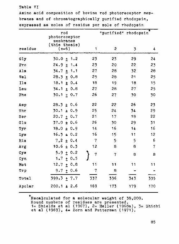
Table VI
Amino acid composition of bovine rod photoreceptor mem
branes and of chromatographically purified rhodopsin,
expressed as moles of residue per mole of rhodopsin
rod "purified" rhodopsin
photoreceptor membranes
(this thesis) residue
Gly
Pro
Ala
Val
Ile
Leu
Phe
Asp
Thr
Ser
Glu
Туг
Lys
His
Arg
Cys
Суп
Met
Тгр
Total
Apolar
(n=
30.0
24.9
34.7
28.3
1Θ.1
34.1
30.1
28.3
30.1
20.7
37.0
18.0
16.3
7,2
10.6
5.9
1.7
12.7
9.7
399.3
200.1
=6;
+
+
+
+
+
+
+
±
±
+
±
±
±
±
±
+
+
+
+
+
±
)
1.2
1.4
1.1
0.8
0.4
0.8
0.7
0.6
0.9
0.7
0.6
0.9
0.2
0.4
0.3
0.2
0.5
0.8
0.6
7.7
2.6
1
23
23
27
25
18
27
26
22
25
21
26
14
16
7
12
) 7
11
7
337
169
2
23
20
28
28
19
28
27
22
24
17
30
16
15
5
8
7
11
8
336
173
3
29
22
32
21
18
27
30
26
34
18
29
14
11
5
8
8
11
343
179
4
24
23
28
25
15
25
30
23
29
22
31
16
12
6
7
8
11
-
335
170
# Recalculated for a molecular weight of 38,000. Round numbers of residues are presented. 1= Shields et al (1967), 2= Heller (1968a), 3= Shichi et al (1969), 4= Zorn and Putterman (1971).
85

in the шешЪгапе preparations have been evaluated. The
ninhydrin reaction has the disadvantage that the observed
molar absorbance coefficients differ markedly for different
amino compounds. This is especially true for membranes,
since these contain both protein and lipid amino groups.
In addition, secondary amino groups interfere with this
assay, and so do the amidine groups resulting from the
reaction of primary amino groups with various imidate
esters (cf. chapter 4), either directly or following
hydrolysis of this acid labile group. In the Sörensen
titration on a micro scale difficulties were encountered
with the formaldehyde reagent, due to its slow oxidation
to formic acid.
This has led us to investigate the suitability of
TNBS for the determination of primary amino groups (Fig.'
13). This reagent has been employed before, e.g. by
Habeeb (1966) and by Pull (1970) who measured the absor
bance at 340 nm of the N-trinitrophenyl derivative, and
by Fields (1971 and 1972) who measured the absorbance at
420 nm of the complex with sulfite ions. Although the
latter variation is well suited to the assay of free amino
compounds, we have observed irreproducible results in the
case of proteins or membranes, apparently due to a sharp
decrease in binding constant of the sulfite complex. Hence,
determination of the absorbance of the N-trinitrophenyl
group at 340 nm has been used. Further points that have
been studied are the molar absorbance coefficients of the
N-trinitrophenyl derivatives in view of the contradictory ( 340 v a l u e s reported by different authors (Satake et al,
1960; Okuyama and Satake, 1960; Goldfarb, 1966; Habeeb,
1966; Kakade and Liener, 1969), the influence on the
absorption spectra of sulfite released during the reaction
(Goldfarb, 1966; Fields, 1971) and the possible reaction
of TNBS with thiol groups (Kotaki et al, 1964; Freedman
and Radda, 196Θ).
86
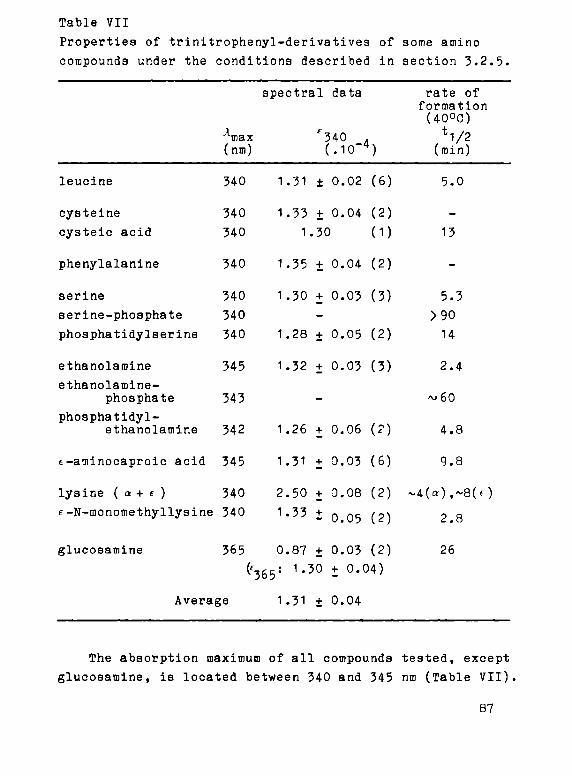
Table VII
Properties of trinitrophenyl-derivatives of some amino
compounds under the conditions described in section 3.2.5.
spectral data rate of formation (40
oC)
лтах
f340 ,
tl/2
(nm) (.10~4) (min)
leucine 340 1.31 ± 0.02 (6) 5.0
cysteine
cysteic acid
phenylalanine
serine
serine-phosphate
phosphatidylserine
ethanolamine
ethanolamine-
phosphate
phosphatidyl-
ethanolamine
t-aminocaproic acid
340
340
340
340
340
340
345
343
342
345
1.33
1,
1.35
1.30
1.28
1.32
1.26
1.31
+ 0.04
.30
+
+
-
+
+
_
+
+
0.04
0.03
0.05
0.03
0.06
0.03
(2)
(1)
(2)
(3)
(2)
(3)
(?)
(6)
-
13
-
5.3
>90
14
2.4
^60
4.8
9.8
lysine ( a+ e ) 340 2.50 + 0.08 (2) ~4(»)f~8(f)
E-N-monomethyllysine 340 1.33 ± Q 05 (2) 2 8
glucosamine 365 0.87 + 0.03 (2) 26
(E
3Sy 1·50 + 0.04)
Average 1.31 ± 0.04
The absorption maximum of all compounds tested, except
glucosamine, is located between 340 and 345 nm (Table VII).
87

Under the conditions indicated (cf. section 3.2.5.)» the
reaction with TNBS proceeds via pseudo first order kinetics,
showing half times of 3-6 min for most a--amino groups and
8-10 min for (-amino groups (Table VII). The values for f340
o f a 1 1 compounds tested are about equal, yielding
an average value of 13,100 (SE 400, n=11). This compares
well with a value of 12,600 reported by Satake et al (i960)
for a variety of amino compounds and the value of 13,500
reported by Habeeb for the f-amino group of lysine. For
glucosamine the same molar absorbance coefficient is found
at its absorption maximum of 365 nm. Cysteine gives results
comparable to the other amino compounds, which implies
that either the sulfhydryl group does not react or the
300 350 400 « 0 300 530 300 350 400 150 500 530 wavelength (nm )
Pig. H . Spectra of trinitrophenylated materials before (dashed line) and after (solid line) acidification. The trinitrophenylation method is described in section 3.2.5.
8Θ

S-trinitrophenyl group is unstable under our conditions,
аз suggested by earlier studies (Kotaki et al, 1964). At
higher pH (6-9) the free amino compounds yield two absorp
tion maxima upon reaction with TNBS, one at 340 nm deriving
from the N-trinitrophenyl group and one at 420 nm deriving
from its complex with liberated sulfite ions. Upon acidi
fication the latter maximum is reduced to a small shoulder
inherent to the free N-trinitrophenyl group (Fig. 14).In
the case of bovine serum albumin (BSA) and photoreceptor
membranes the peak at 420 nm barely develops (Pig. H ) ,
so that the course of the reaction can easily be followed
by observing the absorbance increase at 348 nm. The
reaction with most α-amino groups is complete within
half an hour. The presence of a negatively charged group
as in ethanolamine-phosphate, serine-phosphate and cys-
teic acid markedly decreases the reaction rate (Table VII).
The reactivity of the corresponding phospholipids, phoa-
phatidylethanolamine and phosphatidylserine, is interme
diate between that of the amino compounds and their
phosphate derivatives. In the case of photoreceptor mem
branes the reaction was complete within 1 hr. Determination
of the amount of amino groups present in a commercial
sample of BSA (BDH) by the TNBS procedure, using a value
for £-iän of 13,100 shows good agreement with the amount
calculated from amino acid analysis: Θ06 (SE 11, n=3)
and 810 (SE 14, n=3) nmole/mg respectively. These findings
permit the conclusion that TNBS lends itself for quanti
tative determination of primary amino groupa employing the
procedure described in section 3.2.5. and a value of ε340
= 15»100.
Determination with TNBS yields an average of 424
(SE 4, n=5) nmolee of amino groups per mg of lyophilized
photoreceptor membranes or 52.3 (SE 0.6) moles expressed
per mole of rhodopsin. The amino group containing compounds
have been identified by means of thin layer chromatography
89
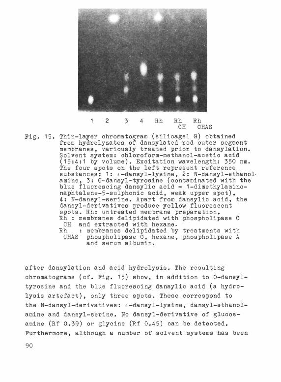
1
Pig. 15. Thin-layer chromatogram from hydrolyzates of da membranes, variously tr Solvent system: chlorof (15:4:1 by volume). Exc The four spots on the 1 substances; 1: (-dansyl amine, 3: O-dansyl-tyro blue fluorescing dansyl naphtalene-5-sulphonic 4·: N-dansyl-aerine. Apa dansyl-derivatives prod spots. Rh: untreated me Rh : membranes delipida
GH and extracted with Rh : membranes delipi CHAS phospholipase G,
and serum albumi
Rh Rh
GH
Rh GHAS
(silicagel G) obtained nsylated rod outer segment eated prior to dansylation. orm-methanol-acetic acid itation wavelength: 550 пш. eft represent reference -lysine, 2: N-dansyl-ethanol-sine (contaminated with the ic acid = l-dimethylamino-acid, weak upper spot), rt from dansylic acid, the uce yellow fluorescent mbrane preparation, ted with phospholipase G
hexane. dated by treatments with hexane, phospholipase A n.
after dansylation and acid hydrolysis. The resulting
chromatograms (cf. Pig. 15) show, in addition to 0-dansyl-
tyrosine and the blue fluorescing dansylic acid (a hydro
lysis artefact), only three spots. These correspond to
the N-dansyl-derivatives: Í-dansyl-lysine, dansyl-ethanol-
amine and dansyl-serine. No dansyl-derivative of glucos
amine (Rf 0.39) or glycine (Rf 0.45) can be detected.
Purthermore, although a number of solvent systems has been
90

Table Vili
Amino acid analysis of rod photoreceptor membranes before
and after dansylation.
Results expressed per mole of rhodopsin. 4 experiments
aiy Asp Thr
Glu Pro
Ala
Val Met
H e
Leu Phe His Arg
before
30.3
30.4
31.4
37.7
25.6
35.3
28.5
12.7
18.2
35.0
30.4
7.2
10.4
after
31.1
29.1
31.9
38.3
23.8
34.9
27.9
13.9
18.1
34.7
31.0
7.2
10.5
¿ w 0
+3Í2
-4І4
+2і2
+2+2
-7+5
-1+1
-2+4
+9±7
-1+4
-1±1
+2±3
0+3
+1+6
Gluc-NH2
Tyr
Lys
Ethanol-amine
Ser(protein)
Ser(lipid)
before
3.6
18.2
16.1
26.9
22.0
8.9
after д(Я0
3.8 +7±10
5.4 -70+6
4.0 -75±2
(0.4)(-98+4)*
4.9 -82ІЗ
(0.6)(-98+5)#
22.4 +2±2
2.3 -74+4
(-0.1)(-101+8)*
Cys (partially) and Try destroyed during hydrolysis 0Д = the difference with absolute standard error be
tween the first two columns, in percentage. * The values between parentheses are obtained after correction for the release of the amino acid from its dansylderivative during hydrolysis (Table IX),
employed, no other or-N-dansyl-derivatives in amounts
exceeding one tenth the amount of rhodopsin were detec
table. The a-N-dansyl-serine derives entirely from phos
pholipids, since it disappears upon enzymatic delipidation
of the photoreceptor membrane (Pig. 15). In confirmation,
amino acid analysis of dansylated membranes shows no
significant decrease in glucosamine, glycine or any other
amino acid except lysine, ethanolamine, serine and tyro
sine (Table VIII).
91

Table IX
Loss of dansyl-derivatives during acid hydrolysis (6.7 N
HCl, 1Θ hr, 110oC) and quantitative thin layer chromato
graphy. η = number of experiments
recovery after recovery acid hydrolysis after TLC
(n=3) (n-4)
original dansyl-compound amide
f-N-dansyl-lysine 77 ± 3 ft traces 94· ± 3 fi
t-N-methyl,f-N-
dansyl-lysine 20-30 JÉ
N-dansyl-serine 49 ± 3 # 24 ± 2 fi 92 ± 3 fi
N-dansyl-ethanolamine 84 Í 3 fi traces 95 ± 4 fi
- : not determined
It thus appears that only three species contribute to
the primary amino group population in the membrane: the
phospholipids phosphatidylserine and phosphatidylethanol-
amine and the protein lysine-residues, implying that in
rhodopsin the N-terminal and glucosamine amino groups
are blocked.
A quantitative determination of the dansylated amino
groups requires the following conditions: (a) dansylation
should be virtually quantitative, (b) corrections should
be applied for losses suffered during hydrolysis (Gray,
1967; Seiler, 1970) and extraction from the TLC plates,
(c) the exact amount of protein originally present should
be determined carefully, preferably by amino acid analysis.
Completeness of dansylation has been checked by amino
acid analysis of dansylated membranes. Hydrolysis losses
have been determined on the pure dansyl compounds (Table
IX). The loss of N-dansyl- derivatives other than «-deriva
tives appears to be mainly due to cleavage of the amide
92

bond, generating dansylic acid and the original amino
compound. In the a-amino-derivativa N-dansyl-aerine,
however, cleavage of the C-N bond appears to occur almost
aa easily as cleavage of the amide bond (Table IX), in
agreement with earlier observations (Seiler, 1970).
When these corrections are applied to the amino acid
analysis, the dansylation appears to be virtually quan
titative (Table VIII). Table IX also shows the relative
amounts of dansyl-compounds recovered after thin-layer
chromatography and extraction. For the amounts used in
our experiments, the loss of different dansyl-derivatives
is independent of the total amount applied. Correction
for these losses may therefore be avoided by the use of
reference substances.
Calculation of the amino compounds present in un
treated membranes upon application of the corrections
from Table IX gives results in good agreement with those
from amino acid analysis (Table X). Likewise, analysis
of delipidated membranes confirms the results reported
by Borggreven et al (1971). In addition, the total amount
of amino groups calculated from both methods agrees well
with the total amount determined by TUBS. The three methods
yield an average number of 420 (SE 10) nmoles of amino
groups per mg of lyophilized material or 52 (SB 1) moles
expressed per mole of rhodopsin (Table X).
3.3.3. Sulfhydryl groups
The number of cysteinic acids determined after per-
formic acid oxidation and amino acid analysis amounts to
10-11 per mole of rhodopsin. After reduction and determi
nation with DTNB or vinylquinoline a total number of 8-10
sulfhydryl groups are detected. The three methods yield
an average of 9.4 (SE 0.9) moles of cysteine expressed
per mole of rhodopsin. Direct sulfhydryl determination in
93

SOS yields 6 cysteine residues per rhodopsin (Table VI).
Since SDS does not attack disulfide bridges, it appears
that about two disulfide bridges due to two cystine resi
dues are present in the photorecptor membrane per rhodopsin
molecule.
5.5.4. Rhodopsin as photoreceptor membrane protein
Prom the sedimentation behaviour of rhodopsin micelles
in digitonin solution, the molecular weight of rhodopsin
Table X
Primary amino groups, including the chromophore binding
group, present per mole of rhodopsin in untreated and
delipidated rod outer segment membranes. Averages with
standard errors from η experiments.
amino-compound
source
untreated
AAA* (n=5)
DansCl (n=4)
TUBS* (n=5)
delipidated
DansCl (n=1)
Borggreven et al (1971)
lysine
protein
16.2 ± 0.2
16.4 ± 0.8
1 7 + 2
-
ethanolamine
phos
26.θ ± 0.6
27.9 ± 1.0
<0.3
0.3 Ì 0.1
serine
pholipid
8.8 ± 0.2
8.2 + 0.6
1.8 ± 0.6
1.9 ± 0.3
total
51.8 ± 0.7
52.5 Í 1.4
52.3 ± 0.6
AAA s amino acid analysis, DansCl = dansylation procedure. TNBS = trinitrophenylation procedure
94

hae been estimated at about 4-0,000 (Hubbard, 1954). Recent
ly, however, a value of about 28,000 has been calculated
from amino acid analysis of chromatographically purified
bovine rhodopsin preparations (Shields et al, 1967; Heller,
1968a). We have tried to resolve this discrepancy by em
ploying electrophoresis on Polyacrylamide gels in the
presence of 0.1$ SOS. This method gives a direct propor
tionality between electrophoretic mobility and the loga
rithm of the molecular weight for both soluble and inso
luble proteins, which do not contain large amounts of
covalently bound carbohydrates (Weber and Osborn, 1969).
SDS-gel electrophoresis of our membrane preparations
shows the presence of only one major band. By calibration
with proteins of different known molecular weights (cata-
lase, MW 60,000; aldolase, MW 40,000; trypsin, MW 23,000;
Д-lactoglobulin, MW 18,000) a molecular weight of 38,000
(SE 1000) is found for this band (Daemen et al, 1972).
This protein clearly represents opsin, since upon reduc
tive fixation of the retinaldehyde group to opsin by
treatment with NaBH. during illumination, the fluorescence
of the resulting retinyl-group is localized in the same
band. This confirms the higher value for the molecular
weight of rhodopsin.
Prom the amino acid analyses we calculate that 44,700
(SE 900, n=6) gram of protein is present per mole of visual
pigment in the photoreceptor membrane. Taking a molecular
weight of 38,000 for opsin, this implies that rhodopsin
is by far the predominant species of the membrane protein
population in the bovine rod photoreceptor membrane,
accounting for 85$ of the total membrane protein (Daemen
et al, 1972). This result has recently been confirmed
for cattle as well as for frog (Heitzmann, 1972; Robinson
et al, 1972).
Calculations of the molar absorbance coefficients of
rhodopsin at 500 nm from the ΔΑΕΛ,Λ, retinaldehyde content
95

and a molecular weight of 38,000 yieida the same value
for membranes, detergent solutions and chromatographically
purified preparations, averaging 40,800 (Баешеп et al,
1972). This again confirms the earlier value (40,600)
obtained in Wald's laboratory (Wald and Brown, 1953)
contrary to the lower value (21,000-23,000) reported by
Heller (1968a).
3.4. Discussion
Our results indicate, that the rod photorecptor mem
brane may be considered as a fairly pure rhodopsin pre
paration (31$ of dry weight, 85$ of protein weight). Since
the use of detergents has some serious drawbacks, which
will be discussed in more detail in the last chapter, we
consider the use of photoreceptormembrane suspensions
for the study of the properties of rhodopsin to be pre
ferable to the use of detergent solubilized rhodopsin
preparations.
The chemical composition of our membrane preparations
is very similar to the one reported by Nielsen et al
(1970). The lower lipid content of other preparations
(Table V) may be due to the presence of contaminations.
We do not confirm recent observations of Mason et al
(1973b), that up to 20$ of the lipids in bovine photo
receptor membranes consist of glycolipids. Since they
also employed the anthrone reaction, their results may
be influenced by the side reaction which we have observed
and for which they have apparently made no correction.
The low sugar content of the membrane further justifies
attempts to purify rhodopsin by means of sugar-binding
proteins like concanavalin A.
We find practically no difference in sugar content
of washed membranes and intact outer segment structures
expressed per dry weight of membranes present. This
96
contradicts the presence of polysaccharides or glycopro
teins in an interdiscal matrix. Electronmicroscopic ob
servations also give no indication for the presence of
a carbohydrate containing matrix between the discs (G.J.
Jones, personal communication; Godfrey, 1973).
The protein/phospholipid ratio of the photoreceptor
membrane is relatively low in comparison to other membranes,
which may be related to the relatively high mobility of
rhodopsin. The amino acid composition confirms the high
degree of hydrophobicity of membrane proteins, about 50$
of the residues having an apolar character. Since purified
rhodopsin shows about the same percentage of apolar
residues (Table VI), this means that the other membrane
proteins must have about the same degree of hydrophobicity
as rhodopsin.
Reporter and Heed (1972) have recently reported the
presence of a high number of methylated basic residues
(histidine, lysine, arginine) in their bovine rhodopsin
preparations. Of the 10 lysines per rhodopsin 2 would
contain a mono- and 4 a di-methylated e-amino-group. They
also report the incorporation of radioactive methyl groups
in lysine, histidine and arginine groups of rhodopsin and
other membrane proteins upon in vitro incubation of entire
retina's in the presence of radioactive methionine. In
order to check this, we have calibrated our amino acid
analyzer with 1-methyl reap, 3-methyl histidine and with
f-monomethyl- andc-dimethyl-lysine and found that these
methylated amino acids could be separated from the common
ones. Upon subsequent careful analysis of our membrane
hydrolyzates we detect no traces of methylated lysine or
histidine. In agreement with this, we find all lysine
residues reacting with TUBS. This reagent reacts with
secondary amino-groups only under more drastic conditions
and very slowly (Means et al, 1972), while under our
assay conditions we detect no reaction with proline or
97
the f-amino-group of <-monomethyl-lyaine. Finally, dansyl-
chloride yields upon reaction with the latter group <-
methyl,<-danaylamino-lysine, which is much less stable
towards acid hydrolysis than f-dansyl-lysine (Table IX)
and has a higher Rf value (0.18) in the solvent system
chloroform-methanol-acetic acid. The number of amino
groups shown in Table X therefore represents only primary
amino groups. These findings raise serious doubts about
the results of Reporter and Reed, which are further
emphasized by the very aspecific nature of their enzymatic
methylation reaction in which all basic residues of all
proteins appear to become methylated.
After dansylation of rod outer segment membranes,
hydrolysis and TLC only f-dansyl-lysine, dansyl-serine
and dansyl-ethanolamine are observed. No dansyl-glucosamine
is detected, which indicates that all four glucosamine-
amino groups present per rhodopsin molecule (Table VIII)
are blocked, as previously reported by Heller and Lawrence
(1970). No significant amounts of a-dansyl-amino acids
have been detected, except for dansyl-serine which derives
entirely from phosphatidylserine. Dansylation of enzyma-
tically delipidated preparations (cf. Borggreven et al,
1971 and 1972) only yields f-N-dansyl-lysine and 0-dansyl-
tyrosine. This indicates that rhodopsin has no free N-
terminal group as has previously concluded from reaction
with dinitrofluorobenzene (Albrecht, 1957; Tsukamoto et
al, 196I). The small peptide with glycine as N-terminal
amino acid present in a 1:2 ratio to rhodopsin in Albrecht's
preparation is apparently not present in our membranes.
The sulfhydryl contents of photoreceptor membranes
and purified rhodopsin preparations reported by various
groups do not give a consistent picture (Heller, 1968a,b;
Shichi et al, 1969; Zorn and Futterman, 1971). This is
presumably due to the rapid air oxidation of sulfhydryl
groups, which we observe in detergent solubilized membrane
98

preparations even at 4 С, the rate being dependent on the
detergent: SDS> СТАВ>Triton X-100>digitonin. In suspen
sion this oxidation does occur, but proceeds very slowly.
At higher temperature of pH the oxidation rate is consi
derably increased.
We detect a total of 6 sulfhydryl groups in the photo
receptor membrane expressed per rhodopsin molecule, a
figure also reported elsewhere (Zorn and Putterman, 1971).
In addition about two disulfide bridges appear to be
present. Heller (1968b) reports two disulfide bridges in
purified rhodopsin preparations, but only one free sulf
hydryl group, suggesting that air oxidation may have
interfered in his experiments. Since rhodopsin represents
85% of all membrane proteins (Tables V and VI) not all 6
cysteine and 2 cystine residues need necessiraly occur
in rhodopsin. However, experiments with sulfhydryl group
reagents indicate that the rhodopsin protein contains
at least 4 sulfhydryl groups (cf. chapter 5). The disulfide
bridges are only reduced under harsh conditions employing
a strong detergent like SDS. In suspension we do not ob
serve any reduction in the presence of mercaptoethanol,
regardless of whether the membranes are illuminated or
not.
99

CHAPTER 4
FUNCTIONAL ANALYSIS OF AKINO GROUPS IN THE НОГ PHOTO
RECEPTOR MEMBRANE
4·1. Introduction
Selective chemical modification of amino acid side
chains has proven to be a valuable tool in studying
protein structure and "active site amino acids". More
recently, modification of functional groups has been
extended to membranes as well (cf. Bretscher, 1973;
Zwaal et al, 1973; Schmidt-Ullrich et al, 1973). However,
in the study of rod outer segment membranes and visual
pigments, this technique has only incidentally been used
so far (Wu and Stryer, 1972; Eratz et al, 1972 and 1973;
Heitzmann and Richards, 1973).
The limitations of this approach should be clearly
borne in mind. The possibility of secondary effects on
the general structure of membranes and/or proteins is
always to be considered. The specificity of group reagents
is rarely absolute. Conditions prevailing within membranes
or at membrane surfaces may differ drastically from the
bulk aqueous phase, thereby altering the reactivity of
modifying agents. Finally, unequivocal assignment of a
specific role to a particular functional group in membranes
or proteins is not well possible as long as the complete
(three-dimensional) structure of the objects under study
is not known. This clearly the case for rod outer segment
membranes and their membrane proteins, e.g. rhodopsin.
Nevertheless, many conditions for useful research
seem to be fulfilled in rod outer segment membranes: 1.
Amino groups in the membrane play a crucial role in the
binding of the chromophoric group, both in rhodopsin as
well as in other membrane components upon migration of the
100

chromophore following illumination. In this context, it
is important to realize that the membrane phospholipids
phosphatidylethanolamine and phosphatidylserine contain
reactive amino groups. 2. Sulfhydryl groups have been
tentatively connected with the excitation process, in view
of their redox properties (Wald, I968). They are also
known to exert catalytic functions in enzymatic activi
ties, which are also thought to be present in the rod
photoreceptor membrane (retinol:NADP oxidoreductase,
ATPase, adenylate cyclase). 3. Aromatic amino acid side
chains may furnish the hydrophobic cleft in which the
chromophoric group is hidden, and may be responsible
for the specific interactions which cause the red shift
of the α-peak of the pigment.
As our first target for chemical modification studies
we have chosen the primary amino groups of the membrane,
since knowledge of these groups seems to be important for
a fuller understanding of the structure and function of
the photoreceptor membrane. There are at least four ways
in which these groups could participate. First, the
characteristic absorption spectrum of rhodopsin is due
to the chromophoric group, 11-cis retinaldehyde, linked
to a primary amino group, presumably by means of a proto-
nated aldimine bond (cf. section 1.3.3.)· Secondly, the
ease with which retinaldehyde in synthetic retinylidene-
imines transiminizes from one amino group to another
(de Pont et al, 1970b; Баетеп et al, 1971) suggests that
after illumination the isomerized chromophoric group may
move by transimization to other amino groups. Thirdly,
primary amino groups could play an important role in
enzymatic activities present in the photoreceptor membrane,
like the retinol:NAOP oxidoreductase. Fourthly, amino
groups contribute to structure and stability of the mem
brane through their role in electrostatic interactions.
As discussed above, in studying effects of chemical
101

modification of specific protein side chains, side effects
like alterations of protein conformation should be preven
ted as much as possible. We therefore sought a mild amino-
group reagent. Methylacetimidate, introduced by Hunter and
Ludwig (1962), seemed to suit our purposes almost perfectly.
It reacts already under mild conditions (pH 7-9) exclusively
with primary amino-groups and it forms an acetamidine group,
which is not much larger than the original amino-group
and is also positively charged (Figs 16 and 17). Hence,
even extensive acetamidination affects protein conformation
H - N - H - t 1
H I • OCH, i ι....μ -¿J
С
Β NH
A'-IIDINATION
pH7-9
N - H ¿ . / 4
R N H 2
R — C H j (methyl acetmmdate)
O / (methylpiccolin-imidate)
FLUOROBENZENE DERIVATIVES
pH β - 1 0
Η - N NO2
R
R -NO2 (dimtroFluorobenzene)
R R -N^CHjî j (trimethylaminomtrofluorobenzene)
SUCCINYLATION
H-N-H 1 ©
H
н,с-с*0 pH6-9
H , C - C .
euccmic anhydride
H-N \ c=o /
C H 2
\ C H 2 /
c - o І-э
о
+ 2Н +
Pig. 16. Overall reaction equations of the amino group modifications described in this chapter.
102

only to a small extent (of. Means and Peeney, 1971).
In evaluating the effects of amino-group modification
we have investigated the following parameters: (1) the
importance of the positive charge on the amino groups,
(2) the effect of the size of the substituent and (3) the
influence of the charge of the modifying agent. In addi
tion to methylacetimidate, we have used the following
reagents (Pigs 16 and 17): methylplccolinimidate (Benisek
and Richards, 1968) which is uncharged at pH 7 (pK~5),
v^ &** ^ ^ F гитггижіші- raes;
MXTAMIDINO- (MA!; PICCOUKEAMIOINO· ( » I )
SUCCINYL- (SA) Ш D 2-N!-R04-TRI4ETH»L-^ Ц Р ^ Г M1INCPHENYL-
Pig. 17. Space-filling models of lysine and its derivatives obtained after reaction with trinitroben-zene sulphonic acid (TNBS), methylacetimidate (MAI), methylpiccolinimldate (MPI), succinic anhydride (SA) and trimethylaminonitrofluorobenzene iodide (TNPB) respectively.
103

introduces an aromatic ring and preserves the positive
charge of the amino-group; Trinitrobenzene sulphonic acid
(TUBS; Fig. 13), which is negatively charged and intro
duces a bulky uncharged substituent; dinitrofluorobenzene
(ONTE) which is uncharged and introduces a large uncharged
group; trimethylamino-nitro-fluorobenzene iodide (TANPB),
which is positively charged and introduces a bulky sub
stituent under preservation of the positive charge; suc
cinic anhydride (SA) , which is uncharged and introduces
a relatively small negatively charged substituent.
The effects of these amino group reagents on the
spectral, photolytic and regenerative properties of rho-
dopsin and opsin, and the activities of the retinol:NADP
oxidoreductase and Na , К - ATPase have been studied.
4.2. Materials and methods
4.2.1. Materials
Methylacetimidate was synthesized according to Hunter
and Ludwig (1962). The final product, a white solid, is
stored as the hydrochloride salt in dry air over solid
KOH at 40C. The purity of this and the other synthesized
compounds has been checked by determining melting points
and by elemental analysis. Methylpiccolinimidate was
synthesized according to Schaefer and Peters (1961). The
colorless liquid is stored in dry air over solid KOH at
4 C. Trimethylamino-nitrofluorobenzene iodide, introduced
by Sutton et al (1972) as a more water soluble, though
less reactive substance than dinitrofluorobenzene, was
synthesized according to Bevan et al (1968). It is stored
at 4 C. All other materials were of the highest purity
available.
104

4.2.2. Amidination
Amidination is performed in aqueous membrane suspen
sions at room temperature. Lyophilized or frozen membranes
(10 mg per ml) are suspended in 0.2 M phosphate buffer
(pH. 7.3) by homogenization in a Potter-Elvehjem tube.
For the reaction with methylacetimidate, an equal amount
of its hydrochloride (20-fold molar excess with respect
to the number of amino groups) is added and the pH is
readjusted to 7.5 by cautious addition of 1 N NaOH under
stirring. After half an hour most of the reagent has been
consumed by reaction with amino groups or by hydrolysis,
and the pH has risen to about 7.8. The soluble reaction
products, mainly acetamide and methanol, are then removed
by sedimentation of the membrane fragments (80,000 χ g,
40C, 30 min). The sediment is either resuspended in the
phosphate buffer to start another treatment, or it is
washed twice with double-distilled water and stored with
or without prior lyophilization at -700C in a light-tight
container. In order to achieve more than 95$ modification
five treatments with methylacetimidate are necessary and
double and quadruple amounts of methylacetimidate should
be added during the fourth and fifth cycle respectively.
Membrane fragments, which have been stored frozen, give
similar results as lyophilized preparations upon amidina
tion. The reaction with methylpiccolinimidate does not
require multiple treatments, since this compound is
hydrolyzed by water at a much slower rate than methyl
acetimidate. To the membrane suspension a 10 to 50-fold
molar excess of methylpiccolinimidate is added. In order
to compensate the alkalinization, the pH is readjusted
to 7.3 every 30 min by cautious addition of 1 N HCl under
shaking. Aliquote, taken from the suspension at various
time intervale, are rapidly diluted with the same phos
phate buffer and centrifuged (80,000 χ g, 40C, 30 min).
The sediment is washed twice, either with the phosphate
105

buffer and stored at 40C, or with double-distilled water,
lyophilized and stored at -70oC. Storage without prior
lyophilization is preferable when larger groups have been
introduced. In all cases appropriate control experiments
are performed.
The various washings during the amidination procedure
cause a 20-30^ loss of material. However, no specific
losses of protein or lipid occur, since amino acid analysis,
phosphorus determination and retinaldehyde determination
(cf. chapters 2 and 3) indicate that no significant changes
in protein-, phospholipid- or retinaldehyde-content take
place during modification, while the amino acid composition
also remains the same. Moreover, Δλ,-ηη and recombination
capacity of the controls do not change during either of
the amidination procedures.
4.2.3. Trinitrophenylation
Modification of rod photoreceptor membranes (0.5-5
mg/ml) with trinitrobenzene sulphonic acid (TNBS) is per
formed in 0.07 M bicarbonate (pH 8.0), either in suspen
sion or in the presence of 0.6$ Triton X-100 reap. SDS.
A 15-fold molar excess of TNBS is employed at various
temperatures. The reaction is followed by recording ab
sorption spectra (280-610 nm, rate: 5 nm/sec) at various
time intervals or by continuous recording the absorbance
at 348 nm,with a Rapid T3 spectrophotometer (Howaldtswerke-
Deutsche Werft, Kiel, W.Germany), which permits measurement
of turbid solutions by placement of the cuvettes immedia
tely in front of the multiplier system,
4.2.4. Modification with fluorobenzene derivatives
Reaction of photoreceptor membranes (2-10 mg/ml) with
2,4-dinltrofluorobenzene or 4-trimethylamino,2-nitrofluoro-
106

benzene iodide is performed in 0.07 N bicarbonate (pH 8.0)
at 20oC or 40
0C. A 10 to 50-fold molar excess of reagent
is employed. After various time intervals aliquota are
taken from the suspension, diluted with 0.2 M phosphate
buffer pH 7.0 and centrifuged (80,000 χ g, 40C, 30 min).
After two washings the sediment is stored at 40C.
4.2.5. Succinylation
Succinylation is performed in the same way as the
reaction with methylacetimidate, except that the incu
bation time of each step is reduced to 15 min in view of
the more rapid hydrolysis of succinic anhydride.
4.2.6. Analytical techniques
The extent of modification obtained by reaction with
the fluorobenzene derivatives can be calculated from
amino acid analysis or be estimated from their own absorp
tion at 360 nm (Hirs, 1968), The amidine groups resulting
from reaction with the imidate esters, do not absorb out
side the ultraviolet and are hydrolyzed to a large extent
under the conditions required for complete acid or basic
hydrolysis of proteins. Therefore, we have measured the
extent of modification in this case by determining the
remaining primary amino groups. The trinitrobenzene
sulphonic acid method (cf. section 3.2.5.) is well suited
for this purpose in view of its reliability and convenience.
Other analytical techniques are performed as described
in the preceding chapters.
4.2.7. Assay of retinol:NAOP oxidoreductase activity
This assay is performed in the light, largely as
described by Rotmans (1973). To 200-300 μΐ of a suspension
of untreated or modified photoreceptor membranes (2-10 mg/ml)
107

in 0.2 M phosphate buffer (pH 6.5-7.0), 25-50 nmoles of
all-trans retinaldehyde in 5 «1 methanol are added. The
sample іэ brought to 370C, whereupon a 25 ,»1 aliquot is
withdrawn for determination of the retinaldehyde concen
tration at t=0. Subsequently a five-fold excess of NADPH
in 10 "1 of water is added (t=0), and at various time
intervals 25 "1 aliquots are removed and frozen immediately
in an acetone-dry ice bath. After the collection of the
aliquote is completed, 300 ml of thiobarbituric acid
reagent (cf. section 2.2.3.) is added to the samples.
The retinaldehyde concentration in the aliquots is deter
mined as described in section 2.2.3. and plotted емиз
time of incubation. From the resulting curves the initial
velocities are calculated.
4.2.8. Photolysis
In order to assess the effect of amino group modifi
cation on the formation of photolytic intermediates,
untreated as well as modified membrane preparations (1-2
mg/ml or 8-16 nmoles of rhodopsin per ml) are suspended
in 0.2 M phosphate buffer (pH 7.0). The suspension is
placed in a water-jacketed cuvette in the Rapid T3 spectro
photometer and flash-bleached by means of a Rollei
Strobofix flasher (t1 AJ
o f ^e fias decay <0.2 msec).
Before the flash and at various time intervals thereafter
spectra are recorded (280-610 nm, 5 nm/sec). Бие to
blinding of the photomultiplier the first spectrum can
only be taken 100-200 msec after the flash. Thus, it has
not been possible to follow the first rapid part of the
photolytic sequence, i.e. up to metarhodopsin II (cf. Pig.
6). Various bandfilters have been employed (Schott-Jena,
W.Germany; thickness 3 mm), cutting off all wavelengths
under reap. 460 nm (GG,-o(0» 510 nm (OGCTQ) and 570 nm
(OGCQQ). Although the extent of bleaching varies somewhat
108

for the various filters, no differences are observed in
the slower part of the photolytic reactions. The tempera
ture (15, 25 and 370C) is regulated Ъу means of a Lauda
Kryoatat equipped with an extra thermostat.
4.3. Results
4.3.1. Anidination
rfhen the amidination with methylacetimidate is carried
out at room temperature and pH 9 in a pH-stat, the results
lack reproducibility. After amidination of 40-70$ of the
amino-groups, the 500 nm absorbance decreases rather
rapidly, while the recombination capacity in some cases
ia abolished at an even lower percentage of amidination.
Amidination at pH 7.5 in phosphate buffer gives reprodu
cible results. Experiments carried out in the dark on
non-illuminated samples yield 68 $ modification after one
treatment (Pig. 18a). Subsequent treatments increase this
percentage more gradually, until after the fifth treatment
about 98$ modification is achieved. This means that 2/3
of the amino groups are very readily accessible. The results
also indicate that amidination up to 80?6 has little impact
on photoreceptor membrane properties: the 500 nm absorbance
and even the entire absorption spectrum remain practically
unchanged (Pig. 19), the recombination capacity is not
significantly decreased and the retinol:NADP oxidoreductase
activity decreases by less than 25 $.
Upon further modification ДА 0
decreases slowly to
about 70$ of the original value, when amidination is
nearly complete. Concurrent release of retinaldehyde is
manifested by an increase of the 380 nm absorbance (Pig.
19). The recombination capacity of the remaining rhodopsin
also decreases slowly from about 90$ to about 70$. The
109

j ι ; ι 1 1 1 1 1 1 >—I
0 50 % 100
percentage of ¿ιι-πιησ ¿roups modified
Fig. 18. Influence of amidination with methylacetimidate on photoreceptor membrane properties. Amidination was performed as described in section 4.2.2. The arrows indicate the start of a new amidination treatment. The percentage of amidination was de-determined by reaction of the unmodified amino-groups with TNBS. a. Amidination performed in darkness. Effect of
amidination on ДАзооД-^—)t recombination
capacity (—Ф—) and retinol:NADP oxidoreduc-tase activity (—A-). The values in non-amidi-nated controls are in each case taken as 100$.
b. Amidination performed in the light. Effect on recombination capacity (—·—). For comparison, the influence of amidination in darkness on the recombination capacity is also shown (—n—). The 4A50O of the preparations before illumination is taken as 100$.
110

10
08
Об « ε Ц
л L О «I
Ч 0 4
0 2
0 2S0 360 440 520 600
wavelength (л m)
Fig. 19. Spectra of rod outer segment membranes, amidinated to various extent with methylacetimidate. Îhe spectra were determined in 2$ digitonin in 67 raM phosphate buffer (pH 6.5) containing 50 шМ hydroxyl-amine, and are recalculated to 1 mg lyophilized material per ml detergent solution. Spectra are shown for non-amidinated (0, —o— ) , twice amidinated (2, -á-, 80$ modification) and five-fold amidinated material (5, - · - , 97$ modification). The peak at 355 nm in the third spectrum derives from retinylidene-oxime} in the absence of hydroxylamine a peak at 380 nm occurs instead.
effect on the retinol:NADP oxidoreductase activity is,
however, drastic: after about 80$ modification it declines
rapidly and is practically abolished at 98$ modification
(Pig. 18).
Although less extensively investigated, a similar
behaviour has been observed for the Na ,K dependent
ATPase activity. In this case, however, the control also
shows a decrease in activity to about 50$ after the last
111

treatment, presumably due to the lower thermal stability
of this enzyme. Upon 70$ amidination the membranes still
contain 60-80$ of the activity of the control. In exten
sively amidinated preparations (> 95$)» no ATPase activity
can be detected any more.
When amidination is carried out in the light on pre
viously illuminated membrane suspensions (15 min, filter
OGC-TQ) , the first two treatments yield results comparable
to those for the unbleached samples (Fig. 18b). Upon
further modification, however, the illuminated membranes
behave quite differently. A fairly rapid decrease in re
combination capacity is observed, and at 97$ modification
this capacity is virtually abolished, while a variable
loss of retinol:NADP oxidoreductase activity is detected.
This light effect is even more clearly demonstrated by
the following experiment (Table XI). A membrane suspension
amidinated to about 92$ in the dark: (four treatments) is
divided in two parts, one of which is further modified to
about 97$ in darkness, while the other part is illuminated
and amidinated to about the same extent in the light. The
Table XI
Dark/light effects upon amidination of photoreceptor
membranes with methylacetimidate.
treatment
MAI*, 4 x in dark
MAI, 5th time,light
MAI,5th time,dark
NH2 blocked
$
92
97 97
rhodopsin
left
$
80 2
70
recombination
capacity
$
75 0 70
R0R +
$
40
15 0
MAI = methylacetimidate +R0R = retinol:NADP oxidoreductase activity
112

membrane fragments treated in the dark retain 70$ of the
original -ÍACQO an<î abou'fc 70$ of their recombiantion capa
city. The fragments, treated in the light during the last
step, retain hardly any ^A^QQ and the recombination capa
city is completely abolished. The retinol:NADP oxidoreduc-
tase activity is partly preserved in the latter case, but
completely abolished in the fragments treated in darkness.
After amidination rhodopsin remains stable at room
temperature towards hydroxylamine (0.1 M), sodium boro-
hydride and an aggressive detergent like cetyltrimethyl-
ammoniumbromide (СТАВ). SOS-gel electrophoresis of the
amidinated samples yields a pattern comparable to that of
untreated membranes (cf. section 3.3.4·.; Daemen et al,
1972): a fast moving lipid band, a main protein band
(opsin) with a molecular weight of about 38,000 and some
weak protein bands of higher molecular weight.
The reactivity of the three amino group species
(f-amino-lysine, phosphatidylserine, phosphatidylethanol-
amine) towards methylacetimidate has been studied by
determining, by means of dansylatlon (section 2.2.3.)»
the amount of these compounds remaining after various
stages of modification. The results are shown in Pigs
20 and 21. It appears that phosphatidylethanolamine is
very rapidly modified. Phosphatidylserine and lysine
show about equal reactivity, although a small part of the
lysine residues appears to be rather resistent.
Extensively dark-amidinated membranes show after
dansylation and hydrolysis a substantial decrease in the
amounts of N-dansyl-derivatives (Fig. 20). As was to be
expected, the amount of O-dansyl-tyrosine remains about
the same. When membranes, illuminated before amidination,
are dansylated, a comparable decrease in N-dansyl-compounds
is observed. However, dansyl-serine is still detected,
while less f-N-dansyl-lysine appears to be left. This has
been confirmed by quantitative determinations (Table XII).
113

1 2 3 Rh Rh Rh Rh 80/0 93/» 97^ аш am am
F i g . 2 0 . See p . 115 f o r l e g e n d .
30 f
s 20
SP -8 io
D einöle ¿»mino group required for binding· o f chromophore
Ly« Eth- 5er NH2 2
W« Eth- 5er NHZ 1»
Ly* Eth- 5er NH2 5
»ucceeeiv« amidinetion etepe
Pig. 21. Relative amounts of various primary amino groups left after various extent of amidination in the dark, аз determined via subsequent aansylation.
114
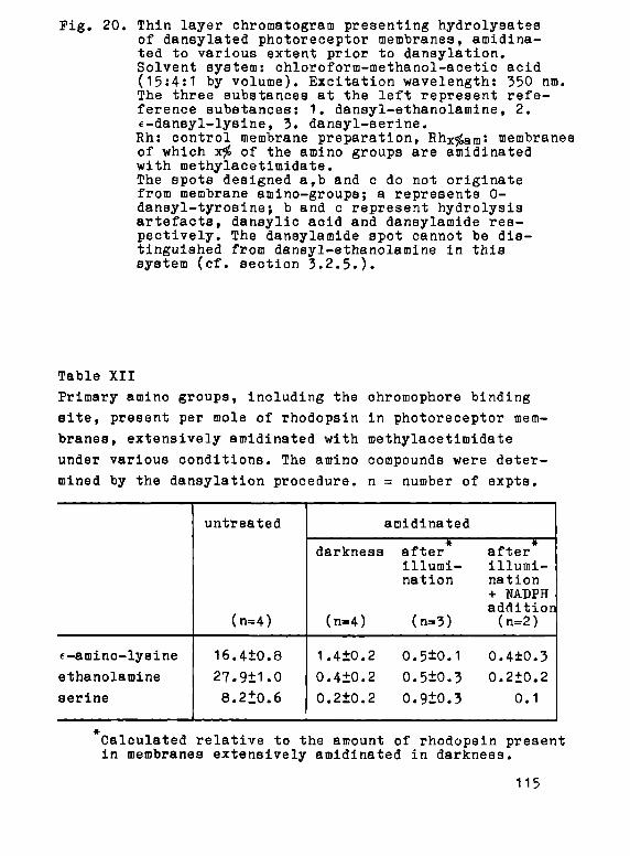
Pig. 20. Thin layer chromatogram presenting hydrolysatea of dansylated photoreceptor membranes, amidina-ted to various extent prior to dansylation. Solvent system: chloroform-methanol-acetic acid (15:4:1 by volume). Excitation wavelength: 350 nm. The three substances at the left represent refe-ference substances: 1. dansyl-ethanolamine, 2. e-dansyl-lysine, 3. dansyl-serine. Rh: control membrane preparation, Rh
x^
a m: membranes
of which x# of the amino groups are amidinated with methylacetimidate. The spots designed a,b and с do not originate from membrane amino-groups; a represents 0-dansyl-tyrosine·, b and с represent hydrolysis artefacts, dansylic acid and dansylamide respectively. The dansylamide spot cannot be distinguished from dansyl-ethanolamine in this system (cf. section 3.2.5.).
Table XII
Primary amino groups, including the chromophore binding
site, present per mole of rhodopsin in photoreceptor mem
branes, extensively amidinated with methylacetimidate
under various conditions. The amino compounds were deter
mined by the dansylation procedure, η = number of expts.
e-amino-lysine
ethanolamine
serine
untreated
(n=4)
16.4+0.8
27.9+1.0
8.2Í0.6
amidinated
darkness
(n=4)
1.4+0.2
0.4+0.2
0.2+0.2
after illumination
(n=3)
0.5±0.1
0.5±0.3
0.9ІО.З
* after illumination + NADPH addition (n=2)
0.4±0.3
0.2+0.2
0.1
Calculated relative to the amount of rhodopsin present in membranes extensively amidinated in darkness.
115

When the amidination is carried out in the dark, only the
ratio lysine/rhodopsin exceeds one. When it is carried
out in the light, the number of f-amino-іуэіпе groups is
decreased,while surprisingly the number of non-amidinated
phosphatidylserine residues is increased. Virtually com
plete modification can only be accomplished in five ami
dination cycles when during illumination NADPH is added
in order to permit reduction of the liberated retinaldehyde
to retinol by the retinol:NAOP oxidoreductase.
The influence of the size of the substituents is
evaluated by carrying out modification with methylpicco-
linimidate and methyl-3,5dinitrobenzimidate. The latter
compound, prepared in the same way as methylacetimidate
except for employing dioxane-ether (1:1) as solvent,
reacts too slowly at pH 7.5-8.0 to be useful. Methyl-
piccolinimidate, however, gives results very similar, and
in some respect, even superior to those obtaimed with
the much smaller methylacetimidate. Presumably as a result
of the much simpler reaction conditions, 96-98$ modifi
cation is achieved in one step and leaves 80-90$ of the
rhodopsin intact, while the recombination capacity
decreases to 70-80$. Extensive modification does not
completely abolish the retinol:NADP oxidoreductase activi
ty, since 10-45$ of the original activity is retained.
Although no quantitative determinations have been performed,
phosphatidylethanolamine again shows the highest modifi
cation rate, while phosphatidylserine appears to be some
what more reactive than the average lysine residue. Thus,
further use of methylpiccolinimidate in amino-group modifi
cation of membranes seems very promising in view of the
simple procedure. This allows experiments with smaller
amounts of material, as well as more accurate kinetic
determinations.
116

ι О 20 40 60 βΟ 100
percentMgß af 3 m i r w g r o u p e modi f i ed
tiKTW ( h )
Pig. 22. a. Influence of trinitrophenylation of rod photoreceptor membranes, auapended in 0.07 M bicarbonate (pH 8.0) in darkness at 40oC, on the spectral integrity of rhodopsin expressed as ДА500 (TNBS, -A-). When performed at room temperature, the reaction virtually stops after about 70$ modification (cf. Pig. 22b) without decrease in ΔΑ500·
P o r comparison, the influ
ence of amidination in darkness on the spectral integrity is also shown (MAI, -+—).
b. Time course of the reaction of TNBS with rod photoreceptor membranes under various conditions. —t— : in darkness at 20
0G
—A- : in darkness at 40oC
-D- : in the light at 400C
- · - : in the light at 40°C in the presence of 0.6% (w/w) Triton X-100.
117
100
eo
: «o
20

4.5.2. Trinitrophenylation
The reaction of rod photoreceptor membranes, suspended
in 0.07 M bicarbonate (pH 8), with a fifteen-fold excess
of TUBS in the dark at 35-400C results in complete modifi
cation after about three hours, concomittant with a complete
loss of the 500 nm absorption and recombination capacity
(Fig. 22a). The absorbance at 500 nm begins to decrease
only after 65-70$ of the amino groups have been modified.
Illumination or addition of Triton X-100 accelerates
the reaction markedly (Fig. 22b). In the presence of SDS
the reaction rate is considerably decreased, as has
previously been observed for soluble proteins (Habeeb,
1966). When trinitrophenylation is performed on membrane
suspensions at 20oC, modification virtually stops at
about 70$ without decrease in 500 nm absorbance (Fig. 22b).
In the presence of Triton X-100, however, nearly complete
modification may even be achieved at 6 C.
Analysis by means of dansylation of the amino compounds
remaining after various stages of modification in suspen
sion at 400C (Fig. 23) shows that the two amino-group
containing phospholipids are rapidly modified, while the
e-aminolysine groups are less reactive. Semilogarithmic
plots of (Δτ.ο-Ατ,.α ) versus t yield no straight lines,
in contrast to model experiments with the single compounds
(section 2.3.2.). This indicates the presence of amino
groups of different reactivity. Viale (1970) has shown
that in the case of a number of simultaneous first order
reactions the change in absorbance with time (A+ .- Α.),
multiplied by the time t after which the first absorbance
measurement has been made, plotted versus the time t,
yields a curve with a number of maxima corresponding to
the various reactive species present. The time ^max at
which a maximum is observed yields an estimate of the
half time of the reaction: t1 /
2 = "^χ·
1 0 2· Similar plots
for the reaction with TUBS under various conditions (Fig. 24)
118

60 во time (rain)
Pig. 23. Percentage of various amino-compounds remaining upon reaction of illuminated photoreceptor membranes with TNBS in suspensión at 40oC. Lye: e-amino-lysine groups, PS: phosphatidyl-éerine, PE: phosphatidylethanolamine.
10 20 30 MO so time (mm)
Fig. 24. Plots of t.(A-t+4 - A-j;) versus t for the reaction of photoreceptor membranes with TNBS under various conditions. The number of maxima represent the number of reactive species, while the tmax gives an estimate of 1/k (Viale, 1970). ·—· : reaction performed in the light 0—0 : reaction performed in darkness The absorbance is measured at 348 nm, Δ is 2.3 minute. The reactions are performed as described in section 4.2.3.
119

show the presence of at least three species with different
reactivity, neglecting the insignificant irregularities
in the broad maxima at t>15 rain. When taken together with
the evidence presented in Pig. 23, the most reactive species
appears to be phosphatidylethanolamine followed by phos-
phatidylserine, while the broad maxima presumably corre
spond to the lysine residues. The reactivity of the
lysine residues in non-illuminated membrane suspensions
appears to be less than that in illuminated membranes
(Figs 22b and 24, Table XIII).
4.3.3« Other reagents
So far, reactions with the other reagents ENFB, TANFB and
succinic anhydride have been less extensively investigated.
Table XIII
Reactivity of membrane amino-groups towards TNBS.
Estimated t.. /ρ values (min) from the reproducible peaks
in plots of t.(A^+A - At) versus t, obtained from reaction of photoreceptor membranes with TNBS under various conditions.
temperature
suspension
in darkness
illuminated
0.6% Triton* X-100
0.5fo SBS
5.50C
-
2 5+; 5
+; »10
-
200C
2 + ; 4
+;>8
> 90
40oC
3+;10 ;>19
3+; 5 ; > H
2+;»6
-15
Non-illuminated and illuminated preparations give rather similar results.
'Tentatively assigned to phospholipids.
120

Some resulta, possibly important in connection with the
data presented above, will be discussed.
Phospholipids show again the highest reactivity
towards all three reagents. Extensive modification is
easily achieved with LNPB аз well as with succinic an
hydride, already at 200C. In both cases the spectral
properties of rhodopsin are ultimately abolished. Upon
complete modification with succinic anhydride, the mem
brane fragments are less easily sedimented, but do not
become soluble even in the presence of θ M urea, as has
been reported for the erythrocyte membrane (Moldow et
al, 1972). Modification with TANPB proceeds rather easily
up to 50-70$. In order to obtain higher modification
percentages, prolonged incubation at higher temperatures
and pH (400C, pH 8.5) is required, during which the
control sample also shows slight denaturation.
4.3.4. Photolysis
Due to technical limitations (section 4.2.8.) the
intermediates in the photolytic chain reaction appearing
before metarhodopsin II can not be observed. Untreated
membrane preparations yield the following picture upon
flash bleaching (Fig. 25): The first intermediate, ob
served about 100 msec after the flash, is metarhodopsin
II (Λ _ 380 nm). Metarhodopsin II is slowly converted max
rf
to metarhodopsin III (Л 460 nm), with a half time of
about 1 min at 370C, 3-4 min at 25
O0 and 12-15 min at
150C; values which are in agreement with earlier reports
(Ebrey, 1968; Bauman, 1972). Assuming equal maximal molar
absorbance coefficients (Weale, 1973)» 20 to 40$ of the
metarhodopsin II is converted to metarhodopsin III as has
also been observed in the excised frog retina (Bauman and
Reinheimer, 1973) and in vivo (Weale, 1973). The other
part may form "N-retinylidene-opsin", a random aldimine
121

300 400 300 600 400 waviel*neth (nm )
500 6 0 0
Pig. 25. Formation of metarhodopsin II (Лш а х 380 nm) and
metarhodopain III (1Шах 460 nm) upon flaeh blea
ching of suspensions (-1 mg/ші in 0.2 M phosphate buffer pH 7.0 at 25
o0) of photoreceptor membranes
amidinated to various extent. AMQ: untreated control preparation, AM2: preparation amidinated to about 80$, AM5: preparation amidinated to about 97#. At the right side difference spectra of the indicated Intermediate spectra are presented. Recording speed: 5 nm/sec. Абіп»
w h i c h varied
between 0.030 and 0.060 was set at zero.
122
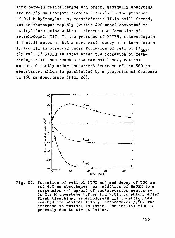
link between retinaldehyde and opsin, maximally absorbing
around 365 nm (compare section 2.3.2.). In the presence
of 0.1 M hydroxylamine, metarhodopsin II .is still formed,
but is thereupon rapidly (within 200 msec) converted to
retinylidene-oxime without intermediate formation of
metarhodopsin III. In the presence of ΝΑΌΡΗ, metarhodopsin
III still appears, but a more rapid decay of metarhodopsin
II and III is observed under formation of retinol (Λ „ :
max
325 nm). If NADPH is added after the formation of meta
rhodopsin III has reached ita maximal level, retinol
appears directly under concurrent decrease of the 380 nm
absorbance, which is parallelled by a proportional decrease
in 46О nm absorbance (Pig. 26).
45 F • ' -^ ' ' ' -—^ 1
I 1 1 1 * 1 1 1 ι -Ο 20 *0 60 80
time (mm)
Pig. 26. Formation of retinol (330 nm) and decay of 380 nm and 4-60 nm absorbance upon addition of NADPH to a suspension (~1 mg/ml) of photoreceptor membranes in 0.2 M phosphate buffer (pH 7.0), in which, aftei flash bleaching, metarhodopsin III formation had reached its maximal level. Temperature: 37
0C. The
decrease in retinol following the initial rise is probably due to air oxidation.
123

Subsequently, the photolytic behaviour has been inves
tigated of photoreceptor membranes, which had been amidi-
nated to various extents in darkness. Acetamidinated and
piccolinamidinated preparations appeared to yield similar
results. Even in extensively amidinated preparations
metarhodopsin II appears (Pig. 25). It is rapidly trans
formed to retinylidene-oxime, when hydroxylamine is present.
Amidination however clearly influences metarhodopsin III
formation. Upon modification of 60-80$ of the amino groups
only 20-30$ as much metarhodopsin III is formed as by
untreated preparations, and metarhodopsin III formation
is completely abolished at 85-90$ amidination (Pig. 25).
4.4. Discussion
4.4.1. Chromophoric binding site
Due to the low reactivity of the binding site of the
chromophoric group in rhodopsin, its identity has been
the subject of some controversy and has never been
established unequivocally. Only recently indications
have been presented for the involvement of an f-amino-
lysine group, but only in a qualitative sense (Daemen et
al, 1971; Pager et al, 1972). Our modification studies
afford an opportunity to identify the chromophoric binding
site in native rhodopsin in a quantitative way. Table
XII clearly shows that after extensive amidination only
primary t-amino-lysine groups are present in sufficient
amount to account for the binding of 11-cis retinaldehyde.
This further emphasized by the fact that in similar pre
parations illuminated before amidination, the number of
i-amino-lysine groups available for dansylation is clearly
reduced (Table XII), the difference being equivalent to
the decrease in the number of rhodopsin molecules. The
124

The retinaldehyde liberated upon illumination remains to
a large extent bound to the membrane via randomly formed
aldimine linke (cf. chapter 2; Bonting et al, 1973), which
explains why nearly complete amidination may only be accom
plished after previous reduction of the liberated retinal
dehyde to retinol (Table XII).
Thus, we have shown that almost complete blocking of
the free amino groups of non-illuminated photoreceptor
membranes can be carried out without major spectral
damage to rhodopsin. Upon subsequent Illumination of the
modified protein, involving release of the ohromophore,
one further f-amino-group of lysine is set free. Blocking
of free amino-groups in illuminated photoreceptor membranes
under the same conditions causes modification of one more
«-amino-lysine group. These data yield in our opinion
incontrovertible proof that the chromophoric group in
native rhodopsin is bound to an €-amino-group of a lysine
residue (de Grip et al, 1973a,Ъ,с).
4-.4.2. Migration of the chromophore following illumination
It is now clear that an «-amino-lysine group functions
as the binding site of the chromophoric group in rhodopsin
as well as in metarhodopsin II (cf. section 1.3.5.). Con
sequently, the question arises whether in both cases the
same residue is involved, or whether the chromophore
migrates during the conversion rhodopsin ——*> metarhodopsin II
from one lysine residue to another (transiminizatinn).
Recent studies in our laboratory argue against the latter
possibility, since reductive fixation of the chromophore
to opsin in the metarhodopsin II stage completely abolishes
the recombination capacity (Rotmans, 1973; Баетеп et al,
1974). This argument is substantiated by our observation
that metarhodopsin II is formed normally upon illumination
of amidinated preparations, which have less than 0.5 free
125
amino group in addition to the chromophore carrying group
(Table XII).
However, complete amidination abolishes the formation
of metarhodopain III (Pig. 25). This clearly suggests a
migration of the chromophore during the metarhodopsin
II • metarhodopsin III transition, which also might
explain the slower rate of this reaction than that of the
formation of metarhodopsin II. These conclusions, i.e.
the same binding site in rhodopsin and metarhodopsin II
and migration during formation of metarhodopsin III, are
further supported by the photolytic behaviour of photo
receptor membranes in the presence of hydroxylamine.
Metarhodopsin II still appears in this situation, in
confirmation of earlier observations of Johnson (1970),
but hydroxylamine in large excess appears to compete
effectively with the binding site in metarhodopsin III.
So far, we have not been able to identify the latter site
positively, but some observations indicate an involvement
of phosphatidylserine. In the first place, metarhodopsin
III formation is already strongly suppressed at 60-70$
modification, implying that an easily modified residue is
involved, and in all our studies the phospholipids appear
to be the more reactive species. In the second place, in
light-amidinated membranes less phosphatidylserine becomes
amidinated than in dark-amidinated membranes, except when
МБРН is present during illumination (Table XII). This
suggests that in the absence of МБРН part of the libe
rated retinaldehyde remains bound to phosphatidylserine.
Under conditions, in which the membrane structure is
denatured by methanol, the liberated retinaldehyde is,
however, predominantly bound to the more reactive phos-
phatidylethanolamine (Poincelot et al, 1970; Баетеп et
al, 1971), of which in addition there are 3 times as many
available. In the third place, phosphatidylserine as a
binding site might explain the red-shifted absorption
126

maximum (460 nm) of mètarhodopsin III through internal
protonation of the retinylidene-imine bond by the phos
phoric acid group of the phospholipid (Daemen and Bonting,
1969).
This would suggest that in the intact membrane phos-
phatidylserine is closely associated with rhodopsin. The
resistance to phospholipase action of one to two phos-
phatidylserine molecules per mole rhodopsin (Borggreven
et al, 1971) points in the same direction. Whether this
phosphatidylserine molecule plays a physiological role
in the "handling" of the liberated retinaldehyde (meta-
rhodopsin III) remains uncertain. The relatively high
resistance of phosphatidylserine in membranes to chemical
(Poensgen and Passow, 1971) and enzymatic (cf. Coleman
et al, 1970) attack seems to be a rather general pheno
menon and this phospholipid also appears to be intimately
associated with other membrane proteins (Wheeler and
Whittam, 1970; Rethy et al, 1972).
The studies, in which reductive fixation of the
chromophore was employed (Rotmans, 1973; Daemen et al,
1974) presented also indications for a migration of the
chromophore to the active site of the retinol:NADP oxido-
reductase, which in the absence of NADPH appeared to be
maximally occupied at 10-15 min after the illumination.
A similar conclusion may be drawn from the results pre
sented in Table XI. The preparation amidinated in dark
ness in 4 treatments to about 92$, still shows 40$ of the
original retinoliNADP oxidoreductase activity. A fifth
modification step performed in darkness abolishes the
activity nearly completely, but if it is performed after
illumination, the enzymatic activity is partly preserved.
This indicates that retinaldehyde liberated during illu
mination has migrated to the enzyme site and protects it
from inactivation. This may further explain the variable
loss in retinólJNABP oxidoreductase activity observed in 4- Ü
127

preparations amidinated extensively in the light.
It is also noteworthy that opsin behaves differently
from rhodopsin during amidination. It looses its recom
bination capacity but only upon extensive amidination
(Pig. 17b), wich is apparently due to modification of the
chromophore-binding lysine amino-group. This would imply
that this group is not too readily accessible, even when
it does not carry the chromophoric group as in opsin.
However, in contrast to rhodopsin, the retinaldehyde bin
ding site in metarhodopsin II is easily accessible to
water-soluble reagents such as sodiumborohydride and
hydroxylamine, which are not much smaller than methyl-
acetimidate. Since transiminization of retinaldehyde
during the formation of metarhodopsin II does not appear
to take place, the following mechanism must be considered.
Upon illumination, the chromophore binding site, which is
shielded in rhodopsin, is displaced due to the isomeri-
zation of the retinaldehyde to a less shielded location.
When the retinaldehyde is subsequently removed by means
of transiminization or hydrolysis, the amino acid residue
"flips back" to its original site, thereby releasing the
strain placed upon the binding area by the isomerization
reaction. This would also explain the fairly rapid reac
tion of opsin with 11-cis retinaldehyde and the low
affinity of opsin for all-trans retinaldehyde (Rotmans,
1973).
In conclusion, the following picture emerges. Illumi
nation of rhodopsin triggers a chain reaction by isomeri
zation of the chromophoric 11-cis retinaldehyde to the
all-trans form. First, the aldimine bond, linking the
chromophore to an t-amino-group of a lysine residue, is
displaced over a distance of maximally 8-10 fi (cf. section
1.3.4.). A number of rapid conformational transitions
follows (Pig. 6), resulting in intermediates, which have
been identified by virtue of their spectral characteristics
128

(cf. Wald, 1968): prelumirhodopain, lumirhodopsin, ше а-
rhodopsin I and metarhodopsin II. Up to the formation of
metarhodopsin II, retinaldehyde remaina linked to its
original binding site, but the aldimine bond assumes a
more exposed position. During the next transition, meta
rhodopsin II • metarhodopsin III, the chromophore
migrates to a new binding site, perhaps a phosphatidyl-
serine residue associated with rhodopsin, while the
original binding site returns to its original shielded
position. Alternatively, migration of retinaldehyde to
the active site of re.tinolrMEP oxidoreductase may occur,
possibly via randomly formed aldimine links. Computer-
adaptation of photolytic kinetics observed in the excised
frog retina shows this picture to be consistent with the
pattern shown in Pig. 6 with metarhodopsin II as well as
metarhodopsin III decaying into opsin with randomly bound
retinaldehyde (Bauman and Reinheimer, 1973).
The role of metarhodopsin III in this context is
obscure. It does not represent the enzyme-substrate
complex of the retinol:NADP oxidoreductase, as has been
suggested by de Pont et al (1970a), since amidination
abolishes metarhodopsin III formation at a much earlier
stage than retinol:NADP oxidoreductase activity (Pigs
18 and 25). Perhaps metarhodopsin III functions as a
temporary store for all-trans retinaldehyde generated
by illumination. This would liberate the original binding
site and permit rhodopsin regeneration to take place.
Another possibility is that the refolding of the opsin
structure occurring during the decay of metarhodopsin II
might stop the release of transmitter substances from the
rod disc.
4.4.3. RetinolrNAOP oxidoreductase
The particulate retinoltNAOP oxidoreductase activity,
129

which catalyses the reduction of retinaldehyde with NABPH
as preferred coenzyme (Putterman, 196?; de Pont et al,
1970a), is rather sensitive to amidination of the photo
receptor membrane with methylacetimidate (Pig. 17a).
Since amidination has a relatively small influence on
the integrity of rhodopsin and since addition of retinal
dehyde can partly prevent the loss of retinol:NADP oxido-
reductase activity, we conclude that the enzyme is inacti
vated by modification of a catalytic amino-group. The
involvement of such a group is not surprising in view of
the easy formation of aldimine links by retinaldehyde,
which would therefore be a suitable way to form an enzyme-
substrate complex. This would also agree with the observed
ability of the retinol:NAOP oxidoreductase to reduce not
only free retinaldehyde, but also retinylidene-imines
(de Pont et al, 1970a). In the latter case, the retinal
dehyde could transiminize rapidly from the carrier amino-
group under formation of the enzyme-substrate complex,
so that the product formation remains the rate-determining
step.
Upon modification with methylpiccolinimidate complete
inactivation of the enzymatic activity is difficult to
accomplish, while at 60-90$ modification even a slight
increase (10-20$) in activity is observed. Plapp (1970)
has reported a twenty-fold activation of horse liver
alcohol dehydrogenase upon 80-90$ modification with
methylpiccolinimidate, while ethylacetinidate caused only
slight activation and showed inhibitory effects at higher
modification. The relatively high activity in extensively
piccolinamidinated preparations may therefore be partly
due to activation of the remaining enzyme activity.
Alternatively, the amino group in the active site of the
retinol:NAOP oxidoreductase may be less accessible to the
bulky methylpiccolinimidate molecule. The effect of TNBS
is difficult to evaluate in this connection, since the
130

enzymatic activity itself is not very resistant towards
the required modification conditions (35-40oC, pH^8).
Further investigations employing other modifying reagents
may yield additional useful information.
4.4·.4·. Structural aspects
It has previously been demonstrated for several
soluble proteins that extensive amidination with methyl-
acetimidate influences the conformation only to a minor
extent (cf. Means and Peeney, 1971). Our results show
that this is also the case for the membrane protein
rhodopsin. Amidination of about 99$ of the free primary
membrane-amino groups, i.e. 97$ of the free membrane-
protein lysine groups, has relatively little effect or.
the 500 nm absorption and the recombination capacity.
The latter of these two parameters is rather sensitive
to conformational changes, as shown by its sensitivity
to detergent action (cf. chapter 6).
Interestingly, the much larger methylpiccolinimidate
yields about the same results. The piccolinamidinated
preparations are also fairly stable at 40 C. On the other
hand, modification with TNBS, БКРВ and succinic anhydride
apparently leads to gradual disruption of the protein
structure and ultimately to denaturation (cf. Fig. 22a).
These results suggest, that introduction of a large
substituent into the protein lysine groups has little
effect on the membrane integrity as long as the positive
charge is retained. This may indicate a more external
location of the bulk of the amino-groups and an important
contribution of the electrostatic interactions of these
groups to the tertiary structure of the protein.
Whereas the phospholipids show under all conditions
a high reactivity towards all amino-group reagents used,
the only reagent which is negatively charged, TNBS, reacts
131

in suspension with the protein £-amino-lysine groups only
relatively slowly (Pigs. 22 and 23, Table XIII). We ten
tatively assign this to a net negative surface charge
present on the rhodopsin molecule, the existence of which
is also consistent with low angle X-ray studies (Blasie,
1972b). The reactivity of model compounds towards TNBS
decreases markedly upon introduction of a negatively
charged substituent in the molecule (Table VII). Sodium-
dodecylsulfate (ЗБЗ), which binds to proteins and lipids
under formation of very negatively charged micelles (cf.
chapter 6), considerably retards the reaction of all
amino-groups containing species in the nembrane w:th
TNBS (Pig. 24, Table XIII). A similar effect of SDS was
Deserved by Khandwala and Kaspar,(1971) with respect to
reaction of microsomal membrane amino-groups with the
cyanate anion. A negative surface charge on rhodopsin
might likewise hamper or even prevent reaction with
TNBS. The decrease in surface charge upon illumination,
concluded from the above mentioned X-ray studies (Blasie,
1972b), is consistent with the higher reactivity of the
lysine side chains in illuminated samples (Table XIII).
A similar low reactivity of protein amino groups was ob
served by Dratz et al (1973) with respect to the negatively
charged fluoresceinisothiocyanate.
The reactivity of the phospholipids in membrane sus
pensions as well as in membrane solutions in Triton X-100
is much higher than that of the corresponding model com
pounds as judged by the half time values (Tables III and
VII). This observation is probably related to the so-
called surface charge effect, which manifests itself at
the surface of highly charged structures like membranes
and lipid layers (Baskin, 1972; Haydon and Myers, 1973;
Seimiya and Ohki, 1973). This effect causes a decrease
of 1-2 log units in acid and basic strength of the cor
responding residues, i.e. acids become less acid, and bases
132

Ъесоше less basic. This should increase the reactivity
of the phospholipids present in such structures towards
amino-group reagents, since the reactions proceed mainly
via the unprotonated ашіпо-group (cf. Preedman and Radda,
1968; Means et al, 1972).
In conclusion, we can tentatively answer the three
points raised in section 4.1. (p. 103): 1. The probable
presence of a net negative charge on the water-exposed
part of rhodopsin implies that the positively charged
amino-groups are required for neutralization of part of
the negative side groups of the protein. If this positive
charge is not retained upon modification (as with TUBS,
DNPB or succinic anhydride) the net negative charge may
rise to a point, where eleotrostatic repulsion leads to
protein unfolding. 2. Introduction of a large substituent
on the protein lysine groups has little effect on the
membrane integrity as long as the positive charge is re
tained, indicating a more external location of the bulk
of the amino groups, in agreement with the first point.
3. Protein amino groups react smoothly with uncharged
reagents, but only slowly with TNBS, which we have inter
preted as indicative for the presence of a net negative
charge on that part of the rhodopsin molecule which is
exposed to the aqueous environment. The phospholipid amino-
groups react rapidly with all modifying agents, possibly
due to a surface charge effect, which decreases the рКат)1:)
of the amino groups, and to the absence of negative
charge concentrations.
133
CHAPTER 5
SULFHYORYL GROUPS IN THE ROB PHOTORECEPTOR MEMBRANE
5.1. Introduction
In 1952 Wald and Brown presented evidence that in dark
adapted bovine rod outer segment membranes, solubilized
in digitonin, about 2 moles of sulfhydryl groups per mole
of rhodopsin are accessible to Ag ions upon amperometric
titration, and that upon illumination two additional sulf
hydryl groups become titratable. This was interpreted as
indicating that a conformational change of the opsin
molecule takes place during photolysis. This concept was
incorporated into a schematic model for the transition
of rhodopsin to opsin (Wald, 1968).
Similar observations were reported by Ostroy et al
(1966), except that they detected up to four sulfhydryl
groups reacting with Ag in darkness, while illumination
"released" another two to four residues. Illumination of
membranes solubilized in Triton X-100 is also reported
to unmask two additional sulfhydryl groups, as determined
by reaction with 5,5'-dithiobis(2-nitrobenzoic acid)(OTNB)
or p-hydroxymercuribenzoic acid (Zorn and Putterman, 1971).
The results of the latter study may, however, have been
affected by air oxidation, since in darkness only one
sulfhydryl group was detected. Earlier determinations in
СТАВ (Heller, 1968a,b) probably suffer from the same effect.
Wald and Brown (1952) further showed that upon reaction
of rhodopsin with the very active sulfhydryl reagent
p-chloromercuribenzoic acid (PCMB) the absorption spectrum
remains intact, but that the capacity of opsin to recom
bine with the chromophoric group, 11-cis retinaldehyde,
is gradually lost. The less active reagents, iodo-acetic
134

acid and iodo-acetamide had no effect on the recombination
capacity.
In addition electronmicroacopic studies, employing
staining by PCMB, revealed that in the excised retina,
which had been fixed in darkness with glutaraldehyde,
illumination exposes about 2 sulfhydryl groups per rhodop-
sin molecule (Patel, 1967).
We decided to reinvestigate the behaviour of the
sulfhydryl groups of the bovine rod photoreceptor membrane
in view of the following points:
1. The possible effects of the detergents, used in all
previous studies to solubilize rhodopsin, have so far
been disregarded. The destabilizing effect of detergents
on rhodopsin (cf. chapter 6) might involve conforma
tional changes and likewise lead to the exposure of
hitherto buried sulfhydryl groups. Hence we have used
both membranes in aqueous suspension and membranes
solubilized in various detergents.
2. Overestimation of the number of sulfhydryl groups
expressed per rhodopsin molecule may have occurred in
all previous studies due to the presence of appreciable
amounts of opsin in bovine photoreceptor membrane pre
parations (cf. chapter 2). In our study we have used
opain-free membranes obtained by treatment with 11-cis
rstinaldehyde during isolation (cf. section 2.2.2.).
3. It seemed worthwile to study the effects of sulfhydryl-
reagents on the retinol:NADP oxidoreductase activity
in photoreceptor membranes, in view of the known
catalytic involvement of sulfhydryl groups in oxido-
reductases.
4. Sulfhydryl groups might be involved in the retinotoxic
effects of iodate, iodoacetate and Perchlorate ions
(Harding, I960; Hird and Yates, 1961; Sorsby, 1962;
Sorsby and Reading, 1964; Orzalesi et al, 1965, 1966,
1967 and 1970).
135

5.2. Mater ia l s and methods
5.2.1. Materials
All reagents are obtained commercially and of the
highest purity available.
5.2.2. Assay of sulfhydryl groups
Sulfhydryl groups are determined with DTNB as described
in section 3.2.2. In addition to sodiumdodecylsulfate
(SBS) the following detergents are used: digitonin, Triton
X-100 and cetyltrimethylammoniumbromide (СТАВ). The same
assay is also performed in suspensions in the absence of
detergent.
5.2.3. Modification of sulfhydryl groups
Modification with PTHB is performed under the same
conditions as in the assay, except that up to tenfold
higher concentrations of the reactants are used. The other
modifying agents used are, together with the reaction
conditions: N-ethylmaleimide (Riordan and Vallee, 196?;
20-fold molar excess, 0.2 M phosphate buffer pH 7.0,
20oC, 30 min), O-methylisourea (Banks and Shafer, 1972;
up to 100-fold molar excess, 0.2 M phosphate buffer pH 7.0,
4 and 20 C), methyl-p-nitrobenzenesulphonate (Heinrikson,
1971; up to 100-fold molar excess, 0.2 M TRIS-HC1 buffer
pH 8.2, 350C) and p-chloromercuribenzoic acid (PCMB;
Riordan and Vallee, 1967; up to 100-fold molar excess,
0.2 M phosphate buffer pH 7.0, 4° and 200G). In all cases
a membrane concentration equivalent to 5-10 mg of lyophi-
lized material per ml is used. The corresponding reaction
equations are presented in Pig. 27.
The number of sulfhydryl groups modified by any of
136

these reagents is calculated as the difference of the
total number of free sulfhydryl groups, assayed with DTNB
in SDS solution, before and after modification. For this
purpose the modifying agents are removed prior to the assay
c-o
1 SH
> ~ -
1 6H
0
H c - <
II >-H c - c
0
1 5H
R - C H 3
RE/
4 о
N-ET
C2H5
Mt
R:
REACTION V
зна
VITH DTNB
1 s
θ2Ν-<θ)-5 * S 4 0 ) - N 0 2 »Η*
Νθ2 CÇ
О
DTNB
HYLSUCCINA
pH 5-б
Э o - c
a m a . - ' ^ n m
NIDATION
1 S \ с .н
"¿TV* o - C ~ ^
C2HS
N-ethylmaleimide
.THYLATION
pH 7-9
H«N
c - o -HN
О
R. O 2 N Y O ) - S - 0
ю τ ι o ^
1 SH
О
J WITH MERCI
pH 7
О
R: -с; 'β
ь R: - S O |
1 s
CH3 » R - H
(O-methyl isourea)
- (methyl-p-nitrobenreneeulphorate)
JRY COMPOUNDS
1 5
( P C M B )
(PCM BS)
Fig. 27. Reactions equations of the sulfhydryl group modifications described in chapter 5.
137

by washing the suspensions twice with 0.2 M phosphate buffer
(pH 7.0; 50,000 χ g, 40C, 30 min).
When the assay or modification of the membrane sulfhy-
dryl groups is performed in the presence of detergents or
at elevated temperature, air is excluded by performing all
steps in a nitrogen atmosphere in order to prevent oxida
tion of aulfhydryl groupa. In Triton X-100, СТАВ and SOS,
and to a less extent in digitonin, rapid oxidation of SH-
groups by air is observed, even at 40C, bleached samples
being even more susceptible than unbleached ones.
5.2.4. Determination of mercury
Mercury is estimated by atomic absorption flame photo
metry (Lfnicam SP 1950) after destruction of the organic
material in a mixture of concentrated HoSO., HC10. and
HNO, (2:3:12 by volume) during 45 min at 180oG. A stan
dard curve is prepared with an aqueous solution of p-
chloromercuribenzene sulphonic acid (PCMBS; 5-50 ppm).
5.2.5. Treatment with iodate
Photoreceptor membranes, suspended in phosphate buffer
(0.07-0.2 M; pH 6.5-7.0; 2-5 mg/mi) are mixed with aqueous
solutions of potassium iodate to a final concentration of -5 -1
10 to 10 M. The resulting suspensions are incubated
either in darkness or in the light at 20oC or 37
0C. After
various time intervals aliquots are taken from the sus
pension and washed twice with the appropriate phosphate
buffer (35,000 χ g, 40C, 30 min). Finally the sediment is
suspended in the same phosphate buffer or washed twice
with distilled water and lyophilized.
The following compounds were tested in a similar way:
sodium fluoride, sodium Perchlorate, sodium chlorate,
sodium iodide and sodium trichloroacetate (10 mM, 20oC).
138

5.3. Reaults
5.3.1. Exposed sulfhydryl groups
Under completely denaturing conditions (1$ ЗБЗ) we
detect 6 sulfhydryl groupa and about 2 disulfide residues
in the photoreceptor membrane exprssed per molecule of
rhodopsin (cf. section 3.3.3.). However, under less drastic
conditions part of the residues do not react with sulf
hydryl reagents in aqueous solutions, presumably because
these residues are buried inside the membrane.
Table XIV shows the sulfhydryl groups, which are
accessible to БТГГВ in dark-adapted and illuminated photo
receptor membranes under various conditions. It makes no
difference in any of these cases, whether illumination
takes place before or after addition of DTNB.
In suspension, the unbleached samples yield about 2
accessible sulfhydryl groups per mole rhodopsin, but no
further increase is observed upon illumination. With N-
ethylmaleimide the same results are obtained as with DTNB;
2 reactive SH-groups before and after illumination (Table
XV). On the other hand, in digitonin solution two SH-groups
per rhodopsin are observed in unbleached samples, and an
additional two become accessible upon illumination (Table
XIV). In Triton X-100, СТАВ and SDS solutions the number
of SH-groups modified in darkness increases to 3.0, 3.6
and 5.9 respectively, in the light to about 6 in all three
cases.
5.3.2. Modification of sulfhydryl groups
Modification of suspensions and of the solutions in
digitonin, Triton X-100 and СТАВ with ΌΤΝΒ in darkness
leaves the absorption spectrum of rhodopsin intact. The
recombination capacity remains unchanged only in the case
139

-F-О
Table XIV
Number of sulfhydryl groups, per mole of rhodopsin, present in bovine photoreceptor
membranes , reacting with DTNB under various conditions.
The number of determinations is given in parentheses.
suspension* digitonin Triton X-100 СТАВ SOS0
ïpenslon üigitonin Triton л-тии иТАЙ sua (5) (4) (5) (2) (5)
In darkness 1.8 ± 0.1 2.0 ± 0.1 3.0 ± 0.1 3.6 ± 0.3 5.9 ± 0.2
After illumination 1.8 ± 0.2 3.9 ± 0.2 6.0 t 0.2 5.8 ± 0.3 5.9 ± 0.2
No difference is observed between frozen and lyophilized photoreceptor membrane preparations.
The reaction is followed for 45-60 min; after this time interval the reaction is over 99?6 complete in detergent solution. In suspension the reaction is then only 90-95$ complete, and reaches its maximal extent in 4-5 hrs. The real number of exposed SH-groups in suspension will therefore be close to 2.
Rhodopsin is not stable in SOS solution at 20oC and is even in darkness
immediately bleached.

of membrane suspensions, both before and after illumination
(Table XV). In digitonin solution the latter parameter
remains unchanged in darkness, but upon modification with
IITNB after illumination the recombination capacity decrea
ses to less than 30$ of the control. In Triton X-100 and
СТАВ the recombination capacity is already abolished by
the detergent alone (Johnson and Williams, 1У70; Snoddeny,
1967).
Under completely denaturing conditions (1$ SDS) we
detect 6 sulfhydryl groups per mole of rhodopsin originally
present (Table XIV). The same number is exposed in Triton
X-100 and СТАВ after illumination. The presence of 8 M
urea accelerates the reaction but without uncovering
additional sulfhydryl groups.
In suspension, reaction with DTNB appears to proceed
somewhat faster in illuminated membranes. Therefore, we
Table XV
Effect of modification of suspended photoreceptor membranes
with DTNB or NEM (N-ethylmaleimide) on some membrane properties.
DTNB (n=5)
NEM (n=2)
In darkness
0 * * +
SH-groups Αςοο recorobi- RQR modified nation
capacity
* # *
1.8 ± 0.1 100 95 t 3 4
1.6 ± 0.3 100 1 0 2 + 5
After illumination
0 * SH-groups recombi-modified nation
capacity
*
1.8 І 0.2 98 ± 4
1.7 ± 0.2 1 0 4 + 6
0Expressed per mole of rhodopsin.
The value of untreated controls is taken as 100$. +R0R = retinol:NADP oxidoreductase activity.
141

have investigated the possibility that illumination changes
the reactivity of the two SH-groups exposed in suspension.
For this purpose we have used two reagents with low reac
tivity: O-methylisourea, which is reported to methylate
the very reactive SH-group in papain at neutral pH (Banks
and Shafer, 1972) and methyl-p-nitrobenzenesulphonate,
which methylates sulfhydryl groups at higher pH (>8.5) and
elevated temperature (40oC; Heinrikson, 1971). The number о
unmodified SH-groups is again determined with DTNB. With
both reagents we find some evidence for increasing reac
tivity of one SH-group after illumination. Membranes, kept
in darkness, do not show any reaction with O-methylisourea
even after prolonged reaction times (7 days, 200C). Illumi
nated membranes, however, react relatively fast (1 day,
40C) up to a certain degree (0.6-1 mole per mole of rhodop-
sin), after which no further reaction is observed. Methyl-
p-nitrobenzenesulphonate is able to methylate about one
SH-group in darkness after long reaction times (6-8 hrs,
pH 8.2, 350C). In illuminated membranes one SH-group is
fairly rapidly (1-2 hrs) modified, the other one reacting
again much more slowly. Some relevant data are collected
in Table XVI. In all cases absorption spectra and recombi
nation capacity remain intact.
Modification with DTNB, in the light as well as in
darkness, markedly inhibits the retinol:NADP oxidoreductase
activity (Table XV). This is not so in the case of O-methyl
isourea. The effect of methyl-p-nitrobenzenesulphonate is
more difficult to evaluate in this context, since methyla-
tion demands more drastic conditions during which the
controls also partly loose their activity. Methylation in
darkness, however, appears to have no influence on this
enzymatic activity.
Reaction with the mercury compound p-chloromercuri-
benzoic acid (PCMB) yields completely divergent results
as compared to the other reagents (Pig. 28). Upon short
H2

Table XVI
Number of sulfhydryl groups (per mole of rhodopsin)
available for modification in bovine photoreceptor
membranes, before and after illumination.
Determination with reagents of different reactivity.
Reagent Before illumination After illumination
DTNB
O-methyliaourea
number of SH-groups modified
2
0
reaction time
4-5 h
7 d
number of SH-groups modified
2
~1
reaction time
3-4 h
1 d
Methyl-p-nitro- л
,1 g ^
2 б
benzenesulphonate
reaction times in darkness with low reagent concentrations
(8.10 M, tenfold molar excess with respect to rhodopsin)
the effects are similar to those observed with ΌΤΝΒ and
N-ethylmaleimide: about 2 sulfhydryl groups are modified
without decrease in recombination capacity (Pig. 28a). If
the reaction is continued, however, more sulfhydryl groups
are modified up to a maximum of 3-3.5. This does not affect
the spectral properties, but markedly decreases the recom
bination capacity. Interestingly, complete removal of the
reagent by treatment with excess dithioerythritol (10 mM,
2 hrs, 20o0), makes two sulfhydryl groups accessible again
to DTNB and partly (70$ vs. 40$) restores the recombination
capacity. At higher reagent concentration (10 mM PCMB) the
spectral properties of the photoreceptor membrane remain
unaffected, but about 4 sulfhydryl groups are readily
modified and the recombination capacity is rapidly abolished.
If the membranes are illuminated prior to reaction with
PCMB, the effect is much more dramatic. All sulfhydryl
143

20 40 time(rtiiri)
1-6 ùf
.̂ α.
ί* *0 Ι Φ Έ? á | η
-h
-
^^^ r~
•
,—·
_-σ
,--0 ' '
^ ^ ^ — · -^^^,·.—-^^^^
100 50 Ο recombination capacity(%)
Pig. 28. a. Number of aulfhydryl groups modified (solid linei and recombination capacity (broken line) of photoreceptor membranes, upon treatment with PCMB (8.10-4M, 20OC) in the light (о—o) and in darkness (·—·). In the latter case the ДА500 remains constant.
b. Plot of recombination capacity versus number of modified sulfhydryl groups, derived from Pig. 28a.
groups are rapidly modified, concomittant with a complete
loss of recombination capacity (Pig. 28). Removal of the
mercury adducts by treatment with dithioerythritol (DTE)
shows that in this case the effect is barely reversible.
After this treatment no recovery of recombination capacity
is observed (4# vs. 0#), and about 3 (rather than the
normal 2) sulfhydryl groups are accessible to DTNB in
these membrane suspensions, suggesting that irreversible
conformational changes have taken place.
Détermination of the mercury content of membranes,
which have been treated in darkness with PCMB and sub
sequently washed twice with buffer, shows that the mem
branes contain a 3 to 6 fold higher amount of mercury
than can be accounted for on the basis of the number of
modified sulfhydryl groups» This may indicate that these
compounds are not completely specific for sulfhydryl groups,
especially if they are employed in large excess.
144

5.3.3. Jiffecta of iodate
Pig. 29 presents the main results obtained upon treat
ment of rod photoreceptor membranes with iodate under
various conditions. Iodate concentrations up to 10~1M show,
even in the presence of 1$ Triton X-100, no significant
effect on the rhodopsin absorption spectrum. However, the
recombination capacity and the retinol:NAOP oxidoreductase
activity are both affected in a concentration-dependent
way. Over a 24 hrs period these effects begin to be noti
ceable at an iodate concentration of 10~'м at 200G and
about 10 M at 370C. Illumination of the membrane suspension
before addition of iodate does not signifcantly influence
the rate of decrease in recombination capacity. Iodate
does also not decrease the rate with which the remaining
active opsin combines with 11-cis retinaldehyde, neither
does it oxidize retinaldehyde. The iodate effect on recom
bination capacity and retinol:NADP oxidoreductase manifests
itself also, and to a similar extent in intact rod outer
segment preparations, indicating that the outer membrane
100 [if ό ' ' ' о '-\ ¿Asoo
\ \ \ 60 - \ Д
0 t 4 ^ recombination cap&aiy
HO- ~~ I -
2 0 - ROS oxidoreductase activity χ
o L 1 1 1 1 L· 0 5 Ю 15 20 25
time(h)
Pig. 29. Effect of 10 mM iodate on properties of the rod photorecptor membrane at 20
oC.
0 = ΔΑ500 • = recombination capacity after treatment in
darkness. Δ = recombination capacity after treatment in
the light. χ = retinol:NAOP oxidoreductase activity
H5

does not present a significant barrier to iodate. This is
not surprising, since Ба зоп and Hollingsworth (1972) show,
that iodate also greatly increases the passive permeability
of the blood-vitreous barrier. Sulfhydryl group determina
tions by means of DTNB indicate that upon treatment with
10 mM iodate at 20 G the two exposed sulfhydryl groups and
probably also 2 buried sulfhydryl groups (expressed per
rhodopsin molecule) are oxidized within 1-2 hrs. SDS-gel
electrophoresis of these preparations in the absence of
mercaptoethanol are indicative for the presence of larger
protein aggregates, which do not enter the gel. No band
corresponding to monomeric opsin (MW 38,000) is observed.
Incubation with DTE (200C, overnight) partly reverses
the latter situation and also leads to partial recovery
of sulfhydryl groups, recombination capacity and retinol:
NADP oxidoreductase activity.
None of these effects is observed upon treatment of
photoreceptor membranes with iodide, fluoride, chlorate,
Perchlorate or trichloroacetate (10 raM, 200C) for up to
24 hrs.
5.4. Discussion
5.4.1. Sulfhydryl groups and photololysis of rhodopsin
Our experiments clearly demonstrate that the use of
more aggressive detergents (digitonin >Triton X-100>0TAB>
SDS) is paralleled by a more extensive modification of
sulfhydryl groups in rod photoreceptor membranes with
DTNB (Table XIV). This finding will be further discussed
in chapter 6. Meanwhile it is clear from Table XIV that
in detergents, which do not affect the absorption spectrum
of rhodopsin, opsin is more susceptible to chemical modi
fication than rhodopsin, confirming earlier reports (cf.
146

section 5.1.) However, this increased availability of SH-
groupa upon illumination is not observed in membrane sus
pensions, either with BTNB and with N-ethylmaleimide as
reagent (Table XV). Since membrane suspensions more closely
approximate the natural environment of rhodopsin than
detergent solutions, it seems very likely that illumination in
vivo will not uncover sulfhydryl groups either. The ob
servation that no additional SH-groups appear upon illumi
nation seema to rule out the hypothesis that a sulfhydryl
residue is involved in the covalent binding of the chromo-
phore (Wald and Brown, 1952; Heller, 1968b). This is also
supported by the Raman scattering data of Rimai et al (1970),
which confirm that in intact photoreceptor membranes, the
chromophoric group is linked via a protonated aldimine band
(cf. Pig. 5). Relevant in this connection may also be the
absence of cysteine residues in a bacterial analogue of
rhodopsin, which shows an absorption maximum at 560 nm
(Oesterhelt and Stoeckenius, 1971) and has 11-cis retinal-
dehyde as chromophore (Wolken and Nakagawa, 1973). Sinoe,
moreover, no direct evidence favoring this hypothesis has
ever been reported, it must be considered untenable.
Our conclusion that illumination does not uncover
sulfhydryl groups in photoreceptor membranes in situ is
not necessarily at variance with the light-induced réaction
of PCMB with glutaraldehyde fixed photoreceptor membranes
(Patel, 1967). Glutaraldehyde brings the membranes into
completely unnatural circumstances by formation of intra-
and inter-molecular crosalinks. This could easily influence
the light-induced conformation change in the visual pigment,
involving the exposure of additional sulfhydryl groups.
Furthermore our results suggest that PCMB is not absolutely
specific for sulfhydryl groups (cf. section 5.4.2.).
While our results indicate that illumination of rhodop
sin in a membrane suspension and presumably also 1η vivo
does not uncover additional sulfhydryl groups, light may
U7

increase to some extent the reactivity of one sulfhydryl
group (Table XVI). This may be due either to an opening
of the structure around this residue, making it more
accessible, or to the uncovering of a strongly basic
group in the neighbourhood of this SH-group. The liberated
e-amino-lysine group (cf. section 5.3.1.) might play such
a role.
5.4.2. Modification with p-chloromercuribenzoate
The results in Pig. 28b suggest the presence of three
categories of two sulfhydryl groups each: (1) two sulfhy
dryl groups are normally exposed, which are reactive and
non-essential for the spectral properties and recombination
capacity of rhodopsin, (2) two residues are not exposed
under normal conditions, but may become reactive following
conformational changes (detergents, PCMB, iodate) with
concomitant loss of recombination capacity, (3) a third
set of sulfhydryl groups remain buried under conditions
leaving the rhodopsin absorption spectrum intact (СТАВ,
PCMB), but they are exposed upon subsequent illumination
or upon denaturation (SBS). A similar classification of
sulfhydryl groups has been presented by Zorn and Putterman
(1971) for partly purified rhodopsin solubilized in deter
gents (Tergitol XH, Triton X-100). The presence of the
latter makes a further comparison with our results, how
ever, not meaningful.
The irreversibility of the PCMB treatment in case of
illuminated membranes illustrates the occurrence of con
formational changes induced by this mercury compound. The
action of PCMB very much resembles that of the detergents
Triton-X-100 and СТАВ (Table XIV). In both cases, 3-4
sulfhydryl groups are accessible in non-illuminated photo
receptor membranes, while all 6 sulfhydryl groups are modi
fied upon illumination and the recombination capacity is
148

abolished. І has recently been reported that high concen
trations of PCMBS (10 шМ) are even able to abolish gradually
the 500 nm absorption of rod photoreceptor membranes,
solubilized in digitonin (Earnshaw and Fujimori, 1973).
Apparently, only a slight excess of mercury compounds
«. 2 mole per mole SH) should be employed in order to avoid
side reactions. These side reactions, which may lead to
partial denaturation, are responsible for the discrepancy
between the results obtained with PCMB and the other sulf-
hydryl reagents. The similarity between the action of PCMB
and detergents is actually not so surprising, since the
structure of this reagent also shows the polarity separation
characteristic of detergents: a rather apolar mercuriphenyl-
tail, combined with a charged headgroup. This similarity
is further expressed by the observation that PCMBS is able
to solubilize up to 40% of the protein present in extensi
vely washed erythrocyte ghosts membranes (Carter, 1973).
5.4.3. Iodate effect
A number of molecules (fluoride, iodate, iodoacetate)
are known to exert a rather specific retinotoxic action,
if injected in the blood stream (cf. Orzalesi et al, 1965,
1966, 1967 and I97O; Grignolo et al, 1966). Fluoride and
iodate ions first affect the pigment epithelium, which
begins to deteriorate after 12 to 24 hrs. Within 48 hrs
the effect spreads out to the rod outer segments, which
gradually loose their structural organization. If kept in
darkness, the rhodopein content of the retina only decreases
gradually. Illumination, however, leads to a rapid loss
of rhodopsin, with a concurrent loss of light-induced
electrical responses in the retina (Grignolo et al, 1966;
Orzalesi et al, 1967; Clifton and Makous, 1973). No clear
changes in the inner segment of the rod are observed.
Iodoacetate, an alkylating reagent for sulfhydryl groups,
149

mainly affects the rod inner and outer segment, ultimately
leading to complete disappearance of this cell type.
The toxic action of fluoride and iodoacetate is presu
mably mediated by blocking of metabolic processes in either
the pigment epithelium or the rod inner segment. Both com
pounds are known as metabolic inhibitors in a more general
context, but the metabolically very active pigment epithe
lium cells and rod inner segments might be particularly
sensitive to this type of inhibition. Besides, the rod
inner segment appears to accumulate iodoacetate ions
(Orzalesi et al, 1970). The specific retina-directed action
of iodate is not well understood, but may also be due to
accumulation of this ion in the retina (Orzalesi and
Calabria, 1967). It has not been settled yet, if iodate
acts directly on the outer segment or if the rod outer
segment metabolism is disorganized, because the degenerated
pigment epithelium is not any longer able to scavenge the
protruding outer segment discs. Since iodate leads to a
decrease in total sulfhydryl content in the retina (Sorsby
and Heading, 1964), and sulfhydryl groups protect against
iodate poisoning (Harding, I960; Sorsby, 1962), we looked
for a possible direct sulfhydryl group mediated effect of
iodate on the photoreceptor membrane.
The observed inhibition of the recombination reaction
by iodate in vitro shows that at least one of the effects
of iodate on the retina in vivo, viz the decrease in rho-
dopsin content only observed upon illumination, may be due
to a direct interaction with the rod photoreceptor membrane.
Prom tracer studies in vivo Orzalesi and Calabria (1967)
calculate an iodate concentration of at least 10 M near
the pigment epithelium and rod outer segments. At this
concentration iodate already gradually decreases the recom
bination capacity of rod photoreceptor membranes in vitro
at 370C (10-20$ after 24 hrs). The actual effect of iodate
on the molecular level is not yet completely cleat·, but
150

sulfhydryl groupa appear to Ъ involved. Since not only
exposed sulfhydryl groups are affected, as is the case
with iodoacetate (Wald and Brown, 1952), DTNB and N-ethyl-
maleimide, the iodate effect more resembles modification
with PCMB. Likewise, both iodate and PCMB adveraily affect
the recombination capacity of photoreceptor membranes,
but iodoacetate, DTNB and N-ethylmaleimide do not. Under
the conditions used, however, the chromophoric site of
the visual pigment is not attacked by any of these reagents.
The ability of iodate to oxidize sulfhydryl groups to
disulfide residues, whereby intermolecular links between
proteins may be formed (Hird and Yates, 1961; Steck, 1972),
and the observed aggregation of iodate treated membranes
upon SDS-gel electrophoresis, suggest that a second pheno
menon is involved: iodate appears to link rhodopain mole
cules together through their sulfhydryl groups under for
mation of disulfide bridges. A similar aggregation has
been observed by Steck (1972) upon treatment of erythrocyte
membranes with iodate. Such an aggregation might put a
strain on the conformation of the individual proteins,
thereby leading to slow conformational changes and inhibi
tion of the recombination reaction. In addition other redox
reactions, for instance involving the highly unsaturated
lipids, may take place, in view of the large increaae in
ultra-violet absorption of iodate-treated membranes (Pig.
30). Lipid oxidation products, which absorb around 250 nm
(Klein, 1970), may affect the protein conformation as well
as combine with protein amino and sulfhydryl groups (Tappel,
1973; Pokorny et al, 1973), which could lead to even larger
aggregates. Oetermination with TNBS (section 3.2.3.) showed
however, no decrease in the amino group content of iodate
treated membranes.
Such a direct iodate effect may even explain the other
phenomena observed in vivo, since the pigment epithelium
cells might be unable to digest membranes containing such
large protein aggregates. This could disorganize their
151

10
ο.β
и 06
I «¡
0 2
250 350 М50 550 650 wavelength (nm)
Fig. 30. Abaorption spectra of untreated (solid line) and iodate-treated (10 mM, 24 hrs, 20
oC; broken
line) photoreceptormembranes, solubilized in 1$ digitonin - 0.07 M phosphate buffer (pH 6.5).
metabolism and thus cause their degeneration. On the
other hand, iodate could also inhibit sulfhydryl enzymes
of the pigment epithelium, just like it does the retinol:
NADP oxidoreductase of the photoreceptor membranes. This
could result in degeneration of the pigment epithelium
cell, thereby halting digestion of old discs and leading
to complete disorganization of the rod outer segment
layer.
5.4.5. Conclusion
In conclusion,our results suggest the presence of
three categories of sulfhydryl residues differing in
accessibility. Under normal conditions two sulfhydryl
groups, which form the most reactive category, are
exposed, before as well as after illumination. The two
V
''.-iodate treated - y,
A - \
|'# untreated
-
I I I . ^ - ^ 1
152

other categories only become exposed following structural
deformation with or without illumination or by complete
denaturation. More detailed studies, employing sequence
analysis and possibly X-ray analysis, will be required
to confirm and extend the insight gained by our modifica
tion studies.
Sulfhydryl groups are apparently not involved in the
binding of the chromophore. Although no additional sulf
hydryl groups are exposed upon illumination of photo
receptor membranes in the absence of detergents, the
reactivity of one residue seems to be enhanced.
In the active site of the retinolrNADP oxidoreductase
probably a sulfhydryl group occurs, which is essential for
the enzymatic activity, in addition to the substrate bin
ding amino-group. This is not surprising since sulfhydryl
groups are generally thought to be involved in this type
of enzymes (cf. Jocelyn, 1972; Twu et al, 1973).
Finally, sulfhydryl groups may mediate the effects of
iodate on the photoreceptor membrane directly and possibly
also through lipid oxidation.
153

CHAPTER 6
GENERAL DISCUSSION
6.1. Introduction
From the very beginning (Kühne, 1878), detergente have
played an important role in studies on visual pigments.
The complete insolubility of visual pigments in water and
salt solutions made it necessary to use detergents in order
to solubilize the pigments for the measurement of their
spectral properties and for their determination by means
of difference spectroscopy before and after illumination.
Consequently, chemical studies of rhodopsin were also,
almost automatically, performed in detergent solution.
Most widely used was digitonin, but recently synthetic
detergents have also been employed. Isolated observations
appeared in the literature, indicating differences in beha
viour of eolubilized rhodopsin compared to non-solubilized
preparations. Our approach to prepare pure rod outer segment
membranes was explicitly intended to eliminate such deter
gent effects. The information obtained in the course of
our experiments with membrane suspensions, with and without
addition of various detergents, appears to be of sufficient
importance to warrant a survey in the first part of this
General Oiscussion (6.2.). The second part (6.3.) is devoted
to a discussion of the results described in the previous
chapters with particular emphasis on the structure-function
relationships in the photoreceptor membrane, derived from
studies in aqueous suspension.
154

6.2. Action of detergents on the rod photoreceptor membrane
6.2.1. Accessibility of amino and aulfhydryl groups
In our studies, the action of detergents on the photo
receptor membrane structure manifests itself most directly
in the accessibility of the sulfhydryl and amino groups
to specific reagents. Thus, the results given in Table XIV
clearly show, that interaction of detergents with the
photoreceptor membrane leads in most cases to the exposure
of additional sulfhydryl groups, in the order suspension^
digitonin< Triton X-100< CTAB<SDS. Likewise, the reacti
vity of primary amino groups towards TNBS at 40oC is clearly
enhanced in the presence of Triton X-100 (Fig. 22b, Table
XIII). In addition, the protein amino groups, which react
only very slowly, if at all, with TNBS at 200C in suspen
sion, become more readily available in the presence of
Triton X-100. Thus the action of detergents leads to an
increase in reactivity of already exposed groups as well
as to the uncovering of hitherto buried residues. The
latter could be due to the removal of lipids, since it has
been suggested that e.g. sulfhydryl groups are involved
in membrane protein-llpid interactions (Robinson, 1966;
Green, 1973). However, the increase in reactivity of the
protein amino groups, most of which are more or less expo
sed to the aqueous environment, suggests that detergents
induce changes in the three-dimensional structure of the
membrane proteins as well, possibly resulting in a partial
unfolding.
6.2.2. Light-induèed detergent effects
Evaluation o£ results obtained from studies involving
detergent-aolubilized photoreceptor fflömbranes is compli
cated by the fact, that the apo-protein of the visual
pigment (opsin) appears to be structurally much more
155

vulnerable than rhodopain itself. Upon illumination in
suspension only one amino group is uncovered, the chro-
mophoric binding site, but no sulfhydryl groups. However,
even a mild detergent like digitonin uncovers more sulf
hydryl groups in opsin as compared to rhodopsin (Table
XIV). This must be due to penetration and expansion of
the protein structure by the detergent. Thus, illumination
of photoreceptor membranes, solubilized in detergents,
also induces additional detergent effects, e.g. exposure
of additional sulfhydryl groups (Table XIV). The lower
susceptibility of rhodopsin as compared to opsin is appa
rently due to a stabilization of the opsin structure by
the chromophore. This suggests that certain regions of
the opsin backbone are tightly held together by the
chromophore, thereby giving that part of the molecule a
condition, which causes the typical rhodopsin absorption
spectrum and which is rather resistant to detergents.
6.2.3. Other detergent effects
A number of other photoreceptor membrane properties
are affected by various detergents in a way similar to
that of the sulfhydryl group reactivity (Table XVII).
Circular dichroism spectrum (Shichi, 1971; Cassim et al,
1972; Rafferty et al, 1972), recombination capacity (Wald
and Brown, 1956; Snodderly, 1967), rate of photolysis
(von Sengbusch, 1971a,b; Baker et al, 1972), thermal sta
bility (Hubbard, 1958; Johnson and Williams, 1970; van
Breugel, unpublished) are only affected to a limited extent
by digitonin (mainly upon illumination), but are markedly
affected by other detergents, in the order: suspension^
digitonin< Triton X-100<CTAB<SDS.
156

Table XVII
Influence of detergents on rod photoreceptor membrane
properties.
1. RETENTION OP THE RHODOPSIN ABSORPTION SPECTRUM
Suspension = digitonin = Triton X-100 = СТАВ »SDS
2. THERMAL STABILITY OP RHODOPSIN
Su3pension> digitonin > Triton X-100 >CTAB> SUS
3. CHANGES IN CIRCULAR DICHROISM SPECTRUM
Suspension< digitonin<Triton X-100~ СТАВ
4. RATE OP PHOTOLYSIS
Suspension^ digitonin< СТАВ
5. ACCESSIBILITY OP FUNCTIONAL GROUPS
Suspension^. digitonin< Triton X-100< CTAB< SES
6. RECOMBINATION CAPACITY
Suspension ~ d i g i t o n i n ^ > T r i t o n X-100-^Emulphogene BC-720 ~ БМО ~ СТАВ
7 . RETINOL:NADP OXIDOREDUCTASE
Suspension > digitonin^· Triton X-100 ~Emulphogene BC-720 > СТАВ
6.2.4. Mechanism of the action of detergents
The interaction pattern of the various classes of deter
gents (nonionic, zwitterionic, cationic, anionic) with so
luble and insoluble macromolecules is not very well under
stood as yet (cf. Tanford, 1968; Reynolds and Tanford,
1970a,b; Green, 1971; Tanford, 1972; Kirkpatriclc and
Sandberg, 1973). Likewise, little is known about the cor
relation between detergent structure and solubilizing
power, although for the nonionic polyoxyethylene deriva
tives, to which Triton X-100 belongs, simple empirical
relationships have been formulated (Marszall, 1973).
Studies by Kondo et al (1973) about the effect of a large
157

variety of detergents on the erythrocyte membrane indicate
that the solubilizing power increaaea in the order
nonionic< cationic<anionic. The nonionic detergenta appear
to form micelles of larger membrane fragments (cf. also
Bonsall and Hunt, 1971), while a strong detergent like SDS
brings about a complete dissociation of non-covalently
bound membrane constituents. Thus the membrane proteins
retain at least part of their lipid environment in the
presence of nonionic detergents. The influence of the
detergent on the membrane conformation is also much more dramat
in the case of anionic detergents (Helenius and Simons,
1972; Makino et al, 1973; Kirkpatrick and Sandberg, 1973),
whereas the tertiary structure of most soluble proteins
is only moderately affected by nonionic and cationic
detergents (Green, 1971; Breuer and Robb, 1972; Makino et
al, 1973). In the presence of high concentratons of Triton
X-100 membrane-bound enzymes are less resistant than soluble
enzymes (Miller, 1970; Heller and Hanahan, 1972; Rotmans,
1973). This difference is probably due to the more amphi-
phatic surface of membrane proteins, which make them more
susceptible to milder detergents (cf. Helenius and Simons,
1972). However, treatment with SOS generally results in
complete denaturation of both soluble and membrane-bound
proteins as indicated by circular dichroism, optical ro
tation dispersion and intrinsic viacoaity data (Reynolds
and Tanford, 1970b; Nelaon, 1971) and by the increased
reactivity of aromatic residues (Khandwala and Kaspar,
1971).
Our present information on the behaviour of the photo
receptor membrane towards various detergents presenta a
more complete picture (cf. Table XVII) than is available
for other membranes. This is also due to the unique
situation that one protein species in the photoreceptor
membrane accounts for at least 85$ of the total protein
population of the membrane, and that this species has in
158

addition well defined spectral properties. The various
photoreceptor membrane properties show a sensitivity
towards the different detergents in the order:
suspension< digitonin< Triton X-100<. CTAB< SOS , which
parallels their solubilizing power towards the erythrocyte
membrane (Kondo et al, 1973). Extensive unfolding of the
protein structure by SDS is evident from the disappearance
of the typical rhodopsin absorption band at 500 nm and
the exposure of all 6 sulfhydryl groups of the membrane.
This is not due to removal of lipids, since the spectral
integrity of rhodopsin is not lipid-dependent (Borggreven
et al, 1971 and 1972). While SDS appears to denature the
membrane structure completely, the other detergents show
less drastic effects, abolishing some properties but
leaving others partly to completely intact (Table XVII).
It seems attractive to call this "partial denaturation".
The latter condition appears to be reversible, since Hong
and Hubbell (1972 and 1973) have shown that removal of
detergent (dodecyltrimethylammonium bromide, DTAB) in the
presence of phospholipids restores e.g. the recombina
tion capacity of rhodopsin preparations. However, the
recombination capacity of rhodopsin is not lipid-dependent
(Hong and Hubbell, 1973; unpublished observations in our
laboratory). This suggests, that the detergent effects on
photoreceptor membranes are primarily due to conformational
changes in the protein part of the membrane. Our experiments
on the accessibility of sulfhydryl groups point in the
same direction. Chromatographical data indicate that
rhodopsin retains part of its lipid environment in the
presence of digitonin and Triton X-100, whereas СТАВ and
its homologue DTAB remove all lipids from the visual pigment
(Heller, 1968a; Hall and Bacharach, 1970; Hong and Hubbell,
1972). Yet, not all sulfhydryl groups are accessible in
CTAB-solution, except after illumination, while in Triton
X-100 all sulfhydryl groups become also accessible upon
159

illumination. In addition, preliminary experiments ahow
that no additional aulfhydryl groups become available
upon enzymatic removal of lipids from photoreceptor
membranes. We tentatively conclude that the mechaniam
of detergent action on photoreceptor membranes also invol
ves a direct interaction with proteins, rather than only
an indirect effect on the lipid-protein interactions in
the membrane.
The reaction of the amino groups with TNBS in SDS solu
tion does not fit into the general pattern of enhanced
lability in anionic detergents. This is presumably due to
the formation of micelles with a large negative charge
(Weber and Osborn, 1969; Reynolds and Tanford, 1970a,b;
Nelson, 1971), which hamper the reaction with the TNBS-
anion (cf. section 4.4.4.). A similar decrease in reacti
vity of amino groups has been observed in SDS-solubilized
microsomal membranes with respect to the cyanate anion
(Khandwala and Kaspar, 1971). The fact that SDS does not
hamper the reaction with DTNE might be due to the follo
wing two facts. First, sulfhydryl groups are much more
reactive towards DTNB than amino groups towards TNBS, as
is evident from the reaction rates of model compounds
(t1/2,SH
1 η t h e o r d e r o f a e c°
n d s. 1/2,ЫН2 in the order
of minutes). This difference may be related to the ioni
zation state of the reacting groups at the prevailing pH,
sulfhydryl groups being partly anionic, partly neutral and
amino groups being largely cationic at pH 8, Secondly,
there may be a surface charge effect on the pKa of the
carboxyl groups of DTNB, as a result of which a small part
of the DTNB will be present in the neutral form. The pos
sible importance of this effect is also indicated by our
observation, that under similar conditions p-chloromercuri-
benzoate reacts at a much higher rate with sulfhydryl groups
than p-chloromercuribenzenesulphonate.
160

6.2.5. Conclusion
A detergent may affect different membranes to a diffe
rent extent, but its mode of action, which also determines
its solubilizing power, presumably obeys a general pattern.
The unique probe properties of rhodopsin make the photo
receptor membrane preeminently suited for the detailed
study of detergent-membrane and detergent-protein inter
actions and for the explicitation and definition of a more
quantitative concept of "solubilizing power".
The use of detergent-free suspensions of photoreceptor
membranes is necessary for the investigation of the dynamic
properties of this membrane, since these properties are
already affected by a mild detergent like digitonin. The
use of detergents should be restricted to the determination
of static properties like absorption spectrum, amino acid
composition and sequence following purification of rhodop
sin, or else to assess the effect of detergents on the
various photoreceptor membrane properties. This does not
imply that all studies on visual pigments, performed in
detergent solution, which represent most of the research
in this field, are invalid. Most of them represent a more
analytical approach, the results of which are not influ
enced by detergents. Other studies have opened up important
fields for further research, but their results should be
interpreted with caution.
6.3. Structure-function relationships in the rod photo
receptor membrane
6.3.1. Introduction
The considerations presented in the foregoing section
emphasize the need for pure and dark-adapted photoreceptor
membranes, prepared without the use of detergents in order
161

to preserve as much as possible the in vivo micro-environ
ment of the membrane. This has led us to the development
of the isolation procedure described in chapter 2. In this
procedure extensive purification of rod outer segments
is accomplished by sucrose density centrifugation, and
all opsin present is converted to rhodopsin by incubation
with 11-cis retinaldehyde in vitro. The purity of the
resulting photoreceptor membranes has been assessed by
aasay of marker enzymes and by electronmicroscopic obser
vations (chapter 2). Their high purity is also indicated
by the constant rhodopsin content and the chemical compo
sition (chapter 3). The high rhodopsin content and the
possibility to eliminate the use of detergents has led us
to the use of these membrane suspensions for our further
research.
6.3.2. Chromophoric site
The results presented in chapter 4 indicate that the
covalent binding of the chromophore in native rhodopsin
occurs by a single £-amino-lysine group. The results of
chapter 5 show, that no sulfhydryl groups are involved
in this binding and reinforce earlier statements that
11-cis retinaldehyde is attached to the protein by a pro-
tonated aldimine link (Morton and Pitt, 1957; Rimai et al,
1970). The three-dimensional structure of opsin appears
to be more labile than, but not much different from, that
of rhodopsin, the chromophoric site presenting an induced-
fit interaction with the chromophore. Binding of the
chromophore markedly stabilizes the protein conformation,
under formation of an apparently hydrophobic, compact and
rather resistant core, which governs the spectral proper
ties of the visual pigment. This core appears to contain
two sulfhydryl groups, which are not exposed under normal
conditions, both before and after illumination. However,
162

after illumination under conditions where partial denatu-
ration occurs (detergents, PGMB), they become exposed.
We cannot exclude that these two residues are involved
in the excitation process, even though they are not invol
ved in the covalent binding of the chromophore.
6.3.3. Binding site of retinaldehyde in photolytic
intermediates
Up to the formation of metarhodopsin II (cf. Fig. 6)
the chromophoric group remains linked to the original
e-amino-lysine group. Бие to the light-induced isomeriza-
tion, however, the aldimine group has switched to a more
exposed position. During the decay of metarhodopsin II
the retinaldehyde transiminizes to either N-retinylidene-
opsin, or to metarhodopsin III. N-retinylidene-opsin is
possibly a mixture of random aldimines of all-trans retinal
dehyde and membrane amino groups. Metarhodopsin III might
represent N-retinylidene-phosphatidylserine, where all-
trans retinaldehyde is linked to a phospholipid specifi
cally associated with rhodopsin.
6.3.4. Recombination
Not surprisingly, the regenerability of opsin is abo
lished by modification of the f-amino-lysine group involved
in the binding of the chromophore (cf. chapter 4). The slow
modification of this group suggests a more buried position,
possibly due to the return of this group to its original
position following the decay of metarhodopsin II. The two
exposed sulfhydryl groups are not essential for the recom
bination reaction, since their modification does not in
fluence the latter (cf. chapter 5). The recombination is,
however, impaired under conditions where a second set of
sulfhydryl groups become exposed (PCMB,iodate). The latter
163

р и с н и ш е u u и lucaj и с ± ± ± u o uj. α и ± ν с .LUL u n e u o o u L L C i i o e U L
conformation changes in that part of the protein structure
which is essential for preservation of the recombination
capacity.
6.3.5. Enzymatic activities
Our modification studies suggest that one or more amino
groups and sulfhydryl groups are actively involved in the
catalytic activity of the retinol:NADP oxidoreductase,
present in the photoreceptor membrane. An amino group
might very well function as binding site for retinaldehyde,
while sulfhydryl groups might take part in the redox reac
tion or in the binding of the coenzyme NADP (cf. Jocelyn,
1972; Twu et al, 1973).
Extensive modification also abolishes the Na+-K
+ acti
vated ATPase activity in the membrane. It is not clear
which properties of this complex enzyme are affected, but
the involvement of amino groups in its catalytic activity
has been suggested (Schwartz et al, 1962; Pull, 1970).
6.3.6. Structural aspects of the photoreceptor membrane
The slow reaction of the protein amino groups in the
photoreceptor membrane with negatively charged reagents
(cf. chapter 4) suggests the presence of a net negative
charge on the water exposed part of the visual pigment,
as has also been deduced from X-ray data (Blasie, 1972b).
The presence of such a charge will prevent association
of neighbouring rhodopsin molecules and keep individual
molecules in the oriented position required for optimal
light absorption. This net negative charge is the diffe
rence between the total number of negative (aspartic acid,
glutamic acid) and positive charges (lysine, arginine).
Thus,modification of lysine without preservation of charge

should increase the net negative charge. The resulting
electrostatic repulsion might overcome the cohesive forces
in the protein structure. This situation and our observa
tion that nearly complete modification of amino-groups can
be accomplished with the bulky methylpiccoliniraidate
without seriously affecting the properties of the membrane
(cf. chapter 4) suggest that most of the lysine groups
are in principle exposed to the aqueous phase.
6.3.7. Perspective
The studies, described in this thesis, have shown
that chemical modification of photoreceptor membranes
with group specific reagents can yield valuable informa
tion on structural and some functional issues of rhodopsin
and of the membrane. However, molecular details of the
most essential process in photoreceptor membranes:
excitation, i.e. the transduction of light into a nervous
impulse, are as yet unknown. Actually, we have hardly
begun to understand its nature. This a fortiori true for
the capacity of this membrane to reisomerize all-trans
retinaldehyde to the 11-eis form, which we are not even
able to mimic in vitro.
As long as there is little hope to prepare rhodopsin
crystals suitable for X-ray analysis, detailed knowledge
of its tertiary structure has to be obtained by indirect
means. One of the promising routes seems chemical modifi
cation with highly specific reagents, including kinetic
approaches. In this context, methylpiccolinimidate might
prove very useful as a fairly stable and very specific
amino-group reagent. The application of other imidate
esters containing bulky groups, negative charge or bifuno-
tional groups could give further information on the func
tional and structural role of amino groups. Such studies
may be extended to include special reporter groups suitable
165

for fluorescence, ESR or NMR measurements. This might be
especially attractive in the case of sulfhydryl group
reagents, since the sulfhydryl groups appear to fall apart
in at least three distinctive groups, of which at least
two are present in rhodopsin. This would permit a fairly
selective labeling by choosing appropriate conditions
(cf. Wu and Stryer, 1972). Modification studies should
further be extended to other functional groups like
carboxyl groups (how important is the negative charge),
tyrosine and tryptophan side chains (chromophoric pocket)
and histidine and arginine groups (positive charge and
possible role in enzymatic activities).
Once analytical approaches to the study of the trans
duction and the reisomerization processes are available,
it may be expected that chemical modification studies
will further contribute effectively to increase our
understanding of the basic mechanisms of the visual process.
166

SUMMARY
In recent years the membrane aspects of the visual
process have been clearly recognized in parallel with
the steadily increasing knowledge of biomembranes in
general. Thus, a current model for the light-induced
excitation of the vertebrate rod cells (Hagins, 1972;
Fig. 7) proposes the light triggered diffusion of a trans
mitter substance (possibly Ca +ions) from the disc membranes,
which contain the visual pigment (Pig. 1), to the outer
membrane of the rod outer segment, where they decrease the
permeability for sodium ions. This causes a reduction in
the "dark current", which is carried by sodium ions cycling
between inner segment and outer segment. This reduction
in the dark current is recognized by the synapse of the
rod cell, leading to stimulation of the bipolar cells,
which signal is further processed in the retina and conduc
ted through the optic nerve to the brain.
Thus, a more detailed overall picture of the excitation
is beginning to emerge. However, information on the mole
cular level is still very limited, except for the initial
photoisomerization of the chromophoric group (Wald, 1968)
The molecular interactions in the photoreceptor membrane
during pigment photolysis, transmitter release and the
subsequent regeneration phase are still poorly understood.
A possible approach to study such structure-function
relationships is chemical modification, which comprises
the specific modification of reactive residues in order
to alter their reactivity and other properties. This
approach has found widespread application in the study of
soluble proteins, to which we owe the development of a
number of highly specific reagents. Meanwhile, chemical
modification has also found application in the study of
membranes. However, it has so far only incidentally been
used fot the study of the photoreceptor membrane. As a
167

start for a more systematic approach, we have investigated
the effect of amino- and sulfhydryl-group modification on
a number of properties associated with the photoreceptor
membrane, viz. the spectra], photolytic and recombination
properties of rhodopsin, and the enzymatic activities:
retinol:NADP oxidoreductase and Na ,K -ATPase.
Chapter 1 describes the current insights on the struc
ture and function of the rod cell, with particular emphasis
on the rod photoreceptor membrane. This membrane appears
to have a relatively fluid lipid bilayer, in which the
visual pigment molecules freely rotate and diffuse. Part
of the pigment molecule is presumably buried in the mem
brane, while the water-exposed part may be very polar due
to a net negative charge. This polarity separation may
maintain the molecules in the optimal position for light
absorption.
Since our studies require the use of pure photorecep
tor membranes without any photolyzed pigment (opsin), we
have developed a procedure (de Grip et al, 1972) for the
isolation of relatively large amounts of pure bovine rod
outer segment membranes with maximal and reproducible
rhodopsin content (chapter 2). Rod outer segments are
separated from other retinal material by sucrose density
centrifugation. Depending on the веа оп from 20 to 50$ of
the pigment is in the bleached state. Incubation of the
rod outer segment suspension with excess 11-cis retinalde-
hyde converts all opsin to rhodopsin. Upon subsequent
addition of NADPH, the excess retinaldehyde is reduced to
retinol by the endogenous particulate retinol:NADP oxido
reductase. The retinol can nearly completely be removed by
a second sucrose density centrifugation. The resulting
membrane preparation appears to be very pure by various
criteria and free of mitochondrial contamination.
Since no other isolation of opsin-free bovine photo
receptor membranes had been reported so far, we have
investigated their chemical composition (chapter 3). The
168

photoreceptor membrane contains 56$ lipids, 42$ protein
and 2$ carbohydrates on a dry weight basis. About Θ0$
of the lipids is formed by phospholipids, the major ones
being phosphatidylcholine (39$), phosphatidylethanolamine
(44$) and phosphatidylserine (14$). SDS-gelelectrophoresis
of the photoreceptor membrane preparations yields one
major protein band with a molecular weight of about 38,000,
corresponding to opsin. This means that rhodopsin repre
sents about 85$ of total membrane protein. Betermination
of the total number of cysteine and primary amino groups
in the membrane by reaction with 5,5,-dithiobis(2-nitro-
benzoic acid) (cysteine) and trinitrobenzene sulphonic
acid, dansylchloride or amino acid analysis (amino-groups)
yields values of 48 and 420 per rag of lyophilized membrane
or 6 and 52 expressed per rhodopsin molecule, respectively.
Only three species contribute to the primary amino-groups:
the phospholipids phosphatidylethanolamine and phosphati
dylserine, and protein f-amino-lysine groups. The rhodop
sin N-terminal and glucosamine amino groups are apparently
blocked. Part of these results have already appeared in
print (Баетеп et al, 1972; de Grip et al, 1973b,с,d).
Chapter 4 describes the modification experiments with
amino-groupa. The use of reagents differing in size and
charge (methylacetimidate, methylpiccolinimidate, trinitro
benzene sulphonic acid, dinitrofluorobenzene, succinic
anhydride) permits the following conclusions on the physio
logical and structural aspects of the photoreceptor
membrane to be drawn:
1. In rhodopsin, the chromophoric group, 11-cis retinal-
dehyde, is linked to an i-amino-lysine residue of opsin.
2. During the fast transitions following illumination of
rhodopsin through the formation of metarhodopsin II
(cf. Pig. 6) the chromophore, which is isomerized by
illumination to the all-trans form, remains linked to
the same ¿-amino-lysine residue. Thereupon it trans-
169

iminizea either randomly or, under formation of metarho-
dopain III, possibly to a phoaphatidylaerine residue
closely associated with rhodopsin.
3. Further migration is poasible to the active site of
the retinoltNABP oxidoreductase, where the retinalde-
hyde ia presumably linked via a primary amino-group.
4. The majority of the amino-groups are exposed to the
aqueous phase, since nearly complete modification
with bulky substituents is poasible without much in
fluence on the membrane integrity, provided the
positive charge on the amino groups is retained. The
amino-groupa of the phoapholipids, especially of phos-
phatidylethanolamine, show a high reactivity towards
all reagents used.
5. The water-exposed part of rhodopsin appears to contain
a net negative charge, which prevents aggregation of
the rhodopsin moleculea, keeps them in a regular array
and hampers reation with negatively charged reagents.
Part of these studies have been published already (de Grip
et al, 1973a,b,с,d).
Chapter 5 describes a similar approach towards the
aulfhydryl groups of the membrane, employing SjS'-dithio-
bis(2-nitrobenzoic acid), N-ethylmaleimide, p-chloromer-
curibenzoate, O-methylisourea, methyl-p-nitrobenzene sul-
phonate and iodate. lodate is a retinotoxic substance,
which specifically destroys pigment epithelium cells and
rod outer segments, when injected into the blood stream.
The six aulfhydryl groups, preaent per rhodopain molecule,
can be divided into three pairs. One pair is exposed in
suspension, before as well as after illumination. Modifi
cation of these sulfhydryl groupa does not impair the re
combination capacity of opsin with ll-cis retinaldehyde
under regeneration of rhodopsin, but completely inhibits
the retinol:NADP oxidoreductase. The two other pairs are
presumably both present in rhodopsin. One pair is exposed
170

upon partial denaturation of the membrane structure as
occurs in the presence of detergents and upon reaction
with p-chloromercuribenzoate or iodate, during which the
500 nm absorption of rhodopsin is retained, but the
recombination reaction is markedly inhibited. Upon illu
mination under the latter conditions, or upon complete
denaturation (1$ sodium dodecylsulfate), the third pair
of sulfhydryl groups is also exposed. The retinotoxic
effect of iodate may at least partly be explained by the
formation of larger aggregates in the membrane due to
oxidation of sulfhydryl groups to disulfide bridges. This
leads to conformational changes, involving gradual loss
of recombination capacity and rapid loss of retinol:NADP
oxidoreductase activiy. These results have also partly
appeared in print (de Grip et al, 1973d).
Chapter 6 presents a general discussion of the results.
From a survey of the effects of detergents on the proper
ties of rhodopsin, it is concluded that results obtained
with detergent solubilized rhodopsin may not be extra
polated to the in vivo situation and that it is preferable
to use aqueous suspensions of photoreceptor membranes in
order to obtain information about the correlation between
structure and function of the pigment in vivo. The results
of our chemical modification studies suggest that valuable
information on the visual process can still be expected
from further application of this approach, and they also
show that photoreceptor membrane preparations are excel
lently suited for the study of general features of biolo
gical membranes, as well as for the study of detergent-
membrane and detergent-protein interactions.
171

SAMENVATTING
Het onderzoek naar het mechanisme van de visuele
excitatie houdt zich bezig met de vraag: Hoe wordt een
lichtprikkel in de retina ingevangen en omgezet in een
zenuwimpuls? Бе membraan-aspecten van dit proces worden
de laatste jaren steeds meer onderkend, mede als gevolg
van een sterk gegroeid inzicht in structuur en functie
van biologische membranen.
Onlangs is een elegant model gepostuleerd voor de,
onder invloed van licht optredende, volgreacties in het
staafje, het schemerzintuig der vertebraten (Hagins, 1972;
Pig. 7). Dit model behelst de door licht geïnduceerde
diffusie van een transmitter substantie, wellicht Ca
ionen, van het zakjes membraan, dat het lichtgevoelige
pigment rhodopsine bevat, naar het celmembraan rond het
staafjes buitensegment (vgl. Pig. 1). Dit leidt tot een
verlaging in de natrium-permeabiliteit van dit celmembraan
en veroorzaakt daardoor een vermindering in de zogenaamde
"donker stroom" van natrium ionen die loopt van het binnen-
segment naar het buitensegment en terug. Deze vermindering
in donker stroom wordt waargenomen door de synaps van de
staafjescel en doorgegeven aan een reeks zenuwcellen in
de retina, die de impuls verder verwerken en via de oog
zenuw doorsturen naar de hersenen.
Dit tamelijk gedetailleerde model voor de visuele
excitatie berust voornamelijk op electrofysiologische
gegevens. Informatie hierover op moleculair niveau ont
breekt echter grotendeels, met uitzondering van de eerste
stap, i.e. de door licht geïnduceerde isomerizatie van de
chromofore groep van rhodopsine, 11-сіз retinaldehyde,
naar all-trans retinaldehyde (Wald, 1968). Voor een derge
lijk, meer moleculair gericht, onderzoek naar relaties
tussen structuur en functie in het fotoreceptor membraan
172
lijkt de techniek: chemische modificatie, d.i. het speci
fiek modificeren van bepaalde reactieve groepen in eiwitten
en membranen om zodoende de rol van deze groepen bij
verschillende processen te kunnen detecteren, zeer geschikt.
Deze techniek is nog slechts weinig toegepast op het foto-
receptor membraan. Om te komen tot een meer systematische
studie, hebben we de invloed van een aantal amino- en
sulfhydryl-groep reagentia op enige eigenschappen van het
fotoreceptor membraan onderzocht, namelijk een drietal
eigenschappen gelocaliseerd in het visuele pigment rho-
dopsine: absorptie-spectrum, reacties volgend op belichting
en recombinatievermogen, en de enzymactiviteiten: retinol:
NADP oxidoreductase en На+,К
+-АТРазе.
Hoofdstuk 1 beschrijft de huidige stand van zaken in
de kennis van structuur en functie van het staafje, waarbij
vooral aandacht wordt geschonken aan het fotoreceptor
membraan. Бе bestanddelen van dit membraan, waaronder
rhodopsine, lijken een vrij grote beweeglijkheid ten op
zichte van elkaar te bezitten dankzij de geringe viscosi
teit van de lipiden in het membraan. Het rhodopsine drijft
als het ware in de dubbellaag van membraanlipiden, waarbij
een apolair gedeelte van het eiwit zich in de lipidenlaag
en een polair, wellicht negatief geladen, deel zich in de
waterige omgeving bevindt.
Aangezien ons onderzoek het gebruik van zuivere mem
braanpreparaten vereist, die geen belicht pigment (opsine)
bevatten, is een procedure ontwikkeld (de Grip et al, 1972)
voor de isolatie van relatief grote hoeveelheden zuivere
runder staafjes fotoreceptor membranen met een maximaal
rhodopsine gehalte. Dit kon worden bereikt door suspensies
van staafjes buitensegmenten, geisoüeerd via dichtheids-
gradientcentrifugatie in sucrose, te incuberen met een
overmaat 11-cis retinaldehyde (de chromofore groep van
rhodopsine), waardoor al het aanwezige opsine werd omgezet
in rhodopsine. De overmaat retinaldehyde bleek te verwijderen
173

door reductie tot retinol met behulp van het endogene
retinol:NADP oxidoreductase en een tweede dichtheids-
gradient. Б uiteindelijk verkregen membraanpreparaten
bleken zeer zuiver en volledig vrij van mitochondriale
verontreiniging.
Aangezien tot nu toe geen procedure bekend was voor
de isolatie van opaine-vrije runder staafjes fotoreceptor
membranen, is de chemische samenstelling ervan onderzocht
(hoofdstuk 3). Op drooggewichtsbasis bevatten zij 56$
lipiden, 42$ eiwit en 2$ suikers. Oneeveer 80$ van de
lipiden bestaat uit fosfolipiden, voornamelijk fosfatidyl-
choline (39$), fosfatidylethanolamine (44$) en fosfatidyl-
serine (14$). SDS-gel electroforese van de membraanprepa
raten geeft een sterk dominerende band met MW ~38,000 te zie
corresponderend met opsine. Aangezien het membraan 44,700
gram eiwit bevat per grammolecuul rhodopsine, betekent dit
dat ongeveer 85$ van het totale membraan eiwit uit
rhodopsine bestaat. Het cysteine- en aminogroep-gehalte
van het membraan is bepaald, via reactie met S.S'-dithio-
bis(2-nitrobenzoezuur) resp. trinitrobenzeensulfonzuur,
dansylchloride en via aminozuuranalyse, op 50 en 420 nmol
per mg drooggewicht of 6 en 52 mol per mol rhodopsine.
De primaire aminogroepen zijn afkomstig van de fosfolipiden
fosfatidylethanolamine en fosfatidylserine en van de eiwit-
f-aminolysine groepen. Бе N-terminale en glucosamine amino
groepen van rhodopsine zijn blijkbaar geblokkeerd. Leze
resultaten werden gedeeltelijk reeds gepubliceerd (Daemen
et al, 1972; de Grip et al, 1973b,c,d).
Hoofdstuk 4 behandelt de resultaten verkregen met
chemische modificatie van aminogroepen. Gebruik van reagen
tia van verschillende grootte en met verschillende lading
(methylaceetimidaat, methylpiccolineimidaat, trinitroben-
zeensulfonzuur, dinitrofluorobenzeen, barnsteenzuuranhy-
dride) leidt tot de volgende conclusies:
1. Бе chromofore groep in rhodopsine, 11-cis retinaldehyde,
174

і gebonden aan een f-aminolysine groep van opsine.
2. Gedurende de snelle reacties volgend op belichting
tot en met de vorming van metarhodopsine II (vgl. Pig.
6), blijft het retinaldehyde, door het licht geisome-
rizeerd tot de all-trans configuratie, gebonden aan het
zelfde lysine residu. Hierna migreert het retinaldehyde
waarbij hetzij een binding met willekeurige amino-groe-
groepen in het membraan wordt aangegaan, of metarhodopsine
III wordt gevormd. De laatste verbinding is wellicht
retinylideen-fosfatidylserine.
3. Hierna kan het retinaldehyde verder migreren naar het
actieve centrum van het retinoliNADP oxidoreductase,
waar het wordt gebonden aan een amino-groep.
4. Het merendeel van de amino-groepen blijkt gemakkelijk
toegankelijk, aangezien vrijwel volledige modificatie
met grote subatituenten mogelijk is, zonder ernstige
aantasting van de rhodopsine struktuur, vooropgesteld
dat de positieve lading op de amino-groep gehandhaafd
blijft. De aminogroepen van de fosfolipiden reageren
gemakkelijk met alle geteste reagentia.
5. Het aan water geëxposeerde gedeelte van rhodopsine
lijkt een negatieve oppervlakte-lading te bezitten, die
aggregatie van de rhodopsine moleculen zal verhinderen
en de reactie van de eiwit amino-groepen met negatief
geladen reagentia vertraagt.
Een gedeelte van deze resultaten ia reeds in druk versche
nen (de Grip et al, 1973a,b,c).
In hoofdstuk 5 wordt een soortgelijke studie van de
sulfhydryl groepen in het membraan beschreven, waarbij
gebruik gemaakt is van 5,5,-dithiobis(2-nitrobenzoezuur),
N-ethylmaleimide, p-chloromercuribenzoezuur, 0-methyl-
isoureum, methyl-p-nitrobenzeensulfonaat en jodaat. Jodaat
is een retinotoxische verbinding, welke na intraveneuze
injectie specifiek de pigmentepitheel cellen en staafjes
buitensegmenten aantast.
175

De zes SH-groepen in het membraan aanwezig, berekend
per mol rhodopsine, blijken in drie paren onderverdeeld
te kunnen worden. Een ervan is toegankelijk in suspensie,
zowel voor als na belichting. Modificatie hiervan heeft
geen invloed op de recombinatie van opsine met 11-cis
retinaldehyde, maar veroorzaakt volledige remming van het
retinol:NAOP oxidoreductase. Бе twee andere paren bevinden
zich vermoedelijk beide in het rhodopsine molecuul. Een
hiervan wordt geexponeerd bij partiele denaturatie van de
membraanatruktuur onder invloed van detergentia, p-chloro-
mercuribenzoezuur of jodaat. Onder deze omstandighede"
blijft de 500 ηra absorptie van rhodopsine rog intact,
maar verliest het rhodopsine zijn recombinatievermogen.
Het derde paar sulfhydryl groepen wordt pas toegankelijk
gemaakt door belichting onder voornoemde condities van
partiele denaturatie of door volledige denaturatie (natrium
dodecylsulfaat). De retinotoxische werking van jodaat
kan ten dele zijn oorzaak vinden in de vorming van eiwit
aggregaten in het membraan door oxydatie van sulfhydryl
groepen tot intermoleculaire disulfide bruggen. Dit kan
leiden tot conformatieveranderingen in de membraanpro-
teinen en een geleidelijk verlies aan recombinatievermogen
en retinol:NADP oxidoreductase activiteit met zich brengen.
Dit onderzoek is eveneens gedeeltelijk gepubliceerd (de
Grip et al, 1973d).
Hoofdstuk 6 biedt een algemene discussie en inventari
satie van de verkregen resultaten. Een overzicht van deter-
gens effecten op de eigenschappen van rhodopsine leidt
tot de conclusie dat resultaten verkregen met rhodopsine
in detergens-oplossing, niet rechtstreeks kunnen worden
geëxtrapoleerd tot de in vivo situatie. Hiertoe zijn
experimenten met gesuspendeerde membranenbeter geschikt.
Deze stelling wordt toegelicht in de daaropvolgende algemene
discussie van de in dit proefschrift beschreven experimenten.
Deze werden ondernomem om door specifieke chemische modifi-
176

catie, relaties tussen struktuur en functie van fotore-
ceptormembraan en rhodopsine op moleculair niveau beter
te gaan begrijpen.
177

REFERENCES
Abrahamson,E.W.(1973): The kinetics of early intermediate processes in the photolysis of visual pigments; in "Biochemistry and physiology of the visual pigments",(Ed. H.Langer); pp.47-56, Springer Verlag, Heidelberg.
Abrahamson,E.W. and Fager,R.S.(1973): The chemistry of vertebrate and invertebrate visual photoreceptors. Curr.Top.Bioenerg. T_ 125-200.
Akhtar.M. and Hirtenstein,M.D.(1969): Chemistry of the active site of rhodopsin. Biochem.J. 115 607-608.
Akhtar.M..Blosse,P.T.and Dewhurst,P.B.(1965): The reduction of a rhodopsin derivative. Life Sci. 4̂ 1221-1226.
Akhtar.M..Blosse,P.T.and Dewhurst,P.B.(1967): The active site of the visual protein rhodopsin. Chem.Commun. 13 631-632.
Akhtar.M..Blosse,P.T.and Dewhurst,P.B.(1968): The nature of the re-tinal-opsin linkage. Biochem.J. 110 693-702.
Albrecht.G.(1957): Terminal amino acids of rhodopsin. J.Biol.Chem. 229 477-487.
Alpern.M.(1971): Rhodopsin kinetics in the human eye. J.Physiol. 217 447-471.
Alpern.M..Holland,M.G.and Ohba.N.(1972): Rhodopsin bleaching signals in essential night blindness. J.Physiol. 225 457-476.
Amer,S.and Akhtar,M.(1972a): Studies on a missing reaction in the visual cycle. Nature New Biol. 237 266-267.
Amer,S.and Akhtar.M.(1972b): The regeneration of rhodopsin from all-trans retinal: Solubilization of an enzyme system involved in the completion of the visual cycle. Biochem.J. 128 987-989.
Amer,S.and Akhtar,M.(1973): Studies on the regeneration of rhodopsin from all-trans-retinal in isolated rat retinae. Nat.New Biol. 245 221-223.
Anderson,R.E.and Maude,M.B.(1970): Phospholipids of bovine rod outer segments. Biochemistry 9 3624-3628.
Anderson,R.E.and Maude,M.B.(1972): Lipids of ocular tissues.VIII.The effects of essential fatty acid deficiency on the phospholipids of the photoreceptor membranes of rat retina. Arch.Biochem.Biophys. 151 270-276.
Anderson,R.E.and Sperling,L.(1971): Lipids of ocular tissues.VII. Positional distribution of the fatty acids in the phospholipids of bovine retina rod outer segments. Arch.Biochem.Biophys. 144 673-677.
Arden,G.B.(1969): The excitation of photoreceptors. Progr.Biophys. Mol.Biol. 29 371-422.
Ashoor.S.H.,Olson,M.F.and Richardson,Th.(1971): Dansylation of casein micelles and purified fractions. Functional group analysis and effect on micelle formation. Agrie.Food Chem. 19 930 934.
Baily,J.L.(1967): Techniques in protein chemistry, pp.100, Elsevier Publishing Company, Amsterdam.
Baker,B.N..Breil,S.J.and Williams,T.P.(1972): Flash bleaching of rhodopsin in cetyltrimethylammonium bromide. Vision Res. 12 1785-1794.
Ball,S..Goodwin,T.W.and Morton,R.A.(1948): Studies on vitamin A. 5. The preparation of retinene.-vitamin A aldehyde. Biochem.J. 42 516-523.
178

Bangham.A.D.(1972): Lipid bilayers and biomembranes. Ann.Rev.Biochem.
4J_ 753-776.
Banks.T.E.and Shafer,J.A.(1972): Inactivation of papain by S-methyla-
tion of its cysteinyl residue with O-methylisourea.
Biochemistry JJ_ 110-114.
Basinger,S.F.and Hall,Μ.Ο.(1973): Studies on rhodopsin biosynthesis
in vitro. Spring meeting of the association for research in vision
and ophthalmology, Sarasota,USA. p.1 Abst.
Baskin,R.J.(1972): Surface charge and calcium binding in sarcoplasmic
reticulum membranes. J.Bioenerg. ^ 249-269.
Baumann,Ch.(1967): Sehpurperbleichung und Stäbchenfunktion in der ізо-liertenFroschnetzhaut. III. Die Dunkeladaptation des skotopischen
Systems nach partieller Sehpurperbleichung. Pflügers Arch.298 70-81.
Baumann,Ch.(1972): Kinetics of slow thermal reactions during the bleaching of rhodopsin in the perfused frog retina. J.Physiol. 222 643-663.
Baumann,Ch.and Reinheimer.R.(1973): Temperature dependence of slow thermal reactions during the bleaching of rhodopsin in the frog retina; in "Biochemistry and physiology of visual pigments'.' (Ed. H.Langer); pp. 89-101, Springer verlag, Heidelberg.
Baumann,Ch.and Scheiber,H.(1968): The dark adaptation of single units in the isolated frog retina following partial bleaching of rhodopsin. Vision Res. 8 1127-1138.
Benisek.W.F.and Richards,F.M.(1968): Attachment of metal-chelating functional groups to hen egg white lysozyme. J.Biol.Chem. 243 4267-4271.
Bevan.C.W.L..Emokpae.T,A.and Hirst,J.(1968): Aromatic nusleophilic replacement.XIV. The mode of transmission of the inductive effect in the benzene ring and the relative activating power of the NMe-O , NMe,, and NO. groups in aromatic nucleophilic replacement reactions. J.Chem.Soc. 238-241.
Bitensky.M.W..Gorman,R.E.and Miller,W.H.(1971): Adenyl cyclase as a link between photon capture and changes in membrane permeability of frog photoreceptors. Proc.Nat.Acad.Sci.USA. 6jJ 561-562.
Blasie.J.K.(1972a): The location of photopigment molecules in the cross-section of frog retinal receptor disk membranes. Biophys.J. _12. 191-204.
Blasie.J.K.(1972b): Net electric charge on photopigment molecules and frog retinal receptor disk membrane structure. Biophys.J. 22 205-213.
Blasie.J.K.$nd Worthington,CR. (1969) : Planar liquid-like arrangement of photopigment molecules in frog retinal receptor disk membranes. J.Mol.Biol. _U 143-152.
Blaurock,A.E.(1972): Locating protein in membranes. Nature 240 556-557.
Blaurock.A.E.and Wilkins,M.H.F.(1969): Structure of frog photoreceptor membranes. Nature 223 906-909.
Blaurock.A.E.and Wilkins.M.H.F.(1972): Structure of retinal photoreceptor membranes. Nature 236 313-314.
179

Bonsall,R.W.and Hunt,S.(1971): Characteristics of interactions between surfactants and the human erythrocyte membrane. Biochim.Biophys.Acta 24£ 266-280.
Bonting.S.L.(1969): The mechanism of the visual process. In current topics in bioenergetics.(Ed. D.R.Sanadi). Vol.3,pp.351-415. Academic Press, New york.
BontingjS.L.(1970): Sodium-potassium activated adenosinetriphosphatase and cation transport, in "Membranes and Ion Transport"(Ed. E.E.Bit-tar) , Vol.1, pp.286-392; Wiley Interscience, New York.
Bonting,S.L.,Caravaggio,L.L.and Canady,M.R.(1964a): Studies on Na-K activated adenosinetriphosphatase X. Occurrence in retinal rods and relation to rhodopsin. Exp.Eye Res. 3 47
Bonting,S.L.,Rotmans,J.P.and Daemen, F.J.M.(1973): Chromophore migration after illumunation of rhodopsin. in:"Biochemistry and physiology of the visual pigments'.' (Ed. H.Langer); pp. 39-44, Springer Verlag, Heidelberg.
Borggreven.J.M.P.M.,Daemen,F.J.M.and Bonting,S.L.(1970): Biochemical aspects of the visual process.VI. The lipid composition of native and hexane extracted cattle rod outer segments. Biochim.Biophys.Acta 202 374-381.
Borggreven.J.M.P.M..Daemen,F.J.M.and Bonting,S.L.(1972): Biochemical aspects of the visual process.XVII. Removal of amino group containing phospholipids from rhodopsin. Arch.Biochem.Biophys. 151 1-7.
Borggreven.J.M.P.M.,Rotmans,J.P.,Bonting,S.L.and Daemen,F.J.M.(1971): Biochemical aspects of the visual process.XIII. The role of phospholipids in cattle rhodopsin studied with phospholipase С
Arch.Biochem.Biophys. _[45 290-299.
Borovjagin,V.L. ,Ivanina,T.A.<md Moshkov.D.A. (1973) : The ultrastruc
tural organization of the photoreceptor membranes and the intra-
disc spaces of the vertebrate retina as revealed by various expe
rimental treatments. Vision Res. _1_3 745-752.
Bouman.M.A.and Velden v.d.,H.A.(1947): The two-quanta explanation
of the dependence of the threshold values and visual acuity on
the visual angle and the time of observation.
J.Opt.Soc.Am. 37 908-919.
Bowmaker.J.K.(1973): Spectral sensitivity and visual pigment absor-
bance. Vision Res. _1_3 783-792.
BowndSjD.(1967): Site of attachment of retinal in rhodopsin.
Nature ZJl · 178-1181.
Bownds.D.and Wald,G.(1965): Reaction of the rhodopsin chromophore
with sodiumborohydride. Nature 205 254-257.
Bownds,D..Gordon-Walker,A..Gaide-Huguenin.A.C.and Robinson,W.(1971):
Characterization and analysis of frog photoreceptor membranes.
J.Gen.Physiol. 58 225-237.
Bownds,D.,Dawes,J..Miller,J.and Stahlman.M.(1972): Phosphorylation of
frog photoreceptor membranes induced by light.
Nat.New Biol. 237 125-127.
Bownds,D.,Dawes,J.and Miller,J.(1973): In vitro physiology of frog pho
to receptormembranes. in "Biochemistry and physiology of visual pig
ments'.' (Ed. H.Langer); pp.267-275, Springer Verlag, Heidelberg.
180

Bowness,J.M.(1959): Purification of rhodopsin using columns containing
calcium-triphosphate. Biochim.Biophys.Acta 3J_ 305-310.
Boycott,B.B.andKolb,H.(1973a): The connections between bipolar cells
and photoreceptors in the retina of the domestic cat.
J.Comp.Neur. UtS 91-114.
Boycott,B.B.and Kolb,H.(1973b): The horizontal cells of the rhesus
monkey retina. J.Comp.Neur. 148 115-140.
Bretscher,M.S.(1973): Membrane structure: Some general principles.
Science _ Ш 622-629.
Breuer,M.M.and Robb,I.D.(1972): Interactions between macromolecules
and detergents. Chemistry and Industry,pp. 530-535.
Bridges,CD.B. (1957) : Cationic extracting agents for rhodopsin and
their mode of action. Biochem.J. ^6 375-383.
Bridges,CD.B. (1962) : Studies on the flash-photolysis of visual pig
ments IV. Dark reactions following the flash irradiation of frog
rhodopsin in suspensions of isolated photoreceptors.
Vision Res. 2 215-232.
Bridges,C.D.B. (1967) : Biochemistry of the visual process, ^"compre
hensive biochemistry" (Ed. M.Florkin).Vol. 27 pp. 31-78.
Elsevier, Amsterdam.
Broekhuyse,R.M.(1968): Phospholipids in tissues of the eye.I. Isola
tion, characterization and quantitative analysis,by two-dimensional
thin-layer chromatography, of diacyl and vinyl-ether phospholipids.
Biochim.Biophys.Acta 152 307-315.
Brown,H.M.,Hagiwara,S.,Koike,H.and Meech,R.M.(1970): Membrane proper
ties of a barnacle photoreceptor examined by the voltage clamp
technique. J.Physiol. 208 385-413.
Brown,P.K.(1972): Rhodopsin rotates in the visual receptor membrane.
Nat.New Biol. 236 35-38.
Carter,J.R.(1973): Role of sulfhydryl groups in erythrocyte membrane
structure. Biochemistry 12 171-176.
Cassim.J.Y.,Rafferty.C.N.and McConnell,D.G.(1972): Ultraviolet cir
cular dichroic studies on retinal photoreceptor outer segment
membranes. Biophys.J. 12 205A Abst.
Chabre.M.and Cavaggioni.A.(1973): Light induced changes of ionic
flux in the retinal rod. Nat.New Biol. 244 118-120.
Chase,A.M.and Haig,C(1938): The absorption spectrum of visual purple.
J.Gen.Physiol. 2J_ 411-430.
Clark,A.W.and Branton.D.(1968): Fracture faces of frozen outer seg
ments from the guinea pig retina. Z.Zellforsch. 9_1_ 586-603.
Clifton,L.and Makous,W.(1973): Iodate poisoning: early effect on
regeneration of rhodopsin and the ERG. Vision Res. J_3 919-924.
Cohen,A.I.(1971): Electronmicroscopic observations on form changes
in photoreceptor outer segments and their saccules in responses
to osmotic stress. J.Cell Biol. 4j) 547-565.
Cohen,A.I.(1973): Chemo-surgical studies on outer segments; in"Bio-
chemistry and physiology of visual pigments"(Ed. H.Langer);
pp.285-294, Springer Verlag, Heidelberg.
Coleman,R.,Finean,J.B.,Knutton,S.and Limbrick.A.R.(1970): A struc
tural study of the modification of erythrocyte ghosts by phos-
pholipase C. Biochim.Biophys.Acta 219 81-92.
161

Collins,F.D.(1953): Rhodopsin and indicator yellow.
Nature УП_ 469-471. Collins,F.D.,Love,R.M.and Morton,R.A.(1952): Studies in rhodopsin.
4. Preparation of rhodopsin. Biochem.J. 5J_ 292-298.
Cone,R.A.(1972): Rotational diffusion of rhodopsin in the visual
receptor membrane. Nat.New Biol. 236 39-43.
Cone.R.A.(1973): The internal transmitter model for visual excitation:
Some quantitative implications. in"Biochemistry physiology of
visual pigments",(Ed. H.Langer); pp.275-285, Springer Verlag,
Heidelberg.
Cone.R.A.and Poo,M.M.(1974): Diffusion of rhodopsin in photoreceptor
membranes.NEI symposium on visual pigments and photoreceptors;
Bethesda,USA. 1973; Exp. Eye Res. J_8 in press.
Crescitelli,F.(1967): Extraction of visual pigments with certain
alkyl phenoy polyethoxy ethanol surface- active compounds.
Vision Res. ]_ 685-693.
Crescitelli,F.and Dartnall,H.J.A.(1953): Human visual purple.
Nature УП 195-196.
Daemen,F.J.M.(1973): Vertebrate rod outer segment membranes.
Biochim.Biophys.Acta 300 255-288.
Daemen,F.J.M.and Bonting.S.L.(1969): Biochemical aspects of the
visual process.V. Internal protonation in retinylidene phospha-
tidyl-ethanolamine and the red-shift in rhodopsin.
Nature 222 879-881.
Daemen.F.J.M.,Borggreven,J.M.P.M.and Bonting.S.L.(1970): Biochemi
cal aspects of the visual mechanism.IX. Molar absorbance of
cattle rhodopsin. Nature ¿27 1259-1260. Daemen.F.L.M..Jansen,P.A.A.and Bonting,S.L.(1971): Biochemical as
pects of the visual process.XIV. The binding of retinaldehyde studied with model aldimines. Arch.Biochem.Biophys. 145 300-309.
Daemen,F.J.M.,de Grip,W.J.and Jansen,?.A.A.(1972): Biochemical aspects of the visual process.XX. The molecular weight of rhodopsin.Biochim.Biophys.Acta; 271 419-428.
Daemen.F.J.M.,Rotmans,J.P.and Bonting.S.L.(1974): On the rhodopsin cycle; NEI symposium on visual pigments and photoreceptors, Bethesda.USA, 1973; Exp.Eye Res. J_8 in press.
DartnalljH.J.A.(1961): Visual pigments before and after extraction from visual cells. Proc.Roy.Soc. В 154 250-266.
Davson.H.and Hollingsworth.J.R.(1972): The effects of iodate on the
blood-vitreous barrier. Exp.Eye Res. 14 21-28.
Denton,E.J.(1959): The contributions of the orientated photosensi
tive and other molecules to the absorption of whole retina.
Proc.Roy. Soc. В J_50 78-94.
Devaux.P.and McÉJonnell ,Η.Μ. (1972) : Lateral diffusion in spin-labeled phosphatidylcholine multilayers. J.Am.Chem;Soc. 94 4475-4481.
Dewey,Μ.Μ..Davis,P.К..Blasie.J.K.and Barr,L.(1969): Localization of
rhodopsin antibody in the retina of the frog.
J.Mol.Biol. 39 395-405.
Donner,K.O.(1973): Rod dark-adaptation and visual pigment photopro-
ducts; in "Biochemistry and physiology of visual pigments1.1
(Ed. H.Langer); pp.205-210, Springer Verlag, Heidelberg.
182

Donner,К.O.and Reuter,!.(1965): The dark-adaptation of single units
in the frog's retina and its relation to the regeneration of
rhodopsin. Vision Res. 5_ 615-632. Donner,Κ.Ο.and Reuter,T.(1967): Dark adaptation processes in the
rhodopsin rods of the frog's retina. Vision Res. 7 17-41.
Donner,Κ.Ο.and Reuter,T.(1968): Visual adaptation of the rhodopsin
rods in the frog's retina. J.Physiol. 199 59-87.
Donner,Κ.Ο.and Reuter,T.(1969): The photoproducts of rhodopsin in
the isolated retina of the frog. Vision Res. 9 815-847.
Dowling.J.E.(1960): Chemistry of visual adaptation in the rat.
Nature J_88 114-1 18.
Dowling.J.E.(1963): Neural and photochemical mechanisms of visual
adaptation in the rat. J.Gen.Physiol. 46 1287-1301.
Dowling.J.E.(1970): Organization of vertebrate retinas.
Inv.Ophthalmol. 9_ 655-680. Dowling.J.E.and Ripps.H.(1973): Effect of magnesium on horizontal
cell activity in the skate retina. Nature 242 101-103.
Dratz.E.A.and Schwartz,S.(1973): Where is rhodopsin?
Nat.New Biol; 242 212-213.
Dratz.E.A.,Gaw,J.E..Schwartz,S.and Ching.W.H.(1972): Molecular or
ganization of photoreceptor membrane of rod outer segments.
Nat.New Biol. ¿37 99-102. Dratz.E..Raubach.R..Nemes,P..Perlman.J.,Madden,K.and Schwartz,S.
(1973): Studies on the molecular organization of rod outer segment membranes. First annual meeting of the American society for photobiology; Sarasota,USA. 103; Abst.
Droz.B.(1963): Dynamic condition of proteins in the visual cells of rats and mice as shown by radio-autography with labelled amino acids. Anat.Rec. 145 157-167.
Dubois,M.,Gilles,K.A.,Hamilton,J.K..Rebers,P.A.and Smith,F.(1956): Colorimetrie method for determination of sugars and related substances. Anal.Chem. 28 350-356.
Earnshaw,W.and Fujimori,E.(1973): Dark bleaching of rhodopsin by organic mercurial. Febs Lett. 34 137-139.
Ebrey.T.G.(1968): The thermal decay of the intermediates of rhodopsin in situ. Vision Res. В 965-982.
Ebrey.T.G.(1971): The use of ammonyx LO in the purification of rho
dopsin and rod outer segments. Vision Res. II 1007-1009.
Ellman.G.L.(1959): Tissue sulfhydryl groups.
Arch.Biochem.Biophys. j}2 70-77.
Ernst,W.and Kemp,CM. (1972) : The effects of rhodopsin decomposition
on P H I responses of isolated rat retinae. Vision Res. 12
1937-1946.
Etingof,R.N.(1972): Distribution, state and translocation of Na and
К in the retinal rod outer segments.(A review) Vision Res. 12
929-941.
Fager.R.S.,Sejnowski,P.and Abrahamson,E.W.(1972): Aqueous cyanohy-
dridoborate reduction of the rhodopsin chromophore.
Biochem.Biophys.Res.Comm. 47̂ 1244-1247.
Falk,G.and Fatt.P.(1969): Distinctive properties of the lamellar
and disk-edge structures of the rod outer segment.
J.Ultrastruct.Res. 28 41-60.

Falk,G.and Fatt.P.(1972): Physical changes induced by light in the
rod outer segment of vertebrates; in "Handbook of sensory phy
siology", Vol. VII/1: Photochemistry of vision (Ed. H.J.A.Dartnall):
pp. 200-245, Springer Verlag, Heidelberg.
Falk,G.and Fatt,P.(1973a): Isolation of components of admittance
change in rod outer segments. J.Physiol. 229 185-220.
Falk,G.and Fatt.P.(1973b): An analysis of light induced admittance
changes in rod outer segments. J.Physiol. 229 221-239.
Fields,R.(1971): The measurement of amino groups in proteins and
peptides. Biochem.J. 124 581-590.
Fields,R.(1972): The rapid determination of amino groups with TNBS.
in "Methods of enzymology",(Ed. C.H.W.Hirs and S.N.Timasheff),
Vol.XXV,pp.464-468. Academic Press,New York.
Fleischer,S.and McConnell(1966): Preliminary observations on the
lipids of bovine retinal outer segment disks. Nature 212
1366-1367.
Folch,J.,Lees,M.and Stanley,G.H.S.(1957): A simple method for the
isolation and purification of total lipids from animal tissues.
J.Biol.Chem. 226 497-509.
Frank,R.N.,Cavanagh,H.D.and Kenyon,K.R.(1973): Light stimulated phos
phorylation of bovine visual pigments by adenosine triphosphate.
J.Biol.Chem. 248 596-609.
Freedman,R.B.and Radda.G.K.(1968): The reaction of 2,4,6-trinitro-
benzenesulphonic acid with amino acids.peptides and proteins.
Biochem.J. J08 383-391.
Futterman.S.(1963): Metabolism of the retina.III. The role of reduced
triphosphopyridine nucleotide in the visual cycle.
J.Biol.Chem. 2J38 1145-1150.
Futterman.S.and Andrews,J.S.(1964): Metabolism of the retina.IV.
The composition of vitamin A ester synthesized by the retina.
J.Biol.Chem. 239 81-84.
Futtennan,S.and Rollins,Μ.Η.(1973): Evidence for the involvement of
a reduced flavin isomerization catalyst in the regeneration of
bleached rhodopsin. Inv.Ophthalmol. 12 234-235.
Futterman.S.and Saslaw.L.D.(1961): The estimation of vitamin A al
dehyde with thiobarbituric acid. J.Biol.Chem. 236 1652-1657.
Futterman,S.,Downer,E.L.and Hendrickson,A.(1971): Effect of essential
fatty acid deficiency on the fatty acid composition, morphology
and electroretinographic response of the retina.
Inv. Ophthalmol. _1_0 151-156.
Gedney.C.,Ward,J.and Ostroy,S.E.(1971): Isolation and study of rho
dopsin and cone responses in the frog retina.
Am.J.Physiol. 22J_ 1754-1759.
Guardi,R. .Sperling,W. .Karle,I.L.and Karle,J. (1971) : Crystal struc
ture of the visual pigment chromophores,11-cis and all-trans
retinal. Nature 232 187-191.
Godfrey,A.J.(1973): A study of the ultrastructure of visual cell
outer segment membranes. J.Ultrastr. Res. 4_3 228-246.
Goldfarb,A.R.(1966): A kinetic study of the reactions of amino acids
and peptides with trinitrobenzenesulphonic acid.
Biochemistry _5 2570-2574.
184

GoridiSjC. ,Virmau2t,N. ,Urban,P.F. and Mandel,P. ( 1973) : Guanyl cyclase
in a mammalian photoreceptor. Febs Lett. 30 163-166.
Gouras,P.(1972): Light and dark adaptation. in"Handbook of sensory
physiology",Vol.VII/2; Physiology of Photoreceptor Organs,(Ed.
M.G.F.Fuortes),pp.609-634; Springer Verlag, Heidelberg.
Gras,W.J.and Worthington.C.R.(1969): X-ray analysis of retinal pho
toreceptors. Proc.Nat.Acad.Sci.USA 63 233-238.
Gray.W.R. ( 1967) : Dansylchloride procedure. on:"Methods in enzymology','
(Ed. C.H.W.Hirs),Vol.XI,pp.139-151; Academic Press, New York.
Green,F.A.(1971): Interactions of a nonionic detergent.II. With
soluble proteins. J.Coll.Interi.Sci. 35 481-485.
Green,F.Α.(1973): Sulfhydryl reagents and lecithin binding to buta-
nal-extracted membranes. Chem.Phys.Lipids 10 309-317.
GrignolOjA. ,0rzalesi,lti and Calabria,G. A. (1966) : Studies on the fine
structure and the rhodopsin cycle of the rabbit retina in experi
mental degeneration induced by sodium iodate. Exp.Eye Res. 5 86-97.
de Grip,W.J.,Daemen,F.J.M.and Bonting,S.L.(1972): Biochemical aspects
of the visual process.XVIII. Enrichment of rhodopsin in rod outer
segment membrane preparations. Vision Res. 12 1697-1707.
de Grip,W.J.,bonting,S.L.and Daemen.F.J.M.(1973a): Biochemical as
pects of the visual process.XXI. The binding site of retinaldehyde
in cattle rhodopsin. Biochim.Biophys.Acta 303 189-193.
de Grip,W.J.,Bonting,S.L.and Daemen.F.J.M.(1973b): The binding site
of retinaldehyde in native rhodopsin. in:"Biochemistry and phy
siology of visual pigments",(Ed. H.Langer),pp.29-38;
Springer Verlag, Heidelberg.
de Grip.W.J.,Daemen,F.J.M.and Bonting.S.L.(1973c): Biochemical as
pects of the visual process.XXII. Amino group modification in
bovine rod photoreceptor membranes. Biochim.Biophys.Acta 323
125-142.
de Grip,W.J.,van de Laar,G.L.M.,Daemen,F.J.M.and Bonting.S.L.(1973d):
Biochemical aspects of the visual process.XXIII. Sulfhydryl
groups and rhodopsin photolysis. Biochim.Biophys.Acta 325
315-322.
Habeeb.A.F.S.A.(1966): Determination of free amino groups in pro
tein by trinitrobenzenesulfonic acid. Anal.Biochem. ^4 328-336.
Hagins,W.A.(1972): The visual process: Excitatory mechanisms in the
primary receptor cells. Ann.Rev.Biophys.Bioeng. 1 131-158.
Hagins,W.A.and Jennings,W.H.(1959): Radiationless migration of elec
tronic excitation in retinal rods. Disc.Far.Soc. 27 180-190.
Hagins,W.A.and Rüppel,H.(1971): Fast photoelectric effects and the properties of vertebrate photoreceptors as electric cables. Fed.Proc. 30 64-68.
Hagins,W.A.,Penn,R.D.and Yoshikami.S.(1970): Dark current and pho-tocurrent in retinal rods. Biophys.J. 10 380-412.
Hall,M.O.and Bacharach.A.D.E.(1970): Linkage of retinal to opsin and absence of phospholipids in purified frog visual pigment,-nr.. Nature 225 637-638.
Hall,M.O.,Bok,D.and Bacharach.A.D.E.(1969): Biosynthesis and assembly of the rod outer segment membrane system. Formation and fate of visual pigment in the frog retina. J.Mol.Biol. 45 397-406.
185

Hall,M.0..Basinger,F.S.and Bok.D.(1973): Studies on the assembly of rod outer segment disc membranes, in:"Biochemistry and physiology of visual pigments";(Ed. H.Langer),pp.319-327, Springer Verlag, Heidelberg.
Hamanaka,!.,Mitsui,T.,Ashida,T.and Kakudo,M.(1972): Crystal structure of all-trans retinal. Acta Crystall. B. 28 214-222.
Harding,R.(1960): Protective effect of cysteine against retinal degeneration induced by iodate and by iodoacetate. Nature j_87 608-609.
Haydon.D.A.and Myers,V.B.(1973): Surface charge,surface dipoles and membrane conductance. Biochim.Biophys.Acta 307 429-443.
Hecht,S.,Shlaer,S.and Pirenne.M.H.(1942): Energy.Quanta and vision. J.Gen.Physiol. 25 819-840.
Heinrikson,R.L.(1971): The selective S-methylation of sulfhydryl groups in proteins and peptides with methyl-p-nitrobenzenesul-fonate. J.Biol.Chem. 246̂ 4090-4096.
Heitzman.H.(1972): Rhodopsin is the predominant protein of rod outer segment membranes. Nat.New Biol. 235 114.
Heitzman,H.and Richards,F.M.(1973): Chemical modification of rhodopsin in bovine rod outer segment membrane. Biophys.J. 13 232 A Abst.
HeleniuSjA.and Simons,K.(1972): The binding of detergents to lipophilic and hydrophilic proteins. J.Biol.Chem. 247 3656-3661.
Heller,J.(1968a): Structure of visual pigments.I. Purification, molecular weight and composition of bovine visual pigment,-.». Biochemistry 2 2906-2913.
Heller,J.(1968b): Structure of visual pigments.II. Binding of retinal and conformational changes on light exposure in bovine visual pigments. Biochemistry ]_ 2914-2920.
Heller,J.and Lawrence,M.A.(1970): Structure of the glycopeptide from bovine visual pigment..-. Biochemistry 9_ 864-869.
Heller,J..Ostwald,T.J.and Bok,D.(1969): The osmotic behaviour of rod photoreceptor outer segment discs. J.CellBiol. 48 633-649.
Hendler,R.W.(1971): Biological membrane ultrastructure. Physiol. Rev. 5j_ 66-97.
Hendriks,T.,de Pont, J.J.H.H.M..Daemen.F.J.M.and Bonting.S.L.(1973): Biochemical aspects of the visual process.XXV. Adenylate cyclase and rod photoreceptor membranes; a critical appraisal. Biochim.Biophys.Acta 330 156-166.
Hird.F.J.R.and Yates,J.R.(1961): The oxidation of protein thiol groups by iodate, bromate and persulphate. Biochem.J. JÎ0 612-616.
Hirs.C.H.W.(1967): Reactions with reactive aryl halides; in:"Methods in enzymology". Vol.XI,(Ed. C.H.W.Hirs),pp.548-555, Academic Press,New York.
Hong,K.and Hubbell.W.L.(1972): Preparation and properties of phospholipid bilayers containing rhodopsin. Proc.Nat.Acad.Sci.USA 69 2617-2621.
Hong,K.and Hubbell.W.L.(1973): Lipid requirements for rhodopsin regenerability. Biochemistry _1_2̂ 4517-4523.
1Θ6

Honig,В.and Karplus,M.(1971): Implications of torsional potential
of retinal isomers for visual excitation. Nature 229 558-560.
Hood.D.C.and Mansfield,A.F.(1972): The isolated receptor potential
of the frog isolated retina: Action spectra before and after
extensive bleaching. Vision Res. _1_2 2109-2119.
Hubbard,R.(1954): The molecular weight of rhodopsin and the nature
of the rhodopsin-digitonin complex. J. Gen.Physiol. У7_ 381-399. Hubbard.R.(1956a): Retinene isomerase. J.Gen.Physiol. 39 935-962.
Hubbard,R.(1956b): Geometrical isomerization of vitamin A, retinene
and retinene oxime. J. Am. Chem.Soc. Tji 4662-4667.
Hubbard,R. (1958):Thermal stability of rhodopsin and opsin.
J.Gen.Physiol. 42 259-280.
Hubbard.R.and Colman,A.D.(1959): Vitamin A content of the frog eye
during light and dark adaptation. Science 130 977-978.
Hubbard.R.and Bowling,J.E.(1962): Formation and utilization of
11-cis vitamin A by the eye tissues during light and dark
adaptation. Nature 193 341-343.
Hubbard,R.and Wald,G.(1952a): Cis-trans isomers of vitamin A and
retinene in the rhodopsin system. J.Gen.Physiol. 36^ 269-315. Hubbard.R.and Wald,G.(1952b): Cis-trans isomers of vitamin A and
retinene in the rhodopsin system. Science 115 60-63.
Hubbell,W.L.and McCpnnell,H.M.(1971): Molecular motion in spin-la
beled phospholipids and membranes. J.Am.Chem.Soc. 93 314-327.
Hunter,M.J.and Ludwig,M.L.(1962): The reaction of imido-esters
with proteins and related small molecules. J.Am.Chem.Soc.
84 3491-3504.
Irving,С S.,Byers,G.W.and Leermakers,P.A.(1970): Spectroscopic
model for the visual pigments. Influence of microenvironmental
polarizability. Biochemistry £ 858-863. Ishikawa.T.and Yamada.E.(1970): The degradation of the photoreceptor
outer segment within the pigment epithelial cell of rat retina. J.Electr.Microsc. \9_ 85-99.
Jocelyn.P.C.(1972): Biochemistry of the SH-group. Academic Press, Londen.
Johnson,R.H.(1970): Absence of effect of hydroxylamine upon production rates of some rhodopsin photo-intermediates. Vision Res. 10 897-900.
Johnson,R.H.and Williams,T.P.(1970): Thermal stability of rhodopsin extracted with Triton-X-100 surfactant. Vision Res. _[£ 85-93.
Kakade,M.L.and Liener,I.E.(1969): Determination of available lysine in proteins. Anal.Biochem. ^7 273-280.
Katz.S.,Shaw,M.E.,Chillag,S.and Miller,J.E.(1972): Protein-ionic detergent interaction. J.Biol.Chem. 247 5228-5233.
Khandwala.A.S.and Kaspar,C.B.(1971): Membrane structure: The reactivity of triptophan, tyrosine and lysine in proteins of the microsomal membrane. Biochim.Biophys.Acta 233 348-357.
King,E.T.(1967): Preparation of succinate dehydrogenase and reconstitution of succinate oxidase. in:"Methods in enzymology", Vol.X,(Ed. R.W.Estabrook and M.E.Pullman);pp.322-331, Academic Press, New York.
187

Kirkpatrick.F.H.and Sandberg,Η.E.(1973): Effect of anionic surfac
tants, nonionic surfactants and neutral salts on the conforma
tion of spin-labeled erythrocyte membrane proteins.
Biochim.Biophys.Acta 298 209-218.
Kissun.R.D.,Graymore,C.K.and Newhouse.P.J.(1972): Coenzyme depen
dency of alcohol dehydrogenase in the retina of the rat.II.
Histochemistry. Exp.Eye Res. j ^ 150-153.
Klein,R.A.(1970): The detection of oxidation in liposome prepara
tions. Biochim.Biophys.Acta 210 486-489.
Kondo,M.,Yoshimura,M.and Okuyama,T.(1972): Solubilization of bovine
erythrocyte membrane by various detergents. J.Jap.Biochem.Soc.
44 849-865.
Korenbrot,J.I.and Cone,R.A.(1972): Dark ionic flux and the effects
of light in isolated rod outer segments. J.Gen.Physiol.
60 20-45.
Korenbrot,J.I..Brown,D.T.and Cone,R.A.(1973): Membrane characteris
tics and osmotic behaviour of isolated rod outer segments.
J.Cell Biol. 56 389-398.
Kotaki.A.,Harada,M.and Yagi.K.(1964): Reaction between sulfhydryl
compounds and 2,4,6-trinitrobenzene-l-sulfonic acid.
J.Biochem. 55 553-561.
Krinsky,N.I.(1958): The enzymatic esterification of vitamin A.
J.Biol.Chem. 232 881-894.
Kropf,A.,Whittenberger,B.P.and Waggener.A.(1974): The chemistry
of visual pigment analogues. Symposium on visual pigments and
photoreceptors,NIH,Bethesda,USA.1973; Exp.Eye Res. in press.
Krull.L.H.,Gibbs,D.E.and Friedman,M.(1971): 2-vinylquinoline, a
reagent to determine protein sulfhydryl groups spectrophotome-
trically. Anal.Biochem. 4£ 80-85.
Kühn,H.and Dreyer,W.J.(1972): Light dependent phosphorylation of rhodopsin by ATP. Febs Lett. 20 1-6.
Kühn,H.,Cook,J.Η.and Dreyer?W.J.(1973): Phosphorylation of rhodopsin in bovine photoreceptor membranes. A dark reaction after illu
mination. Biochemistry _1_2̂ 2495-2502.
Kühne,W.(1878): Zur photochemie der Netzhaut. Unters. Physiol. Inst. Heidelberg. j_ 1-14.
Lam,D.M.K.(1972): Biosynthesis of acetylcholine in turtle photoreceptors. Proc.Nat.Acad. Sci.USA 69 1987-1991.
Lasansky,A.(1972): Cell junctions at the outer synaptic layer of the retina. Inv.Ophthalmol. _П_ 265-275.
Lee,A.G..Birdsall,N.J.M.and Metcalfe,J.С(1973): NMR studies of
biological membranes. Chem.Brit. 9_ 116-123. Lees,M.B.and Paxman.S.(1972): Modification of the Lowry procedure
for the analysis of proteolipid protein. Anal.Biochem.
47 184-192.
Leeson.T.S.(197 1): Freeze-etch studies of rabbit eye.II. Outer seg
ments of retinal photoreceptors. J.Anatomy 108 147-157.
Lewis,D.M.(1957): Regeneration of rhodopsin in the albino rat.
J.Physiol. 236 624-631.
Liebman.P.A.(1962): In situ microspectrophotometric studies on the
pigments of single retinal rods. Biophys.J. 2̂ 161-178.
188

Liebman,P.Α.(1973): Calcium content of frog-rod outer segments.
Spring meeting of the Association for Research in Vision and
Ophthalmology. Sarasota,USA. 21. Abst.
Liebman.P.A.(1973): Birefringence of frog rod outer segments.
Biophys.J. j_3 WPM-L6 Abst.
Lolley.R.N.and Hess,H.H.(1969): The retinal rod outer segment of
the frog: Detachment,isolation.phosphorus fractions and enzyme
activity. J.CellPhysiol. _73 9-24.
Low,W.(1953): The structure and configuration of amino acids, pep
tides and proteins. (Ed. H.Neurath and K.Baily) pp.235-290,
Academic Press, New York.
Lythgoe.R.J.(1937): The absorption spectrum of visual purple and
of indicator yellow. J.Physiol. 8¿ 331-358. Lythgoe,J.N.(1972): List of vertebrate visual pigments; in"Handbook
of sensory physiology", Vol.VII/1, Photochemistry of vision (Ed. H.J.A.Dartnall),pp.604-625; Springer Verlag, Heidelberg.
Mainster.M.A.and White,T.J.(1972): Photoproducts of retinal photo-pigments and visual adaptation. Vision Res. 12 805-823.
Makino.S..Reynolds,J.A.and Tanford.Ch.(1973): The binding of deoxy-cholate and Triton-X-100 to proteins. J.Biol.Chem. 248 4926-4932.
Marchesi,V.T.,Jackon,R.L.,Segrest, J.P.and Kahane.I.(1973): Molecular features of the major glycoprotein of the human erythrocyte membrane. Fed.Proc. 32̂ 1833-1837.
Marshall,J..Mellerio,J.and Palmer,D.A.(1972): Damage to pigeon retinae by moderate illumination from fluorescent lamps. Exp. Eye Res. J_4 164-169.
Marszall,L.(1973): Dependence of hydrophile-lipophile balance of nonionic surfactants on the size of the molecule. Roll.Z.Z.polymère 25J_ 609-610.
Mason,W.T.,Lee,Y.F.,Eager,R.S.and Abrahamson.E.W.(1973a): Ion movements in vertebrate photoreceptors in the dark and upon light exposure. First annual meeting of the American Society for Photobiology,Sarasota,USA; 104. Abst.
Mason,W.T.,Fager,R.S.and Abrahamson.E.W.(1973b): Lipid and fatty acid composition of frog photoreceptor outer segments. Biochemistry _1_2 2147-2150.
McConnell.D.G.(1965): The isolation of retinal outer segment frag ments. J.Cell Biol. 27 459-473.
Means,G.E.and Teeney,R.E.(1971): Chemical modification of proteins. Holden-Day Inc. , San Francisco.
Means,G.E.,Congdon,W.I.and Bender,M.L.(1972): Reactions of 2,4,6-trinitrobenzenesulfonate ion with amines and hydroxide ion. Biochemistry 11 3564-3571.
Miller,D.M.(1970): Total Solubilization of erythrocyte membranes by nonionic detergents. Biochem.Biophys.Res.Coram. ^0 716-722.
Millet,F.,Hargrave,P.A. and Raftery,Μ.Α.(1973): Natural abundance С nuclear magnetic resonance spectra of the lipid in intact
bovine retinal rod outer segment membranes. Biochemistry 12
3591-3592.
189

MoldoWjC.F. .Zucker-FranklirijD. .Gordon.A. ,Hospelhorn,V. and Silber,R.
(1972): Studies on the succinylation of erythrocyte membranes.
Biochim.Biophys.Acta 255 133-148.
Morton,R.A.and Pitt,g.a.j.(1957): Visual pigments, in: Fortschr.
Chem.Organ.Naturstoffe XIV.pp.244-316. Springer, Wien.
Nelson,Ch.A.(1971): The binding of detergents to proteins.
J.Biol.Chem. 246 3895-3901.
Nielsen,N. С.Fleischer,A.and McConnell,D.G.(1970): Lipid composition
of bovine retinal outer segment fragments.
Biochim.Biophys.Acta 211 10-19.
Nillson.S.E.G.(1964): A globular substructure of the retinal recep
tor outer segment membranes and some other cell membranes in the
tadpole. Nature 202 509-510.
O'Brien,P.J.(1972): Incorporation of glucosamine into rhodopsin in
vitro: Spring meeting of the Association for Research in Vision
and Ophthalmology, Sarasota USA.
Oesterhelt,D.and Stoeckenius,W.(1971): Rhodopsin-like protein from
the purple membrane of Halobacterium halobium.
Nat.New.Biol. 233 149-152.
Okuyama.T.and Satake.K.(1960): On the preparation and properties of
2,4,6-trinitrophenyl-amino acids and -peptides.
J.Biochem. UJ. 454-466.
Opienska-Blauth,J.,Charezinsky,M.andBerbec.H.(1963): A new,rapid me
thod of determining tryptophan. Anal.Biochem. 6 69-76.
Orzalesi.N.and Vittone,P.(1965): Ultrastructural findings in experi
mental degenerations of the retina caused by metabolic poisons.
A new approach to the study of retinal function. Atti V Cong.Soc.
Ital. Microscopia Elettronica (Bologna) pp. 206-209.
Orzalesi.N.and Calabria,G.A.(1967): The penetration of I labeled
sodium iodate into the ocular tissues and fluids.
Ophthalm. _153 229-238.
Orzalesi.N.,Grignolo,A.and Calabria,G.A.(1967): Experimental degene
ration of the rabbit retina induced by sodium fluoride.
Exp.Eye Res. b_ 165-170. Orzalesi,N..Calabria,G.A.and Grignolo.A.(1970): Experimental degene
ration of the rabbit retina induced by iodoacetic acid.
Exp.Eye Res. 9. 246-253.
Ostroy.S.E.,Erhardt,F.and Abrahamson.E.W.(1966): The sulfhydryl
groups of rhodopsin. Biochim.Biophys.Acta 126 409-412.
Ostwald.Th.J.and Heller,J.(1972): Properties of a Mg- or Ca-depen-
dent ATP-ase from frog photoreceptor outer segment disks and its
inhibition by illumination. Biochemistry 11 4679-4686.
Pannbacker,R.G.and Schoch.D.R.(1973): Protein kinases of the rod
outer segment. J.Gen.Physiol. 6j 257-258.
Patel,S.С.(1967): Determination of the sites of rhodopsin in the
ultrastructure of the mammalian rod photoreceptor using a selec
tive fixation technique. Brit.J.Physiol.Optics 24 61-102.
Petersen.D.C.and Cone.R.A.(1973): The effect of light on the dipole
moment of rhodopsin. Spring meeting of the Association for Re
search in Vision and Ophthalmology,Sarasota USA 3.
190

Peterson,?.Α.(1971): Characteristics of a Vitamin A-transporting
protein complex occurring in human serum. J.Biol.Chem. 246 34-43.
Piez.K.A.and Morris,L.(I960): A modified procedure for the automatic
analysis of amino acids. Anal.Biochem. 1 187-201.
Plapp.B.V.(1970): Enhancement of the activity of horse liver alcohol
dehydrogenase by modification of amino groups at the active
sites. J.Biol.Chem. 245 1727-1735.
Poensgen,J.and Passow.H.(1971): Action of 1-fluoro-2,4-dinitrobenze-
ne on ion permeability of the human red blood cell.
J.Membrane Biol. 6 210-232.
Poincelot,R.P.and Abrahamson.E.W.(1970): Phospholipid composition
and extractability of bovine rod outer segments and rhodopsin
micelles. Biochemistry 9_ 1820-1825. Poincelot,R.P.and Zull,J.E.(1969): Phospholipid composition and
extractability of light and dark adapted bovine retinal rod ou
ter segments. Vision Res. 9_ 647-651. Poincelot,R.P.,Millar,P.G..Kimbel.R.L.and Abrahamson.E.W.(1970):
Determination of the chromophoric binding site in native bovine
rhodopsin. Biochemistry 9_ 1809-1816. Pokorny,J.,E1-Zeani,B.A.and Janicek,G.(1973): Nonenzymic browning.
VI. Brovming produced by oxydized polyunsaturated lipids on sto
rage with protein in presence of water. Z.Lebensmitt.Untersuch.
Forsch. I5l_ 157-161.
de Pont,J.J.H.H.M.,Daemen,F.J.M.and Bonting.S.L.(1970a): Biochemical
aspects of the visual process.VÎÎI. Enzymatic conversion of re-tinylidene imines by retinoldehydrogenase from rod outer segments. Arch.Biochem.Biophys. 140 275-285.
de Pont,J.J.H.H.M.(Daemen.F.J.M.and Bonting.S.L.(1970b): Biochemical aspects of the visual process.VII. Equilibrium conditions in the formation of retinylidene imines. Arch.Biochem.Biophys. 140 267-274.
Pull,I.(1970): Chemical modification of a cerebral sodium-plus-po tassium ion-stimulated adenosine triphosphatase preparation. Biochem.J. U9_ 377-385.
Radda.G.K.(1971): Enzyme and membrane conformation in biochemical control. Biochem.J. 122 385-396.
Radding.C.M.and Wald,G.(1956): The stability of rhodopsin and opsin. Effects of pH and aging. J.Gen.Physiol. 39 923-933.
Rafferty.Ch.N.,Cassim,J.Y.and McConnell.D.G.(1972): Visible circular dichroic studies on retinal photoreceptor outer segment membranes. Biophys.J. J_2 206A. (Abst)
Raviola.E. and Gilula,N.B.(1973): Gap junctions between photoreceptor cells in the vertebrate retina. Proc.Nat.Acad.Sci. 70 1677-1681.
Reporter,M and Raed,D.W.(1972): Methylation of bovine rhodopsin. Nat.New Biol. 239 210-203.
Rethy.A ,Tanasi,V. .Trevisani,A.and Bamabei.O. (1972) : The role of phosphatidylserine in the hormonal control of adenylate cyclase of rat liver plasma membranes. Biochim.Biophys.Acta 290 58-69.
Reuter,T.(1964): Kinetics of rhodopsin regeneration in the eye of the frog. Nature 202 1119-1120.
191

Reuter,T.(1966): The synthesis of photosensitive pigments in the rods of the frog's retina. Vision Res. 6̂ 15-38.
Rimai,L.,Kilponen,R.G.and Gill,D.(1970): Resonance-enhanced ra-man spectra of visual pigments in intact bovine retinas at low temperatures. Biochem.Biophys.Res.Comm. 41 492-497.
Reynolds,J.A.and Tanford,Ch.(1970a): The gross conformation of protein-sodium dodecyl sulfate complexes. J.Biol.Chem. 245 5161-5165.
Reynolds,J.A.and Tanford.Ch.(1970b): Binding of dodecyl sulfate to proteins at high binding ratios. Possible implications for the state of proteins in biological membranes. Proc.Nat.Acad.Sci. £6 1002-1007.
Riordan.J.F.and Vallee,B.L.(1967): Reactions with N-ethyl malei-mide and p-mercuribenzoate. in:"Methods in Enzymology", XI (Ed. C.H.W.Hirs), pp.541-548, Academic Press,New York.
Ripps.H.and Weale,R.A.(1969): Rhodopsin regeneration in man. Nature 222 775-777.
Robinson,J.D.(1966): Interaction between protein sulfhydryl groups and lipid double bonds in biological membranes. Nature 2_1_2 199-200.
Robinson,W.E..Gordon-Walker,A.and Bownds,D.(1972): Molecular weight of rfog rhodopsin. Nat.New Biol. 235 112-114.
Robyt.J.F.,Ackerman,R.J.and Chittenden,C.G.(1971): Reaction of protein disulfide groups with Ellman's reagent. Arch.Biochem.Biophys. 147 262-269.
Roe.J.H.(1955): The determination of sugar in blood and spinal fluid with anthrone reagent. J.Biol.Chem. 212 335-343.
Rosenkranz,J.(1970): On the fine structure of the frog's rod outer segments, observed by the freeze-etching technique. Z.Zeilforsch. _1_П 228-262.
Rotmans,J.P.(1973): Retinaldehyde as visual pigment chromophore.
Reduction, isomerization and pigment regeneration. Ph.D. the
sis, University of Nijmegen, Nijmegen, The Netherlands.
Rotmans,J.P.,Bonting,S.L.and Daemen.F.J.M.(1972): Biochemical as
pects of the visual process.XV. On the chromophore of rhodop
sin. Vision Res. J_2 337-341.
Rüppel.H.and Hagins,W.A.(1973): Spatial origin of the fast photo-voltage in retinal rods; in:"Biochemistry and physiology of visual pigments",(Ed. H.Langer),pp.257-261 ; Springer Verlag, Heidelberg.
Rushton.W.A.H.(1961): Dark adaptation and the regeneration of rhodopsin. J.Physiol. 156 166-178.
Rushton,W.A.H.(1961): Rhodopsin measurement and dark-adaptation in a subject deficient in cone-vision. J.Physiol. 156 193-205.
Rushton.W.A.H.and Powell,D.S.(1972): The early phase of dark adaptation. Vision Res. J_2 1083-1093.
Rushton.W.A.H.,Campbell,F.W.,Hagins,W.A.and Brindley ,G.S.(1955): The bleaching and regeneration of rhodopsin in the living eye of the albino rabbit and of man. Opt.Acta J_ 183-190.
192
Saito,Ζ.(1938): Isolierung der Stäbchen aussenglieder und spektrale Untersuchung des daraus hergestellten Sehpurpursextrak-tes. Tohuku J.Exptl.Med. 32 432-446.
Satake.K.,Okuyama,T..Ohashi,M.and Shinoda.T.(1960): The spectro-photometric determination of amine, amino acid and peptide with 2,4,6-trinitrobenzene 1-sulfonic acid. J.Biochem. 4^ 654-660.
Schaefer,F.C.and Peters,G.A.(1961): Base-catalyzed reaction of nitriles with alcohols. A convenient route to imidates and amidine salts. J.Org.Chem. 26 412-418.
Schmid,P..Calvert,J.and Steiner,R.(1973): Extraction and purification of lipids:IV, Alternative binary solvent systems to replace chloroform-methanol in studies on biological membranes. Physiol.Chem.Phys. 5 157-166.
Schmidt,W.J.(1938): Polarisationsoptische analyse eines eiweiss-lipoid systems, erläutert am aussenglied der sehzellen. Kolloid Ζ. 85 137-148.
Schmidt-Ulrich,R.,Knüffermann,Η.and Wallach,D.F.Η.(1973): The reaction of l-dimethylaminonaphthalene-5-sulfonyl chloride (dansci
with erythrocyte membranes. A new look at "vectorial" membrane
probes. Biochim.Biophys.Acta 307 353-365.
Schwartz,A..Bachelard,H.S.and McIlwain.H.(1962): The sodium-sti
mulated adenosine-triphosphatase activity and other properties
of cerebral microsomal fractions and subfractions. Biochem.J.
84 626-637.
Segrest,J.P.,Kahane,I.,Jackson,R.L.and Marchesi,V.T.(1973): Major
Glycoprotein of the human erythrocyte membrane: Evidence for
an amphiphatic molecular structure. Arch.Biochem.Biophys.
155 167-183.
Seiler,N.(1970): Use of the dansylreaction in biochemical analysis.
in "Methods of biochemical analysis",(Ed. B.Click),Vol.18;
pp.259-337, Interscience Pub.. New York.
Seiler,N.and Wiechman.M.(1966): Quantitative Bestimmung von Aminen
und von Aminosäuren als l-Dimethylamino-naphthalin-5-sulfon-säureamide auf Dünnschicht-chromatogrammen. Z.Anal.Chem. 220 109-127.
Seimiya.T.and Ohki,S.(1973): Ionic structure of phospholipid membranes and binding of calsium ions. Biochim.Biophys.Acta 298 546-561.
Sengbusch.G.von and Stieve.H.(1971b): Flash photolysis of rhodop-sin.II.Measurements on rhodopsin digitonin solutions and fragments of rod outer segments. Z.Naturforsch. 26 861-862.
Sengbusch,G.von and Stieve,H.(197la): I.Measurements on bovine rod outer segments. Z.Naturforsch. 26B 488-489.
Shaw,E.(1970): Selective chemical modification of proteins. Physiol.Rev. ^0 244-296.
Shichi.H.(1971): Circular dichroism of bovine rhodopsin. Photochem.Photobiol. J_3 499-502.
Shichi,H..Lewis,M.S.,Irreverre,F.and Stone,A.L.(1969): Biochemistry of visual pigments.I. Purification and properties of bovine rhodopsin. J.Biol.Chem. 244 529-536.

Shields,J.E. .Dinovo,E.С. ,Henriksen,R. A. »Kiinbel ,R.L. and Millar,?.G.
(1967): The purification and amino acid composition of bovine
rhodopsin, Biochim.Biophys.Acta 147 238-251.
Sillman.A.J.,Owen,W.G.and Fernandez,H.R.(1973): Rapid dark adap
tation in the frog rod. Vision Res. J_3 393-402.
Singer,S.J.and Nicolson.G.L.(1972): The fluid mosaic model of the
structure of cell membranes. Science 175 720-731.
Sjöstrand.F.S.(1961): Electron microscopy of the retina, in "The structure of the eye" (Ed. G.K.Smelser),pp.1-28; Academic press New York.
Snodderly jr.,D.M.(1967): Reversible and irreversible bleaching of rhodopsin in detergent selutions. Proc.Nat.Acad.Sci.USA 57 1356-1362.
Snyder,A.W.and Richmond,?.(1972): Effect of anomalous dispersion on visual photoreceptors. J.Opt.Soc.Am. 62̂ 1278-1283.
Sorsby.A.(1962): Experimental degeneration of the retina.VII. The protective action of the thiol donors against the retino-toxic effect of sodium iodate. Vision Res. 2̂ 139-148.
Sorsby,A.and Reading,H.W.(1964): Experimental degeneration of the retina.IX. The effect of sodium iodate on retinal -SH levels. Vision Res. 4 511-514.
Spitschan,R.(1971): Eine Verbesserte Methode zur Dünnschicht-chromatografischen Trennung von Mono- und Oligosacchariden. J.Chromatogr. b]_ 169-172.
Steck,Th.L.(1972): Cross-linking of the major proteins of the isolated erythrocyte membrane. J.Mol.Biol. £6 295-305.
Steinemann,A.and Stryer,L.(1973): Accessibility of the carbohydrate moiety of rhodopsin. Biochemistry 12 1499-1502.
Stoeckenius.W.and Engelman,D.M.(1969): Current models for the structure of biological membranes. J,Cell Biol. 42̂ 613-646.
Sutton,D.A.,Drewes,S.E.and Welz,U.(1972): Evaluation of 1-fluoro--2-nitro-4-trimethylammoniobenzene iodide, a protein-solubilizing reagent. Biochem.J. 130 589-595.
Tan.K.E.W.P.(1971): Vision in the ultraviolet; Ph.D. thesis, State University of Utrecht, Utrecht, The Netherlands.
Tanford.C.(1968): Detergents. Advan.Protein Chem. ^3 121. Tanford.C.(1972): Micelle shape and size. J.Phys.Chem. 76 3020-3024. Tansley.K.(1931): The regeneration of visual purple: its relation
to dark adaptation and night blindness. J.Physiol. 71 442-458. TappeljA.L.(1973): Lipid peroxidation damage to cell components.
Fed.Proc. 32 1870-1874. Tazawa.I.and Seaman,A.J.(1972): The electroretinogram of the li
ving extracorporeal bovine eye. The influence of anoxia and hypothermia. Inv.Ophthalmol. J_l_ 691-698.
Timell.T.E..Glaudemans,C.P.J.and Currie.A.L.(1956): Spectropho-tometric method for determination of sugars. Anal.Chem. 28 1916-1920.
Tomita,T.(1970): Electrical activity of vertebrate photoreceptors Quart. Rev.Boiphys. _3 179-222.
194

Tronche,P.,Komar,J.and Couquelet.J.()965): A propos de la régénération du pourpre rétinien dans l'oeil enuclée de bovidés. Compt.Rend.Soc.Biol. J_59 1564-1566.
Tristram,G.R.(1953): The amino acid composition of proteins, in: "The Proteins",(Ed, H.Neurath and K.Bailey) pp.182-234. Academic Press, New York.
Tsukamoto,T..Kanori,T.and Inoue,Y.(1961): Chemical studies on the visual function.VIII. The amino acid composition and N-terminal amino acids of toad scotopsin. Chem.Pharm.Bull. 9_ 563-565.
Twu.J..Chin,C.C.Q.and Wold,F.(1973): Studies on the active-site sulfhydryl groups of Yeast alcohol dehydrogenase. Biochemistry J_2 2856-2862.
Vallee,B.L.and Riordan.J.F.(1969): Chemical approaches to the properties of active sites of enzymes. Ann.Rev.Biochem. 38 733-794.
Viale,R.0.(1970): Rapid method of determining first-order rate constants from experimental data. Anal.Chem. 42̂ 1843-1844.
Wald,G.(1935): Vitamin A in eye tissues. J.Gen.Physiol. 18 905-915.
Wald,G.(1945): Human vision and the spectrum. Science 101 653-658.
Wald,G.(1968): Molecular basis of visual excitation. Science J62 230-239.
Wald,G.and Brown,P.К.(1952): The role of sulfhydryl groups in the bleaching and synthesis of rhodopsin.
J.Gen.Physiol. J35 797-821.
Wald,G.and Brown,P.К.(1953): The molar extinction of rhodopsin.
J.Gen.Physiol. 37 189-200.
Wald,G.and Brown,P.К.(1956): Synthesis and bleaching of rhodopsin.
Nature JTT^ 174-176.
Wald.G.and Brown,P.K. (1958) : Human rhodopsin. Science \Z]_ 222-226.
Wald,G.and Clark,А.В.(1937): Visual adaptation and chemistry of
the rods. J.Gen.Physiol. 2_\_ 93-106. Wald,G.and Hubbard,R.(1960): Enzymic aspects of the visual pro
cesses. in:"The Enzymes",(Eds. P.D.Boyer, H.Lardy and K.Myrbäck) Vol. 3. pp. 369-386; Academic Press, New York.
Wald,G.,Brown,P.К.and Gibbons,I.R.(1963): The problem of visual
excitation. J.Opt.Soc.Am. 53 20-35.
Waleh.A.and Ingraham.L.L.(1973): A molecular orbital study of the
protein-controlled batochromic shift in a model of rhodopsin.
Biochim.Biophys.Acta 156 261-266.
Warren,L.(1959): The thiobarbituric acid assay of sialic acids.
J.Biol.Chem. 234 1971-1975.
Weale.R.A.(1973): Metarhodopsin III. in:"Biochemistry and phy
siology of visual pigments",(Ed. H.Langer),pp.101-105;
Springer Verlag, Heidelberg.
Weber,K.and Osborn.M.(1969): The reliability of molecular Weight
determinations by dodecyl sulfate-polyacrylamide gel electro
phoresis. J.Biol.Chem. 244 4406-4412.
195

Weinstein,G,W.,Hobson,R.R.and Bowling,J.E.(1967): Light and dark adaptation in the isolated rat retina. Nature 215 134-138.
Welch,B.L.and Martin,N,E.(1972): Quantitative analysis of sugars by densitometric inspection of thin-layer chromatograms: Analysis of method. J.Chromatogr. 72 359-364.
Wheeler,K.P.and Whittam.B.(1970): ATPase activity of the sodium pump needs phosphatidylserine. Nature 225 449-450.
Whitten.D.N.and Brown,K.T.(1973): The time courses of late receptor potentials from monkey cones and rods. Vision Res. J_3 107-135.
Wiesenfeld,J.R.and Abrahamson,E.W.(1968): Visual pigments: Their spectra and isomerizations. Photochem.Photobiol. j5 487-493.
Wolken,J.J.and Nakagawa,Ch.S.(1973): Rhodopsin formed from bacterial retinal and cattle opsin. Biochem.Biophys.Res.Comm. 54 1262-1266.
Worthington,C.R.(1971): Structure of photoreceptor membranes. Fed.Proc. J30 57-63.
Wu.C.W.and Stryer,L.(1972): Proximity relationships in rhodopsin. Proc.Nat.Acad.Sci.USA 69̂ 1 104-1 108.
Yoshikami.S.and Hagins,W.A.(1973): Control of the dark current in vertabrate rods and cones; in:"Biochemistry and physiology of visual pigments",(Ed. H.Langer); pp.245-255, Springer Verlag, Heidelberg.
Young,R.W.(1967): The renewal of photoreceptor cell outer segments. J.Cell Biol. 33 61-72.
Young,R.W.(1971 a): The renewal of rod and cone outer segments in the rhesus monkey. J.Cell Biol. 49̂ 303-318.
Young,R.W.(197 lb): An hypothesis to account for a basic distinction between rods and cones. Vision Res. 11 1-5.
Young,R.W.(1971c): Shedding of discs from rod outer segments in the rhesus monkey. J.Ultrastructure Res. 3¡t_ 190-203.
Young,R.W.(1974): Autoradiographic studies on phospholipid renewal in the outer segments of frog rods and cones. NEI Symposium on visual pigments and photoreceptors.Bethesda,USA 1973; Exp.Eye Res. _1_8 in press.
Young,R.W.and Bok,D.(1969): Participation of the retinal pigment epithelium in the rod outer segment renewal process. J.Cell Biol. U2 392-403.
Young,R.W.and Droz,B.(1968): The renewal of protein in retinal rods and cones. J.Cell Biol. 39 169-184.
Young,R.W.and Bibb,C.(1973): Autoradiographic analysis of fatty acid metabolism in the pigment epithelium and visual cells of the frog retina. Spring meeting of the association for research in vision and ophthalmology. 8.
Zahler,W.L.and Cleland,W.W.(1968): A specific and sensitive assay for disulfides. J.Biol.Chem. 243 716-719.
Zimmerman,W.F.(1973): The distribution and proportions of vitamin A compounds in light and dark-adapting rat eyes. Spring meeting of the association for research in vision and ophthalmology.Sarasota,USA p.l (Abst.)
196

Zorn,M.(1971): The reduction of rhodopsin with sodium borohydryde under non-bleaching conditions. Biochim.Biophys.Acta 245 216-220.
Zorn,M.and Futterman.S.(1971): Properties of rhodopsin dependent on associated phospholipid. J.Biol.Chem. 2A6 881-886.
Zorn,M.and Futterman.S.(1973): Extraction, regeneration after bleaching, and ion-exchange chromatography of rhodopsin in Tween 80. Arch.Biochem.Biophys. 157 91-99.
Zuckerman.R.(1971): Mechanisms of photoreceptor current generation in light and darkness. Nat.New Biol. 234 29-31.
Zwaal.R.F.A. .Roelofsen.B. and Colley,CM. ( 1973) : Localization of red cell membrane constituents. Biochim.Biophys.Acta 300 159-182.
197

Curriculum Vitae
W.J.de Grip werd geboren op 16 augustus 1945 te
Apeldoorn. Enige tijd nadien werd in Zetten de lagere
school doorlopen. In 1962 werd het eindexamen Gymnasium/5
behaald (Nijmeegs Lyceum, Nijmegen) en begonnen met de
scheikunde studie aan de Universiteit te Nijmegen. Dit
resulteerde in het kandidaatsexamen Sp in december 1965
en het doctoraalexamen scheikunde in januari 1970,
(hoofdrichting biochemie; bijvakken chemische cytologie
en organische chemie). Sedert 1 februari 1970 is hij als
wetenschappelijk medewerker verbonden aan de afdeling
Biochemie van de Medische Faculteit, waar het hier be
schreven promotieonderzoek uitgevoerd is onder leiding
van Бг. P.J.M.Daemen en Prof. Бг. S.L.Bonting.
198


STELLINGEN
1. Het staafjes fotoreceptor membraan leent zich bij uitstek voor onderzoek aan
de fundamentele aspecten van structuur en dynamiek van biologische membranen
in het algemeen, dankzij de aanwezigheid m hoge concentratie van een licht
gevoelige "probe" met duidelijk gedefinieerde spectrale eigenschappen.
Dit proefschrift, hoofdstuk 2 en 3
2. Bij onderzoek aan membraaneiwitten dient het gebruik van detergentia zoveel
mogelijk vermeden te worden, zelfs indien hierdoor over meer gezuiverde
preparaten zou kunnen worden beschikt
Dit proefschrift, hoofdstuk 6.
3. Het is aanbevelenswaardig om bij chemische modificatie van speciale groepen
in eiwitten en membranen meerdere groepspecifleke reagentia, verschillend in
grootte, lading en reactiviteit, te gebruiken, om de door het reagens zelf
geïntroduceerde parameters zoveel mogelijk te kunnen onderkennen.
Dit proefschrift, hoofdstuk 4 én 5.
4. Om een goede evaluatie mogelijk te maken van de resultaten, dient in publikaties
met betrekking tot het staafjes fotoreceptor membraan het rhodopsine-gehalte van
de membraanpreparaten te worden vermeld, bij voorkeur op drooggewichtsbasis.
Dit proefschrift, hoofdstuk 2.
5. Trayhurn et al leveren geen bewijs voir hun bewering dat het, bij inwerking van
pápame op staafjes fotoreceptor membranen, ontstane splitsmgsprodukt met mole
culairgewicht 25.000, de spectrale eigenschappen van rhodopsine volledig bepaalt
P.Trayhurn, P.Mandel and N Virmaux, FEBS Lett. _38 (1974) 351-353.
6. Bij de interpretatie van hun kinetische gegevens over het vrijkomen van twee
extra SH-groepen bij belichting van m.b.v. digitonme gesolubiliseerd rhodopsine
houden Kimble and Ostroy geen rekening met mogelijke detergens-effecten.
E.A.Kimble and S.E.Ostroy, Biochim. Biophys. Acta 22J_ (1973) 323-331.
7. De experimenten van Mangos and McSherry sluiten een "admixture"-mechanisme, als
mogelijke verklaring voor de flow-afhankelijke amon-samenstelling in de
secretievloeistof van de secretine-gestimuleerde pancreas geenszins uit.
J.A.Mangos and N. R.McSherry, Am. J. Physiol. 22J_ (1971) 496-503.


8. De conclusies van Muldoon et al over de controle van de DNA-replicatie in
Physarum polycephalum zijn, op grond van de door henzelf en door Brewer
gepresenteerde resultaten, aanvechtbaar.
J.J.Muldoon, Th.E.Evans, O.F.Nygaard and H.H.Evans, Biochim. Biophys. Acta
247 (1971) 310-321.
E.N.Brewer, J. Mol. Biol. 6B (1972) 401-412.
9. Aangezien de bijwerkingen van antibiotica in het algemeen onbekend zijn, is
interpretatie van resultaten waarbij sprake is van remming van enigerlei functie
in een complex geheel als levende en delende cellen, vrijwel ondoenlijk.
N.J.Richert and J.D.Hare, Biochem. Biophys. Res. Commun. 4¿ (1972) 5-10.
A.V.Bader, J. Virology J_l_ (1973) 314-325.
10. De antigene eigenschappen van metabolische afbraakproducten van (peptide) hor
monen kunnen een ernstig storende factor vormen bij de immunologische bepaling
van dergelijke hormonen.
G.M.Besser, D.N.Orth, W.E.Nicholson, R.L.Byyny, K.Abe and J.F.Woodham,
J. Clin. Endocrin. 32 (1971) 595-605.
11. Het toeschrijven van divergerende resultaten, verkregen bij onderzoek aan dieren
van eenzelfde species, aan "species variation" kan slechts ironisch bedoeld zijn.
W.T.Mason, R.S.Fager and E.W.Abrahamson, Biochemistry _12. (1973) 2147-2150.
R.E.Anderson and M.Risk, Vision Res. lb_ (1974) 129-131.
t 12. Terwijl de emancipatie van de vrouw o£ het schaakbord opmerkelijk is, laat die
van de vrouw voor het schaakbord nog -te wensen over.
13. Afgaande op de televisie-reclame zou men geneigd zijn te denken dat de STER
iets te maken heeft met hulp aan onderontwikkelde gebieden.
14. Het eventueel afsluiten van de Oosterschelde is, indien men althans de t0B&~
parallelliteit tussen kinder- en dierenbescherming doortrekt, in strijd met
de huidige abortus-wetgeving.
W.J. de Grip
28 februari 1974


Recommended