Diagnostic Musculoskeletal Ultrasound
A Guide for Rehabilitation in Canine Patient
Debra Canapp, DVM, CCRT, CVA Diplomate, American College of Veterinary Sports Medicine and
Rehabilitation Certified Canine Rehabilitation Therapist
Certified Veterinary Acupuncturist
Musculoskeletal Ultrasound Small Animal Medicine
⦿ Clinical Use in Small Animals Increasing
⦿ Currently • Higher resolution equipment • Better image quality • Better diagnostic/monitoring capability for
smaller structures
⦿ Diverse Applications
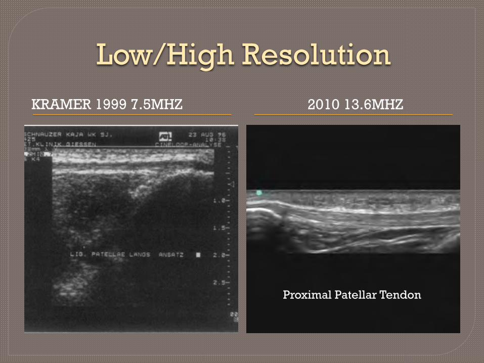
Low/High ResolutionKRAMER 1999 7.5MHZ 2010 13.6MHZ
Proximal Patellar Tendon
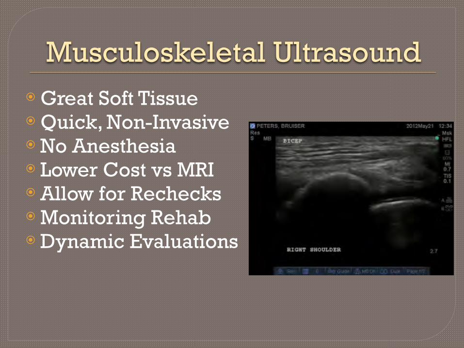
Musculoskeletal Ultrasound⦿ Great Soft Tissue ⦿ Quick, Non-Invasive ⦿ No Anesthesia ⦿ Lower Cost vs MRI ⦿ Allow for Rechecks ⦿ Monitoring Rehab ⦿ Dynamic Evaluations
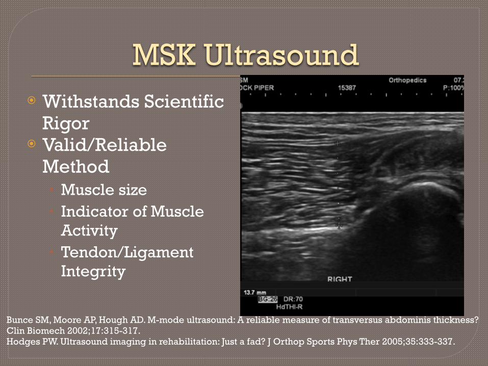
MSK Ultrasound⦿ Withstands Scientific
Rigor ⦿ Valid/Reliable
Method • Muscle size • Indicator of Muscle
Activity • Tendon/Ligament
Integrity
Bunce SM, Moore AP, Hough AD. M-mode ultrasound: A reliable measure of transversus abdominis thickness? Clin Biomech 2002;17:315-317. Hodges PW. Ultrasound imaging in rehabilitation: Just a fad? J Orthop Sports Phys Ther 2005;35:333-337.

Image InterpretationGENERAL GUIDELINES USUAL FACTS
⦿ Tendon, ligament, muscle • Size • Texture ● Homogenous ● Smooth, even
● Non-homogenous ● Mottled
• Echogenicity ● Normal ● Hyperechoic ● More white
● Hypoechoic ● More black
● Anechoic ● Absent
Biceps more hyperechoic than supraspinatus
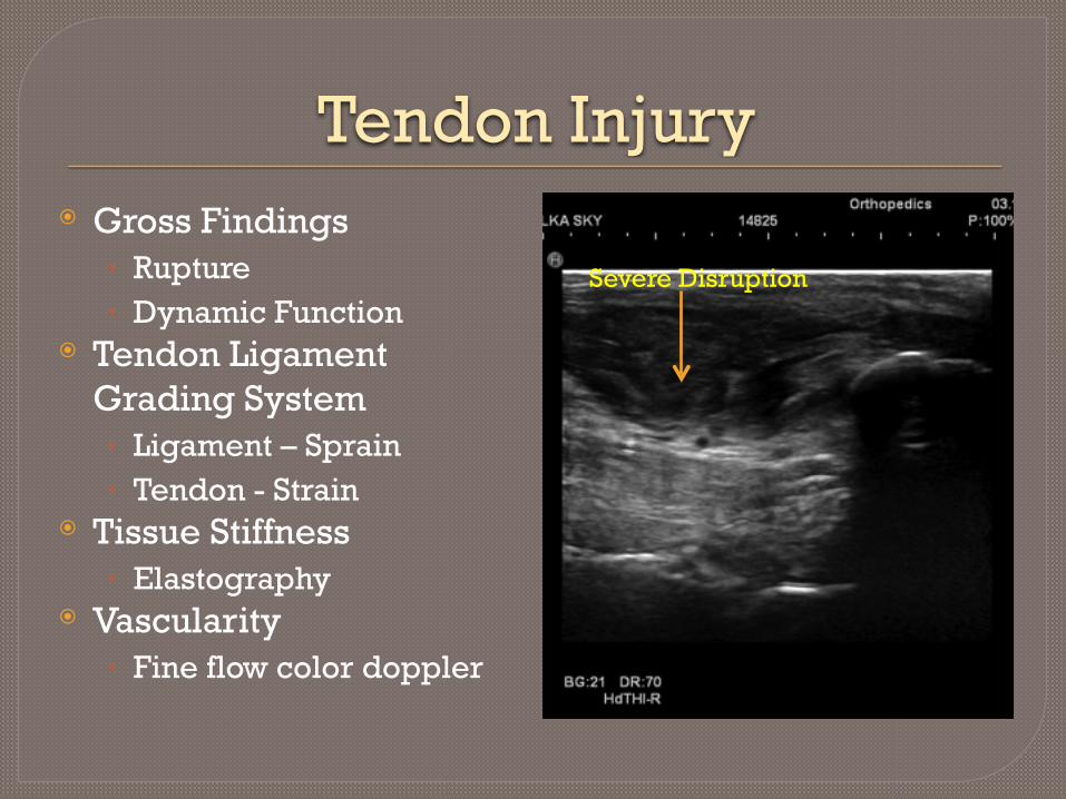
Tendon Injury⦿ Gross Findings
• Rupture • Dynamic Function
⦿ Tendon Ligament Grading System • Ligament – Sprain • Tendon - Strain
⦿ Tissue Stiffness • Elastography
⦿ Vascularity • Fine flow color doppler
Severe Disruption
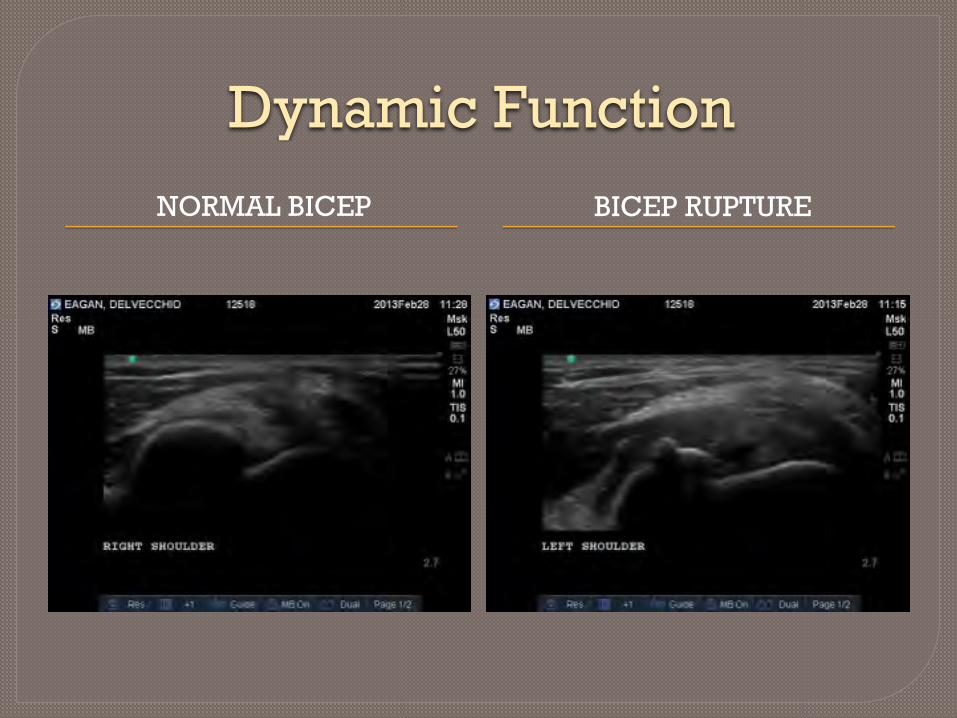
Dynamic FunctionNORMAL BICEP BICEP RUPTURE
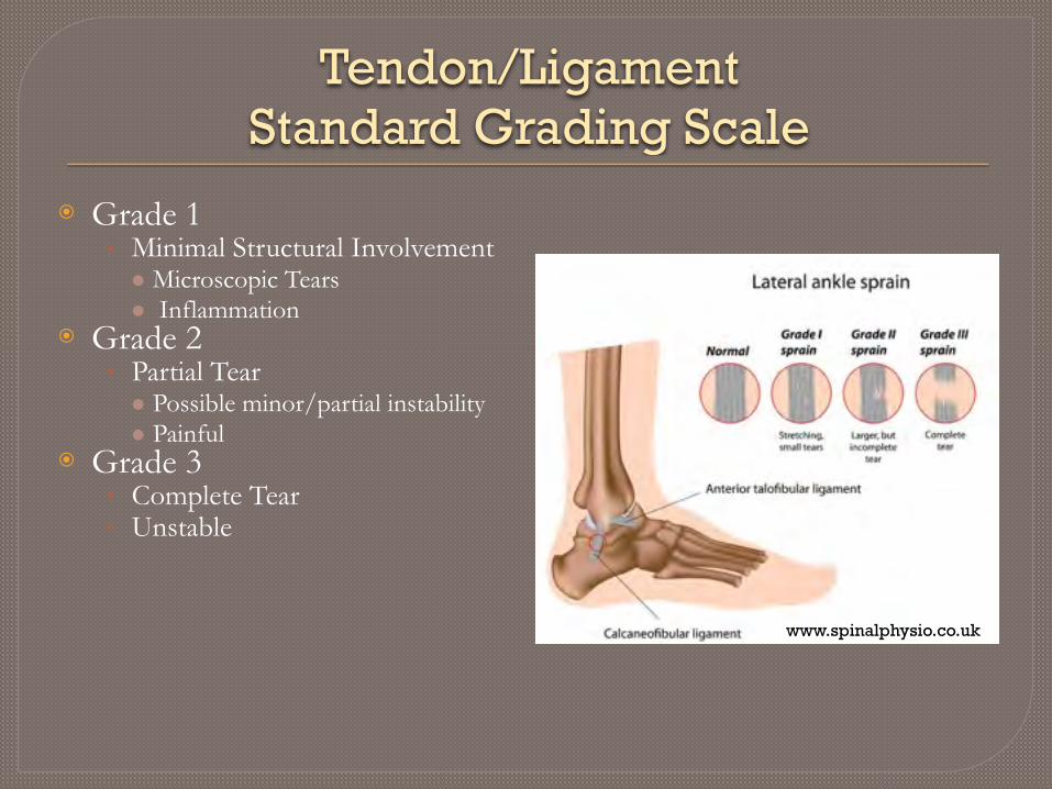
Tendon/Ligament Standard Grading Scale
⦿ Grade 1 • Minimal Structural Involvement ● Microscopic Tears ● Inflammation
⦿ Grade 2 • Partial Tear ● Possible minor/partial instability ● Painful
⦿ Grade 3 • Complete Tear • Unstable
www.spinalphysio.co.uk
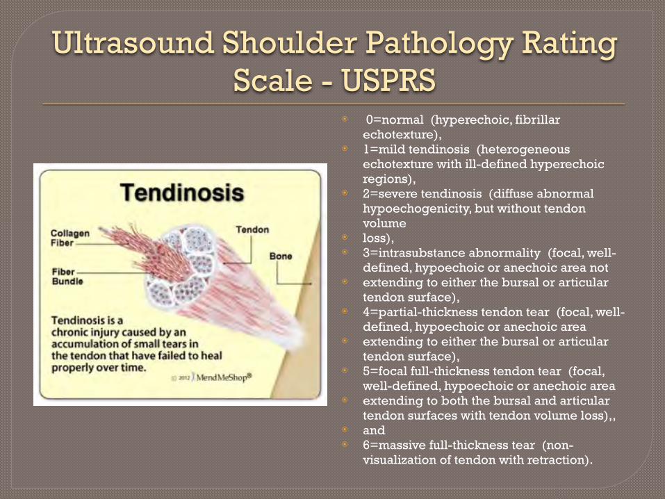
Ultrasound Shoulder Pathology Rating Scale - USPRS
⦿ 0=normal (hyperechoic, fibrillar echotexture),
⦿ 1=mild tendinosis (heterogeneous echotexture with ill-defined hyperechoic regions),
⦿ 2=severe tendinosis (diffuse abnormal hypoechogenicity, but without tendon volume
⦿ loss), ⦿ 3=intrasubstance abnormality (focal, well-
defined, hypoechoic or anechoic area not ⦿ extending to either the bursal or articular
tendon surface), ⦿ 4=partial-thickness tendon tear (focal, well-
defined, hypoechoic or anechoic area ⦿ extending to either the bursal or articular
tendon surface), ⦿ 5=focal full-thickness tendon tear (focal,
well-defined, hypoechoic or anechoic area ⦿ extending to both the bursal and articular
tendon surfaces with tendon volume loss),, ⦿ and ⦿ 6=massive full-thickness tear (non-
visualization of tendon with retraction).
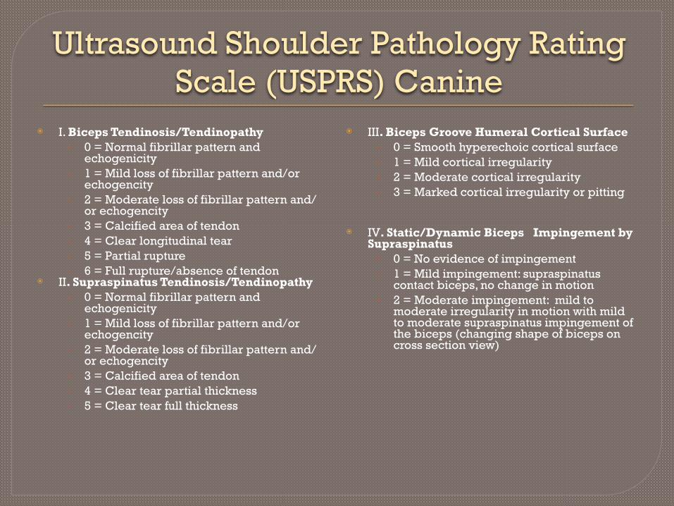
Ultrasound Shoulder Pathology Rating Scale (USPRS) Canine
⦿ I. Biceps Tendinosis/Tendinopathy • 0 = Normal fibrillar pattern and
echogenicity • 1 = Mild loss of fibrillar pattern and/or
echogencity • 2 = Moderate loss of fibrillar pattern and/
or echogencity • 3 = Calcified area of tendon • 4 = Clear longitudinal tear • 5 = Partial rupture • 6 = Full rupture/absence of tendon
⦿ II. Supraspinatus Tendinosis/Tendinopathy • 0 = Normal fibrillar pattern and
echogenicity • 1 = Mild loss of fibrillar pattern and/or
echogencity • 2 = Moderate loss of fibrillar pattern and/
or echogencity • 3 = Calcified area of tendon • 4 = Clear tear partial thickness • 5 = Clear tear full thickness
⦿ III. Biceps Groove Humeral Cortical Surface • 0 = Smooth hyperechoic cortical surface • 1 = Mild cortical irregularity • 2 = Moderate cortical irregularity • 3 = Marked cortical irregularity or pitting
⦿ IV. Static/Dynamic Biceps Impingement by Supraspinatus
• 0 = No evidence of impingement • 1 = Mild impingement: supraspinatus
contact biceps, no change in motion • 2 = Moderate impingement: mild to
moderate irregularity in motion with mild to moderate supraspinatus impingement of the biceps (changing shape of biceps on cross section view)
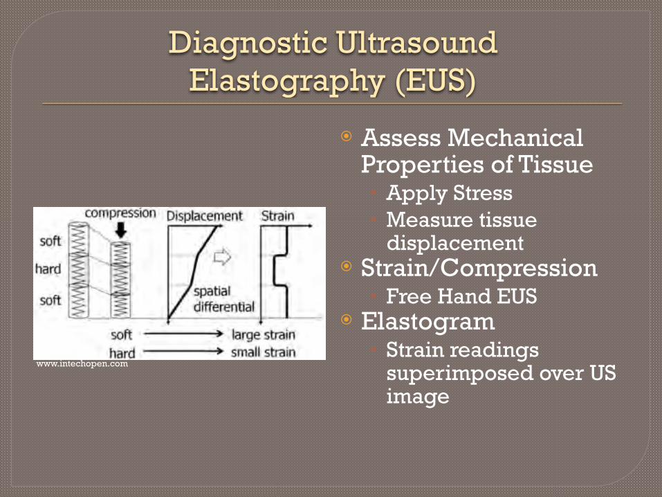
Diagnostic UltrasoundElastography (EUS)
⦿ Assess Mechanical Properties of Tissue • Apply Stress • Measure tissue
displacement ⦿ Strain/Compression
• Free Hand EUS ⦿ Elastogram
• Strain readings superimposed over US image
www.intechopen.com
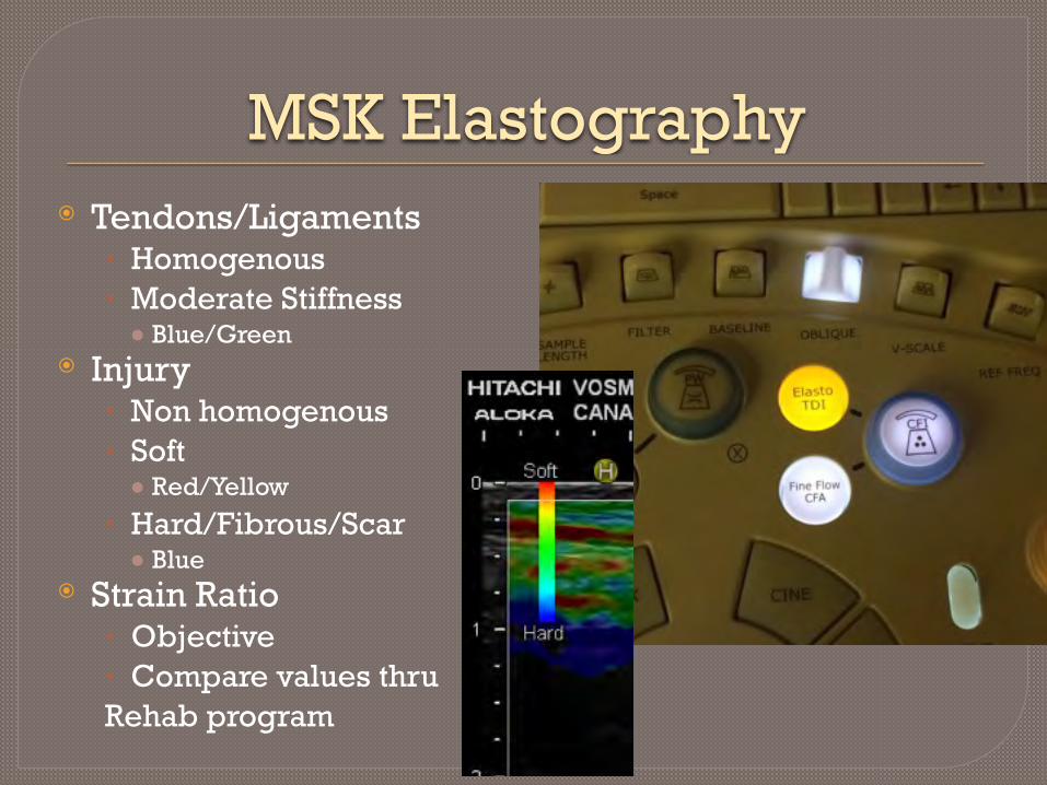
MSK Elastography⦿ Tendons/Ligaments
• Homogenous • Moderate Stiffness ● Blue/Green
⦿ Injury • Non homogenous • Soft ● Red/Yellow
• Hard/Fibrous/Scar ● Blue
⦿ Strain Ratio • Objective • Compare values thru Rehab program
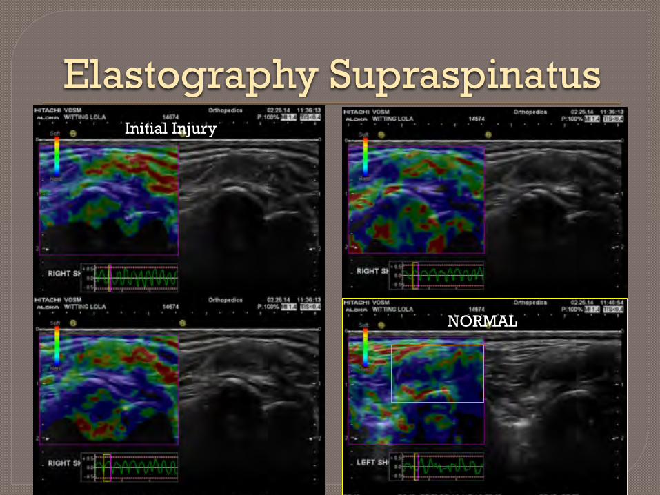
Elastography Supraspinatus
NORMAL
Initial Injury
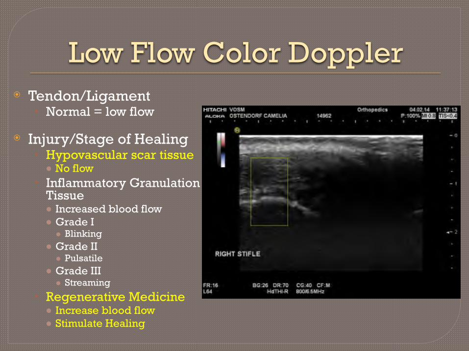
Low Flow Color Doppler⦿ Tendon/Ligament
• Normal = low flow
⦿ Injury/Stage of Healing • Hypovascular scar tissue ● No flow
• Inflammatory Granulation Tissue ● Increased blood flow ● Grade I ● Blinking
● Grade II ● Pulsatile
● Grade III ● Streaming
• Regenerative Medicine ● Increase blood flow ● Stimulate Healing
16
Rehabilitation Case
Examples
Iliopsoas Tendinopathy⦿ “Stella” ⦿ Right Iliopsoas
Insertionopathy ⦿ Right StemCell/PRP
• US guided inj ⦿ Rehab Plan
• Restricted activity ● 8-12 weeks ●Cold Laser, Manual Therapy ●Home Exercise Program
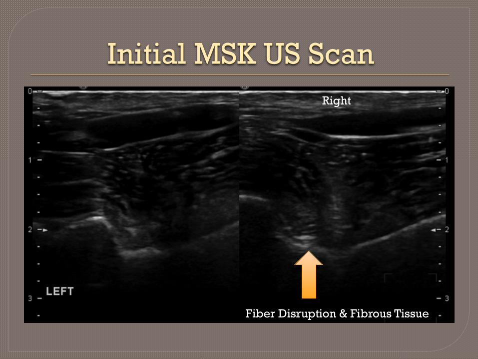
Initial MSK US Scan
Fiber Disruption & Fibrous Tissue
Right
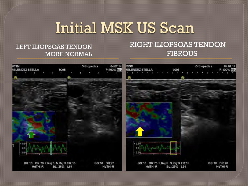
Initial MSK US ScanLEFT ILIOPSOAS TENDON
MORE NORMALRIGHT ILIOPSOAS TENDON
FIBROUS
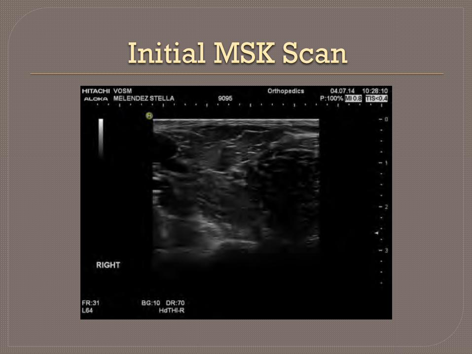
Initial MSK Scan
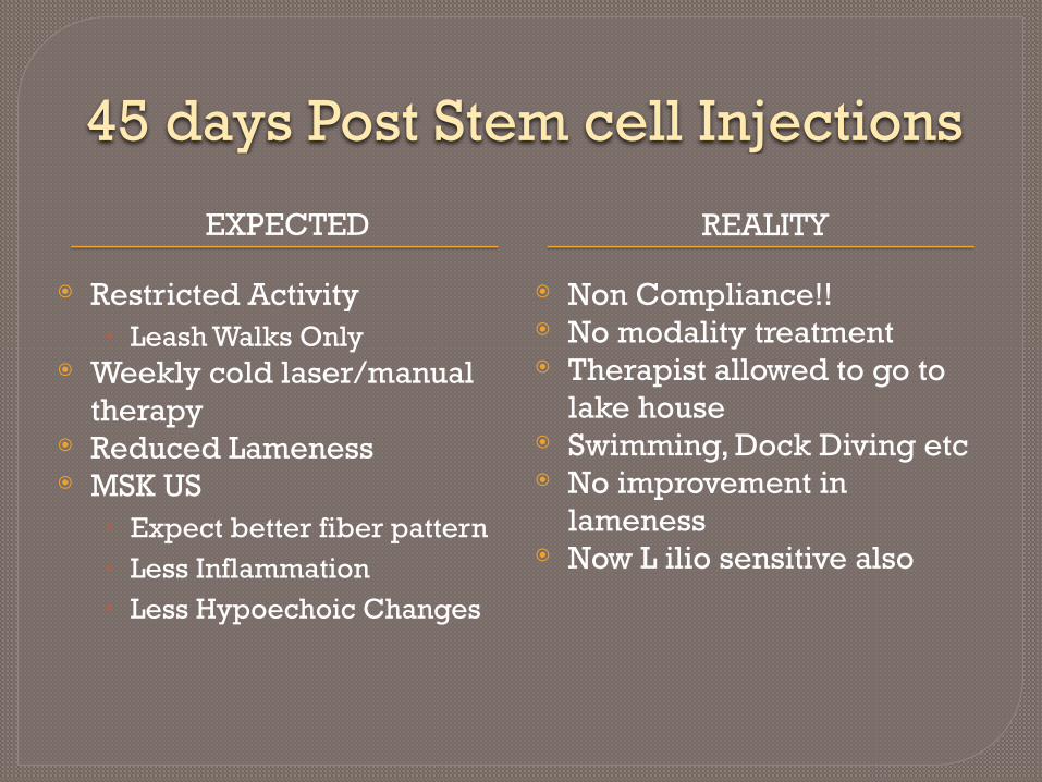
45 days Post Stem cell Injections
EXPECTED REALITY
⦿ Restricted Activity • Leash Walks Only
⦿ Weekly cold laser/manual therapy
⦿ Reduced Lameness ⦿ MSK US
• Expect better fiber pattern • Less Inflammation • Less Hypoechoic Changes
⦿ Non Compliance!! ⦿ No modality treatment ⦿ Therapist allowed to go to
lake house ⦿ Swimming, Dock Diving etc ⦿ No improvement in
lameness ⦿ Now L ilio sensitive also
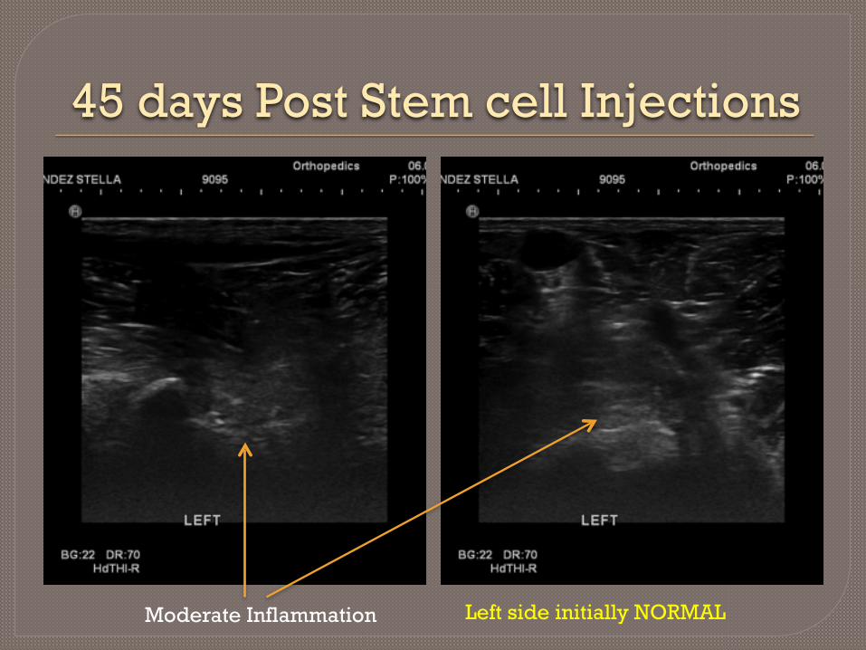
45 days Post Stem cell Injections
Moderate Inflammation Left side initially NORMAL
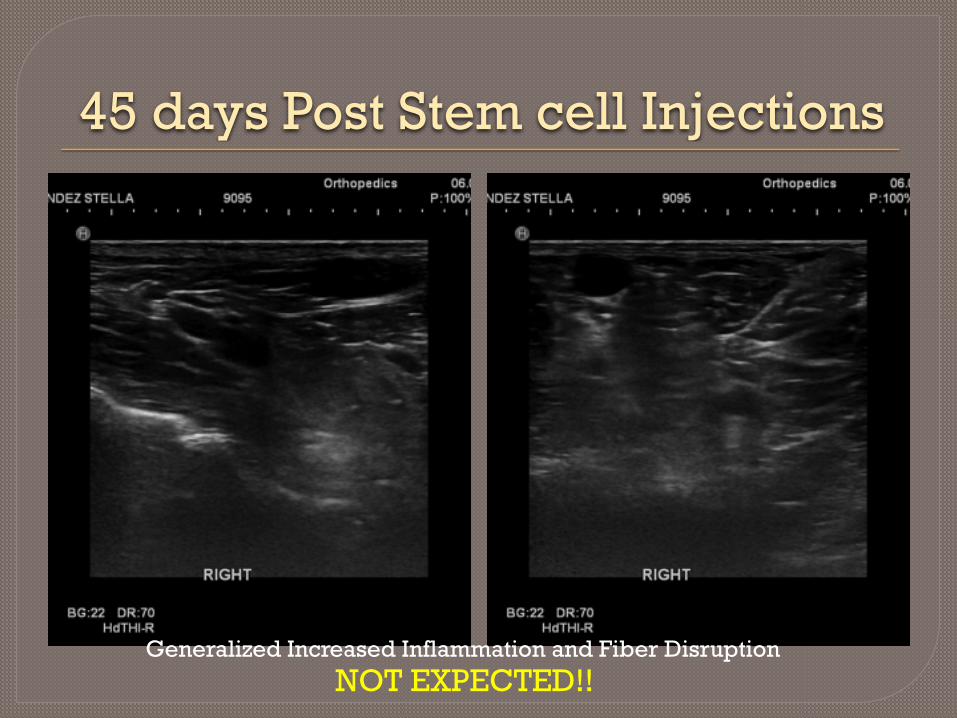
45 days Post Stem cell Injections
Generalized Increased Inflammation and Fiber DisruptionNOT EXPECTED!!
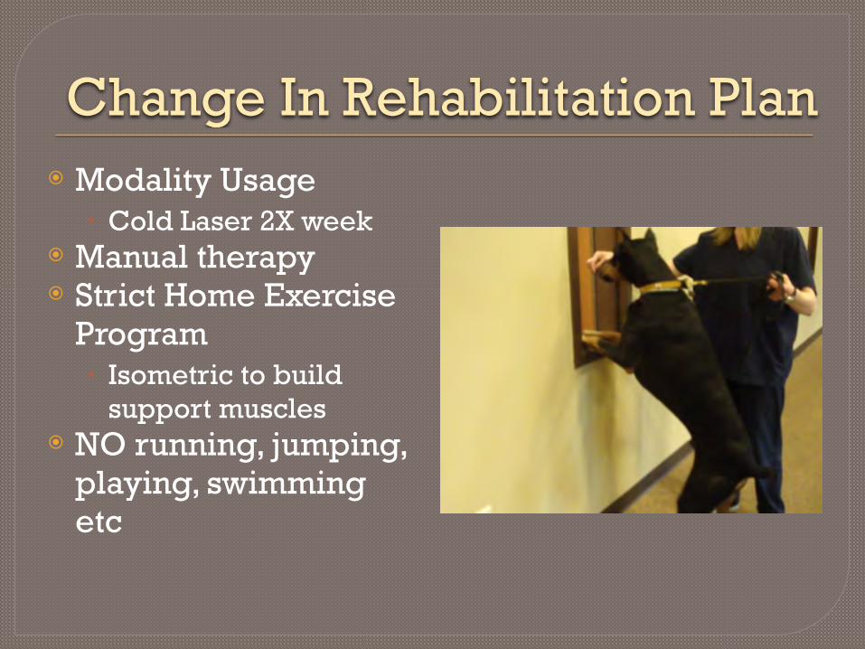
Change In Rehabilitation Plan⦿ Modality Usage
• Cold Laser 2X week ⦿ Manual therapy ⦿ Strict Home Exercise
Program • Isometric to build
support muscles ⦿ NO running, jumping,
playing, swimming etc
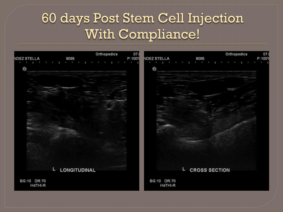
60 days Post Stem Cell Injection With Compliance!
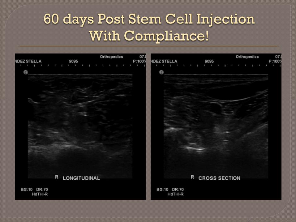
60 days Post Stem Cell Injection With Compliance!
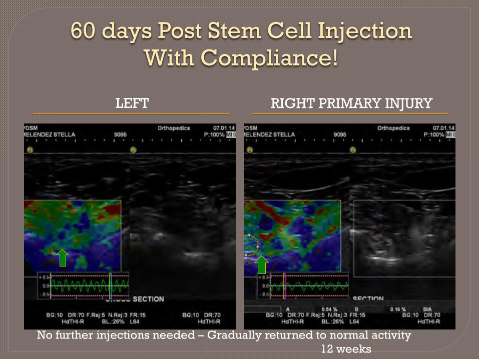
60 days Post Stem Cell Injection With Compliance!
LEFT RIGHT PRIMARY INJURY
No further injections needed – Gradually returned to normal activity 12 weeks
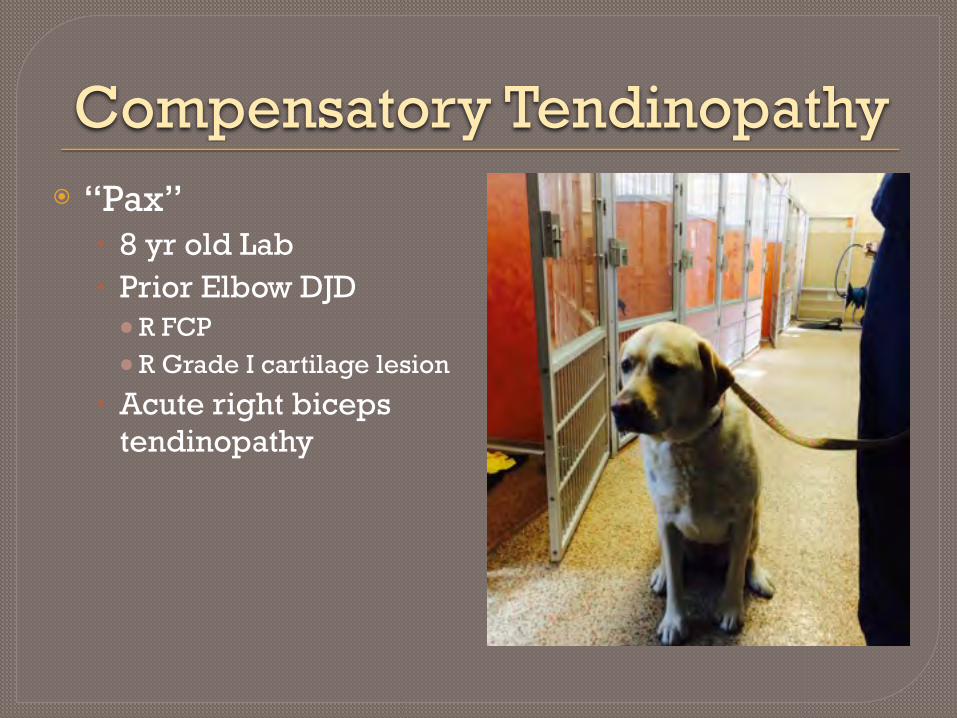
Compensatory Tendinopathy⦿ “Pax”
• 8 yr old Lab • Prior Elbow DJD ●R FCP ●R Grade I cartilage lesion
• Acute right biceps tendinopathy
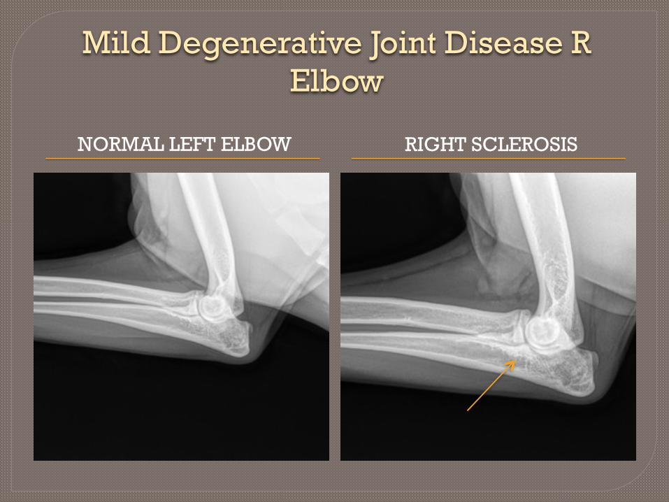
Mild Degenerative Joint Disease R Elbow
NORMAL LEFT ELBOW RIGHT SCLEROSIS
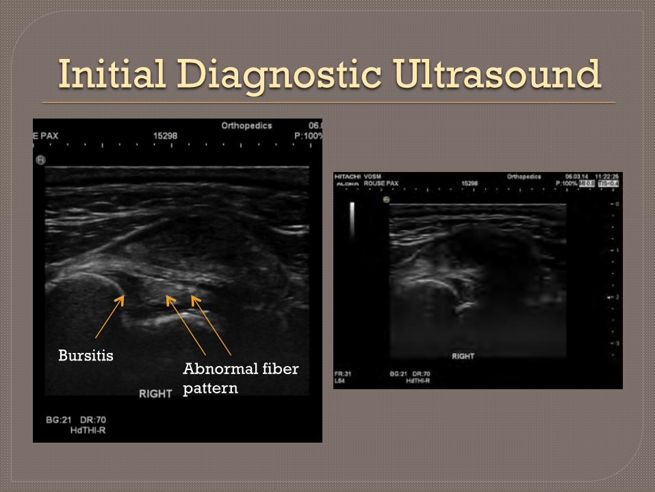
Initial Diagnostic Ultrasound
BursitisAbnormal fiber pattern
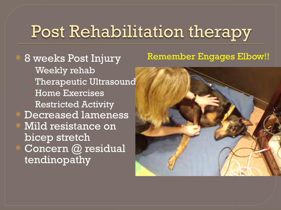
Post Rehabilitation therapy⦿ 8 weeks Post Injury
• Weekly rehab • Therapeutic Ultrasound • Home Exercises • Restricted Activity
⦿ Decreased lameness ⦿ Mild resistance on
bicep stretch ⦿ Concern @ residual
tendinopathy
Remember Engages Elbow!!
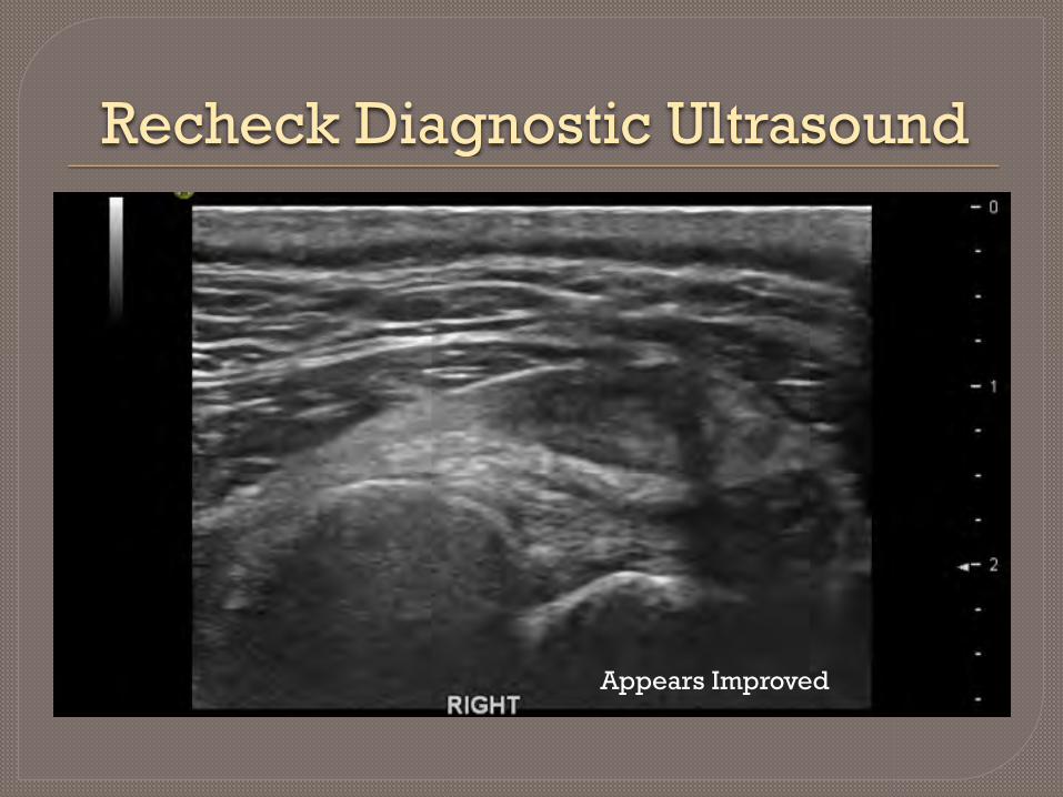
Recheck Diagnostic Ultrasound
Appears Improved
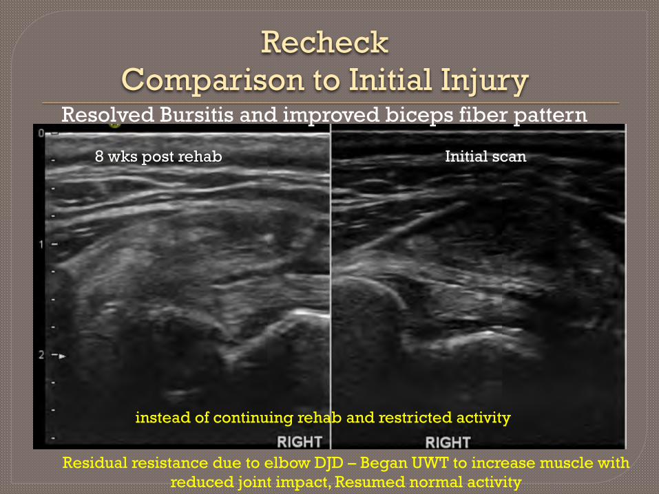
Recheck Comparison to Initial Injury
8 wks post rehab Initial scan
Resolved Bursitis and improved biceps fiber pattern
Residual resistance due to elbow DJD – Began UWT to increase muscle with reduced joint impact, Resumed normal activity
instead of continuing rehab and restricted activity
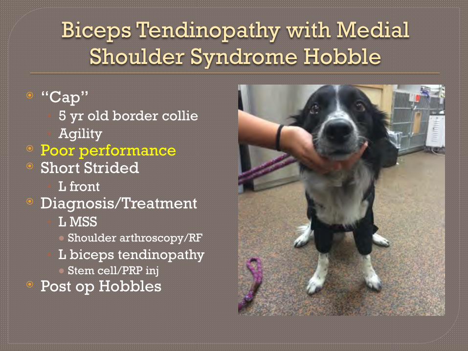
Biceps Tendinopathy with Medial Shoulder Syndrome Hobble
⦿ “Cap” • 5 yr old border collie • Agility
⦿ Poor performance ⦿ Short Strided
• L front ⦿ Diagnosis/Treatment
• L MSS ● Shoulder arthroscopy/RF
• L biceps tendinopathy ● Stem cell/PRP inj
⦿ Post op Hobbles
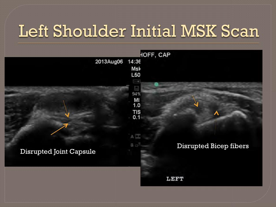
Left Shoulder Initial MSK Scan
Disrupted Joint CapsuleDisrupted Bicep fibers
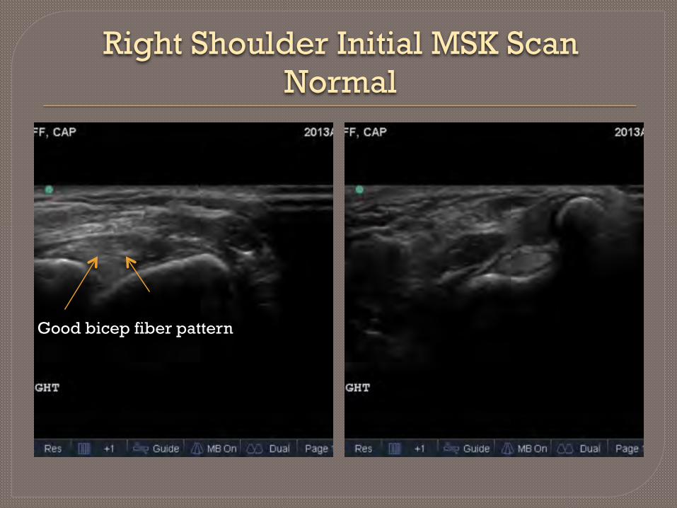
Right Shoulder Initial MSK ScanNormal
Good bicep fiber pattern

90 days Post Treatment/Rehab
STATUS LEFT BICEPS HEALED
⦿ Tendinopathies should be healed
⦿ Equal muscle mass ⦿ Controlled conditioning
performed ⦿ Now
• Gradual return to function/free activity/sport
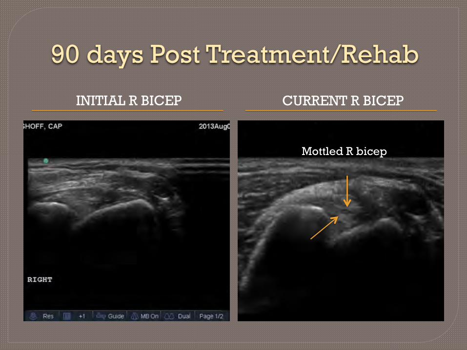
90 days Post Treatment/Rehab
INITIAL R BICEP CURRENT R BICEP
Mottled R bicep
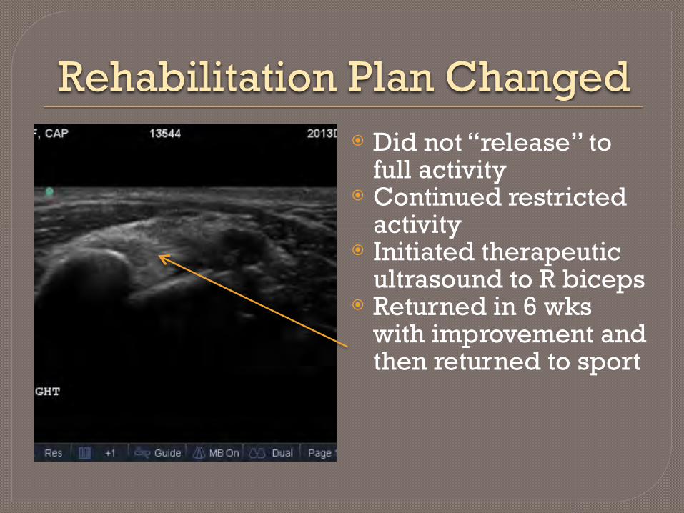
Rehabilitation Plan Changed⦿ Did not “release” to
full activity ⦿ Continued restricted
activity ⦿ Initiated therapeutic
ultrasound to R biceps ⦿ Returned in 6 wks
with improvement and then returned to sport
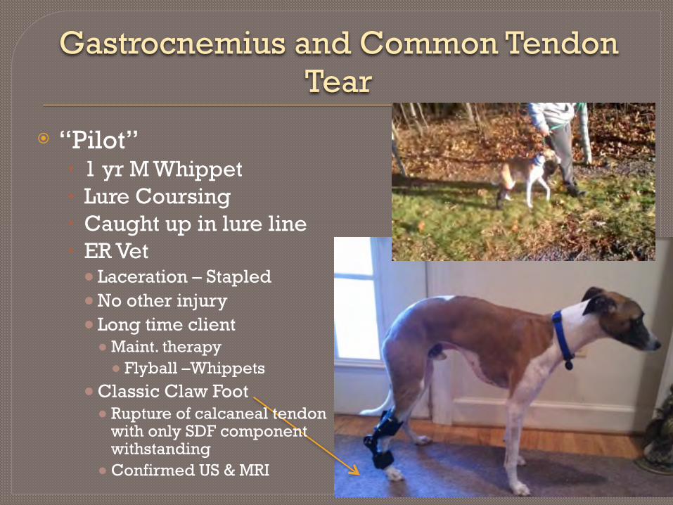
Gastrocnemius and Common Tendon Tear
⦿ “Pilot” • 1 yr M Whippet • Lure Coursing • Caught up in lure line • ER Vet ● Laceration – Stapled ●No other injury ● Long time client ●Maint. therapy ● Flyball –Whippets
●Classic Claw Foot ● Rupture of calcaneal tendon
with only SDF component withstanding
●Confirmed US & MRI
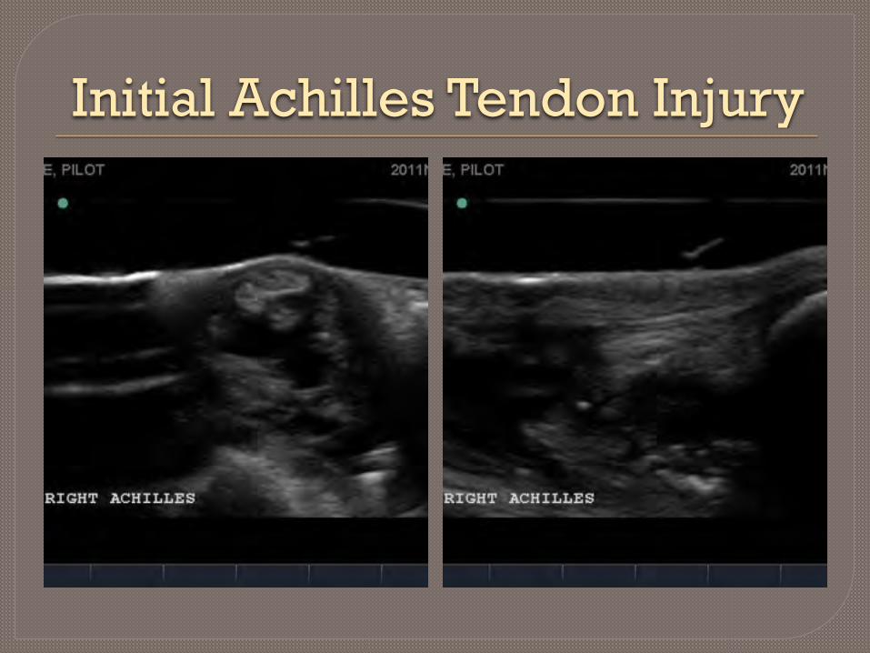
Initial Achilles Tendon Injury
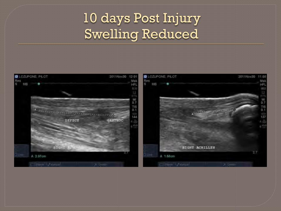
10 days Post Injury Swelling Reduced
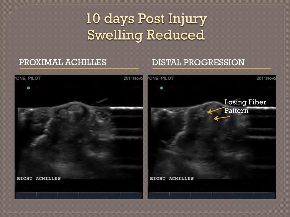
10 days Post Injury Swelling Reduced
PROXIMAL ACHILLES DISTAL PROGRESSION
Losing Fiber Pattern
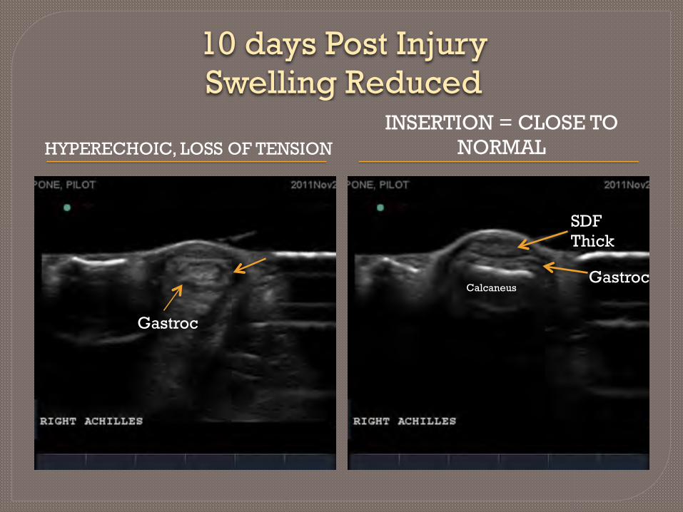
10 days Post Injury Swelling Reduced
HYPERECHOIC, LOSS OF TENSIONINSERTION = CLOSE TO
NORMAL
Calcaneus
SDF Thick
Gastroc
Gastroc
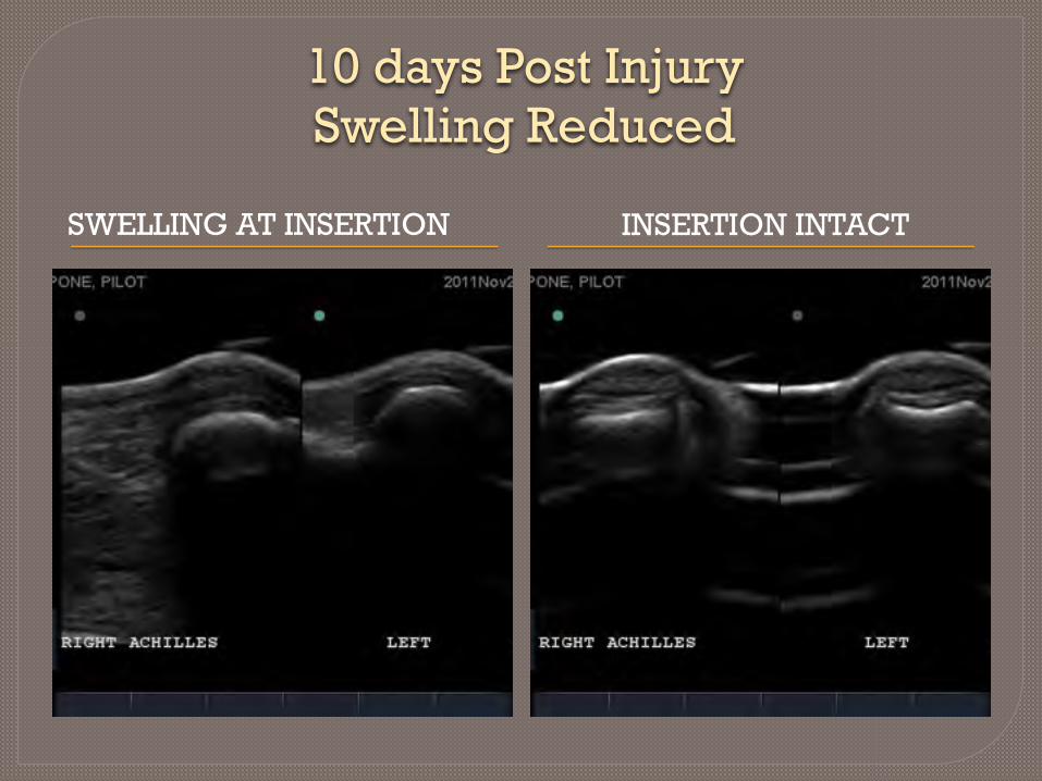
10 days Post Injury Swelling Reduced
SWELLING AT INSERTION INSERTION INTACT
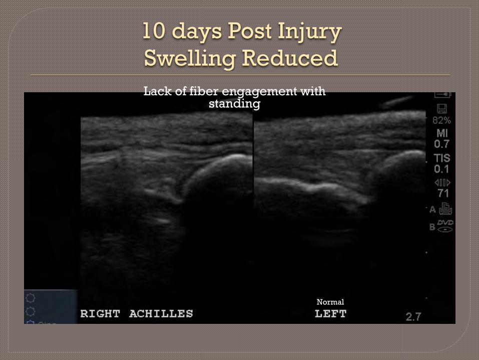
10 days Post Injury Swelling ReducedLack of fiber engagement with
standing
Normal
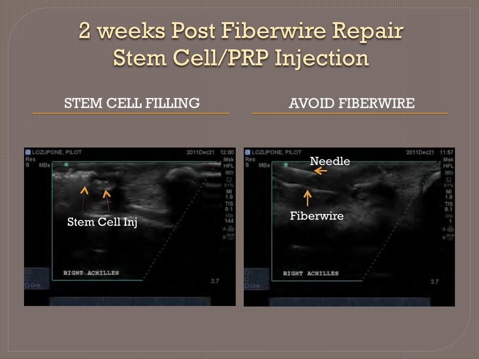
2 weeks Post Fiberwire Repair Stem Cell/PRP Injection
STEM CELL FILLING AVOID FIBERWIRE
Stem Cell Inj Fiberwire
Needle
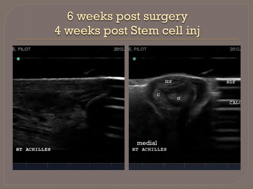
6 weeks post surgery 4 weeks post Stem cell inj
medial
SDF
CG
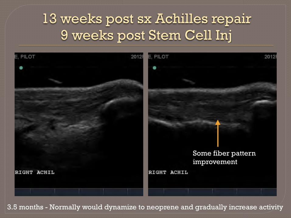
13 weeks post sx Achilles repair 9 weeks post Stem Cell Inj
3.5 months - Normally would dynamize to neoprene and gradually increase activity
Some fiber pattern improvement
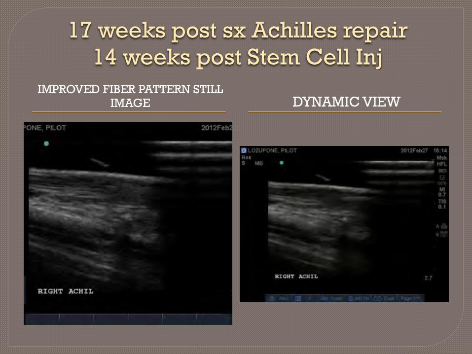
17 weeks post sx Achilles repair 14 weeks post Stem Cell Inj
IMPROVED FIBER PATTERN STILL IMAGE DYNAMIC VIEW
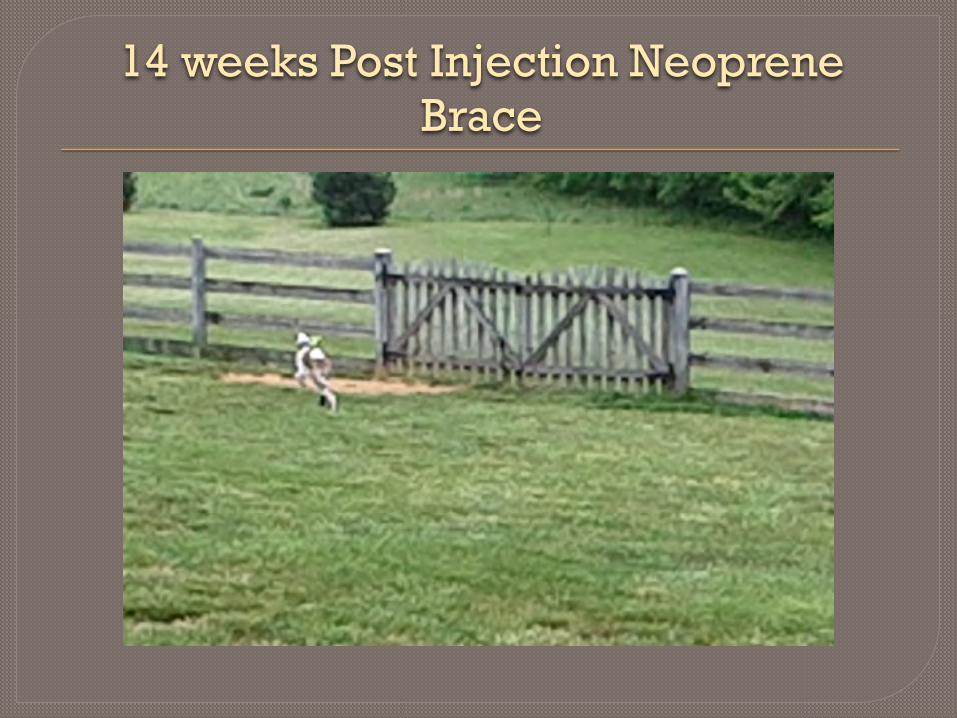
14 weeks Post Injection Neoprene Brace
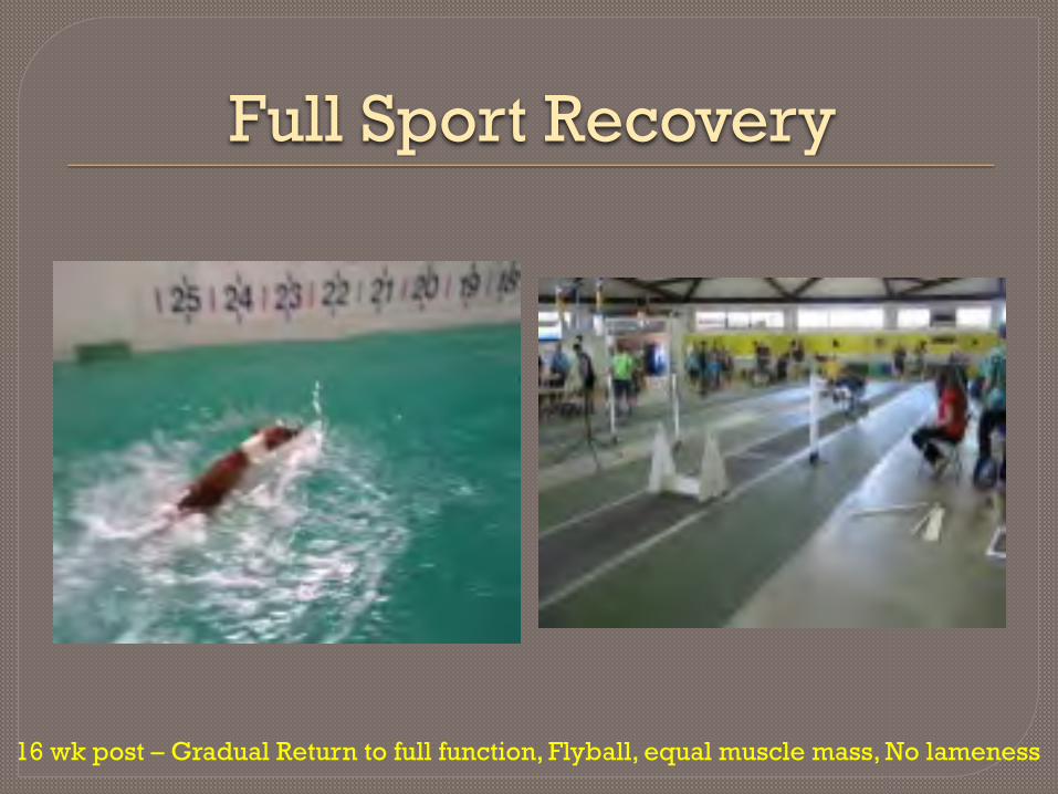
Full Sport Recovery
16 wk post – Gradual Return to full function, Flyball, equal muscle mass, No lameness
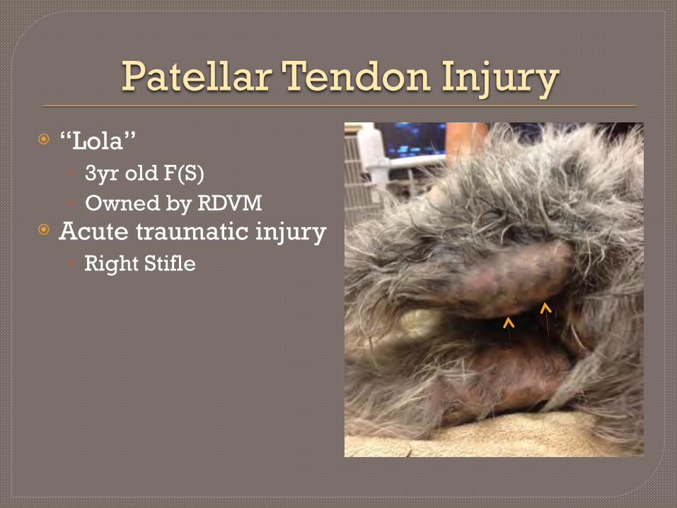
Patellar Tendon Injury⦿ “Lola”
• 3yr old F(S) • Owned by RDVM
⦿ Acute traumatic injury • Right Stifle

Patellar Tendon
Patellar tendon
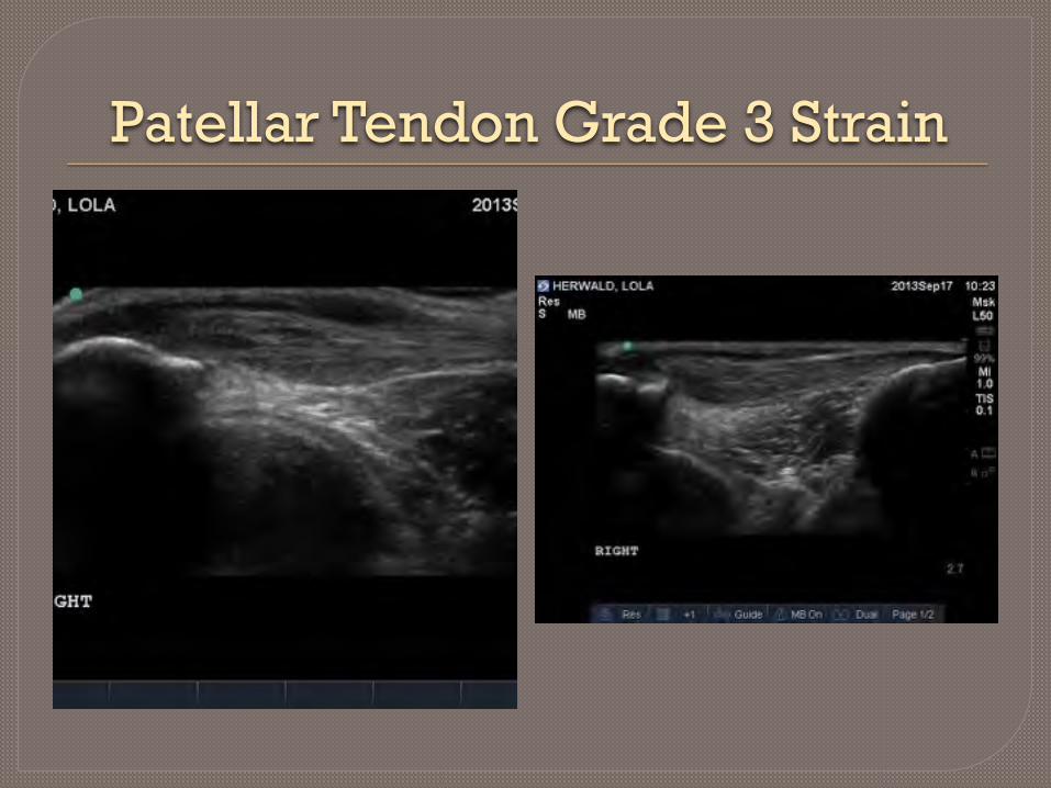
Patellar Tendon Grade 3 Strain
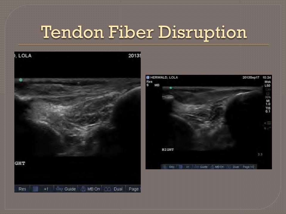
Tendon Fiber Disruption
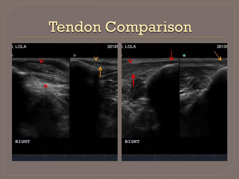
Tendon Comparison
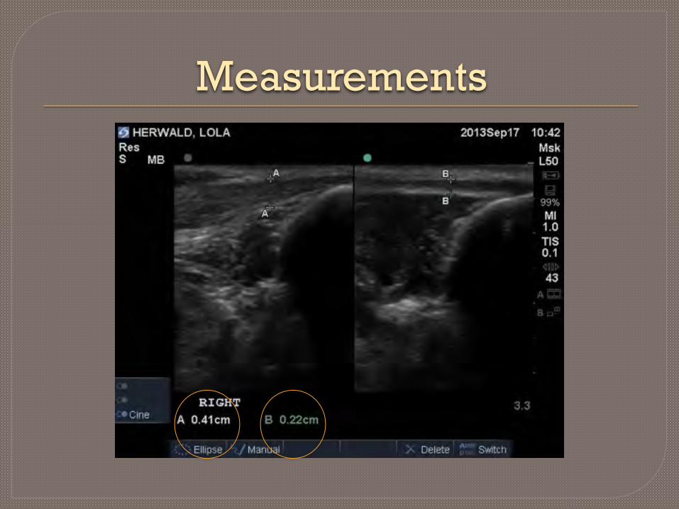
Measurements
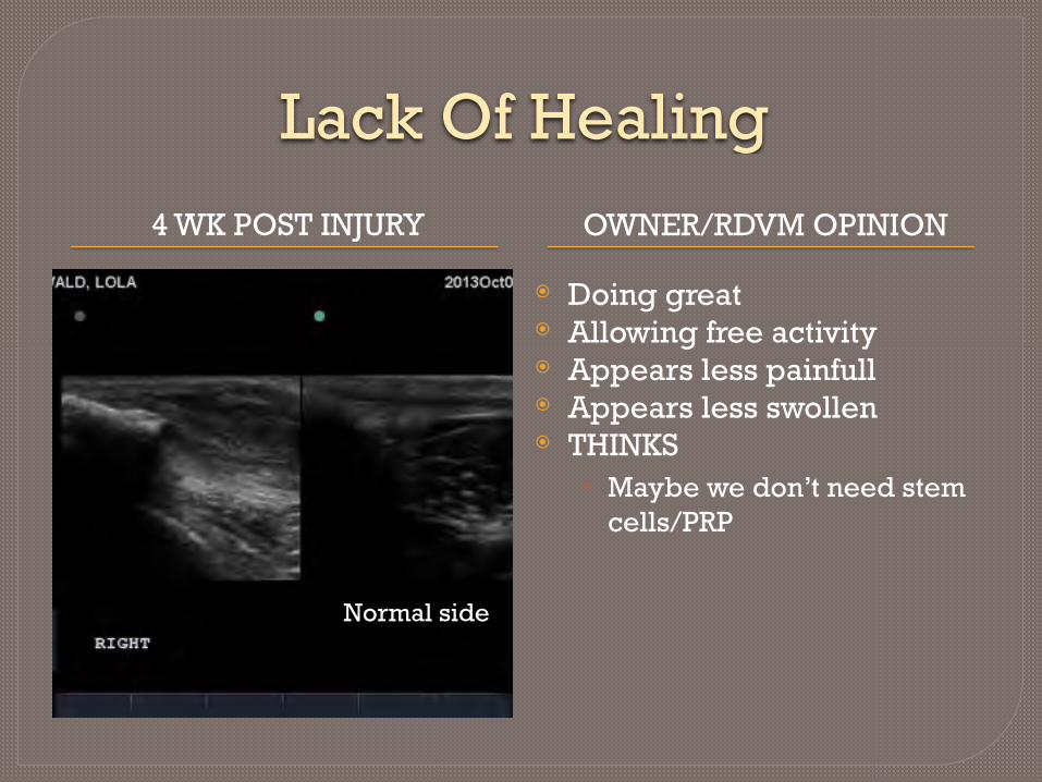
Lack Of Healing4 WK POST INJURY OWNER/RDVM OPINION
⦿ Doing great ⦿ Allowing free activity ⦿ Appears less painfull ⦿ Appears less swollen ⦿ THINKS
• Maybe we don’t need stem cells/PRP
Normal side
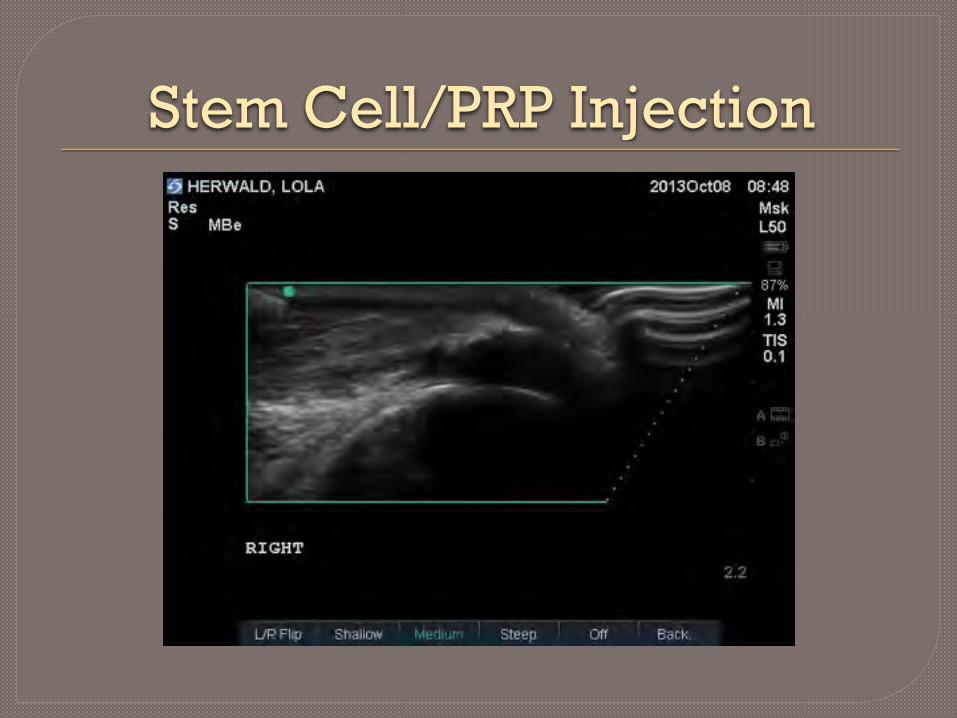
Stem Cell/PRP Injection
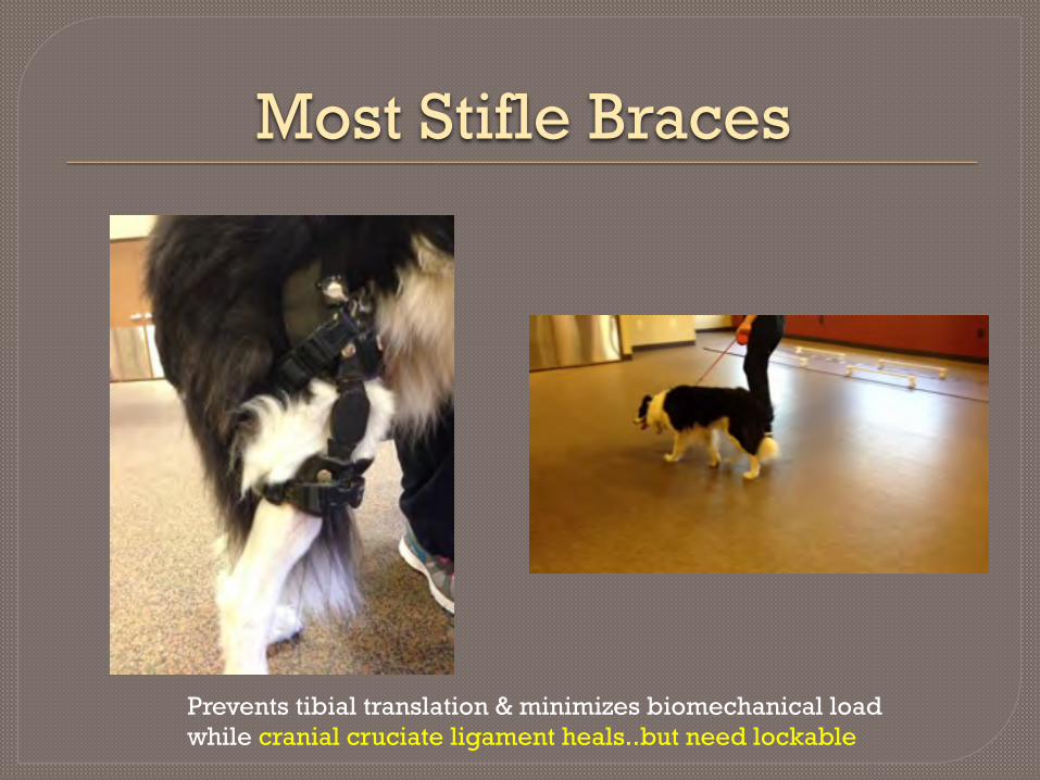
Most Stifle Braces
Prevents tibial translation & minimizes biomechanical load while cranial cruciate ligament heals..but need lockable
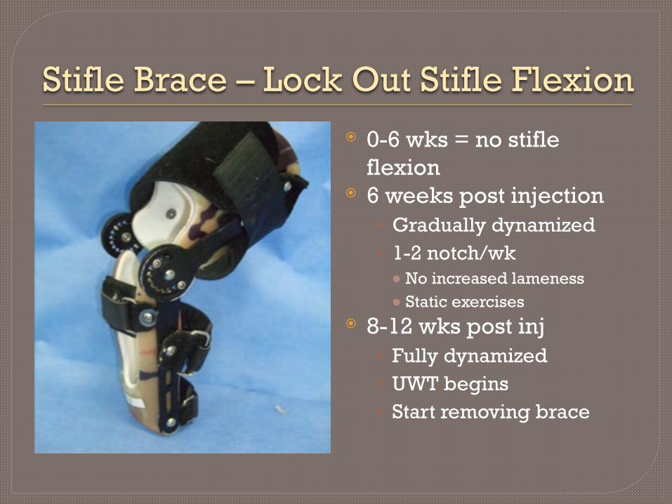
Stifle Brace – Lock Out Stifle Flexion⦿ 0-6 wks = no stifle
flexion ⦿ 6 weeks post injection
• Gradually dynamized • 1-2 notch/wk ● No increased lameness ● Static exercises
⦿ 8-12 wks post inj • Fully dynamized • UWT begins • Start removing brace
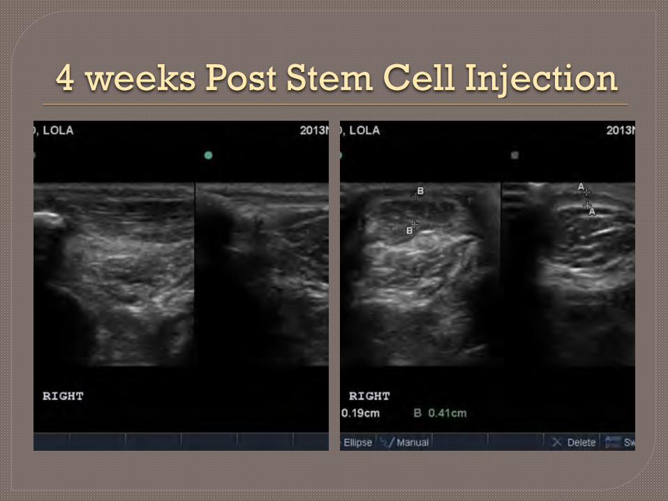
4 weeks Post Stem Cell Injection
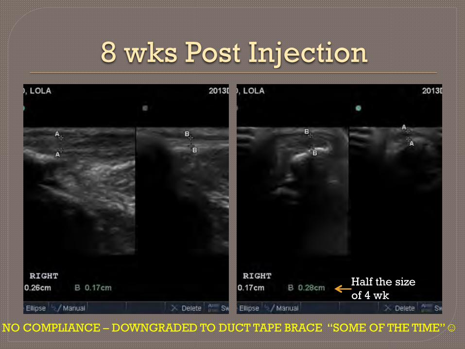
8 wks Post Injection
Half the size of 4 wk
NO COMPLIANCE – DOWNGRADED TO DUCT TAPE BRACE “SOME OF THE TIME”☺
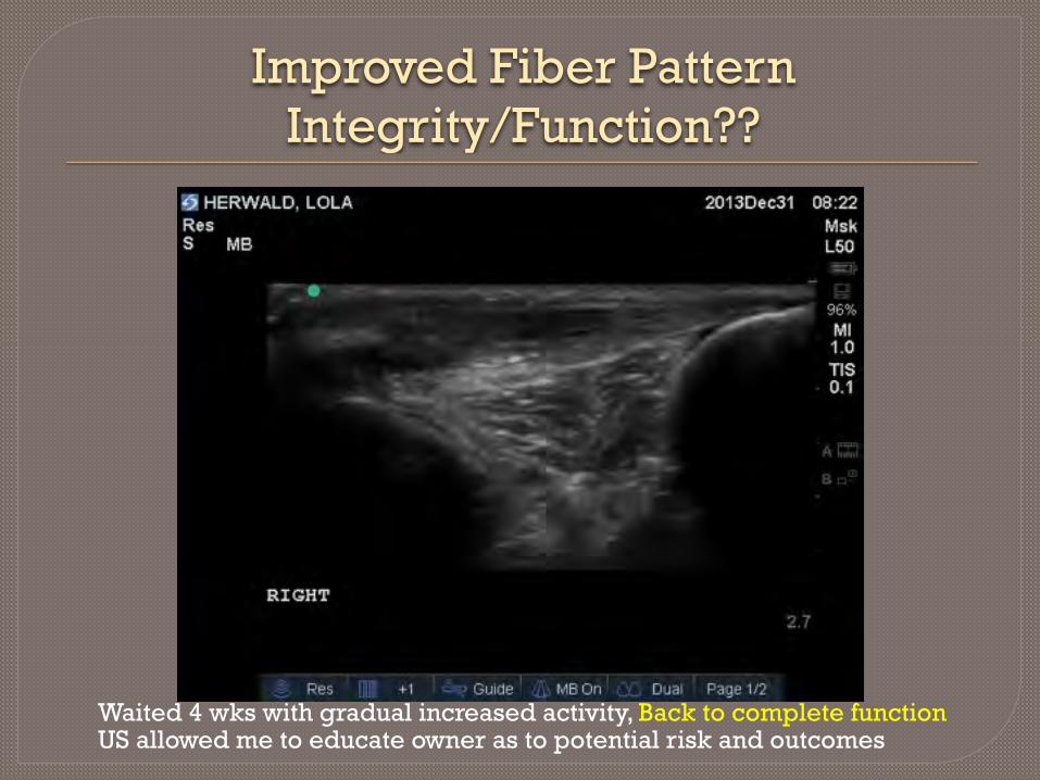
Improved Fiber Pattern Integrity/Function??
Waited 4 wks with gradual increased activity, Back to complete functionUS allowed me to educate owner as to potential risk and outcomes
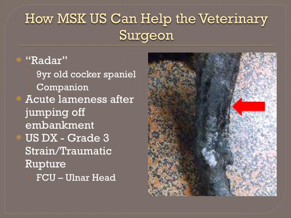
How MSK US Can Help the Veterinary Surgeon
⦿ “Radar” • 9yr old cocker spaniel • Companion
⦿ Acute lameness after jumping off embankment
⦿ US DX - Grade 3 Strain/Traumatic Rupture • FCU – Ulnar Head
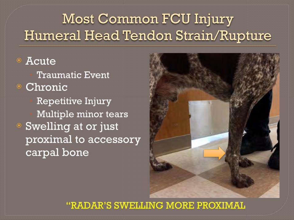
Most Common FCU Injury Humeral Head Tendon Strain/Rupture
⦿ Acute • Traumatic Event
⦿ Chronic • Repetitive Injury • Multiple minor tears
⦿ Swelling at or just proximal to accessory carpal bone
“RADAR’S SWELLING MORE PROXIMAL

Flexor Carpi Ulnaris Injury⦿ FCU
• Humeral Head ●Classic
• Ulnar Head ● Insertion deep to HH
⦿ Injury • Tendon Strain • Avulsion • Muscle Tears

FCU – Ulnar Head Tendon Rupture - Ultrasound
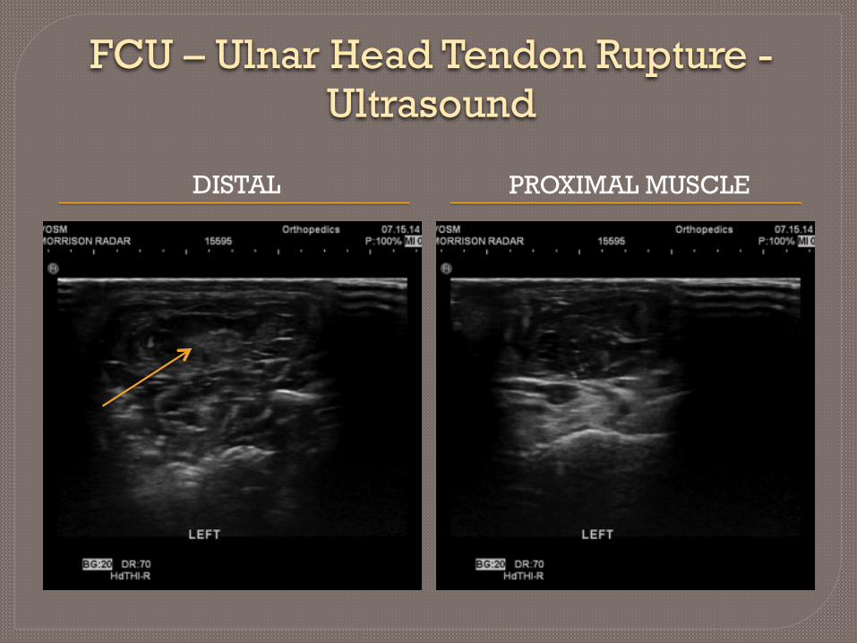
FCU – Ulnar Head Tendon Rupture - Ultrasound
DISTAL PROXIMAL MUSCLE
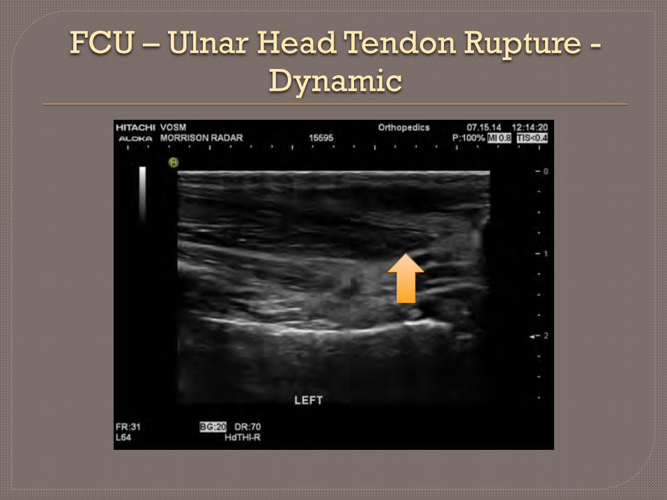
FCU – Ulnar Head Tendon Rupture - Dynamic
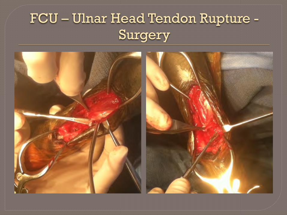
FCU – Ulnar Head Tendon Rupture - Surgery
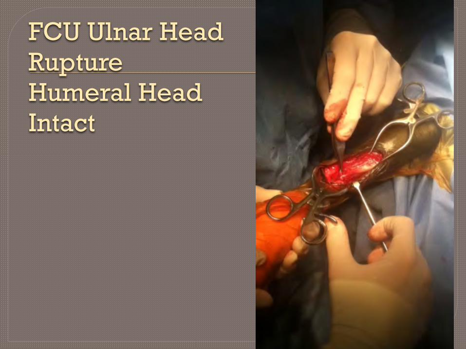
FCU Ulnar HeadRupture Humeral Head Intact
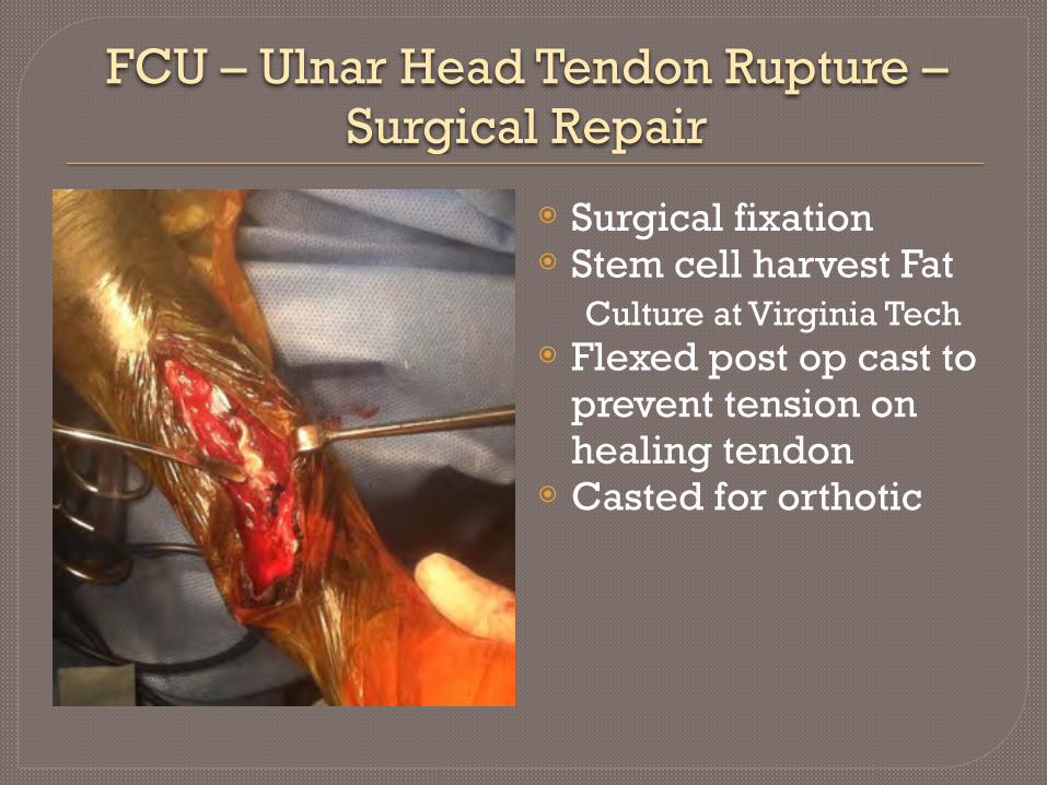
FCU – Ulnar Head Tendon Rupture – Surgical Repair
⦿ Surgical fixation ⦿ Stem cell harvest Fat
Culture at Virginia Tech ⦿ Flexed post op cast to
prevent tension on healing tendon
⦿ Casted for orthotic
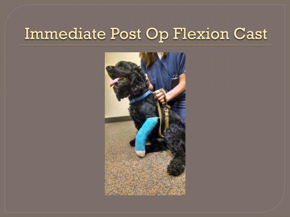
Immediate Post Op Flexion Cast
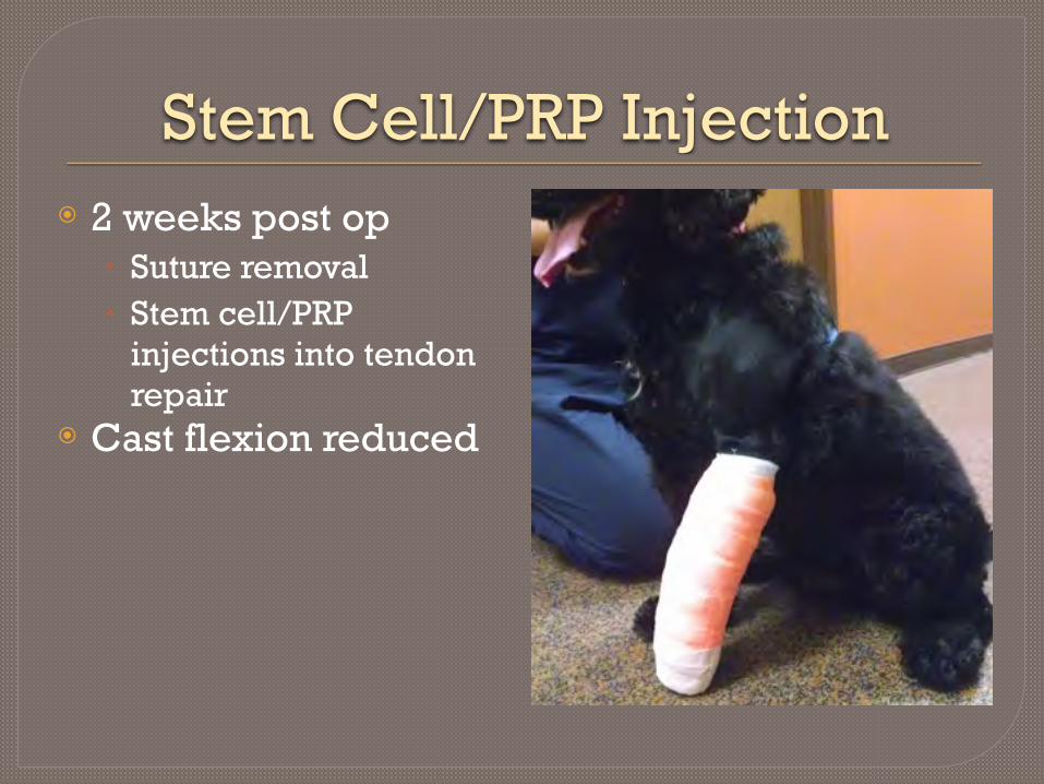
Stem Cell/PRP Injection⦿ 2 weeks post op
• Suture removal • Stem cell/PRP
injections into tendon repair
⦿ Cast flexion reduced
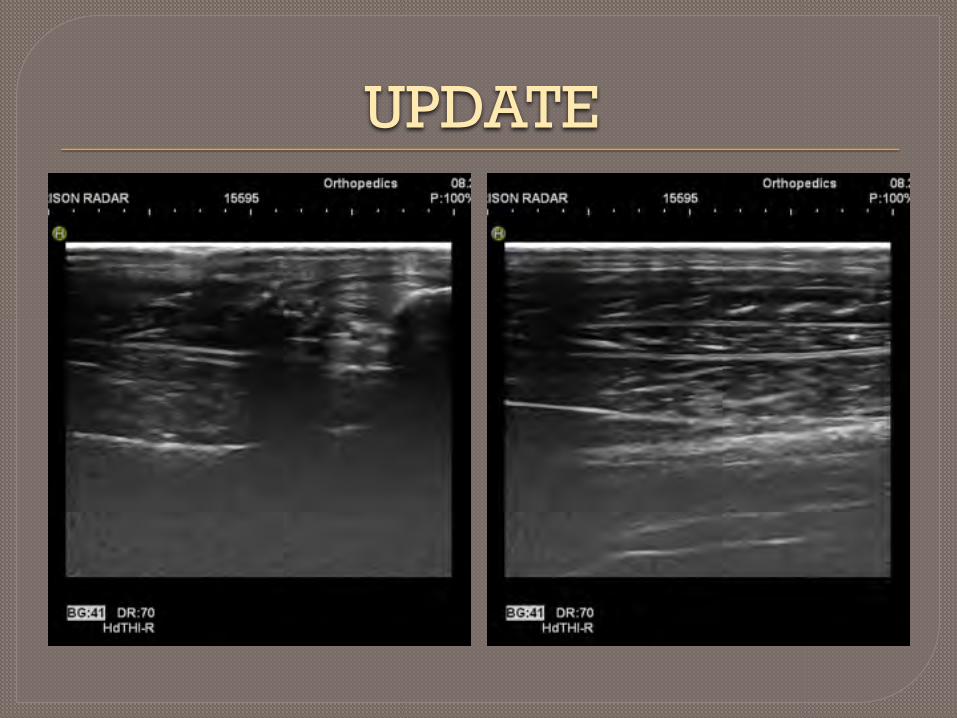
UPDATE
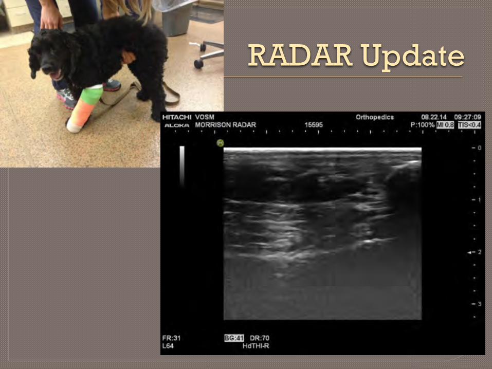
RADAR Update
Conclusion⦿ Canine Musculoskeletal Ultrasound
• Excellent soft tissue diagnostic tool • Valuable in precise regen med application • Excellent tool to monitor healing • Available and economical when compared to
other similar diagnostic tools • INDISPENSABLE TOOL ●Guide in Canine Rehabilitation
So…Hopefully I have convinced..at least some
80
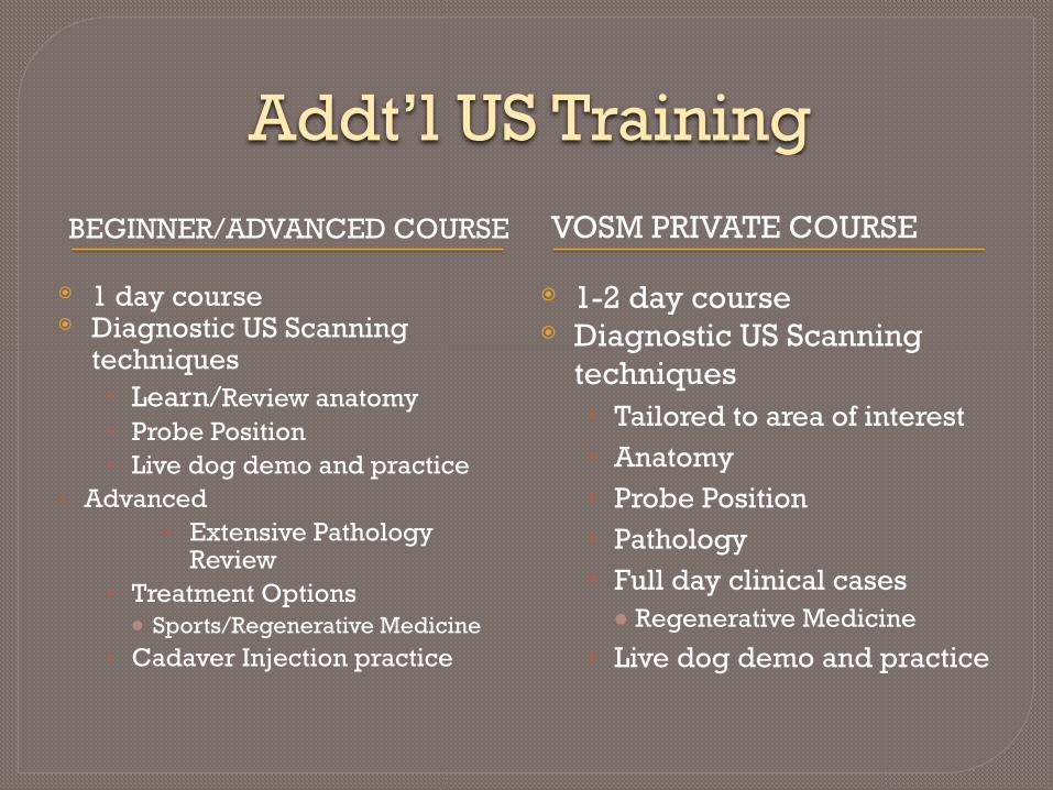
Addt’l US TrainingBEGINNER/ADVANCED COURSE VOSM PRIVATE COURSE
⦿ 1 day course ⦿ Diagnostic US Scanning
techniques • Learn/Review anatomy • Probe Position • Live dog demo and practice
• Advanced • Extensive Pathology
Review • Treatment Options ● Sports/Regenerative Medicine
• Cadaver Injection practice
⦿ 1-2 day course ⦿ Diagnostic US Scanning
techniques • Tailored to area of interest • Anatomy • Probe Position • Pathology • Full day clinical cases ● Regenerative Medicine
• Live dog demo and practice
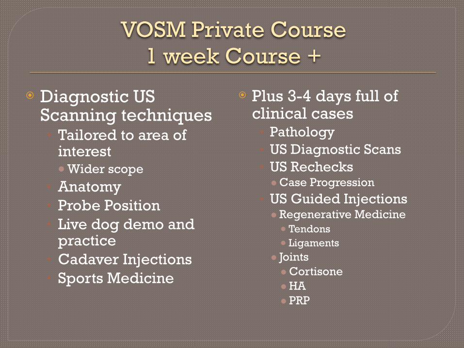
VOSM Private Course1 week Course +
⦿ Diagnostic US Scanning techniques • Tailored to area of
interest ●Wider scope
• Anatomy • Probe Position • Live dog demo and
practice • Cadaver Injections • Sports Medicine
⦿ Plus 3-4 days full of clinical cases
• Pathology • US Diagnostic Scans • US Rechecks ●Case Progression
• US Guided Injections ●Regenerative Medicine ● Tendons ● Ligaments
● Joints ●Cortisone ●HA ● PRP
Thank you!!
Recommended