REVIEWARTICLE
Coronary artery-bronchial artery fistulas: report of two Dutchcases with a review of the literature
S. A. M. Said & R. M. Oortman & J.-H. Hofstra &
P. M. J. Verhorst & R. H. J. A. Slart & M. W. de Haan &
F. Eerens & H. J. G. M. Crijns
Published online: 26 January 2014# The Author(s) 2014. This article is published with open access at Springerlink.com
AbstractBackground Coronary bronchial artery fistulas (CBFs) arerare anomalies, whichmay be isolated or associated with otherdisorders.Materials and methods Two adult patients with CBFs aredescribed and a PubMed search was performed using thekeywords “coronary bronchial artery fistulas” in the periodfrom 2008 to 2013.Results Twenty-seven reviewed subjects resulting in a total of31 fistulas were collected. Asymptomatic presentation wasreported in 5 subjects (19 %), chest pain (n=17) was frequent-ly present followed by haemoptysis (n=7) and dyspnoea (n=5). Concomitant disorders were bronchiectasis (44 %), diabe-tes (33 %) and hypertension (28 %). Multimodality andsingle-modality diagnostic strategies were applied in 56 %
and 44 %, respectively. The origin of the CBFs was the leftcircumflex artery in 61 %, the right coronary artery in 36 %and the left anterior descending artery in 3 %. Managementwas conservative (22 %), surgical ligation (11 %), percutane-ous transcatheter embolisation (30 %), awaiting lung trans-plantation (7 %) or not reported (30 %).Conclusions CBFs may remain clinically silent, or presentwith chest pain or haemoptysis. CBFs are commonly associ-ated with bronchiectasis and usually require a multimodalityapproach to be diagnosed. Several treatment strategies areavailable. This report presents two adult cases with CBFsand a review of the literature.
Keywords Congenital anomaly . Coronary bronchial arteryfistulas . Multi-detector computer tomography . Positronemission tomography/13-ammonia-adenosine scanning .
Management
AbbreviationsAP AnteroposteriorCBF Coronary-bronchial artery fistulaRCA Right coronary artery
Introduction
Coronary bronchial artery fistulas (CBFs) are usually foundincidentally during invasive coronary angiography (CAG) [1].The aetiology of CBFs is uncertain. CBFs are often associatedwith bronchiectasis which may be bilateral [1] or unilateraleither to the left [2] or to the right lung [3]. Sometimes CBFsoccur concomitant with tetralogy of Fallot, supravalvular aor-tic stenosis or Takayasu aortitis [4]. The morphology may beshown using several diagnostic modalities: CAG, magneticresonance imaging (MRI) and multi-detector computed
S. A. M. Said (*)Department of Cardiology, Hospital Group Twente, Geerdinksweg141, 7555 DL Hengelo, the Netherlandse-mail: [email protected]
R. M. Oortman : P. M. J. VerhorstDepartment of Cardiology, Thoraxcentrum Twente, MedischSpectrum Twente, 7513 ER Enschede, the Netherlands
J.<H. HofstraDepartment of Cardiology, Koningin Beatrix Hospital, 7101BN Winterswijk, the Netherlands
R. H. J. A. SlartDepartment of Nuclear Medicine andMolecular Imaging, UniversityMedical Center Groningen, 9713 GZ Groningen, the Netherlands
M. W. de HaanDepartment of Radiology, Maastricht University Medical Center,6229 HX Maastricht, the Netherlands
F. Eerens :H. J. G. M. CrijnsDepartment of Cardiology, Maastricht University Medical Center,6229 HX Maastricht, the Netherlands
Neth Heart J (2014) 22:139–147DOI 10.1007/s12471-014-0518-z
tomography (MDCT). MDCT identified the course of CBFsbetween the circumflex artery (Cx) and the bronchial arteries [2,5] and myocardial perfusion imaging (MPI) revealed reversibledefects [5]. The functional assessment may be obtained byMPIusing technetium-99 m tetrofosmin, MRI and oximetric seriesduring cardiac catheterisation to establish the magnitude of theshunt. CBFs often remain asymptomatic but they can also bethe source of aggravating haemoptysis [6]. CBFs can be man-aged either by a conservative medical regimen, percutaneousocclusion techniques or surgical ligation.
We report two adult patients with angina pectoris in whomCAG demonstrated significant obstructive coronary arterydisease (CAD), and coincidentally CBFs were found. The firstpatient was treated with stenting of the Cx coronary artery, andin a second session the fistulous vessel was occluded bycoiling during a percutaneous transcatheter embolisation(PTE) procedure. The second patient had associated bronchi-ectasis, sustained a subclinical anterior wall myocardial in-farction (MI), and was treated medically for his CBF. Theliterature is briefly reviewed.
Methods
Two adult patients with CBFs are presented and a PubMedsearch was performed using the keywords “coronary bronchi-al artery fistulas” in the period between 2008 and 2013. Thissearch harvested 30 citations. Eighteen relevant papers wereselected and evaluated yielding 27 patients with 31 CBFs.
Case reports
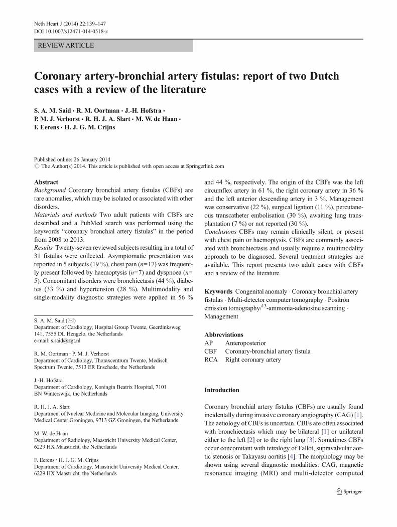
Case 1: A 66-year-old female patient with known arterialhypertension and diaphragmatic hernia was evalu-ated for palpitations, persistent typical and atypicalchest pain which has been present for 3 years. In2008, the patient underwent stenting of the Cx for asignificant lesion. Physical examination was unre-markable except for a bodymass index of 27 kg/m2.A 12-lead ECG was normal and the results of abicycle exercise tolerance test were equivocal. Am-bulatory ECG recording depicted normal rhythmvariations. Echocardiography showed normokineticbiventricular function and normal valvular functionwith an ejection fraction of 60 %. The myocardialperfusion test showed a fixed perfusion defect in theinferior and apical region without reversibility(Fig. 1a). CardiacMRI imaging was normal withoutevidence of delayed enhancement after gadolinium.She had neither haemoptysis nor a pulmonary dis-order. Conventional CAG demonstrated a patentstent in the Cx without other abnormalities. The
myocardial fractional flow reserve for the right cor-onary artery (RCA) was 0.94, proximal left (PL)Cx-II 0.92 and PLCx-I 0.86. A suspicion was raisedsuggesting a fistula between the proximal part of theRCA and the right bronchial artery (Fig. 1b).MDCT documented a fistulous connection betweenthe proximal branch of the RCA and the rightbronchial artery.
Gated adenosine stress/rest 13N-ammonia PET/CT visually demonstrated an apical left ventricular(LV) defect at rest which increased during adenosinestress reaching the basal inferior and inferolateralregions (Fig. 1c). The global stress/rest ratio was0.74 with a high resting flow. The regional stress/rest ratio was LAD 0.81, RCA 0.41 and Cx 0.85.Blood flow through the left anterior descending ar-tery (LAD) and Cx arteries was twofold higher thanthrough the RCA. The RCA was the fistula donorvessel visible on angiography. Normal LV functionwithout stunningwas noticed on gated PET. A severedecrease of the segmental perfusion reserve wasdetected in the basal inferior and basal inferolateralareas. Based on the findings of PET-CT scanning,evidence was delivered for decision-making to per-form an intervention. Percutaneous occlusion of thefistulous vessel was achieved with the application of4 coils. After coiling of the CBF (Fig. 1d), theprocedure was complicated by distal embolisationto the RCA and right ventricular (RV) branch by athrombus formed at the tip of the micro-perfusioncatheter, through which the coils were placed in thefistula, which dislodged on retrieval of the micro-catheter at the end of the procedure. This gave rise tochest pain, ST elevation in the inferior leads, inter-mittent second-degree AV block and cardiogenicshock. She was treated with 7500 IU of intravenousheparin, and an intracoronary bolus of abciximabaccompanied by thrombosuction. The flow in theRCAwas restored with a thrombolysis in myocardialinfarction (TIMI) flow score of 3. Her haemodynam-ic condition stabilised further following the adminis-tration of atropine 1 mg intravenously and fluidexpansion. After full recovery, follow-up trans-thoracic echocardiography (TTE) revealed normalLV systolic function without wall motion distur-bances. During follow-up (now over 2 years) shehas remained free from chest pain while treated withacetylsalicylic acid, omeprazole and perindopril.
Case 2: A 74-year-old male patient with hypertension,chronic obstructive pulmonary disease and bron-chiectasis was evaluated because of angina pectorisdue to subacute anterior wall MI. The clinical ex-amination was unremarkable except for a blood
140 Neth Heart J (2014) 22:139–147
Fig. 1 a Myocardial perfusion imaging (MPI) demonstrating the irre-versible defect of the inferior segment, bCoronary angiographic frame ofRCA demonstrating a CBF between a proximal branch of RCA andbronchial artery before coiling (arrow), c Normal findings on the rest13N-ammonia polar map (left panel) and a large absolute perfusion defect
(dark blue) in the inferior wall after pharmacological stress with adeno-sine (right panel) and d Coronary angiographic frame of RCA demon-strating disappearance of the CBF between a proximal branch of RCAand bronchial artery after coiling (arrow)
Neth Heart J (2014) 22:139–147 141
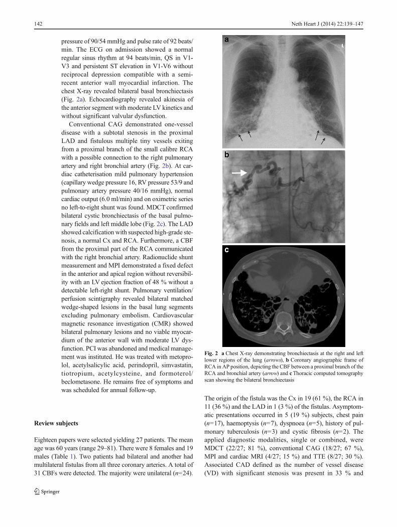
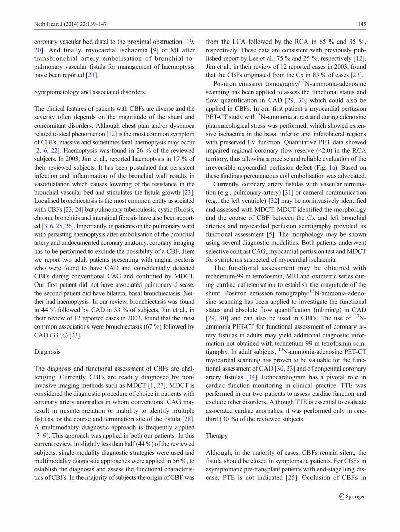
pressure of 90/54 mmHg and pulse rate of 92 beats/min. The ECG on admission showed a normalregular sinus rhythm at 94 beats/min, QS in V1-V3 and persistent ST elevation in V1-V6 withoutreciprocal depression compatible with a semi-recent anterior wall myocardial infarction. Thechest X-ray revealed bilateral basal bronchiectasis(Fig. 2a). Echocardiography revealed akinesia ofthe anterior segment with moderate LV kinetics andwithout significant valvular dysfunction.
Conventional CAG demonstrated one-vesseldisease with a subtotal stenosis in the proximalLAD and fistulous multiple tiny vessels exitingfrom a proximal branch of the small calibre RCAwith a possible connection to the right pulmonaryartery and right bronchial artery (Fig. 2b). At car-diac catheterisation mild pulmonary hypertension(capillary wedge pressure 16, RV pressure 53/9 andpulmonary artery pressure 40/16 mmHg), normalcardiac output (6.0 ml/min) and on oximetric seriesno left-to-right shunt was found. MDCT confirmedbilateral cystic bronchiectasis of the basal pulmo-nary fields and left middle lobe (Fig. 2c). The LADshowed calcification with suspected high-grade ste-nosis, a normal Cx and RCA. Furthermore, a CBFfrom the proximal part of the RCA communicatedwith the right bronchial artery. Radionuclide shuntmeasurement and MPI demonstrated a fixed defectin the anterior and apical region without reversibil-ity with an LV ejection fraction of 48 % without adetectable left-right shunt. Pulmonary ventilation/perfusion scintigraphy revealed bilateral matchedwedge-shaped lesions in the basal lung segmentsexcluding pulmonary embolism. Cardiovascularmagnetic resonance investigation (CMR) showedbilateral pulmonary lesions and no viable myocar-dium of the anterior wall with moderate LV dys-function. PCI was abandoned and medical manage-ment was instituted. He was treated with metopro-lol, acetylsalicylic acid, perindopril, simvastatin,tiotropium, acetylcysteine, and formoterol/beclometasone. He remains free of symptoms andwas scheduled for annual follow-up.
Review subjects
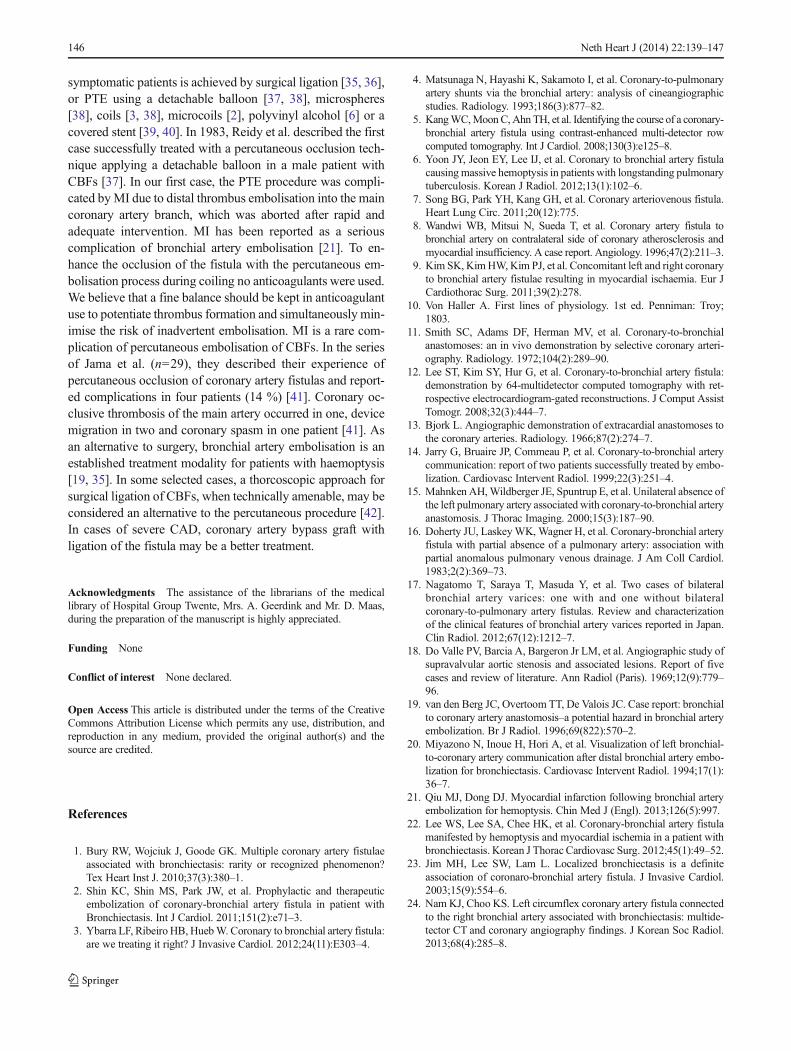
Eighteen papers were selected yielding 27 patients. The meanage was 60 years (range 29–81). There were 8 females and 19males (Table 1). Two patients had bilateral and another hadmultilateral fistulas from all three coronary arteries. A total of31 CBFs were detected. The majority were unilateral (n=24).
The origin of the fistula was the Cx in 19 (61 %), the RCA in11 (36 %) and the LAD in 1 (3 %) of the fistulas. Asymptom-atic presentations occurred in 5 (19 %) subjects, chest pain(n=17), haemoptysis (n=7), dyspnoea (n=5), history of pul-monary tuberculosis (n=3) and cystic fibrosis (n=2). Theapplied diagnostic modalities, single or combined, wereMDCT (22/27; 81 %), conventional CAG (18/27; 67 %),MPI and cardiac MRI (4/27; 15 %) and TTE (8/27; 30 %).Associated CAD defined as the number of vessel disease(VD) with significant stenosis was present in 33 % and
Fig. 2 a Chest X-ray demonstrating bronchiectasis at the right and leftlower regions of the lung (arrows), b Coronary angiographic frame ofRCA in AP position, depicting the CBF between a proximal branch of theRCA and bronchial artery (arrow) and cThoracic computed tomographyscan showing the bilateral bronchiectasis
142 Neth Heart J (2014) 22:139–147
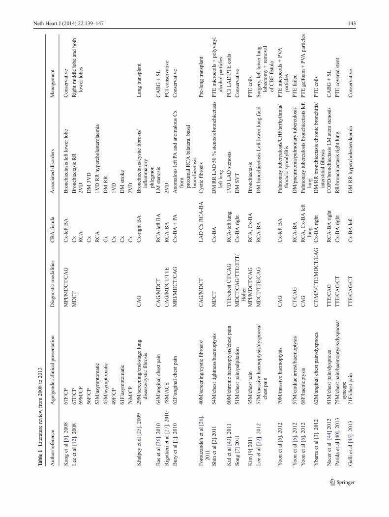
Table1
Literature
review
from
2008
to2013
Author/reference
Age/gender/clinicalpresentatio
nDiagnostic
modalities
CBAfistula
Associateddisorders
Managem
ent
Kangetal[5].2008
67F/CP
MPI/M
DCT/CAG
Cx-leftBA
Bronchiectasisleftlower
lobe
Conservative
Lee
etal[12].2008
67F/CP
MDCT
Cx
BronchiectasisRR
Right
middlelobe
andboth
lower
lobes
69M/CP
RCA
2VD
56F/CP
Cx
DM
3VD
53M/asymptom
atic
RCA
1VDRRhypercholesterolaemia
45M/asymptom
atic
Cx
DM
RR
49F/CP
Cx
1VD
61F/asym
ptom
atic
Cx
DM
stroke
76M/CP
Cx
2VD
Khalpey
etal[25].2009
29M/screening/end-stage
lung
disease/cysticfibrosis
CAG
Cx-rightB
ABronchiectasis/cysticfibrosis/
inflam
matory
phlegm
on
Lungtransplant
Bas
etal[36].2010
64M/anginalchestp
ain
CAG/M
DCT
RCA-leftB
ALM
stenosis
CABG+SL
Rigattierietal[27].2010
78M/ACS
CAG/M
DCT/TTE
RCA-BA
2VD
PCIconservativ
e
Buryetal[1].2010
62F/anginalchestp
ain
MRI/MDCT/CAG
Cx-BA+PA
AnomalousleftPA
andanom
alousCx
from
proxim
alRCAbilateralb
asal
bronchiectasis
Conservative
Forouzandehetal[26].
2011
40M/screening/cystic
fibrosis/
CAG/M
DCT
LADCxRCA-BA
Cystic
fibrosis
Pre-lung
transplant
Shinetal[2].2011
54M/chesttig
htness/haemoptysis
MDCT
Cx-BA
DM
RRLAD50
%stenosisbronchiectasis
leftlung
PTEmicrocoils
+polyvinyl
alcoholp
articles
Kul
etal[43].2011
60M/chronichaem
optysis/chestp
ain
TTE/chestCT/CAG
RCA-leftlung
1VDLADstenosis
PCILADPT
Ecoils
Song
[7]2011
51M/chestpain/palpitatio
nMDCT/CAG/TTE/ETT/
Holter
Cx-BAright
DM
SVT
Conservative
Kim
[9]2011
35M/chestpain
MPI/M
DCT/CAG
RCA,C
x-BA
Bronchiectasis
PTEcoils
Lee
etal[22].2012
57M/m
assive
haem
optysis/dyspnoea/
chestp
ain
MDCT/TTE/CAG
RCA-BA
DM
bronchiectasisLeftlow
erlung
field
Surgery,leftlow
erlung
lobectom
y+removal
ofCBFfistula
Yoonetal[6].2012
70M/m
assive
haem
optysis
CAG
Cx-leftBA
Pulm
onarytuberculosis/CHF/arrhythm
ia/
thoracicspondylitis
PTEmicrocoils
+PVA
particles
Yoonetal[6].2012
57M/cardiac
arrest/haemoptysis
CT/CAG
RCA-BA
DM/pneum
onia/pulmonarytuberculosis
PTEfailed
Yoonetal[6].2012
68F/haem
optysis
CAG
RCA,C
x-BAleft
lung
Pulm
onarytuberculosisbronchiectasisleft
lung
PTEgelfoam+PVAparticles
Ybarraetal[3].2012
62M/anginalchestp
ain/dyspnoea
CT/M
PI/TTE/M
DCT/CAG
Cx-BAright
DM/RRbronchiectasischronicbronchitis/
interstitialfibrosis
PTEcoils
Nacer
etal.[44]2012
81M/chestpain/dyspnoea
TTE/CAG
RCA-BAright
COPD
/bronchiectasisLM
stem
stenosis
CABG+SL
Paridaetal[40].2013
75M/chestpain/haemoptysis/dyspnoea/
syncope
TTE/CAG/CT
Cx-BAright
RR/bronchiectasisrightlung
PTEcoveredstent
Gallietal[45].2013
71F/chestp
ain
TTE/CAG/CT
Cx-BAleft
DM
RRhypercholesterolaemia
Conservative
Neth Heart J (2014) 22:139–147 143
subclassified as follows: 1VD (n=3), 2VD (n=3), 3VD (n=1)and significant left main (LM) disease (n=2). Concomitantdisorders were bronchiectasis (n=12; 44 %), arterial hyper-tension (n=7; 26 %) and diabetes mellitus (n=9; 33 %). Man-agement was conservative medical strategy (n=6; 22 %),awaiting lung transplantation (n=2; 7 %), surgical ligation(n=3; 11 %) combined with coronary artery bypass grafting(n=2) or pulmonary lobectomy (n=1), PTE (n=8; 30 %,including one failure), and was not reported (n=8; 30 %).Materials used, alone or combined, for PTE were microcoils(n=2), coils (n=3) and polyvinyl particles (n=3).
Discussion
History and incidence
CBFs may have a unilateral [7, 8] or bilateral [9] presentation.As early as 1803, a Cx coronary artery to right bronchial arterycommunication was initially described by von Haller [10]. In1972, Smith et al. presented the first angiographic case of aunilateral CBF [11]. The MDCT and conventional CAG inci-dence of CBFs is estimated at 0.61 % (8/1300) [12] and 0.5 %[4], respectively.
Embryology
Small, not functional anastomoses between the bronchial ar-teries and the coronary arteries exist. These anastomoses havebeen regarded as congenital in origin. Coronary angiographicvisualisation was demonstrated by Bjork in 1966; 22 % ofnormal subjects had such anastomoses and it was found in48 % of patients with obstructive CAD [13]. However, thesearterial communications may become enlarged and functionalin a variety of cardiovascular entities including pulmonaryartery hypoplasia, tetralogy of Fallot, supravalvular aorticstenosis and Takayasu arteritis and may be associated withpulmonary disorders such as pulmonary thromboembolism[4, 14–16]. CBFs are probably already present at birth andremain clinically silent in most cases. These congenital vas-cular communications are usually small in size and haemody-namically insignificant. The factors regulating the existence orre-opening and growth of these vascular anastomoses are asfollows. First: disequilibrium of the pressure gradient betweenthe coronary, bronchial and pulmonary vascular trees maylead to growth of CBFs giving rise to increased flow fromthe coronary to the bronchial artery vascular bed [17].Shunting of blood from the coronary to the bronchial circula-tion occurs either when the coronary artery pressure increasesas in supravalvular aortic stenosis [18], or when bronchialartery pressure decreases as in pulmonary atresia and tetralogyof Fallot. Second: in obstructive CAD, the bronchial-to-coronary artery fistula has been demonstrated to fill the distalTa
ble1
(contin
ued)
Author/reference
Age/gender/clinicalpresentatio
nDiagnostic
modalities
CBAfistula
Associateddisorders
Managem
ent
Eryilm
azetal[46].2013
64M/dyspnoeaon
exertio
n/CAG/M
DCT
Cx-BAright
bronchiectasis
Conservative,refusalo
fsurgery
Case1
66F/chestpain
CAG/M
DCT/M
PI/PET-CT
RCA-BA
1VD
PTE/PCICx
Case2
74M/chestpain
CAG/PVscan/M
DCT/M
RI
RCA-BAright
1VD/COPD
/bronchiectasis
Conservative
ACSacutecoronary
syndrome,BAbronchialartery,CABGcoronary
artery
bypass
graftin
g,CAGcoronary
angiography,CBFcoronary
bronchialfistula,CHFcongestiv
eheartfailu
re,COPDchronic
obstructivepulm
onarydisease,CPchestp
ain,
CTcomputedtomography,Cxcircum
flex
coronary
artery,D
Mdiabetes
mellitus,L
ADleftanterior
descending
coronary
artery,L
Mleftmainstem
,MDCT
multi-detector
computedtomography,MPIm
yocardialperfusion
imaging,MRIm
agnetic
resonanceim
aging,PA
pulm
onaryartery,P
CIpercutaneouscoronary
interventio
n,PTE
percutaneous
transcatheter
embolization,PVA
polyvinylalcohol
particles,RCArightcoronaryartery,R
Rhypertension,SLsurgicallig
ation,SV
Tsupraventriculartachycardia,TTE
transthoracicechocardiography,V
Dvesseldisease
144 Neth Heart J (2014) 22:139–147
coronary vascular bed distal to the proximal obstruction [19,20]. And finally, myocardial ischaemia [9] or MI aftertransbronchial artery embolisation of bronchial-to-pulmonary vascular fistula for management of haemoptysishave been reported [21].
Symptomatology and associated disorders
The clinical features of patients with CBFs are diverse and theseverity often depends on the magnitude of the shunt andconcomitant disorders. Although chest pain and/or dyspnoearelated to steal phenomenon [12] is the most common symptomof CBFs, massive and sometimes fatal haemoptysis may occur[2, 6, 22]. Haemoptysis was found in 26 % of the reviewedsubjects. In 2003, Jim et al., reported haemoptysis in 17 % oftheir reviewed subjects. It has been postulated that persistentinfection and inflammation of the bronchial wall results invasodilatation which causes lowering of the resistance in thebronchial vascular bed and stimulates the fistula growth [23].Localised bronchiectasis is the most common entity associatedwith CBFs [23, 24] but pulmonary tuberculosis, cystic fibrosis,chronic bronchitis and interstitial fibrosis have also been report-ed [3, 6, 25, 26]. Importantly, in patients on the pulmonary wardwith persisting haemoptysis after embolisation of the bronchialartery and undocumented coronary anatomy, coronary imaginghas to be performed to exclude the possibility of a CBF. Herewe report two adult patients presenting with angina pectoriswho were found to have CAD and coincidentally detectedCBFs during conventional CAG and confirmed by MDCT.Our first patient did not have associated pulmonary disease,the second patient did have bilateral basal bronchiectasis. Nei-ther had haemoptysis. In our review, bronchiectasis was foundin 44 % followed by CAD in 33 % of subjects. Jim et al., intheir review of 12 reported cases in 2003, found that the mostcommon associations were bronchiectasis (67 %) followed byCAD (33 %) [23].
Diagnosis
The diagnosis and functional assessment of CBFs are chal-lenging. Currently CBFs are readily diagnosed by non-invasive imaging methods such as MDCT [1, 27]. MDCT isconsidered the diagnostic procedure of choice in patients withcoronary artery anomalies in whom conventional CAG mayresult in misinterpretation or inability to identify multiplefistulas, or the course and termination site of the fistula [28].A multimodality diagnostic approach is frequently applied[7–9]. This approach was applied in both our patients. In thiscurrent review, in slightly less than half (44%) of the reviewedsubjects, single-modality diagnostic strategies were used andmultimodality diagnostic approaches were applied in 56 %, toestablish the diagnosis and assess the functional characteris-tics of CBFs. In the majority of subjects the origin of CBFwas
from the LCA followed by the RCA in 65 % and 35 %,respectively. These data are consistent with previously pub-lished report by Lee et al.: 75 % and 25 %, respectively [12].Jim et al., in their review of 12 reported cases in 2003, foundthat the CBFs originated from the Cx in 83 % of cases [23].
Positron emission tomography/13N-ammonia-adenosinescanning has been applied to assess the functional status andflow quantification in CAD [29, 30] which could also beapplied in CBFs. In our first patient a myocardial perfusionPET-CT study with13N-ammonia at rest and during adenosinepharmacological stress was performed, which showed exten-sive ischaemia in the basal inferior and inferolateral regionswith preserved LV function. Quantitative PET data showedimpaired regional coronary flow reserve (<2.0) in the RCAterritory, thus allowing a precise and reliable evaluation of theirreversible myocardial perfusion defect (Fig. 1a). Based onthese findings percutaneous coil embolisation was advocated.
Currently, coronary artery fistulas with vascular termina-tion (e.g., pulmonary artery) [31] or cameral communication(e.g., the left ventricle) [32] may be noninvasively identifiedand assessed with MDCT. MDCT identified the morphologyand the course of CBF between the Cx and left bronchialarteries and myocardial perfusion scintigraphy provided itsfunctional assessment [5]. The morphology may be shownusing several diagnostic modalities. Both patients underwentselective contrast CAG, myocardial perfusion test and MDCTfor symptoms suspected of myocardial ischaemia.
The functional assessment may be obtained withtechnetium-99 m tetrofosmin, MRI and oximetric series dur-ing cardiac catheterisation to establish the magnitude of theshunt. Positron emission tomography/13N-ammonia-adeno-sine scanning has been applied to investigate the functionalstatus and absolute flow quantification (ml/min/g) in CAD[29, 30] and can also be used in CBFs. The use of 13N-ammonia PET-CT for functional assessment of coronary ar-tery fistulas in adults may yield additional diagnostic infor-mation not obtained with technetium-99 m tetrofosmin scin-tigraphy. In adult subjects, 13N-ammonia-adenosine PET-CTmyocardial scanning has proven to be valuable for the func-tional assessment of CAD [30, 33] and of congenital coronaryartery fistulas [34]. Echocardiogram has a pivotal role incardiac function monitoring in clinical practice. TTE wasperformed in our two patients to assess cardiac function andexclude other disorders. Although TTE is essential to evaluateassociated cardiac anomalies, it was performed only in one-third (30 %) of the reviewed subjects.
Therapy
Although, in the majority of cases, CBFs remain silent, thefistula should be closed in symptomatic patients. For CBFs inasymptomatic pre-transplant patients with end-stage lung dis-ease, PTE is not indicated [25]. Occlusion of CBFs in
Neth Heart J (2014) 22:139–147 145
symptomatic patients is achieved by surgical ligation [35, 36],or PTE using a detachable balloon [37, 38], microspheres[38], coils [3, 38], microcoils [2], polyvinyl alcohol [6] or acovered stent [39, 40]. In 1983, Reidy et al. described the firstcase successfully treated with a percutaneous occlusion tech-nique applying a detachable balloon in a male patient withCBFs [37]. In our first case, the PTE procedure was compli-cated byMI due to distal thrombus embolisation into the maincoronary artery branch, which was aborted after rapid andadequate intervention. MI has been reported as a seriouscomplication of bronchial artery embolisation [21]. To en-hance the occlusion of the fistula with the percutaneous em-bolisation process during coiling no anticoagulants were used.We believe that a fine balance should be kept in anticoagulantuse to potentiate thrombus formation and simultaneously min-imise the risk of inadvertent embolisation. MI is a rare com-plication of percutaneous embolisation of CBFs. In the seriesof Jama et al. (n=29), they described their experience ofpercutaneous occlusion of coronary artery fistulas and report-ed complications in four patients (14 %) [41]. Coronary oc-clusive thrombosis of the main artery occurred in one, devicemigration in two and coronary spasm in one patient [41]. Asan alternative to surgery, bronchial artery embolisation is anestablished treatment modality for patients with haemoptysis[19, 35]. In some selected cases, a thorcoscopic approach forsurgical ligation of CBFs, when technically amenable, may beconsidered an alternative to the percutaneous procedure [42].In cases of severe CAD, coronary artery bypass graft withligation of the fistula may be a better treatment.
Acknowledgments The assistance of the librarians of the medicallibrary of Hospital Group Twente, Mrs. A. Geerdink and Mr. D. Maas,during the preparation of the manuscript is highly appreciated.
Funding None
Conflict of interest None declared.
Open Access This article is distributed under the terms of the CreativeCommons Attribution License which permits any use, distribution, andreproduction in any medium, provided the original author(s) and thesource are credited.
References
1. Bury RW, Wojciuk J, Goode GK. Multiple coronary artery fistulaeassociated with bronchiectasis: rarity or recognized phenomenon?Tex Heart Inst J. 2010;37(3):380–1.
2. Shin KC, Shin MS, Park JW, et al. Prophylactic and therapeuticembolization of coronary-bronchial artery fistula in patient withBronchiectasis. Int J Cardiol. 2011;151(2):e71–3.
3. Ybarra LF, Ribeiro HB, HuebW. Coronary to bronchial artery fistula:are we treating it right? J Invasive Cardiol. 2012;24(11):E303–4.
4. Matsunaga N, Hayashi K, Sakamoto I, et al. Coronary-to-pulmonaryartery shunts via the bronchial artery: analysis of cineangiographicstudies. Radiology. 1993;186(3):877–82.
5. KangWC,MoonC,Ahn TH, et al. Identifying the course of a coronary-bronchial artery fistula using contrast-enhanced multi-detector rowcomputed tomography. Int J Cardiol. 2008;130(3):e125–8.
6. Yoon JY, Jeon EY, Lee IJ, et al. Coronary to bronchial artery fistulacausingmassive hemoptysis in patients with longstanding pulmonarytuberculosis. Korean J Radiol. 2012;13(1):102–6.
7. Song BG, Park YH, Kang GH, et al. Coronary arteriovenous fistula.Heart Lung Circ. 2011;20(12):775.
8. Wandwi WB, Mitsui N, Sueda T, et al. Coronary artery fistula tobronchial artery on contralateral side of coronary atherosclerosis andmyocardial insufficiency. A case report. Angiology. 1996;47(2):211–3.
9. Kim SK, KimHW,Kim PJ, et al. Concomitant left and right coronaryto bronchial artery fistulae resulting in myocardial ischaemia. Eur JCardiothorac Surg. 2011;39(2):278.
10. Von Haller A. First lines of physiology. 1st ed. Penniman: Troy;1803.
11. Smith SC, Adams DF, Herman MV, et al. Coronary-to-bronchialanastomoses: an in vivo demonstration by selective coronary arteri-ography. Radiology. 1972;104(2):289–90.
12. Lee ST, Kim SY, Hur G, et al. Coronary-to-bronchial artery fistula:demonstration by 64-multidetector computed tomography with ret-rospective electrocardiogram-gated reconstructions. J Comput AssistTomogr. 2008;32(3):444–7.
13. Bjork L. Angiographic demonstration of extracardial anastomoses tothe coronary arteries. Radiology. 1966;87(2):274–7.
14. Jarry G, Bruaire JP, Commeau P, et al. Coronary-to-bronchial arterycommunication: report of two patients successfully treated by embo-lization. Cardiovasc Intervent Radiol. 1999;22(3):251–4.
15. Mahnken AH,Wildberger JE, Spuntrup E, et al. Unilateral absence ofthe left pulmonary artery associated with coronary-to-bronchial arteryanastomosis. J Thorac Imaging. 2000;15(3):187–90.
16. Doherty JU, LaskeyWK,Wagner H, et al. Coronary-bronchial arteryfistula with partial absence of a pulmonary artery: association withpartial anomalous pulmonary venous drainage. J Am Coll Cardiol.1983;2(2):369–73.
17. Nagatomo T, Saraya T, Masuda Y, et al. Two cases of bilateralbronchial artery varices: one with and one without bilateralcoronary-to-pulmonary artery fistulas. Review and characterizationof the clinical features of bronchial artery varices reported in Japan.Clin Radiol. 2012;67(12):1212–7.
18. Do Valle PV, Barcia A, Bargeron Jr LM, et al. Angiographic study ofsupravalvular aortic stenosis and associated lesions. Report of fivecases and review of literature. Ann Radiol (Paris). 1969;12(9):779–96.
19. van den Berg JC, Overtoom TT, De Valois JC. Case report: bronchialto coronary artery anastomosis–a potential hazard in bronchial arteryembolization. Br J Radiol. 1996;69(822):570–2.
20. Miyazono N, Inoue H, Hori A, et al. Visualization of left bronchial-to-coronary artery communication after distal bronchial artery embo-lization for bronchiectasis. Cardiovasc Intervent Radiol. 1994;17(1):36–7.
21. Qiu MJ, Dong DJ. Myocardial infarction following bronchial arteryembolization for hemoptysis. Chin Med J (Engl). 2013;126(5):997.
22. Lee WS, Lee SA, Chee HK, et al. Coronary-bronchial artery fistulamanifested by hemoptysis and myocardial ischemia in a patient withbronchiectasis. Korean J Thorac Cardiovasc Surg. 2012;45(1):49–52.
23. Jim MH, Lee SW, Lam L. Localized bronchiectasis is a definiteassociation of coronaro-bronchial artery fistula. J Invasive Cardiol.2003;15(9):554–6.
24. Nam KJ, Choo KS. Left circumflex coronary artery fistula connectedto the right bronchial artery associated with bronchiectasis: multide-tector CT and coronary angiography findings. J Korean Soc Radiol.2013;68(4):285–8.
146 Neth Heart J (2014) 22:139–147
25. Khalpey Z, Camp Jr P, Jaklitsch MT. Left circumflex to bronchialartery fistula. Ann Thorac Surg. 2009;88(1):303.
26. Forouzandeh F, Krim SR, Estep J, et al. A case of all 3 coronary tobronchial arteries fistulas. J Am Coll Cardiol. 2011;58(9):987.
27. Rigattieri S, Fedele S, Sperandio M, et al. Coronary-to-bronchialartery fistula in a patient with multivessel coronary disease treatedby percutaneous coronary intervention. J Cardiovasc Med(Hagerstown). 2010;11(8):625–7.
28. Schmid M, Achenbach S, Ludwig J, et al. Visualization of coronaryartery anomalies by contrast-enhanced multi-detector row spiral com-puted tomography. Int J Cardiol. 2006;111(3):430–5.
29. Hasbak P, Kjaer A. Positron emission tomography to replace myo-cardial scintigraphy. Ugeskr Laeger. 2011;173(8):567–72.
30. Tio RA, Dabeshlim A, Siebelink HM, et al. Comparison between theprognostic value of left ventricular function andmyocardial perfusionreserve in patients with ischemic heart disease. J Nucl Med.2009;50(2):214–9.
31. Chen ML, Lo HS, Su HY, et al. Coronary artery fistula: assessmentwith multidetector computed tomography and stress myocardial sin-gle photon emission computed tomography. Clin Nucl Med.2009;34(2):96–8.
32. Barone-Rochette G, Vanzetto G, Saunier C, et al. Combination ofanatomic and perfusion imaging for decision making in a profession-al soccer player with giant coronary artery to left ventricle fistula. JNucl Cardiol. 2009;16(4):640–3.
33. Juarez-Orozco LE, Glauche J, Alexanderson E, et al. Myocardialperfusion reserve in spared myocardium: correlation with infarct sizeand left ventricular ejection fraction. Eur J Nucl Med Mol Imaging.2013;40(8):1148–54.
34. Said SA, Nijhuis RL, Op den Akker JW, et al. Diagnostic andtherapeutic approach of congenital solitary coronary artery fistulasin adults: Dutch case series and review of literature. Neth Heart J.2011;19(4):183–91.
35. St John Sutton MG, Miller GA, Kerr IH, et al. Coronary artery stealvia large coronary artery to bronchial artery anastomosis successfullytreated by operation. Br Heart J. 1980;44(4):460–3.
36. Bas S, Yiginer O, Atalay M, et al. Coronary-to-bronchial arteryfistula with conventional and multi-detector computed tomographyangiographic images. Hellenic J Cardiol. 2010;51(2):164–5.
37. Reidy JF, Sowton E, RossDN. Transcatheter occlusion of coronary tobronchial anastomosis by detachable balloon combined with coro-nary angioplasty at same procedure. Br Heart J. 1983;49(3):284–7.
38. Brenot P, Riou JY, Losay J, et al. [Endovascular treatment of coronaryarterial fistulae in children and adults]. Arch Mal Coeur Vaiss.2007;100(5):373–9.
39. Saijo Y, Izutsu K, Sonobe T, et al. Successful closure of coronary-bronchial artery fistula with vein graft-coated stent. CatheterCardiovasc Interv. 1999;46(2):214–7.
40. Parida AK, Lodh M, Sanyal J, et al. Coronary bronchial steal. J CaseRep. 2013;3(1):39–43.
41. Jama A, Barsoum M, Bjarnason H, et al. Percutaneous closure ofcongenital coronary artery fistulae: results and angiographic follow-up. JACC Cardiovasc Interv. 2011;4(7):814–21.
42. Fortunato Junior JA, Branco Filho AA, Granzotto PC, et al. Video-thoracoscopy closure of coronary artery fistula: case report. Rev BrasCir Cardiovasc. 2010;25(1):109–11.
43. Kul S, Canga Y, Guvenc TS, et al. A hidden cause of hemoptysis:coronary artery-to-pulmonary parenchyma fistula. Turk KardiyolDern Ars. 2011;39(8):686–9.
44. Nacar AB, Yorgun H, Tuncer C. Right coronary-to-bronchial arteryfistula on the contralateral side of coronary stenosis. Arch Turk SocCardiol. 2012;40(2):196.
45. Galli E, Rizza A, Remoli E, et al. Coronary-to-bronchial artery fistulain a patient with angina. J Cardiol Cases. 2013;7:e45–7.
46. Eryilmaz U, Gungor H, Uyar S, et al. Circumflex-to-bronchial arteryfistula with saccular aneurysm. Postep Kardiol Inter. 2013;9(33):296–7.
Neth Heart J (2014) 22:139–147 147
Recommended


![BRONCHIAL AND NONBRONCHIAL SYSTEMIC …...In 1996 Ramakantan et al [5] treated 140 cases of haemoptysis with bronchial artery embolisation with immediate control achieved in 102 cases](https://img.dokumen.tips/doc/110x75/5e846f76ec21c031c35b498e/bronchial-and-nonbronchial-systemic-in-1996-ramakantan-et-al-5-treated-140.jpg)