© 2016 The Journal of Indian Prosthodontic Society | Published by Wolters Kluwer - Medknow 193
Clinical acceptability of metal‑ceramic fixed partial dental prosthesis fabricated with direct metal laser sintering technique‑5 year follow‑up
Radhakrishnan Prabhu, Geetha Prabhu, Eswaran Baskaran, Eswaran M. ArumugamDepartment of Prosthodontics, Thai Moogambigai Dental College and Hospital, Mogappair, Chennai, Tamil Nadu, India
INTRODUCTION
A significant change from conventional castings is the
introduction of direct metal laser sintered (DMLS) technology used for metal‑ceramic restoration.[1‑5] Various in vitro studies
Statement of Problem: In recent years, direct metal laser sintered (DMLS) metal-ceramic-based fixed partial denture prostheses have been used as an alternative to conventional metal-ceramic fixed partial denture prostheses. However, clinical studies for evaluating their long-term clinical survivability and acceptability are limited.Aims and Objective: The aim of this study was to assess the efficacy of metal-ceramic fixed dental prosthesis fabricated with DMLS technique, and its clinical acceptance on long-term clinical use.Materials and Methods: The study group consisted of 45 patients who were restored with posterior three-unit fixed partial denture prosthesis made using direct laser sintered metal-ceramic restorations. Patient recall and clinical examination of the restorations were done after 6months and every 12 months thereafter for the period of 60 months. Clinical examination for evaluation of longevity of restorations was done using modified Ryge criteria which included chipping of the veneered ceramic, connector failure occurring in the fixed partial denture prosthesis, discoloration at the marginal areas of the veneered ceramic, and marginal adaptation of the metal and ceramic of the fixed denture prosthesis. Periapical status was assessed using periodical radiographs during the study period. Survival analysis was made using the Kaplan–Meier method.Results: None of the patients had failure of the connector of the fixed partial denture prostheses during the study period. Two exhibited biological changes which included periapical changes and proximal caries adjacent to the abutments.Conclusion: DMLS metal-ceramic fixed partial denture prosthesis had a survival rate of 95.5% and yielded promising results during the 5-year clinical study.
Key Words: Direct metal laser sintered crowns, fixed partial denture prosthesis, laser sintered crowns
Address for correspondence: Dr. Radhakrishnan Prabhu, Department of Prosthodontics, Thai Moogambigai Dental College and Hospital, Mogappair, Chennai ‑ 600 107, Tamil Nadu, India. E‑mail: [email protected]: 31st July, 2015, Accepted: 18th November, 2015
Access this article onlineQuick Response Code:
Website:
www.j‑ips.org
DOI:
10.4103/0972‑4052.176526
Original Article
Abstract
How to cite this article: Prabhu R, Prabhu G, Baskaran E, Arumugam EM. Clinical acceptability of metal-ceramic fixed partial dental prosthesis fabricated with direct metal laser sintering technique-5 year follow-up. J Indian Prosthodont Soc 2016;16:193‑7.
This is an open access article distributed under the terms of the Creative Commons Attribution‑NonCommercial‑ShareAlike 3.0 License, which allows others to remix, tweak, and build upon the work non‑commercially, as long as the author is credited and the new creations are licensed under the identical terms.
For reprints contact: [email protected]
[Downloaded free from http://www.j-ips.org on Saturday, April 02, 2016, IP: 49.206.1.43]

Prabhu, et al.: Clinical acceptability of laser sintered metal‑ceramic fixed dental prosthesis
194 The Journal of Indian Prosthodontic Society | Apr-Jun 2016 | Vol 16 | Issue 2
on DMLS have yielded promising results for its wider clinical usage. Yet studies regarding clinical longevity and the survival rates for posterior metal‑ceramic fixed partial dentures done with DMLS technique is lacking. Hence, this study was undertaken to assess the clinical acceptability of posterior metal‑ceramic fixed partial denture prosthesis made with DMLS technique.
MATERIALS AND METHODS
Forty‑five patients with the mean age group of 40 years with missing maxillary or mandibular second premolar or first molar who reported to the Department of Prosthodontics, Thai Moogambigai Dental College and Hospital and were in need of three‑unit fixed partial denture formed the study group. The study was approved by the Ethical Committee of Dr. MGR. Educational and Research Institute University and all the patients were provided with informed consent. The criteria for case selection include the missing teeth that were removed due to irreversible pulpal reasons. Only the vital abutment teeth were selected, and they were evaluated for proper positioning in the dental arch without any rotation, tipping, malalignment, and periodontal problems. It was also made sure that abutment teeth were opposed to natural dentition and had no supraeruption. Abutments which were not satisfying these criteria were excluded from the study group. Radiographic evaluation was done to rule out any periodontal or periapical pathologies of the abutments. Occlusal examination was done to rule out any para functional habits and temporomandibular joint ailments. All the abutment preparations were done by the same prosthodontist to standardize the preparations. The preparation design protocols were followed based on the study done by Tara et al.[6] The preparation design had an occlusal reduction of 1.5 mm, which was evaluated using wax check‑bite and measured using wax calipers. The preparation had a circumferential chamfer finish line design with a circumferential reduction of 0.8 mm and a total convergence of 6°. All internal angles were carefully rounded. Impression was made using addition polyvinyl siloxane material (Aquasil Soft Putty/Regular Set, Dentsply De Trey GmbH, Germany) and poured with Type IV die stone (Fuji rock, GC). The cast was sent to Dent Care Dental Lab (Muvattupuzha, Kerala, India) for the construction of DMLS posterior three‑unit metal‑ceramic fixed partial denture. Provisional restoration was done using poly methyl methacrylate (Dental Products India [DPI], Rapid Repair Cold Cure, DPI, Mumbai). The digital construction of the metal framework was done using computer software, and the laser sintered processing was done by the laser sintering unit (EOSINT M 270, Eos Germany) where a high energy focused laser beam directly fuses a localized region of a thin layer of cobalt–chromium metal powder to build up the restoration gradually. The thickness of the metal copings was a minimum of 0.35 mm with a connector thickness of
3 mm. The thickness of the veneered ceramic (VITA VM 13 ceramic) was 1.15 mm occlusally and 0.8 mm cervically. Sandblasting was done using 50 µm alumina. Intraoral evaluation of the restorations were made for marginal integrity, and occlusal contacts were evaluated with articulating film and adjustments were made using porcelain polishing kit to attain contacts in maximum intercuspation, and to eliminate lateral interferences. Cementation was done using Type I glass ionomer cement (GC Corporation Tokyo, Japan). Post‑insertion oral hygiene instructions including interdental brushing were explained to the patients and recommended to follow regularly. Recall visits were made at 6, 12 months interval and annually thereafter for the next 60 months to follow‑up the restorations that were made. Though recall visits were done at 6 months interval, only annual evaluation was done to assess the longevity of the restorations. The clinical evaluation was done by qualified prosthodontists by visual and clinical examination using conventional dental diagnostic instruments. To standardize the assessment on the longevity of restorations, the restorations in the study group were evaluated using the recommended clinical indices called the modified Ryge clinical criteria [Table 1].[7] These criteria through visual and probing examination assesses the fracture resistance of the veneered ceramic, connector failure occurring in the fixed partial denture prosthesis, discoloration at the marginal areas of the veneered ceramic, and marginal integrity of the fixed denture prosthesis. According to the criteria, all categories were given scores namely Alpha, Bravo, Charlie, and Delta ratings to determine whether the restorations is in excellent state or failing during the study period. Radiographic periapical assessment of the abutments and proximal caries assessments were also done during the annual evaluation. The variables were graded based on the clinical evaluation and the
Table 1: Clinical evaluation of restorations using modified Ryge criteriaClinical evaluation of the restorations in terms of fracture measurements
Alpha - A. Smooth surface of the restoration (shiny after air drying)Bravo - B. Dull surface and/or chipping of porcelain that does not impair functionCharlie - C. Chipping of veneering porcelain impairing esthetics and function and/or exposing framework materialDelta - D. Fracture of connector between the pontic and retainer and/or fracture through frame work material
Clinical evaluation for marginal adaptationsAlpha - A. No visible evidences of crevice along the margins; no catch or penetration of the explorerBravo - B. Visible evidence of crevice and/or catch of explorer; no penetration of the explorerCharlie - C. Visible evidence of crevice and penetration of the explorerDelta - D. Restoration is mobile, fractured, or missing
Clinical evaluation for marginal discolorationAlpha - A. No discoloration not penetrating in pulpal directionBravo - B. Superficial discoloration but not penetrating in pulpal directionCharlie - C. Discoloration and penetrating in pulpal direction
[Downloaded free from http://www.j-ips.org on Saturday, April 02, 2016, IP: 49.206.1.43]
Prabhu, et al.: Clinical acceptability of laser sintered metal‑ceramic fixed dental prosthesis
The Journal of Indian Prosthodontic Society | Apr-Jun 2016 | Vol 16 | Issue 2 195
probability distributions of these were calculated. An analysis of survival using the Kaplan–Meier method with approximate 95% confidence intervals was performed for the survival of these posterior fixed partial dentures.[8]
RESULTS
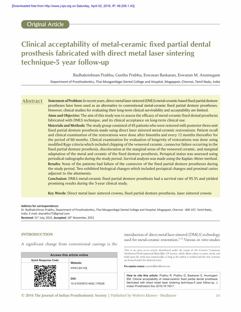
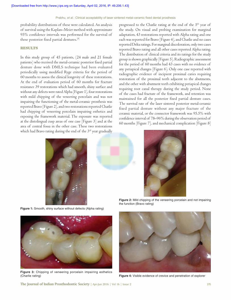
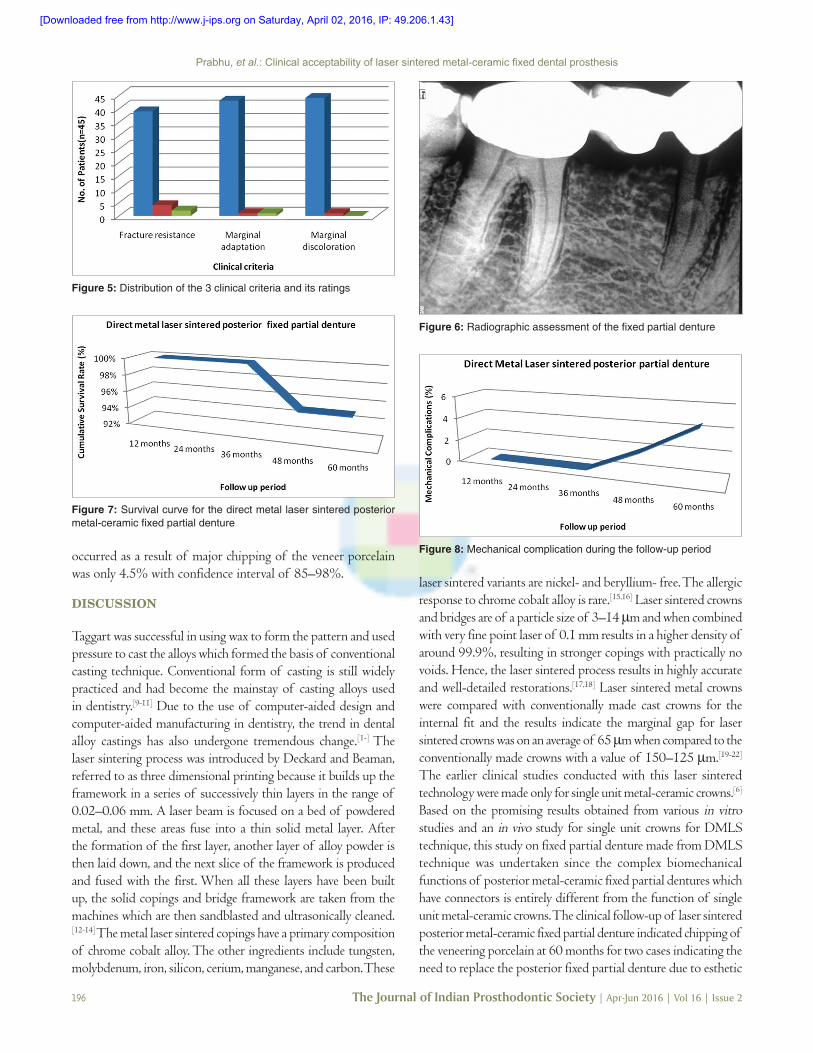
In this study group of 45 patients, (24 male and 21 female patients) who received the metal‑ceramic posterior fixed partial denture done with DMLS technique had been evaluated periodically using modified Ryge criteria for the period of 60 months to assess the clinical longevity of these restorations. At the end of evaluation period of 60 months for fracture resistance 39 restorations which had smooth, shiny surface and without any defects were rated Alpha [Figure 1], four restorations with mild chipping of the veneering porcelain and was not impairing the functioning of the metal‑ceramic prosthesis was reported Bravo [Figure 2], and two restorations reported Charlie had chipping of veneering porcelain impairing esthetics and exposing the framework material. The exposure was reported at the distolingual cusp areas of one case [Figure 3] and at the area of central fossa in the other case. These two restorations which had Bravo rating during the end of the 3rd year gradually
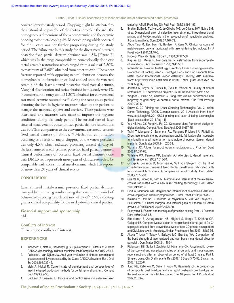
progressed to the Charlie rating at the end of the 5th year of the study. On visual and probing examination for marginal adaptation, 43 restorations reported with Alpha rating and one each was reported for Bravo [Figure 4], and Charlie and no cases reported Delta ratings. For marginal discoloration, only two cases reported Bravo rating and all other cases reported Alpha rating. The distribution of clinical criteria and its ratings for the study group is shown graphically [Figure 5]. Radiographic assessment for the period of 60 months had 43 cases with no evidence of any periapical changes [Figure 6]. Only one case reported with radiographic evidence of incipient proximal caries requiring restoration of the proximal teeth adjacent to the abutments, and the other with abutment teeth exhibiting periapical changes requiring root canal therapy during the study period. None of the cases had fracture of the framework, and retention was maintained for all the posterior fixed partial denture cases. The survival rate of the laser sintered posterior metal‑ceramic fixed partial denture without any major fracture of the ceramic material, or the connector framework was 95.5% with confidence interval of 78–96% during the observation period of 60 months [Figure 7], and mechanical complication [Figure 8]
Figure 1: Smooth, shiny surface without defects (Alpha rating)
Figure 2: Mild chipping of the veneering porcelain and not impairing the function (Bravo rating)
Figure 3: Chipping of veneering porcelain impairing esthetics (Charlie rating) Figure 4: Visible evidence of crevice and penetration of explorer
[Downloaded free from http://www.j-ips.org on Saturday, April 02, 2016, IP: 49.206.1.43]
Prabhu, et al.: Clinical acceptability of laser sintered metal‑ceramic fixed dental prosthesis
196 The Journal of Indian Prosthodontic Society | Apr-Jun 2016 | Vol 16 | Issue 2
occurred as a result of major chipping of the veneer porcelain was only 4.5% with confidence interval of 85–98%.
DISCUSSION
Taggart was successful in using wax to form the pattern and used pressure to cast the alloys which formed the basis of conventional casting technique. Conventional form of casting is still widely practiced and had become the mainstay of casting alloys used in dentistry.[9‑11] Due to the use of computer‑aided design and computer‑aided manufacturing in dentistry, the trend in dental alloy castings has also undergone tremendous change.[1‑] The laser sintering process was introduced by Deckard and Beaman, referred to as three dimensional printing because it builds up the framework in a series of successively thin layers in the range of 0.02–0.06 mm. A laser beam is focused on a bed of powdered metal, and these areas fuse into a thin solid metal layer. After the formation of the first layer, another layer of alloy powder is then laid down, and the next slice of the framework is produced and fused with the first. When all these layers have been built up, the solid copings and bridge framework are taken from the machines which are then sandblasted and ultrasonically cleaned.[12‑14] The metal laser sintered copings have a primary composition of chrome cobalt alloy. The other ingredients include tungsten, molybdenum, iron, silicon, cerium, manganese, and carbon. These
laser sintered variants are nickel‑ and beryllium‑ free. The allergic response to chrome cobalt alloy is rare.[15,16] Laser sintered crowns and bridges are of a particle size of 3–14 µm and when combined with very fine point laser of 0.1 mm results in a higher density of around 99.9%, resulting in stronger copings with practically no voids. Hence, the laser sintered process results in highly accurate and well‑detailed restorations.[17,18] Laser sintered metal crowns were compared with conventionally made cast crowns for the internal fit and the results indicate the marginal gap for laser sintered crowns was on an average of 65 µm when compared to the conventionally made crowns with a value of 150–125 µm.[19‑22] The earlier clinical studies conducted with this laser sintered technology were made only for single unit metal‑ceramic crowns.[6] Based on the promising results obtained from various in vitro studies and an in vivo study for single unit crowns for DMLS technique, this study on fixed partial denture made from DMLS technique was undertaken since the complex biomechanical functions of posterior metal‑ceramic fixed partial dentures which have connectors is entirely different from the function of single unit metal‑ceramic crowns. The clinical follow‑up of laser sintered posterior metal‑ceramic fixed partial denture indicated chipping of the veneering porcelain at 60 months for two cases indicating the need to replace the posterior fixed partial denture due to esthetic
Figure 5: Distribution of the 3 clinical criteria and its ratings
Figure 6: Radiographic assessment of the fixed partial denture
Figure 7: Survival curve for the direct metal laser sintered posterior metal-ceramic fixed partial denture
Figure 8: Mechanical complication during the follow-up period
[Downloaded free from http://www.j-ips.org on Saturday, April 02, 2016, IP: 49.206.1.43]
Prabhu, et al.: Clinical acceptability of laser sintered metal‑ceramic fixed dental prosthesis
The Journal of Indian Prosthodontic Society | Apr-Jun 2016 | Vol 16 | Issue 2 197
concerns over the study period. Chipping might be attributed to the anatomical preparation of the abutment teeth in the arch, the homogeneous dimensions of the veneer ceramic, and the ceramic bonding to the metal copings.[23] Minor chipping which occurred for the 4 cases was not further progressing during the study period. The failure rate in this study for the direct metal sintered posterior fixed partial denture obtained was 4.5% [Figure 7] which was in the range comparable to conventionally done cast metal‑ceramic restorations which ranged from a value of 2.50% to maximum of 7.60% obtained from other study.[24] The veneer fracture reported with opposing natural dentition denotes the biomechanical differentiation of load applied onto the veneered ceramic of the laser sintered posterior fixed partial denture. Marginal discoloration and caries obtained in this study were 4% in comparison to range up to 21.20% obtained for conventional cast metal‑ceramic restorations[24] during the same study period denoting the lack in hygienic measures taken by the patient to manage the marginal gingiva and crevices. These patients were instructed, and measures were made to improve the hygienic conditions during the study period. The survival rate of laser sintered metal‑ceramic posterior fixed partial denture restorations was 95.5% in comparison to the conventional cast metal‑ceramic fixed partial denture of 84.3%.[25] Mechanical complication occurring as a result of major chipping of the veneer porcelain was only 4.5% which indicated promising clinical efficacy of the laser sintered metal‑ceramic posterior fixed partial dentures. Clinical performance of posterior fixed partial dentures done with DMLS technique needs more years of clinical research to be comparable with conventional metal‑ceramic which has reports of more than 20 years of clinical service.
CONCLUSION
Laser sintered metal‑ceramic posterior fixed partial dentures have yielded promising results during the observation period of 60 months by proving their clinical survival rate of 95.5% indicating greater clinical acceptability for use in day‑to‑day clinical practice.
Financial support and sponsorshipNil.
Conflicts of interestThere are no conflicts of interest.
REFERENCES
1. Tinschert J,NattG,HassenpflugS,SpiekermannH.Status of current CAD/CAMtechnologyindentalmedicine.IntJComputDent2004;7:25‑45.
2. PallesenU,vanDijkenJW.An8‑yearevaluationofsinteredceramicandglassceramicinlaysprocessedbytheCerecCAD/CAMsystem.EurJOralSci2000;108:239‑46.
3. MehlA,HickelR.Current state of development and perspectives ofmachine‑basedproductionmethodsfordentalrestorations.IntJComputDent1999;2:9‑35.
4. DeckardC,Beaman JJ.Process and control issues in selective laser
sintering.ASMEProdEngDivPublPed1988;33:191‑197.5. IbrahimD,BroiloTL,HeitzC,deOliveiraMG,deOliveiraHW,NobreSM,
et al.Dimensional error of selective laser sintering, three‑dimensionalprintingandPolyJetmodels inthereproductionofmandibularanatomy.JCraniomaxillofacSurg2009;37:167‑73.
6. AbouTaraM, EschbachS, Bohlsen F, KernM.Clinical outcome ofmetal‑ceramic crowns fabricatedwith laser‑sintering technology. Int JProsthodont2011;24:46‑8.
7. RygeG.Clinicalcriteria.IntDentJ1980;30:347‑58.8. Kaplan EL, Meier P. Nonparametric estimation from incomplete
observations.JAmStatAssoc1958;53:457‑81.9. International PowderMetallurgy Directory Laser Sintering‑Versatile
Production ofTooling Inserts,PrototypeParts andEndProducts fromMetalPowder.InternationalPowderMetallurgyDirectory;2011.Availablefrom: http://www.ipmd.net/articles/0010887.html. [Last accessed on2014Aug14].
10. JokstadA, BayneS, BlunckU,TyasM,WilsonN.Quality of dentalrestorations.FDIcommissionproject2‑95.IntDentJ2001;51:117‑58.
11. Wagner J,Hiller KA,SchmalzG. Long‑term clinical performanceandlongevity of gold alloy vs ceramic partial crowns. ClinOral Investig2003;7:80‑5.
12. BrownC. 3DPrinting andLaserSinteringTechnologies.Vol. 2. InsideDentalTechnology,AEGISCommunications;2011.Availablefrom:http://www.dentalaegis/idt/2011/08/3d‑printing‑and‑laser‑sintering‑technologies [Lastaccessedon2014Aug14].
13. YauHT,HsuCY,PengHL,PaiCC.Computer‑aidedframeworkdesignfordigitaldentistry.ComputAidedDesAppl2008;5:667‑75.
14. TrainiT,ManganoC,SammonsRL,ManganoF,MacchiA,PiattelliA.Directlasermetalsinteringasanewapproachtofabricationofanisoelasticfunctionally gradedmaterial formanufacture of porous titaniumdentalimplants.DentMater2008;24:1525‑33.
15. Wataha JC.Alloys for prosthodontic restorations. J Prosthet Dent2002;87:351‑63.
16. WiltshireWA, FerreiraMR, LigthelmAJ.Allergies to dentalmaterials.QuintessenceInt1996;27:513‑20.
17. ÖrtorpA, Jönsson D, MouhsenA, Vult von Steyern P. The fit ofcobalt‑chromium three‑unit fixed dental prostheses fabricated withfour different techniques:A comparative in vitro study. DentMater2011;27:356‑63.
18. QuanteK,LudwigK,KernM.Marginalandinternalfitofmetal‑ceramiccrowns fabricatedwith a new lasermelting technology. DentMater2008;24:1311‑5.
19. BindlA,MörmannWH.Marginalandinternalfitofall‑ceramicCAD/CAMcrown‑copingsonchamferpreparations.JOralRehabil2005;32:441‑7.
20. KokuboY,OhkuboC, TsumitaM,MiyashitaA, Vult von Steyern P,FukushimaS.Clinicalmarginal and internal gapsofProceraAllCeramcrowns.JOralRehabil2005;32:526‑30.
21. FusayamaT.FactorsandtechniqueofprecisioncastingPartI.JProsthetDent1959;9:468‑85.
22. Bhaskaran E,Azhagarasan NS, Miglani S, Ilango T, Krishna GP,GajapathiB.ComparativeevaluationofmarginalandinternalgapofCo‑Crcopingsfabricatedfromconventionalwaxpattern,3DprintedresinpatternandDMLStech:An in vitro study.JIndianProsthodontSoc2013;13:189‑95.
23. AkovaT,UcarY,TukayA,BalkayaMC,BrantleyWA.Comparison ofthebondstrengthoflaser‑sinteredandcastbasemetaldentalalloystoporcelain.DentMater2008;24:1400‑4.
24. PjeturssonBE,SailerI,ZwahlenM,HämmerleCH.Asystematicreviewof thesurvivalandcomplication ratesofall‑ceramicandmetal‑ceramicreconstructionsafter an observation period of at least 3 years.Part I:Singlecrowns.ClinOralImplantsRes2007;18Suppl3:73‑85.Erratumin:2008;19:326‑8.
25. JungRE,KalksteinO,Sailer I,RoosM,HämmerleCH.A comparisonof composite post buildups and cast gold post‑and‑core buildups forthe restoration of nonvital teeth after 5 to 10 years. Int JProsthodont2007;20:63‑9.
[Downloaded free from http://www.j-ips.org on Saturday, April 02, 2016, IP: 49.206.1.43]
Recommended


![Partial Replacement of Aggregates with Ceramic Tiles and … · Partial Replacement of Aggregates with Ceramic Tiles and Rebutted Tyre Waste in Concrete P. Ramanaidu [1], P ... aggregate,](https://img.dokumen.tips/doc/110x75/5f07fa667e708231d41fb73a/partial-replacement-of-aggregates-with-ceramic-tiles-and-partial-replacement-of.jpg)