Case report
Open Access
Primitive neuroectodermal tumor of the kidney in an adult:a case reportAdrian Businger1, Andreas Zettl2, Stefan Sonnet3, Robin Ruszat4
and Markus von Flüe1*
Addresses: 1Department of Surgery, St. Claraspital, Kleinriehenstrasse 30, CH-4016 Basel, Switzerland, 2Pathology Viollier, Jacob Burckhardt-Strasse86, Postfach, CH-4002 Basel, Switzerland, 3Department of Radiology, St. Claraspital, Kleinriehenstrasse 30, CH-4016 Basel, Switzerland and4Urology Unit, St. Claraspital, Kleinriehenstrasse 30, CH-4016 Basel, Switzerland
Email: AB - [email protected]; AZ - [email protected]; SS - [email protected]; RR - [email protected];MVF* - [email protected]
*Corresponding author
Received: 23 March 2009 Accepted: 1 April 2009 Published: 5 June 2009
Cases Journal 2009, 2:6791 doi: 10.4076/1757-1626-2-6791
This article is available from: http://casesjournal.com/casesjournal/article/view/6791
© 2009 Businger et al; licensee Cases Network Ltd.This is an Open Access article distributed under the terms of the Creative Commons Attribution License (http://creativecommons.org/licenses/by/3.0),which permits unrestricted use, distribution, and reproduction in any medium, provided the original work is properly cited.
Abstract
Introduction: Primitive neuroectodermal tumors (PNETs) occur predominantly in childhoodpreferentially in the soft tissues of the lower extremity and the paraspinal region. We present here arare case of a PNET of the kidney in an adult.
Case presentation: A tumor adjacent to the right kidney was detected by ultrasound coincidentallyat a routine check-up in a 46-year-old woman with irritable bowel syndrome in her medical history.The patient had no clinical signs. Contrast-enhanced computerized tomography scan of the abdomendemonstrated a highly vascularized renal tumor. A retroperitonealectomy with en-bloc resection ofthe kidney was performed, and histopathological work-up showed a primitive neuroectodermaltumor of the kidney with the characteristic translocation t(11;22)(q24;q12).
Conclusion: This tumor entity must be accurately distinguished from other renal neoplasmsbecause of the prognostic and therapeutic impact.
IntroductionSmall round cell tumors of the kidney encompass a broadrange of neoplasms including lymphoma, clear cellsarcoma of the kidney, neuroblastoma, monophasicWilms’ tumor, carcinoid, desmoplastic small round celltumor, synovial sarcoma and extraskeletal Ewing sarcoma/primitive neuroectodermal tumor (ES/PNET) with over-lapping morphologic features but different therapeutic
regimes. The latter is a rare neoplasm with early regionaland distant (lungs, liver, bone) metastasis [1]. Theprognosis is poor with a 5-year disease-free survival rateof about 50% despite multimodal therapy [2]. Thediagnosis and differential diagnosis is based on immuno-histochemical phenotypes and molecular studies [3]. In1994, Mor described the first case of a ES/PNET in thekidney [4].
Page 1 of 4(page number not for citation purposes)

Here, we report another patient with a primary renalES/PNET.
Case presentationDuring medical examination on the occasion of a routinesonography, an asymptomatic tumor directly near theright kidney and with possible renal origin was detected ina 46-year-old Caucasian woman and Swiss citizen with ahistory of former smoking with 10 pack-years, irritablebowel syndrome with recurring mild abdominal pain, anda past fibroadenoma excision in the right breast five yearsago. The patient complained neither of micturitionsymptoms nor of fever or feebleness. She had no historyof weight loss, but she suffered from night sweats for onemonth. She had no medication history, and physicalfindings and laboratory tests were normal. Contrast-enhanced CT scan of the abdomen showed a highlyvascularized, well-defined renal tumor with dimensions of45 × 65 × 102 mm. The mass extended towards the renalpelvis and the right kidney itself was therefore laterallydisplaced. There was an apparent infiltration of therenal parenchyma or the urinary tract collection system(Figure 1). The 3D-reconstruction of abdominal CTangiography at arterial phase showed an extensive tumorvascularization with feeding and draining vessels andarteriovenous shunt formation (Figure 2). Abdominal MRIconfirmed the tumor with an extensive vascularization. Weperformed an open retroperitonealectomy with removal ofthe right kidney en-bloc with a caudo-cranial-mobilizationof the tumor and several ligations of the ovarian vein, theduplicated renal artery, and the likewise duplicated renal
vein. The tumor showed extremely dilated veins on itssurface.
Macroscopically, the nephrectomy specimen measured15 × 12 × 6 cm and weighted 468 g. Almost the wholekidney was infiltrated by a tan to grey, soft tumor masswith multiple foci of necrosis. The tumor had spread to therenal hilus with encasement of the renal artery and vein;the perirenal fatty tissue was free of tumor. The resectionmargins were free of tumor.
Histological examination revealed a uniform population oftumor cells with round to oval nuclei, very small nucleoli,smoky chromatin, slightly irregular nuclear membranesand small, intensely PAS+ cytoplasm (Figure 2c). Numer-ous signs of mitosis and scattered apoptosis as wellas geographic zones of necrosis were encountered(Figures 3-5).
Immunohistochemical stainings were performed using theautomated Bond-maX system (Vision Biosystems, Novo-castra). The tumor cells showed strong positivity for CD99(1:200, Novocastra) (Figure 6), vimentin (1:1000, Dako)and NSE (neuron specific enolase, 1:200, Novocastra).
Figure 1. Contrast-enhanced CT scan of the abdomen showsa well-defined, highly vascularized renal tumor. The massextends towards the renal pelvis and the right kidney islaterally displaced. Tumor borders are indicated by arrows.
Figure 2. CT angiography of the abdomen at arterial phaseshows an extensive tumor vascularization with feeding anddraining vessels and arteriovenous shunt formation: Feedingrenal arteries (white arrows). Main draining vein (blackarrowhead). Pathological vessels (white arrowheads).
Page 2 of 4(page number not for citation purposes)
Cases Journal 2009, 2:6791 http://casesjournal.com/casesjournal/article/view/6791
Immunostainings for cytokeratin AE1/3 [(1:800, Dako),EMA (1:50, Dako), S100 (1:24000, Dako), LCA (1:800,DAKO)], CD117 (1:50, Dako), and CD34 (1:400, Dako)were negative.
In molecular studies (performed by L. Guillou, Universityof Lausanne, Switzerland), the tumor showed the char-acteristic translocation t(11;22)(q24;q12) with a EWSR1/FLI1 fusion transcript. A diagnosis of primary ES/PNET ofthe kidney was rendered [4].
Postoperatively, the patient recovered well and wasdischarged from the hospital after 14 days. She was treatedaccording to the EURO/E.W.I.N.G. 99-protocol withone course of VAI (vincristine, actinomycin D andifosfamide), followed by seven chemotherapy coursesaccording to the FVAC schema (vincristine, actinomycinand Endoxan).
DiscussionES/PNETs of the kidney are extremely rare diseaseentities [5] and are morphologically and immunophe-notypically indistinct from extrarenal ES/PNET [6]. They
Figure 3. At scanning magnification (HE stain, ×40), a highlycellular neoplasm is seen infiltrating the kidney.
Figure 4. At higher magnification (×200), the neoplasmappears to be composed of tumor cells with round to ovalnuclei, very small nucleoli, smoky chromatin, slightly irregularnuclear membranes.
Figure 5. At higher magnification (×200), the neoplasmappears to be composed of tumor cells with round to ovalnuclei, very small nucleoli, smoky chromatin, slightly irregularnuclear membranes and small, intensely PAS+ cytoplasm.
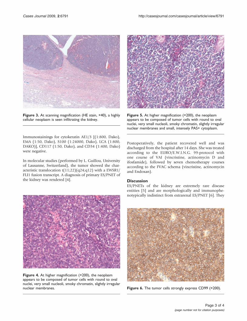
Figure 6. The tumor cells strongly express CD99 (×200).
Page 3 of 4(page number not for citation purposes)
Cases Journal 2009, 2:6791 http://casesjournal.com/casesjournal/article/view/6791

show the same gene fusions [7] and have a similar pooroutcome. In 1994, Mor described the first case of a renalES/PNET [4]. Subsequently, several case reports andsmall series of renal PNET were reported in the literature[6,8-10]. Reportedly, renal ES/PNET usually arises inyoung adults and shows a slight male predominance.Clinically, the tumors usually behave aggressively withfrequent metastasis and tumor-related death. In ourcase, the tumor was asymptomatic and only detectedcoincidentally. Histologically, several other small roundcell tumors enter into the differential diagnosis of renalES/PNET. Morphological, immunophenotypical andmolecular studies are needed to exclude a blastema-predominant Wilms’ tumor (which may show areas ofstromal or epithelial differentiation, is rarely CD99positive but frequently shows WT1 expression), meta-static neuroblastoma and clear cell sarcoma (whichusually arise in younger patients), synovial sarcoma(which commonly expresses cytokeratin or EMA andshows the characteristic t(X;18)), and small cell carci-noma and lymphoma (which show completely differentimmunophenotypes). Nonuniform nomenclature of theentity “renal ES/PNET” may have led to underreportingof this entity [11].
ConclusionRenal ES/PNET is rare. Differentiation of small round celltumors of the kidney may be challenging, and asympto-matic lesions in this location are often detected only bychance. In spite of surgical removal and the applicationof chemotherapy in this chemosensitive tumor with thesame nosologic form as Ewing’s sarcoma [12], disease-freesurvival is poor.
List of abbreviationsNSE, neuron specific enolase; CT, computed tomography;MRI, magnetic resonance imaging; ES, Ewing sarcoma;PNET, primitive neuroectodermal tumor.
ConsentWritten informed consent was obtained from the patientfor publication of this case report and accompanyingimages. A copy of the written consent is available forreview by the Editor-in-Chief of this journal.
Competing interestsThe authors declare that they have no competing interests.
Authors’ contributionsAB, SS, RR and MvF treated the patient and wereresponsible for writing the paper and looking up thebackground references. AZ was the pathologist in charge.MvF was responsible for over all coordination and finalproofreading of the manuscript. All the above-mentionedauthors read and approved the final manuscript.
AcknowledgmentsThe authors acknowledge Martin Buess, oncologist, St.Claraspital, Basel, Switzerland for his valuable intellectualinput, Robert Lemoine, MD, Pathology Viollier WeintraubSA, Geneva, Switzerland for his initial diagnosis, and LouisGuillou, MD, Department of Pathology, Centre Hospita-lier Universitaire Vaudois CHUV, Lausanne, Switzerlandfor his consultation diagnosis and the molecular char-acterization of the tumor.
References1. Marina NM, Etcubanas E, Parham DM, Bowman LC, Green A:
Peripheral primitive neuroectodermal tumor (peripheralneuroepithelioma) in children. A review of the St. Judeexperience and controversies in diagnosis and management.Cancer 1989, 64:1952-1960.
2. Kushner BH, Hajdu SI, Gulati SC, Erlandson RA, Exelby PR,Lieberman PH: Extracranial primitive neuroectodermaltumors. The Memorial Sloan-Kettering Cancer Centerexperience. Cancer 1991, 67:1825-1829.
3. Friedrichs N, Vorreuther R, Poremba C, Schafer KL, Böcking A,Buettner R, Zhou H: Primitive neuroectodermal tumor(PNET) in the differential diagnosis of malignant kidneytumors. Pathol Res Pract 2002, 198:563-569.
4. Mor Y, Nass D, Raviv G, Neumann Y, Nativ O, Goldwasser B:Malignant peripheral primitive neuroectodermal tumor(PNET) of the kidney. Med Pediatr Oncol 1994, 23:437-440.
5. Antoneli CB, Costa CM, de Camargo B, Sredni ST, Alfer W Jr,Chojniak R: Primitive neuroectodermal tumor (PNET)/extra-osseous Ewing sarcoma of the kidney. Med Pediatr Oncol 1998,30:303-307.
6. Jimenez RE, Folpe AL, Lapham RL, Ro JY, O’Shea PA, Weiss SW,Amin MB: Primary Ewing’s sarcoma/primitive neuroectoder-mal tumor of the kidney: a clinicopathologic and immuno-histochemical analysis of 11 cases. Am J Surg Pathol 2002,26:320-327.
7. Parham DM, Roloson GJ, Feely M, Green DM, Bridge JA, Beckwith JB:Primary malignant neuroepithelial tumors of the kidney: aclinicopathologic analysis of 146 adult and pediatric casesfrom the National Wilms’ Tumor Study Group PathologyCenter. Am J Surg Pathol 2001, 25:133-146.
8. Thyavihally YB, Tongaonkar HB, Gupta S, Kurkure PA, Amare P,Muckaden MA, Desai SB: Primitive neuroectodermal tumor ofthe kidney: a single institute series of 16 patients. Urology 2008,71:292-296.
9. Chu WC, Reznikov B, Lee EY, Grant RM, Cheng FW, Babyn P:Primitive neuroectodermal tumour (PNET) of the kidney: arare renal tumour in adolescents with seemingly character-istic radiological features. Pediatr Radiol 2008, 38:1089-1094.
10. Ellinger J, Bastian PJ, Hauser S, Biermann K, Müller SC: Primitiveneuroectodermal tumor: rare, highly aggressive differentialdiagnosis in urologic malignancies. Urology 2006, 68:257-262.
11. Chan YF, Llewellyn H: Intrarenal primitive neuroectodermaltumour. Br J Urol 1994, 73:326-327.
12. Marley EF, Liapis H, Humphrey PA, Nadler RB, Siegel CL, Zhu X,Brandt JM, Dehner LP: Primitive neuroectodermal tumor of thekidney–another enigma: a pathologic, immunohistochemical,and molecular diagnostic study. Am J Surg Pathol 1997,21:354-359.
Page 4 of 4(page number not for citation purposes)
Cases Journal 2009, 2:6791 http://casesjournal.com/casesjournal/article/view/6791
Recommended