Case ReportPregnancy after Uterine Artery Embolization:A Case Report in a Woman with Leiomyomata
Helena Isabel Lopes, Maria Isabel Sá, and Rosa Maria Rodrigues
Department of Obstetrics and Gynecology, Centro Hospitalar do Porto, 4099-001 Porto, Portugal
Correspondence should be addressed to Helena Isabel Lopes; lena [email protected]
Received 20 November 2014; Accepted 21 January 2015
Academic Editor: Olivier Picone
Copyright © 2015 Helena Isabel Lopes et al. This is an open access article distributed under the Creative Commons AttributionLicense, which permits unrestricted use, distribution, and reproduction in any medium, provided the original work is properlycited.
Background. Several pregnancies have been reported after embolization of uterine artery.This procedure is an accepted nonsurgicaltreatment for symptomatic uterine fibroids but its safety in women desiring future childbearing is not well established. CaseReport. We present a 40-year-old woman with leiomyomata who became pregnant after previously undergone uterine arteryembolization for three times. The placenta was previa and the fetus was in transverse position. She had a cesarean delivery ofan appropriately grown fetus at 37 weeks, which was followed by uterine atony requiring hysterectomy. Conclusion. Althoughpregnancy-related outcomes remain understudied, the available reports evidence that pregnancies after uterine artery embolizationmay be at significantly increased risk for postpartum hemorrhage, cesarean delivery, abnormal placentation, and malpresentation.In patients who are undergoing this type of treatment and contemplating pregnancy, the possibility of adverse complications shouldbe taken in consideration and women should be appropriately advised.
1. Introduction
Uterine fibroids are one of the most common benign tumors,with a prevalence of 30% among women of reproductiveage. Although most women are asymptomatic, there areothers that experience bothersome bulk-related symptomslike pelvic pain, pelvic pressure, urinary frequency, andabnormal uterine bleeding [1]. Uterine artery embolization(UAE) is a nonsurgical treatment option for these womenand who wish to retain their uterus and avoid surgery [2].Recommendation of UAE in women desiring pregnancy iscontroversial since there are concerns about its effects onfertility and pregnancy [3].
This paper reports a case of pregnancy and its outcomesin a woman who had previously submitted to three uterineartery embolizations for symptomatic leiomyomata.
2. Case Presentation
We present a case of a 40-year-old woman, gravida 4para 1, with antenatal surveillance of her pregnancy in ourinstitution. Her first pregnancy and labor occurred normally,
fourteen years ago. Some years later, she started experiencedysmenorrhea, pelvic pain, and heavy menstrual bleeding.The diagnosis of leiomyomata was made by ultrasonographyand magnetic resonance imaging (MRI), showing a uterinevolume of 580 cc with a dominant intramural fibroid of 105 ccvolume in the anterior wall. She suffered two miscarriagesafter this diagnosis. After counseling about her differentoptions of treatment, she always refused any type of surgery,trying to preserve her uterus and expecting a future preg-nancy.Then, uterine artery embolizationwas performedwithdecrease in fibroid size of 24% after 6 months and improve-ment of symptoms. However, one year after, the symptomsrecurred and uterinemyomas returned to a previous size. Sheundergone for more 2 UAE in another center. The procedurewas repeated in the second time because only 30% degreeof fibroid ischemia was achieved after the first embolization.Moreover, MRI revealed some contributing circulation fromthe left ovarian artery.
Three months after embolization she became pregnantspontaneously. On antenatal ultrasound scans several uterineintramural fibroids were evident including the dominantone placed at the lower segment of anterior wall measuring
Hindawi Publishing CorporationCase Reports in Obstetrics and GynecologyVolume 2015, Article ID 235312, 3 pageshttp://dx.doi.org/10.1155/2015/235312
2 Case Reports in Obstetrics and Gynecology
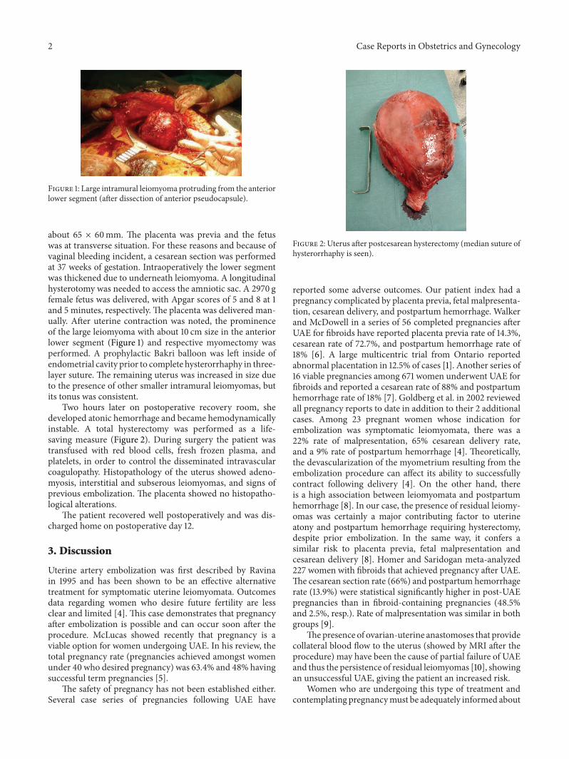
Figure 1: Large intramural leiomyoma protruding from the anteriorlower segment (after dissection of anterior pseudocapsule).
about 65 × 60mm. The placenta was previa and the fetuswas at transverse situation. For these reasons and because ofvaginal bleeding incident, a cesarean section was performedat 37 weeks of gestation. Intraoperatively the lower segmentwas thickened due to underneath leiomyoma. A longitudinalhysterotomy was needed to access the amniotic sac. A 2970 gfemale fetus was delivered, with Apgar scores of 5 and 8 at 1and 5 minutes, respectively. The placenta was delivered man-ually. After uterine contraction was noted, the prominenceof the large leiomyoma with about 10 cm size in the anteriorlower segment (Figure 1) and respective myomectomy wasperformed. A prophylactic Bakri balloon was left inside ofendometrial cavity prior to complete hysterorrhaphy in three-layer suture. The remaining uterus was increased in size dueto the presence of other smaller intramural leiomyomas, butits tonus was consistent.
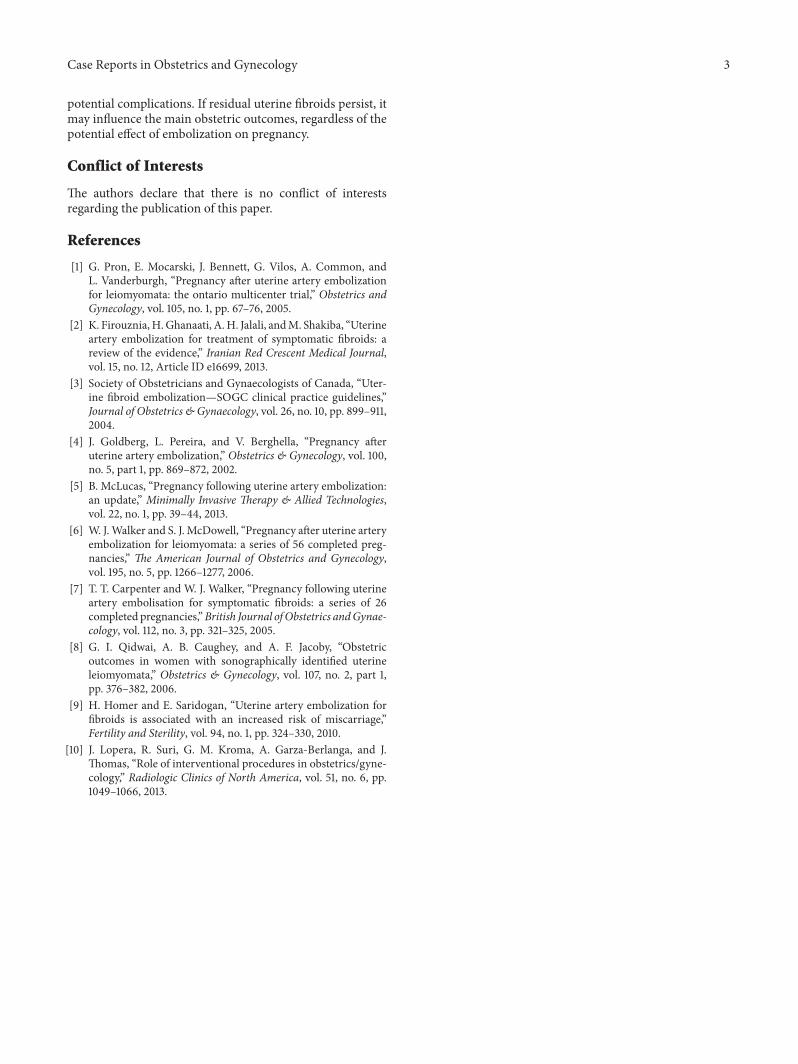
Two hours later on postoperative recovery room, shedeveloped atonic hemorrhage and became hemodynamicallyinstable. A total hysterectomy was performed as a life-saving measure (Figure 2). During surgery the patient wastransfused with red blood cells, fresh frozen plasma, andplatelets, in order to control the disseminated intravascularcoagulopathy. Histopathology of the uterus showed adeno-myosis, interstitial and subserous leiomyomas, and signs ofprevious embolization. The placenta showed no histopatho-logical alterations.
The patient recovered well postoperatively and was dis-charged home on postoperative day 12.
3. Discussion
Uterine artery embolization was first described by Ravinain 1995 and has been shown to be an effective alternativetreatment for symptomatic uterine leiomyomata. Outcomesdata regarding women who desire future fertility are lessclear and limited [4]. This case demonstrates that pregnancyafter embolization is possible and can occur soon after theprocedure. McLucas showed recently that pregnancy is aviable option for women undergoing UAE. In his review, thetotal pregnancy rate (pregnancies achieved amongst womenunder 40 who desired pregnancy) was 63.4% and 48% havingsuccessful term pregnancies [5].
The safety of pregnancy has not been established either.Several case series of pregnancies following UAE have
Figure 2: Uterus after postcesarean hysterectomy (median suture ofhysterorrhaphy is seen).
reported some adverse outcomes. Our patient index had apregnancy complicated by placenta previa, fetal malpresenta-tion, cesarean delivery, and postpartum hemorrhage. Walkerand McDowell in a series of 56 completed pregnancies afterUAE for fibroids have reported placenta previa rate of 14.3%,cesarean rate of 72.7%, and postpartum hemorrhage rate of18% [6]. A large multicentric trial from Ontario reportedabnormal placentation in 12.5% of cases [1]. Another series of16 viable pregnancies among 671 women underwent UAE forfibroids and reported a cesarean rate of 88% and postpartumhemorrhage rate of 18% [7]. Goldberg et al. in 2002 reviewedall pregnancy reports to date in addition to their 2 additionalcases. Among 23 pregnant women whose indication forembolization was symptomatic leiomyomata, there was a22% rate of malpresentation, 65% cesarean delivery rate,and a 9% rate of postpartum hemorrhage [4]. Theoretically,the devascularization of the myometrium resulting from theembolization procedure can affect its ability to successfullycontract following delivery [4]. On the other hand, thereis a high association between leiomyomata and postpartumhemorrhage [8]. In our case, the presence of residual leiomy-omas was certainly a major contributing factor to uterineatony and postpartum hemorrhage requiring hysterectomy,despite prior embolization. In the same way, it confers asimilar risk to placenta previa, fetal malpresentation andcesarean delivery [8]. Homer and Saridogan meta-analyzed227 women with fibroids that achieved pregnancy after UAE.The cesarean section rate (66%) and postpartum hemorrhagerate (13.9%) were statistical significantly higher in post-UAEpregnancies than in fibroid-containing pregnancies (48.5%and 2.5%, resp.). Rate of malpresentation was similar in bothgroups [9].
Thepresence of ovarian-uterine anastomoses that providecollateral blood flow to the uterus (showed by MRI after theprocedure) may have been the cause of partial failure of UAEand thus the persistence of residual leiomyomas [10], showingan unsuccessful UAE, giving the patient an increased risk.
Women who are undergoing this type of treatment andcontemplating pregnancymust be adequately informed about
Case Reports in Obstetrics and Gynecology 3
potential complications. If residual uterine fibroids persist, itmay influence the main obstetric outcomes, regardless of thepotential effect of embolization on pregnancy.
Conflict of Interests
The authors declare that there is no conflict of interestsregarding the publication of this paper.
References
[1] G. Pron, E. Mocarski, J. Bennett, G. Vilos, A. Common, andL. Vanderburgh, “Pregnancy after uterine artery embolizationfor leiomyomata: the ontario multicenter trial,” Obstetrics andGynecology, vol. 105, no. 1, pp. 67–76, 2005.
[2] K. Firouznia,H.Ghanaati, A.H. Jalali, andM. Shakiba, “Uterineartery embolization for treatment of symptomatic fibroids: areview of the evidence,” Iranian Red Crescent Medical Journal,vol. 15, no. 12, Article ID e16699, 2013.
[3] Society of Obstetricians and Gynaecologists of Canada, “Uter-ine fibroid embolization—SOGC clinical practice guidelines,”Journal of Obstetrics & Gynaecology, vol. 26, no. 10, pp. 899–911,2004.
[4] J. Goldberg, L. Pereira, and V. Berghella, “Pregnancy afteruterine artery embolization,” Obstetrics & Gynecology, vol. 100,no. 5, part 1, pp. 869–872, 2002.
[5] B. McLucas, “Pregnancy following uterine artery embolization:an update,” Minimally Invasive Therapy & Allied Technologies,vol. 22, no. 1, pp. 39–44, 2013.
[6] W. J.Walker and S. J. McDowell, “Pregnancy after uterine arteryembolization for leiomyomata: a series of 56 completed preg-nancies,” The American Journal of Obstetrics and Gynecology,vol. 195, no. 5, pp. 1266–1277, 2006.
[7] T. T. Carpenter and W. J. Walker, “Pregnancy following uterineartery embolisation for symptomatic fibroids: a series of 26completed pregnancies,”British Journal ofObstetrics andGynae-cology, vol. 112, no. 3, pp. 321–325, 2005.
[8] G. I. Qidwai, A. B. Caughey, and A. F. Jacoby, “Obstetricoutcomes in women with sonographically identified uterineleiomyomata,” Obstetrics & Gynecology, vol. 107, no. 2, part 1,pp. 376–382, 2006.
[9] H. Homer and E. Saridogan, “Uterine artery embolization forfibroids is associated with an increased risk of miscarriage,”Fertility and Sterility, vol. 94, no. 1, pp. 324–330, 2010.
[10] J. Lopera, R. Suri, G. M. Kroma, A. Garza-Berlanga, and J.Thomas, “Role of interventional procedures in obstetrics/gyne-cology,” Radiologic Clinics of North America, vol. 51, no. 6, pp.1049–1066, 2013.

Submit your manuscripts athttp://www.hindawi.com
Stem CellsInternational
Hindawi Publishing Corporationhttp://www.hindawi.com Volume 2014
Hindawi Publishing Corporationhttp://www.hindawi.com Volume 2014
MEDIATORSINFLAMMATION
of
Hindawi Publishing Corporationhttp://www.hindawi.com Volume 2014
Behavioural Neurology
EndocrinologyInternational Journal of
Hindawi Publishing Corporationhttp://www.hindawi.com Volume 2014
Hindawi Publishing Corporationhttp://www.hindawi.com Volume 2014
Disease Markers
Hindawi Publishing Corporationhttp://www.hindawi.com Volume 2014
BioMed Research International
OncologyJournal of
Hindawi Publishing Corporationhttp://www.hindawi.com Volume 2014
Hindawi Publishing Corporationhttp://www.hindawi.com Volume 2014
Oxidative Medicine and Cellular Longevity
Hindawi Publishing Corporationhttp://www.hindawi.com Volume 2014
PPAR Research
The Scientific World JournalHindawi Publishing Corporation http://www.hindawi.com Volume 2014
Immunology ResearchHindawi Publishing Corporationhttp://www.hindawi.com Volume 2014
Journal of
ObesityJournal of
Hindawi Publishing Corporationhttp://www.hindawi.com Volume 2014
Hindawi Publishing Corporationhttp://www.hindawi.com Volume 2014
Computational and Mathematical Methods in Medicine
OphthalmologyJournal of
Hindawi Publishing Corporationhttp://www.hindawi.com Volume 2014
Diabetes ResearchJournal of
Hindawi Publishing Corporationhttp://www.hindawi.com Volume 2014
Hindawi Publishing Corporationhttp://www.hindawi.com Volume 2014
Research and TreatmentAIDS
Hindawi Publishing Corporationhttp://www.hindawi.com Volume 2014
Gastroenterology Research and Practice
Hindawi Publishing Corporationhttp://www.hindawi.com Volume 2014
Parkinson’s Disease
Evidence-Based Complementary and Alternative Medicine
Volume 2014Hindawi Publishing Corporationhttp://www.hindawi.com
Recommended