For OSUWMC USE ONLY. To license, please contact the OSU Technology Commercialization Office at https://tco.osu.edu.
ANTERIOR CRUCIATE LIGAMENT (ACL) RECONSTRUCTION: PATELLAR TENDON AUTOGRAFT Background The following anterior cruciate ligament (ACL) reconstruction rehabilitation protocol is specific to patients with a patellar tendon autograft. If a hamstring autograft was used, please refer to the “Anterior Crucial Ligament (ACL) Reconstruction: Hamstring Autograft” protocol on the OSU Sports Medicine website. The rehabilitation recommendations below are based upon the guidance of content experts, evidence-based practice and the Multicenter Orthopaedic Outcomes Network (MOON) group. Progression through each phase is based on the patient demonstrating readiness by achieving functional criteria rather than the time elapsed from surgery. The times frames identified after each phase are approximate times for the average patient, NOT guidelines for progression. Disclaimer Progression is time and criterion-based, dependent on soft tissue healing, patient demographics and clinician evaluation. If you are working with an Ohio State Sports Medicine patient and questions arise, please contact the author by calling our office at (614) 293-2385.
For OSUWMC USE ONLY. To license, please contact the OSU Technology Commercialization Office at https://tco.osu.edu.
Summary of Recommendations Precautions • No testing of repaired or reconstructed ligaments (Lachman, Anterior/Posterior
Drawer, Varus/Valgus Stress) prior to 12 WEEKS post-operative • No loaded open kinetic chain knee extension beyond 45 degrees x 8 WEEKS • Meniscus Repair:
a. No forced flexion ROM beyond 90º x4 WEEKS b. No CKC exercises >90º x 8 WEEKS c. PWBing x 4 weeks for concomitant root, radial and/or horizontal cleavage
meniscus repairs only i. All other types of meniscus repairs will be FWBing ii. Please refer to the “post-op plan” section of the operative note for
clarification
Outcome Tools
Collect the Lower Extremity Functional Scale (LEFS) at each visit Consider collecting one of the following outcome tools at initial evaluation, monthly and discharge. Be consistent with which outcome tool is collected each time.
1. IKDC 2. KOOS
You may choose to include ACL-RSI, Tegner or other questionnaires specific to your patient’s needs.
Strength Testing
1. Isometric testing anytime- fixed at 90º 2. Isokinetic testing no earlier than 12 weeks
Criteria to Discharge Assistive Device
1. ROM: Full active knee extension equivalent to healthy, contralateral limb; no pain on passive overpressure
2. Strength: Able to perform strong quad isometric with full tetany and superior patellar glide and able to perform 2x10 supine SLR without quad lag
3. Effusion: ≤1+ is preferred (2+ acceptable if all other criteria are met) 4. Weight Bearing: Demonstrates pain-free ambulation without visible gait deviation
Criteria to Initiate Running and Jumping
1. ROM: full, pain-free knee ROM, symmetrical with the uninvolved limb 2. Strength: Isokinetic testing 80% or greater for hamstring and quad at 60º/sec and
300º/sec (Appendix D) 3. Effusion: ≤ 1+ 4. Weight Bearing: normalized gait and jogging mechanics 5. Neuromuscular Control: Pain-free hopping in place
Criteria for Return to Sport
1. ROM: full, pain-free knee ROM, symmetrical with the uninvolved limb 2. Strength: Isokinetic testing 90% or greater for hamstring and quad at 60º/sec and
300º/sec (Appendix D) 3. Effusion: No reactive effusion ≤ 1+ with sport-specific activity 4. Weight Bearing: normalized gait and jogging mechanics 5. Neuromuscular control: appropriate mechanics and force attenuation strategies with
high level agility, plyometrics, and high impact movements 6. Functional Hop Testing: LSI ≥ 90% for all tests (Appendix E) 7. Physician Clearance
For OSUWMC USE ONLY. To license, please contact the OSU Technology Commercialization Office at https://tco.osu.edu.
RED/YELLOW FLAGS Red flags are signs/symptoms that require immediate referral for re-evaluation. Yellow flags are signs/symptoms that require modification to plan of care. Red Flags • Signs of DVT (Refer directly to ED)
o Localized tenderness along the distribution of deep venous system o Entire LE swelling o Calf swelling >3cm compared to asymptomatic limb o Pitting edema o Collateral superficial veins
• Lack of full knee extension by 4 weeks (Refer to surgeon for re-evaluation) • Mechanical block or clunk (Refer to surgeon for re-evaluation)
Yellow Flags
• Persistent reactive pain or effusion following therapy or ADLs o Decrease intensity of therapy interventions, continue effusion management and
provide patient education regarding activity modification until reactive symptoms resolve
Protection Phase (Post-ACLR – 4 weeks) Appointments • Goal: Restore ROM, minimize effusion and pain.
• Post-operative evaluation should be performed 3-5 days following surgery. Follow-up appointments 1-2x per week, depending on progression towards goals.
Precautions a. No testing of repaired or reconstructed ligaments (Lachman, Anterior/Posterior Drawer, Varus/Valgus Stress) prior to 12 WEEKS post-operative
b. No loaded open kinetic chain knee extension beyond 45 ° x 8 WEEKS • Meniscus Repair:
a. No forced flexion ROM beyond 90º x4 WEEKS b. No CKC exercises >90º x 8 WEEKS c. PWBing x 4 weeks for concomitant root, radial and/or horizontal cleavage
meniscus repairs only i. All other types of meniscus repairs will be FWBing ii. Please refer to the “post-op plan” section of the operative note for
clarification
Pain and Effusion
≤2+ (using Modified Stroke Test) • Effusion management strategies: cryotherapy and compression (ie. Donut,
ace wrap) and limited WB therapeutic exercise as appropriate
ROM Strong emphasis on patellar mobilizations (superior/inferior > medial/lateral) to regain full knee ROM Extension: Emphasis on achieving full knee extension immediately following surgery. If full extension is not achieved by 4 weeks, contact surgeon regarding ROM concerns. Utilize low load, long duration stretching (Appendix A) Flexion: No forced flexion past 90º for all meniscus repairs.
ACLR and meniscectomy are able to push for symmetrical flexion as appropriate.
For OSUWMC USE ONLY. To license, please contact the OSU Technology Commercialization Office at https://tco.osu.edu.
Therapeutic Exercise
• Emphasis on quad activation without gluteal co-contraction • Restore patellar mobility • Symmetrical ROM • Decrease effusion • Ambulation with appropriate joint loading and without obvious gait deviation
Suggested Interventions
• Extension ROM: bag hangs or prone hangs (Appendix A) • Flexion ROM: heel slides, wall slides, upright bike • Patellar mobilization: superior, inferior, medial, lateral • Quad Isometrics; SLR 4-way • TKE: prone and standing • Prone hamstring curls • Weight shifting, SL balance • Neuromuscular re-education using electrical stimulation (NMES) at 60º knee
flexion (Appendix B)
NMES Parameters (Appendix B)
• NMES pads are placed on the proximal and distal quadriceps • Patient: Seated with the knee in at least 60º flexion, shank secured with strap and
back support with thigh strap preferred. The ankle pad/belt should be two finger widths superior to the lateral malleoli
• The patient is instructed to relax while the e-stim generates at least 50% of their max volitional contraction against a fixed resistance OR maximal tolerable amperage without knee joint pain
• 10-20 seconds on/ 50 seconds off x 15 min
Criteria to Discharge Assistive Device
1. ROM: Full active knee extension; no pain on passive overpressure 2. Strength: Able to perform strong quad isometric with full tetany and superior
patellar glide and able to perform 2x10 supine SLR without quad lag 3. Effusion: ≤ 1+ is preferred (2+ acceptable if all other criteria are met) 4. Weight Bearing: Demonstrates pain-free ambulation without visible gait deviation
Criteria to Progress to Early Loading Phase
Goals: (These do not limit progression to next phase; however, should be addressed with interventions) ROM: ≥ 0-120 degrees Strength: Quadriceps set with normal superior patellar translation, SLR x 10 seconds without extensor lag Effusion: ≤ 2+ with Modified stroke test Weight Bearing: Able to tolerate CKC therex program without increased pain and ≤ 2+ effusion

For OSUWMC USE ONLY. To license, please contact the OSU Technology Commercialization Office at https://tco.osu.edu.
Early Loading Phase (4-8 weeks) Appointments • Goal: to improve LE loading symmetry, increase strength and normalize gait mechanics.
• 1-2 visits per week with emphasis on HEP compliance (2-3 days per week outside of therapy).
Precautions If full AROM knee extension is not achieved by 4 weeks, contact surgeon Open Chain knee extension: • Initiate un-resisted LAQ at 4 weeks (partial full range) • Initiate multi-angle isometrics (from 90-60⁰) at 4 weeks • Begin isotonic open chain knee extensions through protected ROM (90-45°) at 6 weeks
Pain and Effusion
Cryotherapy/compression as needed for reactive effusion. Patellar taping and/or Cho-Pat strap to reduce PF symptoms if present
ROM • Monitor and progress knee ROM, patellar mobility, and LE flexibility • Continued emphasis on end-range ROM • Continue bike for ROM and warm up
Suggested Interventions and timelines
• Multi-angle knee isometrics from 90-60⁰ • Initiate open chain knee extension exercises
o Initiate unresisted LAQ at 4 weeks (partial full range) o Begin isotonic open chain knee extensions through protected ROM (90-45°) at 6
weeks • Progress WB quadriceps exercises with emphasis on proper LE mechanics • Hamstring curls (prone, machine or physioball) • Progress gluteal and lumbopelvic strength and stability • Progress single leg balance • Endurance: low impact - treadmill walking, stepper, elliptical (6 weeks) • NMES (see parameters in week 1-4)
Criteria to d/c NMES
• <20% quadriceps deficit on isometric testing OR- If a Biodex machine is not available: 1. 10 SLR without quad lag 2. Normal gait 3. 10 heel taps to to 60 degrees with good quality 4. 10 rep max on LP and similar effort bilaterally 5. Inability to break quad MMT
Criteria to Progress to Strength and Power Phase
1. ROM: Maintain full, pain free AROM including PF mobility 2. Effusion: ≤ 1+ 3. Strength: See criteria to discharge NMES 4. Weight Bearing: Able to tolerate therapeutic exercise program without increased pain or
>1+ effusion 5. Neuromuscular Control: Demonstrates proper lower extremity mechanics with all
therapeutic exercises (bilaterally)

For OSUWMC USE ONLY. To license, please contact the OSU Technology Commercialization Office at https://tco.osu.edu.
Strength and Power Phase (8-12 weeks) Appointments • Goal to increase lower extremity strength and power.
• 1-2 visits per week with emphasis on patient compliance with resistance training as part of HEP (2-3 days per week outside of therapy).
Precautions Open Chain knee extension: resisted open chain knee extension (protected ROM full ROM)
Pain and Effusion
Cryotherapy/compression as needed for reactive effusion. Patellar taping and/or Cho-Pat strap to reduce PF symptoms if present
ROM • Monitor and progress knee ROM, patellar mobility, and LE flexibility • Consider higher level warm ups, including bike sprints or versaclimber • Continue aggressive techniques to achieve/maintain full knee extension if necessary (i.e.
weighted bag hang) as needed • If full AROM knee extension is not achieved by 4 weeks, contact surgeon regarding ROM
concerns.
Suggested Interventions and timelines
• Multi-angle knee isometrics from 90-0⁰ • Progress isotonic open chain knee extensions through full range (90-0⁰) • Continue isolated hamstring interventions
o RDL o Nordic hamstring curls
• Progress gluteal and lumbopelvic strength and stability • Progress single leg balance • Initiate PWB plyometrics on shuttle (8-10 weeks) • NMES if appropriate (see parameters in week 1-4)
Criteria to initiate Running and Jumping
1. ROM: full, pain-free knee ROM, symmetrical with the uninvolved limb 2. Strength: Isokinetic testing 80% or greater for hamstring and quad at 60º/sec and
300º/sec (See Appendix C and D) 3. Effusion: ≤ 1+ 4. Weight Bearing: normalized gait and jogging mechanics 5. Neuromuscular Control: Pain-free hopping in place
Criteria to Progress to Return to Function Phase
6. ROM: Maintain full, pain free AROM including PF mobility 7. Effusion: ≤ 1+ 8. Strength: Isometric or isokinetic quadriceps and hamstrings strength >/= 80% 9. Weight Bearing: Able to tolerate therapeutic exercise program, including jogging
progression, without increased pain or >1+ effusion 10. Neuromuscular Control: Demonstrates proper lower extremity mechanics with all
therapeutic exercises (bilaterally) 11. Outcome Tools: ≥ 7/10 on #10 IKDC Questionnaire

For OSUWMC USE ONLY. To license, please contact the OSU Technology Commercialization Office at https://tco.osu.edu.
Return to Function Phase (12 weeks-Return to Sport) Appointments Increased frequency from previous stage to 1-2x per week when appropriate to initiate
plyometric training and return to running program.
Precautions Criteria to initiate hopping • Full, pain free ROM • ≤ 1+ effusion • ≥ 7 /10 on #10 IKDC Questionnaire • ≥ 80% isokinetic strength symmetry (hamstrings and quadriceps) OR ≥ 80%
limb symmetry on acceptable isokinetic alternative (See Appendix D)
Criteria to initiate jogging (in addition to above criteria) • Hop downs with appropriate landing mechanics • Audible rhythmic strike patterns and no gross visual compensation
Pain and Effusion
Effusion may increase with increased activity, ≤1+ and/or non-reactive effusion for progression of plyometrics
ROM Full, symmetrical to contralateral limb, and pain free with overpressure
Therapeutic Exercise
• Performance of the quadriceps, hamstrings and trunk dynamic stability • Muscle power generation and absorption via plyometrics • Sport- and position-specific activities • Begin agility exercises between 50-75% effort (utilize visual feedback to improve
mechanics as needed) • Advance plyometrics: Bilateral to single leg, progress by altering surfaces, adding
ball toss, 3D rotations, etc.
Suggested Interventions
Therapeutic Exercise/Neuromuscular Re-education • Squats, leg extension, leg curl, leg press, deadlifts, lunges (multi-direction),
crunches, rotational trunk exercises on static and dynamic surfaces, monster walks, PWB to FWB jumping
• Single-leg squats on BOSU with manual perturbation to trunk or legs, Single-leg BOSU balance, single-leg BOSU Romanian deadlift
Agility • Side shuffling, Carioca, Figure 8, Zig-zags, Resisted jogging (Sports Cord) in
straight planes, backpedaling Plyometrics • Single-leg hop downs from increasing height (up to 12” box), Single-leg hop-holds,
Double and single-leg hopping onto unstable surface, Double and single-leg jump-turns, Repeated tuck jumps
Criteria for Return to Sport
1. ROM: full, pain free knee ROM, symmetrical with the uninvolved limb 2. Strength: Isokinetic testing 90% or greater for hamstring and quad at 60º/sec and
300º/sec 3. Effusion: No reactive effusion ≥ 1+ with sport-specific activity 4. Weight Bearing: normalized gait and jogging mechanics 5. Neuromuscular control: appropriate mechanics and force attenuation strategies
with high level agility, plyometrics, and high impact movements 6. Functional Hop Testing: LSI 90% or greater for all tests (Appendix E) 7. Physician Clearance
For OSUWMC USE ONLY. To license, please contact the OSU Technology Commercialization Office at https://tco.osu.edu.
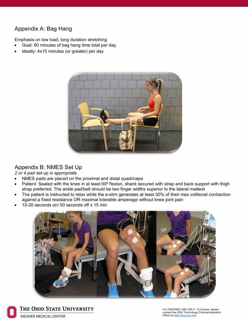
Appendix A: Bag Hang Emphasis on low load, long duration stretching • Goal: 60 minutes of bag hang time total per day. • Ideally: 4x15 minutes (or greater) per day
Appendix B: NMES Set Up 2 or 4 pad set-up is appropriate • NMES pads are placed on the proximal and distal quadriceps • Patient: Seated with the knee in at least 60º flexion, shank secured with strap and back support with thigh
strap preferred. The ankle pad/belt should be two finger widths superior to the lateral malleoli • The patient is instructed to relax while the e-stim generates at least 50% of their max volitional contraction
against a fixed resistance OR maximal tolerable amperage without knee joint pain • 10-20 seconds on/ 50 seconds off x 15 min

For OSUWMC USE ONLY. To license, please contact the OSU Technology Commercialization Office at https://tco.osu.edu.
Appendix C: Isokinetic Data Interpretation
Definition Clinical Impact What to do A Peak Torque (ft-lbs) Peak torque during
repetitions Symmetry criteria (see ‘E’- this is the data represented in pie charts)
If <80%; continue unilateral, high resistance strength training
B Coefficient of Variance (%)
Between repetition variability
Goal: < 15% If >15%, consider retest
C Total Work (ft-lbs) Torque over all repetitions
Possible indicator of fatigue If >10%; consider high volume training
D Agonist/Antagonist Ratio (%)
Hamstring/Quadriceps Ratio
Goal: >60% <60%; ensure 1:1 quadriceps:hamstring exercise ratio
E Limb Symmetry Pie Charts
Strength relative to involved limb
Goal: <10% asymmetry (either direction- deficit OR stronger on involved limb)
If <80%, continue NMES in addition to strength training If <90%, continue unilateral > bilateral strength training emphasis
For OSUWMC USE ONLY. To license, please contact the OSU Technology Commercialization Office at https://tco.osu.edu.
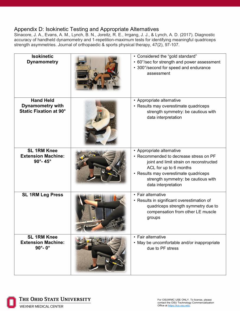
Appendix D: Isokinetic Testing and Appropriate Alternatives Sinacore, J. A., Evans, A. M., Lynch, B. N., Joreitz, R. E., Irrgang, J. J., & Lynch, A. D. (2017). Diagnostic accuracy of handheld dynamometry and 1-repetition-maximum tests for identifying meaningful quadriceps strength asymmetries. Journal of orthopaedic & sports physical therapy, 47(2), 97-107.
Isokinetic Dynamometry
• Considered the “gold standard” • 60°/sec for strength and power assessment • 300°/second for speed and endurance
assessment
Hand Held Dynamometry with
Static Fixation at 90°
• Appropriate alternative • Results may overestimate quadriceps
strength symmetry: be cautious with data interpretation
SL 1RM Knee Extension Machine:
90°- 45°
• Appropriate alternative • Recommended to decrease stress on PF
joint and limit strain on reconstructed ACL for up to 6 months
• Results may overestimate quadriceps strength symmetry: be cautious with data interpretation
SL 1RM Leg Press
• Fair alternative • Results in significant overestimation of
quadriceps strength symmetry due to compensation from other LE muscle groups
SL 1RM Knee Extension Machine:
90°- 0°
• Fair alternative • May be uncomfortable and/or inappropriate
due to PF stress

For OSUWMC USE ONLY. To license, please contact the OSU Technology Commercialization Office at https://tco.osu.edu.
Appendix E: Single Leg Hop Series

For OSUWMC USE ONLY. To license, please contact the OSU Technology Commercialization Office at https://tco.osu.edu.
Authors: Caroline Brunst, PT, DPT, SCS, AT Reviewers: Mary Montalto, PT, DPT; Vickie Otto, PT, DPT; Stephanie Di Stasi, PT, PhD, OCS; Laura Schmitt, PT, PhD; John Dewitt, PT; Robert Magnussen, MD; David Flanigan, MD; Christopher Kaeding, MD References: Adams, D., Logerstedt, D., Hunter-Giordano, A., Axe, M. J., & Snyder-Mackler, L. (2012). Current Concepts for
Anterior Cruciate Ligament Reconstruction: A Criterion-Based Rehabilitation Progression. Journal of Orthopaedic & Sports Physical Therapy, 42(7), 601–614. https://doi.org/10.2519/jospt.2012.3871
Di Stasi, S., Myer, G. D., & Hewett, T. E. (2013). Neuromuscular Training to Target Deficits Associated With Second Anterior Cruciate Ligament Injury. Journal of Orthopaedic & Sports Physical Therapy, 43(11), 777-A11. https://doi.org/10.2519/jospt.2013.4693
Grindem, H., Snyder-Mackler, L., Moksnes, H., Engebretsen, L., & Risberg, M. A. (2016). Simple decision rules can reduce reinjury risk by 84% after ACL reconstruction: the Delaware-Oslo ACL cohort study. British Journal of Sports Medicine, 50(13), 804–808. https://doi.org/10.1136/bjsports-2016-096031
Hewett, T. E., Myer, G. D., Ford, K. R., Heidt, R. S., Colosimo, A. J., McLean, S. G., … Succop, P. (2005). Biomechanical measures of neuromuscular control and valgus loading of the knee predict anterior cruciate ligament injury risk in female athletes: A prospective study. American Journal of Sports Medicine, 33(4), 492–501. https://doi.org/10.1177/0363546504269591
Hewett, T. E., Myer, G. D., Ford, K. R., Paterno, M. V., & Quatman, C. E. (2012). The 2012 ABJS nicolas andry award: The sequence of prevention: A systematic approach to prevent anterior cruciate ligament injury knee. Clinical Orthopaedics and Related Research, 470(10), 2930–2940. https://doi.org/10.1007/s11999-012-2440-2
Myer, G. D., Chu, D. A., Brent, J. L., & Hewett, T. E. Trunk and Hip Control Neuromuscular Training for the Prevention of Knee Joint Injury, 27 Clinics in Sports Medicine § (2008). Elsevier. https://doi.org/10.1016/j.csm.2008.02.006
Schmitt, L. C., Paterno, M. V., & Hewett, T. E. (2012). The Impact of Quadriceps Femoris Strength Asymmetry on Functional Performance at Return to Sport Following Anterior Cruciate Ligament Reconstruction. Journal of Orthopaedic & Sports Physical Therapy, 42(9), 750–759. https://doi.org/10.2519/jospt.2012.4194
Sinacore, J. A., Evans, A. M., Lynch, B. N., Joreitz, R. E., Irrgang, J. J., & Lynch, A. D. (2017). Diagnostic Accuracy of Handheld Dynamometry and 1-Repetition-Maximum Tests for Identifying Meaningful Quadriceps Strength Asymmetries. Journal of Orthopaedic & Sports Physical Therapy, 47(2), 97–107. https://doi.org/10.2519/jospt.2017.6651
Wright, R. W., Haas, A. K., Anderson, J., Calabrese, G., Cavanaugh, J., Hewett, T. E., … Wolf, B. R. (2015). Anterior Cruciate Ligament Reconstruction Rehabilitation. Sports Health: A Multidisciplinary Approach, 7(3), 239–243. https://doi.org/10.1177/1941738113517855
Zazulak, B. T., Hewett, T. E., Reeves, N. P., Goldberg, B., & Cholewicki, J. (2007a). Deficits in Neuromuscular Control of the Trunk Predict Knee Injury Risk. The American Journal of Sports Medicine, 35(7), 1123–1130. https://doi.org/10.1177/0363546507301585
Zazulak, B. T., Hewett, T. E., Reeves, N. P., Goldberg, B., & Cholewicki, J. (2007b). The Effects of Core Proprioception on Knee Injury. The American Journal of Sports Medicine, 35(3), 368–373. https://doi.org/10.1177/0363546506297909
Recommended


