Antenatal iron supplementation, FGF23, and bone metabolism inKenyan women and their offspring: secondary analysis of arandomized controlled trial
Vickie S Braithwaite,1,2 Martin N Mwangi,3,4 Kerry S Jones,5 Ayse Y Demir,6 Ann Prentice,1,7 Andrew M Prentice,7
Pauline EA Andang’o,8 and Hans Verhoef3,9
1Medical Research Council (MRC) Nutrition and Bone Health Research Group, Clifford Allbutt Building, University of Cambridge, Cambridge BiomedicalCampus, Cambridge, CB2 0AH, United Kingdom (formerly the MRC Elsie Widdowson Laboratory, 120 Fulbourn Road, Cambridge, CB1 9NL, UnitedKingdom); 2Medical Research Council (MRC) Epidemiology Unit, University of Cambridge School of Clinical Medicine, Cambridge Biomedical Campus,Cambridge, CB2 0QQ, United Kingdom; 3Wageningen University, Division of Human Nutrition and Health, P.O. Box 17, 6700 AA Wageningen, TheNetherlands; 4University of Malawi, College of Medicine, Training and Research Unit of Excellence, Private Bag 360, BT 3, Chichiri, Blantyre, Malawi;5National Institute for Health Research (NIHR) Biomedical Research Centre Nutritional Biomarker Laboratory, MRC Epidemiology Unit, University ofCambridge, Cambridge Biomedical Campus , Cambridge, CB2 0AH, United Kingdom,; 6Meander Medical Centre, Laboratory for Clinical Chemistry andHematology, P.O. Box 1502 , 3800 BM Amersfoort, The Netherlands; 7Medical Research Council (MRC) Unit The Gambia at London School of Hygiene &Tropical Medicine, Atlantic Boulevard , Fajara, Banjul, The Gambia; 8Maseno University, School of Public Health and Community Development, Maseno,Kenya; and 9Wageningen University, Cell Biology and Immunology Group, P.O. Box 338,, 6708 WD Wageningen, The Netherlands
ABSTRACTBackground: Fibroblast growth factor-23 (FGF23) regulates bodyphosphate homeostasis primarily by increasing phosphaturia. It alsoacts as a vitamin D-regulating hormone. Maternal iron deficiency isassociated with perturbed expression and/or regulation of FGF23 andhence might be implicated in the pathogenesis of hypophosphatemia-driven rickets in their offspring.Objectives: We aimed to determine the effect of antenatal oral ironsupplementation on FGF23 concentration and maternal and infantmarkers of bone-mineral regulation.Methods: We performed a secondary analysis of a trial in which470 rural Kenyan women with singleton pregnancies and hemoglobinconcentrations ≥ 90 g/L were randomly allocated to daily, supervisedsupplementation with 60 mg elemental iron as ferrous fumarateor placebo from 13–23 weeks of gestation until 1 mo postpartum.As previously reported, iron supplementation improved iron statusin mothers and neonates. For the present study, we reanalyzed allavailable plasma samples collected in mothers and neonates at birth,with primary outcomes being concentrations of FGF23, measuredby 2 assays: 1 that detects intact hormone and C-terminal cleavageproducts (total-FGF23) and another that detects the intact hormoneonly (intact-FGF23).Results: Analysis was performed on 433 women (n = 216, irongroup; n = 217, placebo group) and 414 neonates (n = 207, irongroup; n = 207, placebo group). Antenatal iron supplementationreduced geometric mean total-FGF23 concentrations in mothers andneonates by 62.6% (95% CI: 53.0%, 70.3%) and 15.2% (95% CI:−0.3%, 28.4%, P = 0.06), respectively. In addition, it increasedgeometric mean neonatal intact-FGF23 concentrations by 21.6%(95% CI: 1.2%, 46.1%), increased geometric mean maternal hepcidin
concentrations by 136.4% (95% CI: 86.1%, 200.3%), and decreasedmean maternal 25-hydroxyvitamin D concentrations by 6.1 nmol/L(95% CI: −11.0, −1.2 nmol/L).Conclusions: Analysis of this randomized trial confirms that ironsupplementation can reverse elevated FGF23 production causedby iron deficiency in iron-deficient mothers and their neonates.Further investigations are warranted to assess to what extent ironsupplementation can prevent FGF23-mediated hypophosphatemicrickets or osteomalacia. Am J Clin Nutr 2021;113:1104–1114.
Keywords: iron deficiency anemia, fibroblast growth factor(FGF23), phosphate, vitamin D, bone, Africa, pregnancy
IntroductionThere is increasing evidence that iron deficiency in children
with chronically inadequate calcium intake may predispose torickets, one of the most common noncommunicable childhooddiseases in developing countries (1). In this pathogenic pathway(Figure 1), there seems to be a central role for fibroblast growthfactor-23 (FGF23), a key regulator of circulating concentrationsof phosphate and 1,25-dihydroxyvitamin D [1,25(OH)2D—the bioactive form of vitamin D] (2). In vitamin D-repleteGambian children, rickets was associated with elevated plasmaconcentrations of total-FGF23 (as measured by an assay whichdetects both the intact hormone and the C-terminal fragmentsthat result from its cleavage), 1,25(OH)2D, and total alkalinephosphatase (a marker of dysregulated bone formation that iselevated in rickets and osteomalacia) and with lower plasma
1104 Am J Clin Nutr 2021;113:1104–1114. Printed in USA. © The Author(s)2021. Published by Oxford University Press on behalf of the American Society for Nutrition. This is an Open Access article distributed under the terms of theCreative Commons Attribution License (http://creativecommons.org/licenses/by/4.0/), which permits unrestricted reuse, distribution, and reproduction in anymedium, provided the original work is properly cited.
Effect of oral antenatal iron on FGF23 and bone 1105
concentrations of phosphate (3). Gambian children with a historyof rickets-like bone deformities had a higher prevalence ofanemia than community controls, and total-FGF23 concentrationwas negatively associated with hemoglobin concentration (4).In an iron supplementation study among Gambian children,plasma total-FGF23 concentration at baseline was inverselyassociated with markers of iron (plasma ferritin concentration,hemoglobin concentration), whereas at 3 mo after the start ofiron supplementation, plasma total-FGF23 concentrations werelower than at the start of the intervention (5). As with mostsuch studies, ethics constraints precluded the use of a placebocontrol group. In a prospective cohort study, Gambian childrenborn to mothers who were iron deficient during pregnancyhad higher concentrations of total-FGF23 and total alkalinephosphatase than those whose mothers had normal iron statusand this difference persisted ≤2 y of age (6). In mice, diet-induced iron deficiency during pregnancy and nursing resultedin elevated serum concentrations of total-FGF23 and intact-FGF23 (as measured using an assay which only detects the intacthormone), hypophosphatemia, and reduced serum concentrationsof 1,25(OH)2D in their pups (7). These data suggest thatmaternal iron deficiency may lead to perturbations in FGF23regulation that may in turn lead to hypophosphatemia-drivenrickets in the offspring. In addition, maternal bone mineraldensity decreases by 3%–5% during pregnancy and lactation(8). This physiological change, coupled with iron-deficiencyphosphate loss, may put additional pressures on the maternalskeleton, thus predisposing iron-deficient women to poor bonehealth in later life.
FGF23 gene expression occurs predominantly within osteo-cytes but the FGF23 hormone regulates circulating phosphateconcentrations via the FGF receptor in renal tubule cells whereFGF23 signaling causes a reduction in phosphate resorption from
Supported by Royal Society project grant RG2015R2 (to VSB) and theMedical Research Council (MRC) and the Department for InternationalDevelopment (DFID) under the MRC/DFID Concordat agreement via MRCprogrammes U105960371 (to AP) and MC-A760-5QX00 (to AMP). Theoriginal study was supported by the INSTAPA project which received fundingfrom European Union Seventh Framework Programme FP7/2007/2013 grant211484 (to MNM). KSJ is supported by National Institute for Health Research(NIHR) Cambridge Biomedical Research Centre grant IS-BRC-1215-20014.The NIHR Cambridge Biomedical Research Centre is a partnership betweenCambridge University Hospitals NHS Foundation Trust and the Universityof Cambridge, funded by the NIHR. The views expressed are those of theauthors and not necessarily those of the NHS, the NIHR, or the Departmentof Health and Social Care.
Supplemental Methods, Supplemental Table 1, and Supplemental Figure1 are available from the “Supplementary data” link in the online posting ofthe article and from the same link in the online table of contents at https://academic.oup.com/ajcn/.
Address correspondence to VSB (e-mail: [email protected]).Abbreviations used: β-Crosslaps, β-C-terminal telopeptide; CRP, C-
reactive protein; eGFR, estimated glomerular filtration rate; EPO, erythropoi-etin; FGF23, fibroblast growth factor-23; GSD, geometric SD; intact-FGF23,intact fibroblast growth factor-23 hormone; PTH, intact parathyroid hormone;sTfR, soluble transferrin receptor; total-FGF23, intact fibroblast growthfactor-23 hormone and the C-terminal fragments that result from its cleavage;1,25(OH)2D, 1,25-dihydroxyvitamin D; 25(OH)D, 25-hydroxyvitamin D.
Received September 3, 2020. Accepted for publication December 10, 2020.First published online March 1, 2021; doi: https://doi.org/10.1093/ajcn/
nqaa417.
the glomerular filtrate and thus renal phosphate excretion. Theintact FGF23 hormone undergoes proteolytic cleavage into its C-and N-terminal fragments. Although the intact FGF23 hormoneis known to have effects on phosphate and vitamin D metabolism,there is conflicting evidence as to whether the C-terminal FGF23fragment is active, acts in the same way (9), or acts in the oppositeway to the intact hormone (10).
The requirement for iron and the demands on the kidneyincrease during pregnancy, and the risk of developing irondeficiency and/or anemia increases substantially. Iron absorptionand thus the effect of iron interventions on systemic biomarkers(e.g., hemoglobin concentration) are known to depend on ironstatus. This process is mediated by circulating concentrationsof hepcidin, the master regulator of body iron homeostasis.Causality of the relations between iron deficiency and bonemetabolism has not yet been shown, because evidence has so farbeen obtained exclusively from observational studies and animalstudies. Such an effect would be pertinent to developing coun-tries, where iron deficiency often affects the majority of pregnantwomen and a substantial proportion of young children (11).
The aim of this current study was to investigate the effect ofantenatal oral iron supplementation on circulating concentrationsof FGF23 and hence on markers of phosphate and bonemetabolism (including vitamin D) and kidney function at birthin pregnant Kenyan women and their offspring. In addition,we explored to what extent the response of these markers tointervention depended on initial iron status.
Methods
Study design, participants, randomization, and blinding
The present study used samples and data that were collectedas part of a randomized placebo-controlled trial in rural Kenya(NCT01308112) originally designed to measure the effectof antenatal iron supplementation on maternal Plasmodiuminfection risk, maternal iron status, and neonatal outcomes.The recruitment for the original trial was conducted between2011 and 2013. Study details and main results have beenpublished elsewhere (12). In brief, pregnant women aged 15–45 y with singleton pregnancies, gestational age of 13–23 wk(determined by obstetric ultrasonography), and hemoglobinconcentration ≥ 90 g/L were individually randomly assignedat a 1:1 allocation ratio to 60 mg of elemental iron as ferrousfumarate or placebo until 1 mo postpartum. The supplementscontained no other micronutrients and their neonates did notreceive any additional supplements. From screening until theend of the intervention, local mill operators added fortificantiron (target dose: 20 mg/kg flour) to grain routinely broughtfor milling by homestead members of all the participatingwomen. Based on weighed intake studies, we estimate thatfortification supplied on average 5.7 mg of elemental iron asferric sodium ethylenediaminetetraacetate daily to the pregnantwomen in both arms of the intervention. Participants and fieldstaff were blinded to the randomization and intervention until dataanalysis.
All women provided written informed consent. Approvalfor the original study was obtained from ethics committees atthe London School of Hygiene and Tropical Medicine, UnitedKingdom, and the Kenyatta National Hospital/University of
1106 Braithwaite et al.
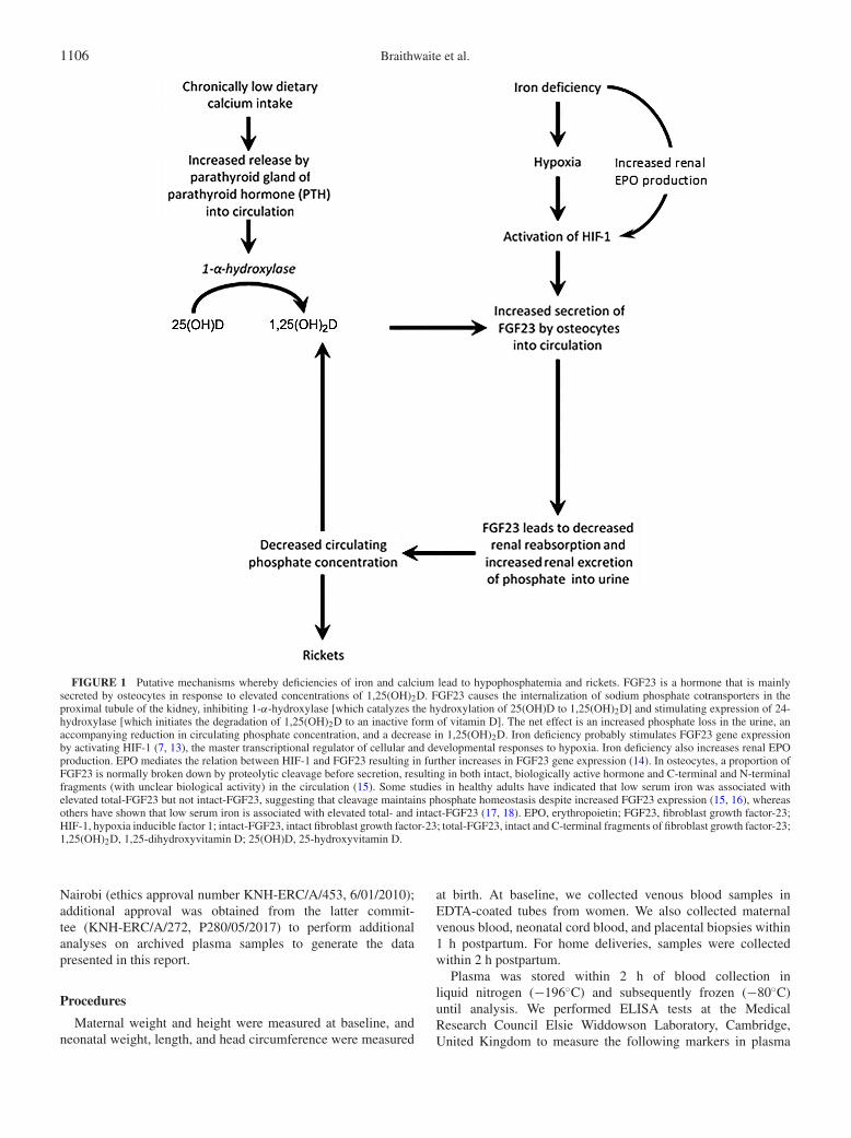
FIGURE 1 Putative mechanisms whereby deficiencies of iron and calcium lead to hypophosphatemia and rickets. FGF23 is a hormone that is mainlysecreted by osteocytes in response to elevated concentrations of 1,25(OH)2D. FGF23 causes the internalization of sodium phosphate cotransporters in theproximal tubule of the kidney, inhibiting 1-α-hydroxylase [which catalyzes the hydroxylation of 25(OH)D to 1,25(OH)2D] and stimulating expression of 24-hydroxylase [which initiates the degradation of 1,25(OH)2D to an inactive form of vitamin D]. The net effect is an increased phosphate loss in the urine, anaccompanying reduction in circulating phosphate concentration, and a decrease in 1,25(OH)2D. Iron deficiency probably stimulates FGF23 gene expressionby activating HIF-1 (7, 13), the master transcriptional regulator of cellular and developmental responses to hypoxia. Iron deficiency also increases renal EPOproduction. EPO mediates the relation between HIF-1 and FGF23 resulting in further increases in FGF23 gene expression (14). In osteocytes, a proportion ofFGF23 is normally broken down by proteolytic cleavage before secretion, resulting in both intact, biologically active hormone and C-terminal and N-terminalfragments (with unclear biological activity) in the circulation (15). Some studies in healthy adults have indicated that low serum iron was associated withelevated total-FGF23 but not intact-FGF23, suggesting that cleavage maintains phosphate homeostasis despite increased FGF23 expression (15, 16), whereasothers have shown that low serum iron is associated with elevated total- and intact-FGF23 (17, 18). EPO, erythropoietin; FGF23, fibroblast growth factor-23;HIF-1, hypoxia inducible factor 1; intact-FGF23, intact fibroblast growth factor-23; total-FGF23, intact and C-terminal fragments of fibroblast growth factor-23;1,25(OH)2D, 1,25-dihydroxyvitamin D; 25(OH)D, 25-hydroxyvitamin D.
Nairobi (ethics approval number KNH-ERC/A/453, 6/01/2010);additional approval was obtained from the latter commit-tee (KNH-ERC/A/272, P280/05/2017) to perform additionalanalyses on archived plasma samples to generate the datapresented in this report.
Procedures
Maternal weight and height were measured at baseline, andneonatal weight, length, and head circumference were measured
at birth. At baseline, we collected venous blood samples inEDTA-coated tubes from women. We also collected maternalvenous blood, neonatal cord blood, and placental biopsies within1 h postpartum. For home deliveries, samples were collectedwithin 2 h postpartum.
Plasma was stored within 2 h of blood collection inliquid nitrogen (−196◦C) and subsequently frozen (−80◦C)until analysis. We performed ELISA tests at the MedicalResearch Council Elsie Widdowson Laboratory, Cambridge,United Kingdom to measure the following markers in plasma
Effect of oral antenatal iron on FGF23 and bone 1107
collected at delivery: total-FGF23 using the C-terminal assay(60-6100) which detects both the C-terminal fragment and theintact hormone, and intact-FGF23 using the intact-FGF23 assay(60-6600) which detects only the intact form of the hormone(Immutopics); 1,25(OH)2D (the active metabolite of vitaminD; Immunodiagnostic Systems); β-C-terminal telopeptide (β-Crosslaps—a marker of bone resorption that is elevated inincreased bone resorption; Immunodiagnostic Systems); andhepcidin (a regulator of iron metabolism that is low in irondeficiency and raised by inflammation; Hepcidin-25, Bioac-tive). Hemoglobin concentration was measured in the field byphotometer (HemoCue 301, Radiometer) and plasma α1-acidglycoprotein (a marker of inflammation) and soluble transferrinreceptor (sTfR—a marker of iron status) were measured atMeander Medical Centre, Amersfoort, Netherlands (UniCel DxC880i analyzer, Beckman Coulter) as part of the original trial (12).Plasma concentrations of ferritin, C-reactive protein (CRP—a marker of inflammation), 25-hydroxyvitamin D [25(OH)D—marker of vitamin D status], total alkaline phosphatase, intactparathyroid hormone (PTH—a primary regulatory hormone ofcalcium that is elevated in calcium deficiency), cystatin C(a marker that is elevated in kidney dysfunction), and phos-phate were measured at Meander Medical Centre, Amersfoort,Netherlands (Architect c16000 and Architect i2000SR, Abbott).ELISA assay accuracy was monitored across the working rangeof assays using kit controls supplied by the manufacturers.The UK laboratory was accredited by the Vitamin D ExternalQuality Assessment Scheme (http://www.deqas.org/) and the UKNational External Quality Assessment Service (https://ukneqas.org.uk/) and the laboratory in the Netherlands by the ExternalQuality Assurance System (https://www.eurl-ar.eu/eqas.aspx)and German Society for Clinical Chemistry and LaboratoryMedicine (DGKL) (https://www.dgkl.de/en/). In addition, analiquot of a pooled plasma sample was assayed in each batchto monitor possible drift in measurements over time. Intra- andinterassay CVs were <7% and <6% for total-FGF23, intact-FGF23, β-Crosslaps, 1,25(OH)2D, 25(OH)D, PTH, ferritin, CRP,phosphate, total alkaline phosphatase, and cystatin C and <5%and <17% for hepcidin, respectively.
Estimated glomerular filtration rate (eGFR—a measure ofkidney glomerular function) was calculated using equationsapproved by the National Kidney Foundation (https://www.kidney.org/professionals/KDOQI/gfr)—the Chronic Kidney DiseaseEpidemiology Collaboration (CKD-EPI) cystatin C equationsfor those aged >18 y: eGFR, mL·min−1·1.73 m−2 = 123.9× [cystatin C (mg/L)/0.8]−1.328 × 0.996age and for those aged≤18 y: eGFR, mL·min−1·1.73 m−2 = 70.69 × [cystatin C(mg/L)]−0.931. At baseline, dipstick tests (Access Bio) wereused to detect histidine-rich protein-2 and lactate dehydrogenasespecific either to P. falciparum or to nonfalciparum human Plas-modium species (12). qPCR was used to detect P. falciparum–specific DNA in erythrocytes, and mothers were also testedfor HIV infection. HIV-infected mothers continued or wereoffered antiretroviral treatment as part of their standard clinicalcare.
Outcomes
The primary analysis concerned group differences in plasmaconcentrations of total-FGF23 and intact-FGF23 in maternal
blood samples at delivery and neonatal cord blood samples.As secondary outcomes, we studied other biomarkers of bonemetabolism and kidney function, namely, plasma concentrationsof phosphate, PTH, total alkaline phosphatase, β-Crosslaps,25(OH)D, 1,25(OH)2D, cystatin C, and eGFR.
Statistical analysis
Sample size requirements were calculated based on the effectof iron supplementation on Plasmodium infection risk (12) aspart of the original trial design; because they are not relevant tothe current study, they are not reported here. The current studyperformed analysis on all available plasma samples from mothersat birth (n = 433) and from infant cord blood (n = 414) atdelivery.
Anthropometric z scores for the neonates at birth werederived with Kenyan children as a reference (19). The followingdefinitions were used: prematurity–being born at or before37 completed weeks of gestation (<259 days of gestation ascalculated by early pregnancy ultrasound); anemia: hemoglobinconcentration < 110 g/L for pregnant women (19); iron defi-ciency (depleted iron stores)–plasma ferritin concentration <15μg/L for women and <12 μg/L for neonates (20); inflammation–plasma CRP > 10 mg/L (21); vitamin D insufficiency–25(OH)Dconcentration <50 nmol/L; and vitamin D deficiency–25(OH)Dconcentration <30 nmol/L (22). There are no establishedthresholds for intact-FGF23 or total-FGF23 concentrations or foreGFR in pregnancy and neonates, or for anemia in neonates, andso these variables were not dichotomized. Plasmodium infectionwas defined as past or present maternal infection assessed atparturition, regardless of species, as indicated by ≥1 positive testresults for the presence of Plasmodium lactate dehydrogenaseor P. falciparum–specific histidine-rich protein-2 in plasma orby placental histopathology or P. falciparum DNA in maternalerythrocytes from venous or placental blood by a PCR test.
Statistical analysis was performed using Stata version 16.0(StataCorp). We visually inspected histograms to assess the shapeof the distribution and to identify possible outliers. Skewed datawere normalized by log transformation as appropriate. Groupswere described using mean ± SD for normally distributed dataand geometric mean ± geometric SD (GSD) for log-transformeddata. GSD was calculated as the exponentiated SD of the log-transformed variable. It is a dimensionless factor that indicatesvariation that is equivalent to subtraction or addition of 1 SD on alog-transformed scale. For plasma CRP concentration at baseline,we computed descriptive statistics with a Tobit model to accountfor data being left-censored at the limit of quantification (1 mg/L).
Plasma concentrations of ferritin and soluble transferrinreceptor (sTfR) are affected by infection and inflammationindependently of iron status. We used multiple regression modelsto adjust the iron markers for such effects (SupplementalMethods). We used adjusted plasma iron markers to calculatethe body iron index, i.e., the ln of the ratio of the adjusted ferritinconcentration to the adjusted sTfR concentration. This indicatorhas been shown to be linearly associated with quantitativeestimates of the size of the body iron store in iron-replete adults,and with the size of the functional deficit that would need to becorrected before iron could again be accumulated in the store iniron-deficient individuals (23).
1108 Braithwaite et al.
Crude intervention effects on continuous outcomes wereestimated by simple linear regression. To assess the potentialrole of confounding due to imbalances in baseline variables,we used multiple fractional polynomial regression to estimateintervention effects adjusted for maternal characteristics as-sessed at randomization, i.e., hemoglobin concentration, bodyiron index, age, BMI, gestational age at delivery (calculatedfrom early pregnancy ultrasonography), parity, HIV infection,and Plasmodium infection. Both in simple linear regressionmodels and in fractional polynomial models, we accounted forheteroscedasticity of the error terms as appropriate (P valuesfor Breusch–Pagan/Cook–Weisberg tests < 0.05). Interventioneffects are reported as absolute differences in means for normallydistributed outcomes, or as relative differences in geometricmeans for log-transformed outcomes.
For unadjusted prevalence differences, we used Newcombe’smethod to estimate 95% CIs and “N−1” chi-square teststo compute P values (24). We used log-binomial regressionmodels (adjrr command in Stata package st0306.pkg) to estimateprevalence differences adjusted for baseline characteristics, andto estimate unadjusted prevalence differences when contingencytables contained cells with expected values <10.
In a preplanned analysis, we used multiple fractional poly-nomial regression analysis to explore to what extent ironstatus at baseline modified the magnitude of the effect ofiron supplementation on FGF23 and selected markers of bonemetabolism at delivery, anticipating that iron absorption andthus the response to administered iron would be larger in iron-deficient women than in their iron-replete peers. We examinedsuch effect modification with a single independent variable (bodyiron index) and 10 outcomes (see the Outcomes section), in bothmothers and neonates, which resulted in 20 analyses. We used themfpi procedure in Stata with the “flex(3)” specification to definethe flexibility of the main effects and interaction models withadjustment for potentially influential maternal characteristicsassessed at randomization, i.e., hemoglobin concentration, BMI,gestational age, parity, HIV infection, and Plasmodium infection.We used a nominal significance level of 0.05 for selection ofvariables and power functions; selection of linear, first-degree,or second-degree polynomials was based on the lowest value forAkaike’s information criterion. To check for possible overfittingin the interaction models, we examined to what extent possibletrends in intervention effects observed across quintiles of bodyiron index were consistent with effect modification as measuredby fractional polynomial regression.
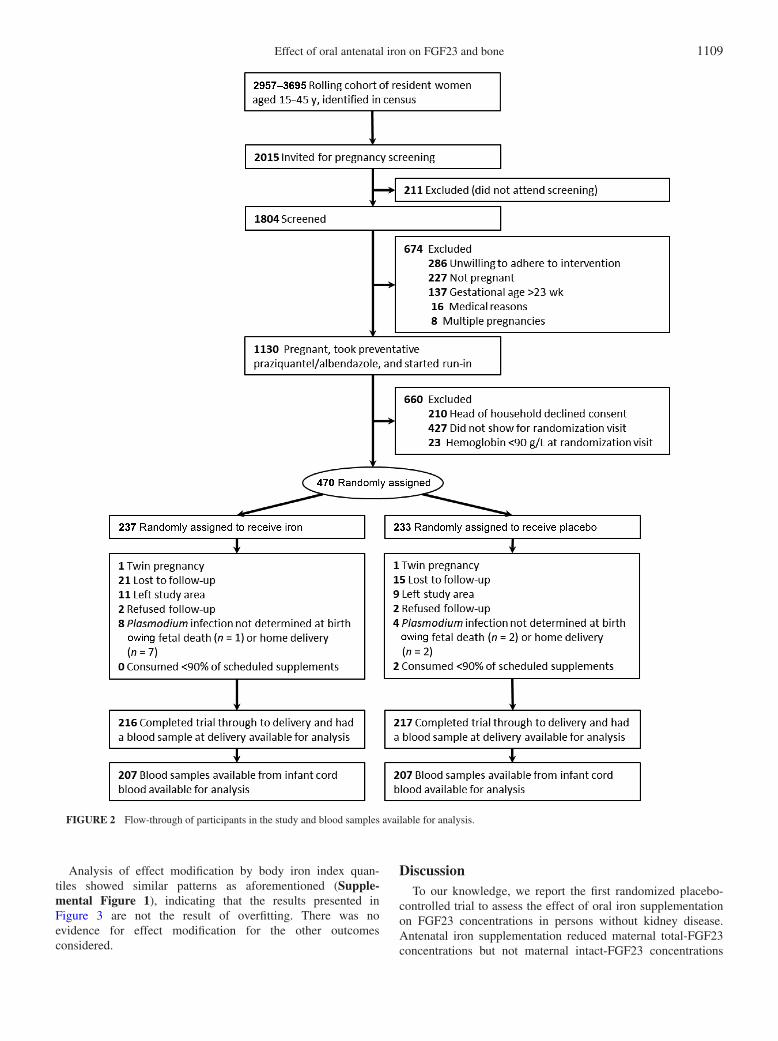
ResultsPrimary outcome data were available for 433 women (92%
of 470 women randomly assigned to the intervention). Samplesizes <433 reported in the subsequent sections were due to insuf-ficient plasma volume being available for biochemical analysis(Figure 2).
Maternal and neonatal biomarkers
At baseline, women had a mean age of 25.1 y, 16.2% werenulliparous, 38.3% were anemic, 99.1% were iron deficient(plasma ferritin concentration < 15 μg/L; values adjusted for
inflammation and Plasmodium infection), 34.4% had a current orrecent Plasmodium infection, and 21.7% were HIV-infected. Atbaseline, anthropometric indexes, iron status, and infection statuswere similar between intervention groups (Table 1).
Antenatal oral iron supplementation reduced the geometricmean maternal concentration of total-FGF23 (by −62.6%) andmean maternal concentration of 25(OH)D (by −6.1 nmol/L)(Table 2). It also improved maternal iron status as shown byan increased geometric mean concentration of hepcidin (by136.4%); increased mean concentration of hemoglobin (by 9.0g/L); increased geometric mean concentration of ferritin (by95.6%); and a reduced prevalence of anemia (by 29.1%) and irondeficiency (by 27.1%).
Antenatal oral iron supplementation reduced the geometricmean neonatal total-FGF23 concentration (by 15.2%), whereas itincreased the geometric mean neonatal intact-FGF23 concentra-tion (by 21.6%) (Table 2). It improved neonatal iron stores (meanferritin concentration increased by 23.3%) (Table 2) and otherneonatal outcomes (birth weight, length at birth, and gestationalduration increased by 141 g, 0.9 cm, and 3.4 d, respectively)described in our previous publication (12).
No marked group differences were seen in the other maternalor neonatal biomarkers of bone, kidney, or vitamin D metabolism.As might be expected with a randomized trial of this size,adjustment of treatment effects for baseline variables that wereconsidered a priori to be prognostic for outcome did not markedlychange effect estimates (Supplemental Table 1), showing thateffects are attributable to the intervention alone and not toselection bias.
Influence of baseline variables on intervention effects
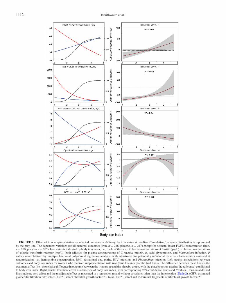
Figure 3 shows intervention × body iron index interactions forselected outcomes. In women who received placebo (left panels,red lines), low iron status at baseline (as indicated by body ironindex values) was associated with higher maternal concentrationsof total-FGF23, intact-FGF23, and cystatin C, as well as lowerneonatal intact-FGF23 concentration and lower maternal eGFR.Consistent with these observations, we found that antenatal oraliron supplementation reduced maternal total-FGF23 concentra-tions (overall: by 62.6%) (Table 2), with the magnitude of thiseffect being inversely associated with maternal iron status atbaseline (Figure 3, second right panel; P = 0.004). Similarly, wefound that antenatal oral iron supplementation increased neonatalintact-FGF23 concentration, with the magnitude of the effectbeing inversely associated with maternal iron status at baseline(P = 0.04).
For maternal intact-FGF23 concentration, maternal cystatinC concentration, and maternal eGFR, the effect of antenataliron supplementation also varied by initial maternal iron status(Figure 3, right panels), but cumulative frequency distributions(Figure 3, left panels, gray lines) indicated that most womenhad body iron index values around the cutoffs that deter-mined whether the effect of intervention was either positiveor negative (intact-FGF23, eGFR) or body iron index valueswithin the range of no effect (cystatin C). This may explainthe absence of marked effects in the unadjusted analysis(Table 2; also indicated in Figure 3, right panels, dashedlines).
Effect of oral antenatal iron on FGF23 and bone 1109
FIGURE 2 Flow-through of participants in the study and blood samples available for analysis.
Analysis of effect modification by body iron index quan-tiles showed similar patterns as aforementioned (Supple-mental Figure 1), indicating that the results presented inFigure 3 are not the result of overfitting. There was noevidence for effect modification for the other outcomesconsidered.
DiscussionTo our knowledge, we report the first randomized placebo-
controlled trial to assess the effect of oral iron supplementationon FGF23 concentrations in persons without kidney disease.Antenatal iron supplementation reduced maternal total-FGF23concentrations but not maternal intact-FGF23 concentrations
1110 Braithwaite et al.
TABLE 1 Maternal characteristics at baseline, by intervention group1
Characteristic Placebo (n = 217) Iron (n = 216)
Age, y 25.0 ± 6.1 25.1 ± 6.1Parity,2 n 2 (0–10) 2 (0–9)Nullipara 19.8 [43] 12.5 [27]Height, cm 162.4 ± 6.8 162.5 ± 5.9Weight, kg 57.4 ± 7.4 58.0 ± 7.5Gestational age at randomization,3 wk 17.5 ± 1.2 17.9 ± 1.2Hemoglobin concentration, g/L 112.4 ± 11.8 113.4 ± 10.7Anemia (hemoglobin concentration < 110 g/L) 40.5 [88] 36.1 [78]Plasma ferritin concentration,3 μg/L 15.9 ± 2.5 16.3 ± 2.4Iron deficiency (plasma ferritin concentration < 15 μg/L)
Without adjustment for inflammation and infection4 53.5 [116] 53.7 [116]With adjustment for inflammation and infection4 99.5 [216] 98.6 [213]
Plasma CRP concentration,3,5 mg/L 4.1 ± 3.72 4.0 ± 3.79Plasma AGP concentration, g/L 0.78 ± 0.27 0.76 ± 0.26Inflammation (plasma CRP ≥ 10 mg/L) 26.7 [58] 23.6 [51]Current Plasmodium infection 36.4 [79] 32.4 [70]HIV infection 21.7 [47] 21.8 [47]
1Values are mean ± SD or percentage [n] unless otherwise indicated. AGP, α1-acid glycoprotein; CRP,C-reactive protein.
2Median (range).3Geometric mean ± geometric SD.4Adjustment based on plasma CRP concentration, plasma AGP concentration, and Plasmodium infection (see
Supplemental Methods).5Based on log-transformed data and a Tobit model to account for left-censoring at the limit of quantification
(1 mg/L).
at delivery. The magnitude of the effect on maternal total-FGF23 concentrations was inversely associated with initialmaternal iron status. Iron supplementation decreased total-FGF23 concentration and increased intact-FGF23 concentrationin neonates, and there was evidence that the magnitude and thedirection of the intervention effect on neonatal intact-FGF23concentration depended on initial maternal iron status. Althoughthere was no measurable overall effect of iron supplementationon maternal intact-FGF23 concentration, maternal cystatin Cconcentration, and maternal eGFR, we found that initial maternaliron status may have influenced the direction and magnitude ofthese effects. Lastly, iron supplementation decreased maternal25(OH)D concentration, but there was no evidence that this effectdepended on initial maternal iron status (12).
The strengths of this study include its randomized design;large sample size; supervised administration of supplements withexcellent adherence (iron: 100%; placebo: 99.1%) (12); blindingof participants, field staff, and laboratory staff to the intervention;the use of a placebo as a comparator; and the inclusion of iron-deficient and anemic women in the trial. One study limitationis that we assessed intervention effects on multiple endpoints,which has the inherent risk of multiplicity. On the otherhand, effects on related outcomes can be mutually reinforcinginstead of undermining (25). This point is underscored by ourfinding that iron supplementation led to increased maternalferritin concentrations (Table 2), which lends credence to theconcurrent increase in maternal hemoglobin concentrations: bothfindings indicate that the intervention improved maternal ironstatus. Results of the intervention × baseline body iron indexinteractions should be interpreted with caution and considered asexploratory because of the potential instability of the functionsfound, and because further studies are needed to validate the
body iron index, adjusted for inflammation and infection, asan indicator of body iron status. In addition, in light of thedecreases in 25(OH)D concentrations and the known effects ofiron supplements on intestinal calcium absorption, it is likely thatiron supplementation affects calcium metabolism. This findingcould not be further investigated because blood samples werecollected in EDTA-coated tubes which inhibits accurate calciummeasurement. Plasma erythropoietin (EPO) concentration wasnot measured owing to constraints in available plasma volumeand so could not be investigated in the causal pathway betweeniron supplementation and total-FGF23 reduction. An additionallimitation is that certain characteristics that may have affected theoutcome were not measured at baseline. However, with samplesizes >200/group, simple randomization normally yields onlymild disparities in sample sizes between groups (26). Thus,it is unlikely that group imbalances in prognostic variables atbaseline led to substantial selection bias in our effect estimates.The marked reduction in maternal and neonatal total-FGF23concentrations after antenatal iron supplementation is in keepingwith previous studies (6, 7) and is likely caused by a reversalof hypoxia and/or EPO-driven FGF23 gene expression inosteocytes that occurs in iron deficiency (7). Consistent withthis hypothesis, we found that the reduction in total-FGF23was most pronounced in mothers with the lowest body ironstatus.
Our findings corroborate emerging evidence that orallyadministered iron does not normally lead to measurable changesin maternal plasma concentrations of intact-FGF23 or phosphate(6). Iron deficiency results in increased concentrations ofhypoxia-inducible factor 1α, which stimulates FGF23 transcrip-tion through increased EPO production and perhaps also bydirectly binding to the FGF23 promoter (15). The resulting
Effect of oral antenatal iron on FGF23 and bone 1111
TABLE 2 Effect of antenatal iron supplementation on maternal and neonatal biomarkers at birth1
Maternal blood Neonatal (cord) blood
Biomarker concentration or status n Mean ± SD Effect (95% CI) P value n Mean ± SD Effect (95% CI) P value
Total-FGF23,2 RU/mLPlacebo 217 370.3 ± 3.62 Reference 207 647.3 ± 2.29 ReferenceIron 216 138.4 ± 3.09 − 62.6% (−70.3%, −53.0%) <0.0005 207 548.6 ± 2.50 − 15.2% (−28.4%, 0.3%) 0.06
Intact-FGF23,2 ng/LPlacebo 217 41.3 ± 1.54 Reference 205 6.1 ± 2.65 ReferenceIron 216 39.4 ± 1.46 − 4.5% (−11.5%, 3.1%) 0.24 200 7.4 ± 2.47 21.6% (1.2%, 46.1%) 0.04
25-hydroxyvitamin D, nmol/LPlacebo 217 99.6 ± 28.7 Reference 204 63.1 ± 19.3 ReferenceIron 216 93.5 ± 23.0 − 6.1 (−11.0, −1.2) 0.01 206 61.5 ± 17.7 − 1.6 (−5.2, 2.0) 0.37
Inadequate 25-hydroxyvitamin D (<50 nmol/L)Placebo 217 2.8 [6] Reference 204 23.5 [48] ReferenceIron 216 3.7 [8] 0.9% (−3.0%, 5.0%) 0.96 206 25.2 [52] − 1.7% (−10.0%, 6.6%) 0.97
1,25-dihydroxyvitamin D, pmol/LPlacebo 203 351.0 ± 78.2 Reference 195 203.0 ± 62.9 ReferenceIron 200 351.1 ± 84.4 0.1 (−15.8, 16.0) 0.98 195 203.9 ± 52.7 0.8 (−10.6, 12.4) 0.88
Parathyroid hormone,2 pmol/LPlacebo 217 4.0 ± 1.94 Reference 205 0.5 ± 2.69 ReferenceIron 216 3.9 ± 1.92 − 2.0% (−13.4%, 10.9%) 0.75 206 0.5 ± 2.97 − 3.3% (−21.0%, 18.3%) 0.74
Phosphate, mmol/LPlacebo 217 1.27 ± 0.32 Reference 205 2.05 ± 0.64 ReferenceIron 216 1.29 ± 0.24 0.03 (−0.03, 0.08) 0.30 206 2.11 ± 0.65 0.06 (−0.07, 0.18) 0.38
Total alkaline phosphatase,2 U/LPlacebo 217 102.3 ± 1.88 Reference 205 17.3 ± 1.74 ReferenceIron 215 96.1 ± 1.86 − 6.0% (−16.5%, 5.7%) 0.30 206 17.6 ± 2.22 2.6% (−10.2%, 10.2%) 0.77
β-Crosslaps,2 μg/LPlacebo 210 0.6 ± 1.81 Reference 206 0.8 ± 1.22 ReferenceIron 209 0.7 ± 1.70 7.2% (−3.7%, 19.4%) 0.20 206 0.8 ± 1.27 1.5% (−2.7%, 5.9%) 0.48
Cystatin C, mg/LPlacebo 217 1.26 ± 0.29 Reference 205 2.06 ± 0.40 ReferenceIron 216 1.25 ± 0.30 0.00 (−0.06, 0.05) 0.90 206 2.06 ± 0.37 0.00 (−0.07, 0.08) 0.97
eGFR,2 mL · min−1 · 1.73 m−2
Placebo 217 62.1 ± 1.32 Reference 205 36.6 ± 1.18 ReferenceIron 216 62.4 ± 1.32 0.5% (−4.6%, 5.8%) 0.86 206 36.6 ± 1.19 0.0% (−3.3%, 3.4%) 1.00
Hepcidin,2 μg/LPlacebo 217 1.9 ± 3.41 Reference 207 8.1 ± 2.30 ReferenceIron 216 4.4 ± 3.69 136.4% (86.1%, 200.3%) <0.0005 207 9.1 ± 2.24 12.2% (−4.3%, 31.4%) 0.16
Hemoglobin, g/LPlacebo 214 111.6 ± 19.0 Reference 209 150.6 ± 21.0 ReferenceIron 215 120.7 ± 16.4 9.0 (5.7, 12.4) <0.0005 206 153.8 ± 21.7 3.2 (−1.0, 7.3) 0.13
Anemia (hemoglobin < 110 g/L for mothers)Placebo 214 50.5 [108] Reference —Iron 215 21.4 [46] − 29.1% (−37.4%, −20.1%) <0.001 —3
Ferritin,2 μg/LPlacebo 217 19.0 ± 2.61 Reference 205 103.0 ± 2.11 ReferenceIron 216 37.1 ± 2.55 95.6% (63.6%, 133.9%) <0.0005 206 127.0 ± 2.14 23.3% (6.6%, 42.7%) 0.005
Iron deficiency (ferritin ≤ 15 μg/L mothers and <12 μg/L for neonates)Placebo 217 43.3 [94] Reference 205 0.9 [2] ReferenceIron 216 16.2 [35] − 27.1% (−35.0%, −18.7%) <0.001 205 0.0 [0] —4 ND
CRP, mg/LPlacebo 217 6.7 ± 3.93 Reference 131 0.2 [0.2–0.3] ReferenceIron 216 7.7 ± 3.69 16.4% (−9.6%, 49.8%) 0.24 128 0.2 [0.2–0.3] —5 0.62
Inflammation (CRP > 10 mg/L)Placebo 217 38.2 [83] Reference 131 5.3 [7] ReferenceIron 216 39.8 [86] 1.6% (−7.6%, 10.7%) 0.74 128 1.6 [2] − 3.8% (−8.2%, 0.6%) 0.09
1Values are mean ± SD or % [n] unless indicated otherwise. Effects are reported as absolute difference in means, relative difference (%) in geometric means, or difference in prevalence,with placebo as the reference group. Group estimates are medians [IQRs]; group differences in distributions were compared by independent-samples Mann–Whitney U test, which yields a Pvalue only. For continuous outcomes, P values were obtained by simple linear regression analysis, accounting for heteroscedasticity. For binary outcomes, we used Newcombe’s method toestimate 95% CIs and “N−1” chi-square tests to compute P values. We used log-binomial regression models to estimate prevalence differences when contingency tables contained cells withexpected values <10. P values indicate the probability of data occurring as observed or being more extreme than observed under the assumption of no effect, i.e., outcomes being identicallydistributed for groups that received supplementation with either placebo or iron. β-Crosslaps, β-C-terminal telopeptide; CRP, C-reactive protein; eGFR, estimated glomerular filtration rate;intact-FGF23, intact fibroblast growth factor-23; ND, not determined; total-FGF23, intact and C-terminal fragments of fibroblast growth factor-23.
2Geometric mean ± geometric SD.3Effects on anemia were not calculated in neonates because anemia is poorly defined in this group.4Not determined as there were too few cases of iron deficiency to allow analyses.5Plasma CRP concentration in cord blood was highly skewed and could not be normalized by log transformation.
increase in FGF23 production does not normally result inmaternal hypophosphatemia; excess FGF23 is proteolyticallycleaved within osteocytes, resulting in increased secretion of C-terminal FGF23 fragments into the circulation (which is reflected
in total-FGF23 concentrations), whereas concentrations of thebiologically active intact hormone remain largely unaffected(27). By contrast, some intravenous iron preparations (e.g.,ferric carboxymaltose, saccharated ferric oxide) appear to
1112 Braithwaite et al.
FIGURE 3 Effect of iron supplementation on selected outcomes at delivery, by iron status at baseline. Cumulative frequency distribution is representedby the gray line. The dependent variables are all maternal outcomes (iron, n = 216; placebo, n = 217) except for neonatal intact-FGF23 concentration (iron,n = 200; placebo, n = 205). Iron status is indicated by body iron index, i.e., the ln of the ratio of plasma concentrations of ferritin (μg/L) to plasma concentrationsof soluble transferrin receptor (mg/L), both adjusted for plasma concentrations of C-reactive protein, α1-acid glycoprotein, and Plasmodium infection. Pvalues were obtained by multiple fractional polynomial regression analysis, with adjustment for potentially influential maternal characteristics assessed atrandomization, i.e., hemoglobin concentration, BMI, gestational age, parity, HIV infection, and Plasmodium infection. Left panels: associations betweenoutcomes and body iron index for women who received supplementation with iron (blue lines) or placebo (red lines). The difference between these lines is thetreatment effect (i.e., the relative difference in outcome between the iron group and the placebo group, with the placebo group used as the reference) conditionalto body iron index. Right panels: treatment effect as a function of body iron index, with corresponding 95% confidence bands and P values. Horizontal dashedlines indicate zero effect and the unadjusted effect as measured in a regression model without covariates other than the intervention (Table 2). eGFR, estimatedglomerular filtration rate; intact-FGF23, intact fibroblast growth factor-23; total-FGF23, intact and C-terminal fragments of fibroblast growth factor-23.
Effect of oral antenatal iron on FGF23 and bone 1113
block cleavage of FGF23, thus leading to FGF23-mediatedphosphaturia and a concomitant decrease in circulating phosphateconcentrations (15). Although hypophosphatemia due to single-dose administration of intravenous iron is often transient andasymptomatic, it can persist for >1 mo, and some patientsdevelop severe and symptomatic hypophosphatemia with seriousmusculoskeletal complications, including osteomalacia, fragilityfractures, and hypoxemia (28).
In the present study, the effect of the iron interventionon maternal intact-FGF23 concentration varied with baselineiron status (Figure 3), suggesting that FGF23 transcriptionand cleavage are uncoupled in women with poor iron status.In our study, this occurred in ∼40% of women (the lowest2 quintiles; see Supplemental Figure 1). Chronically elevatedconcentrations of intact-FGF23 result in phosphaturia andeventually hypophosphatemia, which in turn can lead to seriousmusculoskeletal complications, including rickets in childrenand osteomalacia fragility fractures in adults. However, overallwe found no evidence that iron affected plasma phosphateconcentrations or bone turnover, although the intervention period(∼5 mo) may have been insufficient to detect changes in thesemarkers. Further studies are needed to discount the possibilitythat moderate to severe iron deficiency causes hyperphosphaturiaand hypophosphatemia in pregnant women. Special caution iswarranted when prescribing intravenous ferric carboxymaltose toindividuals with iron deficiency.
Our findings suggest that iron supplementation leads toincreased glomerular filtration rates in women with poor ironstatus. This discovery is in agreement with a Mendelianrandomization study that showed serum iron concentration to beassociated with glomerular filtration rates (29). In a randomizedcontrolled trial in Bangladesh, children whose mothers hadantenatally received a daily oral supplement containing 60 mgFe/d had higher glomerular filtration rates at 4.5 y of age thanthose whose mothers received 30 mg Fe/d (30). Children witha history of rickets had a reduced glomerular filtration rate asestimated from plasma cystatin C concentration (4). Further workis needed to elucidate a possible role of kidney function along thecausal pathway between iron and rickets.
In a prospective cohort study, increased total-FGF23 concen-tration was associated with an increase in all-cause mortalityin a general population of older US adults (31). Although thisassociation may have been in part confounded by the effectof inflammation on FGF23 expression, it is possible that thereduction that is seen in maternal and infant total-FGF23 afterantenatal iron supplementation confers a general health benefit.
Intriguingly, and in contrast to effects observed in pregnantwomen, antenatal iron supplementation led to increased intact-FGF23 concentrations in neonates, which conversely suggeststhat maternal iron deficiency during pregnancy leads to decreasedneonatal intact-FGF23 concentrations, without an apparent effecton neonatal serum phosphate concentration. These findingscontradict reports from a study in neonatal pups born to femalewild-type mice which were fed an iron-deficient diet duringthe third week of pregnancy (corresponding to the human thirdtrimester) and during lactation, and which developed increasedconcentrations of both intact-FGF23 and total-FGF23, as wellas decreased phosphate concentrations (7). From that report (7),however, it is not clear whether these outcomes were measuredimmediately after birth.
In an earlier prospective cohort study, poor maternal iron statusduring pregnancy was associated with a higher total-FGF23concentration but similar intact-FGF23 concentrations in infantsand young children (6). Unfortunately, however, these markerswere not assessed at delivery. In contrast to the critical role thatFGF23 plays in regulating phosphorus and skeletal metabolism inadults and children, murine studies suggest that FGF23 may notbe required to regulate fetal phosphorus homeostasis, placentalphosphorus transport, or skeletal development, perhaps becausethe placenta’s role to actively transport phosphorus and otherminerals from the maternal circulation is independent of FGF23(32). In mice, fetal phosphate concentrations are relatively highand decrease within a few days after birth, concurrently withthe cessation of placental phosphate influx. Within hours to daysafter loss of the placental phosphorus pump, neonatal FGF23becomes an important regulator of renal phosphorus excretionand intestinal phosphorus absorption (32). It is possible that theeffects of antenatal iron supplementation on offspring FGF23and phosphate metabolism may only become apparent later ininfancy, and not at birth and so were not fully captured in thisstudy.
Antenatal iron supplementation also had an effect on vitaminD metabolism by decreasing maternal 25(OH)D concentration.Emerging evidence has found a biological link between vitaminD and iron metabolism. The vitamin D response element hasbeen identified on HAMP (the gene encoding hepcidin), and1,25(OH)2D has been shown to suppress hepcidin expressionand thereby increase the amount of iron absorbed from thediet and released from stores (33). It is possible that there is anegative feedback loop whereby when hepcidin concentrationsare high (in iron repletion, or with antenatal iron supplementationor with inflammation), 25(OH)D concentration decreases. Analternative pathway for decreases in maternal 25(OH)D isthrough direct effects of FGF23 on vitamin D metabolism.Higher FGF23 concentrations are known to downregulateCytochrome P450 Family 27 Subfamily B Member 1 (CYP27B1)expression and upregulate Cytochrome P450 Family 24 Sub-family A Member 1 (CYP24A1) expression with the potentialto lower 1,25(OH)2D and raise 24,25(OH)2D, respectively(34).
We conclude that antenatal oral iron supplementation canredress perturbances in maternal and neonatal FGF23 metabolisminduced by maternal iron deficiency during pregnancy. Thissuggests the possibility that iron supplementation could helpprevent rickets and osteomalacia in individuals with an impairedability to regulate FGF23 concentration.
We thank all of the study participants and staff involved in the trial, thelaboratory staff at the Meander Medical Centre and Medical Research CouncilElsie Widdowson laboratory—in particular Mrs. J Bennet, Mrs. A Laidlaw,and Dr. S Nigdikar, and Mr. Philip Ndemwa for his assistance in obtainingethical approval.
The authors’ responsibilities were as follows—MNM and HV: designedthe original trial; MNM, PEA, and HV: conducted the original trial; VSBand HV: designed the research and take responsibility for the integrity of thedata analysis; VSB, AYD, and HV: conducted the research; VSB, AP, AYD,HV, and MNM: analyzed the data; VSB, AP, KSJ, AMP, and HV: interpretedthe data; VSB, KSJ, AMP, and HV: wrote the paper; and all authors:read and approved the final manuscript. The authors report no conflicts ofinterest.
1114 Braithwaite et al.
Data AvailabilityData described in the article may be made available upon
request pending application and approval.
References1. Prentice A. Nutritional rickets around the world. J Steroid Biochem Mol
Biol 2013;136:201–6.2. Pool LR, Wolf M. FGF23 and nutritional metabolism. Annu Rev Nutr
2017;37:247–68.3. Prentice A, Ceesay M, Nigdikar S, Allen SJ, Pettifor JM. FGF23 is
elevated in Gambian children with rickets. Bone 2008;42:788–97.4. Braithwaite V, Jarjou LMA, Goldberg GR, Prentice A. Iron status and
fibroblast growth factor-23 in Gambian children. Bone 2012;50:1351–6.
5. Braithwaite V, Prentice A, Doherty C, Prentice A. FGF23 is correlatedwith iron status but not with inflammation and decreases after ironsupplementation: a supplementation study. Int J Pediatr Endocrinol2012;2012(1):27.
6. Braithwaite VS, Prentice A, Darboe MK, Prentice AM, Moore SE. Theeffects of maternal iron deficiency on infant fibroblast growth factor-23and mineral metabolism. Bone 2016;83:1–8.
7. Clinkenbeard EL, Farrow EG, Summers LJ, Cass TA, Roberts JL, BaytCA, Lahm T, Albrecht M, Allen MR, Peacock M, et al. Neonataliron deficiency causes abnormal phosphate metabolism by elevatingFGF23 in normal and ADHR mice. J Bone Miner Res 2014;29(2):361–9.
8. Olausson H, Goldberg GR, Laskey MA, Schoenmakers I, Jarjou LM,Prentice A. Calcium economy in human pregnancy and lactation. NutrRes Rev 2012;25(1):40–67.
9. Berndt TJ, Craig TA, McCormick DJ, Lanske B, Sitara D, RazzaqueMS, Pragnell M, Bowe AE, O’Brien SP, Schiavi SC, et al. Biologicalactivity of FGF-23 fragments. Pflugers Arch 2007;454(4):615–23.
10. Goetz R, Nakada Y, Hu MC, Kurosu H, Wang L, Nakatani T, Shi M,Eliseenkova AV, Razzaque MS, Moe OW, et al. Isolated C-terminal tailof FGF23 alleviates hypophosphatemia by inhibiting FGF23-FGFR-Klotho complex formation. Proc Natl Acad Sci U S A 2010;107(1):407–12.
11. Mwangi MN, Prentice AM, Verhoef H. Safety and benefits of antenataloral iron supplementation in low-income countries: a review. Br JHaematol 2017;177(6):884–95.
12. Mwangi MN, Roth JM, Smit MR, Trijsburg L, Mwangi AM, DemirAY, Wielders JP, Mens PF, Verweij JJ, Cox SE, et al. Effect of dailyantenatal iron supplementation on Plasmodium infection in Kenyanwomen: a randomized clinical trial. JAMA 2015;314(10):1009–20.
13. Farrow EG, Yu X, Summers LJ, Davis SI, Fleet JC, Allen MR, RoblingAG, Stayrook KR, Jideonwo V, Magers MJ, et al. Iron deficiency drivesan autosomal dominant hypophosphatemic rickets (ADHR) phenotypein fibroblast growth factor-23 (Fgf23) knock-in mice. Proc Natl AcadSci U S A 2011;108(46):E1146–E55.
14. David V, Francis C, Babitt JL. Ironing out the cross talk between FGF23and inflammation. Am J Physiol Renal Physiol 2017;312(1):F1–8.
15. Imel EA, Peacock M, Gray AK, Padgett LR, Hui SL, Econs MJ.Iron modifies plasma FGF23 differently in autosomal dominanthypophosphatemic rickets and healthy humans. J Clin EndocrinolMetab 2011;96(11):3541–9.
16. Wolf M, White KE. Coupling fibroblast growth factor 23 productionand cleavage: iron deficiency, rickets, and kidney disease. Curr OpinNephrol Hypertens 2014;23(4):411–19.
17. Lewerin C, Ljunggren O, Nilsson-Ehle H, Karlsson MK, Herlitz H,Lorentzon M, Ohlsson C, Mellström D. Low serum iron is associatedwith high serum intact FGF23 in elderly men: the Swedish MrOS study.Bone 2017;98:1–8.
18. Bozentowicz-Wikarek M, Kocelak P, Owczarek A, Olszanecka-Glinianowicz M, Mossakowska M, Skalska A, Więcek A, ChudekJ. Plasma fibroblast growth factor 23 concentration and iron status.Does the relationship exist in the elderly population? Clin Biochem2015;48(6):431–6.
19. WHO. Haemoglobin concentrations for the diagnosis of anaemia andassessment of severity. Vitamin and Mineral Nutrition InformationSystem [Internet]. Geneva, Switzerland: World Health Organization;2011, [accessed 2020 August 1]. Available from: http://www.who.int/vmnis/indicators/haemoglobin.
20. WHO. Serum ferritin concentrations for the assessment of iron statusand iron deficiency in populations [Internet]. Geneva, Switzerland:World Health Organization; 2011, [accessed 2020 August 1]. Availablefrom: http://www.who.int/vmnis/indicators/serum_ferritin.
21. Nielsen FR, Bek KM, Rasmussen PE, Qvist I, Tobiassen M. C-reactiveprotein during normal pregnancy. Eur J Obstet Gynecol Reprod Biol1990;35(1):23–7.
22. Roth DE, Abrams SA, Aloia J, Bergeron G, Bourassa MW, BrownKH, Calvo MS, Cashman KD, Combs G, De-Regil LM, et al. Globalprevalence and disease burden of vitamin D deficiency: a roadmapfor action in low- and middle-income countries. Ann N Y Acad Sci2018;1430(1):44–79.
23. Skikne BS, Flowers CH, Cook JD. Serum transferrin receptor: aquantitative measure of tissue iron deficiency. Blood 1990;75(9):1870–6.
24. Richardson JTE. The analysis of 2 × 2 contingency tables—yet again.Statist Med 2011;30(8):890.
25. Schulz KF, Grimes DA. Multiplicity in randomised trials I: endpointsand treatments. Lancet 2005;365(9470):1591–5.
26. Schulz KF, Grimes DA. Unequal group sizes in randomised trials:guarding against guessing. Lancet 2002;359(9310):966–70.
27. Wheeler JA, Clinkenbeard EL. Regulation of fibroblast growthfactor 23 by iron, EPO, and HIF. Curr Mol Biol Rep 2019;5(1):8–17.
28. Wolf M, Chertow GM, Macdougall IC, Kaper R, Krop J, Strauss W.Randomized trial of intravenous iron-induced hypophosphatemia. JCIInsight 2018;3(23):e124486.
29. Del Greco MF, Foco L, Pichler I, Eller P, Eller K, BenyaminB, Whitfield JB; Genetics of Iron Status Consortium; CKDGenConsortium, Pramstaller PP, et al. Serum iron level and kidneyfunction: a Mendelian randomization study. Nephrol Dial Transplant2017;32(2):273–8.
30. Hawkesworth S, Wagatsuma Y, Kahn AI, Hawlader MD, FulfordAJ, Arifeen SE, Persson LA, Moore SE. Combined food andmicronutrient supplements during pregnancy have limited impact onchild blood pressure and kidney function in rural Bangladesh. J Nutr2013;143(5):728–34.
31. Souma N, Isakova T, Lipiszko D, Sacco RL, Elkind MS, DeRosaJT, Silverberg SJ, Mendez AJ, Dong C, Wright CB, et al.Fibroblast growth factor 23 and cause-specific mortality in the generalpopulation: the Northern Manhattan Study. J Clin Endocrinol Metab2016;101(10):3779–86.
32. Ma Y, Kirby BJ, Fairbridge NA, Karaplis AC, Lanske B, KovacsCS. FGF23 is not required to regulate fetal phosphorus metabolismbut exerts effects within 12 hours after birth. Endocrinology2017;158(2):252–63.
33. Smith EM, Alvarez JA, Kearns MD, Hao L, Sloan JH, Konrad RJ,Ziegler TR, Zughaier SM, Tangpricha V. High-dose vitamin D3 reducescirculating hepcidin concentrations: a pilot, randomized, double-blind,placebo-controlled trial in healthy adults. Clin Nutr 2017;36(4):980–5.
34. Dusso AS, Rodriguez M. Enhanced induction of Cyp24a1 by FGF23 butlow serum 24,25-dihydroxyvitamin D in CKD: implications for therapy.Kidney Int 2012;82(10):1046–9.
Recommended


![MINISTRY OF HEALTH - Home | K4Health · IFA supplementation for pregnant women in the country is one of the routine services provided within Focused Antenatal Care [FANC]. It is important](https://img.dokumen.tips/doc/110x75/5be4ebab09d3f28a428b5991/ministry-of-health-home-k4health-ifa-supplementation-for-pregnant-women.jpg)