An Adrenaline (and Gold?) Rush for the GPCRCommunityKendall J. Blumer†,* and Jeremy Thorner‡,*†Department of Cell Biology and Physiology, Washington University School of Medicine, St. Louis, Missouri 63110 and‡Department of Molecular and Cell Biology, University of California, Berkeley, California 94720
I In 1823, the music world was as-tounded by the publication of Ludwigvan Beethoven’s monumental 33 Varia-
tions on a Waltz by Anton Diabelli. So it is to-day, eight years after Palczewski et al. un-veiled the structure of the first G-protein-coupled receptor (GPCR), retinal rhodopsin(reviewed in ref 1), that we are now dazzledagain by the beauty of the first variation onthe GPCR theme revealed by publication ofcrystal structures of the �-2-adrenergic re-ceptor (�2AR) (2–4), which responds to thecatecholamine neurotransmitter, epineph-rine (formerly adrenaline). What do the�2AR structures solved by Kobilka, Weis,Stevens, and their colleagues tell us that wedid not know from rhodopsin? Much as thecollection of Beethoven’s Diabelli Variationsillustrated the depth of his genius, the �2ARstructures provide important insights thatboth reinforce our understanding of coremechanistic themes and illuminate the un-derlying basis for the astonishing functionaldiversity displayed by the GPCR family, thelargest and most clinically important class ofreceptors in the eukaryotic kingdom (5).While intrinsically beautiful, the structuralprinciples now established also hold practi-cal promise for the design of new drugs tar-geted to specific GPCRs.
Similarities and differences between rho-dopsin and �2AR biology make these recep-tors ideal for structural comparison. Like allvertebrate photoreceptors, rhodopsin con-tains a covalently linked chromophore (reti-nal) that undergoes light-driven cis–trans
isomerization (1). In the dark, cis-retinal sta-bilizes rhodopsin in its inactive conforma-tion, completely suppressing spontaneousactivity. Upon photon absorption, retinalisomerizes to the trans form, releasing theconstraints that hold rhodopsin in the inac-tive state and stabilizing its active conforma-tion, producing a biological response withinmilliseconds. In contrast, like essentially allother GPCRs, �2AR binds a diffusible ligandnoncovalently, producing biological re-sponses on a slower time scale (seconds-to-minutes) (6). Despite such differences, rho-dopsin and �2AR share with all GPCRs acommon mechanism of action: catalyzingGDP�GTP exchange on the �-subunit of amembrane-bound receptor-associated(“coupled”) heterotrimeric G protein. Thus,a central challenge has been to determinethe structural changes evoked by photonabsorption or by binding of a specific diffus-ible ligand and how they alter receptorconformation and bring about G proteinactivation.
Because most GPCRs are inherently con-formationally flexible (7), especially whenextracted with detergent from their nativeplasma membrane environment, and be-cause �2AR in particular has limited polarsurfaces available to form crystal contacts(6), determining its 3D structure by proteincrystallography presented some enormoustechnical challenges. Kobilka, Weis, andStevens have solved these problems usingtactics that represent a tour de force in thebiochemistry, engineering, and crystallogra-
*Corresponding authors,[email protected],[email protected].
Published online December 21, 2007
10.1021/cb7002412 CCC: $37.00
© 2007 American Chemical Society
ABSTRACT G-Protein-coupled receptors areone of the largest protein families found in meta-zoans. Using several novel strategies, the firstatomic resolution structures of a receptor that isactivated by a diffusible ligand have beendetermined.
Point ofVIEW
www.acschemicalbiology.org VOL.2 NO.12 • ACS CHEMICAL BIOLOGY 783
phy of an integral membrane protein. Ineach of the two �2AR structures (3, 4) the re-ceptor is bound to a partial inverse agonist(carazolol), stabilizing an inactive conforma-tion. To provide further stability and in-crease polar surface for crystallization, Ko-bilka and Weis bound a monoclonalantibody to the third cytoplasmic loop ofthe receptor (3, 8). In a second complemen-tary approach, Kobilka, Weis, and Stevensreplaced part of the third cytoplasmic loopwith T4 lysozyme (2, 4). Given the potentialfor introducing non-native perturbations thatmight have arisen from the use of thesestrategies and the other extraordinary mea-sures (e.g., necessity for cholesterol) thatwere used for crystallization, it is importantto emphasize and rather reassuring that thedetails of the two structures are remarkablycongruent, an indication that the salientstructural features of �2AR have been pre-served. Nonetheless, even though theiroverall folds are quite similar, more of thetransmembrane helices and loops of the re-ceptor were resolved and the bound ligandwas more clearly visible in the structure ofthe �2AR-T4 lysozyme chimera than in thestructure of native �2AR that was immobi-
lized by binding of a Fab antibody fragmentdirected against the third intracellular loop.
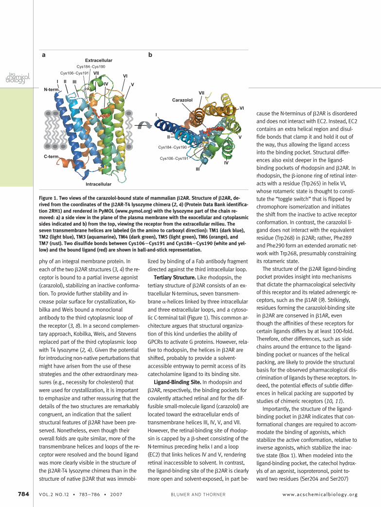
Tertiary Structure. Like rhodopsin, thetertiary structure of �2AR consists of an ex-tracellular N-terminus, seven transmem-brane �-helices linked by three intracellularand three extracellular loops, and a cytoso-lic C-terminal tail (Figure 1). This common ar-chitecture argues that structural organiza-tion of this kind underlies the ability ofGPCRs to activate G proteins. However, rela-tive to rhodopsin, the helices in �2AR areshifted, probably to provide a solvent-accessible entryway to permit access of itscatecholamine ligand to its binding site.
Ligand-Binding Site. In rhodopsin and�2AR, respectively, the binding pockets forcovalently attached retinal and for the dif-fusible small-molecule ligand (carazolol) arelocated toward the extracellular ends oftransmembrane helices III, IV, V, and VII.However, the retinal-binding site of rhodop-sin is capped by a �-sheet consisting of theN-terminus preceding helix I and a loop(EC2) that links helices IV and V, renderingretinal inaccessible to solvent. In contrast,the ligand-binding site of the �2AR is clearlymore open and solvent-exposed, in part be-
cause the N-terminus of �2AR is disorderedand does not interact with EC2. Instead, EC2contains an extra helical region and disul-fide bonds that clamp it and hold it out ofthe way, thus allowing the ligand accessinto the binding pocket. Structural differ-ences also exist deeper in the ligand-binding pockets of rhodopsin and �2AR. Inrhodopsin, the �-ionone ring of retinal inter-acts with a residue (Trp265) in helix VI,whose rotameric state is thought to consti-tute the “toggle switch” that is flipped bychromophore isomerization and initiatesthe shift from the inactive to active receptorconformation. In contrast, the carazolol li-gand does not interact with the equivalentresidue (Trp268) in �2AR; rather, Phe289and Phe290 form an extended aromatic net-work with Trp268, presumably constrainingits rotameric state.
The structure of the �2AR ligand-bindingpocket provides insight into mechanismsthat dictate the pharmacological selectivityof this receptor and its related adrenergic re-ceptors, such as the �1AR (9). Strikingly,residues forming the carazolol-binding sitein �2AR are conserved in �1AR, eventhough the affinities of these receptors forcertain ligands differs by at least 100-fold.Therefore, other differences, such as sidechains around the entrance to the ligand-binding pocket or nuances of the helicalpacking, are likely to provide the structuralbasis for the observed pharmacological dis-crimination of ligands by these receptors. In-deed, the potential effects of subtle differ-ences in helical packing are supported bystudies of chimeric receptors (10, 11).
Importantly, the structure of the ligand-binding pocket in �2AR indicates that con-formational changes are required to accom-modate the binding of agonists, whichstabilize the active conformation, relative toinverse agonists, which stabilize the inac-tive state (Box 1). When modeled into theligand-binding pocket, the catechol hydrox-yls of an agonist, isoproteronol, point to-ward two residues (Ser204 and Ser207)
I
II
IIIIV
V
VI
Cys184−Cys190
Cys106−Cys191
VIICarazolol
I
C-term.
N-term.
II III IV V
VIVIICys184−Cys190
Cys106−Cys191
Extracellular
Intracellular
a b
Figure 1. Two views of the carazolol-bound state of mammalian �2AR. Structure of �2AR, de-rived from the coordinates of the �2AR-T4 lysozyme chimera (2, 4) (Protein Data Bank identifica-tion 2RH1) and rendered in PyMOL (www.pymol.org) with the lysozyme part of the chain re-moved: a) a side view in the plane of the plasma membrane with the exocellular and cytoplasmicsides indicated and b) from the top, viewing the receptor from the extracellular milieu. Theseven transmembrane helices are labeled (in the amino to carboxyl direction): TM1 (dark blue),TM2 (light blue), TM3 (aquamarine), TM4 (dark green), TM5 (light green), TM6 (orange), andTM7 (rust). Two disulfide bonds between Cys106�Cys191 and Cys184�Cys190 (white and yel-low) and the bound ligand (red) are shown in ball-and-stick representation.
784 VOL.2 NO.12 • 783–786 • 2007 www.acschemicalbiology.orgBLUMER AND THORNER
critical for catecholamine binding; however,in the inactive state, the distance is too longfor hydrogen bonding. Likewise, residuesimplicated in selective binding of agonists(Asn293 and Tyr308) are also too distant tointeract productively with the modeled iso-proteronol ligand. Thus, significant confor-mational remodeling of �2AR presumablyoccurs to accommodate agonist occupancyof this binding pocket.
GPCR Activation. Spectroscopy studiesprovide evidence that both rhodopsin (12)and �2AR (13) undergo activation-associ-ated conformational changes. Despite awealth of such biophysical data, the struc-tural mechanisms of GPCR activation remainpoorly understood. Results obtained byelectron paramagnetic resonance and fluo-rescence spectroscopy (14) suggest thatrhodopsin activation involves large rigid-body movements of transmembrane heli-ces III and VI. In contrast, much smallertransmembrane domain movements aresuggested when crystal structures of rho-dopsin in its inactive state versus a photo-activated intermediate conformation arecompared (1), although the structural rela-tionship between this intermediate and fullyactive (meta II) rhodopsin is not clear.
Although the structures of �2AR boundto an inverse agonist define an inactive con-
formation of this receptor, they neverthe-less provide some insight into potentialmechanisms whereby agonist binding couldchange the conformation of the cytoplas-mic domains of the receptor to cause G pro-tein activation. In this regard, one interest-ing feature that can be inferred from thestructures is that side chain interactions in-volving transmembrane helices in the intra-cellular half of the receptor form a networkthat is more tightly packed than the networkof interactions involving transmembrane he-lices within the extracellular half of the re-ceptor. Thus, receptor activation may in-volve rearrangement of the contacts withinthis network of interactions rather than iso-lated rigid body movement of transmem-brane helices per se. Indeed, fluorescencespectroscopy data indicate that various ago-nists stabilize �2AR in structurally distinctactive states, indicative of conformationalplasticity (13).
Support for the conclusion that subtlerather than gross structural rearrangementsaccompany �2AR activation is provided byamino acid substitutions that lead to el-evated agonist-independent activity (i.e.,constitutively active mutations, CAMs) or toimpaired agonist-induced G-protein activa-tion (i.e., uncoupling mutations, UCMs).CAMs are thought to define residues whose
interactions stabilize the inactive conforma-tion of the receptor. In the �2AR structure,residues of the CAM class cluster centrallyon helices III and VI (15). UCMs identify resi-dues whose interactions are not requiredfor high-affinity ligand binding but are none-theless required to stabilize (or for the func-tion of) the active state of the receptor (16).Indeed, UCMs include a cluster of residuesnear the cytoplasmic end of helix VII. Intrigu-ingly, the structures reveal that the resi-dues pinpointed as CAMs and UCMs seemto be linked via packing interactions, suchthat agonist-induced movements of onewould be propagated through a chain of in-teractions to affect the others. One such net-work of side chains packs near the toggleswitch residue (Trp286), such that changesin the rotameric state of this side chaincould be propagated to the cytoplasmicends of the helices by affecting the packingof the residues defined as CAMs and UCMs.Regrettably, the disposition of parts of theintracellular loops and the C-terminal cyto-solic tail are unknown because they weredisordered in the crystal structures. Theymay be revealed if crystals of complexes be-tween �2AR and its cognate heterotrimericG-protein (Gs) can now be obtained andanalyzed by X-ray diffraction methods.
Another intriguing hypothesis suggestedby the rhodopsin and now the �2AR struc-tures is that a relatively loosely packed,water-filled region facilitates conformationaltransitions in response to agonist binding.This kind of environment presumably limitsconstraints on and thus lowers the energybarrier for changes in side chain repacking.In �2AR, this region contains a network ofpotential hydrogen bond interactions thatlink the indole ring proton of Trp268 withseveral other conserved residues (Asn322,Pro323, and Tyr326) that extend toward thecytoplasmic ends of transmembrane heli-ces. The potential importance of this water-filled network is underscored by the identifi-cation of UCMs that correspond to theseconserved residues (6).
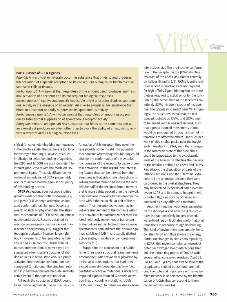
Box 1. Classes of GPCR LigandsAgonist: Any artificial or naturally occurring substance that binds to and producesfull activation of a specific receptor and its consequent biological or biochemical re-sponse in cells or tissues.Partial agonist: Any agonist that, regardless of the amount used, produces submaxi-mal activation of a receptor and its consequent biological responses.Inverse agonist (negative antagonist): Applicable only if a receptor displays spontane-ous activity in the absence of an agonist. An inverse agonist is any substance thatbinds to a receptor and fully suppresses its spontaneous activity.Partial inverse agonist: Any inverse agonist that, regardless of amount used, pro-duces submaximal suppression of spontaneous receptor activity.Antagonist (neutral antagonist): Any substance that binds to the same receptor asan agonist yet produces no effect other than to block the ability of an agonist to acti-vate a receptor and its biological responses.
www.acschemicalbiology.org VOL.2 NO.12 • 783–786 • 2007 785
Point ofVIEW
GPCR Dimers. �2AR, rhodopsin, andother GPCRs appear to have the capacity toform dimers or higher-order oligomers, andevidence indicates that such associationsare of physiological relevance (17–19).However, the �2AR crystal structures showthat, at least when held in its inactive con-formation by binding to a partial inverseagonist, the receptor exists in a stablyfolded monomeric state that shows onlyminimal interactions with other receptors inthe lattice. Of course, this observation couldmean that one effect of such a ligand is toprevent receptor�receptor association. Un-til the structure of �2AR bound to a fullagonist is obtained, the role of receptor�receptor contact in �2AR function and howthe agonist-occupied form of the receptorstimulates G-protein activation will both re-main matters of conjecture. Thus, althoughthe recently determined 3D views of �2ARare exciting and important, determinationof the structure of an agonist�receptor-G-protein complex remains the “holy grail” ofGPCR biology.
Future Prospects. Because the vast ma-jority of GPCRs bind diffusible ligands, thesereceptors must either have ligand-bindingpockets that are always solvent-accessibleor must display sufficient conformationalflexibility that they “flicker” frequently intoa state where the entrance to the pocket istransiently opened. The properties of �2ARand the features revealed by the recent de-termination of many of its structural ele-ments at atomic resolution strongly suggestthat, in the short term, it will be a much bet-ter template than rhodopsin for generatingmodels of clinically relevant GPCRs whosestructures have not been solved. Further-more and in the long term, the path-findingapproaches used by Kobilka, Weiss, andStevens to acquire structures for �2AR willundoubtedly be applied to other GPCRs.Hence, we can all look forward to viewingother equally stirring variations on the GPCRtheme.
Acknowledgment: We thank MarkusSeeliger for assistance with preparation of the fig-ure. The authors are supported by National Insti-tutes of Health and Department of Defense Re-search Grants GM44592, HL753632, andBC60037 (K.J.B.) and GM21841 (J.T.).
REFERENCES1. Ridge, K. D., and Palczewski, K. (2007) Visual rho-
dopsin sees the light: structure and mechanism ofG protein signaling, J. Biol. Chem. 282, 9297–9301.
2. Cherezov, V., Rosenbaum, D. M, Hanson, M. A., Ras-mussen, S. G., Thian, F. S., Kobilka, T. S., Choi, H. J.,Kuhn, P., Weis, W. I., Kobilka, B. K., and Stevens,R. C. (2007) High-resolution crystal structure of anengineered human {beta}2-adrenergic G proteincoupled receptor, Science 318, 1258–1265.
3. Rasmussen, S. G., Choi, H. J., Rosenbaum, D. M., Ko-bilka, T. S., Thian, F. S., Edwards, P. C., Burgham-mer, M., Ratnala, V. R., Sanishvili, R., Fischetti, R. F.,Schertler, G. F., Weis, W. I., and Kobilka, B. K.(2007) Crystal structure of the human beta(2) adren-ergic G-protein-coupled receptor, Nature 450, 383–387.
4. Rosenbaum, D. M., Cherezov, V., Hanson, M. A., Ras-mussen, S. G., Thian, F. S., Kobilka, T. S., Choi, H. J.,Yao, X. J., Weis, W. I., Stevens, R. C., and Kobilka,B. K. (2007) GPCR engineering yields high-resolutionstructural insights into {beta}2 adrenergic receptorfunction, Science 318, 1266–1273.
5. Watling, K. J. E., Ed. (2001) The Sigma-RBI Hand-book of Receptor Classification and Signal Transduc-tion, 4th ed., Sigma-RBI, St. Louis, MO.
6. Ostrowski, J., Kjelsberg, M. A., Caron, M. G., andLefkowitz, R. J. (1992) Mutagenesis of the beta2-adrenergic receptor: how structure elucidatesfunction, Annu. Rev. Pharmacol. Toxicol. 32, 167–183.
7. Kjelsberg, M. A., Cotecchia, S., Ostrowski, J., Caron,M. G., and Lefkowitz, R. J. (1992) Constitutive activa-tion of the alpha 1B-adrenergic receptor by allamino acid substitutions at a single site. Evidencefor a region which constrains receptor activation,J. Biol. Chem. 267, 1430–1433.
8. Day, P. W., Rasmussen, S. G., Parnot, C., Fung, J. J.,Masood, A., Kobilka, T. S., Yao, X. J., Choi, H. J., Weis,W. I., Rohrer, D. K., and Kobilka, B. K. (2007) Amonoclonal antibody for G protein-coupled receptorcrystallography, Nat. Methods 4, 927–929.
9. Lefkowitz, R. J., and Caron, M. G. (1990) The adrener-gic receptors, Adv. Second Messenger Phosphopro-tein Res. 24, 1–8.
10. Frielle, T., Daniel, K. W., Caron, M. G., and Lefkowitz,R. J. (1988) Structural basis of beta-adrenergic recep-tor subtype specificity studied with chimeric beta1/beta 2-adrenergic receptors, Proc. Natl. Acad. Sci.U.S.A. 85, 9494–9498.
11. Kobilka, B. K., Kobilka, T. S., Daniel, K., Regan, J. W.,Caron, M. G., and Lefkowitz, R. J. (1988) Chimeric al-pha 2-,beta 2-adrenergic receptors: delineation ofdomains involved in effector coupling and ligandbinding specificity, Science 240, 1310–1316.
12. Mathies, R. A. (1999) Photons, femtoseconds anddipolar interactions: a molecular picture of the pri-mary events in vision, Novartis Found. Symp. 224,70–101.
13. Kobilka, B. K. (2002) Agonist-induced conforma-tional changes in the beta2 adrenergic receptor,J. Pept. Res. 60, 317–321.
14. Hubbell, W. L., Altenbach, C., Hubbell, C. M., andKhorana, H. G. (2003) Rhodopsin structure, dynam-ics, and activation: a perspective from crystallogra-phy, site-directed spin labeling, sulfhydryl reactivity,and disulfide cross-linking, Adv. Protein Chem. 63,243–290.
15. Ren, Q., Kurose, H., Lefkowitz, R. J., and Cotecchia,S. (1993) Constitutively active mutants of the alpha2-adrenergic receptor, J. Biol. Chem. 268, 16483–16487.
16. Hausdorff, W. P., Hnatowich, M., O’Dowd, B. F., Ca-ron, M. G., and Lefkowitz, R. J. (1990) A mutation ofthe beta 2-adrenergic receptor impairs agonist ac-tivation of adenylyl cyclase without affecting high af-finity agonist binding. Distinct molecular determi-nants of the receptor are involved in physicalcoupling to and functional activation of Gs, J. Biol.Chem. 265, 1388–1393.
17. Floyd, D. H., Geva, A., Bruinsma, S. P., Overton, M. C.,Blumer, K. J., and Baranski, T. J. (2003) C5a receptoroligomerization. II. Fluorescence resonance energytransfer studies of a human G protein-coupled recep-tor expressed in yeast, J. Biol. Chem. 278, 35354–35361.
18. Milligan, G., Canals, M., Pediani, J. D., Ellis, J., andLopez-Gimenez, J. F. (2006) The role of GPCR dimeri-sation/oligomerisation in receptor signalling, ErnstSchering Found. Symp. Proc. 2, 145–161.
19. Overton, M. C., Chinault, S. L., and Blumer, K. J.(2003) Oligomerization, biogenesis, and signalingis promoted by a glycophorin A-like dimerization mo-tif in transmembrane domain 1 of a yeast G protein-coupled receptor, J. Biol. Chem. 278,49369–49377.
786 VOL.2 NO.12 • 783–786 • 2007 www.acschemicalbiology.orgBLUMER AND THORNER
Recommended