Lee et al. BMC Complementary and Alternative Medicine 2014, 14:338http://www.biomedcentral.com/1472-6882/14/338
RESEARCH ARTICLE Open Access
Acupuncture stimulation improvesscopolamine-induced cognitive impairment viaactivation of cholinergic system and regulationof BDNF and CREB expressions in ratsBombi Lee1*, Bongjun Sur1, Jaegul Shim1, Dae-Hyun Hahm1,2 and Hyejung Lee1,2*
Abstract
Background: Acupuncture is an alternative therapy that is widely used to treat various neurodegenerative diseasesand effectively improve cognitive and memory impairment. The aim of this study was to examine whetheracupuncture stimulation at the Baihui (GV20) acupoint improves memory defects caused by scopolamine (SCO)administration in rats. We also investigated the effects of acupuncture stimulation at GV20 on the cholinergicsystem as well as the expression of brain-derived neurotrophic factor (BDNF) and cAMP-response element-bindingprotein (CREB) in the hippocampus.
Methods: SCO (2 mg/kg, i.p.) was administered to male rats once daily for 14 days. Acupuncture stimulation atGV20 was performed for 5 min before SCO injection. After inducing cognitive impairment via SCO administration,we conducted a passive avoidance test (PAT) and the Morris water maze (MWM) test to assess behavior.
Results: Acupuncture stimulation at GV20 improved memory impairment as measured by the PAT and reduced theescape latency for finding the platform in the MWM test. Acupuncture stimulation at GV20 significantly alleviatedmemory-associated decreases in the levels of choline acetyltransferase (ChAT), BDNF and CREB proteins in thehippocampus. Additionally, acupuncture stimulation at GV20 significantly restored the expression of cholinetransporter 1 (CHT1), vesicular acetylcholine transporter (VAChT), BDNF and CREB mRNA in the hippocampus.These results demonstrate that acupuncture stimulation at GV20 exerts significant neuroprotective effects againstSCO-induced neuronal impairment and memory dysfunction in rats.
Conclusions: These findings suggest that acupuncture stimulation at GV20 might be useful in variousneurodegenerative diseases to improve cognitive functioning via stimulating cholinergic enzyme activities andregulating BDNF and CREB expression in the brain.
Keywords: Scopolamine, Memory, Cholinergic neurons, Brain-derived neurotrophic factor, cAMP-responseelement-binding protein
* Correspondence: [email protected]; [email protected] and Meridian Science Research Center, College of KoreanMedicine, Kyung Hee University, 26 Kyungheedae-ro, Dongdaemun-gu, Seoul130-701, Republic of Korea2BK21 PLUS Korean Medicine Science Center, College of Korean Medicine,Kyung Hee University, Seoul 130-701, Korea
© 2014 Lee et al.; licensee BioMed Central Ltd. This is an Open Access article distributed under the terms of the CreativeCommons Attribution License (http://creativecommons.org/licenses/by/4.0), which permits unrestricted use, distribution, andreproduction in any medium, provided the original work is properly credited. The Creative Commons Public DomainDedication waiver (http://creativecommons.org/publicdomain/zero/1.0/) applies to the data made available in this article,unless otherwise stated.
Lee et al. BMC Complementary and Alternative Medicine 2014, 14:338 Page 2 of 14http://www.biomedcentral.com/1472-6882/14/338
BackgroundAlzheimer’s disease (AD) is a progressive neurodegenerativedisorder of the brain that is characterized by deteriorationof memory and cognitive function due to cholinergicnervous system dysfunction [1]. Decreased cholinergicfunction in the brain, as primarily observed in patients withAD, can result in a decline in memory and cognitivefunction [2]. Accordingly, various cholinergic drugs havebeen approved to treat or alleviate AD, and they exert theirtherapeutic effects by counteracting acetylcholine (ACh)deficits and consequently enhancing ACh levels in thebrain [3]. In fact, the most common therapy for ADis administration of acetylcholinesterase (AChE) inhibitors,such as donepezil, galantamine, and rivastigmine, whichtemporarily increase the availability of ACh at cholinergicsynapses [4]. Nevertheless, new drugs used to treatAD patients are limited due to their short half-livesand excessive side effects caused by peripheral cholinergicsystem activation and hepatotoxicity, the most frequent andcritical side-effect of these drugs [5]. Thus, an alternativetreatment modality for AD patients is required.Scopolamine (SCO) is a tropane alkaloid drug that
exhibits competitive antagonism at muscarinic acetylcholinereceptors (mAChRs) by interfering with cholinergictransmission in the central nervous system CNS; [6].Therefore, SCO administration to animals is used asan experimental model of the cognitive deteriorationand memory disturbances in AD; this animal model isfrequently used to screen for drugs that have potentialtherapeutic value in AD-type dementia patients [7-9]. SCOadministration not only induces dysregulation of thecholinergic neuronal pathway and memory circuits inthe CNS but also reduces the expression of brain-derivedneurotrophic factor (BDNF) and cAMP-response element-binding protein (CREB) in the brain; thus, BDNF and CREBmay act a novel therapeutics to treat hippocampal dysfunc-tion and memory deficits [10,11]. BDNF is believed to beresponsible for synaptic plasticity and memory perform-ance, particularly in the water maze test, and is coupled toCREB activation [12,13]. Several studies demonstrated thathippocampal BDNF and CREB are important in long-termmemory formation [14], and play important roles in patho-logical conditions and neurodegenerative diseases such asAD [15,16].In East Asian nations, acupuncture is used widely to
treat many neurodegenerative disorders, including AD,Parkinson’s disease (PD) and dementia [17]. Its therapeuticeffects and mechanism of action have been investigated inboth clinical and animal studies. This alternative therapy isknown to modulate biochemical balance in the CNS and tomaintain homeostasis [18]. Specifically, Baihui (GV20) isone of the most important acupoints targeted to alleviateneurodegenerative disorders and cognitive impairment inacupuncture treatment [19]. Several studies showed that
acupuncture stimulation at GV20 reduces cerebral infarctand increases dopamine levels in the brain tissue ofischemia-reperfusion injured rats [20], and reduces theamount of apoptotic neurons in the hippocampal CA1 areaof rats with vascular dementia [19]. Although a brief reportof the anti-dementia or anti-ischemic activity of GV20acupuncture stimulation has been published, whetherGV20 acupuncture stimulation therapeutic efficacy inalleviating spatial cognitive function following repeatedSCO-induced neuronal impairment is due to cholinergicsystem regulation or BDNF and CREB expression remainsunknown [21]. Indeed, one study failed to demonstrate atherapeutic effect of acupuncture on AD [22]. Such majordifferences could be attributable to differences in acupunc-ture stimulation parameters, as selection of appropriatestimulation parameters is a crucial factor in the efficacy ofacupuncture [23]. Therefore, it is appropriate to investigatechanges in spatial cognitive function and neuronalbiomarkers in SCO-treated cognitive impairment tobetter understand the therapeutic effects and mechanismsof action of acupuncture stimulation at GV20.The aim of the present study was to evaluate the ability
of acupuncture stimulation at GV20 to improve learningand memory in rats exposed to repeated SCO-inducedmemory deficits as measured by their performance in thepassive avoidance test (PAT) and the Morris water maze(MWM) test. We also investigated how these activities wererelated to the cholinergic system and the expression ofCREB and BDNF in the CNS, and whether acupuncturestimulation exerted anti-AD activity in this model to eluci-date neural mechanisms underlying the memory-enhancingeffect of acupuncture stimulation.
MethodsAnimalsAdult male Sprague–Dawley (SD) rats weighing 220–240 g were obtained from Samtako Animal Co. (Seoul,Korea). The rats were housed in a limited-access rodentfacility with up to five rats per polycarbonate cage. Theroom controls were set to maintain the temperatureat 22 ± 2°C and the relative humidity at 55 ± 15%. Cages werelit by artificial light for 12 h each day. Sterilized drinkingwater and standard chow diet were supplied ad libitum toeach cage during the experiments. The animal experimentswere conducted in accordance with the National Institutes ofHealth Guide for the Care and Use of Laboratory Animals(NIH Publications No. 80–23), revised in 1996, and wereapproved by the Kyung Hee University Institutional AnimalCare and Use Committee. All animal experiments began atleast 7 days after the animals arrived.
Experimental groupsIn order to develop learning and memory deficits in thebrain, the rats were intraperitoneally injected with 2 mg/kg

Lee et al. BMC Complementary and Alternative Medicine 2014, 14:338 Page 3 of 14http://www.biomedcentral.com/1472-6882/14/338
SCO, dissolved in physiological saline, once a day for14 days. Normal animals received saline instead of SCO asa vehicle control. Different rats in an experimentalgroup were subjected to either behavioral testing orimmunohistochemistry. The rats were randomly dividedinto five groups of six or seven individuals as follows:normal group (SAL group, n = 7), the SCO-induced andsaline-treated group (SCO group as a control, n = 7), theSCO-induced and Baihui (GV20) acupoint-stimulatedgroup (SCO +GV20 group, n = 7), the SCO-induced andYangji (TE4) acupoint-stimulated group (SCO+TH4group, n = 6), and SCO-induced and non-acupoint (onthe tail)-stimulated group (SCO+TA group, n = 6) everydaily for 5 min before the SCO injection. Scopolaminehydrobromide were purchased from Sigma-AldrichChemical Co. (St Louis, MO, USA). The experimentalschedule of drug administration, acupuncture stimulationand behavioral tests is shown in Figure 1.
Acupuncture stimulationAcupuncture stimulation was bilaterally performedevery day for 5 min before the SCO injection duringthe SCO-injection period. The acupuncture stimulationwas performed as previously described [24]. The GV20acupoint, meeting point on the Governing vessel with thesix yang channels, is anatomically located on the midsagittalline, at the intersection of a line connecting the right andleft ear apices [25]. The acupoint on tail and the Yangjiacupoint were selected as a non-acupoint and a comparisonacupoint, respectively. As a comparable control acupoint,we also performed the stimulation to another acupoint, theTE4 acupoint, on hand Shaoyang triple energizer meridian,is located at the midpoint of the dorsal crease of the wrist,in the depression on the ulnar side of the tendon ofthe extensor muscle of the finger [26,27]. In addition,the non-acupoint needling was performed at one-fifthpoint of tail length from the proximal end of the tail toavoid the tail acupoints located at proximal or distalregion of the tail. Sterilized disposable stainless steelacupuncture needles (0.30 × 25 mm, Suzhou Kangnian
Figure 1 Experimental schedules for scopolamine (SCO) administratiowas designed to explore the efficacy of acupuncture therapy in healing chusing behavioral and neurobiological methodologies. All rats excluding therats were subjected to the Morris water maze (MWM) and the passive avoitissues was collected immediately. SCO: scopolamine; IHC: Immunohistoche
Medical Devices Co., Ltd., Shzhou, China) were insertedperpendicularly as deep as 2–3 mm at GV20 or TE4acupoint. The depth of needle insertion at each acupointwas arbitrarily determined based on several previouslystudies [28] and in the animal acupuncture atlas [21].During the acupuncture procedure, rats were gentlyhandled entirely to minimize stress in the rats. Wedid not carry out sham acupuncture for control. TheSAL and SCO groups were handled for 5 min to acalming effect, instead of acupuncture stimulation,similar to what we described previously [24].
Passive avoidance testThe test was basically performed according to thestep-through method. The Gemini Avoidance System(SD Instruments., San Diego, CA, USA) was used forthis experiment. Basically, the step-through passiveavoidance apparatus (PAA) consists of a tilting flooracrylic box divided into two-compartments, a lightenedcompartment connected to a darkened compartments byan automatic guillotine door and a control unit generatingan electric shock (Behbood Pardaz Co., Ghaem, Iran). Theelectric shock can be delivered to the grid floor, made ofstainless steel rods (3 mm diameter) spaced 1 cm apart, inboth compartments. First, the rats were given trials toacquisition test in the apparatus. In the training session, arat was placed in a lightened compartment of the PAAfacing away from the entrance to the dark compartment,and then the guillotine door was opened. Because ofintrinsic preference to the dark environment, the ratimmediately entered the dark compartment and thedoor was closed. During acquisition test, the latencytime before entry into the dark compartment was recordedfor each rat. After 30 min, the rats were placed inthe lightened compartment once again. After enteringthe dark compartment, the guillotine door was closedand subsequently a mild electrical shock (0.5 mA)was applied for 3 s. The retention test was started24 h after the acquisition trial for training. The rat wasagain placed in the lightened compartment and the
n to induce spatial memory impairments in rats. The experimentronic SCO-induced spatial memory impairment in an animal modelSAL group received SCO injection. Two weeks after SCO injection, the
dance test (PAT). After behavioral testing, rats were sacrificed and brainmistry.
Lee et al. BMC Complementary and Alternative Medicine 2014, 14:338 Page 4 of 14http://www.biomedcentral.com/1472-6882/14/338
guillotine door was opened. In the retention test, therat was placed in the PAA as previously describedand the time required for the rat to enter the darkcompartment was measured for a maximum period of3 min in the same method with the acquisition test.The rat did not enter the dark compartment withinthis period received a latency time of 180 s.
Morris water maze testThe MWM test was performed in a small circular pool(2.0 m in diameter and 0.35 m deep) made of polypropyleneand internally painted white. The pool was half-filled withwater to a depth of 30 cm. The water in the pool was madeopaque by adding 1 kg skim milk powder and continuouslymaintained at 22 ± 2°C. The pool was divided into fourquadrants of equal area. During the MWM test, an escapeplatform (15 cm in diameter) was located in one of foursections of the pool, hidden 1.5 cm below the water surfaceand approximately 50 cm from the sidewalls. Several visualcues were placed around the pool in plain sight of theanimals. A digital camera was mounted to the ceilingstraight above the center of the pool and was connected toa computerized recording system equipped with a trackingprogram (S-MART; PanLab Co., Barcelona, Spain), whichpermitted on- and off-line automated tracking of the pathstaken by the rats. The MWM test was initiated on the 14th
day after the SCO administration commenced. The animalsreceived three trials per day. The rats were trained to findthe hidden platform, which remained in a fixed locationthroughout the test. The trials lasted for a maximum of180 s, and the escape latency was expressed by theswimming time to find the submerged platform in the pool.The animals were tested with three trials per day for 5 days,and they received a 60-s probe trial on the sixth day.Finding the platform was defined as staying on it for at least4 s before the acquisition time of 180 s ended. When therat failed to find the platform in the limited time in first trialof hidden platform test, the rats should be placed on theplatform for 20 s and assigned a latency of 180 s. Betweenone trail and the next, the water in the pool was stirred toremove olfactory traces of previous swim patterns. Theentire schedule proceeded for 6 days and each animal hadthree trials for training per day with a 40–50 min inter-trialinterval. For the probe trial, a rat was placed in the quadrantlocated diagonally from the target quadrant and allowed toswim to the quadrant from which the escape platform hadbeen removed for a maximum of 60 s. The probe trial wasexpressed by the ratio of the time spent (or the distancetraveled) in searching for the platform in the targetquadrant to the total duration spent swimming in the pool.
Open field testPrior to water maze testing, the rats were individuallyhoused in a rectangular container that was made of dark
polyethylene (60 × 60 × 30 cm) to provide the bestcontrast to the white rats in a dimly lit room equipped witha video camera above the center of the room, and theirlocomotor activities (animal’s movements) were then mea-sured. The locomotor activity indicated by the speed anddistance of movements was monitored by a computerizedvideo-tracking system using the S-MART program (PanLabCo., Barcelona, Spain). After 5 min of adaptation, thedistance they traveled in the container was recorded foranother 5 min. The locomotor activity was measured incentimeters. The floor surface of each chamber wasthoroughly cleaned with 70% ethanol between tests.
ImmunohistochemistryFor immunohistochemical studies, three rats in eachgroups were deeply anesthetized with sodium pentobar-bital (80 mg/kg, by intraperitoneal injection) and perfusedthrough the ascending aorta with normal saline (0.9%),followed by 300 ml (per rat) of 4% paraformaldehyde in0.1 M phosphate-buffered saline (PBS). The brains wereremoved in a randomized order, post-fixed over-night,and cryoprotected with 20% sucrose in 0.1 M PBS at 4°C.Coronal sections 30 μm thick were serially cut throughthe hippocampus using a cryostat (Leica CM1850; LeicaMicrosystems Ltd., Nussloch, Germany). The sectionswere obtained according to the rat atlas of Paxinos andWatson [29]. The primary antibodies against the followingspecific antigen were used: choline acetyltransferase (ChAT;sheep polyclonal ChAT, 1:2,000 dilution, CambridgeResearch Biochemicals Co., Bellingham, UK), acetyl-cholinesterase (AchE; goat polyclonal AchE, 1:2,000dilution, Santa Cruz Biotechnology Inc., CA, USA),BDNF (rabbit polyclonal BDNF, 1:200 dilution, Cellsignaling., Bostron, MA, USA) and CREB (rabbit polyclonalCREB, 1:250 dilution, Cell signaling., Bostron, MA, USA).Briefly, the sections were incubated with primary antiserumin PBST (PBS plus 0.3% Triton X-100) for 72 h at 4°C. Thesections were incubated for 120 min at room temperaturewith secondary antibody. The secondary antibodies wereobtained from Vector Laboratories Co. (Burlingame, CA,USA) and diluted 1:200 in PBST containing 2% normalserum. To visualize immunoreactivity, the sections wereincubated for 90 min in avidin-biotin-peroxidase complex(ABC) reagent (Vectastain Elite ABC kit; Vector Labs. Co.),and incubated in a solution containing 3,3’-diaminobenzi-dine (DAB; Sigma) and 0.01% H2O2 for 1 min. Finally, thetissues were washed in PBS, followed by a brief rinsein distilled water, and mounted individually ontoslides. Images were captured using the AxioVision 3.0imaging system (Carl Zeiss, Inc., Oberkochen, Germany)and processed using Adobe Photoshop (Adobe Systems,Inc., San Jose, CA, USA). The sections were viewed at400× magnification, and the numbers of ChAT-, AchE-,BDNF-, and CREB-labeled cells was quantified in the CA1
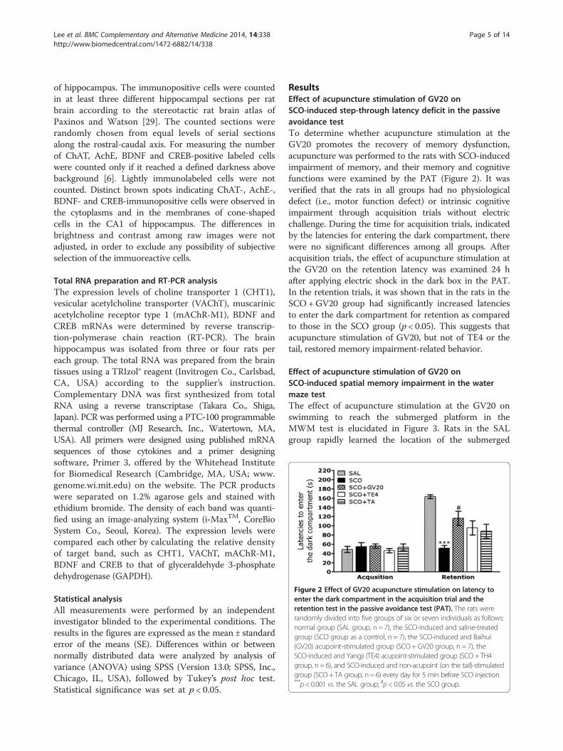
Figure 2 Effect of GV20 acupuncture stimulation on latency toenter the dark compartment in the acquisition trial and theretention test in the passive avoidance test (PAT). The rats wererandomly divided into five groups of six or seven individuals as follows:normal group (SAL group, n = 7), the SCO-induced and saline-treatedgroup (SCO group as a control, n = 7), the SCO-induced and Baihui(GV20) acupoint-stimulated group (SCO + GV20 group, n = 7), theSCO-induced and Yangji (TE4) acupoint-stimulated group (SCO+ TH4group, n = 6), and SCO-induced and non-acupoint (on the tail)-stimulatedgroup (SCO+ TA group, n = 6) every day for 5 min before SCO injection.***p< 0.001 vs. the SAL group; #p< 0.05 vs. the SCO group.
Lee et al. BMC Complementary and Alternative Medicine 2014, 14:338 Page 5 of 14http://www.biomedcentral.com/1472-6882/14/338
of hippocampus. The immunopositive cells were countedin at least three different hippocampal sections per ratbrain according to the stereotactic rat brain atlas ofPaxinos and Watson [29]. The counted sections wererandomly chosen from equal levels of serial sectionsalong the rostral-caudal axis. For measuring the numberof ChAT, AchE, BDNF and CREB-positive labeled cellswere counted only if it reached a defined darkness abovebackground [6]. Lightly immunolabeled cells were notcounted. Distinct brown spots indicating ChAT-, AchE-,BDNF- and CREB-immunopositive cells were observed inthe cytoplasms and in the membranes of cone-shapedcells in the CA1 of hippocampus. The differences inbrightness and contrast among raw images were notadjusted, in order to exclude any possibility of subjectiveselection of the immuoreactive cells.
Total RNA preparation and RT-PCR analysisThe expression levels of choline transporter 1 (CHT1),vesicular acetylcholine transporter (VAChT), muscarinicacetylcholine receptor type 1 (mAChR-M1), BDNF andCREB mRNAs were determined by reverse transcrip-tion-polymerase chain reaction (RT-PCR). The brainhippocampus was isolated from three or four rats pereach group. The total RNA was prepared from the braintissues using a TRIzol® reagent (Invitrogen Co., Carlsbad,CA, USA) according to the supplier’s instruction.Complementary DNA was first synthesized from totalRNA using a reverse transcriptase (Takara Co., Shiga,Japan). PCR was performed using a PTC-100 programmablethermal controller (MJ Research, Inc., Watertown, MA,USA). All primers were designed using published mRNAsequences of those cytokines and a primer designingsoftware, Primer 3, offered by the Whitehead Institutefor Biomedical Research (Cambridge, MA, USA; www.genome.wi.mit.edu) on the website. The PCR productswere separated on 1.2% agarose gels and stained withethidium bromide. The density of each band was quanti-fied using an image-analyzing system (i-MaxTM, CoreBioSystem Co., Seoul, Korea). The expression levels werecompared each other by calculating the relative densityof target band, such as CHT1, VAChT, mAChR-M1,BDNF and CREB to that of glyceraldehyde 3-phosphatedehydrogenase (GAPDH).
Statistical analysisAll measurements were performed by an independentinvestigator blinded to the experimental conditions. Theresults in the figures are expressed as the mean ± standarderror of the means (SE). Differences within or betweennormally distributed data were analyzed by analysis ofvariance (ANOVA) using SPSS (Version 13.0; SPSS, Inc.,Chicago, IL, USA), followed by Tukey’s post hoc test.Statistical significance was set at p < 0.05.
ResultsEffect of acupuncture stimulation of GV20 onSCO-induced step-through latency deficit in the passiveavoidance testTo determine whether acupuncture stimulation at theGV20 promotes the recovery of memory dysfunction,acupuncture was performed to the rats with SCO-inducedimpairment of memory, and their memory and cognitivefunctions were examined by the PAT (Figure 2). It wasverified that the rats in all groups had no physiologicaldefect (i.e., motor function defect) or intrinsic cognitiveimpairment through acquisition trials without electricchallenge. During the time for acquisition trials, indicatedby the latencies for entering the dark compartment, therewere no significant differences among all groups. Afteracquisition trials, the effect of acupuncture stimulation atthe GV20 on the retention latency was examined 24 hafter applying electric shock in the dark box in the PAT.In the retention trials, it was shown that in the rats in theSCO +GV20 group had significantly increased latenciesto enter the dark compartment for retention as comparedto those in the SCO group (p < 0.05). This suggests thatacupuncture stimulation of GV20, but not of TE4 or thetail, restored memory impairment-related behavior.
Effect of acupuncture stimulation of GV20 onSCO-induced spatial memory impairment in the watermaze testThe effect of acupuncture stimulation at the GV20 onswimming to reach the submerged platform in theMWM test is elucidated in Figure 3. Rats in the SALgroup rapidly learned the location of the submerged
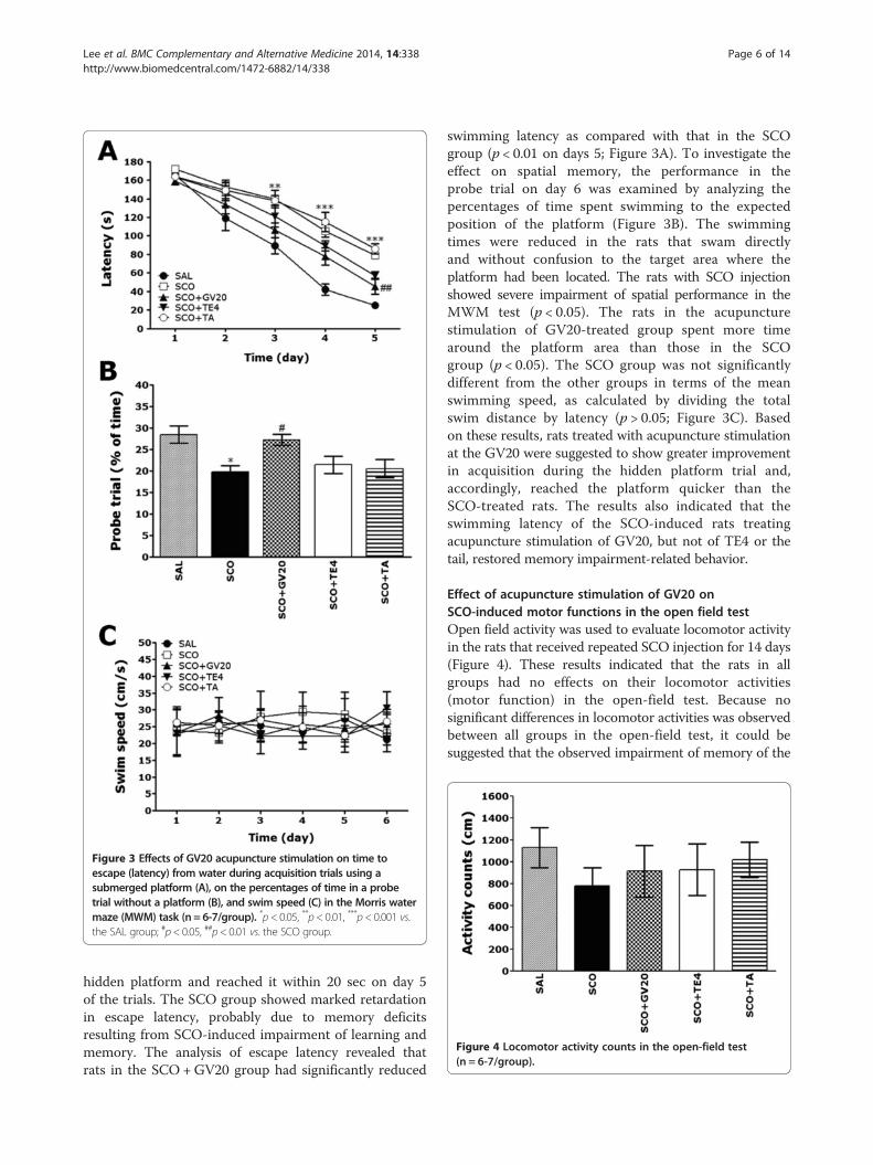
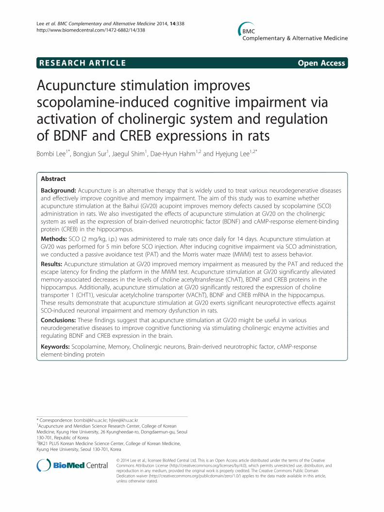
Figure 3 Effects of GV20 acupuncture stimulation on time toescape (latency) from water during acquisition trials using asubmerged platform (A), on the percentages of time in a probetrial without a platform (B), and swim speed (C) in the Morris watermaze (MWM) task (n = 6-7/group). *p< 0.05, **p< 0.01, ***p< 0.001 vs.the SAL group; #p< 0.05, ##p< 0.01 vs. the SCO group.
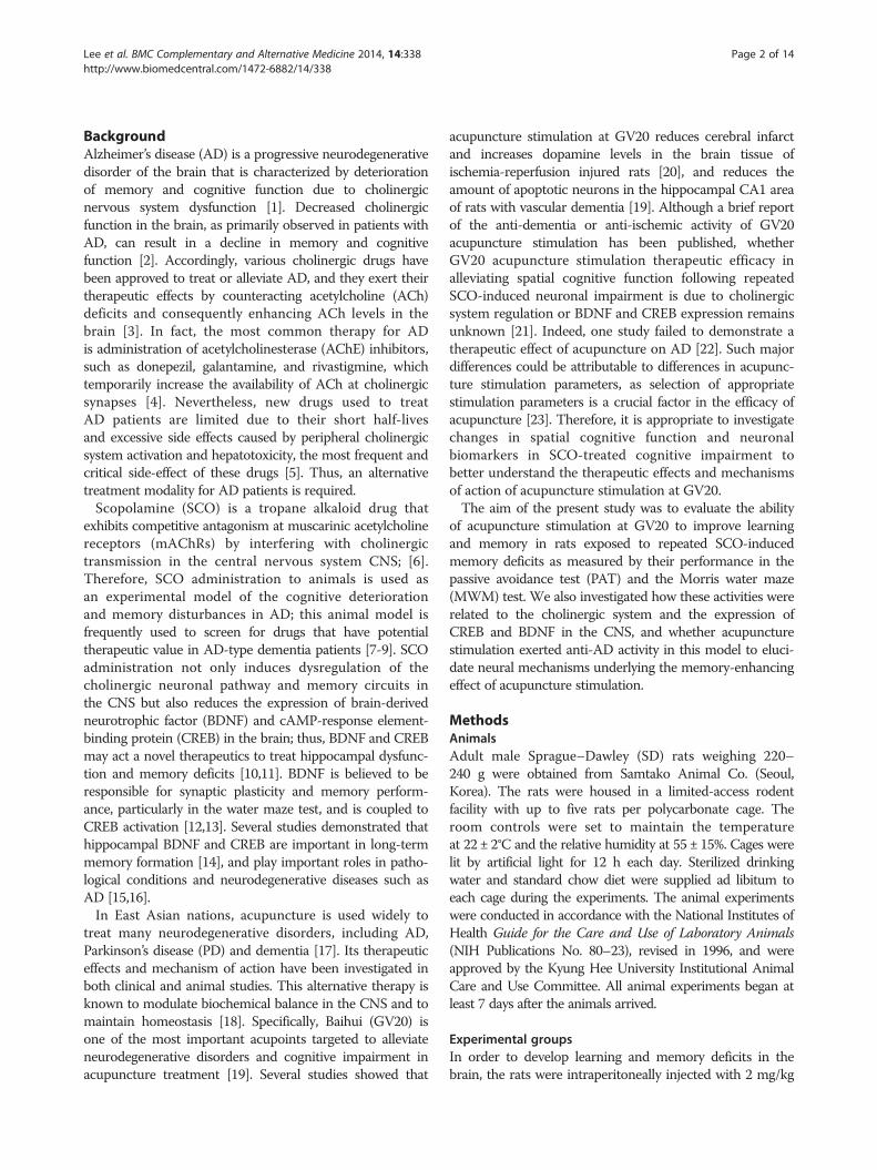
Figure 4 Locomotor activity counts in the open-field test(n = 6-7/group).
Lee et al. BMC Complementary and Alternative Medicine 2014, 14:338 Page 6 of 14http://www.biomedcentral.com/1472-6882/14/338
hidden platform and reached it within 20 sec on day 5of the trials. The SCO group showed marked retardationin escape latency, probably due to memory deficitsresulting from SCO-induced impairment of learning andmemory. The analysis of escape latency revealed thatrats in the SCO +GV20 group had significantly reduced
swimming latency as compared with that in the SCOgroup (p < 0.01 on days 5; Figure 3A). To investigate theeffect on spatial memory, the performance in theprobe trial on day 6 was examined by analyzing thepercentages of time spent swimming to the expectedposition of the platform (Figure 3B). The swimmingtimes were reduced in the rats that swam directlyand without confusion to the target area where theplatform had been located. The rats with SCO injectionshowed severe impairment of spatial performance in theMWM test (p < 0.05). The rats in the acupuncturestimulation of GV20-treated group spent more timearound the platform area than those in the SCOgroup (p < 0.05). The SCO group was not significantlydifferent from the other groups in terms of the meanswimming speed, as calculated by dividing the totalswim distance by latency (p > 0.05; Figure 3C). Basedon these results, rats treated with acupuncture stimulationat the GV20 were suggested to show greater improvementin acquisition during the hidden platform trial and,accordingly, reached the platform quicker than theSCO-treated rats. The results also indicated that theswimming latency of the SCO-induced rats treatingacupuncture stimulation of GV20, but not of TE4 or thetail, restored memory impairment-related behavior.
Effect of acupuncture stimulation of GV20 onSCO-induced motor functions in the open field testOpen field activity was used to evaluate locomotor activityin the rats that received repeated SCO injection for 14 days(Figure 4). These results indicated that the rats in allgroups had no effects on their locomotor activities(motor function) in the open-field test. Because nosignificant differences in locomotor activities was observedbetween all groups in the open-field test, it could besuggested that the observed impairment of memory of the
Lee et al. BMC Complementary and Alternative Medicine 2014, 14:338 Page 7 of 14http://www.biomedcentral.com/1472-6882/14/338
rats with repeated SCO injection was not attributed to thedifferences in their locomotion activities. This may reflectan active response and water avoidance stress when theanimal is confronted with a MWM test. However, ourresults suggest that the rats in all groups displayed noanxiolytic-like behavior in the open-field test after apretest stress exposure in the MWM test. This indicatesthat acupuncture stimulation at the GV20 did not affectthe active responses or psychomotor function as measuredby the rats’ performance in the MWM test.
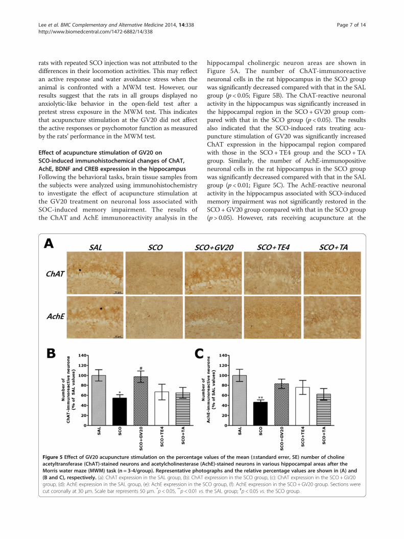
Effect of acupuncture stimulation of GV20 onSCO-induced immunohistochemical changes of ChAT,AchE, BDNF and CREB expression in the hippocampusFollowing the behavioral tasks, brain tissue samples fromthe subjects were analyzed using immunohistochemistryto investigate the effect of acupuncture stimulation atthe GV20 treatment on neuronal loss associated withSOC-induced memory impairment. The results ofthe ChAT and AchE immunoreactivity analysis in the
Figure 5 Effect of GV20 acupuncture stimulation on the percentage vacetyltransferase (ChAT)-stained neurons and acetylcholinesterase (AMorris water maze (MWM) task (n = 3-4/group). Representative photo(B and C), respectively. (a): ChAT expression in the SAL group, (b): ChAT egroup, (d): AchE expression in the SAL group, (e): AchE expression in the SCcut coronally at 30 μm. Scale bar represents 50 μm. *p < 0.05, **p < 0.01 vs.
hippocampal cholinergic neuron areas are shown inFigure 5A. The number of ChAT-immunoreactiveneuronal cells in the rat hippocampus in the SCO groupwas significantly decreased compared with that in the SALgroup (p < 0.05; Figure 5B). The ChAT-reactive neuronalactivity in the hippocampus was significantly increased inthe hippocampal region in the SCO+GV20 group com-pared with that in the SCO group (p < 0.05). The resultsalso indicated that the SCO-induced rats treating acu-puncture stimulation of GV20 was significantly increasedChAT expression in the hippocampal region comparedwith those in the SCO+TE4 group and the SCO +TAgroup. Similarly, the number of AchE-immunopositiveneuronal cells in the rat hippocampus in the SCO groupwas significantly decreased compared with that in the SALgroup (p < 0.01; Figure 5C). The AchE-reactive neuronalactivity in the hippocampus associated with SCO-inducedmemory impairment was not significantly restored in theSCO +GV20 group compared with that in the SCO group(p > 0.05). However, rats receiving acupuncture at the
alues of the mean (±standard errer, SE) number of cholinechE)-stained neurons in various hippocampal areas after thegraphs and the relative percentage values are shown in (A) andxpression in the SCO group, (c): ChAT expression in the SCO + GV20O group, (f): AchE expression in the SCO + GV20 group. Sections werethe SAL group; #p < 0.05 vs. the SCO group.
Lee et al. BMC Complementary and Alternative Medicine 2014, 14:338 Page 8 of 14http://www.biomedcentral.com/1472-6882/14/338
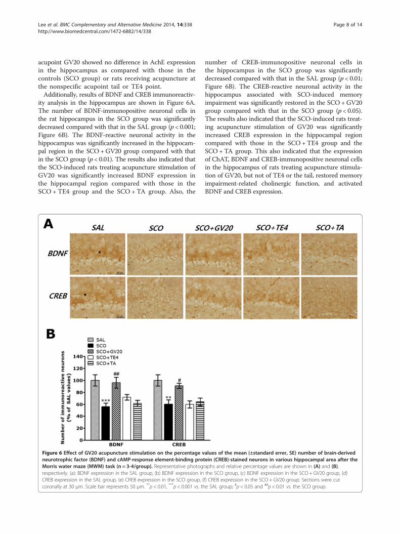
acupoint GV20 showed no difference in AchE expressionin the hippocampus as compared with those in thecontrols (SCO group) or rats receiving acupuncture atthe nonspecific acupoint tail or TE4 point.Additionally, results of BDNF and CREB immunoreactiv-
ity analysis in the hippocampus are shown in Figure 6A.The number of BDNF-immunopositive neuronal cells inthe rat hippocampus in the SCO group was significantlydecreased compared with that in the SAL group (p < 0.001;Figure 6B). The BDNF-reactive neuronal activity in thehippocampus was significantly increased in the hippocam-pal region in the SCO+GV20 group compared with thatin the SCO group (p < 0.01). The results also indicated thatthe SCO-induced rats treating acupuncture stimulation ofGV20 was significantly increased BDNF expression inthe hippocampal region compared with those in theSCO + TE4 group and the SCO + TA group. Also, the
Figure 6 Effect of GV20 acupuncture stimulation on the percentage vneurotrophic factor (BDNF) and cAMP-response element-binding protMorris water maze (MWM) task (n = 3-4/group). Representative photogrespectively. (a): BDNF expression in the SAL group, (b) BDNF expression inCREB expression in the SAL group, (e) CREB expression in the SCO group, (coronally at 30 μm. Scale bar represents 50 μm. **p < 0.01, ***p < 0.001 vs. th
number of CREB-immunopositive neuronal cells inthe hippocampus in the SCO group was significantlydecreased compared with that in the SAL group (p < 0.01;Figure 6B). The CREB-reactive neuronal activity in thehippocampus associated with SCO-induced memoryimpairment was significantly restored in the SCO +GV20group compared with that in the SCO group (p < 0.05).The results also indicated that the SCO-induced rats treat-ing acupuncture stimulation of GV20 was significantlyincreased CREB expression in the hippocampal regioncompared with those in the SCO +TE4 group and theSCO +TA group. This also indicated that the expressionof ChAT, BDNF and CREB-immunopositive neuronal cellsin the hippocampus of rats treating acupuncture stimula-tion of GV20, but not of TE4 or the tail, restored memoryimpairment-related cholinergic function, and activatedBDNF and CREB expression.
alues of the mean (±standard errer, SE) number of brain-derivedein (CREB)-stained neurons in various hippocampal area after theraphs and relative percentage values are shown in (A) and (B),the SCO group, (c) BDNF expression in the SCO + GV20 group, (d)f) CREB expression in the SCO + GV20 group. Sections were cute SAL group; #p < 0.05 and ##p < 0.01 vs. the SCO group.
Lee et al. BMC Complementary and Alternative Medicine 2014, 14:338 Page 9 of 14http://www.biomedcentral.com/1472-6882/14/338
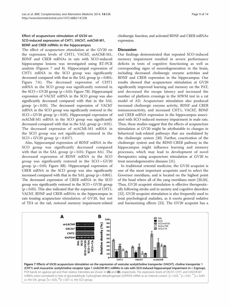
Effect of acupuncture stimulation of GV20 onSCO-induced expression of CHT1, VAChT, mAChR-M1,BDNF and CREB mRNAs in the hippocampusThe effect of acupuncture stimulation at the GV20 onthe expression levels of CHT1, VAChT, mAChR-M1,BDNF and CREB mRNAs in rats with SCO-inducedhippocampus lesions was investigated using RT-PCRanalysis (Figures 7 and 8). Hippocampal expression ofCHT1 mRNA in the SCO group was significantlydecreased compared with that in the SAL group (p < 0.001;Figure 7A). The decreased expression of CHT1mRNA in the SCO group was significantly restored inthe SCO+GV20 group (p < 0.01; Figure 7B). Hippocampalexpression of VAChT mRNA in the SCO group was alsosignificantly decreased compared with that in the SALgroup (p < 0.05). The decreased expression of VAChTmRNA in the SCO group was significantly restored in theSCO+GV20 group (p < 0.05). Hippocampal expression ofmAChR-M1 mRNA in the SCO group was significantlydecreased compared with that in the SAL group (p < 0.01).The decreased expression of mAChR-M1 mRNA inthe SCO group was not significantly restored in theSCO + GV20 group (p = 0.837).Also, hippocampal expression of BDNF mRNA in the
SCO group was significantly decreased comparedwith that in the SAL group (p < 0.01; Figure 8A). Thedecreased expression of BDNF mRNA in the SCOgroup was significantly restored in the SCO + GV20group (p < 0.05; Figure 8B). Hippocampal expression ofCREB mRNA in the SCO group was also significantlyincreased compared with that in the SAL group (p < 0.001).The decreased expression of CREB mRNA in the SCOgroup was significantly restored in the SCO+GV20 group(p < 0.05). This also indicated that the expression of CHT1,VAChT, BDNF and CREB mRNAs in the hippocampus inrats treating acupuncture stimulation of GV20, but notof TE4 or the tail, restored memory impairment-related
Figure 7 Effects of GV20 acupuncture stimulation on the expression of(CHT1) and muscarinic acetylcholine receptor type 1 (mAChR-M1) mRNPCR bands on agarose gel and their relative intensities are shown in (A) andmRNAs were normalized to that of glyceraldehyde 3-phosphate dehydrogenavs. the SAL group; #p < 0.05, ##p < 0.01 vs. the SCO group.
cholinergic function, and activated BDNF and CREB mRNAsexpression.
DiscussionOur findings demonstrated that repeated SCO-inducedmemory impairment resulted in severe performancedeficits in tests of cognitive functioning as well ascorresponding signs of neurodegeneration in the brain,including decreased cholinergic enzyme activities andBDNF and CREB expression in the hippocampus. Ourresults showed that acupuncture stimulation at GV20significantly improved learning and memory on the PAT,and decreased the escape latency and increased thenumber of platform crossings in the MWM test in a ratmodel of AD. Acupuncture stimulation also producedincreased cholinergic enzyme activity, BDNF and CREBimmunoreactivity, and increased CHT1, VAChT, BDNFand CREB mRNA expression in the hippocampus associ-ated with SCO-induced memory impairment in male rats.Thus, these studies suggest that the effects of acupuncturestimulation at GV20 might be attributable to changes inbehavioral task-related pathways that are modulated bythe cholinergic system [30]. Further, coactivation of thecholinergic system and the BDNF-CREB pathway in thehippocampus might influence learning and memoryprocesses, which may lead to development of noveltherapeutics using acupuncture stimulation at GV20 totreat neurodegenerative diseases [31].In traditional oriental medicine, the GV20 acupoint is
one of the most important acupoints used to select theGovernor meridians, and is located on the highest pointof the head where all of the yang meridians meet [20,26].Thus, GV20 acupoint stimulation is effective therapeutic-ally following stroke and in anxiety and cognitive disorders[32]. GV20 acupoint stimulation is also frequently used totreat psychological maladies, as it exerts general sedativeand harmonizing effects [33]. The GV20 acupoint has a
vesicular acetylcholine transporter (VAChT), choline transporter 1As in rats with SCO-induced hippocampal impairment (n = 3/group).(B), respectively. The expression levels of VAChT, CHT1 and mAChR-M1se (GAPDH) mRNA as an internal control. *p < 0.05, **p < 0.01, ***p < 0.001
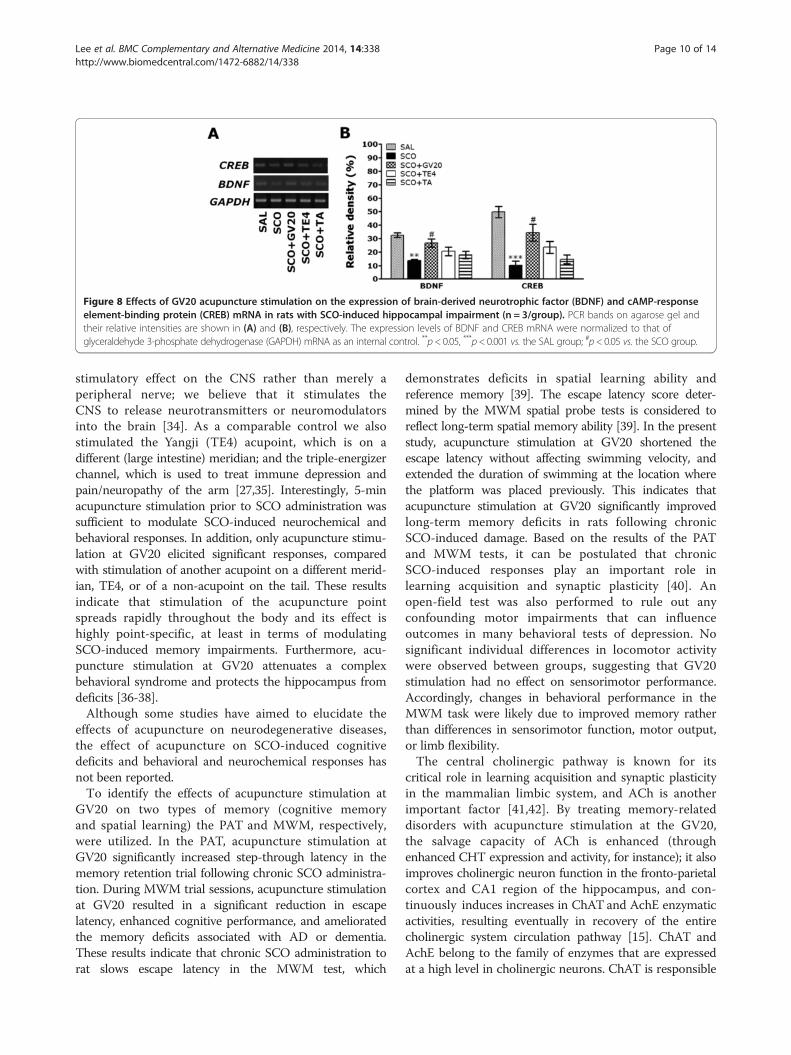
Figure 8 Effects of GV20 acupuncture stimulation on the expression of brain-derived neurotrophic factor (BDNF) and cAMP-responseelement-binding protein (CREB) mRNA in rats with SCO-induced hippocampal impairment (n = 3/group). PCR bands on agarose gel andtheir relative intensities are shown in (A) and (B), respectively. The expression levels of BDNF and CREB mRNA were normalized to that ofglyceraldehyde 3-phosphate dehydrogenase (GAPDH) mRNA as an internal control. **p< 0.05, ***p< 0.001 vs. the SAL group; #p< 0.05 vs. the SCO group.
Lee et al. BMC Complementary and Alternative Medicine 2014, 14:338 Page 10 of 14http://www.biomedcentral.com/1472-6882/14/338
stimulatory effect on the CNS rather than merely aperipheral nerve; we believe that it stimulates theCNS to release neurotransmitters or neuromodulatorsinto the brain [34]. As a comparable control we alsostimulated the Yangji (TE4) acupoint, which is on adifferent (large intestine) meridian; and the triple-energizerchannel, which is used to treat immune depression andpain/neuropathy of the arm [27,35]. Interestingly, 5-minacupuncture stimulation prior to SCO administration wassufficient to modulate SCO-induced neurochemical andbehavioral responses. In addition, only acupuncture stimu-lation at GV20 elicited significant responses, comparedwith stimulation of another acupoint on a different merid-ian, TE4, or of a non-acupoint on the tail. These resultsindicate that stimulation of the acupuncture pointspreads rapidly throughout the body and its effect ishighly point-specific, at least in terms of modulatingSCO-induced memory impairments. Furthermore, acu-puncture stimulation at GV20 attenuates a complexbehavioral syndrome and protects the hippocampus fromdeficits [36-38].Although some studies have aimed to elucidate the
effects of acupuncture on neurodegenerative diseases,the effect of acupuncture on SCO-induced cognitivedeficits and behavioral and neurochemical responses hasnot been reported.To identify the effects of acupuncture stimulation at
GV20 on two types of memory (cognitive memoryand spatial learning) the PAT and MWM, respectively,were utilized. In the PAT, acupuncture stimulation atGV20 significantly increased step-through latency in thememory retention trial following chronic SCO administra-tion. During MWM trial sessions, acupuncture stimulationat GV20 resulted in a significant reduction in escapelatency, enhanced cognitive performance, and amelioratedthe memory deficits associated with AD or dementia.These results indicate that chronic SCO administration torat slows escape latency in the MWM test, which
demonstrates deficits in spatial learning ability andreference memory [39]. The escape latency score deter-mined by the MWM spatial probe tests is considered toreflect long-term spatial memory ability [39]. In the presentstudy, acupuncture stimulation at GV20 shortened theescape latency without affecting swimming velocity, andextended the duration of swimming at the location wherethe platform was placed previously. This indicates thatacupuncture stimulation at GV20 significantly improvedlong-term memory deficits in rats following chronicSCO-induced damage. Based on the results of the PATand MWM tests, it can be postulated that chronicSCO-induced responses play an important role inlearning acquisition and synaptic plasticity [40]. Anopen-field test was also performed to rule out anyconfounding motor impairments that can influenceoutcomes in many behavioral tests of depression. Nosignificant individual differences in locomotor activitywere observed between groups, suggesting that GV20stimulation had no effect on sensorimotor performance.Accordingly, changes in behavioral performance in theMWM task were likely due to improved memory ratherthan differences in sensorimotor function, motor output,or limb flexibility.The central cholinergic pathway is known for its
critical role in learning acquisition and synaptic plasticityin the mammalian limbic system, and ACh is anotherimportant factor [41,42]. By treating memory-relateddisorders with acupuncture stimulation at the GV20,the salvage capacity of ACh is enhanced (throughenhanced CHT expression and activity, for instance); it alsoimproves cholinergic neuron function in the fronto-parietalcortex and CA1 region of the hippocampus, and con-tinuously induces increases in ChAT and AchE enzymaticactivities, resulting eventually in recovery of the entirecholinergic system circulation pathway [15]. ChAT andAchE belong to the family of enzymes that are expressedat a high level in cholinergic neurons. ChAT is responsible
Lee et al. BMC Complementary and Alternative Medicine 2014, 14:338 Page 11 of 14http://www.biomedcentral.com/1472-6882/14/338
for ACh biosynthesis and is required for cholinergicneurotransmission in the central and peripheral nervoussystems [43]. Because ACh is rapidly hydrolyzed by AchE,the duration of ACh action in the synaptic cleft isdependent on AchE activity [42]. According to thecholinergic hypothesis, memory impairment in patientswith senile dementia is due to selective and irreversible def-icits in cholinergic function or alternation in hippocampalfunctioning in the brain [41,43]. The expression andactivation of AchE and ChAT regulate the dynamicconcentration of ACh in the cholinergic synapses inAD brain [41]. Thus, we propose that the beneficialeffects of acupuncture stimulation at GV20 in amelioratingmemory impairment could be related to an increase incentral cholinergic functioning. We also demonstrated asignificant decrease in CHT1, VAChT and mAChR-M1mRNA levels expression in rat hippocampal tissuefollowing SCO-induced memory impairment in malerats, and acupuncture stimulation at GV20 induced anincrease in CHT1 and VAChT mRNA levels, which mightcontribute to increased cholinergic activity. This studystrongly suggests a direct correlation between reducedCHT1, VAChT and mAChR-M1 mRNA levels in thehippocampus and impaired cognition, which supportssome of the results of the work presented brain [44]. Themajority of ACh is rapidly hydrolyzed by AchE to choline.High-affinity CHT1 recycles choline from the synapticcleft back to presynaptic terminals for ACh resynthesis[45]. Of the orchestrated events leading to the regulationof ACh release and cholinergic neurotransmission, therate-limiting step is the recycling of choline by CHT [46].In the presynaptic terminals, CHTs, present in endo-somes, predominantly localize in synaptic vesicles [46].Also, VAChT packages newly formed ACh into synapticvesicles to prepare its release to the synaptic cleft. VAChTactivity may affect generation of the readily releasableACh pool [47,48]. Specifically, in neurons that possesssmall releasable pools of synaptic vesicles, includingcentral cholinergic neurons, the rate of filling recyclingvesicles may be directly influenced by the level of VAChT[37,48]. Therefore, new findings concerning the use ofCHT1 and VAChT activation in the brain to develop noveltherapeutics for healing memory deficits have beenreported in several basic and clinical studies [49]. Also,mAChRs are G protein-coupled receptors that mediatethe actions of the neurotransmitter, acetylcholine [50].Although ubiquitously expressed in the CNS, thehighest M1 protein expression is found in the cortex,hippocampus, and striatum, both presynaptically andpostsynaptically [50]. Some studies suggest that mAChR-M1receptors have a number of functions, including inlearning and memory, and are implicated in a numberof human diseases, including AD and dementia [50].Thus, M1 agonists, in addition to their expected use
as a cholinergic replacement strategy, might be of uniquevalue in delaying the progression of memory deficits dueto repeated SCO injection in male rats [51]. Therefore,chronic SCO-induced memory deficits result in significantreductions in mAChR-M1 expression in the hippocampusand poor performance on hippocampus-dependent tasks[50]. However, our results showed significant decreases inmAChR-M1 mRNA expression in rat hippocampal tissuefollowing SCO-induced memory deficits and showed thatacupuncture stimulation at GV20 did not significantlyrestore mAChR-M1 mRNA levels. Despite the lack ofstatistical significance, acupuncture stimulation at GV20resulted in a trend toward recovery of the mAChR-M1mRNA level. The reason for the observed differences inmAChR-M1 mRNA levels following GV20 acupuncturestimulation of cholinergic neuron action produced bySCO-induced memory impairment in male rats also shouldbe investigated further.Many studies strongly support the role of BDNF in
modulating of synaptic function and plasticity in theCNS during learning and memory processes in additionto its actions related to neuronal cell survival andprevention of neurodegeneration [52]. Further, sufficientevidence exists to indicate that CREB regulates theexpression of genes involved in neuroplasticity, cell survival,and long-term memory formation [53,54]. Thus, BDNFtranscription, regulated by CREB, may also be a criticalplayer in the adaptive neuronal responses underlyinglearning and memory function [12]. Thus, a previousstudy suggested a close correlation between reducedexpression of BDNF and CREB in the hippocampuswith cognitive impairment [11]. SCO-induced memorydeficits were associated with significant reductions inBDNF and CREB expression in the hippocampus anddisruption in hippocampal function during workingmemory [9,14]. We thus propose that acupuncture stimu-lation at GV20 significantly prevented SCO-inducedreductions in BDNF and CREB expression. These findingssuggest that the beneficial effects of acupuncture stimula-tion at GV20 include decreased SCO-induced memoryand learning deficits due to increased BDNF expressionvia the CREB signaling pathway and could be relatedto increased neuronal functioning [14]. We also demon-strated significantly decreased BDNF and CREB mRNAlevels in rat hippocampal tissue following SCO-inducedmemory deficits, and showed that acupuncture stimulationat GV20 restored the BDNF and CREB mRNA levels. Thisstudy strongly suggests a close correlation between reducedBDNF and CREB protein levels and gene expression in thehippocampus.According to our results, acupuncture stimulation at
GV20 in ameliorating memory impairment could be relatedto an increased in central cholinergic functioning. Thus, weconcluded that the effect of acupuncture stimulation at
Lee et al. BMC Complementary and Alternative Medicine 2014, 14:338 Page 12 of 14http://www.biomedcentral.com/1472-6882/14/338
GV20 is interrelated with primary alternation of ChATand AchE activities as changes of the cholinergic system,furthermore, there are several major signaling pathwaysimplicated in learning and memory [55]. Therefore, wealso demonstrated that the levels of BDNF and CREBexpression in the hippocampus involved after acupuncturestimulation with SCO to investigate the mechanism of themodulating action of the cholinergic system. Our resultshave shown altered BDNF and CREB expression levels inthe hippocampus with dementia in rats, accompanied bythe impairment of cognition in behavioral tests, whichmight be attributed to the reduction of cholinergic activity[56]. Some studies suggested that the cholinergic systemacted on hippocampal newborn cells via CREB signaling[15]. The cholinergic system might activate the CREBsignaling via a change of the BDNF level in the hippocam-pus. BDNF is known to enhance the phosphorylation ofCREB and the survival of newborn cells [15]. In the presentstudy, SCO reduced the BDNF level in hippocampus,consistent with the previous studies that the deficiency ofcentral cholinergic system reduced the level of hippocam-pal BDNF mRNA [6]. Therefore, our results suggestedthat cholinergic activation by acupuncture stimulationregulated the mRNA of BDNF in the hippocampus.Taken together, we suggested that acupuncture stimulationmight have the effect on the preserving of neuronal activityof the CREB-regulated BNDF pathway in hippocampalcholinergic neurons by restoring ACh level.Acupuncture improves reversible malfunctions of the
body via direct activation of various brain pathways andthus contributes to the restoration of normal systemicbalance, probably due to regulation of neurotransmitters,including Ach [30]. Currently, acupuncture is a relevantcomplementary and alternative therapy for managingvarious cognitive disorders and psychosomatic diseases,such as stress, ischemia, and dementia [55]. Several studiesdemonstrated that electroacupuncture stimulation (EA)at GV20 and Dazhui (GV14) improves motor recovery,and stimulates BDNF/trkB expression in rats with focalcerebral ischemia [14,57]. Acupuncture stimulation atShenmen (HT7), Zusanil (ST36), Fenglong (ST40) andTaixi (KI3) improves brain function in AD patients [58],and acupuncture stimulation at ST36 protects againstcognitive impairment caused by cerebral multi-infarctdementia in rats [35]. Acupuncture stimulation at GV20alleviates the spatial memory impairment induced bycerebral multi-infarction, as evaluated by shortenedescape latency and increased swimming time in the targetquadrant in rats [59]. Therefore, these findings suggestthat acupuncture stimulation at GV20 can amelioratememory-related performance in many behavioral tests aswell as modulate cholinergic neurons and regulate BDNFand CREB expression, which supports some of the resultsof this study.
Some studies suggest that only 5-min acupuncturestimulation can improve memory function in animalmodels and it appears to have a therapeutic effect onseveral pathological disorders. For example, the long-termelectroacupuncture at acupoints Baihui (DU20) and Zusanli(ST36) for 5 min relieves the increased mean arterialpressure (MAP) and cerebral abnormality in both structureand function in spontaneously hypertensive rats (SHR),this beneficial action is most likely mediated via inhibitionof angiotensin II type I receptor (AT1R)-endothelinreceptor (ETAR)-endothelin-1 (ET-1) pathway [56]. Also,acupuncture stimulation to the HT7 acupoint for 5 minsignificantly ameliorated learning and memory deficitsthrough recovery of the acetylcholine system. Acupunctureimproved performance on the spatial memory test andprotected septohippocampal cholinergic neurons fromexogenous corticosterone-induced destruction [24].Therefore, we think that only 5-min acupuncture stimula-tion to GV20 acupoint clearly well elicited effective re-sponses in learning and memory functions in our results.Our results indicated that only 5-min acupuncture to GV20was capable of attenuating impairments of memory andcognition and protecting the hippocampus from deficits.Our study is basically intended to test effect of
acupuncture stimulation at GV20 on prevention ofrepeated SCO-induced memory impairment rather thecure. Therefore, in order to test acupuncture stimulation atGV20 as the therapeutic method for improve learning andmemory in rats exposed to repeated SCO-induced memorydeficits, the acupuncture stimulation at GV20 was bilat-erally applied before the SCO injection, and thereby we didexamine to evaluate the ability of acupuncture stimulationat GV20 on prevention as measured by their performancein the PAT and the MWM test. Many studies suggest thatmanual acupuncture treatment at Kunlun (BL60) acupointbefore formalin injection showed significant inhibited notonly flinching behavior in the late phase of the formalin testmodel but also c-Fos expression in the spinal dorsal horn[60]. Also, Yanggu (SI5) acupuncture for 1 min immediatelyprior to morphine injection has been shown to suppressthe reinstatement of morphine-seeking behavior selectivelythrough reducing the motivation for drug via GABAreceptor system [61]. Therefore, our results showed thatacupuncture stimulation at GV20 for 5 min immediatelyprior to SCO injection might be attributable to changes inbehavioral task-related pathways that are modulated bythe cholinergic system.
ConclusionThe present study demonstrated that the cognitive deficitsand memory impairment observed after SCO-inducedhippocampal lesion are closely related to the degenerationof cholinergic neurons and the BDNF-CREB pathway;acupuncture stimulation at GV20 significantly ameliorated
Lee et al. BMC Complementary and Alternative Medicine 2014, 14:338 Page 13 of 14http://www.biomedcentral.com/1472-6882/14/338
spatial memory deficits through recovery of the AChsystem. Moreover, the improvement in SCO-inducedcognitive decline following acupuncture stimulation atGV20 could also be due to the alleviation of cholinergicneurochemical abnormalities and activation of BDNF andCREB expression. In conclusion, acupuncture stimulationat GV20 may be a useful alternative therapy for AD-typedementia.
AbbreviationsGV20: Baihui; SCO: Scopolamine; BDNF: Brain-derived neurotrophic factor;CREB: cAMP-response element-binding protein; PAT: Passive avoidance test;MWM: Morris water maze; ChAT: Choline acetyltransferase; CHT1: Cholinetransporter 1; VAChT: Vesicular acetylcholine transporter; AD: Alzheimer’sdisease; ACh: Acetylcholine; AChE: Acetylcholinesterase; CNS: Central nervoussystem; PD: Parkinson disease; PBS: Phosphate-buffered saline;ABC: Avidin-biotin-peroxidase complex; DAB: 3,3’-diaminobenzidine;mAChR-M1: Muscarinic acetylcholine receptor type 1; RT-PCR: Reversetranscription-polymerase chain reaction; GAPDH: Glyceraldehyde 3-phosphate dehydrogenase; ANOVA: Analysis of variance; TE4: Yangji;EA: Electroacupuncture stimulation; GV14: Dazhui; HT7: Shenmen;ST36: Zusanil; ST40: Fenglong; KI3: Taixi.
Competing interestsThe authors declared that they have no competing interests.
Authors’ contributionsAuthor contributions to the study and manuscript preparation are as follows.Conception and design: BL. Carried out the experiments: BL, BS and JS.Acquisition of data: BL. Analysis and interpretation: BL and DHH. Drafting thearticle: BL. Statistical analysis: BL and BS. Study supervision: HL. All authorsread and approved the final manuscript.
AcknowledgementsThis research was supported by a grant of the National Research Foundationof Korea funded by the Korean government (MEST) (2013R1A1A2063051),Republic of Korea.
Received: 22 June 2014 Accepted: 28 August 2014Published: 17 September 2014
References1. Scarpini E, Scheltens P, Feldman H: Treatment of Alzheimer’s disease:
current status and new perspectives. Lancet Neurol 2003, 2(9):539–547.2. Hasselmo ME: The role of acetylcholine in learning and memory.
Curr Opin Neurobiol 2006, 16(6):710–715.3. Drever BD, Anderson WG, Johnson H, O’Callaghan M, Seo S, Choi DY, Riedel G,
Platt B: Memantine acts as a cholinergic stimulant in the mousehippocampus. J Alzheimers Dis 2007, 12(4):319–333.
4. Lanctôt KL, Rajaram RD, Herrmann N: Therapy for Alzheimer’s disease:how effective are current treatments? Ther Adv Neurol Disord 2009,2(3):163–180.
5. Lahiri DK, Farlow MR, Sambamurti K, Greig NH, Giacobini E, Schneider LS: Acritical analysis of new molecular targets and strategies for drugdevelopments in Alzheimer’s disease. Curr Drug Targets 2003,4(2):97–112. Review.
6. Lee B, Sur B, Shim I, Lee H, Hahm DH: Phellodendron amurense and ItsMajor Alkaloid Compound, Berberine Ameliorates Scopolamine-InducedNeuronal Impairment and Memory Dysfunction in Rats. Korean J PhysiolPharmacol 2012, 16(2):79–89.
7. Ahmed T, Gilani AH: Inhibitory effect of curcuminoids onacetylcholinesterase activity and attenuation of scopolamine-inducedamnesia may explain medicinal use of turmeric in Alzheimer's disease.Pharmacol Biochem Behav 2009, 91(4):554–559.
8. Uchida S, Endo S, Akita K, Ohta T, Fukuda S: The cyanine Dye NK-4improves scopolamine-induced memory impairments in mice. Biol PharmBull 2012, 35(10):1831–1835.
9. Hong SW, Yang JH, Joh EH, Kim HJ, Kim DH: Gypenoside TN-2 amelioratesscopolamine-induced learning deficit in mice. J Ethnopharmacol 2011,134(3):1010–1013.
10. Willians CM, El Mohsen MA, Vauzour D, Rendeiro C, Butler LT, Ellis JA,Whiteman M, Spencer JP: Blueberry-induced changes in spatial workingmemory correlate with changes in hippocampal CREB phosphorylationand brain-derived neurotrophic factor (BDNF) levels. Free Radic Biol Med2008, 45(3):295–305.
11. Xu J, Rong S, Xie B, Sun Z, Deng Q, Wu H, Bao W, Wang D, Yao P, Huang F,Liu L: Memory impairment in cognitively impaired aged rats associatedwith decreased hippocampal CREB phosphorylation: reversal byprocyanidins extracted from the lotus seedpod. J Gerontol A Biol Sci MedSci 2010, 65(9):933–940.
12. Tyler WJ, Alonso M, Bramham CR, Pozzo-miller LD: From acquisition toconsolidation: on the role of brain-derived neurotrophic factor signaling inhippocampal-dependent learning. Learn Mem 2002, 9(5):224–237. Reveiw.
13. Kim EJ, Jung IH, Van Le TK, Jeong JJ, Kim NJ, Kim DH: Ginsenosides Rg5 andRh3 protect scopolamine-induced memory deficits in mice. J Ethnopharmacol2013, 146(1):294–299.
14. Bekinschtein P, Cammarota M, Katche C, Slipczuk L, Rossato JI, Goldin A,Izquierdo I, Medina JH: BDNF is essential to promote persistence oflong-term memory storage. Proc Natl Acad Sci U S A 2008, 105(7):2711–2716.
15. Kotani S, Yamauchi T, Teramoto T, Ogura H: Donepezil, an acetylcholinesteraseinhibitor, enhances adult hippocampal neurogenesis. Chem Biol Interact 2008,175(1–3):227–230.
16. Kotani S, Yamauchi T, Teramoto T, Ogura H: Pharmacological evidence ofcholinergic involvement in adult hippocampal neurogenesis in rats.Neuroscience 2004, 142(2):505–514.
17. He X, Yan T, Chen R, Ran D: Acute effects of electro-acupuncture (EA) onhippocampal long term potentiation (LTP) of perforant path-dentategyrus granule cells synapse related to memory. Acupunct Electrother Res2012, 37(2–3):89–101.
18. Lee JD, Jang MH, Kim EH, Kim CJ: Acupuncture decreases neuropeptide Yexpression in the hypothalamus of rats with Streptozotocin-induceddiabetes. Acupunct Electrother Res 2004, 29(1–2):73–82.
19. Zhu Y, Zeng Y: Electroacupuncture protected pyramidal cells inhippocampal CA1 region of vascular dementia rats by inhibiting theexpression of p53 and Noxa. CNS Neurosci Ther 2011, 17(6):599–604.
20. Chunag CM, Hsieh CL, Li TC, Lin JG: Acupuncture stimulation at Baihuiacupoint reduced cerebral infarct and increased dopamine levels inchronic cerebral hypoterfusion and ischemia-reperfusion injuredSprague–Dawley rats. Am J Chin Med 2007, 35(5):779–791.
21. Shen EY, Chen FJ, Chen YY, Lin MF: Locating the Acupoint Baihui (GV20)Beneath the Cerebral Cortex with MRI Reconstructed 3D Neuroimages.Evid Based Complement Alternat Med 2011, 362494.
22. Lee MS, Shin BS, Ernst E: Acupuncture for Alzheimer's disease: asystematic review. Int J Clin Pract 2009, 63(6):874–879.
23. Zhou F, Guo J, Cheng J, Wu G, Xia Y: Electroacupuncture increasedcerebral blood flow and reduced ischemia brain injury: dependenceon stimulation intensity and frequency. J Appl Physiol 2011,111(6):1877–1887.
24. Lee B, Sur BJ, Kwon S, Jung E, Shim I, Lee H, Hahm DH: Acupuncturestimulation alleviates corticosterone-induced impairments of spatialmemory and cholinergic neurons in rats. Evid Based Complement AlternatMed 2012, 2012:670536.
25. Fang Z, Ning J, Xiong C, Shulin Y: Effects of electroacupuncture at headpoints on the function of cerebral motor areas in stroke patients: A PETstudy. Evid Based Complement Alternat Med 2012, 2012:902413.
26. Wang Q, Wang F, Li X, Yang Q, Li X, Xu N, Huang Y, Zhang Q, Gou X, Chen S,Xiong L: Electroacupuncture pretreatment attenuates cerebral ischemicinjury through α7 nicotinic acetylcholine receptor-mediated inhibition ofhigh-mobility group box 1 release in rats. J Neuroinflammation 2012,26(9):24–36.
27. Youn D, Na C: Hepatoprotective effects of elector-acupuncture atTaechung (LR3) and Yangji (TE4) on experimental liver injury in rats.Korean J Acupunct 2006, 23(2):167–176.
28. Zhao RJ, Yoon SS, Lee BH, Kwon YK, Kim KJ, Shim I, Choi KH, Kim MR,Golden GT, Yang CH: Acupuncture normalizes the release of accumbaldopamine during the withdrawal period and after the ethanolchallenge in chronic ethanol-treated rats. Neurosci Lett 2006,395(1):28–32.
Lee et al. BMC Complementary and Alternative Medicine 2014, 14:338 Page 14 of 14http://www.biomedcentral.com/1472-6882/14/338
29. Paxinos G, Watson C: The rat brain in stereotaxic coordinates. 3rd edition.New York, USA: Academic Press; 1986:54–85.
30. Sun Z, Jia J, Gong X, Jia Y, Deng J, Wang X, Wang X: Inhibition of glutamateand acetylcholine release in behavioral improvement induced byelectroacupuncture in parkinsonian rats. Neurosci Lett 2012, 520(1):32–37.
31. Lin YW, Hsieh CL: Electroacupuncture at Baihui acupoint (GV20) reversesbehavior deficit and long-term potentiation through N-methyl-d-aspartateand transient receptor potential vanilloid subtype 1 receptors in middlecerebral artery occlusion rats. J Integr Neurosci 2010, 9(3):269–282.
32. Satoh H: Acute effects of acupuncture treatment with baihui (GV20) onhuman arterial stiffness andwave reflection. J Acupunct Meridian Stud2009, 2(2):130–134.
33. Hwang IK, Chung JY, Yoo DY, Yi SS, Youn HY, Seong JK, Yoon YS: Effects ofelectroacupuncture at Zusanli and Baihui on brain-derived neurotrophicfactor and cyclic AMP response element-binding protein in the hippocampaldentate gyrus. J Vet Med Sci 2010, 72(11):1431–1436.
34. Liu SY, Hsieh CL, Wei TS, Liu PT, Chang YJ, Li TC: Acupuncture stimulationimproves balance function in stroke patients: a single-blinded controlled,randomized study. Am J Chin Med 2009, 37(3):483–494.
35. Yu J, Liu C, Zhang X, Han J: Acupuncture improved cognitive impairmentcaused by multi-infarct dementia in rats. Physiol Behav 2005, 86(4):434–441.
36. Kim JH, Choi KH, Jang YJ, Bae SS, Shin BC, Choi BT, Shin HK:Electroacupuncture Acutely Improves Cerebral Blood Flow andAttenuates Moderate Ischemic Injury via an Endothelial Mechanism inMice. PLoS One 2013, 8(2):e56736.
37. Prado MA, Reis RA, Prado VF, de Mello MC, Gomez MV, de Mello FG:Regulation of acetylcholine synthesis and storage. Neurochem Int 2002,41(5):291–299. Review.
38. Klinkenberg I, Blokland A: The validity of scopolamine as apharmacological model for cognitive impairment: a review of animalbehavioral studies. Neurosci Biobehav Rev 2010, 34(8):1307–1350.
39. Doguc DK, Delibas N, Vural H, Altuntas I, Sutcu R, Sonmez Y: Effects ofchronic scopolamine administration on spatial working memory andhippocampal receptors related to learning. Behav Pharmacol 2012,23(8):762–770.
40. Khakpai F, Nasehi M, Haeri-Rohani A, Eidi A, Zarrindast MR: Scopolamineinduced memory impairment; possible involvement of NMDA receptormechanisms of dorsal hippocampus and/or septum. Behav Brain Res2012, 213(1):1–10.
41. Giacobini E: Long term stabilizing effect of cholinesterase inhibitors in thetherapy of Alzheimer’s disease. J Neural Transm Suppl 2002, 62:181–187.
42. Hut RA, Van der Zee EA: The cholinergic system, circadian rhythmicity,and time memory. Behav Brain Res 2011, 221(2):466–480.
43. White KG, Ruske AC: Memory deficits in Alzheimer's disease: theencoding hypothesis and cholinergic function. Psychon Bull Rev 2002,9(3):426–437. Review.
44. Lee B, Sur BJ, Han JJ, Shim I, Her S, Lee HJ, Hahm DH: Krill phosphatidylserineimproves learning and memory in Morris water maze in aged rats.Prog Neuropsychopharmacol Biol Psychiatry 2010, 34(6):1085–1093.
45. Sarter M, Parikh V: Choline transporters, cholinergic transmission andcognition. Nat Rev Neurosci 2005, 6(1):48–56.
46. Ferguson SM, Savchenko V, Apparsundaram S, Zwick M, Wright J, Heilman CJ, YiH, Levey AI, Blakely RD: Vesicular localization and activity-dependent traffickingof presynaptic choline transporters. J Neurosci 2003, 23(30):9697–9709.
47. De Jaeger X, Cammarota M, Prado MA, Izquierdo I, Prado VF, Pereira GS:Decreased acetylcholine release delays the consolidation of objectrecognition memory. Behav Brain Res 2013, 1(238):62–68.
48. Nagy PM, Aubert I: Overexpression of the vesicular acetylcholinetransporter increased acetylcholine release in the hippocampus.Neuroscience 2012, 30(218):1–11.
49. Sase A, Khan D, Höger H, Lubec G: Intraperitoneal injection of salinemodulates hippocampal brain receptor complex levels but does notimpair performance in the Morris Water Maze. Amino Acids 2012,43(2):783–792.
50. Veeraragavan S, Bui N, Perkins JR, Yuva-Paylor LA, Carpenter RL, Paylor R:Modulation of behavioral phenotypes by a muscarinic M1 antagonist ina mouse model of fragile X syndrome. Psychopharmacology (Berl) 2011,217(1):143–151.
51. Parfitt GM, Campos RC, Barbosa AK, Koth AP, Barros DM: Participation ofhippocampal cholinergic system in memory persistence for inhibitoryavoidance in rats. Neurobiol Learn Mem 2012, 97(2):183–188.
52. Vaynman S, Ying Z, Gomez-Pinilla F: Interplay between brain-derivedneurotrophic factor and signal transduction modulators in the regulationof the effects of exercise on synaptic-plasticity. Neuroscience 2003,122(3):647–657.
53. Kida S, Josselyn SAV, de Ortiz SP, Kogan JH, Chevere I, Masushige S: CREBrequired for the stability of new and reactivated fear memories. Nat Neurosci2002, 5(4):348–355.
54. Guzowski JF, McGaugh JL: Antisense oligodeoxynucleotide-mediateddisruption of hippocampal cAMP response element binding proteinlevels impairs consolidation of memory for water maze training. Proc NatlAcad Sci U S A 1997, 94(1):2693–2698.
55. Feng S, Wang Q, Wang H, Peng Y, Wang L, Lu Y, Shi T, Xiong L:Electroacupuncture pretreatment ameliorates hypergravity-inducedimpairment of learning and memory and apoptosis of hippocampalneurons in rats. Neurosci Lett 2010, 478(3):150–155.
56. Liu R, Li JZ, Song JK, Zhou D, Huang C, Bai XY, Xie T, Zhang X, Li YJ, Wu CX,Zhang L, Li L, Zhang TT, Du GH: Pinocembrin improves cognition andprotects the neurovascular unit in Alzheimer related deficits. Neurobiol Aging2014, 35(6):1275–1285.
57. Kim MW, Chung YC, Jung HC, Park MS, Han YM, Chung YA, Maeng LS,Park SI, Lim J, Im WS, Chung JY, Kim M, Mook I, Kim M: Electroacupunctureenhances motor recovery performance with brain-derived neurotrophicfactor expression in rats with cerebral infarction. Acupunct Med 2012,30(3):222–226.
58. Zhou Y, Jin J: Effect of acupuncture given at the HT 7, ST 36, ST 40 andKI 3 acupoints on various parts of the brains of Alzheimer’ s diseasepatients. Acupunct Electrother Res 2008, 33(1–2):9–17.
59. Wang T, Liu CZ, Yu JC, Jiang W, Han JX: Acupuncture protected cerebralmulti-infarction rats from memory impairment by regulating theexpression of apoptosis related genes Bcl-2 and Bax in hippocampus.Physiol Behav 2009, 96(1):155–161.
60. Chang KH, Bai SJ, Lee H, Lee BH: Effects of acupuncture stimulation atdifferent acupoints on formalin-induced pain in rats. Korean J PhysiolPharmacol 2014, 18(2):121–127.
61. Lee BH, Lim SC, Jeon HJ, Kim JS, Lee YK, Lee HJ, In S, Kim HY, Yoon SS, Yang CH:Acupuncture suppresses reinstatement of morphine-seeking behaviorinduced by a complex cue in rats. Neurosci Lett 2012, 26(548):126–131.
doi:10.1186/1472-6882-14-338Cite this article as: Lee et al.: Acupuncture stimulation improvesscopolamine-induced cognitive impairment via activation of cholinergicsystem and regulation of BDNF and CREB expressions in rats. BMC Com-plementary and Alternative Medicine 2014 14:338.
Submit your next manuscript to BioMed Centraland take full advantage of:
• Convenient online submission
• Thorough peer review
• No space constraints or color figure charges
• Immediate publication on acceptance
• Inclusion in PubMed, CAS, Scopus and Google Scholar
• Research which is freely available for redistribution
Submit your manuscript at www.biomedcentral.com/submit
Recommended