Research
Activation of b2-adrenoceptor enhances synapticpotentiation and behavioral memory via cAMP-PKAsignaling in the medial prefrontal cortex of rats
Hou-Cheng Zhou,1 Yan-Yan Sun,1 Wei Cai,1 Xiao-Ting He,1 Feng Yi,1 Bao-Ming Li,1,2,3
and Xue-Han Zhang1,3
1Institute of Neurobiology and State Key Laboratory of Medical Neurobiology, Institutes of Brain Science, Fudan University, Shanghai
200032, China; 2Center for Neuropsychiatric Diseases, Institute of Life Science, Nanchang University, Nanchang 330031, China
The prefrontal cortex (PFC) plays a critical role in cognitive functions, including working memory, attention regulation,
behavioral inhibition, as well as memory storage. The functions of PFC are very sensitive to norepinephrine (NE), and
even low levels of endogenously released NE exert a dramatic influence on the functioning of the PFC. Activation of b-adre-
noceptors (b-ARs) facilitates synaptic potentiation and enhances memory in the hippocampus. However, little is known
regarding these processes in the PFC. In the present study, we investigate the role of b2-AR in synaptic plasticity and behav-
ioral memory. Our results show that b2-AR selective agonist clenbuterol facilitates spike-timing-dependent long-term po-
tentiation (tLTP) under the physiological conditions with intact GABAergic inhibition, and such facilitation is prevented by
co-application with the cAMP inhibitor Rp-cAMPS. Loading postsynaptic pyramidal cells with Rp-cAMPS, the PKA inhib-
itor PKI5-24, or the G protein inhibitor GDP-b-S significantly decreases, but does not eliminate, the effect of clenbuterol.
Clenbuterol suppresses the GABAergic transmission, while blocking GABAergic transmission by the GABAA receptor
blocker partially mimics the effect of clenbuterol. In behavioral tests, a post-training infusion of clenbuterol into mPFC en-
hances 24-h trace fear memory. In summary, we observed that prefrontal cortical b2-AR activation by clenbuterol facili-
tates tLTP and enhances trace fear memory. The mechanism underlying tLTP facilitation involves stimulating postsynaptic
cAMP-PKA signaling cascades and suppressing GABAergic circuit activities.
[Supplemental material is available for this article.]
The prefrontal cortex (PFC) plays a critical role in several aspectsof cognition, including attention regulation, behavioral inhi-bition, learning, and memory (Goldman-Rakic 1987; Miller andCohen 2001; Fuster 2003). The PFC guides behavior and thoughtusing working memory (Goldman-Rakic 1987) and is involvedin the encoding and retrieval of memories, such as the inhibitionof proactive interference and the prevention of memories andthoughts from becoming distracting. Additionally, the PFC partic-ipates in the association of temporally separated events (Fusteret al. 2000) and the long-term storage of information involvingtemporal relationships (Runyan et al. 2004). Damage to the PFCresults in an inability to select, maintain, and associate temporallydisconnected stimuli (Dias et al. 1997).
Norepinephrine (NE), a neuromodulatory transmitter, is se-creted in response to arousing or novel stimuli (Sara and Segal1991; Berridge and Waterhouse 2003). The release of low to mod-erate levels of NE activates a-2A adrenoceptors and improvesPFC functions, such as working memory performance and at-tention (Arnsten and Goldman-Rakic 1985; Arnsten et al. 1988;Franowicz and Arnsten 1998; Li et al. 1999; Franowicz et al.2002; Wang et al. 2007). By contrast, higher levels of NE, as wellas stress, activate a-1 adrenoceptors and impair PFC functions(Arnsten and Jentsch 1997; Birnbaum et al. 1999; Mao et al.1999; Marzo et al. 2010).
b-Adrenoceptors (b-ARs) are G-protein-coupled receptorsthat mediate physiological responses to NE. b-ARs include threesubtypes, b1, b2, and b3, which are all present in the nervous sys-tem. Extensive behavioral and physiological studies have demon-strated that b-AR activation facilitates long-term potentiation(LTP) (Katsuki et al. 1997; Lin et al. 2003; Straube et al. 2003;Gelinas and Nguyen 2005; O’Dell et al. 2010; Connor et al. 2011)and enhances memory (Ji et al. 2003; Tronel et al. 2004; Lemonet al. 2009; Miranda et al. 2011).
The stimulation of b2-AR has been shown to enhance LTPand memory in the hippocampus and amygdala. For example,Qian et al. (2012) reported that b2-AR activation supports pro-longed hippocampal u-tetanus-LTP (Qian et al. 2012), which isan important form of synaptic plasticity for hippocampal func-tions. McGaugh and co-workers (1991) reported that the infusionof the b2-AR agonist clenbuterol into the amygdala immediatelyafter training enhances memory retention in an inhibitory avoid-ance task (Introini-Collison et al. 1991). By contrast, the functionsof prefrontal cortical b2-AR in synaptic plasticity and behavioralmemory, particularly the cellular mechanisms involved in thesefunctions, remain poorly studied.
Excitatory glutamatergic synapses in the PFC are plastic,and changes in synaptic strength occur in the PFC during be-havioral tasks, particularly during working-memory-related tasks(Laroche et al. 1990; Jay et al. 1995). Changes in the strengthof cortical synapses are presumed to occur depending on the pre-cise timing of pre- and postsynaptic activity, a process knownas spike-timing-dependent plasticity (Magee and Johnston 1997;Markram et al. 1997; Bi and Poo 2001). The relative timing
3Corresponding authorsE-mail [email protected] [email protected] is online at http://www.learnmem.org/cgi/doi/10.1101/lm.030411.113.
20:274–284 # 2013 Cold Spring Harbor Laboratory PressISSN 1549-5485/13; www.learnmem.org
274 Learning & Memory
Cold Spring Harbor Laboratory Press on May 19, 2018 - Published by learnmem.cshlp.orgDownloaded from
of pre- and postsynaptic activity determines whether synapticstrength will increase or decrease (Sjostrom and Nelson 2002).Therefore, prefrontal cortical spike-timing-dependent plasticityhas been proposed as an important cellular mechanism underly-ing PFC cognitive functions.
Trace conditioning is a form of learning that requires the as-sociation of a conditioned stimulus (CS) and an unconditionedstimulus (US) separated by time, which differs from the classic de-lay paradigm in that the animal must sustain attention during thetrace interval to learn the CS–US association (Huerta et al. 2000;Han et al. 2003). The PFC has been proposed to be involved inthe acquisition and the storage of trace fear memory (Sacchettiet al. 2002; Runyan et al. 2004; Gilmartin and McEchron 2005).Synaptic plasticity within the mPFC is critical for the storage oftrace fear memory (Gilmartin and Helmstetter 2010). However,the effect of mPFC noradrenergic modulation on trace fear mem-ory remains unknown.
The present study aimed to characterize the effect of prefron-tal cortical b2-AR activation on synaptic potentiation and behav-ioral memory. We examined the effect of the b2-AR agonistclenbuterol on spike-timing-dependent LTP (tLTP) using an in vi-tro whole-cell patch-clamp recording technique. Furthermore, weexamined the effect of injecting clenbuterol directly into rats’mPFC immediately after trace fear conditioning. The resultsfrom both studies suggest that the activation of b2-AR by clenbu-terol enhances tLTP and memory retention and that these en-hancements can be reversed by the b2-AR antagonist ICI 118551.
Results
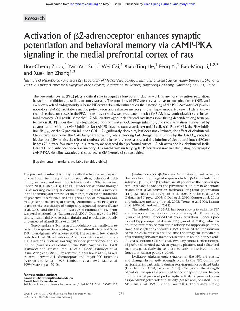
The b2-AR is present in pyramidal cells of the mPFCb2-AR expression in the PFC of humans (Kalaria et al. 1989) andnonhuman primates (Flugge et al. 1997) has been extensively in-vestigated. However, the distribution of b2-AR in the rodent PFCis unknown. We examined the expression of b2-AR in pyramidalcells of the rat PFC using a double immunofluorescence label-ing technique. CaMKII is a pyramidal cell marker (Muller et al.2006). Confocal images showed that b2-AR is expressed uniform-ly in pyramidal cells of both superficial layers (layer 2/3) and deeplayers (layer 5/6) of mPFC. Figure 1 shows that nearly all CaMKII-positive cells stained positively for b2-AR, which indicates thatb2-AR is widely present in pyramidal cells of the mPFC.
The activation of the b2-AR facilitates tLTPThe wide distribution of b2-AR in the pyramidal cells of the mPFCimplies that b2-AR has an important role in physiological func-tion. Therefore, we inquired whether activation of b2-AR could al-ter the synaptic plasticity of the mPFC. To test this question, weperformed whole-cell patch-clamp recordings from visually iden-tified pyramidal neurons in layer 2/3 of the mPFC slices and stim-ulated input by extracellular stimulation in layer 2/3 (Fig. 2A). Weidentified pyramidal cells by injecting depolarized currents intothe neurons to induce action potentials. The typical firing patternof the pyramidal cells showed significant firing frequency adapta-tions (Tsvetkov et al. 2002) (Fig. 2B, left). The injection of luciferyellow into the intracellular solution allowed us to identify pyra-midal cells based on the pyramid shape of their somata and theirapical dendrites (Fig. 2B, right).
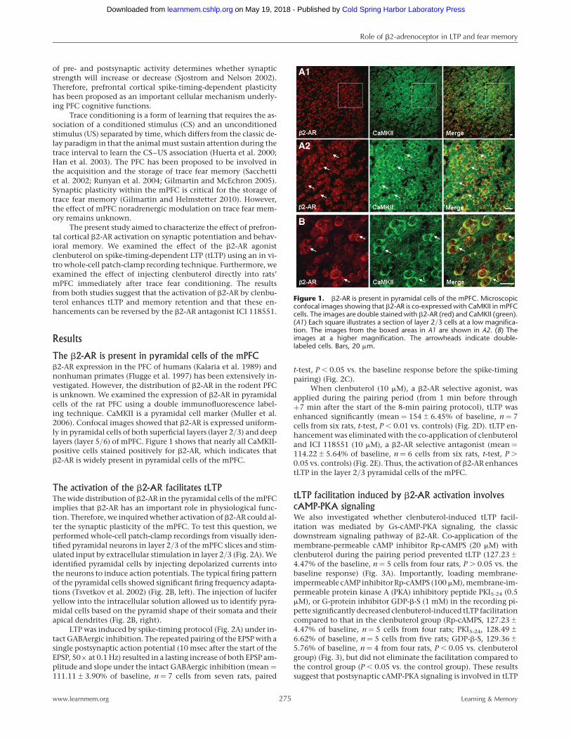
LTP was induced by spike-timing protocol (Fig. 2A) under in-tact GABAergic inhibition. The repeated pairing of the EPSP with asingle postsynaptic action potential (10 msec after the start of theEPSP, 50× at 0.1 Hz) resulted in a lasting increase of both EPSP am-plitude and slope under the intact GABAergic inhibition (mean ¼111.11+3.90% of baseline, n ¼ 7 cells from seven rats, paired
t-test, P , 0.05 vs. the baseline response before the spike-timingpairing) (Fig. 2C).
When clenbuterol (10 mM), a b2-AR selective agonist, wasapplied during the pairing period (from 1 min before through+7 min after the start of the 8-min pairing protocol), tLTP wasenhanced significantly (mean ¼ 154+6.45% of baseline, n ¼ 7cells from six rats, t-test, P , 0.01 vs. controls) (Fig. 2D). tLTP en-hancement was eliminated with the co-application of clenbuteroland ICI 118551 (10 mM), a b2-AR selective antagonist (mean ¼114.22+5.64% of baseline, n ¼ 6 cells from six rats, t-test, P .
0.05 vs. controls) (Fig. 2E). Thus, the activation of b2-AR enhancestLTP in the layer 2/3 pyramidal cells of the mPFC.
tLTP facilitation induced by b2-AR activation involves
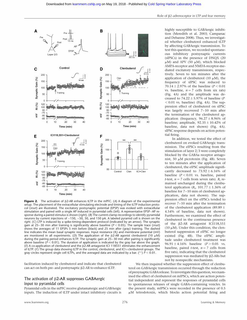
cAMP-PKA signalingWe also investigated whether clenbuterol-induced tLTP facil-itation was mediated by Gs-cAMP-PKA signaling, the classicdownstream signaling pathway of b2-AR. Co-application of themembrane-permeable cAMP inhibitor Rp-cAMPS (20 mM) withclenbuterol during the pairing period prevented tLTP (127.23+
4.47% of the baseline, n ¼ 5 cells from four rats, P . 0.05 vs. thebaseline response) (Fig. 3A). Importantly, loading membrane-impermeable cAMP inhibitor Rp-cAMPS (100mM), membrane-im-permeable protein kinase A (PKA) inhibitory peptide PKI5-24 (0.5mM), or G-protein inhibitor GDP-b-S (1 mM) in the recording pi-pette significantly decreased clenbuterol-induced tLTP facilitationcompared to that in the clenbuterol group (Rp-cAMPS, 127.23+
4.47% of baseline, n ¼ 5 cells from four rats; PKI5-24, 128.49+
6.62% of baseline, n ¼ 5 cells from five rats; GDP-b-S, 129.36+
5.76% of baseline, n ¼ 4 from four rats, P , 0.05 vs. clenbuterolgroup) (Fig. 3), but did not eliminate the facilitation compared tothe control group (P , 0.05 vs. the control group). These resultssuggest that postsynaptic cAMP-PKA signaling is involved in tLTP
Figure 1. b2-AR is present in pyramidal cells of the mPFC. Microscopicconfocal images showing that b2-AR is co-expressed with CaMKII in mPFCcells. The images are double stained with b2-AR (red) and CaMKII (green).(A1) Each square illustrates a section of layer 2/3 cells at a low magnifica-tion. The images from the boxed areas in A1 are shown in A2. (B) Theimages at a higher magnification. The arrowheads indicate double-labeled cells. Bars, 20 mm.
Role of b2-adrenoceptor in LTP and fear memory
www.learnmem.org 275 Learning & Memory
Cold Spring Harbor Laboratory Press on May 19, 2018 - Published by learnmem.cshlp.orgDownloaded from
facilitation induced by clenbuterol and indicate that clenbuterolcan act on both pre- and postsynaptic b2-AR to enhance tLTP.
The activation of b2-AR suppresses GABAergic
input to pyramidal cellsPyramidal cells in the mPFC receive glutamatergic and GABAergicinputs. The induction of tLTP under intact inhibitory circuits is
highly susceptible to GABAergic inhibi-tion (Meredith et al. 2003; Campanacand Debanne 2008). Thus, we investigat-ed whether clenbuterol enhanced tLTPby affecting GABAergic transmission. Totest this question, we recorded spontane-ous inhibitory postsynaptic currents(sIPSCs) in the presence of DNQX (20mM) and APV (50 mM), which blockedAMPA-receptor and NMDA-receptor-me-diated excitatory transmissions, respec-tively. Seven to ten minutes after theapplication of clenbuterol (10 mM), thefrequency of sIPSC was reduced to70.14+2.97% of the baseline (P , 0.01vs. baseline, n ¼ 7 cells from six rats)(Fig. 4A) and the amplitude was de-creased to 74.22+5.97% of baseline (P, 0.01 vs. baseline) (Fig. 4A). The sup-pression effect of clenbuterol on sIPSCwas largely recovered 7–10 min afterthe termination of the clenbuterol ap-plication (frequency, 96.27+8.96% ofbaseline; amplitude, 92.35+10.42% ofbaseline, data not shown) (Fig. 4A).sIPSC response depends on action poten-tial firing.
In addition, we tested the effect ofclenbuterol on evoked GABAergic trans-mission. The eIPSCs resulting from thestimulation of layer 2/3 were completelyblocked by the GABAa receptor antago-nist, 50 mM picrotoxin (Fig. 4B). Sevento ten minutes after the application ofclenbuterol, the eIPSC amplitude signifi-cantly decreased to 73.92+6.16% ofbaseline (P , 0.01 vs. baseline, pairedt-test, n ¼ 7 cells from seven rats). Ri re-mained unchanged during the clenbu-terol application (Ri, 101.77+1.36% ofbaseline for 7–10 min of clenbuterol ap-plication, data not shown). The sup-pression effect on the eIPSCs tended torecover 7–10 min after the terminationof the clenbuterol application (89.89+
4.54% of baseline, data not shown).Furthermore, we examined the effect ofclenbuterol in the continuous presenceof the b2-AR antagonist ICI118,551(10 mM). Under this condition, the clen-buterol suppression of eIPSC no longerexisted (Fig. 4B). The eIPSC ampli-tude under clenbuterol treatment was96.91+4.16% baseline (P . 0.05 vs.baseline, paired t-test, n ¼ 7 cells fromfive rats), indicating that the clenbuterolsuppression was mediated by b2-ARs butnot by nonspecific mechanisms.
We then inquired whether the suppression effect of clenbu-terol on GABAergic transmission occurred through the reductionofpresynapticGABArelease.To investigate thisquestion,weexam-ined the effect of clenbuterol on mIPSCs, which are action poten-tial independent and represent the responses of pyramidal cellsto spontaneous releases of single GABA-containing vesicles. Inthe present study, mIPSCs were recorded in the presence of 0.5mM tetrodotoxin, which blocks action potential firing and
Figure 2. The activation of b2-AR enhances tLTP in the mPFC. (A) A diagram of the experimentalsetup. The placement of the extracellular stimulating electrode and timing of the tLTP induction proto-col (inset) are illustrated. The excitatory postsynaptic potential (EPSP) was evoked with extracellularstimulation and paired with a single AP induced in pyramidal cells (left). A representative EPSP–AP re-sponse during a paired stimulus is shown (right). (B) The current-clamp recordings to identify pyramidalneurons by current injections of –150, –50, 50, and 150 pA. A labeled pyramid cell is shown on theright. (C) LTP is induced by a spike-timing-dependent protocol (indicated by an arrow). The synapticgain at 25–30 min after training is significantly above baseline (P , 0.05). The sample trace (inset)shows the averages of 11 EPSPs 5 min before (black) and 25 min after (gray) training. The dashedline indicates the mean basal synaptic responses. Input resistance (Ri) and membrane potential (mV)are monitored in all experiments. (D) The application of the b2-AR agonist clenbuterol (10 mM)during the pairing period enhances tLTP. The synaptic gain at 25–30 min after pairing is significantlyabove baseline (P , 0.01). The duration of application is indicated by the gray bar above the graph.(E) A co-application of clenbuterol and the b2-AR antagonist ICI 118551 eliminates the enhancementof tLTP. (F) The group data showing tLTP in the control, clenbuterol, and ICI+clenbuterol groups. Thegray circles represent single cell tLTPs, and the averaged data are indicated by a bar. (∗∗) P , 0.01.
Role of b2-adrenoceptor in LTP and fear memory
www.learnmem.org 276 Learning & Memory
Cold Spring Harbor Laboratory Press on May 19, 2018 - Published by learnmem.cshlp.orgDownloaded from
propagation.Figure4Cshowsthatduringa7–10 min bathapplica-tion of clenbuterol, the mIPSC frequency decreased to 74.92+
3.36% of baseline (P , 0.01 vs. controls) (Fig. 4B, middle) andmIPSC amplitude decreased to 82.83+2.02% of baseline (P ,
0.01 vs. controls, paired t-test, n ¼ 8 cells from eight rats) (Fig. 4B,right). The suppression effect of clenbuterol on mIPSCs was largelywashed out after the termination of the clenbuterol application(frequency, 90.60+5.38% of baseline; amplitude, 92.07+3.78%of controls, data not shown). These data suggest that clenbuterolacts on both pre- and postsynaptic b2-AR to suppress GABAergictransmission.
Taken together, these results demonstrate that clenbuterolsuppresses inhibitory input into layer 2/3 pyramidal cells by act-ing on b2-AR in both postsynaptic pyramidal cells and presynap-tic GABAergic terminals.
The activation of b2-AR has no effect on glutamatergic
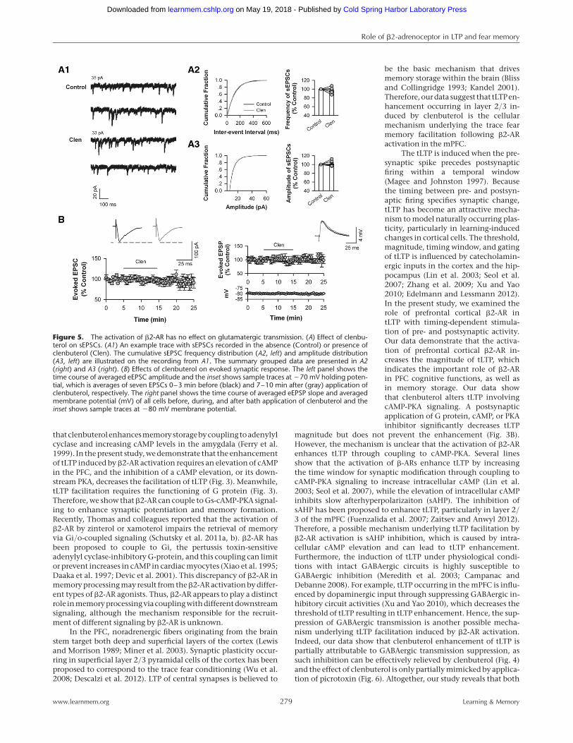
transmission to pyramidal cellsClenbuterol may also enhance tLTP through enhancing the excit-atory synaptic input into layer 2/3 pyramidal cells. To investigatethis mechanism, we recorded spontaneous excitatory postsynap-tic currents (sEPSCs) in the presence of picrotoxin (50 mM) toblock GABAA-mediated inhibitory transmission. Figure 5A showsthat both the frequency and amplitude of sEPSCs remained un-changed 7–10 min after the application of clenbuterol (10 mM)
(frequency, 97.21+2.89% of baseline,P . 0.05 vs. controls; amplitude,101.56+2.71% of baseline, P . 0.05vs. controls; n ¼ 7 cells from six rats).We further examined the effect of clen-buterol on evoked synaptic response.Figure 5B shows that clenbuterol hasno effect on both the amplitude ofevoked EPSC (eEPSC) and the slope ofevoked EPSP (eEPSP) (eEPSC, 97.53+
3.64% baseline, P . 0.05 vs. controls,n ¼ 8 from seven rats; eEPSP, 100.11+
8.9% baseline, P . 0.05 vs. controls,n ¼ 7 cells from four rats). These resultssuggest that the activation of b2-AR didnot alter the excitatory glutamatergicinput to the pyramidal cells.
The suppression of GABAergic
transmission partially mimics the
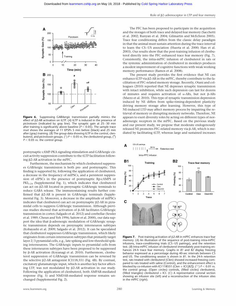
effect of b2-AR activation on tLTPtLTP induction at many excitatorysynapses is sensitive to GABAergic inhi-bition (Meredith et al. 2003; Campanacand Debanne 2008). The presented datashow that activation of b2-AR suppress-es GABAergic transmission. Then, weaskwhether thereductionofGABAergictransmission plays a dominant role intLTP facilitation induced by b2-AR acti-vation. To test this question, we addedGABAA receptor blocker picrotoxin(50 mM) in the perfusion artificial cere-brospinal fluid (ACSF) to block theGABAergic inhibitory circuit. Figure 6showsthat tLTP was induced in thepres-ence of picrotoxin (mean ¼ 124.26+
3.61% of baseline, n ¼ 5 cells from fourrats, paired t-test, P , 0.05 vs. the base-
line) (Fig. 6A) response before the spike-timing pairing and tLTPmagnitude was augmented significantly comparing with thecontrols, suggesting that GABAergic inhibition constrains tLTPin layer 2/3 pyramidal cells of mPFC (P , 0.05 vs. controls) (Fig.6B). Moreover, the magnitude of tLTP in the presence of picro-toxin dramatically lowered the magnitude of tLTP induced byclenbuterol (P , 0.01 vs. clenbuterol group) (Fig. 6B), suggestingthat suppression of GABAergic transmission partially but notcompletely mimics the effect of clenbuterol on tLTP. Togetherour data indicate that tLTP enhancement induced by b2-AR acti-vation under intact inhibitory circuit is partially mediated by sup-pressing GABAergic inhibition.
Post-training activation of b2-AR enhances trace fear
memory in the mPFCSynaptic plasticity within the cortex is critical for trace fear mem-ory (Gilmartin and Helmstetter 2010). LTP enhancement in layer2/3 pyramidal cells of the cortex has been proposed to correspondwith enhancement in trace fear conditioning (Wu et al. 2008).Thus, the enhancement of tLTP induced by clenbuterol maylead to an improved retention of trace fear memory. Therefore,we bilaterally infused clenbuterol (1 mg in 0.5 mL saline on eachside) into the prelimbic (PrL) area of the mPFC immediately post-training to examine the freezing response in a trace fear memorybehavioral task.
Figure 3. The activation of b2-AR enhances tLTP via cAMP-PKA signaling. (A) The co-application ofclenbuterol with permeable cAMP inhibitor Rp-cAMPS prevents tLTP. (B) Clenbuterol-potentiatedtLTP is significantly decreased but not completely eliminated by recording pipette-loaded Rp-cAMPS(Rp-cAMP(loaded)) (B1), PKI5-24 (PKI(loaded)) (B2), and GDP-b-S (GDP-b-S(loaded)) (B3). (C) A summaryof tLTP. (∗) P , 0.05 vs. the clenbuterol group, (#) P , 0.05 vs. the control group.
Role of b2-adrenoceptor in LTP and fear memory
www.learnmem.org 277 Learning & Memory
Cold Spring Harbor Laboratory Press on May 19, 2018 - Published by learnmem.cshlp.orgDownloaded from
During the seven CS–US pairings training, all rats showedcomparable increased freezing throughout the training sessions(Fig. 7B1). Trace fear memory retention was examined 24 h aftertraining. Before the tone was delivered, all rats displayed similarmovements in the testing chamber (F(2,27) ¼ 0.0729, P . 0.05,
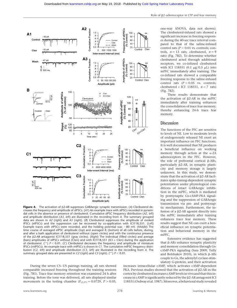
one-way ANOVA, data not shown).The clenbuterol-infused rats showed asignificant increase in freezing respons-es during the 40-sec trace interval com-pared to that of the saline-infusedcontrol rats (P , 0.01 vs. controls; con-trols, n ¼ 13 rats; clenbuterol, n ¼ 9rats) (Fig. 7B2). To determine whetherclenbuterol acted through additionalreceptors, we co-infused clenbuterolwith ICI 118551 (0.1 mg/0.5 mL) intomPFC immediately after training. Theco-infused rats showed a comparablefreezing response to the saline-infusedcontrol rats (P . 0.05 vs. controls;clenbuterol + ICI 118551, n ¼ 7 rats)(Fig. 7B2).
These results demonstrate thatthe activation of b2-AR in the mPFCimmediately after training enhancesthe consolidation of trace fear memory,thereby enhancing 24-h trace fearmemory.
Discussion
The functions of the PFC are sensitiveto levels of NE. Low to moderate levelsof endogenously released NE exert animportant influence on PFC functions.It is well documented that NE producesa beneficial influence on workingmemory through action at the a-2Aadrenoceptors in the PFC. However,the role of prefrontal cortical b-ARs,particularly b2-AR, in synaptic plasti-city and memory storage is largelyunknown. In this study, we demon-strate that the activation of b2-AR facil-itates spike-timing-dependent synapticpotentiation under physiological con-ditions of intact GABAergic inhibi-tion in the mPFC, which is mediatedby postsynaptic Gs-cAMP-PKA signal-ing and the suppression of GABAergictransmission via pre- and postsynap-tic mechanisms. Furthermore, the in-fusion of a b2-AR agonist directly intothe mPFC immediately after trainingenhances trace fear memory. Theseresults suggest that NE can exert a ben-eficial influence on synaptic potentia-tion and behavioral memory in themPFC.
Extensive evidence has suggestedthat b-ARs enhance synaptic plasticityand memory consolidation through Gs-cAMP-PKA signaling (Sara 2009; Tullyand Bolshakov 2010), in which b-ARscouple to Gs, the adenylyl cyclase-stim-ulatory G-protein, and their activation
increases intracellular cAMP, which activates cAMP-dependentPKA. Previous studies showed that the activation of b2-AR in thecortexbyclenbuterol increasescAMPlevels invivoandthat this in-crease in cAMP is significantly reduced by theb2-AR antagonist ICI118551(Ordwayetal.1987).Moreover, abehavioral studyrevealed
Figure 4. The activation of b2-AR suppresses GABAergic synaptic transmission. (A) Clenbuterol de-creases the frequency and amplitude of sIPSCs. (A1) An example trace with sIPSCs recorded in pyrami-dal cells in the absence or presence of clenbuterol. Cumulative sIPSC frequency distribution (A2, left)and amplitude distribution (A3, left) are illustrated in the recording from A. The summary groupeddata are shown in A2 (right) and A3 (right). (B) Clenbuterol suppresses the amplitude of evokedIPSCs (eIPSCs) and the suppression can be recovered by co-application with ICI118,551. (Left)Example traces with eIPSCs were recorded, and the holding potential was 280 mV. (Middle) Thetime course of averaged eIPSC amplitude (top) and averaged Ri (bottom) of all cells before, during,and after a bath application of clenbuterol without (open circles) and with the continuous presenceof the b2-AR antagonist ICI118,551 (gray circles). (Right) The individual (filled circles) and average(bars) amplitudes of eIPSCs without (Clen) and with ICI118,551 (ICI + Clen) during the applicationof clenbuterol. (∗∗) P , 0.01. (C) Clenbuterol decreases the frequency and amplitude of miniatureIPSCs (mIPSCs). An example trace with mIPSCs is shown in C1. The cumulative mIPSC frequency distri-bution (C2, left) and amplitude distribution (C3, left) are illustrated in the recording from A. Thesummary grouped data are presented in C2 (right) and C3 (right). (∗∗) P , 0.01.
Role of b2-adrenoceptor in LTP and fear memory
www.learnmem.org 278 Learning & Memory
Cold Spring Harbor Laboratory Press on May 19, 2018 - Published by learnmem.cshlp.orgDownloaded from
thatclenbuterolenhancesmemorystoragebycouplingtoadenylylcyclase and increasing cAMP levels in the amygdala (Ferry et al.1999). In the present study, we demonstrate that the enhancementof tLTP induced byb2-AR activation requires an elevation of cAMPin the PFC, and the inhibition of a cAMP elevation, or its down-stream PKA, decreases the facilitation of tLTP (Fig. 3). Meanwhile,tLTP facilitation requires the functioning of G protein (Fig. 3).Therefore, we show thatb2-AR can couple to Gs-cAMP-PKA signal-ing to enhance synaptic potentiation and memory formation.Recently, Thomas and colleagues reported that the activation ofb2-AR by zinterol or xamoterol impairs the retrieval of memoryvia Gi/o-coupled signaling (Schutsky et al. 2011a, b). b2-AR hasbeen proposed to couple to Gi, the pertussis toxin-sensitiveadenylyl cyclase-inhibitory G-protein, and this coupling can limitor prevent increases in cAMP in cardiac myocytes (Xiao et al. 1995;Daaka et al. 1997; Devic et al. 2001). This discrepancy of b2-AR inmemory processing mayresult from theb2-AR activation by differ-ent types of b2-AR agonists. Thus, b2-AR appears to play a distinctrole inmemoryprocessingviacouplingwithdifferentdownstreamsignaling, although the mechanism responsible for the recruit-ment of different signaling by b2-AR is unknown.
In the PFC, noradrenergic fibers originating from the brainstem target both deep and superficial layers of the cortex (Lewisand Morrison 1989; Miner et al. 2003). Synaptic plasticity occur-ring in superficial layer 2/3 pyramidal cells of the cortex has beenproposed to correspond to the trace fear conditioning (Wu et al.2008; Descalzi et al. 2012). LTP of central synapses is believed to
be the basic mechanism that drivesmemory storage within the brain (Blissand Collingridge 1993; Kandel 2001).Therefore,ourdata suggest that tLTPen-hancement occurring in layer 2/3 in-duced by clenbuterol is the cellularmechanism underlying the trace fearmemory facilitation following b2-ARactivation in the mPFC.
The tLTP is induced when the pre-synaptic spike precedes postsynapticfiring within a temporal window(Magee and Johnston 1997). Becausethe timing between pre- and postsyn-aptic firing specifies synaptic change,tLTP has become an attractive mecha-nism to model naturally occurring plas-ticity, particularly in learning-inducedchanges in cortical cells. The threshold,magnitude, timing window, and gatingof tLTP is influenced by catecholamin-ergic inputs in the cortex and the hip-pocampus (Lin et al. 2003; Seol et al.2007; Zhang et al. 2009; Xu and Yao2010; Edelmann and Lessmann 2012).In the present study, we examined therole of prefrontal cortical b2-AR intLTP with timing-dependent stimula-tion of pre- and postsynaptic activity.Our data demonstrate that the activa-tion of prefrontal cortical b2-AR in-creases the magnitude of tLTP, whichindicates the important role of b2-ARin PFC cognitive functions, as well asin memory storage. Our data showthat clenbuterol alters tLTP involvingcAMP-PKA signaling. A postsynapticapplication of G protein, cAMP, or PKAinhibitor significantly decreases tLTP
magnitude but does not prevent the enhancement (Fig. 3B).However, the mechanism is unclear that the activation of b2-ARenhances tLTP through coupling to cAMP-PKA. Several linesshow that the activation of b-ARs enhance tLTP by increasingthe time window for synaptic modification through coupling tocAMP-PKA signaling to increase intracellular cAMP (Lin et al.2003; Seol et al. 2007), while the elevation of intracellular cAMPinhibits slow afterhyperpolarizaiton (sAHP). The inhibition ofsAHP has been proposed to enhance tLTP, particularly in layer 2/
3 of the mPFC (Fuenzalida et al. 2007; Zaitsev and Anwyl 2012).Therefore, a possible mechanism underlying tLTP facilitation byb2-AR activation is sAHP inhibition, which is caused by intra-cellular cAMP elevation and can lead to tLTP enhancement.Furthermore, the induction of tLTP under physiological condi-tions with intact GABAergic circuits is highly susceptible toGABAergic inhibition (Meredith et al. 2003; Campanac andDebanne 2008). For example, tLTP occurring in the mPFC is influ-enced by dopaminergic input through suppressing GABAergic in-hibitory circuit activities (Xu and Yao 2010), which decreases thethreshold of tLTP resulting in tLTP enhancement. Hence, the sup-pression of GABAergic transmission is another possible mecha-nism underlying tLTP facilitation induced by b2-AR activation.Indeed, our data show that clenbuterol enhancement of tLTP ispartially attributable to GABAergic transmission suppression, assuch inhibition can be effectively relieved by clenbuterol (Fig. 4)and the effect of clenbuterol is only partially mimicked by applica-tion of picrotoxin (Fig. 6). Altogether, our study reveals that both
Figure 5. The activation of b2-AR has no effect on glutamatergic transmission. (A) Effect of clenbu-terol on sEPSCs. (A1) An example trace with sEPSCs recorded in the absence (Control) or presence ofclenbuterol (Clen). The cumulative sEPSC frequency distribution (A2, left) and amplitude distribution(A3, left) are illustrated on the recording from A1. The summary grouped data are presented in A2(right) and A3 (right). (B) Effects of clenbuterol on evoked synaptic response. The left panel shows thetime course of averaged eEPSC amplitude and the inset shows sample traces at 270 mV holding poten-tial, which is averages of seven EPSCs 0–3 min before (black) and 7–10 min after (gray) application ofclenbuterol, respectively. The right panel shows the time course of averaged eEPSP slope and averagedmembrane potential (mV) of all cells before, during, and after bath application of clenbuterol and theinset shows sample traces at 280 mV membrane potential.
Role of b2-adrenoceptor in LTP and fear memory
www.learnmem.org 279 Learning & Memory
Cold Spring Harbor Laboratory Press on May 19, 2018 - Published by learnmem.cshlp.orgDownloaded from
postsynaptic cAMP-PKA signaling stimulation and GABAergic cir-cuit activity suppression contribute to the tLTP facilitation follow-ing b2-AR activation in the mPFC.
Furthermore, the mechanism by which clenbuterol suppress-es GABAergic transmission is both pre- and postsynaptic. Thisfinding is supported by, following the application of clenbuterol,a decrease in the frequency of mIPSCs, and a persistent suppres-sion of eIPSCs in the presence of postsynaptic Rp-cAMPS orPKI5-24 (Supplemental Fig. 1), which indicates that clenbuterolcan act on b2-AR located in presynaptic GABAergic terminals toreduce GABA release. The immunostaining results further con-firmed that b2-AR is present in GABAergic terminals (Supple-mental Fig. 3). Moreover, a decrease in the amplitude of mIPSCsindicates that clenbuterol can act on postsynaptic b2-AR in pyra-midal cells to suppress GABAergic transmission. Although previ-ous studies showed that activation of b-AR facilitates GABAergictransmission in cortex (Salgado et al. 2012) and cerebellar (Sessleret al. 1989; Cheun and Yeh 1996; Saitow et al. 2000), our data sup-port the idea that b-adrenergic modulation of GABAergic synap-tic transmission depends on presynaptic interneuron subtypes(Kobayashi et al. 2009; Salgado et al. 2012). It can be speculatedthat clenbuterol suppresses GABAergic transmission, which likelyoriginates from certain interneuron subtypes that primarily targetlayer 2/3 pyramidal cells, e.g., late spiking and low-threshold spik-ing interneurons. The GABAergic inputs to pyramidal cells fromthese interneuron subtypes have been proposed to be suppressedby b-AR activation (Koyanagi et al. 2010). Furthermore, clenbu-terol suppression of GABAergic transmission can be reversed bythe selective b2-AR antagonist ICI118,551 (Fig. 4B). By contrast,excitatory glutamatergic input, which is another key factor affect-ing LTP, was not modulated by b2-AR activation in our study.Following the application of clenbuterol, both AMPAR-mediatedresponse (Fig. 5) and NMDAR-mediated response remains un-changed (Supplemental Fig. 2).
The PFC has been proposed to participate in the acquisitionand the storages of both trace and delayed fear memory (Sacchettiet al. 2002; Runyan et al. 2004; Gilmartin and McEchron 2005).Trace fear conditioning differs from the classic delay paradigmin that the animal must sustain attention during the trace intervalto learn the CS–US association (Huerta et al. 2000; Han et al.2003). Our results show that the post-training infusion of clenbu-terol directly into the PFC enhanced trace fear memory (Fig. 7).Consistently, the intra-mPFC infusion of clenbuterol in rats orthe systemic administration of clenbuterol in monkeys producesa modest improvement of cognitive functions with weak workingmemory performance (Ramos et al. 2008).
The present study provides the first evidence that NE canenhance tLTP via b2-AR in the mPFC, thereby contribute to the fa-cilitation of PFC-related memory storage. Recently, Otani and col-leagues (2010) reported that NE depresses synaptic transmissionwith intact inhibition, while such depression can last for dozensof minutes and requires activation of a-ARs, but not b-ARs(Marzo et al. 2010). This type of synaptic transmission depressioninduced by NE differs from spike-timing-dependent plasticitydriving memory storage after learning. However, this type ofNE-induced LTD may affect memory process by impairing the re-trieval of memory or disrupting memory networks. Therefore, NEappears to exert diversity roles by acting on different types of nor-adrenergic receptors in the mPFC. Based on the previous studyand our present study, we propose that moderate endogenouslyreleased NE promotes PFC-related memory via b-AR, which is me-diated by facilitating tLTP, whereas large and sustained increases
Figure 6. Suppressing GABAergic transmission partially mimics theeffect of b2-AR activation on tLTP. (A) tLTP is induced in the presence ofpictrotoxin (indicated by gray line). The synaptic gain at 25–30 minafter training is significantly above baseline (P , 0.05). The sample traceinset shows the averages of 11 EPSPs 5 min before (black) and 25 minafter (gray) training. (B) The group data showing tLTP in the control, clen-buterol, and pictrotoxin groups. (∗) P , 0.05 vs. the clenbuterol group, (#)P , 0.05 vs. the control group.
Figure 7. Post-training activation of b2-AR in mPFC enhances trace fearmemory. (A) An illustration of the sequence of post-training intra-mPFCinfusions, trace-conditioning trials (CS–US pairings), and the retentiontest. (B) Intra-mPFC infusion of clenbuterol immediately post-training en-hances 24-h trace fear memory. Graphs in B1 and B2 display freezingbehavior expressed as a percentage during 40-sec intervals between CSand US. The conditioning session is shown in B1. In the 24-h retentiontest, rats treated with clenbuterol (Clen) showed increased freezing com-pared to rats treated with saline (Control), and the enhancement effect isblocked by co-infusion with ICI 118551 (Clen + ICI [B2]). (∗∗) P , 0.01 vs.the control group. (Open circles) controls, (filled circles) clenbuterol,(filled triangles) clenbuterol + ICI. (C) A representative coronal sectionshowing an infusion site (left) and a reconstruction of the infusion sitesin the mPFC (right).
Role of b2-adrenoceptor in LTP and fear memory
www.learnmem.org 280 Learning & Memory
Cold Spring Harbor Laboratory Press on May 19, 2018 - Published by learnmem.cshlp.orgDownloaded from

of endogenous NE (e.g., stress) impede PFC-related memory viaa-AR, which is caused by impairing the retrieval of memory or dis-rupting memory networks.
In conclusion, the present study demonstrates that prefron-tal cortical b2-AR facilitates tLTP and enhances fear memory stor-age elicited by trace fear conditioning. Moreover, activation ofb2-AR enhances tLTP by coupling to postsynaptic cAMP-PKA cas-cades and suppressing inhibitory circuits with the involvement ofboth pre- and postsynaptic b2-AR.
Materials and Methods
Electrophysiological experiment
Subjects
Male Sprague-Dawley rats that were 4–6 wk old (100–120 g)were purchased from SLACCAS, Shanghai, China. The rats werehoused under a 12-h light/dark cycle; food and water were avail-able ad libitum. The experiments in the present study wereperformed in accordance with the guidelines published in theNIH Guide for the Care and Use of Laboratory Animals. Effortswere made to minimize the number of animals used and theirsuffering.
Slice preparation
Coronal brain slices (300 mm) containing PFC were preparedusing standard methods (Ji et al. 2008). The slices were trans-ferred to a submerged recovery chamber containing oxygen-ated (95% O2 and 5% CO2) ACSF (124 mM NaCl, 2.5 mMKCl, 2.5 mM CaCl2, 1.3 mM MgCl2, 26.2 mM NaHCO3, 1.25mM NaH2PO4, and 11 mM glucose) at room temperature for atleast 1 h.
Whole-cell recordings
Whole-cell current-clamp recordings were performed using stan-dard procedures at room temperature (Couey et al. 2007). Slicescontaining PFC were positioned in a perfusion chamber attachedto the fixed stage of an Olympus BX51 microscope (Olympus)with infrared DIC optics for visualizing the whole-cell patch-clamp recordings. eEPSPs were generated with a pulse from astimulation isolation unit controlled by a Master-8 pulse gene-rator (A.M.P.I. Instruments). EPSPs were recorded from pyrami-dal cells in layer 2/3 using an Axon 200B amplifier (AxonInstruments), and stimulations were delivered using a bipolartungsten stimulating electrode placed in layer 2/3 approximately100 mm lateral to the recorded pyramidal cell (Fig. 1A). EPSPswere evoked every 30 sec, and the neuron membrane potentialswere maintained at 280 mV. The recording pipettes (3–5 MV)were filled with a solution containing 145 mM K-gluconate, 5mM NaCl, 1 mM MgCl2, 0.2 mM EGTA, 10 mM HEPES, 2 mMMg-ATP, 0.1 mM Na3-GTP, and 10 mM phosphocreatine (adjust-ed to pH 7.2 with KOH). After obtaining stable EPSPs for 10 min,a tLTP induction paradigm was used within 12 min after estab-lishing the whole-cell configuration to prevent a washout effecton the tLTP induction. The slope of the initial 2.5 msec of theEPSP was analyzed to ensure that the data reflected only themonosynaptic component of each experiment (Froemke et al.2005). Synaptic gain was measured as the change in the averageEPSP slope; namely, when a 5-min period that occurred 25–30-min post-pairing was compared to the EPSP slope measuredin the last 5 min of the 10-min baseline recordings. During thepairing period, pre- and postsynaptic stimulus pairing was re-peated 50 times, with a 10-sec interval between each pairing(Couey et al. 2007). Throughout the experiments, cell Ri wasmonitored by applying a 10 pA, 500 msec hyperpolarizing pulseat the end of each sweep. Data were not included in the analysisif the cell Ri varied by more than +20% during the experiment.To assess the effect of b2-AR stimulation in these experiments,the b2-AR agonist clenbuterol was applied during the pairing pe-
riod (from 1 min before through +7 min after the start of the8-min pairing protocol). For the experiments with recording pi-pette loading drugs, recordings were begun at least 10 min afterrupturing the cell membrane. The Wilcoxon signed-rank testand Mann–Whitney U-test were used to assess significance inthe tLTP data.
For the IPSC recordings, the pipette solution contained (inmM) 70 K-gluconate, 70 KCl, 20 HEPES, 0.5 CaCl2, 5.0 EGTA,and 5 Mg-ATP, and the pH was adjusted to 7.2 with KOH. Thecell membrane potentials were maintained at 270 mV. The fre-quencies and amplitudes of sIPSC/mIPSC/sEPSC were analyzedusing the Mini Analysis software package (v8.0, Synaptosoft,http://www.synaptosoft.com). Drug-induced changes in cumu-lative fractions of sIPSC/mIPSC/sEPSC amplitudes and inter-event intervals were analyzed for statistical significance usingthe Kolmogorov–Smirnov (K-S) test (Mini Analysis v8.0). Allgrouped data were analyzed using paired or unpaired t-testsand a critical probability of P , 0.05 (STATISTICA 6.0).
Behavioral experiment
Subjects
Male Sprague-Dawley rats (7–8 wk, 180–200 g) were purchasedfrom SLACCAS, Shanghai, China. The rats were housed in plasticcages (1–2 per cage) and placed on a 12-h light/dark cycle. Foodand water were provided ad libitum throughout the experiment.
Surgery
The rats were anesthetized with sodium pentobarbital (40 mg/kgi.p.). Stainless steel guide cannulae (23-gauge) were bilaterally po-sitioned above the PrL area of the mPFC based on the coordinates(Paxinos and Watson 1986): bregma +3.2 mm, lateral +0.75 mm,and a depth of 21.5 mm from the skull surface. The guide cannu-lae were fixed to the skull with dental cement. Dummy cannulae,cut 0.5 mm longer than the guide cannulae, were inserted into theguide cannulae to prevent clogging and reduce the risk of infec-tion. The rats were provided a minimum recovery of 5 d beforethe experimental procedures were performed.
Drug administration
The b2-AR agonist clenbuterol (1 mg in 0.5 mL saline per side) andthe b2-AR antagonist ICI 118551 (0.1 mg in 0.5 mL saline per side)were prepared as concentrated stock solutions in distilled water.For drug administration, the rats were gently held while the dum-my cannulae were exchanged with 30-gauge infusion cannulae.The tip of the injection needle was 2.7 mm beyond that of theguide cannula, which yielded a total distance of 4.2 mm fromthe skull surface and reached to the PrL area. The drug solutionwas infused bilaterally with an infusion pump at a rate of 0.2 mLper min. Each side of the mPFC was infused with a total volumeof 0.5 mL drug solution. After the infusion, the infusion cannulaewere left in place for an additional 2 min to allow the drug solu-tion to diffuse from the tip of the cannulae. The dummy cannulaewere then replaced and the rats were returned to their home cages.The infusions were performed immediately after training.
Behavioral procedure
All behavioral tests were performed by an investigator who wasblind to the treatment groups. A trace fear conditioning protocol,with a minor modification, was performed as previously described(Runyan et al. 2004). The animals were briefly placed in the train-ing context (Med Associates Inc.) and provided a 120-sec habitu-ation period. The conditioning trials began with a 10-sec tone (2.2kHz, 80 dB, CS) followed by a 40-sec trace period before the animalreceived a 0.5-sec (0.5 mA) foot shock (US). The rats’ freezingbehavior was monitored by a camera. The percentage of freezingwas measured every 1 sec during the trace period (40 sec) afterthe tone. The conditioning trial was followed by a pseudo-randomintertrial interval (ITI) that varied between 1 and 3 min. The
Role of b2-adrenoceptor in LTP and fear memory
www.learnmem.org 281 Learning & Memory
Cold Spring Harbor Laboratory Press on May 19, 2018 - Published by learnmem.cshlp.orgDownloaded from
conditioning trial was repeated seven times during a 16-min train-ing period.
The retention of a CS–US association was measured at 24 hby monitoring freezing behavior. During trace memory reten-tion testing, the animal was placed in a novel context and pro-vided with a 120-sec habituation period. In the absence of afoot shock, a 10-sec tone was produced followed by a varied ITIperiod. The retention trial was repeated four times. Only three re-tention trials were used for the analyses. The percentage of freez-ing was measured every 1 sec during the trace period (40 sec) afterthe tone.
ChemicalsAll reagents were purchased from Sigma-Aldrich with the ex-ceptions of Rp-cAMPS and PKI5-24 (the PKA inhibitor frag-ment 5-24), which were purchased from Tocris Bioscience. Allchannel blockers were prepared as concentrated stock solutionsin distilled water or DMSO and were either added immediatelyto the ACSF in working concentrations or stored at 220˚C for sub-sequent use.
ImmunostainingThe rats (approximately 30-d old and 100 g) were deeply anesthe-tized with pentobarbital sodium (45 mg/kg, i.p.), and a transcar-dial perfusion was performed with 300 mL of 37˚C salinefollowed by 200 mL of 4% ice-cold paraformaldehyde (PFA) dis-solved in phosphate-buffered saline (PBS, pH 7.4). The brainswere removed and fixed for 2 h in PFA at 4˚C. The brains werethen placed in 30% (w/v) sucrose solution. Coronal sections (35mm) were cut using a cryostat (Leica CM900). The sections wererinsed with 0.01 M PBS and incubated in a solution of 0.5%Triton-X in PBS for 30 min followed by 10% normal goat serum(Invitrogen Immunodetection) for 2 h at room temperature.
For double immunofluorescence, the sections were incubatedin a mixture of rabbit anti-b2-AR (1:25; Catalogue No. SC-9042,Santa Cruz) and mouse anti-CaMKII (1:120, Catalogue No.MAB8699, Lot No. LV1377413; Chemicon International), whichis a pyramidal cell marker (Muller et al. 2006). The primary anti-bodies were diluted in goat serum and incubated for 48 h at 4˚C.After a 48-h incubation, the sections were rinsed with PBS andthe appropriate secondary antibody was applied. Fluorescentsecondary antibodies (whole-IgG affinity-purified antibodies,Alexa Fluor 568 goat anti-rabbit, and Alexa Fluor 488 goat anti-mouse, from Jackson ImmunoResearch Laboratories) were appliedto the tissues at a dilution of 1:400 in normal blocking serum for 2h at 4˚C.
The immunolabeled sections were examined and all imageswere obtained using a confocal laser-scanning microscope system(Olympus FV 1000). Twelve-bit images were captured at a resolu-tion of 1024 × 1024 pixels using 20 × PlanApo and 40×PlanApoimmersion (1.2 NA) objectives. Immunoreactivity was examinedunder a condition of optimal resolution. The pinhole diameterwas set to 105mm to significantly reduce the contribution of cyto-plasmic fluorescence. Z-sectioning was performed at 1 to 2 mm in-tervals, and stacks of optical sections on the z-axis were acquired.Confocal photomicrographs were further processed using AdobePhotoshop. The pictures were not modified except for adjustmentsof scaling, levels, brightness, and contrast. No immunolabelingwas observed in the control slices in which the primary antibodywas omitted. A multi-tracking configuration was used to eliminatecrosstalk between the fluorescent detection channels.
Data analysis and statisticsData are expressed as the mean+ SEM in all cases, with P , 0.05indicating significance. Data between two groups were statisti-cally compared using t-tests, while data among the groups wereanalyzed using a one-way analysis of variance (ANOVA) withplanned comparisons as post hoc analyses using STATISTICA(StatSoft).
AcknowledgmentsThis work was supported by grants from the National NaturalScience Foundation of China (31271171, 30990263, and30821002 to B.-M.L.; 31121061 and 30700218 to X.-H.Z.) and theMinistry of Science and Technology of China (2011CBA00406to B.-M.L.).
ReferencesArnsten AF, Goldman-Rakic PS. 1985. a2-adrenergic mechanisms in
prefrontal cortex associated with cognitive decline in aged nonhumanprimates. Science 230: 1273–1276.
Arnsten AF, Jentsch JD. 1997. The a-1 adrenergic agonist, cirazoline,impairs spatial working memory performance in aged monkeys.Pharmacol Biochem Behav 58: 55–59.
Arnsten AF, Cai JX, Goldman-Rakic PS. 1988. The a-2 adrenergic agonistguanfacine improves memory in aged monkeys without sedative orhypotensive side effects: Evidence for a-2 receptor subtypes. J Neurosci8: 4287–4298.
Berridge CW, Waterhouse BD. 2003. The locus coeruleus–noradrenergicsystem: Modulation of behavioral state and state-dependent cognitiveprocesses. Brain Res 42: 33–84.
Bi G, Poo M. 2001. Synaptic modification by correlated activity: Hebb’spostulate revisited. Annu Rev Neurosci 24: 139–166.
Birnbaum S, Gobeske KT, Auerbach J, Taylor JR, Arnsten AF. 1999. A role fornorepinephrine in stress-induced cognitive deficits: a-1-adrenoceptormediation in the prefrontal cortex. Biol Psychiatry 46: 1266–1274.
Bliss TV, Collingridge GL. 1993. A synaptic model of memory: Long-termpotentiation in the hippocampus. Nature 361: 31–39.
Campanac E, Debanne D. 2008. Spike timing-dependent plasticity: Alearning rule for dendritic integration in rat CA1 pyramidal neurons.J Physiol 586: 779–793.
Cheun JE, Yeh HH. 1996. Noradrenergic potentiation of cerebellar Purkinjecell responses to GABA: Cyclic AMP as intracellular intermediary.Neuroscience 74: 835–844.
Connor SA, Wang YT, Nguyen PV. 2011. Activation of b-adrenergicreceptors facilitates heterosynaptic translation-dependent long-termpotentiation. J Physiol 589: 4321–4340.
Couey JJ, Meredith RM, Spijker S, Poorthuis RB, Smit AB, Brussaard AB,Mansvelder HD. 2007. Distributed network actions by nicotine increasethe threshold for spike-timing-dependent plasticity in prefrontalcortex. Neuron 54: 73–87.
Daaka Y, Luttrell LM, Lefkowitz RJ. 1997. Switching of the coupling of theb2-adrenergic receptor to different G proteins by protein kinase A.Nature 390: 88–91.
Descalzi G, Li XY, Chen T, Mercaldo V, Koga K, Zhuo M. 2012. Rapidsynaptic potentiation within the anterior cingulate cortex mediatestrace fear learning. Mol Brain 5: 6.
Devic E, Xiang Y, Gould D, Kobilka B. 2001. b-adrenergic receptorsubtype-specific signaling in cardiac myocytes from b(1) and b(2)adrenoceptor knockout mice. Mol Pharmacol 60: 577–583.
Dias R, Robbins TW, Roberts AC. 1997. Dissociable forms of inhibitorycontrol within prefrontal cortex with an analog of the Wisconsin CardSort Test: Restriction to novel situations and independence from“on-line” processing. J Neurosci 17: 9285–9297.
Edelmann E, Lessmann V. 2012. Dopamine modulates spiketiming-dependent plasticity and action potential properties in CA1pyramidal neurons of acute rat hippocampal slices. Front SynapticNeurosci 3: 6.
Ferry B, Roozendaal B, McGaugh JL. 1999. Basolateral amygdalanoradrenergic influences on memory storage are mediated by aninteraction between b- and a1-adrenoceptors. J Neurosci 19:5119–5123.
Flugge G, Ahrens O, Fuchs E. 1997. b-adrenoceptors in the tree shrewbrain. I. Distribution and characterization of [125I]iodocyanopindololbinding sites. Cell Mol Neurobiol 17: 401–415.
Franowicz JS, Arnsten AF. 1998. The a-2a noradrenergic agonist,guanfacine, improves delayed response performance in young adultrhesus monkeys. Psychopharmacology 136: 8–14.
Franowicz JS, Kessler LE, Borja CM, Kobilka BK, Limbird LE, Arnsten AF.2002. Mutation of the a2A-adrenoceptor impairs working memoryperformance and annuls cognitive enhancement by guanfacine. JNeurosci 22: 8771–8777.
Froemke RC, Poo MM, Dan Y. 2005. Spike-timing-dependent synapticplasticity depends on dendritic location. Nature 434: 221–225.
Fuenzalida M, Fernandez de Sevilla D, Buno W. 2007. Changes of the EPSPwaveform regulate the temporal window for spike-timing-dependentplasticity. J Neurosci 27: 11940–11948.
Fuster JM. 2003. Cortex and mind: unifying cognition, pp. 155–164. OxfordUniversity Press, New York.
Role of b2-adrenoceptor in LTP and fear memory
www.learnmem.org 282 Learning & Memory
Cold Spring Harbor Laboratory Press on May 19, 2018 - Published by learnmem.cshlp.orgDownloaded from
Fuster JM, Bodner M, Kroger JK. 2000. Cross-modal and cross-temporalassociation in neurons of frontal cortex. Nature 405: 347–351.
Gelinas JN, Nguyen PV. 2005. b-adrenergic receptor activation facilitatesinduction of a protein synthesis-dependent late phase of long-termpotentiation. J Neurosci 25: 3294–3303.
Gilmartin MR, Helmstetter FJ. 2010. Trace and contextual fearconditioning require neural activity and NMDA receptor-dependent transmission in the medial prefrontal cortex. Learn Mem17: 289–296.
Gilmartin MR, McEchron MD. 2005. Single neurons in the medialprefrontal cortex of the rat exhibit tonic and phasic coding during tracefear conditioning. Behav Neurosci 119: 1496–1510.
Goldman-Rakic PS. 1987. Circuitry of the primate prefrontal cortex and theregulation of behavior by representational memory. In Handbook ofphysiology V (ed. Plum F), pp. 373–417. American Physiological Society,Bethesda.
Han CJ, O’Tuathaigh CM, van Trigt L, Quinn JJ, Fanselow MS, Mongeau R,Koch C, Anderson DJ. 2003. Trace but not delay fear conditioningrequires attention and the anterior cingulate cortex. Proc Natl Acad Sci100: 13087–13092.
Huerta PT, Sun LD, Wilson MA, Tonegawa S. 2000. Formation of temporalmemory requires NMDA receptors within CA1 pyramidal neurons.Neuron 25: 473–480.
Introini-Collison IB, Miyazaki B, McGaugh JL. 1991. Involvement of theamygdala in the memory-enhancing effects of clenbuterol.Psychopharmacology 104: 541–544.
Jay TM, Burette F, Laroche S. 1995. NMDA receptor-dependent long-termpotentiation in the hippocampal afferent fibre system to the prefrontalcortex in the rat. Eur J Neurosci 7: 247–250.
Ji JZ, Wang XM, Li BM. 2003. Deficit in long-term contextual fear memoryinduced by blockade of b-adrenoceptors in hippocampal CA1 region.Eur J Neurosci 17: 1947–1952.
Ji XH, Cao XH, Zhang CL, Feng ZJ, Zhang XH, Ma L, Li BM. 2008. Pre- andpostsynaptic b-adrenergic activation enhances excitatory synaptictransmission in layer V/VI pyramidal neurons of the medial prefrontalcortex of rats. Cereb Cortex 18: 1506–1520.
Kalaria RN, Andorn AC, Tabaton M, Whitehouse PJ, Harik SI, Unnerstall JR.1989. Adrenergic receptors in aging and Alzheimer’s disease: Increasedb 2-receptors in prefrontal cortex and hippocampus. J Neurochem 53:1772–1781.
Kandel ER. 2001. The molecular biology of memory storage: A dialogbetween genes and synapses. Biosci Rep 21: 565–611.
Katsuki H, Izumi Y, Zorumski CF. 1997. Noradrenergic regulation ofsynaptic plasticity in the hippocampal CA1 region. J Neurophysiol77: 3013–3020.
Kobayashi M, Kojima M, Koyanagi Y, Adachi K, Imamura K,Koshikawa N. 2009. Presynaptic and postsynaptic modulation ofglutamatergic synaptic transmission by activation of a(1)- andb-adrenoceptors in layer V pyramidal neurons of rat cerebral cortex.Synapse 63: 269–281.
Koyanagi Y, Yamamoto K, Oi Y, Koshikawa N, Kobayashi M. 2010.Presynaptic interneuron subtype- and age-dependent modulation ofGABAergic synaptic transmission by b-adrenoceptors in rat insularcortex. J Neurophysiol 103: 2876–2888.
Laroche S, Jay TM, Thierry AM. 1990. Long-term potentiation in theprefrontal cortex following stimulation of the hippocampal CA1/subicular region. Neurosci Lett 114: 184–190.
Lemon N, Aydin-Abidin S, Funke K, Manahan-Vaughan D. 2009. Locuscoeruleus activation facilitates memory encoding and induceshippocampal LTD that depends on b-adrenergic receptor activation.Cereb Cortex 19: 2827–2837.
Lewis DA, Morrison JH. 1989. Noradrenergic innervation of monkeyprefrontal cortex: A dopamine-b-hydroxylase immunohistochemicalstudy. J Comp Neurol 282: 317–330.
Li BM, Mao ZM, Wang M, Mei ZT. 1999. a-2 adrenergic modulation ofprefrontal cortical neuronal activity related to spatial working memoryin monkeys. Neuropsychopharmacology 21: 601–610.
Lin YW, Min MY, Chiu TH, Yang HW. 2003. Enhancement of associativelong-term potentiation by activation of b-adrenergic receptors at CA1synapses in rat hippocampal slices. J Neurosci 23: 4173–4181.
Magee JC, Johnston D. 1997. A synaptically controlled, associativesignal for Hebbian plasticity in hippocampal neurons. Science 275:209–213.
Mao ZM, Arnsten AF, Li BM. 1999. Local infusion of an a-1 adrenergicagonist into the prefrontal cortex impairs spatial working memoryperformance in monkeys. Biol Psychiatry 46: 1259–1265.
Markram H, Lubke J, Frotscher M, Sakmann B. 1997. Regulation of synapticefficacy by coincidence of postsynaptic APs and EPSPs. Science 275:213–215.
Marzo A, Bai J, Caboche J, Vanhoutte P, Otani S. 2010. Cellular mechanismsof long-term depression induced by noradrenaline in rat prefrontalneurons. Neuroscience 169: 74–86.
Meredith RM, Floyer-Lea AM, Paulsen O. 2003. Maturation of long-termpotentiation induction rules in rodent hippocampus: Role ofGABAergic inhibition. J Neurosci 23: 11142–11146.
Miller EK, Cohen JD. 2001. An integrative theory of prefrontal cortexfunction. Annu Rev Neurosci 24: 167–202.
Miner LH, Schroeter S, Blakely RD, Sesack SR. 2003. Ultrastructurallocalization of the norepinephrine transporter in superficial and deeplayers of the rat prelimbic prefrontal cortex and its spatial relationshipto probable dopamine terminals. J Comp Neurol 466: 478–494.
Miranda MI, Sabath E, Nunez-Jaramillo L, Puron-Sierra L. 2011.b-Adrenergic receptors in the insular cortex are differentially involvedin aversive vs. incidental context memory formation. Learn Mem 18:502–507.
Muller JF, Mascagni F, McDonald AJ. 2006. Pyramidal cells of the ratbasolateral amygdala: Synaptology and innervation by parvalbumin-immunoreactive interneurons. J Comp Neurol 494: 635–650.
O’Dell TJ, Connor SA, Gelinas JN, Nguyen PV. 2010. Viagra for yoursynapses: Enhancement of hippocampal long-term potentiation byactivation of b-adrenergic receptors. Cell Signal 22: 728–736.
Ordway GA, O’Donnell JM, Frazer A. 1987. Effects of clenbuterol on centralb-1 and b-2 adrenergic receptors of the rat. J Pharmacol Exp Ther 241:187–195.
Paxinos G, Watson C. 1986. The rat brain in stereotaxic coordinates.Academic Press, San Diego, CA.
Qian H, Matt L, Zhang M, Nguyen M, Patriarchi T, Koval OM, Anderson ME,He K, Lee HK, Hell JW. 2012. b2-Adrenergic receptor supportsprolonged u tetanus-induced LTP. J Neurophysiol 107: 2703–2712.
Ramos BP, Colgan LA, Nou E, Arnsten AF. 2008. b2 adrenergic agonist,clenbuterol, enhances working memory performance in aging animals.Neurobiol Aging 29: 1060–1069.
Runyan JD, Moore AN, Dash PK. 2004. A role for prefrontal cortexin memory storage for trace fear conditioning. J Neurosci 24:1288–1295.
Sacchetti B, Baldi E, Lorenzini CA, Bucherelli C. 2002. Differentialcontribution of some cortical sites to the formation of memory tracessupporting fear conditioning. Exp Brain Res 146: 223–232.
Saitow F, Satake S, Yamada J, Konishi S. 2000. b-adrenergicreceptor-mediated presynaptic facilitation of inhibitory GABAergictransmission at cerebellar interneuron-Purkinje cell synapses. JNeurophysiol 84: 2016–2025.
Salgado H, Garcia-Oscos F, Martinolich L, Hall S, Restom R, Tseng KY,Atzori M. 2012. Pre- and postsynaptic effects of norepinephrine ongamma-aminobutyric acid-mediated synaptic transmission in layer 2/3of the rat auditory cortex. Synapse 66: 20–28.
Sara SJ. 2009. The locus coeruleus and noradrenergic modulation ofcognition. Nat Rev 10: 211–223.
Sara SJ, Segal M. 1991. Plasticity of sensory responses of locus coeruleusneurons in the behaving rat: Implications for cognition. Prog Brain Res88: 571–585.
Schutsky K, Ouyang M, Castelino CB, Zhang L, Thomas SA. 2011a. Stressand glucocorticoids impair memory retrieval via b2-adrenergic,Gi/o-coupled suppression of cAMP signaling. J Neurosci 31:14172–14181.
Schutsky K, Ouyang M, Thomas SA. 2011b. Xamoterol impairshippocampus-dependent emotional memory retrieval via Gi/o-coupled b2-adrenergic signaling. Learn Mem 18: 598–604.
Seol GH, Ziburkus J, Huang S, Song L, Kim IT, Takamiya K, Huganir RL,Lee HK, Kirkwood A. 2007. Neuromodulators control the polarity ofspike-timing-dependent synaptic plasticity. Neuron 55: 919–929.
Sessler FM, Mouradian RD, Cheng JT, Yeh HH, Liu WM, Waterhouse BD.1989. Noradrenergic potentiation of cerebellar Purkinje cell responsesto GABA: Evidence for mediation through the b-adrenoceptor-coupledcyclic AMP system. Brain Res 499: 27–38.
Sjostrom PJ, Nelson SB. 2002. Spike timing, calcium signals and synapticplasticity. Curr Opin Neurobiol 12: 305–314.
Straube T, Korz V, Balschun D, Frey JU. 2003. Requirement of b-adrenergicreceptor activation and protein synthesis for LTP-reinforcement bynovelty in rat dentate gyrus. J Physiol 552: 953–960.
Tronel S, Feenstra MG, Sara SJ. 2004. Noradrenergic action in prefrontalcortex in the late stage of memory consolidation. Learn Mem 11:453–458.
Tsvetkov E, Carlezon WA, Benes FM, Kandel ER, Bolshakov VY. 2002. Fearconditioning occludes LTP-induced presynaptic enhancement ofsynaptic transmission in the cortical pathway to the lateral amygdala.Neuron 34: 289–300.
Tully K, Bolshakov VY. 2010. Emotional enhancement of memory: Hownorepinephrine enables synaptic plasticity. Mol Brain 3: 15.
Wang M, Ramos BP, Paspalas CD, Shu Y, Simen A, Duque A,Vijayraghavan S, Brennan A, Dudley A, Nou E, et al. 2007.a2A-adrenoceptors strengthen working memory networks byinhibiting cAMP-HCN channel signaling in prefrontal cortex. Cell129: 397–410.
Role of b2-adrenoceptor in LTP and fear memory
www.learnmem.org 283 Learning & Memory
Cold Spring Harbor Laboratory Press on May 19, 2018 - Published by learnmem.cshlp.orgDownloaded from

Wu LJ, Zhang XH, Fukushima H, Zhang F, Wang H, Toyoda H, Li BM,Kida S, Zhuo M. 2008. Genetic enhancement of trace fear memoryand cingulate potentiation in mice overexpressing Ca2+/calmodulin-dependent protein kinase IV. Eur J Neurosci 27: 1923–1932.
Xiao RP, Ji X, Lakatta EG. 1995. Functional coupling of theb2-adrenoceptor to a pertussis toxin-sensitive G protein in cardiacmyocytes. Mol Pharmacol 47: 322–329.
Xu TX, Yao WD. 2010. D1 and D2 dopamine receptors in separate circuitscooperate to drive associative long-term potentiation in the prefrontalcortex. Proc Natl Acad Sci 107: 16366–16371.
Zaitsev AV, Anwyl R. 2012. Inhibition of the slow afterhyperpolarizationrestores the classical spike timing-dependent plasticity rule obeyed inlayer 2/3 pyramidal cells of the prefrontal cortex. J Neurophysiol 107:205–215.
Zhang JC, Lau PM, Bi GQ. 2009. Gain in sensitivity and loss in temporalcontrast of STDP by dopaminergic modulation at hippocampalsynapses. Proc Natl Acad Sci 106: 13028–13033.
Received January 16, 2013; accepted in revised form February 20, 2013.
Role of b2-adrenoceptor in LTP and fear memory
www.learnmem.org 284 Learning & Memory
Cold Spring Harbor Laboratory Press on May 19, 2018 - Published by learnmem.cshlp.orgDownloaded from
10.1101/lm.030411.113Access the most recent version at doi: 20:2013, Learn. Mem.
Hou-Cheng Zhou, Yan-Yan Sun, Wei Cai, et al. prefrontal cortex of ratsbehavioral memory via cAMP-PKA signaling in the medial
2-adrenoceptor enhances synaptic potentiation andβActivation of
Material
Supplemental
http://learnmem.cshlp.org/content/suppl/2013/04/17/20.5.274.DC1
References
http://learnmem.cshlp.org/content/20/5/274.full.html#ref-list-1
This article cites 76 articles, 24 of which can be accessed free at:
License
ServiceEmail Alerting
click here.top right corner of the article or
Receive free email alerts when new articles cite this article - sign up in the box at the
© 2013 Cold Spring Harbor Laboratory Press
Cold Spring Harbor Laboratory Press on May 19, 2018 - Published by learnmem.cshlp.orgDownloaded from
Recommended


![Adrenoceptor Agents [Compatibility Mode]](https://img.dokumen.tips/doc/110x75/577d26cc1a28ab4e1ea236f9/adrenoceptor-agents-compatibility-mode.jpg)

