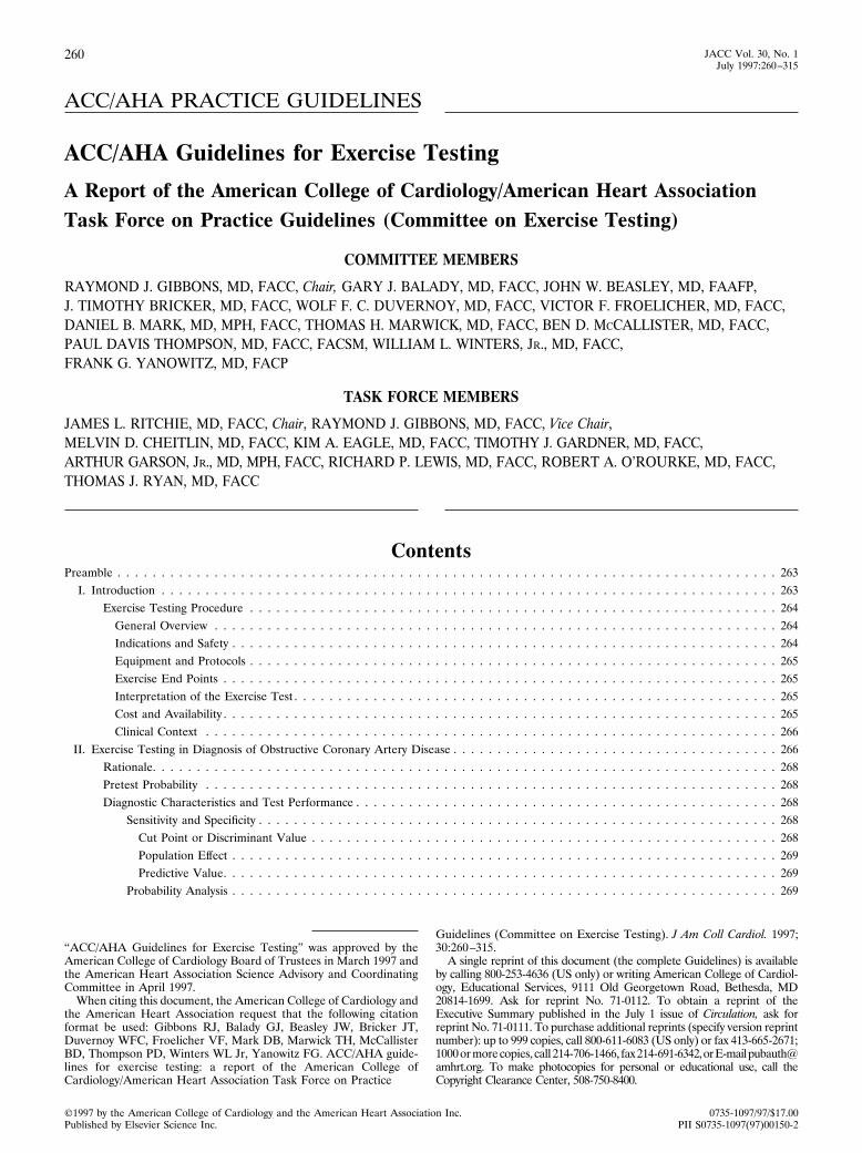
ACC/AHA PRACTICE GUIDELINES
ACC/AHA Guidelines for Exercise Testing
A Report of the American College of Cardiology/American Heart AssociationTask Force on Practice Guidelines (Committee on Exercise Testing)
COMMITTEE MEMBERS
RAYMOND J. GIBBONS, MD, FACC, Chair, GARY J. BALADY, MD, FACC, JOHN W. BEASLEY, MD, FAAFP,J. TIMOTHY BRICKER, MD, FACC, WOLF F. C. DUVERNOY, MD, FACC, VICTOR F. FROELICHER, MD, FACC,DANIEL B. MARK, MD, MPH, FACC, THOMAS H. MARWICK, MD, FACC, BEN D. MCCALLISTER, MD, FACC,PAUL DAVIS THOMPSON, MD, FACC, FACSM, WILLIAM L. WINTERS, JR., MD, FACC,FRANK G. YANOWITZ, MD, FACP
TASK FORCE MEMBERS
JAMES L. RITCHIE, MD, FACC, Chair, RAYMOND J. GIBBONS, MD, FACC, Vice Chair,MELVIN D. CHEITLIN, MD, FACC, KIM A. EAGLE, MD, FACC, TIMOTHY J. GARDNER, MD, FACC,ARTHUR GARSON, JR., MD, MPH, FACC, RICHARD P. LEWIS, MD, FACC, ROBERT A. O’ROURKE, MD, FACC,THOMAS J. RYAN, MD, FACC
ContentsPreamble . . . . . . . . . . . . . . . . . . . . . . . . . . . . . . . . . . . . . . . . . . . . . . . . . . . . . . . . . . . . . . . . . . . . . . . . . . . 263
I. Introduction . . . . . . . . . . . . . . . . . . . . . . . . . . . . . . . . . . . . . . . . . . . . . . . . . . . . . . . . . . . . . . . . . . . . . . 263Exercise Testing Procedure . . . . . . . . . . . . . . . . . . . . . . . . . . . . . . . . . . . . . . . . . . . . . . . . . . . . . . . . . . . . 264
General Overview . . . . . . . . . . . . . . . . . . . . . . . . . . . . . . . . . . . . . . . . . . . . . . . . . . . . . . . . . . . . . . . . 264Indications and Safety . . . . . . . . . . . . . . . . . . . . . . . . . . . . . . . . . . . . . . . . . . . . . . . . . . . . . . . . . . . . . . 264Equipment and Protocols . . . . . . . . . . . . . . . . . . . . . . . . . . . . . . . . . . . . . . . . . . . . . . . . . . . . . . . . . . . . 265Exercise End Points . . . . . . . . . . . . . . . . . . . . . . . . . . . . . . . . . . . . . . . . . . . . . . . . . . . . . . . . . . . . . . . 265Interpretation of the Exercise Test . . . . . . . . . . . . . . . . . . . . . . . . . . . . . . . . . . . . . . . . . . . . . . . . . . . . . . . 265Cost and Availability . . . . . . . . . . . . . . . . . . . . . . . . . . . . . . . . . . . . . . . . . . . . . . . . . . . . . . . . . . . . . . . 265Clinical Context . . . . . . . . . . . . . . . . . . . . . . . . . . . . . . . . . . . . . . . . . . . . . . . . . . . . . . . . . . . . . . . . . 266
II. Exercise Testing in Diagnosis of Obstructive Coronary Artery Disease . . . . . . . . . . . . . . . . . . . . . . . . . . . . . . . . . . . . . 266Rationale. . . . . . . . . . . . . . . . . . . . . . . . . . . . . . . . . . . . . . . . . . . . . . . . . . . . . . . . . . . . . . . . . . . . . . . 268Pretest Probability . . . . . . . . . . . . . . . . . . . . . . . . . . . . . . . . . . . . . . . . . . . . . . . . . . . . . . . . . . . . . . . . . 268Diagnostic Characteristics and Test Performance . . . . . . . . . . . . . . . . . . . . . . . . . . . . . . . . . . . . . . . . . . . . . . . . 268
Sensitivity and Specificity . . . . . . . . . . . . . . . . . . . . . . . . . . . . . . . . . . . . . . . . . . . . . . . . . . . . . . . . . . . 268Cut Point or Discriminant Value . . . . . . . . . . . . . . . . . . . . . . . . . . . . . . . . . . . . . . . . . . . . . . . . . . . . . 268Population Effect . . . . . . . . . . . . . . . . . . . . . . . . . . . . . . . . . . . . . . . . . . . . . . . . . . . . . . . . . . . . . . 269Predictive Value. . . . . . . . . . . . . . . . . . . . . . . . . . . . . . . . . . . . . . . . . . . . . . . . . . . . . . . . . . . . . . . 269
Probability Analysis . . . . . . . . . . . . . . . . . . . . . . . . . . . . . . . . . . . . . . . . . . . . . . . . . . . . . . . . . . . . . . 269
“ACC/AHA Guidelines for Exercise Testing” was approved by theAmerican College of Cardiology Board of Trustees in March 1997 andthe American Heart Association Science Advisory and CoordinatingCommittee in April 1997.
When citing this document, the American College of Cardiology andthe American Heart Association request that the following citationformat be used: Gibbons RJ, Balady GJ, Beasley JW, Bricker JT,Duvernoy WFC, Froelicher VF, Mark DB, Marwick TH, McCallisterBD, Thompson PD, Winters WL Jr, Yanowitz FG. ACC/AHA guide-lines for exercise testing: a report of the American College ofCardiology/American Heart Association Task Force on Practice
Guidelines (Committee on Exercise Testing). J Am Coll Cardiol. 1997;30:260–315.
A single reprint of this document (the complete Guidelines) is availableby calling 800-253-4636 (US only) or writing American College of Cardiol-ogy, Educational Services, 9111 Old Georgetown Road, Bethesda, MD20814-1699. Ask for reprint No. 71-0112. To obtain a reprint of theExecutive Summary published in the July 1 issue of Circulation, ask forreprint No. 71-0111. To purchase additional reprints (specify version reprintnumber): up to 999 copies, call 800-611-6083 (US only) or fax 413-665-2671;1000 or more copies, call 214-706-1466, fax 214-691-6342,[email protected]. To make photocopies for personal or educational use, call theCopyright Clearance Center, 508-750-8400.
JACC Vol. 30, No. 1July 1997:260–315
260
©1997 by the American College of Cardiology and the American Heart Association Inc. 0735-1097/97/$17.00Published by Elsevier Science Inc. PII S0735-1097(97)00150-2
Believability Criteria for Diagnostic Tests . . . . . . . . . . . . . . . . . . . . . . . . . . . . . . . . . . . . . . . . . . . . . . . . . . . 270
Diagnostic Accuracy of the Standard Exercise Test . . . . . . . . . . . . . . . . . . . . . . . . . . . . . . . . . . . . . . . . . . . . . 270
Sensitivity From Meta-Analysis . . . . . . . . . . . . . . . . . . . . . . . . . . . . . . . . . . . . . . . . . . . . . . . . . . . . . . . 271
Specificity From Meta-Analysis . . . . . . . . . . . . . . . . . . . . . . . . . . . . . . . . . . . . . . . . . . . . . . . . . . . . . . . 271
Influence of Other Factors on Test Performance. . . . . . . . . . . . . . . . . . . . . . . . . . . . . . . . . . . . . . . . . . . . . . . 272
Drugs . . . . . . . . . . . . . . . . . . . . . . . . . . . . . . . . . . . . . . . . . . . . . . . . . . . . . . . . . . . . . . . . . . . . . . 272
Digoxin. . . . . . . . . . . . . . . . . . . . . . . . . . . . . . . . . . . . . . . . . . . . . . . . . . . . . . . . . . . . . . . . . . . . 272b-Blocker Therapy . . . . . . . . . . . . . . . . . . . . . . . . . . . . . . . . . . . . . . . . . . . . . . . . . . . . . . . . . . . . . 272Other Drugs . . . . . . . . . . . . . . . . . . . . . . . . . . . . . . . . . . . . . . . . . . . . . . . . . . . . . . . . . . . . . . . . . 272
Electrocardiographic Abnormalities . . . . . . . . . . . . . . . . . . . . . . . . . . . . . . . . . . . . . . . . . . . . . . . . . . . . . . 272Left Bundle Branch Block . . . . . . . . . . . . . . . . . . . . . . . . . . . . . . . . . . . . . . . . . . . . . . . . . . . . . . . . . . 272Right Bundle Branch Block . . . . . . . . . . . . . . . . . . . . . . . . . . . . . . . . . . . . . . . . . . . . . . . . . . . . . . . . . 272Left Ventricular Hypertrophy With Repolarization Abnormalities . . . . . . . . . . . . . . . . . . . . . . . . . . . . . . . . . . . 272Resting ST Depression . . . . . . . . . . . . . . . . . . . . . . . . . . . . . . . . . . . . . . . . . . . . . . . . . . . . . . . . . . . . 272
Overview of Confounders: Digoxin, Resting ST Depression, Left Ventricular Hypertrophy. . . . . . . . . . . . . . . . . . . . . . . 273ST-Segment Interpretation Issues . . . . . . . . . . . . . . . . . . . . . . . . . . . . . . . . . . . . . . . . . . . . . . . . . . . . . . . 273
Lead Selection . . . . . . . . . . . . . . . . . . . . . . . . . . . . . . . . . . . . . . . . . . . . . . . . . . . . . . . . . . . . . . . . . 273Upsloping ST Depression. . . . . . . . . . . . . . . . . . . . . . . . . . . . . . . . . . . . . . . . . . . . . . . . . . . . . . . . . . . 273ST Elevation . . . . . . . . . . . . . . . . . . . . . . . . . . . . . . . . . . . . . . . . . . . . . . . . . . . . . . . . . . . . . . . . . . 273R-Wave Changes . . . . . . . . . . . . . . . . . . . . . . . . . . . . . . . . . . . . . . . . . . . . . . . . . . . . . . . . . . . . . . . 273Heart Rate Adjustment . . . . . . . . . . . . . . . . . . . . . . . . . . . . . . . . . . . . . . . . . . . . . . . . . . . . . . . . . . . . 273Computer Processing . . . . . . . . . . . . . . . . . . . . . . . . . . . . . . . . . . . . . . . . . . . . . . . . . . . . . . . . . . . . . 274
III. Risk Assessment and Prognosis in Patients With Symptoms or a Prior History of Coronary Artery Disease . . . . . . . . . . . . . . . . 274Risk Stratification: General Considerations . . . . . . . . . . . . . . . . . . . . . . . . . . . . . . . . . . . . . . . . . . . . . . . . . . 274Prognosis of Coronary Artery Disease: General Considerations . . . . . . . . . . . . . . . . . . . . . . . . . . . . . . . . . . . . . . 274Risk Stratification With the Exercise Test . . . . . . . . . . . . . . . . . . . . . . . . . . . . . . . . . . . . . . . . . . . . . . . . . . . 275
Symptomatic Patients With Nonacute Coronary Artery Disease . . . . . . . . . . . . . . . . . . . . . . . . . . . . . . . . . . . . . 276Patients With Unstable Angina . . . . . . . . . . . . . . . . . . . . . . . . . . . . . . . . . . . . . . . . . . . . . . . . . . . . . . . 279
Use of Exercise Test Results in Patient Treatment. . . . . . . . . . . . . . . . . . . . . . . . . . . . . . . . . . . . . . . . . . . . . . 280IV. After Myocardial Infarction . . . . . . . . . . . . . . . . . . . . . . . . . . . . . . . . . . . . . . . . . . . . . . . . . . . . . . . . . . . . . 280
Exercise Test Logistics. . . . . . . . . . . . . . . . . . . . . . . . . . . . . . . . . . . . . . . . . . . . . . . . . . . . . . . . . . . . . . 281Exclusions From Testing . . . . . . . . . . . . . . . . . . . . . . . . . . . . . . . . . . . . . . . . . . . . . . . . . . . . . . . . . . . 281Timing and Protocol . . . . . . . . . . . . . . . . . . . . . . . . . . . . . . . . . . . . . . . . . . . . . . . . . . . . . . . . . . . . . 282Safety . . . . . . . . . . . . . . . . . . . . . . . . . . . . . . . . . . . . . . . . . . . . . . . . . . . . . . . . . . . . . . . . . . . . . . 282
Risk Stratification and Prognosis . . . . . . . . . . . . . . . . . . . . . . . . . . . . . . . . . . . . . . . . . . . . . . . . . . . . . . . . 282Inability to Exercise . . . . . . . . . . . . . . . . . . . . . . . . . . . . . . . . . . . . . . . . . . . . . . . . . . . . . . . . . . . . . . 282Exercise-Induced Ischemia . . . . . . . . . . . . . . . . . . . . . . . . . . . . . . . . . . . . . . . . . . . . . . . . . . . . . . . . . . 282Exercise Capacity . . . . . . . . . . . . . . . . . . . . . . . . . . . . . . . . . . . . . . . . . . . . . . . . . . . . . . . . . . . . . . . 283Blood Pressure . . . . . . . . . . . . . . . . . . . . . . . . . . . . . . . . . . . . . . . . . . . . . . . . . . . . . . . . . . . . . . . . . 283Other Variables . . . . . . . . . . . . . . . . . . . . . . . . . . . . . . . . . . . . . . . . . . . . . . . . . . . . . . . . . . . . . . . . 283
Activity Counseling. . . . . . . . . . . . . . . . . . . . . . . . . . . . . . . . . . . . . . . . . . . . . . . . . . . . . . . . . . . . . . . . 284Cardiac Rehabilitation . . . . . . . . . . . . . . . . . . . . . . . . . . . . . . . . . . . . . . . . . . . . . . . . . . . . . . . . . . . . . . 285Summary . . . . . . . . . . . . . . . . . . . . . . . . . . . . . . . . . . . . . . . . . . . . . . . . . . . . . . . . . . . . . . . . . . . . . 285
V. Exercise Testing Using Ventilatory Gas Analysis . . . . . . . . . . . . . . . . . . . . . . . . . . . . . . . . . . . . . . . . . . . . . . . . . 285VI. Special Groups: Women, Asymptomatic Individuals, and Postrevascularization Patients . . . . . . . . . . . . . . . . . . . . . . . . . . . 287
Women . . . . . . . . . . . . . . . . . . . . . . . . . . . . . . . . . . . . . . . . . . . . . . . . . . . . . . . . . . . . . . . . . . . . . . 287Rationale . . . . . . . . . . . . . . . . . . . . . . . . . . . . . . . . . . . . . . . . . . . . . . . . . . . . . . . . . . . . . . . . . . . . 287
Accuracy of Electrocardiographic Analysis in Women . . . . . . . . . . . . . . . . . . . . . . . . . . . . . . . . . . . . . . . . . 287Non-ECG End Points . . . . . . . . . . . . . . . . . . . . . . . . . . . . . . . . . . . . . . . . . . . . . . . . . . . . . . . . . . . 288Conclusion . . . . . . . . . . . . . . . . . . . . . . . . . . . . . . . . . . . . . . . . . . . . . . . . . . . . . . . . . . . . . . . . . . 288
Diagnosis of Coronary Artery Disease in the Elderly . . . . . . . . . . . . . . . . . . . . . . . . . . . . . . . . . . . . . . . . . . . . 289Rationale . . . . . . . . . . . . . . . . . . . . . . . . . . . . . . . . . . . . . . . . . . . . . . . . . . . . . . . . . . . . . . . . . . . . 289
Exercise Testing in Asymptomatic Individuals Without Known Coronary Artery Disease . . . . . . . . . . . . . . . . . . . . . . . . 290Rationale . . . . . . . . . . . . . . . . . . . . . . . . . . . . . . . . . . . . . . . . . . . . . . . . . . . . . . . . . . . . . . . . . . . . 290
Background . . . . . . . . . . . . . . . . . . . . . . . . . . . . . . . . . . . . . . . . . . . . . . . . . . . . . . . . . . . . . . . . . 290Diagnostic Considerations . . . . . . . . . . . . . . . . . . . . . . . . . . . . . . . . . . . . . . . . . . . . . . . . . . . . . . . . . 290
261JACC Vol. 30, No. 1 GIBBONS ET AL.July 1997:260–315 EXERCISE TESTING GUIDELINES

Prognostic Evaluation . . . . . . . . . . . . . . . . . . . . . . . . . . . . . . . . . . . . . . . . . . . . . . . . . . . . . . . . . . . 290
ST-Segment Response . . . . . . . . . . . . . . . . . . . . . . . . . . . . . . . . . . . . . . . . . . . . . . . . . . . . . . . . . . 291
Exercise Capacity. . . . . . . . . . . . . . . . . . . . . . . . . . . . . . . . . . . . . . . . . . . . . . . . . . . . . . . . . . . . . 291
Risk Factors. . . . . . . . . . . . . . . . . . . . . . . . . . . . . . . . . . . . . . . . . . . . . . . . . . . . . . . . . . . . . . . . 291
Stress Imaging Tests . . . . . . . . . . . . . . . . . . . . . . . . . . . . . . . . . . . . . . . . . . . . . . . . . . . . . . . . . . . 291Who to Screen? . . . . . . . . . . . . . . . . . . . . . . . . . . . . . . . . . . . . . . . . . . . . . . . . . . . . . . . . . . . . . . . 291
Population Screening . . . . . . . . . . . . . . . . . . . . . . . . . . . . . . . . . . . . . . . . . . . . . . . . . . . . . . . . . . 291Screening in Patients With Coronary Artery Disease Risk Factors . . . . . . . . . . . . . . . . . . . . . . . . . . . . . . . . 291Screening in Other Patient Groups at High Risk of Coronary Artery Disease . . . . . . . . . . . . . . . . . . . . . . . . . . 291Before Fitness Program . . . . . . . . . . . . . . . . . . . . . . . . . . . . . . . . . . . . . . . . . . . . . . . . . . . . . . . . . 293Special Groups . . . . . . . . . . . . . . . . . . . . . . . . . . . . . . . . . . . . . . . . . . . . . . . . . . . . . . . . . . . . . . 293
Implications for Clinical Practice . . . . . . . . . . . . . . . . . . . . . . . . . . . . . . . . . . . . . . . . . . . . . . . . . . . . . 293Valvular Heart Disease . . . . . . . . . . . . . . . . . . . . . . . . . . . . . . . . . . . . . . . . . . . . . . . . . . . . . . . . . . . . . 293
Rationale . . . . . . . . . . . . . . . . . . . . . . . . . . . . . . . . . . . . . . . . . . . . . . . . . . . . . . . . . . . . . . . . . . . . 293Uses of Exercise Testing in Patients With Valvular Heart Disease . . . . . . . . . . . . . . . . . . . . . . . . . . . . . . . . . . 293Aortic Stenosis . . . . . . . . . . . . . . . . . . . . . . . . . . . . . . . . . . . . . . . . . . . . . . . . . . . . . . . . . . . . . . . 293Mitral Stenosis . . . . . . . . . . . . . . . . . . . . . . . . . . . . . . . . . . . . . . . . . . . . . . . . . . . . . . . . . . . . . . . 294Aortic Regurgitation . . . . . . . . . . . . . . . . . . . . . . . . . . . . . . . . . . . . . . . . . . . . . . . . . . . . . . . . . . . . 294Mitral Regurgitation . . . . . . . . . . . . . . . . . . . . . . . . . . . . . . . . . . . . . . . . . . . . . . . . . . . . . . . . . . . . 294
Exercise Testing Before and After Revascularization . . . . . . . . . . . . . . . . . . . . . . . . . . . . . . . . . . . . . . . . . . . . 294Rationale . . . . . . . . . . . . . . . . . . . . . . . . . . . . . . . . . . . . . . . . . . . . . . . . . . . . . . . . . . . . . . . . . . . . 294
Exercise Testing Before Revascularization . . . . . . . . . . . . . . . . . . . . . . . . . . . . . . . . . . . . . . . . . . . . . . . . 294Exercise Testing After Revascularization. . . . . . . . . . . . . . . . . . . . . . . . . . . . . . . . . . . . . . . . . . . . . . . . . 294Exercise Testing After Coronary Bypass Graft Surgery. . . . . . . . . . . . . . . . . . . . . . . . . . . . . . . . . . . . . . . . . 294Exercise Testing After Percutaneous Transluminal Coronary Angioplasty . . . . . . . . . . . . . . . . . . . . . . . . . . . . . . 295
Investigation of Heart Rhythm Disorders . . . . . . . . . . . . . . . . . . . . . . . . . . . . . . . . . . . . . . . . . . . . . . . . . . . 296Evaluation of Patients With Known or Suspected Exercise-Induced Arrhythmias . . . . . . . . . . . . . . . . . . . . . . . . . . . 296
Ventricular Arrhythmias . . . . . . . . . . . . . . . . . . . . . . . . . . . . . . . . . . . . . . . . . . . . . . . . . . . . . . . . . . 296Supraventricular Arrhythmias . . . . . . . . . . . . . . . . . . . . . . . . . . . . . . . . . . . . . . . . . . . . . . . . . . . . . . . 296Sinus Node Dysfunction . . . . . . . . . . . . . . . . . . . . . . . . . . . . . . . . . . . . . . . . . . . . . . . . . . . . . . . . . . 296
Cardiac Pacemakers . . . . . . . . . . . . . . . . . . . . . . . . . . . . . . . . . . . . . . . . . . . . . . . . . . . . . . . . . . . . . . 297VII. Pediatric Testing: Exercise Testing in Children and Adolescents . . . . . . . . . . . . . . . . . . . . . . . . . . . . . . . . . . . . . . . . 297
Differences Between Pediatric and Adult Testing . . . . . . . . . . . . . . . . . . . . . . . . . . . . . . . . . . . . . . . . . . . . . . 297Exercise Testing for Specific Pediatric and Congenital Cardiac Problems . . . . . . . . . . . . . . . . . . . . . . . . . . . . . . . . . 298
Exercise Testing of Children and Adolescents With Chest Pain . . . . . . . . . . . . . . . . . . . . . . . . . . . . . . . . . . . . . 298Exercise Testing of Patients With Unoperated Left-to-Right Shunts . . . . . . . . . . . . . . . . . . . . . . . . . . . . . . . . . . 298Exercise Testing in Patients With Postoperative Left-to-Right Shunts . . . . . . . . . . . . . . . . . . . . . . . . . . . . . . . . . 298Exercise Testing With Unoperated or Palliated Cyanotic Congenital Cardiac Defects . . . . . . . . . . . . . . . . . . . . . . . . 298Exercise Testing for Patients With Coarctation of the Aorta . . . . . . . . . . . . . . . . . . . . . . . . . . . . . . . . . . . . . . . 298Exercise Testing for the Child or Adolescent With Pulmonary Stenosis . . . . . . . . . . . . . . . . . . . . . . . . . . . . . . . . 300Exercise Testing for the Child or Adolescent With Aortic Stenosis or Regurgitation . . . . . . . . . . . . . . . . . . . . . . . . . 300Exercise Testing After Surgery for Tetralogy of Fallot . . . . . . . . . . . . . . . . . . . . . . . . . . . . . . . . . . . . . . . . . . 300Exercise Testing After the Fontan Operation (Total Systemic Venous to Pulmonary Connection) . . . . . . . . . . . . . . . . . 300Exercise Testing of Patients With Cardiomyopathy . . . . . . . . . . . . . . . . . . . . . . . . . . . . . . . . . . . . . . . . . . . . 301Exercise Testing of Children or Adolescents With Syncope . . . . . . . . . . . . . . . . . . . . . . . . . . . . . . . . . . . . . . . 301Exercise Testing of Children or Adolescents With Atrial Arrhythmias . . . . . . . . . . . . . . . . . . . . . . . . . . . . . . . . . 301Exercise Testing of Children or Adolescents With Ventricular Arrhythmias . . . . . . . . . . . . . . . . . . . . . . . . . . . . . . 301Exercise Testing of Children or Adolescents With Conduction Abnormalities and in Pacemaker Follow-up . . . . . . . . . . . . 301Exercise Testing of Children or Adolescents With Known or Suspected Coronary Artery Disease . . . . . . . . . . . . . . . . . 301Exercise Testing of Children or Adolescents With Cardiac Transplantation . . . . . . . . . . . . . . . . . . . . . . . . . . . . . . 301Exercise Testing After an Operation to Correct Transposition of the Great Arteries . . . . . . . . . . . . . . . . . . . . . . . . . 301
Appendixes . . . . . . . . . . . . . . . . . . . . . . . . . . . . . . . . . . . . . . . . . . . . . . . . . . . . . . . . . . . . . . . . . . . . . . . 302References . . . . . . . . . . . . . . . . . . . . . . . . . . . . . . . . . . . . . . . . . . . . . . . . . . . . . . . . . . . . . . . . . . . . . . . 303Index . . . . . . . . . . . . . . . . . . . . . . . . . . . . . . . . . . . . . . . . . . . . . . . . . . . . . . . . . . . . . . . . . . . . . . . . . . 312
262 GIBBONS ET AL. JACC Vol. 30, No. 1EXERCISE TESTING GUIDELINES July 1997:260–315
Preamble
It is important that the medical profession play a significantrole in critically evaluating the use of diagnostic proceduresand therapies in the management or prevention of diseasestates. Rigorous and expert analysis of the available datadocumenting relative benefits and risks of those proceduresand therapies can produce helpful guidelines that improve theeffectiveness of care, optimize patient outcomes, and impactthe overall cost of care favorably by focusing resources on themost effective strategies.
The American College of Cardiology (ACC) and the Amer-ican Heart Association (AHA) have jointly engaged in theproduction of such guidelines in the area of cardiovasculardisease since 1980. This effort is directed by the ACC/AHATask Force on Practice Guidelines, whose charge is to developand revise practice guidelines for important cardiovasculardiseases and procedures. Experts in the subject underconsideration are selected from both organizations to exam-ine subject-specific data and write guidelines. The processincludes additional representatives from other medical prac-titioner and specialty groups where appropriate. Writinggroups are specifically charged to perform a formal litera-ture review, weigh the strength of evidence for or against aparticular treatment or procedure, and include estimates ofexpected health outcomes when data exist. Patient-specificmodifiers, comorbidities, and issues of patient preferencethat might influence the choice of particular tests or thera-pies are considered as well as frequency of follow-up andcost-effectiveness.
The ACC/AHA Task Force on Practice Guidelines makesevery effort to avoid any actual or potential conflicts of interestthat might arise as a result of an outside relationship orpersonal interest of a member of the writing panel. Specificallyall members of the writing panel are asked to provide disclo-sure statements of all such relationships that might be per-ceived as real or potential conflicts of interest. These state-ments are reviewed by the parent task force, reported orally toall members of the writing panel at the first meeting, andupdated yearly and as changes occur.
These practice guidelines are intended to assist physiciansin clinical decision making by describing a range of generallyacceptable approaches for the diagnosis, management, orprevention of specific diseases or conditions. These guidelinesattempt to define practices that meet the needs of mostpatients in most circumstances. The ultimate judgment regard-ing care of a particular patient must be made by the physicianand patient in light of all of the circumstances presented bythat patient.
The executive summary and recommendations are pub-lished in the July 1 issue of Circulation. The full text ispublished in Journal of the American College of Cardiology.Reprints of the full text and the executive summary areavailable from both organizations.
These guidelines have been officially endorsed by theAmerican College of Sports Medicine, the American Societyof Echocardiography and the American Society of NuclearCardiology.
James L. Ritchie, MD, FACCChair, ACC/AHA Task Force on Practice Guidelines
I. IntroductionThe American College of Cardiology/American Heart As-
sociation Task Force on Practice Guidelines was formed tomake recommendations regarding the appropriate use oftesting in the diagnosis and treatment of patients with known orsuspected cardiovascular disease. Exercise testing is widely avail-able and relatively low cost. For the purposes of this document,exercise testing is a cardiovascular stress test using treadmill orbicycle exercise and electrocardiographic and blood pressuremonitoring. Pharmacological stress and the use of imaging mo-dalities (radionuclide imaging, echocardiography) are beyond thescope of these guidelines.
The current committee was given the task of reviewing andrevising the guidelines for exercise testing published in Sep-tember 1986. Since that report, many new studies have beenpublished regarding the usefulness of exercise testing forprediction of outcome in both symptomatic and asymptomaticpatients. The usefulness of oxygen consumption measurementsin association with exercise testing to identify patients who arecandidates for cardiac transplantation has been recognized.The usefulness and cost-effectiveness of exercise testing hasbeen compared with more expensive imaging procedures inselected patient subsets. All of these developments are consid-ered in these guidelines.
In considering the use of exercise testing in individualpatients, the following factors are important:
1. The quality, expertise, and experience of the professionaland technical staff performing and interpreting the study
2. The sensitivity, specificity, and accuracy of the technique3. The cost and accuracy of the technique as compared with
more expensive imaging procedures4. The effect of positive or negative results on clinical decision
making5. The potential psychological benefits of patient reassurance
The format of these guidelines includes a brief descriptionof exercise testing followed by a discussion of its usefulness inspecific clinical situations. Usefulness is considered for (1)diagnosis; (2) severity of disease/risk assessment/prognosis inpatients with known or suspected chronic coronary artery disease(CAD); (3) risk assessment of patients early after myocardialinfarction; (4) specific clinical populations identified by gender,age, other cardiac disease, or prior coronary revascularization;and (5) pediatric populations. The recommendations for partic-ular situations are summarized in each section.
The committee reviewed and compiled all pertinent pub-
263JACC Vol. 30, No. 1 GIBBONS ET AL.July 1997:260–315 EXERCISE TESTING GUIDELINES
lished reports (excluding abstracts) through a computerizedsearch of the English-language literature since 1975 and amanual search of final articles. Specific attention was devotedto identification and compilation of appropriate meta-analyses.Detailed evidence tables were developed whenever necessaryusing specific criteria detailed in the guidelines. The meta-analyses and evidence tables were extensively reviewed by anexpert in methodologies. Inaccuracies and inconsistencies inthe original publications were identified and corrected when-ever possible. The recommendations made are based primarilyon these published data. Because there are essentially norandomized trials assessing health outcomes for diagnostictests, the committee has not ranked the available scientificevidence in an A, B, or C fashion (as was done in otherACC/AHA documents). When few or no data exist, this isnoted in the text, and the recommendations are based on theexpert consensus of the committee.
The ACC/AHA classifications I, II, and III are used tosummarize indications as follows:
Class I: Conditions for which there is evidence and/orgeneral agreement that a given procedure or treatmentis useful and effective.
Class II: Conditions for which there is conflicting evidenceand/or a divergence of opinion about the usefulness/efficacy of a procedure or treatment.
IIa: Weight of evidence/opinion is in favor ofusefulness/efficacy.
IIb: Usefulness/efficacy is less well established byevidence/opinion.
Class III: Conditions for which there is evidence and/orgeneral agreement that the procedure/treatment is notuseful/effective and in some cases may be harmful.
A complete list of the hundreds of publications coveringmany decades of exercise testing is beyond the scope of theseguidelines, and only selected references are included. Thecommittee consisted of acknowledged experts in exercise test-ing, as well as general cardiologists, a general internist, a familymedicine physician, and cardiologists with expertise in the useof stress imaging modalities. The committee included repre-sentatives of the American Academy of Family Physicians, theAmerican College of Sports Medicine, and the AmericanCollege of Physicians. Both the academic and private practicesectors, as well as both adult and pediatric expertise, wererepresented. This document was reviewed by three outsidereviewers nominated by the ACC and by three outside review-ers nominated by the AHA, as well as by outside reviewersnominated by the American Academy of Family Physicians,the American College of Physicians, the American College ofSports Medicine, the American Society of Echocardiography,and the American Society of Nuclear Cardiology. This docu-ment will be reviewed 2 years after publication and yearlythereafter by the task force to determine whether a revision isneeded. These guidelines will be considered current unless thetask force revises or withdraws them from distribution.
This report overlaps with several previously publishedACC/AHA guidelines for patient treatment that potentiallyinvolve exercise testing, including guidelines for perioperativecardiovascular evaluation for noncardiac surgery,1 guidelinesfor management of patients with acute myocardial infarction,2guidelines for percutaneous transluminal coronary angio-plasty,3 and guidelines and indications for coronary arterybypass graft surgery.4 These guidelines are not intended toinclude information previously covered in guidelines for theuse of noninvasive imaging modalities. This report does notinclude a discussion of radionuclide angiography, myocardialperfusion imaging, or positron emission tomography, which arecovered in the recently published guidelines for clinical use ofcardiac radionuclide imaging.5 This report also does not in-clude any discussion of stress echocardiography, which iscovered in the recently published guidelines for clinical appli-cation of echocardiography.6 For clarity, there are occasionalreferences to the use of both radionuclide and echocardio-graphic imaging techniques. However, these brief referencesare not intended to provide a comprehensive understanding ofthe use of these imaging modalities. For such an understand-ing, the reader is referred to the other published guidelines.These guidelines do apply to both adults and children.
Exercise Testing ProcedureGeneral Overview Exercise testing is a well-established
procedure that has been in widespread clinical use for manydecades. It is beyond the scope of this document to provide adetailed “how-to” description of this procedure. Such a de-scription is available in previous publications from the AHA,including the statement on exercise standards,7 guidelines forclinical exercise testing laboratories,8 and guidelines for exer-cise testing in the pediatric age group,9 to which interestedreaders are referred. This section is intended to provide a briefoverview of the exercise testing procedure.
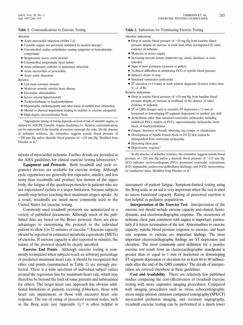
Indications and Safety Although exercise testing is gener-ally a safe procedure, both myocardial infarction and deathhave been reported and can be expected to occur at a rate ofup to 1 per 2500 tests.10 Good clinical judgment shouldtherefore be used in deciding which patients should undergoexercise testing. Absolute and relative contraindications toexercise testing are summarized in Table 1.
Exercise testing should be supervised by an appropriatelytrained physician. As indicated in the ACP/ACC/AHA taskforce statement on clinical competence in exercise testing,11
exercise testing in selected patients can be safely performed byproperly trained nurses, exercise physiologists, physical thera-pists, or medical technicians working directly under the super-vision of a physician, who should be in the immediate vicinityand available for emergencies. The electrocardiogram, heartrate, and blood pressure should be carefully monitored andrecorded during each stage of exercise as well as duringST-segment abnormalities and chest pain. The patient shouldbe continuously monitored for transient rhythm disturbances,ST-segment changes, and other electrocardiographic manifes-
264 GIBBONS ET AL. JACC Vol. 30, No. 1EXERCISE TESTING GUIDELINES July 1997:260–315
tations of myocardial ischemia. Further details are provided inthe AHA guidelines for clinical exercise testing laboratories.8
Equipment and Protocols Both treadmill and cycle er-gometer devices are available for exercise testing. Althoughcycle ergometers are generally less expensive, smaller, and lessnoisy than treadmills and produce less motion of the upperbody, the fatigue of the quadriceps muscles in patients who arenot experienced cyclists is a major limitation, because subjectsusually stop before reaching their maximum oxygen uptake. Asa result, treadmills are much more commonly used in theUnited States for exercise testing.
Commonly used treadmill protocols are summarized in avariety of published documents. Although much of the pub-lished data are based on the Bruce protocol, there are clearadvantages to customizing the protocol to the individualpatient to allow 6 to 12 minutes of exercise.12 Exercise capacityshould be reported in estimated metabolic equivalents (METs)of exercise. If exercise capacity is also reported in minutes, thenature of the protocol should be clearly specified.
Exercise End Points Although exercise testing is com-monly terminated when subjects reach an arbitrary percentageof predicted maximum heart rate, it should be recognized thatother end points (summarized in Table 2) are strongly pre-ferred. There is a wide spectrum of individual subject valuesaround the regression line for maximum heart rate, which maytherefore be beyond the limit of some patients and submaximalfor others. The target heart rate approach has obvious addi-tional limitations in patients receiving b-blockers, those withheart rate impairment, or those with excessive heart rateresponse. The use of rating of perceived exertion scales, suchas the Borg scale (see Appendix 1),13 is often helpful in
assessment of patient fatigue. Symptom-limited testing usingthe Borg scale as an aid is very important when the test is usedto assess functional capacity. Rating of perceived exertion isless helpful in pediatric populations.
Interpretation of the Exercise Test Interpretation of theexercise test should include exercise capacity and clinical, hemo-dynamic, and electrocardiographic response. The occurrence ofischemic chest pain consistent with angina is important, particu-larly if it forces termination of the test. Abnormalities in exercisecapacity, systolic blood pressure response to exercise, and heartrate response to exercise are important findings. The mostimportant electrocardiographic findings are ST depression andelevation. The most commonly used definition for a positiveexercise test result from an electrocardiographic standpoint isgreater than or equal to 1 mm of horizontal or downslopingST-segment depression or elevation for at least 60 to 80 millisec-onds after the end of the QRS complex.4 The details of interpre-tation are covered elsewhere in these guidelines.
Cost and Availability There are relatively few publishedstudies comparing the cost-effectiveness of treadmill exercisetesting with more expensive imaging procedures. Comparedwith imaging procedures such as stress echocardiography,stress single-photon emission computed tomography (SPECT)myocardial perfusion imaging, and coronary angiography,treadmill exercise testing can be performed at a much lower
Table 1. Contraindications to Exercise Testing
Absolute● Acute myocardial infarction (within 2 d)● Unstable angina not previously stabilized by medical therapy*● Uncontrolled cardiac arrhythmias causing symptoms or hemodynamic
compromise● Symptomatic severe aortic stenosis● Uncontrolled symptomatic heart failure● Acute pulmonary embolus or pulmonary infarction● Acute myocarditis or pericarditis● Acute aortic dissection
Relative†● Left main coronary stenosis● Moderate stenotic valvular heart disease● Electrolyte abnormalities● Severe arterial hypertension‡● Tachyarrhythmias or bradyarrhythmias● Hypertrophic cardiomyopathy and other forms of outflow tract obstruction● Mental or physical impairment leading to inability to exercise adequately● High-degree atrioventricular block
*Appropriate timing of testing depends on level of risk of unstable angina, asdefined by AHCPR Unstable Angina Guidelines.14 †Relative contraindicationscan be superseded if the benefits of exercise outweigh the risks. ‡In the absenceof definitive evidence, the committee suggests systolic blood pressure of.200 mm Hg and/or diastolic blood pressure of .110 mm Hg. Modified fromFletcher et al.7
Table 2. Indications for Terminating Exercise Testing
Absolute indications● Drop in systolic blood pressure of .10 mg Hg from baseline blood
pressure despite an increase in work load, when accompanied by otherevidence of ischemia
● Moderate to severe angina● Increasing nervous system symptoms (eg, ataxia, dizziness, or near-
syncope)● Signs of poor perfusion (cyanosis or pallor)● Technical difficulties in monitoring ECG or systolic blood pressure● Subject’s desire to stop● Sustained ventricular tachycardia● ST elevation ($1.0 mm) in leads without diagnostic Q-waves (other than
V1 or aVR)Relative indications
● Drop in systolic blood pressure of $10 mm Hg from baseline bloodpressure despite an increase in workload, in the absence of otherevidence of ischemia
● ST or QRS changes such as excessive ST depression (.2 mm ofhorizontal or downsloping ST-segment depression) or marked axis shift
● Arrhythmias other than sustained ventricular tachycardia, includingmultifocal PVCs, triplets of PVCs, supraventricular tachycardia, heartblock, or bradyarrhythmias
● Fatigue, shortness of breath, wheezing, leg cramps, or claudication● Development of bundle branch block or IVCD that cannot be
distinguished from ventricular tachycardia● Increasing chest pain● Hypertensive response*
*In the absence of definitive evidence, the committee suggests systolic bloodpressure of .250 mm Hg and/or a diastolic blood pressure of .115 mm Hg.ECG indicates electrocardiogram; PVCs, premature ventricular contractions;ICD, implantable cardioverter-defibrillator discharge; and IVCD, intraventricu-lar conduction delay. Modified from Fletcher et al.7
265JACC Vol. 30, No. 1 GIBBONS ET AL.July 1997:260–315 EXERCISE TESTING GUIDELINES
cost. Table 3 is a comparison of 1996 Medicare RVUs (relativevalue units, professional and technical) for treadmill exercisetesting and selected imaging procedures. These RVUs providean estimate of relative costs. Compared with the treadmillexercise test, the cost of stress echocardiography is at least 2.4times higher, stress SPECT myocardial imaging 5.3 timeshigher, and coronary angiography 20 times higher. Lower costof the treadmill exercise test alone does not necessarily resultin a lower overall cost of patient care, as the cost of additionaltesting and interventions may be higher when the initialtreadmill exercise test is less accurate than these more sophis-ticated procedures.
Treadmill exercise testing is performed frequently. Asshown in Table 3, treadmill exercise tests are performed aboutas often as the most frequent imaging procedure (stressSPECT myocardial perfusion imaging). An estimated twothirds of the treadmill exercise tests charged to Medicare in1994 were performed as office procedures, and 33% of thecharges were submitted by noncardiologists. Thus, treadmillexercise tests are more widely performed, do not alwaysrequire a cardiologist, and are convenient for the patientbecause they are often an office-based procedure.
Clinical Context The vast majority of treadmill exercisetesting is performed in adults with symptoms of known orsuspected ischemic heart disease. Special groups who repre-sent exceptions to this norm are discussed in detail in sectionsVI and VII. Sections II through IV reflect the variety ofpatients and clinical decisions (so-called “nodal points”) forwhich exercise testing is used. Although this document is notintended to be a guideline for the management of stable chestpain, the committee thought that it was important to providean overall context for the use of exercise testing to facilitate theuse of these guidelines (Fig 1).
Patients who are candidates for exercise testing may havestable symptoms of chest pain, may be stabilized by medicaltherapy following symptoms of unstable chest pain, or may bepost–myocardial infarction or postrevascularization patients.The clinician should first address whether the diagnosis ofCAD is certain, based on the patient’s history, electrocardio-gram, and symptoms of chest pain. The important factorsinvolved in addressing this question are covered in section II of
this document, which focuses on the use of treadmill exercisetesting for diagnosis.
Even in patients for whom the diagnosis of CAD is certain,based on age, gender, description of chest pain, and history ofprior myocardial infarction, there usually is a clinical need forrisk or prognostic assessment to determine the need forpossible coronary angiography or revascularization. The po-tential role of treadmill exercise testing in such patients isdetailed in section III.
Post–myocardial infarction patients represent a commonfirst presentation of ischemic heart disease. They are a subsetof patients who may need risk or prognostic assessment. Thissubgroup is considered in detail in section IV, which includesa discussion of the implications of acute reperfusion therapyfor interpretation of exercise testing in this population.
II. Exercise Testing in Diagnosis ofObstructive Coronary Artery Disease
Class I
1. Adult patients (including those with complete right bundlebranch block or less than 1 mm of resting ST depression)with an intermediate pretest probability of CAD (Table 4),based on gender, age, and symptoms (specific exceptionsare noted under Classes II and III below).
Class IIa
1. Patients with vasospastic angina.
Class IIb
1. Patients with a high pretest probability of CAD by age,symptoms, and gender.
2. Patients with a low pretest probability of CAD by age,symptoms, and gender.
3. Patients with less than 1 mm of baseline ST depression andtaking digoxin.
4. Patients with electrocardiographic criteria for left ventricu-lar hypertrophy (LVH) and less than 1 mm of baseline STdepression.
Table 3. Medicare Fees and Volumes of Commonly Used Diagnostic Procedures
Procedure 1996 CPT Code(s)
1996 Total(Professional and Technical)
Medicare RVUs
1994 Medicare Data
NumberPerformed
Percent Chargedby Cardiologists
PercentOffice-Based
Treadmill exercise test 93015 or 93016–93018 3.30 875,780* 67* 66*Stress echocardiography 93350, 93015 7.95
(plus any Doppler charge)213,404 78 67
Stress SPECT myocardial perfusion imaging 78465, 93015 17.45(plus isotope charge)
889,319 † 34
Left heart catheterization with leftventriculogram and coronary angiography
93510, 93543, 93545,93555, 93556
66.83 728,763 88 ,1
*These numbers are estimates, after excluding treadmill exercise tests performed with perfusion imaging. †There are no reliable data regarding this percentage.CPT indicates current procedural terminology; RVUs, relative value units; and SPECT, single-photon emission computed tomography.
266 GIBBONS ET AL. JACC Vol. 30, No. 1EXERCISE TESTING GUIDELINES July 1997:260–315
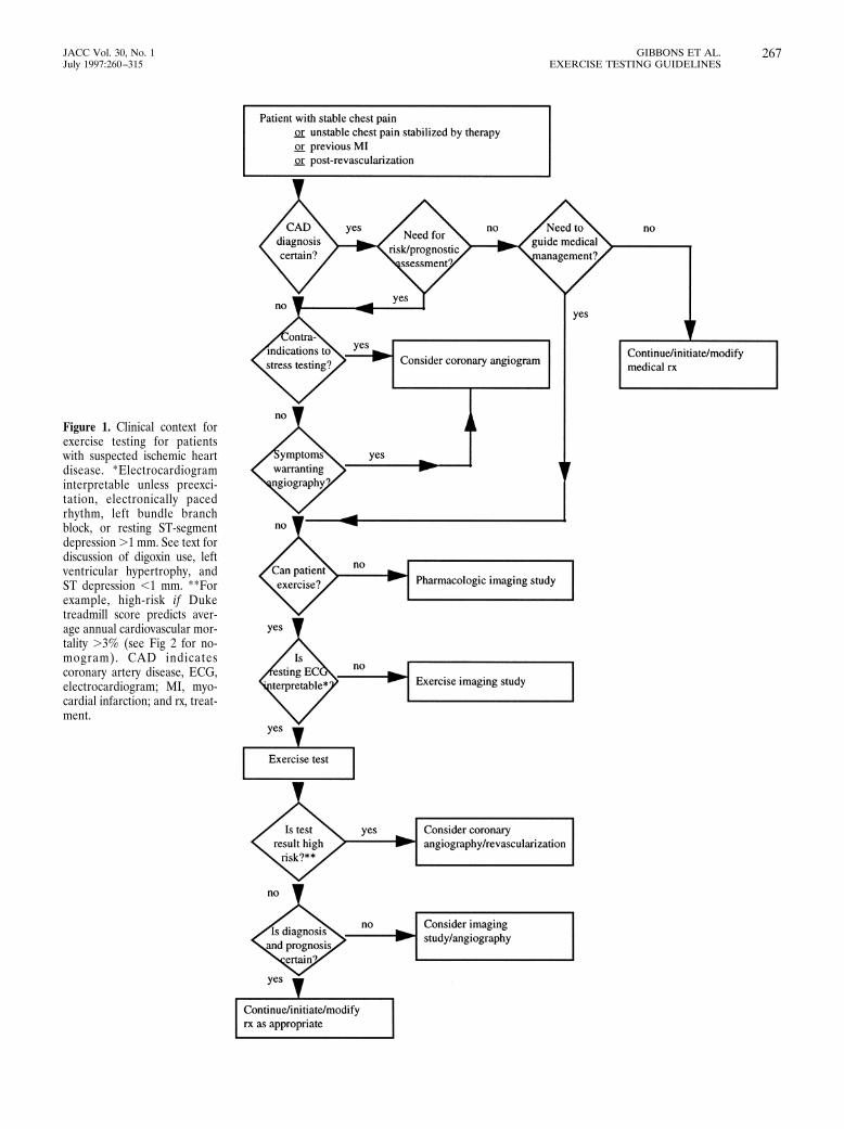
Figure 1. Clinical context forexercise testing for patientswith suspected ischemic heartdisease. *Electrocardiograminterpretable unless preexci-tation, electronically pacedrhythm, left bundle branchblock, or resting ST-segmentdepression .1 mm. See text fordiscussion of digoxin use, leftventricular hypertrophy, andST depression ,1 mm. **Forexample, high-risk if Duketreadmill score predicts aver-age annual cardiovascular mor-tality .3% (see Fig 2 for no-mogram). CAD indicatescoronary artery disease, ECG,electrocardiogram; MI, myo-cardial infarction; and rx, treat-ment.
267JACC Vol. 30, No. 1 GIBBONS ET AL.July 1997:260–315 EXERCISE TESTING GUIDELINES
Class III
1. Patients with the following baseline ECG abnormalities:● Preexcitation (Wolff-Parkinson-White) syndrome● Electronically paced ventricular rhythm● Greater than 1 mm of resting ST depression● Complete left bundle branch block
2. Patients with a documented myocardial infarction or priorcoronary angiography demonstrating significant diseasehave an established diagnosis of CAD; however, ischemiaand risk can be determined by testing (see sections III andIV).
RationaleThe exercise test may be used if the diagnosis of CAD is
uncertain. Although other clinical findings, such as dyspnea onexertion, resting ECG abnormalities, or multiple risk factorsfor atherosclerosis may suggest the possibility of CAD, themost predictive clinical finding is a history of chest pain ordiscomfort. Myocardial ischemia is the most important causeof chest pain and is most commonly a consequence of under-lying coronary disease. CAD that has not resulted in sufficientluminal occlusion to cause ischemia during stress15 can stilllead to ischemic events through spasm, plaque rupture, andthrombosis, but most catastrophic events are associated withextensive atherosclerosis. These nonobstructive lesions explainsome of the events that occur after a normal exercise test (seesection III). Although the coronary angiogram has obviouslimitations,16 angiographic lesions remain the clinical goldstandard. Results of correlative studies have been divided overthe use of 50% or 70% luminal occlusion. Meta-analysis of thestudies has not demonstrated that the criteria affect the testcharacteristics.
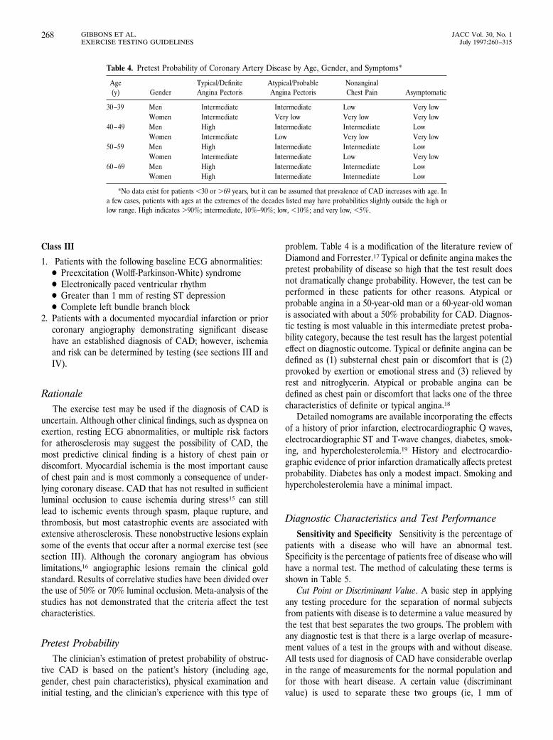
Pretest ProbabilityThe clinician’s estimation of pretest probability of obstruc-
tive CAD is based on the patient’s history (including age,gender, chest pain characteristics), physical examination andinitial testing, and the clinician’s experience with this type of
problem. Table 4 is a modification of the literature review ofDiamond and Forrester.17 Typical or definite angina makes thepretest probability of disease so high that the test result doesnot dramatically change probability. However, the test can beperformed in these patients for other reasons. Atypical orprobable angina in a 50-year-old man or a 60-year-old womanis associated with about a 50% probability for CAD. Diagnos-tic testing is most valuable in this intermediate pretest proba-bility category, because the test result has the largest potentialeffect on diagnostic outcome. Typical or definite angina can bedefined as (1) substernal chest pain or discomfort that is (2)provoked by exertion or emotional stress and (3) relieved byrest and nitroglycerin. Atypical or probable angina can bedefined as chest pain or discomfort that lacks one of the threecharacteristics of definite or typical angina.18
Detailed nomograms are available incorporating the effectsof a history of prior infarction, electrocardiographic Q waves,electrocardiographic ST and T-wave changes, diabetes, smok-ing, and hypercholesterolemia.19 History and electrocardio-graphic evidence of prior infarction dramatically affects pretestprobability. Diabetes has only a modest impact. Smoking andhypercholesterolemia have a minimal impact.
Diagnostic Characteristics and Test PerformanceSensitivity and Specificity Sensitivity is the percentage of
patients with a disease who will have an abnormal test.Specificity is the percentage of patients free of disease who willhave a normal test. The method of calculating these terms isshown in Table 5.
Cut Point or Discriminant Value. A basic step in applyingany testing procedure for the separation of normal subjectsfrom patients with disease is to determine a value measured bythe test that best separates the two groups. The problem withany diagnostic test is that there is a large overlap of measure-ment values of a test in the groups with and without disease.All tests used for diagnosis of CAD have considerable overlapin the range of measurements for the normal population andfor those with heart disease. A certain value (discriminantvalue) is used to separate these two groups (ie, 1 mm of
Table 4. Pretest Probability of Coronary Artery Disease by Age, Gender, and Symptoms*
Age(y) Gender
Typical/DefiniteAngina Pectoris
Atypical/ProbableAngina Pectoris
NonanginalChest Pain Asymptomatic
30–39 Men Intermediate Intermediate Low Very lowWomen Intermediate Very low Very low Very low
40–49 Men High Intermediate Intermediate LowWomen Intermediate Low Very low Very low
50–59 Men High Intermediate Intermediate LowWomen Intermediate Intermediate Low Very low
60–69 Men High Intermediate Intermediate LowWomen High Intermediate Intermediate Low
*No data exist for patients ,30 or .69 years, but it can be assumed that prevalence of CAD increases with age. Ina few cases, patients with ages at the extremes of the decades listed may have probabilities slightly outside the high orlow range. High indicates .90%; intermediate, 10%–90%; low, ,10%; and very low, ,5%.
268 GIBBONS ET AL. JACC Vol. 30, No. 1EXERCISE TESTING GUIDELINES July 1997:260–315
ST-segment depression). If the value is set high (ie, 2 mm ofST-segment depression) to ensure that nearly all normalsubjects have a normal test, giving the test a high specificity,then a substantial number of those with the disease appear tobe normal, reducing the test sensitivity. There may be reasonsfor wanting to adjust a test to have a relatively higher sensitiv-ity, but sensitivity and specificity are inversely related.
Population Effect. Sensitivity and specificity are inverselyrelated, affected by the population tested, and determined bythe choice of a cut point or discriminant value. Once adiscriminant value that determines the specificity and sensitiv-ity of a test is chosen, then the population tested must beconsidered. If the population is skewed toward persons with agreater severity of disease, then the test will have a highersensitivity for any cut point chosen. For instance, the exercisetest has a higher sensitivity in the elderly and persons withthree-vessel disease than in younger persons and those withone-vessel disease. A test can have a lower specificity if it isused in persons in whom false-positive results are more likely,such as those with valvular heart disease, LVH, resting STdepression, and patients taking digoxin.
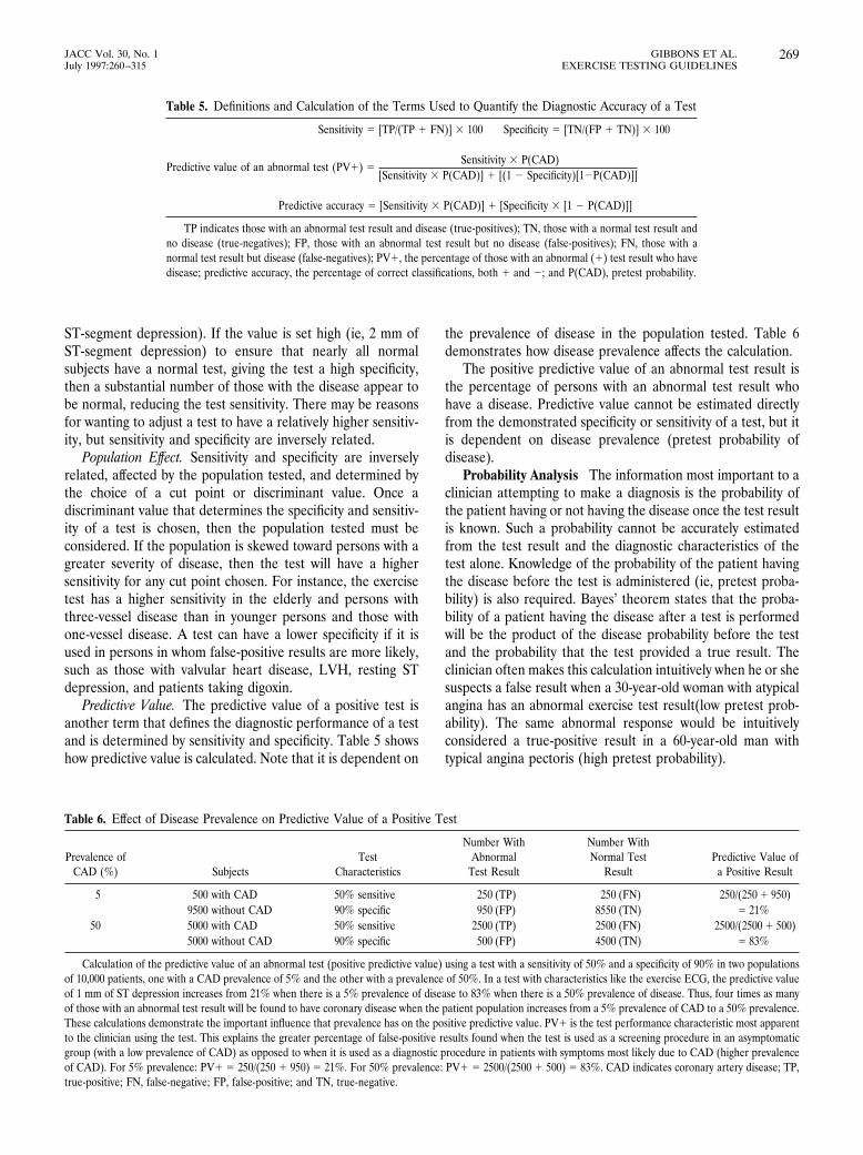
Predictive Value. The predictive value of a positive test isanother term that defines the diagnostic performance of a testand is determined by sensitivity and specificity. Table 5 showshow predictive value is calculated. Note that it is dependent on
the prevalence of disease in the population tested. Table 6demonstrates how disease prevalence affects the calculation.
The positive predictive value of an abnormal test result isthe percentage of persons with an abnormal test result whohave a disease. Predictive value cannot be estimated directlyfrom the demonstrated specificity or sensitivity of a test, but itis dependent on disease prevalence (pretest probability ofdisease).
Probability Analysis The information most important to aclinician attempting to make a diagnosis is the probability ofthe patient having or not having the disease once the test resultis known. Such a probability cannot be accurately estimatedfrom the test result and the diagnostic characteristics of thetest alone. Knowledge of the probability of the patient havingthe disease before the test is administered (ie, pretest proba-bility) is also required. Bayes’ theorem states that the proba-bility of a patient having the disease after a test is performedwill be the product of the disease probability before the testand the probability that the test provided a true result. Theclinician often makes this calculation intuitively when he or shesuspects a false result when a 30-year-old woman with atypicalangina has an abnormal exercise test result(low pretest prob-ability). The same abnormal response would be intuitivelyconsidered a true-positive result in a 60-year-old man withtypical angina pectoris (high pretest probability).
Table 6. Effect of Disease Prevalence on Predictive Value of a Positive Test
Prevalence ofCAD (%) Subjects
TestCharacteristics
Number WithAbnormal
Test Result
Number WithNormal Test
ResultPredictive Value of
a Positive Result
5 500 with CAD 50% sensitive 250 (TP) 250 (FN) 250/(250 1 950)9500 without CAD 90% specific 950 (FP) 8550 (TN) 5 21%
50 5000 with CAD 50% sensitive 2500 (TP) 2500 (FN) 2500/(2500 1 500)5000 without CAD 90% specific 500 (FP) 4500 (TN) 5 83%
Calculation of the predictive value of an abnormal test (positive predictive value) using a test with a sensitivity of 50% and a specificity of 90% in two populationsof 10,000 patients, one with a CAD prevalence of 5% and the other with a prevalence of 50%. In a test with characteristics like the exercise ECG, the predictive valueof 1 mm of ST depression increases from 21% when there is a 5% prevalence of disease to 83% when there is a 50% prevalence of disease. Thus, four times as manyof those with an abnormal test result will be found to have coronary disease when the patient population increases from a 5% prevalence of CAD to a 50% prevalence.These calculations demonstrate the important influence that prevalence has on the positive predictive value. PV1 is the test performance characteristic most apparentto the clinician using the test. This explains the greater percentage of false-positive results found when the test is used as a screening procedure in an asymptomaticgroup (with a low prevalence of CAD) as opposed to when it is used as a diagnostic procedure in patients with symptoms most likely due to CAD (higher prevalenceof CAD). For 5% prevalence: PV1 5 250/(250 1 950) 5 21%. For 50% prevalence: PV1 5 2500/(2500 1 500) 5 83%. CAD indicates coronary artery disease; TP,true-positive; FN, false-negative; FP, false-positive; and TN, true-negative.
Table 5. Definitions and Calculation of the Terms Used to Quantify the Diagnostic Accuracy of a Test
Sensitivity 5 [TP/(TP 1 FN)] 3 100 Specificity 5 [TN/(FP 1 TN)] 3 100
Predictive value of an abnormal test (PV1) 5Sensitivity 3 P(CAD)
[Sensitivity 3 P(CAD)] 1 [(1 2 Specificity)[12P(CAD)]]
Predictive accuracy 5 [Sensitivity 3 P(CAD)] 1 [Specificity 3 [1 2 P(CAD)]]
TP indicates those with an abnormal test result and disease (true-positives); TN, those with a normal test result andno disease (true-negatives); FP, those with an abnormal test result but no disease (false-positives); FN, those with anormal test result but disease (false-negatives); PV1, the percentage of those with an abnormal (1) test result who havedisease; predictive accuracy, the percentage of correct classifications, both 1 and 2; and P(CAD), pretest probability.
269JACC Vol. 30, No. 1 GIBBONS ET AL.July 1997:260–315 EXERCISE TESTING GUIDELINES
Scores developed from multivariable analysis of clinical andexercise test variables provide superior discrimination com-pared with using only the ST-segment response to diagnoseCAD. Such scores can provide probabilities of CAD that aremore accurate than ST measurements alone.20,21 However,diagnostic interpretation of the exercise test still centersaround the ST response, because the clinician remains uncer-tain about which other variables to apply and how to includethem in prediction. Although the statistical models proposedhave proved to be superior, the available equations havediffered as to variables and coefficients chosen. In addition, theequations were usually derived in study populations with ahigher prevalence of disease than seen in clinical settingsbecause of work-up bias, ie, the results of the exercise test wereused to decide who would undergo cardiac catheterization. Forthese reasons, use of these equations remains controversialand limited. Several such equations are shown in Appendix 2.However, when these computational techniques have beencompared with the judgment of experienced clinical cardiolo-gists, the predictions have been comparable.22,23 Physicians areoften urged to “use” more than just the ST segment ininterpreting the exercise test; these equations provide the onlyscientific means to do so.
Believability Criteria for Diagnostic TestsStudies should include consecutive or randomly selected
patients for whom the diagnosis is in doubt.24 Any diagnostictest appears to function well if obviously normal subjects arecompared with those who obviously have the disease in ques-tion (a “limited challenge”). The more relevant issue is toevaluate patients who are suspected but not known to have thedisease of interest and to differentiate those who do from thosewho do not. If the patients enrolled in the study do notrepresent this diagnostic dilemma group, the test may performwell in the study but not in clinical practice. Problems arisewhen patients who most certainly have the disease (ie, post–myocardial infarction patients) are included in this diagnosticsample. Post–myocardial infarction patients may be includedin studies to predict disease severity but should not be included
in studies attempting to distinguish those with disease fromthose without disease.
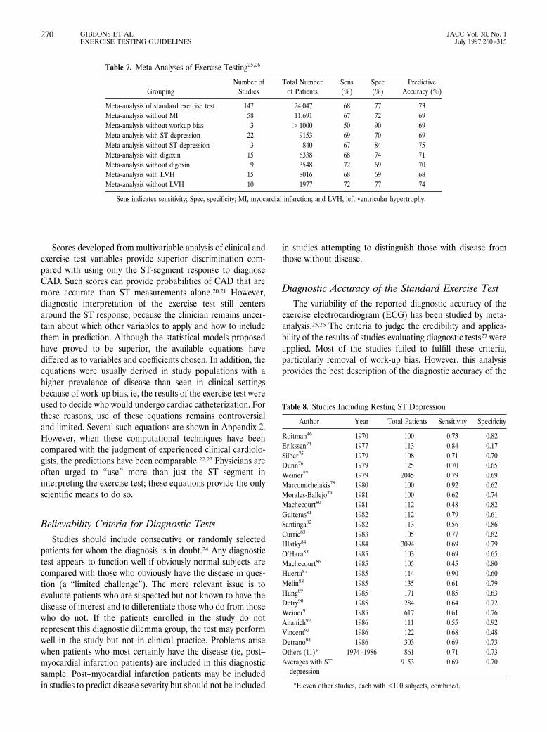
Diagnostic Accuracy of the Standard Exercise TestThe variability of the reported diagnostic accuracy of the
exercise electrocardiogram (ECG) has been studied by meta-analysis.25,26 The criteria to judge the credibility and applica-bility of the results of studies evaluating diagnostic tests27 wereapplied. Most of the studies failed to fulfill these criteria,particularly removal of work-up bias. However, this analysisprovides the best description of the diagnostic accuracy of the
Table 7. Meta-Analyses of Exercise Testing25,26
GroupingNumber of
StudiesTotal Number
of PatientsSens(%)
Spec(%)
PredictiveAccuracy (%)
Meta-analysis of standard exercise test 147 24,047 68 77 73Meta-analysis without MI 58 11,691 67 72 69Meta-analysis without workup bias 3 . 1000 50 90 69Meta-analysis with ST depression 22 9153 69 70 69Meta-analysis without ST depression 3 840 67 84 75Meta-analysis with digoxin 15 6338 68 74 71Meta-analysis without digoxin 9 3548 72 69 70Meta-analysis with LVH 15 8016 68 69 68Meta-analysis without LVH 10 1977 72 77 74
Sens indicates sensitivity; Spec, specificity; MI, myocardial infarction; and LVH, left ventricular hypertrophy.
Table 8. Studies Including Resting ST Depression
Author Year Total Patients Sensitivity Specificity
Roitman46 1970 100 0.73 0.82Erikssen74 1977 113 0.84 0.17Silber75 1979 108 0.71 0.70Dunn76 1979 125 0.70 0.65Weiner77 1979 2045 0.79 0.69Marcomichelakis78 1980 100 0.92 0.62Morales-Ballejo79 1981 100 0.62 0.74Machecourt80 1981 112 0.48 0.82Guiteras81 1982 112 0.79 0.61Santinga82 1982 113 0.56 0.86Currie83 1983 105 0.77 0.82Hlatky84 1984 3094 0.69 0.79O’Hara85 1985 103 0.69 0.65Machecourt86 1985 105 0.45 0.80Huerta87 1985 114 0.90 0.60Melin88 1985 135 0.61 0.79Hung89 1985 171 0.85 0.63Detry90 1985 284 0.64 0.72Weiner91 1985 617 0.61 0.76Ananich92 1986 111 0.55 0.92Vincent93 1986 122 0.68 0.48Detrano94 1986 303 0.69 0.73Others (11)* 1974–1986 861 0.71 0.73Averages with ST
depression9153 0.69 0.70
*Eleven other studies, each with ,100 subjects, combined.
270 GIBBONS ET AL. JACC Vol. 30, No. 1EXERCISE TESTING GUIDELINES July 1997:260–315
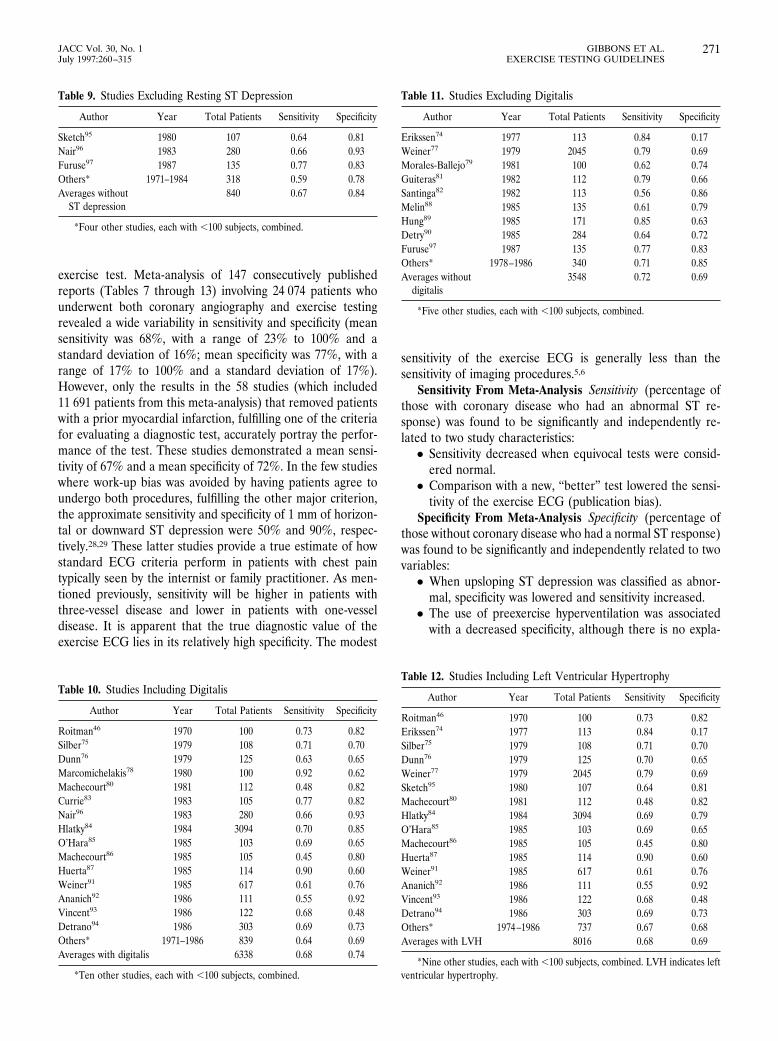
exercise test. Meta-analysis of 147 consecutively publishedreports (Tables 7 through 13) involving 24 074 patients whounderwent both coronary angiography and exercise testingrevealed a wide variability in sensitivity and specificity (meansensitivity was 68%, with a range of 23% to 100% and astandard deviation of 16%; mean specificity was 77%, with arange of 17% to 100% and a standard deviation of 17%).However, only the results in the 58 studies (which included11 691 patients from this meta-analysis) that removed patientswith a prior myocardial infarction, fulfilling one of the criteriafor evaluating a diagnostic test, accurately portray the perfor-mance of the test. These studies demonstrated a mean sensi-tivity of 67% and a mean specificity of 72%. In the few studieswhere work-up bias was avoided by having patients agree toundergo both procedures, fulfilling the other major criterion,the approximate sensitivity and specificity of 1 mm of horizon-tal or downward ST depression were 50% and 90%, respec-tively.28,29 These latter studies provide a true estimate of howstandard ECG criteria perform in patients with chest paintypically seen by the internist or family practitioner. As men-tioned previously, sensitivity will be higher in patients withthree-vessel disease and lower in patients with one-vesseldisease. It is apparent that the true diagnostic value of theexercise ECG lies in its relatively high specificity. The modest
sensitivity of the exercise ECG is generally less than thesensitivity of imaging procedures.5,6
Sensitivity From Meta-Analysis Sensitivity (percentage ofthose with coronary disease who had an abnormal ST re-sponse) was found to be significantly and independently re-lated to two study characteristics:
● Sensitivity decreased when equivocal tests were consid-ered normal.
● Comparison with a new, “better” test lowered the sensi-tivity of the exercise ECG (publication bias).
Specificity From Meta-Analysis Specificity (percentage ofthose without coronary disease who had a normal ST response)was found to be significantly and independently related to twovariables:
● When upsloping ST depression was classified as abnor-mal, specificity was lowered and sensitivity increased.
● The use of preexercise hyperventilation was associatedwith a decreased specificity, although there is no expla-
Table 9. Studies Excluding Resting ST Depression
Author Year Total Patients Sensitivity Specificity
Sketch95 1980 107 0.64 0.81Nair96 1983 280 0.66 0.93Furuse97 1987 135 0.77 0.83Others* 1971–1984 318 0.59 0.78Averages without
ST depression840 0.67 0.84
*Four other studies, each with ,100 subjects, combined.
Table 10. Studies Including Digitalis
Author Year Total Patients Sensitivity Specificity
Roitman46 1970 100 0.73 0.82Silber75 1979 108 0.71 0.70Dunn76 1979 125 0.63 0.65Marcomichelakis78 1980 100 0.92 0.62Machecourt80 1981 112 0.48 0.82Currie83 1983 105 0.77 0.82Nair96 1983 280 0.66 0.93Hlatky84 1984 3094 0.70 0.85O’Hara85 1985 103 0.69 0.65Machecourt86 1985 105 0.45 0.80Huerta87 1985 114 0.90 0.60Weiner91 1985 617 0.61 0.76Ananich92 1986 111 0.55 0.92Vincent93 1986 122 0.68 0.48Detrano94 1986 303 0.69 0.73Others* 1971–1986 839 0.64 0.69Averages with digitalis 6338 0.68 0.74
*Ten other studies, each with ,100 subjects, combined.
Table 11. Studies Excluding Digitalis
Author Year Total Patients Sensitivity Specificity
Erikssen74 1977 113 0.84 0.17Weiner77 1979 2045 0.79 0.69Morales-Ballejo79 1981 100 0.62 0.74Guiteras81 1982 112 0.79 0.66Santinga82 1982 113 0.56 0.86Melin88 1985 135 0.61 0.79Hung89 1985 171 0.85 0.63Detry90 1985 284 0.64 0.72Furuse97 1987 135 0.77 0.83Others* 1978–1986 340 0.71 0.85Averages without
digitalis3548 0.72 0.69
*Five other studies, each with ,100 subjects, combined.
Table 12. Studies Including Left Ventricular Hypertrophy
Author Year Total Patients Sensitivity Specificity
Roitman46 1970 100 0.73 0.82Erikssen74 1977 113 0.84 0.17Silber75 1979 108 0.71 0.70Dunn76 1979 125 0.70 0.65Weiner77 1979 2045 0.79 0.69Sketch95 1980 107 0.64 0.81Machecourt80 1981 112 0.48 0.82Hlatky84 1984 3094 0.69 0.79O’Hara85 1985 103 0.69 0.65Machecourt86 1985 105 0.45 0.80Huerta87 1985 114 0.90 0.60Weiner91 1985 617 0.61 0.76Ananich92 1986 111 0.55 0.92Vincent93 1986 122 0.68 0.48Detrano94 1986 303 0.69 0.73Others* 1974–1986 737 0.67 0.68Averages with LVH 8016 0.68 0.69
*Nine other studies, each with ,100 subjects, combined. LVH indicates leftventricular hypertrophy.
271JACC Vol. 30, No. 1 GIBBONS ET AL.July 1997:260–315 EXERCISE TESTING GUIDELINES
nation for this association. Hyperventilation was oncethought to reveal false-positive ST responders by bringingout ST depression with a stimulus other than ischemia;however, this has not been validated, and it is no longerrecommended as a routine to be performed beforestandard testing.26
The Tables 8 to 13 in the appendix were developed forresolving the issues of LVH, resting ST depression, anddigoxin. Of the 58 studies, only those that provided sensitivity,specificity, and total patient numbers were considered, andonly those with more than 100 patients were consideredseparately. Regarding the effect of resting ECG abnormalities,the studies that included patients with LVH had a meansensitivity of 68% and a mean specificity of 69%; the studiesthat excluded them had a mean sensitivity of 72% and a meanspecificity of 77%. Studies that included patients with restingST depression had a mean sensitivity of 69% and a meanspecificity of 70%; studies that excluded them had a meansensitivity of 67% and a mean specificity of 84%. Studies thatincluded patients receiving digoxin had a mean sensitivity of68% and a mean specificity of 74%; studies that excludedpatients on digoxin had a mean sensitivity of 72% and a meanspecificity of 69%. Comparing these results with the averagesensitivity of 67% and specificity of 72% as well as to them-selves, only LVH and resting ST depression appear to lowerspecificity. However, other studies in apparently healthy persons(see below) have suggested that digoxin also lowers specificity.
These meta-analyses provide only indirect evidence regard-ing these potentially important factors, because they assumethat the study populations were otherwise equal with respect tocharacteristics that might influence test performance. Thiscritical assumption has not been confirmed and may not betrue.
The wide variability in test performance apparent from thismeta-analysis makes it important that clinicians use propermethods for testing and analysis. Upsloping ST depressionshould be considered borderline or negative. Hyperventilationis no longer routinely recommended before testing.
Influence of Other Factors on Test PerformanceDrugs Digoxin. Digoxin produces abnormal exercise-
induced ST depression in 25% to 40% of apparently healthynormal subjects.30,31 The prevalence of abnormal responses isdirectly related to age. Although patients must be off themedication for at least 2 weeks for its effect to be gone, it is notnecessary to do so before diagnostic testing.32
b-Blocker Therapy. Despite the marked effect of b-blockerson maximal exercise heart rate, with patients subgroupedaccording to b-blocker administration initiated by their refer-ring physician, no differences in test performance were foundin a consecutive group of men being evaluated for possibleCAD.33 For routine exercise testing, it appears unnecessary forphysicians to accept the risk of stopping b-blockers beforetesting when a patient exhibits possible symptoms of ischemia.However, exercise testing in patients on b-blockers may havereduced diagnostic value due to inadequate heart rate re-sponse.
Other Drugs. Various medications, including antihyperten-sive agents and vasodilators, can affect test performance byaltering the hemodynamic response of blood pressure. Acuteadministration of nitrates can attenuate the angina and STdepression associated with myocardial ischemia. Flecainidehas been associated with exercise-induced ventricular tachy-cardia (VT).34,35
Electrocardiographic AbnormalitiesLeft Bundle Branch Block Exercise-induced ST depression
usually occurs with left bundle branch block and has noassociation with ischemia.36 Even up to 1 cm of ST depressioncan occur in healthy normal subjects.
Right Bundle Branch Block Exercise-induced ST depres-sion usually occurs with right bundle branch block in theanterior chest leads (V1 through V3) and is not associated withischemia.37 However, in the left chest leads (V5 and V6) orinferior leads (II and aVF), its test characteristics are similar tothose of a normal resting ECG.
Left Ventricular Hypertrophy With Repolarization Abnor-malities As discussed previously, this ECG abnormality isassociated with a decreased specificity of exercise testing, butsensitivity is unaffected. Therefore, a standard exercise testmay still be the first test, with referrals for further tests onlyindicated in patients with an abnormal test result.
Resting ST Depression Resting ST-segment depressionhas been identified as a marker for adverse cardiac events inpatients with and without known CAD.38–42 Miranda et al43
performed a retrospective study of 223 patients without clinicalor electrocardiographic evidence of prior myocardial infarc-tion. Women, patients with resting ECGs showing left bundlebranch block or LVH, and those on digoxin or with valvular orcongenital heart disease were excluded. Ten percent hadpersistent resting ST-segment depression and nearly twice theprevalence of severe coronary disease (30%) than those with-out resting ST-segment depression (16%). Two millimetersof additional exercise-induced ST-segment depression or
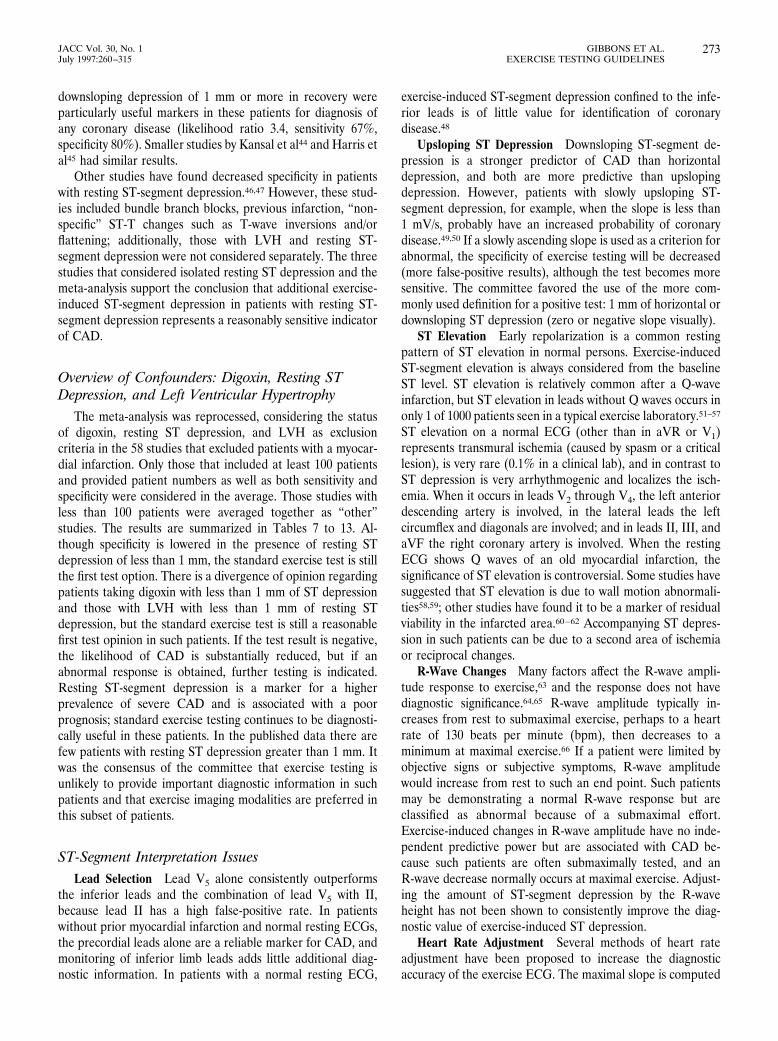
Table 13. Studies Excluding Left Ventricular Hypertrophy
Author Year Total Patients Sensitivity Specificity
Marcomichelakis78 1980 100 0.92 0.62Morales-Ballejo79 1981 100 0.62 0.74Guiteras81 1982 112 0.79 0.66Santinga82 1982 113 0.56 0.86Currie83 1983 105 0.77 0.82Nair96 1983 280 0.66 0.93Melin88 1985 135 0.61 0.79Hung89 1985 171 0.85 0.63Detry90 1985 284 0.64 0.72Furuse97 1987 135 0.77 0.83Others* 1971–1983 442 0.69 0.84Averages without LVH 1977 0.72 0.77
*Six other studies, each with ,100 subjects, combined. LVH indicates leftventricular hypertrophy.
272 GIBBONS ET AL. JACC Vol. 30, No. 1EXERCISE TESTING GUIDELINES July 1997:260–315
downsloping depression of 1 mm or more in recovery wereparticularly useful markers in these patients for diagnosis ofany coronary disease (likelihood ratio 3.4, sensitivity 67%,specificity 80%). Smaller studies by Kansal et al44 and Harris etal45 had similar results.
Other studies have found decreased specificity in patientswith resting ST-segment depression.46,47 However, these stud-ies included bundle branch blocks, previous infarction, “non-specific” ST-T changes such as T-wave inversions and/orflattening; additionally, those with LVH and resting ST-segment depression were not considered separately. The threestudies that considered isolated resting ST depression and themeta-analysis support the conclusion that additional exercise-induced ST-segment depression in patients with resting ST-segment depression represents a reasonably sensitive indicatorof CAD.
Overview of Confounders: Digoxin, Resting STDepression, and Left Ventricular Hypertrophy
The meta-analysis was reprocessed, considering the statusof digoxin, resting ST depression, and LVH as exclusioncriteria in the 58 studies that excluded patients with a myocar-dial infarction. Only those that included at least 100 patientsand provided patient numbers as well as both sensitivity andspecificity were considered in the average. Those studies withless than 100 patients were averaged together as “other”studies. The results are summarized in Tables 7 to 13. Al-though specificity is lowered in the presence of resting STdepression of less than 1 mm, the standard exercise test is stillthe first test option. There is a divergence of opinion regardingpatients taking digoxin with less than 1 mm of ST depressionand those with LVH with less than 1 mm of resting STdepression, but the standard exercise test is still a reasonablefirst test opinion in such patients. If the test result is negative,the likelihood of CAD is substantially reduced, but if anabnormal response is obtained, further testing is indicated.Resting ST-segment depression is a marker for a higherprevalence of severe CAD and is associated with a poorprognosis; standard exercise testing continues to be diagnosti-cally useful in these patients. In the published data there arefew patients with resting ST depression greater than 1 mm. Itwas the consensus of the committee that exercise testing isunlikely to provide important diagnostic information in suchpatients and that exercise imaging modalities are preferred inthis subset of patients.
ST-Segment Interpretation IssuesLead Selection Lead V5 alone consistently outperforms
the inferior leads and the combination of lead V5 with II,because lead II has a high false-positive rate. In patientswithout prior myocardial infarction and normal resting ECGs,the precordial leads alone are a reliable marker for CAD, andmonitoring of inferior limb leads adds little additional diag-nostic information. In patients with a normal resting ECG,
exercise-induced ST-segment depression confined to the infe-rior leads is of little value for identification of coronarydisease.48
Upsloping ST Depression Downsloping ST-segment de-pression is a stronger predictor of CAD than horizontaldepression, and both are more predictive than upslopingdepression. However, patients with slowly upsloping ST-segment depression, for example, when the slope is less than1 mV/s, probably have an increased probability of coronarydisease.49,50 If a slowly ascending slope is used as a criterion forabnormal, the specificity of exercise testing will be decreased(more false-positive results), although the test becomes moresensitive. The committee favored the use of the more com-monly used definition for a positive test: 1 mm of horizontal ordownsloping ST depression (zero or negative slope visually).
ST Elevation Early repolarization is a common restingpattern of ST elevation in normal persons. Exercise-inducedST-segment elevation is always considered from the baselineST level. ST elevation is relatively common after a Q-waveinfarction, but ST elevation in leads without Q waves occurs inonly 1 of 1000 patients seen in a typical exercise laboratory.51–57
ST elevation on a normal ECG (other than in aVR or V1)represents transmural ischemia (caused by spasm or a criticallesion), is very rare (0.1% in a clinical lab), and in contrast toST depression is very arrhythmogenic and localizes the isch-emia. When it occurs in leads V2 through V4, the left anteriordescending artery is involved, in the lateral leads the leftcircumflex and diagonals are involved; and in leads II, III, andaVF the right coronary artery is involved. When the restingECG shows Q waves of an old myocardial infarction, thesignificance of ST elevation is controversial. Some studies havesuggested that ST elevation is due to wall motion abnormali-ties58,59; other studies have found it to be a marker of residualviability in the infarcted area.60–62 Accompanying ST depres-sion in such patients can be due to a second area of ischemiaor reciprocal changes.
R-Wave Changes Many factors affect the R-wave ampli-tude response to exercise,63 and the response does not havediagnostic significance.64,65 R-wave amplitude typically in-creases from rest to submaximal exercise, perhaps to a heartrate of 130 beats per minute (bpm), then decreases to aminimum at maximal exercise.66 If a patient were limited byobjective signs or subjective symptoms, R-wave amplitudewould increase from rest to such an end point. Such patientsmay be demonstrating a normal R-wave response but areclassified as abnormal because of a submaximal effort.Exercise-induced changes in R-wave amplitude have no inde-pendent predictive power but are associated with CAD be-cause such patients are often submaximally tested, and anR-wave decrease normally occurs at maximal exercise. Adjust-ing the amount of ST-segment depression by the R-waveheight has not been shown to consistently improve the diag-nostic value of exercise-induced ST depression.
Heart Rate Adjustment Several methods of heart rateadjustment have been proposed to increase the diagnosticaccuracy of the exercise ECG. The maximal slope is computed
273JACC Vol. 30, No. 1 GIBBONS ET AL.July 1997:260–315 EXERCISE TESTING GUIDELINES
either manually 67 or by computer.68 A second techniquedivides the difference between ST depression at peak exerciseby the exercise-induced increase in heart rate.69,70 Althoughthe initial reports were promising, neither meta-analysis25 nora subsequent study71 found convincing evidence of benefit. Thepotential explanations for these discordant findings are de-tailed elsewhere.71,72 As described in sections III and IV, it ismore important to consider exercise capacity rather thanexercise heart rate in interpretation of exercise tests.
Computer Processing Although computer processing ofthe exercise ECG can be helpful, it can result in a false-positiveindication of ST depression.73 To avoid this problem, thephysician should always be provided with ECG recordings ofthe raw unprocessed ECG data for comparison with anyaverages the exercise test monitor generates. It is preferablethat averages always be contiguously preceded by the raw ECGdata. The degree of filtering and preprocessing should alwaysbe presented along with the ECG recordings and should becompared with the AHA recommendations (0 to 100 Hz usingnotched power line frequency filters). It is preferable that theAHA standards be the default setting. All averages should becarefully labeled and explained, particularly those that simu-late raw data. Simulation of raw data with averaged datashould be avoided. Obvious breaks should be inserted betweenaveraged ECG complexes. Averages should be check markedto indicate the PR isoelectric line as well as the ST measure-ment points. None of the computerized scores or measure-ments have been sufficiently validated to recommend theirwidespread use.
III. Risk Assessment and Prognosis inPatients With Symptoms or a Prior History
of Coronary Artery Disease
Class I
1. Patients undergoing initial evaluation with suspected orknown CAD. Specific exceptions are noted below in ClassIIb.
2. Patients with suspected or known CAD previously evalu-ated with significant change in clinical status.
Class IIb
1. Patients with the following ECG abnormalities:● Preexcitation (Wolff-Parkinson-White) syndrome● Electronically paced ventricular rhythm● Greater than 1 mm of resting ST depression● Complete left bundle branch block
2. Patients with a stable clinical course who undergo periodicmonitoring to guide treatment.
Class III
1. Patients with severe comorbidity likely to limit life expect-ancy and/or candidacy for revascularization.
Risk Stratification: General ConsiderationsRisk or prognostic stratification is one of the pivotal
activities in medical practice. Virtually all patient managementdecisions are driven by the clinician’s assessment of thepatient’s prognosis. During the initial encounter, the physiciancollects a standard data set of history, physical exam, andlaboratory test data items. Using these data, the physicianformulates a working diagnosis and risk assessment and selectsan initial management strategy.98 This strategy may consist ofadditional noninvasive testing, referral for prompt cardiaccatheterization, or performance of a therapeutic trial. Theadditional data resulting from these management steps mayaffirm the initial risk assessment, cause it to be modified, orresult in a completely revised risk assessment. The updated riskassessment in turn may indicate the need for further testingand/or therapy. Each additional patient-physician encounterprovides an opportunity to update the risk assessment andmodify the therapeutic plan appropriately.
The most important implication of the foregoing for theseguidelines is that risk stratification with the exercise test doesnot take place in isolation but as part of a process that includesmore readily accessible (and sometimes less expensive) datafrom the clinical exam and other laboratory tests. Thus, thevalue of exercise testing for risk stratification must be consid-ered in light of what is added to that which is already knownabout the patient’s risk status.
Whereas prognosis typically refers to probability of survival,other outcomes such as freedom from myocardial infarction,symptom status, functional capacity, and other aspects ofquality of life are equally important to many patients. Mostresearch on exercise testing, however, has concentrated on therelation between test parameters and future survival (and, to alesser extent, freedom from myocardial infarction). Theseoutcomes will be primarily considered in this section of theguidelines.
Prognosis of Coronary Artery Disease:General Considerations
CAD is a chronic disorder with a natural history that spansmultiple decades. In each affected individual the diseasetypically cycles in and out of a number of clinically definedphases: asymptomatic or presymptomatic, stable angina, pro-gressive angina, unstable angina, or acute myocardial infarc-tion. Although the specific approach to risk stratification of thecoronary disease patient can vary according to the phase of thedisease in which the patient presents, some general conceptsapply across the coronary disease spectrum.
Conceptually the probability of cardiac death in a patientwith CAD can be viewed as the sum of the risks at the time ofevaluation (the current risk state) and the risk that the diseasewill progress over time to a higher or lower risk state. Thepatient’s current risk state is a function of five major types ofprognostic measures (Table 14). The strongest predictor oflong-term survival with CAD is function of the left ventricle. Inparticular, the extent of damage or dysfunction and the success
274 GIBBONS ET AL. JACC Vol. 30, No. 1EXERCISE TESTING GUIDELINES July 1997:260–315
of mechanisms used by the cardiovascular system to compen-sate for that damage are of paramount importance. Manydifferent clinical and laboratory parameters provide informa-tion about the extent of left ventricular dysfunction (Table 14).Ejection fraction is the most commonly used measure, but italone does not completely describe the prognostic informationin left ventricular function. Another group of prognosticfactors describes the anatomic extent and severity of athero-sclerotic involvement of the coronary tree. The number ofdiseased vessels is the most common measure of this domain.More details about the coronary anatomy add importantprognostic information to this simple measure. A third groupof prognostic factors provides evidence of a recent coronaryplaque rupture, indicating a substantially increased short-termrisk for cardiac death or nonfatal myocardial infarction. Wors-ening clinical symptoms with unstable features is the majorclinical marker of a plaque event. The fourth group of prog-nostic factors is related to the presence of electrical instabilityof the myocardium and the propensity for malignant ventric-ular arrhythmia. The final group of prognostic factors de-scribes general health and noncoronary comorbidity.
The probability that a given patient will progress to a
higher- or lower-risk disease state depends primarily on factorsrelated to the aggressiveness of the underlying atheroscleroticprocess (Table 14). Patients with the major cardiac risk factors,including smoking, hypercholesterolemia, diabetes mellitus,and hypertension, are most likely to evidence progressiveatherosclerosis with repeated coronary plaque events. Patientswith symptomatic coronary disease at a younger age also mayhave a more aggressive disease process.
A growing body of pathological, angiographic, angioscopic,and intravascular ultrasonographic data support a pathophys-iological model in which most major cardiac events (suddendeath, acute myocardial infarction, unstable angina) are initi-ated by microscopic ruptures of high-risk or vulnerable athero-sclerotic plaques. Characteristically, vulnerable plaques have acholesterol gruel core and a thin fibrous cap. Various nonspe-cific factors may act as triggers and cause a vulnerable plaqueto rupture at thinned sites around the shoulders of the cap.This exposes inner plaque material to the flowing intra-arterialblood and initiates formation of a platelet-fibrin thrombus overthe area of rupture. Clinically the rupture may seal withoutdetectable sequelae, or the patient may experience worseningangina, acute myocardial infarction, or sudden cardiac death.Several lines of evidence have shown that the majority ofvulnerable plaques appear “angiographically insignificant” be-fore rupture (ie, less than 75% diameter stenosis). In contrast,most “significant” plaques (greater than or equal to 75%stenosis) visualized at angiography are at low risk for plaquerupture. Thus, the ability of stress testing of any type to detectvulnerable atherosclerotic lesions may be limited by thesmaller size and lesser effect on coronary blood flow of theseplaques and may explain the occasional acute coronary eventthat may occur not long after a negative treadmill test.
Risk Stratification With the Exercise TestThe major exercise ECG testing measures that have been
proposed as prognostic markers are listed in Table 15. Becausethe exercise test is a diagnostic tool rather than a therapy, itseffect on patient outcomes is necessarily indirect. To the extentthat the test guides clinicians to select more appropriate oreffective therapies, the exercise test will improve outcomes.However, no randomized trials of exercise testing versus noexercise testing have been performed. The entire evidencebase for exercise testing therefore consists of observationalstudies. No direct evidence links different exercise testingstrategies with differing outcomes.
As described previously, the risks of exercise testing inappropriately selected candidates are extremely low. Thus, themain arguments for not performing an exercise test in manyclinical situations are that the information provided would notjustify the extra costs of obtaining that information (ie, the testwould not be cost-effective in that given situation) and/or thetest might provide misleading information that could lead toinappropriate or unnecessary additional testing or therapy(both of which may have higher risks than exercise testing).
In reviewing the published evidence in this area, the
Table 14. Prognostic Factors for Patients With Coronary Disease
Prognostic factors for current risk stateLeft ventricular function/damage
History of prior MIPathologic Q waves on the resting ECGCongestive heart failure symptomsCardiomegaly on the chest x-rayEjection fractionEnd-systolic volumeRegional LV wall motion abnormalitiesConduction disturbances on the ECGMitral regurgitationExercise duration/tolerance
Severity of CADAnatomic extent and severity of CADCollateral vessels presentTransient ischemia on ambulatory monitorExercise- or stress-induced ST deviation
Coronary plaque eventProgressive or unstable ischemic symptomsTransient ischemia on resting ECG
Electrical stabilityVentricular arrhythmias
General healthAgeNoncoronary comorbidity
Prognostic factors for change in risk stateFactors predisposing to disease progression
SmokingHyperlipidemiaDiabetes mellitusHypertension
Other genetic/metabolic factors
MI indicates myocardial infarction; ECG, electrocardiogram; LV, left ven-tricular; and CAD, coronary artery disease.
275JACC Vol. 30, No. 1 GIBBONS ET AL.July 1997:260–315 EXERCISE TESTING GUIDELINES
subcommittee focused on studies that examined hard cardiacoutcome events (death alone or death plus myocardial infarc-tion) and had at least five (and preferably 10) outcome eventsfor every candidate variable evaluated. Use of appropriatemultivariable statistical techniques was also a requirement forselection. Special emphasis was given to studies that evaluatedthe incremental effects of the exercise test beyond the prog-nostic information available from the clinical evaluation (his-tory, physical examination, and resting 12-lead ECG).
Symptomatic Patients With Nonacute Coronary ArteryDisease Unless cardiac catheterization is indicated, patientswith suspected or known CAD and new or changing symptomssuggesting ischemia should generally undergo exercise testingto assess the risk of future cardiac events. As described in theACC/AHA guidelines for percutaneous transluminal coronaryangioplasty and for coronary artery bypass grafting, documen-tation of exercise- or stress-induced ischemia is desirable formost patients who are being evaluated for revascularization.3,4
Choice of initial stress testing modality should be based onevaluation of the patient’s resting ECG, physical ability toperform exercise, and local expertise and technology. For riskassessment, the exercise test should be the standard initialmode of stress testing used in patients with a normal ECG whoare not taking digoxin.99–101 Patients with widespread restingST depression (greater than or equal to 1 mm), complete leftbundle branch block, ventricular paced rhythm, or preexcita-tion should usually be tested with an imaging modality. Exer-cise testing may still provide useful prognostic information inpatients with these ECG changes but cannot be used to identifyischemia. Patients unable to exercise because of physical limita-tions affecting exercise capacity (eg, arthritis, amputations, severeperipheral vascular disease, severe chronic obstructive pulmonary
disease, general debility), should undergo pharmacological stresstesting in combination with imaging.
In patients with suspected or known symptomatic coronarydisease, exercise testing can be used to estimate prognosis andassist in management decisions. The primary evidence in thisarea consists of seven observational studies of the prognosticvalue of the exercise ECG (Table 16). An overview of theavailable literature has shown some inconsistency among stud-ies in the exercise variables identified as independent prognos-tic factors. These differences are at least partially attributableto differences in the spectrum of patients referred for testing,the amount of crossover to coronary revascularization, and thesample size/statistical power of the analysis.109
One of the strongest and most consistent prognostic mark-ers identified in exercise testing is maximum exercise capacity,which is at least partly influenced by the extent of resting leftventricular dysfunction and the amount of further LV dysfunc-tion induced by exercise. However, the relation betweenexercise capacity and LV function is complex, because exercisecapacity is also affected by age, general physical conditioning,comorbidities, and psychological state (especially the presenceof depression).110 Several exercise parameters can be used asmarkers of exercise capacity (Table 15), including maximalexercise duration, maximum MET level achieved, maximumworkload achieved, maximum heart rate, and double product.When interpreting the exercise test, it is very important to takeexercise capacity into account; the specific variable used tosummarize this aspect of test performance is less important.The translation of exercise duration or workload into METs(oxygen uptake expressed in multiples of basal oxygen uptake,3.5 O2 mL/kg per minute) has the advantage of providing acommon measure of performance regardless of the type ofexercise test or protocol used. Although such translations arebased on approximations and are not as accurate for individualpatients as measured maximal oxygen uptake (VO2max),VO2max has not been studied for prognostic purposes in largeseries of patients with chest pain.
A second group of prognostic exercise testing markersrelates to exercise-induced ischemia. These markers includeexercise-induced ST-segment depression, exercise-induced ST-segment elevation (in leads without pathological Q waves andnot in aVR), and exercise-induced angina. In a large exercisetesting cohort, exercise ST deviation (elevation or depression)best summarized the prognostic information from this area.103
Other less powerful prognostic ST variables included thenumber of leads showing significant ST-segment depression,configuration of the exercise-induced ST depression (down-sloping, horizontal, or upsloping), and duration of ST devia-tion into the recovery phase of the test.
Two early influential studies of exercise treadmill testingand prognosis were reported from the Duke CardiovascularDisease Databank and the Coronary Artery Surgery StudyRegistry. Using the Duke database, McNeer and coworkers111
demonstrated that an “early positive” exercise test result (STdepression greater than or equal to 1 mm in the first two stagesof the Bruce protocol) identified a high-risk population,
Table 15. Measurements Available From the ExerciseTreadmill Test
ElectrocardiographicMaximum ST depressionMaximum ST elevationST-depression slope (downsloping, horizontal, upsloping)Number of leads showing ST changesDuration of ST deviation into recoveryST/HR indexesExercise-induced ventricular arrhythmiasTime to onset of ST deviation
HemodynamicMaximum exercise heart rateMaximum exercise systolic blood pressureMaximum exercise double product (HR 3 BP)Total exercise durationExertional hypotension (drop below preexercise value)Chronotropic incompetence
SymptomaticExercise-induced anginaExercise-limiting symptomsTime to onset of angina
HR indicates heart rate; and BP, blood pressure.
276 GIBBONS ET AL. JACC Vol. 30, No. 1EXERCISE TESTING GUIDELINES July 1997:260–315
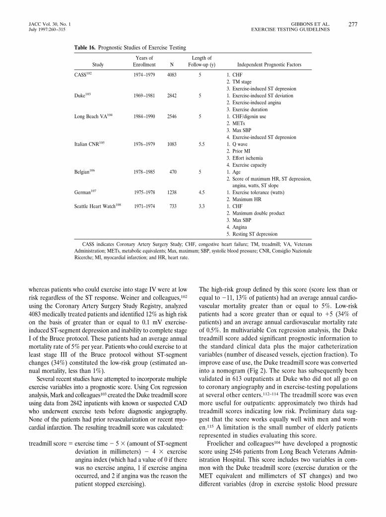
whereas patients who could exercise into stage IV were at lowrisk regardless of the ST response. Weiner and colleagues,102
using the Coronary Artery Surgery Study Registry, analyzed4083 medically treated patients and identified 12% as high riskon the basis of greater than or equal to 0.1 mV exercise-induced ST-segment depression and inability to complete stageI of the Bruce protocol. These patients had an average annualmortality rate of 5% per year. Patients who could exercise to atleast stage III of the Bruce protocol without ST-segmentchanges (34%) constituted the low-risk group (estimated an-nual mortality, less than 1%).
Several recent studies have attempted to incorporate multipleexercise variables into a prognostic score. Using Cox regressionanalysis, Mark and colleagues103 created the Duke treadmill scoreusing data from 2842 inpatients with known or suspected CADwho underwent exercise tests before diagnostic angiography.None of the patients had prior revascularization or recent myo-cardial infarction. The resulting treadmill score was calculated:
treadmill score 5 exercise time 2 5 3 (amount of ST-segmentdeviation in millimeters) 2 4 3 exerciseangina index (which had a value of 0 if therewas no exercise angina, 1 if exercise anginaoccurred, and 2 if angina was the reason thepatient stopped exercising).
The high-risk group defined by this score (score less than orequal to 211, 13% of patients) had an average annual cardio-vascular mortality greater than or equal to 5%. Low-riskpatients had a score greater than or equal to 15 (34% ofpatients) and an average annual cardiovascular mortality rateof 0.5%. In multivariable Cox regression analysis, the Duketreadmill score added significant prognostic information tothe standard clinical data plus the major catheterizationvariables (number of diseased vessels, ejection fraction). Toimprove ease of use, the Duke treadmill score was convertedinto a nomogram (Fig 2). The score has subsequently beenvalidated in 613 outpatients at Duke who did not all go onto coronary angiography and in exercise-testing populationsat several other centers.112–114 The treadmill score was evenmore useful for outpatients: approximately two thirds hadtreadmill scores indicating low risk. Preliminary data sug-gest that the score works equally well with men and wom-en.115 A limitation is the small number of elderly patientsrepresented in studies evaluating this score.
Froelicher and colleagues104 have developed a prognosticscore using 2546 patients from Long Beach Veterans Admin-istration Hospital. This score includes two variables in com-mon with the Duke treadmill score (exercise duration or theMET equivalent and millimeters of ST changes) and twodifferent variables (drop in exercise systolic blood pressure
Table 16. Prognostic Studies of Exercise Testing
StudyYears of
Enrollment NLength of
Follow-up (y) Independent Prognostic Factors
CASS102 1974–1979 4083 5 1. CHF2. TM stage3. Exercise-induced ST depression
Duke103 1969–1981 2842 5 1. Exercise-induced ST deviation2. Exercise-induced angina3. Exercise duration
Long Beach VA104 1984–1990 2546 5 1. CHF/digoxin use2. METs3. Max SBP4. Exercise-induced ST depression
Italian CNR105 1976–1979 1083 5.5 1. Q wave2. Prior MI3. Effort ischemia4. Exercise capacity
Belgian106 1978–1985 470 5 1. Age2. Score of maximum HR, ST depression,
angina, watts, ST slopeGerman107 1975–1978 1238 4.5 1. Exercise tolerance (watts)
2. Maximum HRSeattle Heart Watch108 1971–1974 733 3.3 1. CHF
2. Maximum double product3. Max SBP4. Angina5. Resting ST depression
CASS indicates Coronary Artery Surgery Study; CHF, congestive heart failure; TM, treadmill; VA, VeteransAdministration; METs, metabolic equivalents; Max, maximum; SBP, systolic blood pressure; CNR, Consiglio NazionaleRicerche; MI, myocardial infarction; and HR, heart rate.
277JACC Vol. 30, No. 1 GIBBONS ET AL.July 1997:260–315 EXERCISE TESTING GUIDELINES
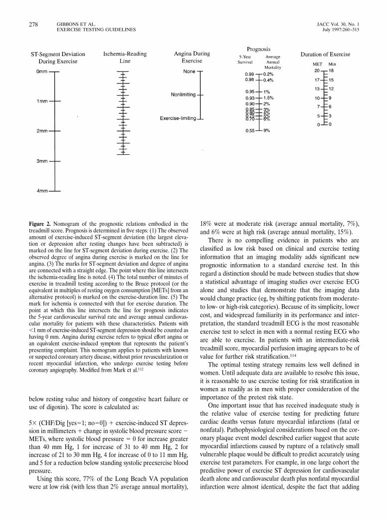
below resting value and history of congestive heart failure oruse of digoxin). The score is calculated as:
53 (CHF/Dig [yes51; no50]) 1 exercise-induced ST depres-sion in millimeters 1 change in systolic blood pressure score 2METs, where systolic blood pressure 5 0 for increase greaterthan 40 mm Hg, 1 for increase of 31 to 40 mm Hg, 2 forincrease of 21 to 30 mm Hg, 4 for increase of 0 to 11 mm Hg,and 5 for a reduction below standing systolic preexercise bloodpressure.
Using this score, 77% of the Long Beach VA populationwere at low risk (with less than 2% average annual mortality),
18% were at moderate risk (average annual mortality, 7%),and 6% were at high risk (average annual mortality, 15%).
There is no compelling evidence in patients who areclassified as low risk based on clinical and exercise testinginformation that an imaging modality adds significant newprognostic information to a standard exercise test. In thisregard a distinction should be made between studies that showa statistical advantage of imaging studies over exercise ECGalone and studies that demonstrate that the imaging datawould change practice (eg, by shifting patients from moderate-to low- or high-risk categories). Because of its simplicity, lowercost, and widespread familiarity in its performance and inter-pretation, the standard treadmill ECG is the most reasonableexercise test to select in men with a normal resting ECG whoare able to exercise. In patients with an intermediate-risktreadmill score, myocardial perfusion imaging appears to be ofvalue for further risk stratification.114
The optimal testing strategy remains less well defined inwomen. Until adequate data are available to resolve this issue,it is reasonable to use exercise testing for risk stratification inwomen as readily as in men with proper consideration of theimportance of the pretest risk state.
One important issue that has received inadequate study isthe relative value of exercise testing for predicting futurecardiac deaths versus future myocardial infarctions (fatal ornonfatal). Pathophysiological considerations based on the cor-onary plaque event model described earlier suggest that acutemyocardial infarctions caused by rupture of a relatively smallvulnerable plaque would be difficult to predict accurately usingexercise test parameters. For example, in one large cohort thepredictive power of exercise ST depression for cardiovasculardeath alone and cardiovascular death plus nonfatal myocardialinfarction were almost identical, despite the fact that adding
Figure 2. Nomogram of the prognostic relations embodied in thetreadmill score. Prognosis is determined in five steps: (1) The observedamount of exercise-induced ST-segment deviation (the largest eleva-tion or depression after resting changes have been subtracted) ismarked on the line for ST-segment deviation during exercise. (2) Theobserved degree of angina during exercise is marked on the line forangina. (3) The marks for ST-segment deviation and degree of anginaare connected with a straight edge. The point where this line intersectsthe ischemia-reading line is noted. (4) The total number of minutes ofexercise in treadmill testing according to the Bruce protocol (or theequivalent in multiples of resting oxygen consumption [METs] from analternative protocol) is marked on the exercise-duration line. (5) Themark for ischemia is connected with that for exercise duration. Thepoint at which this line intersects the line for prognosis indicatesthe 5-year cardiovascular survival rate and average annual cardiovas-cular mortality for patients with these characteristics. Patients with,1 mm of exercise-induced ST-segment depression should be counted ashaving 0 mm. Angina during exercise refers to typical effort angina oran equivalent exercise-induced symptom that represents the patient’spresenting complaint. This nomogram applies to patients with knownor suspected coronary artery disease, without prior revascularization orrecent myocardial infarction, who undergo exercise testing beforecoronary angiography. Modified from Mark et al.112
278 GIBBONS ET AL. JACC Vol. 30, No. 1EXERCISE TESTING GUIDELINES July 1997:260–315
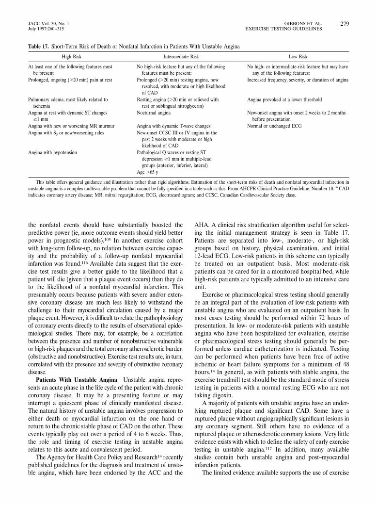
the nonfatal events should have substantially boosted thepredictive power (ie, more outcome events should yield betterpower in prognostic models).103 In another exercise cohortwith long-term follow-up, no relation between exercise capac-ity and the probability of a follow-up nonfatal myocardialinfarction was found.116 Available data suggest that the exer-cise test results give a better guide to the likelihood that apatient will die (given that a plaque event occurs) than they doto the likelihood of a nonfatal myocardial infarction. Thispresumably occurs because patients with severe and/or exten-sive coronary disease are much less likely to withstand thechallenge to their myocardial circulation caused by a majorplaque event. However, it is difficult to relate the pathophysiologyof coronary events directly to the results of observational epide-miological studies. There may, for example, be a correlationbetween the presence and number of nonobstructive vulnerableor high-risk plaques and the total coronary atherosclerotic burden(obstructive and nonobstructive). Exercise test results are, in turn,correlated with the presence and severity of obstructive coronarydisease.
Patients With Unstable Angina Unstable angina repre-sents an acute phase in the life cycle of the patient with chroniccoronary disease. It may be a presenting feature or mayinterrupt a quiescent phase of clinically manifested disease.The natural history of unstable angina involves progression toeither death or myocardial infarction on the one hand orreturn to the chronic stable phase of CAD on the other. Theseevents typically play out over a period of 4 to 6 weeks. Thus,the role and timing of exercise testing in unstable anginarelates to this acute and convalescent period.
The Agency for Health Care Policy and Research14 recentlypublished guidelines for the diagnosis and treatment of unsta-ble angina, which have been endorsed by the ACC and the
AHA. A clinical risk stratification algorithm useful for select-ing the initial management strategy is seen in Table 17.Patients are separated into low-, moderate-, or high-riskgroups based on history, physical examination, and initial12-lead ECG. Low-risk patients in this scheme can typicallybe treated on an outpatient basis. Most moderate-riskpatients can be cared for in a monitored hospital bed, whilehigh-risk patients are typically admitted to an intensive careunit.
Exercise or pharmacological stress testing should generallybe an integral part of the evaluation of low-risk patients withunstable angina who are evaluated on an outpatient basis. Inmost cases testing should be performed within 72 hours ofpresentation. In low- or moderate-risk patients with unstableangina who have been hospitalized for evaluation, exerciseor pharmacological stress testing should generally be per-formed unless cardiac catheterization is indicated. Testingcan be performed when patients have been free of activeischemic or heart failure symptoms for a minimum of 48hours.14 In general, as with patients with stable angina, theexercise treadmill test should be the standard mode of stresstesting in patients with a normal resting ECG who are nottaking digoxin.
A majority of patients with unstable angina have an under-lying ruptured plaque and significant CAD. Some have aruptured plaque without angiographically significant lesions inany coronary segment. Still others have no evidence of aruptured plaque or atherosclerotic coronary lesions. Very littleevidence exists with which to define the safety of early exercisetesting in unstable angina.117 In addition, many availablestudies contain both unstable angina and post–myocardialinfarction patients.
The limited evidence available supports the use of exercise
Table 17. Short-Term Risk of Death or Nonfatal Infarction in Patients With Unstable Angina
High Risk Intermediate Risk Low Risk
At least one of the following features mustbe present
No high-risk feature but any of the followingfeatures must be present:
No high- or intermediate-risk feature but may haveany of the following features:
Prolonged, ongoing (.20 min) pain at rest Prolonged (.20 min) resting angina, nowresolved, with moderate or high likelihoodof CAD
Increased frequency, severity, or duration of angina
Pulmonary edema, most likely related toischemia
Resting angina (.20 min or relieved withrest or sublingual nitroglycerin)
Angina provoked at a lower threshold
Angina at rest with dynamic ST changes$1 mm
Nocturnal angina New-onset angina with onset 2 weeks to 2 monthsbefore presentation
Angina with new or worsening MR murmur Angina with dynamic T-wave changes Normal or unchanged ECGAngina with S3 or new/worsening rales New-onset CCSC III or IV angina in the
past 2 weeks with moderate or highlikelihood of CAD
Angina with hypotension Pathological Q waves or resting STdepression $1 mm in multiple-leadgroups (anterior, inferior, lateral)
Age .65 y
This table offers general guidance and illustration rather than rigid algorithms. Estimation of the short-term risks of death and nonfatal myocardial infarction inunstable angina is a complex multivariable problem that cannot be fully specified in a table such as this. From AHCPR Clinical Practice Guideline, Number 10.14 CADindicates coronary artery disease; MR, mitral regurgitation; ECG, electrocardiogram; and CCSC, Canadian Cardiovascular Society class.
279JACC Vol. 30, No. 1 GIBBONS ET AL.July 1997:260–315 EXERCISE TESTING GUIDELINES
testing in patients with appropriate indications as soon as thepatient has stabilized clinically. Larsson and colleagues118
compared a symptom-limited predischarge (3 to 7 days) exer-cise test with a test performed at 1 month in 189 patients withunstable angina or non–Q-wave infarction. The prognosticvalue of the two tests was similar, but the earlier test identifiedadditional patients who would experience events during theperiod before the 1-month exercise test. In this populationthese earlier events represented one half of all events occur-ring during the first year.
The Research on Instability in Coronary Artery Disease(RISC) study group119 examined the use of predischargesymptom-limited bicycle exercise testing in 740 men admittedwith unstable angina (51%) or non–Q-wave myocardial infarc-tion (49%). The major independent predictors of 1-yearinfarction-free survival in multivariable regression analysiswere the number of leads with ischemic ST-segment depres-sion and peak exercise workload achieved.
Use of Exercise Test Results in Patient Treatment
As a diagnostic technique, exercise testing has no directeffect on patient outcomes. It is only through judicious use ofthe information gained that the test is linked with improvedoutcomes. Thus, the post–exercise test prognosis or risk pointsto a particular management strategy that is viewed as mostappropriate (based on expected outcomes).
There is little evidence linking different exercise-definedrisk groups with alternative classes of medical therapy. How-ever, the results of exercise testing may be used to titratemedical therapy up to a desired level. The other major manage-ment step addressed by exercise testing is whether to proceedwith additional testing (which might ultimately lead to revascu-larization). An important caveat is that decisions about additionaltesting (especially cardiac catheterization) must take into accountpatient preferences and comorbidity. Patients with severe coex-isting diseases that make them poor candidates for revasculariza-tion in general should be managed without invasive evaluation,regardless of the results of stress testing.
Patients with a low-risk exercise test result (eg, those with apredicted average annual cardiac mortality less than or equalto 1% per year) can be treated medically without need for referralto cardiac catheterization. Patients with a high-risk exercise testresult (eg, patients with a strongly positive test result in Fig 2 orpredicted average annual cardiac mortality greater than or equalto 4% per year) should usually be referred for cardiac cathe-terization. Patients with an intermediate-risk exercise testresult (eg, predicted average annual cardiac mortality of 2% to3% per year) should be referred for additional testing, eithercardiac catheterization or an exercise imaging study. Anintermediate-risk stress test result in a patient with evidence ofleft ventricular dysfunction should usually prompt referral forcardiac catheterization.
IV. After Myocardial Infarction
Class I
1. Before discharge for prognostic assessment, activity pre-scription, evaluation of medical therapy (submaximal atabout 4 to 7 days).*
2. Early after discharge for prognostic assessment, activityprescription, evaluation of medical therapy, and cardiacrehabilitation if the predischarge exercise test was not done(symptom-limited/about 14 to 21 days).*
3. Late after discharge for prognostic assessment, activityprescription, evaluation of medical therapy, and cardiacrehabilitation if the early exercise test was submaximal(symptom-limited/about 3 to 6 weeks).*
Class IIa
1. After discharge for activity counseling and/or exercise train-ing as part of cardiac rehabilitation in patients who haveundergone coronary revascularization.
Class IIb
1. Before discharge in patients who have undergone cardiaccatheterization to identify ischemia in the distribution of acoronary lesion of borderline severity.
2. Patients with the following ECG abnormalities:
● Complete left bundle branch block● Preexcitation syndrome● Left ventricular hypertrophy● Digoxin therapy● Greater than 1 mm of resting ST-segment depression● Electronically paced ventricular rhythm
3. Periodic monitoring in patients who continue to participatein exercise training or cardiac rehabilitation.
Class III
1. Severe comorbidity likely to limit life expectancy and/orcandidacy for revascularization.
The above recommendations, the text, and Fig 3 are largelybased on the ACC/AHA guidelines for the management ofpatients with acute myocardial infarction.2 Although some ofthe evidence is presented in more detail here and a few recentreferences are added, the committee did not feel that there wassufficient new evidence to justify a major revision of thepreviously published recommendations.
Exercise testing is useful in evaluation and treatment ofpatients after myocardial infarction. As therapies and treat-ment strategies for myocardial infarction have changed dra-matically, particularly over the past decade, the current role ofexercise testing must be viewed in the context of the patientswho present for testing. Shorter hospital stays, widespread use
*Exceptions are noted under Classes IIb and III.
280 GIBBONS ET AL. JACC Vol. 30, No. 1EXERCISE TESTING GUIDELINES July 1997:260–315
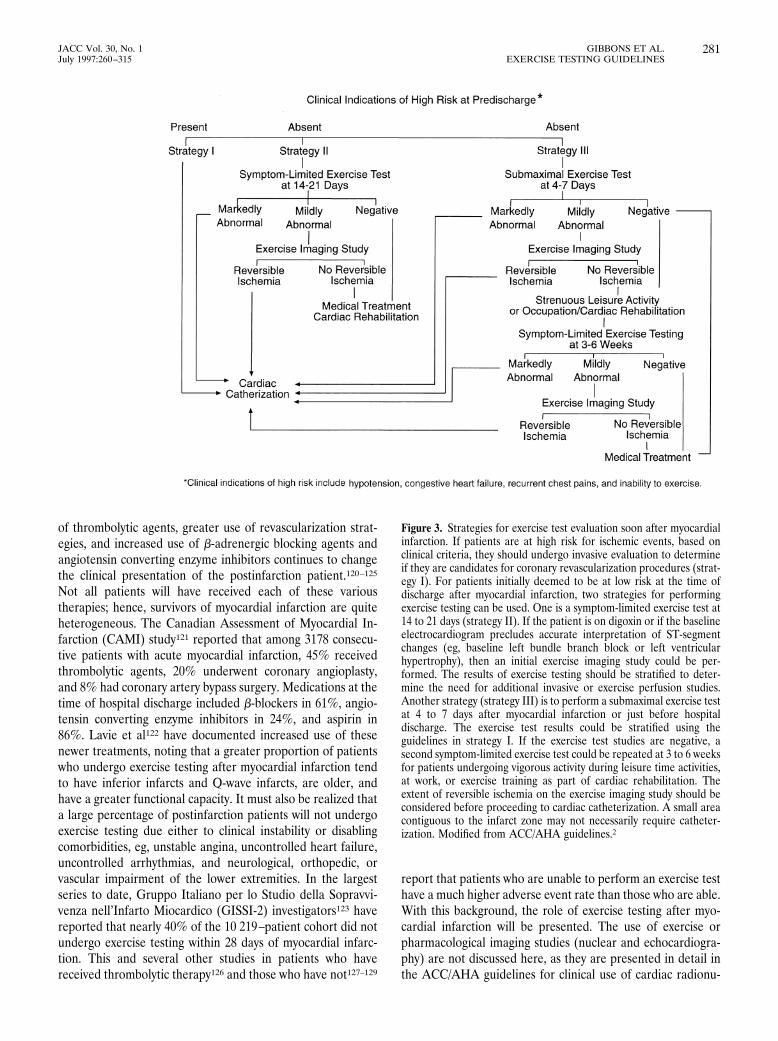
of thrombolytic agents, greater use of revascularization strat-egies, and increased use of b-adrenergic blocking agents andangiotensin converting enzyme inhibitors continues to changethe clinical presentation of the postinfarction patient.120–125
Not all patients will have received each of these varioustherapies; hence, survivors of myocardial infarction are quiteheterogeneous. The Canadian Assessment of Myocardial In-farction (CAMI) study121 reported that among 3178 consecu-tive patients with acute myocardial infarction, 45% receivedthrombolytic agents, 20% underwent coronary angioplasty,and 8% had coronary artery bypass surgery. Medications at thetime of hospital discharge included b-blockers in 61%, angio-tensin converting enzyme inhibitors in 24%, and aspirin in86%. Lavie et al122 have documented increased use of thesenewer treatments, noting that a greater proportion of patientswho undergo exercise testing after myocardial infarction tendto have inferior infarcts and Q-wave infarcts, are older, andhave a greater functional capacity. It must also be realized thata large percentage of postinfarction patients will not undergoexercise testing due either to clinical instability or disablingcomorbidities, eg, unstable angina, uncontrolled heart failure,uncontrolled arrhythmias, and neurological, orthopedic, orvascular impairment of the lower extremities. In the largestseries to date, Gruppo Italiano per lo Studio della Sopravvi-venza nell’Infarto Miocardico (GISSI-2) investigators123 havereported that nearly 40% of the 10 219–patient cohort did notundergo exercise testing within 28 days of myocardial infarc-tion. This and several other studies in patients who havereceived thrombolytic therapy126 and those who have not127–129
report that patients who are unable to perform an exercise testhave a much higher adverse event rate than those who are able.With this background, the role of exercise testing after myo-cardial infarction will be presented. The use of exercise orpharmacological imaging studies (nuclear and echocardiogra-phy) are not discussed here, as they are presented in detail inthe ACC/AHA guidelines for clinical use of cardiac radionu-
Figure 3. Strategies for exercise test evaluation soon after myocardialinfarction. If patients are at high risk for ischemic events, based onclinical criteria, they should undergo invasive evaluation to determineif they are candidates for coronary revascularization procedures (strat-egy I). For patients initially deemed to be at low risk at the time ofdischarge after myocardial infarction, two strategies for performingexercise testing can be used. One is a symptom-limited exercise test at14 to 21 days (strategy II). If the patient is on digoxin or if the baselineelectrocardiogram precludes accurate interpretation of ST-segmentchanges (eg, baseline left bundle branch block or left ventricularhypertrophy), then an initial exercise imaging study could be per-formed. The results of exercise testing should be stratified to deter-mine the need for additional invasive or exercise perfusion studies.Another strategy (strategy III) is to perform a submaximal exercise testat 4 to 7 days after myocardial infarction or just before hospitaldischarge. The exercise test results could be stratified using theguidelines in strategy I. If the exercise test studies are negative, asecond symptom-limited exercise test could be repeated at 3 to 6 weeksfor patients undergoing vigorous activity during leisure time activities,at work, or exercise training as part of cardiac rehabilitation. Theextent of reversible ischemia on the exercise imaging study should beconsidered before proceeding to cardiac catheterization. A small areacontiguous to the infarct zone may not necessarily require catheter-ization. Modified from ACC/AHA guidelines.2
281JACC Vol. 30, No. 1 GIBBONS ET AL.July 1997:260–315 EXERCISE TESTING GUIDELINES
clide imaging,5 guidelines for clinical application of echocardi-ography,6 and guidelines for management of acute myocardialinfarction.2
Exercise testing after myocardial infarction yields informa-tion in the following areas: (1) risk stratification and assess-ment of prognosis; (2) functional capacity for activity pre-scription after hospital discharge, including domestic andoccupational work evaluation and exercise training as part ofcomprehensive cardiac risk reduction and rehabilitation; and(3) assessment of adequacy of medical therapy and the need toemploy other diagnostic or treatment options.
Exercise Test LogisticsExclusions From Testing The absolute and relative con-
traindications to exercise testing are presented in Table 1. Inpatients with an abnormal resting ECG due to left bundlebranch block, preexcitation syndrome, LVH, digoxin therapy,or those demonstrating major (greater than 1 mm) ST-segmentdepression or elevation, an exercise or pharmacological imag-ing study should be considered, as the accuracy of the exerciseECG in detecting provokeable ischemia is reduced.
Timing and Protocol Exercise tests can be characterizedaccording to the time after myocardial infarction when the testis performed and the protocol used. The timing of the predis-charge exercise test continues to shorten, as does the hospitalstay for patients with an uncomplicated myocardial infarction.Predischarge exercise tests in the recent literature range from5 to 26 days after infarction.126,129–132 Exercise tests have beenperformed as early as 3 days after myocardial infarction124;however, the safety of this very early protocol has not beenestablished. Postdischarge tests have been performed early (14to 21 days), at 6 weeks,133 or at 6 months after infarction.134
The exercise protocols can be either submaximal or symptom-limited. Submaximal protocols have a predetermined endpoint often defined as a peak heart rate of 120 bpm, or 70%predicted maximum heart rate, or a peak MET level of 5.131
Symptom-limited tests are designed to continue until thepatient demonstrates signs and/or symptoms that necessitatetermination of exercise (ie, angina, fatigue, greater than orequal to 2 mm of ST-segment depression, ventricular arrhyth-mias, or greater than or equal to a 10 mm Hg drop in systolicblood pressure from the resting blood pressure).135 The mostcommonly used treadmill protocols are the modified Bruce,the modified Naughton, and the standard Bruce.131 The ramptreadmill or cycle ergometer protocols offer the advantage ofsteady gradual increases in work rate and better estimation offunctional capacity136 but have not been widely studied inpatients early after myocardial infarction.
Recent studies have evaluated symptom-limited protocolsat 4 to 7 days after myocardial infarction and have includedpatients treated with thrombolytic agents. These studies dem-onstrate that such testing yields ischemic responses nearlytwice as often as submaximal tests and are a better estimate ofpeak functional capacity.130,135,137 Thus, early symptom-limitedtests have the potential to be more useful in activity prescrip-
tion before discharge. However, the additive prognostic valuefrom information obtained from the performance of symptom-limited protocols within days rather than weeks after myocar-dial infarction has not yet been established.
Safety Exercise testing after myocardial infarction appearsto be safe. The incidence of fatal cardiac events, including fatalmyocardial infarction and cardiac rupture, is 0.03%, nonfatalmyocardial infarction and successfully resuscitated cardiacarrest is 0.09%, and complex arrhythmias, including VT, is1.4%. Symptom-limited protocols have an event rate which is1.9 times that of submaximal tests, although the overall fatalevent rate is quite low.130,131,135 The majority of the safety dataare based on exercise testing performed more than 7 days aftermyocardial infarction. The number of patients reported at 4 to7 days is more limited, and typically time is reported as a meanvalue or a range so that it is impossible to determine how manypatients were studied at 4 days.
Risk Stratification and PrognosisThe prognosis among survivors of myocardial infarction
continues to improve, particularly in patients who have re-ceived thrombolytic therapy and revascularization during hos-pitalization. One-year postdischarge mortality in the CAMIstudy121 was 8.4% and was distinctly lower in the 45% ofpatients who received thrombolytic therapy (3.7% mortality)and in the 28% who underwent coronary angioplasty (3%mortality) or coronary artery bypass surgery (3.7% mortality).Data from the Global Utilization of Streptokinase and TPAfor Occluded Arteries (GUSTO) trial138 demonstrate that 57%of the 41 021 patients who received thrombolytic therapy hadno complications (no recurrent ischemia, reinfarction, heartfailure, stroke, or invasive procedures) at 4 days after myocar-dial infarction. The mortality rate at 1 month was 1% and at 1year, 3.6%. Recurrent ischemia occurred in 7% of this group.
The improvement in 1-year mortality in patients who havereceived thrombolytic therapy is multifactorial. Such patientsare (1) less likely to have severe three-vessel CAD; (2) have asmaller infarct size; and (3) frequently undergo coronaryangiography in lieu of exercise testing. Consequently thepatient population that presently undergoes predischarge ex-ercise testing in clinical trials of thrombolytic therapy is fardifferent from less selected historical populations or concur-rent patient populations not treated with thrombolytic therapy.Their low cardiac event rate following discharge is thereforenot surprising and substantially reduces the predictive accuracyof early exercise testing.
There is limited evidence on the ability of exercise testing torisk stratify patients who have not received reperfusion in thecurrent era. Although their subsequent mortality rates arelower than in patients treated in the prethrombolytic erabecause of therapeutic advances and revascularization, theirabsolute event rates are higher than in patients who havereceived thrombolytic therapy. Although the available evi-dence is limited, exercise testing presumably can still assist inthe risk stratification of such patients.
282 GIBBONS ET AL. JACC Vol. 30, No. 1EXERCISE TESTING GUIDELINES July 1997:260–315
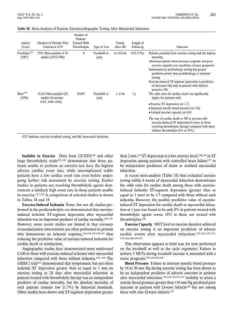
Inability to Exercise Data from GUSTO138 and otherlarge thrombolytic trials123,126 demonstrate that those pa-tients unable to perform an exercise test have the highestadverse cardiac event rate, while uncomplicated stablepatients have a low cardiac event rate even before under-going further risk assessment by exercise testing. Earlierstudies in patients not receiving thrombolytic agents dem-onstrate a similarly high event rate in those patients unableto exercise.127,129 A comparison of selected studies is shownin Tables 18 and 19.
Exercise-Induced Ischemia Some, but not all, studies per-formed in the prethrombolytic era demonstrated that exercise-induced ischemic ST-segment depression after myocardialinfarction was an important predictor of cardiac mortality.139–141
However, more recent studies are limited in that coronaryrevascularization interventions are often performed in personswho demonstrate an ischemic response,126,129,135,142,143 thusreducing the predictive value of exercise-induced ischemia forcardiac death or reinfarction.
Angiographic studies have demonstrated more multivesselCAD in those with exercise-induced ischemia after myocardialinfarction compared with those without ischemia.144–146 TheGISSI-2 trial123 demonstrated that symptomatic but not silentischemic ST depression greater than or equal to 1 mm onexercise testing at 28 days after myocardial infarction inpatients treated with thrombolytic therapy was an independentpredictor of cardiac mortality, but the absolute mortality ofsuch patients remains low (1.7%) by historical standards.Other studies have shown only ST-segment depression greater
than 2 mm,147 ST depression at a low exercise level,148,149 or STdepression among patients with controlled heart failure127 tobe independent predictors of death or nonfatal myocardialinfarction.
A recent meta-analysis (Table 18) that evaluated exercisetesting within 6 weeks of myocardial infarction demonstratesthe odds ratio for cardiac death among those with exercise-induced ischemic ST-segment depression (greater than orequal to 1 mm) to be 1.7 compared with those without suchischemia. However, the positive predictive value of exercise-induced ST depression for cardiac death or myocardial infarc-tion at 1 year was found to be only 8% in patients treated withthrombolytic agents versus 18% in those not treated withthrombolytics.150
Exercise Capacity MET level or exercise duration achievedon exercise testing is an important predictors of adversecardiac events after myocardial infarction.123,125,129,132,
134,143,148,149,151
This observation appears to hold true for tests performedon the treadmill as well as the cycle ergometer. Failure toachieve 5 METs during treadmill exercise is associated with aworse prognosis.129,134,147,148
Blood Pressure Failure to increase systolic blood pressureby 10 to 30 mm Hg during exercise testing has been shown tobe an independent predictor of adverse outcome in patientsafter myocardial infarction.123,132,134,152,153 Inability to attain asystolic blood pressure greater than 110 mm Hg predicted pooroutcome in patients with Q-wave infarcts129 but not amongthose with non–Q-wave infarcts.127
Table 18. Meta-Analyses of Exercise Electrocardiographic Testing After Myocardial Infarction
Author(Year)
Number of Patients WhoUnderwent ETT
Number ofPatients
Treated WithThrombolysis Type of Test
TimingAfter MI
Length ofFollow-up Outcome
Froelicher141
(1987)5331 Meta-analysis of 24
studies (1973–1986)0 Treadmill or
cycle1.6–9.0 wk 0.25–5.70 y Patients excluded from exercise testing had the highest
mortality.Abnormal systolic blood pressure response and poor
exercise capacity were predictive of poor prognosis.Submaximal or predischarge testing has greater
predictive power than postdischarge or maximaltesting.
Exercise-induced ST-segment depression is predictiveof increased risk only in patients with inferior-posterior MI.
Shaw150
(1996)15,613 Meta-analysis (28
studies of exercise-10,067 Treadmill or
cycle1–6 wk 1 y The odds ratio for cardiac death was significantly
higher for patients with:ETT, 1980–1995)
● Exercise ST depression (or 1.7)● Impaired systolic blood pressure (or 4.0)● Limited exercise capacity (or 4.0)
The rate of cardiac death or MI in persons withexercise-induced ST depression is lower in thosereceiving thrombolytic therapy compared with thosewithout thrombolysis (8% vs 18%).
ETT indicates exercise treadmill testing, and MI, myocardial infarction.
283JACC Vol. 30, No. 1 GIBBONS ET AL.July 1997:260–315 EXERCISE TESTING GUIDELINES
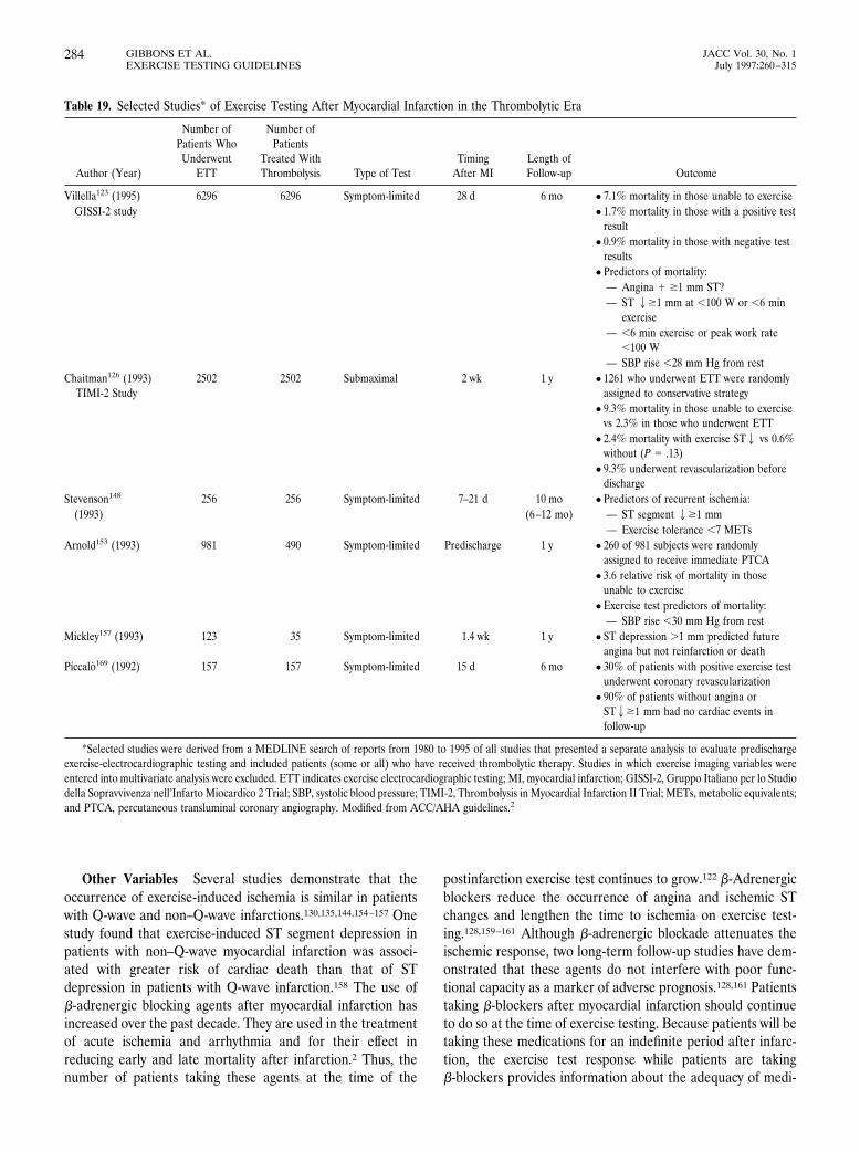
Other Variables Several studies demonstrate that theoccurrence of exercise-induced ischemia is similar in patientswith Q-wave and non–Q-wave infarctions.130,135,144,154–157 Onestudy found that exercise-induced ST segment depression inpatients with non–Q-wave myocardial infarction was associ-ated with greater risk of cardiac death than that of STdepression in patients with Q-wave infarction.158 The use ofb-adrenergic blocking agents after myocardial infarction hasincreased over the past decade. They are used in the treatmentof acute ischemia and arrhythmia and for their effect inreducing early and late mortality after infarction.2 Thus, thenumber of patients taking these agents at the time of the
postinfarction exercise test continues to grow.122 b-Adrenergicblockers reduce the occurrence of angina and ischemic STchanges and lengthen the time to ischemia on exercise test-ing.128,159–161 Although b-adrenergic blockade attenuates theischemic response, two long-term follow-up studies have dem-onstrated that these agents do not interfere with poor func-tional capacity as a marker of adverse prognosis.128,161 Patientstaking b-blockers after myocardial infarction should continueto do so at the time of exercise testing. Because patients will betaking these medications for an indefinite period after infarc-tion, the exercise test response while patients are takingb-blockers provides information about the adequacy of medi-
Table 19. Selected Studies* of Exercise Testing After Myocardial Infarction in the Thrombolytic Era
Author (Year)
Number ofPatients Who
UnderwentETT
Number ofPatients
Treated WithThrombolysis Type of Test
TimingAfter MI
Length ofFollow-up Outcome
Villella123 (1995) 6296 6296 Symptom-limited 28 d 6 mo ● 7.1% mortality in those unable to exerciseGISSI-2 study ● 1.7% mortality in those with a positive test
result● 0.9% mortality in those with negative test
results● Predictors of mortality:
— Angina 1 $1 mm ST?— ST 2$1 mm at ,100 W or ,6 min
exercise— ,6 min exercise or peak work rate
,100 W— SBP rise ,28 mm Hg from rest
Chaitman126 (1993)TIMI-2 Study
2502 2502 Submaximal 2 wk 1 y ● 1261 who underwent ETT were randomlyassigned to conservative strategy
● 9.3% mortality in those unable to exercisevs 2.3% in those who underwent ETT
● 2.4% mortality with exercise ST2 vs 0.6%without (P 5 .13)
● 9.3% underwent revascularization beforedischarge
Stevenson148 256 256 Symptom-limited 7–21 d 10 mo ● Predictors of recurrent ischemia:(1993) (6–12 mo) — ST segment 2$1 mm
— Exercise tolerance ,7 METsArnold153 (1993) 981 490 Symptom-limited Predischarge 1 y ● 260 of 981 subjects were randomly
assigned to receive immediate PTCA● 3.6 relative risk of mortality in those
unable to exercise● Exercise test predictors of mortality:
— SBP rise ,30 mm Hg from restMickley157 (1993) 123 35 Symptom-limited 1.4 wk 1 y ● ST depression .1 mm predicted future
angina but not reinfarction or deathPiccalo169 (1992) 157 157 Symptom-limited 15 d 6 mo ● 30% of patients with positive exercise test
underwent coronary revascularization● 90% of patients without angina or
ST2$1 mm had no cardiac events infollow-up
*Selected studies were derived from a MEDLINE search of reports from 1980 to 1995 of all studies that presented a separate analysis to evaluate predischargeexercise-electrocardiographic testing and included patients (some or all) who have received thrombolytic therapy. Studies in which exercise imaging variables wereentered into multivariate analysis were excluded. ETT indicates exercise electrocardiographic testing; MI, myocardial infarction; GISSI-2, Gruppo Italiano per lo Studiodella Sopravvivenza nell’Infarto Miocardico 2 Trial; SBP, systolic blood pressure; TIMI-2, Thrombolysis in Myocardial Infarction II Trial; METs, metabolic equivalents;and PTCA, percutaneous transluminal coronary angiography. Modified from ACC/AHA guidelines.2
284 GIBBONS ET AL. JACC Vol. 30, No. 1EXERCISE TESTING GUIDELINES July 1997:260–315
cal therapy in preventing ischemia and arrhythmias as well ascontrolling heart rate and blood pressure response duringexercise.
Activity CounselingExercise testing after myocardial infarction is useful for
counseling patients and their families about domestic, recre-ational, and occupational activities that can be safely per-formed after discharge from the hospital. Functional capacityin METs derived from the exercise test can be used to estimatetolerance for specific activities. Published charts that providean estimate of energy requirements for various activities areavailable7 but should be used only as a guide, with theknowledge that the intensity at which activities are performedwill directly influence the amount of energy required. Mostdomestic chores and activities require less than 5 METs;hence, a submaximal test at the time of hospital discharge canbe useful in counseling regarding the first several weeks aftermyocardial infarction.
The follow-up symptom-limited testing performed 3 to 6weeks after myocardial infarction can assist in further activityprescription and issues concerning return to work. Most occu-pational activities require less than 5 METs. In the 15% ofpersons in the labor force whose work involves heavy manuallabor,162 the exercise test data should not be used as the solecriterion for recommendations regarding return to work. En-ergy demands for lifting heavy objects, temperature, andenvironmental and psychological stresses are not assessed byroutine exercise tests and must be taken into consideration.Simulated work tests can be performed in patients with lowfunctional capacity, left ventricular dysfunction, exercise-induced ischemia, and those who are otherwise apprehensiveabout returning to a physically demanding occupation.163–165
Cardiac RehabilitationCardiac rehabilitation combines prescriptive exercise train-
ing with coronary risk factor modification in patients with heartdisease. It is considered standard care that should be integratedinto the treatment plan of patients with CAD.166 Randomizedtrials of cardiac rehabilitation after myocardial infarction showconsistent trends toward survival benefit among patients enrolledin cardiac rehabilitation programs.162,166 Meta-analyses of thesetrials have calculated a significant 20% to 25% reduction incardiovascular death in patients enrolled in such programs.167
Moreover, higher levels of physical fitness when measured on anexercise tolerance test are associated with reduced subsequentmortality.123,129,132,134,143,148,149,151 Exercise training improves ex-ercise capacity among cardiac patients by 11% to 66% after 3 to6 months of training, with the greatest benefits among those whoare most unfit.166
Exercise testing in cardiac rehabilitation is essential indevelopment of the exercise prescription to establish a safe andeffective training intensity, in risk stratification of patients todetermine the level of supervision and monitoring required
during exercise training sessions, and in evaluation of trainingprogram outcome.7,164,168 For these reasons, symptom-limitedexercise testing before program initiation is needed for allpatients in whom cardiac rehabilitation is recommended (re-cent myocardial infarction, recent coronary artery bypass sur-gery, recent coronary angioplasty, chronic stable angina, con-trolled heart failure).7,166
Exercise testing in the stable cardiac patient who continuesan exercise training program is often performed after theinitial 8 to 12 weeks of exercise training and periodicallythereafter, although there are no available studies to assess itsvalue. Such testing may be useful to rewrite the exerciseprescription, evaluate improvement in functional capacity, andprovide feedback to the patient.166
SummaryContemporary treatment of the patient with acute myocar-
dial infarction includes one or more of the following: medicaltherapy, thrombolytic agents, and coronary revascularization.These interventions have led to marked improvement in theprognosis of the postinfarction patient, particularly those whohave been treated with reperfusion. The patient populationeligible for predischarge exercise testing in clinical trials ofthrombolytic therapy is therefore far different from less selectedhistorical populations. Their low cardiac event rate substantiallyreduces the predictive accuracy of early exercise testing. However,there is limited evidence on the ability of exercise testing to riskstratify patients who have not received reperfusion therapy in thecurrent era. Their mortality rates are higher than those whoreceived either thrombolytic therapy or who have undergonecoronary revascularization. Thus, exercise testing presumably canstill assist in risk stratification of such patients. Patients who havenot undergone coronary revascularization and are unable toundergo exercise testing have the worst prognosis.
Exercise testing after myocardial infarction is safe. Sub-maximal testing can be performed at about 4 to 7 days; about3 to 6 weeks later a symptom-limited exercise test can beperformed. Alternatively, symptom-limited tests can be con-ducted early after discharge at about 14 to 21 days. Strategiesfor exercise test evaluation after myocardial infarction areoutlined in Fig 3.3
Exercise test predictors of adverse outcome in the postin-farction patient include ischemic ST-segment depression greaterthan or equal to 1 mm, particularly if accompanied by symptoms,at a low level of exercise, or in the presence of controlled heartfailure; functional capacity less than 5 METs; inadequate bloodpressure response (peak systolic blood pressure less than110 mm Hg or less than 30 mm increase from resting level).
Exercise testing is useful in activity counseling after dis-charge from the hospital. Exercise testing is also an importanttool in exercise training as part of comprehensive cardiacrehabilitation. It is used to develop and modify the exerciseprescription and assess the patient’s response to and progressin the exercise training program.
285JACC Vol. 30, No. 1 GIBBONS ET AL.July 1997:260–315 EXERCISE TESTING GUIDELINES
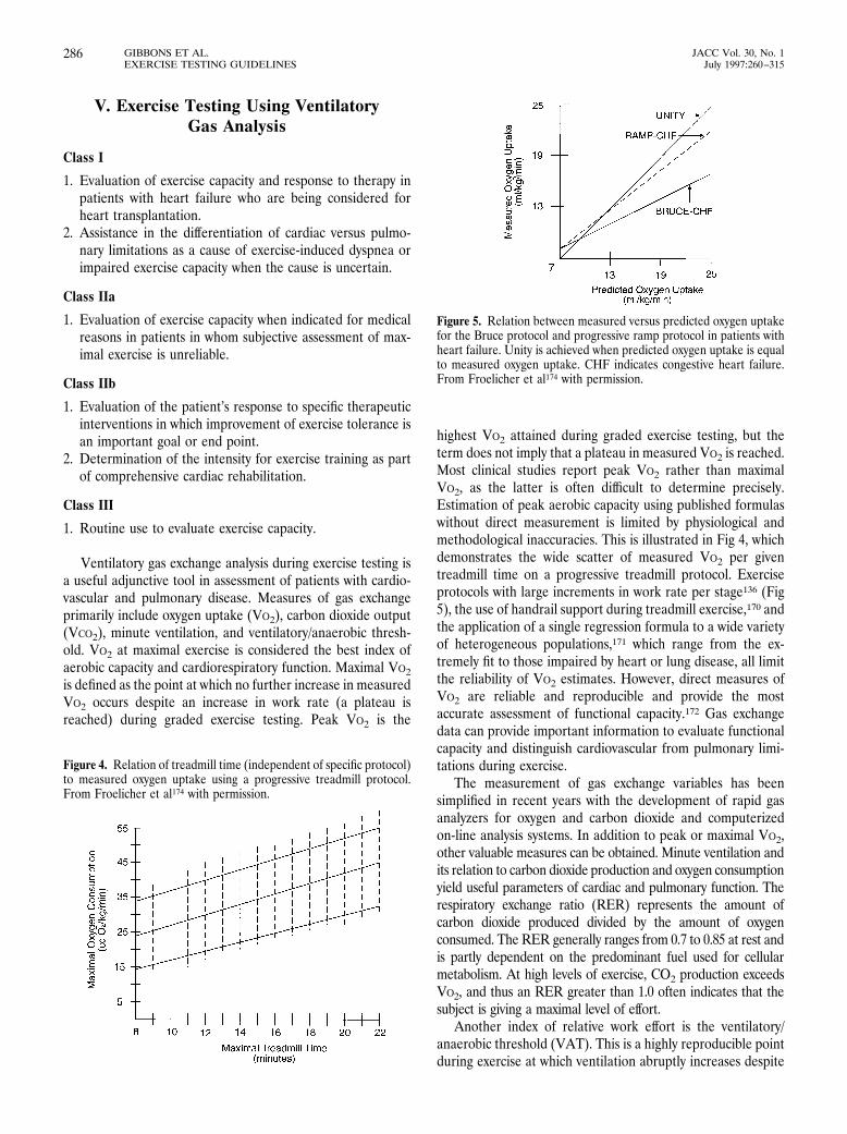
V. Exercise Testing Using VentilatoryGas Analysis
Class I
1. Evaluation of exercise capacity and response to therapy inpatients with heart failure who are being considered forheart transplantation.
2. Assistance in the differentiation of cardiac versus pulmo-nary limitations as a cause of exercise-induced dyspnea orimpaired exercise capacity when the cause is uncertain.
Class IIa
1. Evaluation of exercise capacity when indicated for medicalreasons in patients in whom subjective assessment of max-imal exercise is unreliable.
Class IIb
1. Evaluation of the patient’s response to specific therapeuticinterventions in which improvement of exercise tolerance isan important goal or end point.
2. Determination of the intensity for exercise training as partof comprehensive cardiac rehabilitation.
Class III
1. Routine use to evaluate exercise capacity.
Ventilatory gas exchange analysis during exercise testing isa useful adjunctive tool in assessment of patients with cardio-vascular and pulmonary disease. Measures of gas exchangeprimarily include oxygen uptake (VO2), carbon dioxide output(VCO2), minute ventilation, and ventilatory/anaerobic thresh-old. VO2 at maximal exercise is considered the best index ofaerobic capacity and cardiorespiratory function. Maximal VO2is defined as the point at which no further increase in measuredVO2 occurs despite an increase in work rate (a plateau isreached) during graded exercise testing. Peak VO2 is the
highest VO2 attained during graded exercise testing, but theterm does not imply that a plateau in measured VO2 is reached.Most clinical studies report peak VO2 rather than maximalVO2, as the latter is often difficult to determine precisely.Estimation of peak aerobic capacity using published formulaswithout direct measurement is limited by physiological andmethodological inaccuracies. This is illustrated in Fig 4, whichdemonstrates the wide scatter of measured VO2 per giventreadmill time on a progressive treadmill protocol. Exerciseprotocols with large increments in work rate per stage136 (Fig5), the use of handrail support during treadmill exercise,170 andthe application of a single regression formula to a wide varietyof heterogeneous populations,171 which range from the ex-tremely fit to those impaired by heart or lung disease, all limitthe reliability of VO2 estimates. However, direct measures ofVO2 are reliable and reproducible and provide the mostaccurate assessment of functional capacity.172 Gas exchangedata can provide important information to evaluate functionalcapacity and distinguish cardiovascular from pulmonary limi-tations during exercise.
The measurement of gas exchange variables has beensimplified in recent years with the development of rapid gasanalyzers for oxygen and carbon dioxide and computerizedon-line analysis systems. In addition to peak or maximal VO2,other valuable measures can be obtained. Minute ventilation andits relation to carbon dioxide production and oxygen consumptionyield useful parameters of cardiac and pulmonary function. Therespiratory exchange ratio (RER) represents the amount ofcarbon dioxide produced divided by the amount of oxygenconsumed. The RER generally ranges from 0.7 to 0.85 at rest andis partly dependent on the predominant fuel used for cellularmetabolism. At high levels of exercise, CO2 production exceedsVO2, and thus an RER greater than 1.0 often indicates that thesubject is giving a maximal level of effort.
Another index of relative work effort is the ventilatory/anaerobic threshold (VAT). This is a highly reproducible pointduring exercise at which ventilation abruptly increases despite
Figure 4. Relation of treadmill time (independent of specific protocol)to measured oxygen uptake using a progressive treadmill protocol.From Froelicher et al174 with permission.
Figure 5. Relation between measured versus predicted oxygen uptakefor the Bruce protocol and progressive ramp protocol in patients withheart failure. Unity is achieved when predicted oxygen uptake is equalto measured oxygen uptake. CHF indicates congestive heart failure.From Froelicher et al174 with permission.
286 GIBBONS ET AL. JACC Vol. 30, No. 1EXERCISE TESTING GUIDELINES July 1997:260–315
linear increases in work rate. Shortly beyond the anaerobicthreshold, fatigue usually ensues. The term anaerobic thresh-old is based on the hypothesis that at a given work rate, theoxygen supplied to exercising muscles does not meet theoxygen requirements. This imbalance increases anaerobic gly-colysis for energy generation, yielding lactate as a metabolicbyproduct.173 Although the anaerobic threshold is a definedend point that can be established by several different methods,the actual cause of the observed abrupt rise in minute venti-lation remains controversial. This hypothesis is supported bythe fact that measured lactate levels increase at the point atwhich minute ventilation begins its curvilinear relation to workrate. However, whether muscle hypoxia is a main stimulus forincreased lactate production is not yet clear. Thus, the trueanaerobic threshold at the muscle cell level, the onset of bloodlactate accumulation, and the VAT are separate but relatedevents that occur during exercise.
The VAT is determined by several easily recognized mea-surements that can be obtained during respiratory gas analysis.These include (1) a departure of linearity of minute ventilation(VE) and VCO2 with increasing work rates and an abruptincrease in the RER and fraction of O2 in expired air (FEO2);(2) an increase in VE/VO2 without an increase in VE/VCO2, andan increase in FEO2 without a decrease in the fraction of CO2 inexpired air; (3) the lowest VE/VO2 value measured duringexercise; and (4) a curvilinear increase in VE and VCO2 with alinear increase in VO2 (Fig 6).173,174 Further details on themethodology and interpretation of data obtained during ven-tilatory gas analysis are available.8,174,175
Measurement of expiratory gases during exercise testingcan (1) provide the best estimate of functional capacity; (2)grade the severity of functional impairment; (3) objectivelyevaluate the response to interventions that may affect exercisecapacity; (4) objectively track the progression of disease thatmay limit exercise capacity; and (5) assist in differentiatingcardiac from pulmonary limitations in exercise capacity.176
Normal values for maximal oxygen uptake among healthyadults at different ages are available7 and may serve as a usefulreference in the evaluation of exercise capacity. The VAT hasbeen proposed as a more sensitive index of fitness than maximalVO2, heart rate, or total fitness in children. Normal values forVAT in children are provided elsewhere.9 Determination ofexercise training intensity to maintain or improve health andfitness among persons with or without heart disease can bederived from direct measurements of peak oxygen consumptionas shown in Table 20.177 This may be most useful when the heartrate response to exercise is not a reliable indicator of exerciseintensity (eg, patients with fixed-rate pacemakers). Rating ofperceived exertion is also helpful in this setting.
Data derived from exercise testing with ventilatory gasanalysis have proved to be reliable and important measures inthe evaluation of patients with heart failure.178–181 The exercisecapacity of patients with heart failure based on their peak VO2and VAT can be divided into four classes as shown in Table21.182 This classification system is limited in that age andgender are not taken into account. Moreover, peak exercise
capacity does not necessarily reflect the daily activities of heartfailure patients. Stratification of ambulatory heart failurepatients using this technique has improved ability to identifythose with the poorest prognosis, who should be considered forheart transplantation.183,184 (See Table 22.)
The technique of ventilatory gas measurement has a num-ber of potential limitations that hinder its broad applicability.Gas exchange measurement systems are costly and require me-ticulous maintenance and calibration for optimal use.170 Person-nel who administer tests and interpret results must be trained andproficient in this technique. Finally, the test requires additionalcost and time as well as patient cooperation.8
VI. Special Groups: Women, AsymptomaticIndividuals, and Postrevascularization PatientsWomen
Rationale Cardiovascular disease is one of the principalcauses of death in women, exceeding mortality due to breastcancer by a factor of 11.185 The probability of coronary diseasein women, based on age, gender, and the nature of symp-toms,17 is most commonly in the low to intermediate probabil-ity range, especially in premenopausal women. While typicalangina is as meaningful in women older than 60 years as inmen,186 the clinical diagnosis of coronary disease in womenmay be difficult to make: almost half the women with anginalsymptoms in the Coronary Artery Surgery Study (CASS)187
(who were younger than 65) had normal coronary arterio-grams. From a Bayesian standpoint, the low prevalence ofCAD presents a particularly difficult situation for noninvasivetesting. Moreover, the results of functional testing—exercisecapacity, ST-segment changes, and imaging tests—may beinfluenced by gender.
Accuracy of Electrocardiographic Analysis in Women. TheST response to exercise appears to be gender-related from anearly age, with ST-segment abnormalities more commonlyreported in third-grade girls than boys.188 Studies examiningthe accuracy of ST-segment interpretation for the diagnosis ofcoronary disease according to gender are summarized in Table23.84,88,186,189–192,194–199 Variations in results in women may bedue to the use of different criteria for defining coronarydisease, differences in population selection (including preva-lence of prior myocardial infarction and multivessel disease),and differences in performance of the test, including criteriafor ST-segment positivity, and type of exercise.
Exercise-induced ST depression is less sensitive in womenthan men,84 reflecting a lower prevalence of severe CAD andthe inability of many women to exercise to maximum aerobiccapacity.201,202 The exercise ECG is commonly viewed as lessspecific in women than men, although Table 23 demonstratesthat this finding has not been uniform. Even after patients withreferral bias were excluded, the ST-segment response wasfound to be less accurate in women.28 Significant genderdifferences were reported in unbiased estimates of sensitivityand specificity. However, these were modest (less than 10%),
287JACC Vol. 30, No. 1 GIBBONS ET AL.July 1997:260–315 EXERCISE TESTING GUIDELINES
and did not appear to preclude the use of treadmill exercisetesting in women.28 A careful analysis of the incrementaldiagnostic value of treadmill testing found similar value in menand women.200 Studies that have documented lower specificityin women have cited both lower disease prevalence andnon-Bayesian factors,192 which might include the greater prev-alence of mitral valve prolapse and syndrome X in women,differences in microvascular function (leading perhaps to cor-onary spasm), and possibly hormonal differences.
The standard approach to exercise testing involves catego-rization of the ST-segment response as “positive” or “nega-tive” results. The accuracy of exercise testing in women may beenhanced by attention to features other than the absolute levelof ST depression. The ST/heart-rate relation has recently beenshown to be of value203 but awaits widespread application.Avoidance of identifying ST depression in the inferior leadsand identification of test positivity based on persistentchanges204 enhance the predictive value of a positive test butmay compromise the predictive value of a negative test.Finally, as the ST-segment response is a continuous variable,continuous analysis of the ST segment may recover the infor-mation lost from its analysis as a dichotomous variable. Thisanalysis has been combined with non-ECG end points intomultivariate models (see below).
Non-ECG End Points. The exercise test provides a wealthof other material, including exercise capacity, hemodynamicresponse to exercise, and the presence of cardiac symptoms,which are used in interpretation of the test result and are notapparent in ST analysis alone. The diagnostic contribution ofthese findings has been calculated in multivariate models,resulting in development of equations giving the likelihood ofdisease. The accuracy of exercise testing was significantly
Figure 6. Measurements used to determine the gas exchange anaero-bic threshold (Atge). VE indicates minute ventilation; VCO2, carbondioxide production; VO2, oxygen uptake; and FeO2, fraction of expiredair that is oxygen. From Froelicher et al174 with permission.
Table 20. Classification of Exercise Intensity Based onOxygen Uptake177
Intensity % VO2max
Very light , 25Light 25–44Moderate 45–59Hard 60–84Very hard $ 85Maximal 100
VO2max indicates maximal oxygen uptake.
Table 21. Classification of Exercise Capacity in Patients WithHeart Failure, Based on Peak Oxygen Uptake and VentilatoryAnaerobic Threshold182
Class ImpairmentPeak VO2
(mL/kg/min)VAT
(mL/kg/min)
A None to mild . 20 . 14B Mild to moderate 16–20 11–14C Moderate to severe 10–16 8–11D Severe , 10 , 8
VO2 indicates oxygen uptake; and VAT, ventilatory anaerobic threshold.
288 GIBBONS ET AL. JACC Vol. 30, No. 1EXERCISE TESTING GUIDELINES July 1997:260–315
increased by the use of a multivariate model compared withST-segment evaluation alone.196 However, not all centers havereported these favorable findings,198 and although the exercisescore concept is attractive, its clinical application in women hasremained limited.
Conclusion. The diagnosis of CAD in women presentsdifficulties that are not experienced in the investigation of men.These problems reflect differences in exercise physiology, bodyhabitus, coronary physiology, and prevalence of CAD betweenmen and women.
The accuracy of the exercise ECG for diagnosis of coronarydisease in women may have important limitations. Physiciansmust be cognizant of the influence of submaximal exercise onsensitivity; patients likely to exercise submaximally shouldundergo pharmacological stress testing. Concern about false-positive ST-segment responses may be addressed by carefulassessment of posttest probability and selective use of stressimaging tests before proceeding to angiography.88 On theother hand, the difficulties posed by clinical evaluation ofprobability of CAD in women have led to speculation thatstress imaging approaches may be an efficient initial alternativeto the exercise ECG in women.199 Although the optimalstrategy for circumventing false-positive test results for diag-nosis of CAD in women remains to be defined, there are
currently insufficient data to justify routine stress imaging testsas the initial test for CAD in women.
Diagnosis of Coronary Artery Disease in the ElderlyRationale Patients older than 65 years are usually defined
as “elderly.” One possible subdivision of this group205 is the“young old” (65 to 75 years) and the “old old” (older than 75);CAD is highly prevalent in symptomatic patients in bothgroups. With the aging of the population, the former nowconstitute a substantial proportion of patients; thus, this sec-tion focuses on patients older than 75 years. Unfortunately,few data have been published with respect to the use ofexercise testing for diagnostic and prognostic assessment ofCAD in this group. For example, tables used for the identifi-cation of pretest disease probability do not consider patientsolder than 70, and scores for assessing prognosis have notincluded elderly patients. Presumably the prevalence and riskof coronary disease increase with advancing age, but nonethe-less, in 1989 the National Health Interview Survey206 reportedthat the prevalence of diagnosed CAD was 1.8% in men overthe age of 75 and 1.5% in women over 75. This disease iscommonly occult, silent ischemia being estimated to be presentin 15% of 80-year-olds.207 On Bayesian grounds, the highprevalence and greater severity of coronary stenoses in thisgroup increase the sensitivity of testing but make it harder torule out significant disease.
The performance of exercise testing poses several problemsin the elderly, but it is certainly not contraindicated in thisgroup. Functional capacity is often compromised due to mus-cle weakness and deconditioning, and therefore the decisionwhether to send the patient for an exercise or pharmacologicalstress test is more important than in younger patients. In somepatients with problems of gait and coordination, a bicycleexercise test may be more attractive than a treadmill exercise
Table 22. Guidelines for Peak Exercise Oxygen Uptake as aCriterion for Cardiac Transplantation184
Category forTransplant
Peak VO2
(mL/kg/min)
Accepted indication , 10Probable indication , 14Inadequate indication . 15
VO2 indicates oxygen uptake.
Table 23. Sensitivity and Specificity of Exercise Electrocardiography in Women*
Author (year)n
(Women)Mean Age
(y)Definition of
CADMultivesselCAD (%)
PositiveExercise TestResult (% of
Women)
Sensitivity: Women(n 5 PatientsWith CAD)
Specificity: Women(n 5 Patients
Without CAD)
Guiteras189 (1972) 112 49 . 70% dia 12 38 79%, n 5 42 66%, n 5 70Linhart190 (1974) 98 46 . 50% dia na 34 71%, n 5 24 78%, n 5 74Sketch191 (1975) 56 50 . 75% dia na 27 50%, n 5 10 78%, n 5 46Barolsky192 (1979) 92 50 . 50% dia 16 41 60%, n 5 30 68%, n 5 62Weiner193 (1979) 580 na . 70% dia 16 48 76%, n 5 169 64%, n 5 411Ilsley194 (1982) 62 51 . 50% dia 27 44 67%, n 5 27 74%, n 5 35Hung195 (1984) 92 51 . 70% dia 16 51 75%, n 5 28 59%, n 5 64Hlatky84 (1984) 613 na . 75% dia na na 57%, n 5 194 86%, n 5 419Melin88 (1985) 93 51 . 50% dia 20 30 58%, n 5 24 80%, n 5 69Robert196 (1991) 135 53 . 50% dia 29 37 68%, n 5 56 48%, n 5 79Chae197 (1993) 114 na . 50% dia na 54 66%, n 5 71 60%, n 5 43Williams198 (1994) 70 60 . 50% dia 19 57 67%, n 5 33 51%, n 5 37Marwick199 (1995) 118 60 . 50% dia 17 58 77%, n 5 48 56%, n 5 70Morise200 (1995)† 264 56 . 50% dia 27 33 46%, n 5 81 74%, n 5 151Morise200 (1995)‡ 288 57 . 50% dia 26 36 55%, n 5 106 74%, n 5 159
*Studies of .50 women. †Derivation set. ‡Validation set. CAD indicates coronary artery disease; dia, diameter stenosis; and na, not available.
289JACC Vol. 30, No. 1 GIBBONS ET AL.July 1997:260–315 EXERCISE TESTING GUIDELINES
test,208 but in older patients, bicycle exercise is often limited byunfamiliarity. Certainly, if treadmill exercise is used, moreattention must be given to the mechanical hazards of exercisein elderly patients. More gradual protocols should be favoredin selection of a treadmill exercise protocol in elderly pa-tients.209 Elderly patients are much more likely to hold on tothe handrails tightly, reducing the validity of treadmill time forestimating METs.
Interpretation of exercise testing in the elderly differsslightly from that in the young. Resting ECG abnormalities,including prior myocardial infarction and intraventricular con-duction delays, may compromise the availability of diagnosticdata from the ECG. Nonetheless, the application of standardST-segment response criteria to elderly subjects is not associ-ated with significantly different accuracy from younger peo-ple.84 Due to the greater prevalence of both CAD and severeCAD, it is not surprising that the exercise ECG in this grouphas a slightly higher sensitivity (84%) and lower specificity(70%) than in younger patients.210 These false-positive resultsmay also reflect the coexistence of LVH due to valvular diseaseand hypertension as well as conduction disturbances. Althoughthe risk of coronary angiography may be greater in the elderlyand the justification for coronary intervention may be less, theresults of exercise testing in the elderly remain important,because medical therapy may itself carry risks in this group.
In addition to ST-segment criteria, attention should be paidto chronotropic and inotropic responses to exercise, exercise-induced arrhythmias, and exercise capacity. Arrhythmias occurmore frequently with increasing age, especially at higherworkloads but are not necessarily an adverse feature unlessassociated with evidence of ischemia.209 Chronotropic incom-petence (failure to achieve 85% of age-predicted maximumheart rate) and a hypotensive response to exercise are ominousfeatures, as shown in other age groups. The presence of STdepression in asymptomatic elderly patients is not associatedwith high event rates,211 and the positive predictive value ofthese features may be enhanced by consideration of otherexercise parameters as well as a stepwise approach combinedwith stress imaging tests, discussed in the section on screening.
Exercise Testing in Asymptomatic Persons WithoutKnown Coronary Artery Disease
Class I
1. None.
Class IIb
1. Evaluation of persons with multiple risk factors.*
2. Evaluation of asymptomatic men older than 40 years andwomen older than 50 years:
● Who plan to start vigorous exercise (especially if seden-tary)or
● Who are involved in occupations in which impairmentmight impact public safetyor
● Who are at high risk for CAD due to other diseases (eg,chronic renal failure)
Class III
1. Routine screening of asymptomatic men or women.
Rationale Background. Studies of the natural history ofCAD have shown early changes of atherosclerosis to beprominent in young, presumably asymptomatic military per-sonnel and civilians dying from other causes.214 CAD isresponsible for over half a million deaths each year and 1.5million hospitalizations for myocardial infarction, at a cost ofmore than $100 billion a year in the United States.185 In lightof these human and economic costs, attention has turned tothe early diagnosis of CAD in the hope that treatment mayavoid complications and reduce the cost of acute treatment.
The purpose of screening is to either prolong life orimprove its quality because of early detection of disease.215 Inasymptomatic patients with severe coronary disease, data fromthe CASS and the Asymptomatic Cardiac Ischemia Pilot(ACIP) study suggest that revascularization may prolonglife.216,217 However, the ACIP was a pilot study, and an NHLBIfollow-up study has suggested that acute cardiac events inpredominantly low-risk patients are unpredictable.218 Diagno-sis of ischemia may stratify patients for the intensity of riskfactor modification.219 Although this may seem inconsistentwith the current position that simple risk reduction should beattempted in all patients,220 the identification of functionalimpairment may motivate patients to be more compliant withrisk factor modification.113
On the other hand, the use of exercise testing to screen forCAD poses problems from standpoints of both positive andnegative predictive value. First, because these tests are used forthe diagnosis of coronary disease in asymptomatic persons,mild coronary disease, which is prognostically benign, may beidentified. Conversely, because many coronary events occurdue to plaque rupture involving minor stenoses, the absence offlow-limiting stenoses (associated with a negative exercise test)does not preclude the occurrence of subsequent myocardialinfarction.
Diagnostic Considerations. As discussed earlier, the posttestprobability of coronary disease is dependent on the accuracy ofthe test and the pretest probability of disease. Unfortunately,the accuracy of exercise testing in asymptomatic persons hasnever been defined and probably never will be, because thesepersons could not undergo angiography. An alternative, obser-vational approach involves analysis of the predictive value of a
*Multiple risk factors are defined212 as hypercholesterolemia (greater than240 mg/dL), hypertension (systolic blood pressure greater than 140 mm Hg ordiastolic blood pressure greater than 90 mm Hg), smoking, diabetes, and familyhistory of heart attack or sudden cardiac death in a first-degree relative youngerthan 60 years. An alternative approach might be to select patients with aFramingham risk score consistent with at least a moderate risk of serious cardiacevents within 5 years.213
290 GIBBONS ET AL. JACC Vol. 30, No. 1EXERCISE TESTING GUIDELINES July 1997:260–315
positive test, which has ranged between 25%221 and 72%.222
These numbers are obviously influenced by work-up bias.Nonetheless, the predictive value of a positive test may beenhanced by consideration of not only the ST-segment re-sponse but also other exercise variables,223 although attemptsto enhance the predictive value of a positive test usuallycompromise the predictive value of a negative test. Nonethe-less, additional risk stratification is possible by taking intoaccount the severity of ST-segment depression and bloodpressure response to exercise.224
Prognostic Evaluation. Despite these observations, the realissue is not to identify coronary disease but to predict outcome.Traditionally the prediction of myocardial infarction and deathare considered the most important end points of screening,although these have been addressed in only the Seattle HeartWatch,212 the Multiple Risk Factor Intervention Trial(MRFIT),225 and Lipid Research Clinics (LRC)226 studies.Angina is a less important end point, because intervention canbe postponed until its onset without harming the patient. Inaddition, the use of angina as an end point has a methodolog-ical weakness, because the presence of a previous positiveexercise test may make it more likely that chest pain symptomsare interpreted as anginal. Nonetheless, in the era of managedcare, the likelihood of re-presentation with progressive symp-toms may carry important cost implications, and for this reasonother studies using a composite end point including anginahave been included in Table 24.212,225–227,230,232,233 In general,the relative risk of a subsequent event is increased in patientswith a positive exercise test result, although the absolute risk ofa cardiac event in an asymptomatic population remains in onlythe 1% range,225 even if ST changes are associated with riskfactors. A positive exercise test result is more predictive oflater development of angina than occurrence of a major event.Even taking all end points (including subsequent angina) intoaccount, a minority of patients with a positive test resultexperience cardiac events, but those with a positive test resultmay suffer from being labeled at risk, as they may undergounnecessary, expensive, and potentially hazardous interven-tions.
Furthermore, most patients with subsequent cardiovasculardeath have a negative test result, because the sensitivity fordetecting subsequent cardiovascular death is low. Because ofthe role of false-positive test results, several studies haverecommended consideration of other data complementary tothe presence of greater than 1 mm of ST-segment depression.Taking other factors into account in a multivariate analysis hasshown exercise testing to be predictive of hard events,225,226,232
with relative risks in the range of 4:1 or 5:1. These includeother aspects of the ST-segment response, other exerciseparameters, risk factors, and the results of stress-imaging tests.
ST-SEGMENT RESPONSE. More recent studies have replacedor supplemented use of greater than 0.1 mV of ST-segmentdepression with the ST integral225,226 and the ST heart rateslope. The latter was predictive of outcome despite the factthat ST-segment analysis alone was not predictive of outcomein the Framingham study.232
EXERCISE CAPACITY. Interestingly there appears to be norelation between the performance of maximal or submaximaltesting and the predictive value of the ST-segment response.However, the development of evidence of ischemia at low workload is associated with a relatively high risk of subsequentevents. ST-segment depression occurring after fewer than 6minutes of the Bruce protocol has been associated with arelative risk of 6.7 in men and 3.6 in women,212,226 and ischemiaat less than 5 minutes of exercise has been associated with arelative risk of 14.7 in men and 5.6 in women.230
RISK FACTORS. The Bayesian issues posed by testing patientswith a low probability of CAD may be reduced a little byscreening a slightly higher-risk group. This can be done byapplying the test only to patients with risk factors for CAD (seenext section).
STRESS IMAGING TESTS. Recently exercise testing was shownto be of value for screening patients with a family history ofcoronary disease. This study used a composite end point ratherthan hard events, and the addition of thallium imaging to theexercise test substantially increased the predictive value of theexercise data alone.234
Who to Screen? POPULATION SCREENING. General screeningprograms, for example, attempting to identify young patientswith early disease, have the limitation that severe CAD(requiring intervention) in asymptomatic patients is exceed-ingly rare.17 Although the physical risks of exercise testing arenegligible,7 false-positive test results may engender inappro-priate anxiety and may have serious adverse consequences inrelation to work and insurance. For these reasons, the use ofexercise testing in healthy asymptomatic persons has not beenrecommended.235,236
SCREENING IN PATIENTS WITH CORONARY ARTERY DISEASE RISK
FACTORS. The importance of accounting for the clinical situa-tion of patients with a positive test result was best illustrated inthe Seattle Heart Watch Study.212 In this study the results ofexercise testing were not predictive of outcome in the group asa whole, but in patients with one or more risk factors and twoabnormal features on exercise testing (chest pain, exercise lessthan 6 minutes, attainment of less than 90% of predicted heartrate, or ST-segment depression), there was a 30-fold incrementof cardiac risk, even though this group accounted for a smallfraction (less than 10%) of the study population. Similarly, inthe MRFIT study, although the intervention group showedonly a trend to more favorable outcome compared with theusual care group, patients with a positive exercise test resultsignificantly benefited by risk factor modification.237
Based on prognostic considerations, asymptomatic malepatients older than 40 years with one or more risk factors(hypercholesterolemia, hypertension, smoking, diabetes, orfamily history of premature CAD) may obtain useful prognos-tic information from exercise testing. The greater the numberof risk factors (ie, pretest probability), the more likely thepatient will profit from screening. For these purposes, riskfactors should be strictly defined: hypercholesterolemia as totalcholesterol greater than 240 mg/dL, hypertension as systolicblood pressure greater than 140 mm Hg or diastolic blood
291JACC Vol. 30, No. 1 GIBBONS ET AL.July 1997:260–315 EXERCISE TESTING GUIDELINES
Tabl
e24
.Pr
edic
tion
ofC
ardi
acE
vent
sby
Exe
rcise
Test
ing
inSt
udie
sof
.50
0A
sym
ptom
atic
Indi
vidu
als
Aut
hor
orSt
udy
(yea
r)n
Wom
en(%
)Fi
rstE
CG
Prot
ocol
Exe
rcise
End
Poin
tC
riter
ia
Prev
alen
ceof
STD
epre
ssio
n(%
)E
vent
sE
vent
spe
r10
00Pa
tient
/YR
elat
ive
Risk
for
Eve
nts
Follo
w-up
(y)
Froe
liche
r227
(197
4)13
900
1965
Var
ious
Max
imal
STde
pres
sion
10.1
AP1
MI1
SCD
5.3
14.3
6.3
Bruc
e212
(198
0)23
650
Befo
re19
75Br
uce
Max
imal
Che
stpa
in,S
Tde
pres
sion,
exer
cise
dura
tion,
MH
R,
90%
pred
icte
d
11.1
AP1
MI1
SCD
3.5
29fo
r2
of4
exer
cise
crite
ria,*
3.4
for
STde
pres
sion
5.6
McH
enry
228
(198
4)91
60
1968
Mod
ified
Balk
eM
axim
alST
depr
essio
n2.
5A
P1M
I1SC
D5.
64.
912
.7
Bruc
e229
(198
3)41
5813
1971
Bruc
eM
axim
alC
hest
pain
,ST
depr
essio
n,ex
erci
sedu
ratio
n,M
HR
,90
%pr
edic
ted,
RPP
,80
%pr
edic
ted
14.6
AP
(23)
1M
I(1
1)1
SCD
(34)
MI5
3.4,
AP
57.
1,co
mpo
site
10.5
Exe
rcise
dura
tion
56.
7M
,3.6
W;c
hest
pain
53.
5M
,3.3
W;
MH
R5
2.4
M,
1.8
W;R
PP5
2.4
M,
1.3
W;S
T5
2.6
M,
6.7
W
6.1
Gia
gnon
i226
(198
3)51
427
1971
Var
ious
Subm
axim
alST
depr
essio
n26
.2M
IA
PA
P1M
I1SC
D
n/a
13.4
(uni
varia
te,M
I)3.
4(u
niva
riate
AP)
5.6
(mul
tivar
iate
)
6.3
Alle
n230
(198
0)88
835
1973
Elle
stad
Max
imal
STde
pres
sion,
R-w
ave
resp
onse
,exe
rcise
dura
tion
11.8
AP1
MI1
SCD
10.8
STde
pres
sion
52.
4M
,1.
9W
;Rwa
ve5
2.7
M,1
.9W
;ex
erci
sedu
ratio
n5
5.6
M,1
4.7
WLR
C(G
ordo
n,19
86)23
131
780
1972
Mod
ified
Bruc
eM
axim
alST
resp
onse
5.7
CH
Dde
ath
2.1
Stro
ngpo
sitiv
e5
5.0,
allp
ositi
ve5
4.6
8.4
MR
FIT
(Rau
taha
rju,1
986)
225
6008
019
72V
ario
usSu
bmax
imal
STde
pres
sion
12.2
CH
Dde
ath
AP
MI
2.6
18.2 5.1
3.7
1.5
1.2
7
Fram
ingh
am(O
kin,
1991
)232
3168
5219
71Br
uce
Max
imal
STde
pres
sion,
ST/H
Rin
dex,
reco
very
loop
14.6
AP1
MI1
SCD
4.8
STde
pres
sion
51.
2;ST
/HR
52.
2;re
cove
rylo
op5
2.1;
com
bine
d5
3.6
4.3
*Bru
cepr
otoc
ol:T
hefo
urex
erci
secr
iteria
were
ches
tpai
nwi
thex
erci
se,s
hort
dura
tion,
hear
trat
eim
pairm
ent.
10%
,isc
hem
icST
depr
essio
n.E
CG
indi
cate
sele
ctro
card
iogr
am;A
P,an
gina
pect
oris;
SCD
,sud
den
card
iac
deat
h;M
I,m
yoca
rdia
linf
arct
ion;
MH
R,m
axim
alhe
artr
ate;
RPP
,rat
e–pr
essu
repr
oduc
t;C
HD
,cor
onar
yhe
artd
iseas
e;an
dH
R,h
eart
rate
.Mod
ified
from
Sada
M,D
etra
noR
.Scr
eeni
ngfo
rco
rona
ryar
tery
dise
ase.
In:
Mar
wick
T,ed
.Car
diac
Stre
ssTe
sting
and
Imag
ing.
New
Yor
k:C
hurc
hill
Livi
ngst
one,
1996
.
292 GIBBONS ET AL. JACC Vol. 30, No. 1EXERCISE TESTING GUIDELINES July 1997:260–315
pressure greater than 90 mm Hg, smoking, diabetes, history ofheart attack or sudden cardiac death in a first-degree relativeless than 60 years old. An alternative approach would be tostudy patients with a certain level of cardiovascular riskexpressed as a continuous variable and therefore accountingfor not only the presence but also the severity of risk factors.Such data have been derived in asymptomatic persons from theFramingham study.213 Attempts to extend screening to personswith lower degrees of risk are not recommended, as screeningis unlikely to improve patient outcome.
SCREENING IN OTHER PATIENT GROUPS AT HIGH RISK OF
CORONARY ARTERY DISEASE. Some patient subgroups areknown to be at particularly high risk of coronary disease andare often asymptomatic in the presence of this disease. Inaddition to patients with diabetes and peripheral vasculardisease,238 they include persons with previous cardiac trans-plantation239 and chronic renal failure.240 These patients aremore likely to have established coronary disease requiringintervention. Unfortunately, however, partly because of theprevalence of coexisting LVH, functional testing is oftennondiagnostic, and standard noninvasive tests have provedparticularly insensitive for detection of coronary artery vascu-lopathy after cardiac transplantation.241 In these groups, stressimaging tests may be of more value for risk stratification.
BEFORE FITNESS PROGRAM. A distinction must be madebetween asymptomatic patients with and without a history ofcardiac disease. Many asymptomatic patients presenting foradvice about becoming fit are doing so because of the devel-opment of symptoms that they either deny or ascribe tononcardiac causes. Although small, the risk of sudden deathduring exercise in patients with cardiac disease (which hasbeen estimated at 1 per 784 000 hours) is higher than that ofthe general population.242 In those with a history of cardiacdisease (including CAD), exercise testing is recommended as ameans of stratifying risk.243 Similarly, patients on antihyper-tensive therapy may benefit from exercise testing before train-ing as a means of adjusting their exercise prescription.
Cardiac arrest is more likely to occur during exercise than atrest, and this association is much greater in sedentary thanactive persons.244 Thus, when a sedentary person starts anexercise program, there is presumably a period of increasedrisk. For this reason, it has been recommended that middle-aged or older (ie, older than 40 to 50 years) men shouldundergo a screening exercise test if an exercise program morevigorous than walking is to be pursued. However, in asymp-tomatic patients without known cardiac disease, the absoluterisk of a major cardiac event during activity is still small,245 andthere are no data to justify or criticize testing. In the LRC studyof 3617 hypercholesterolemic men, the predictive value of apositive exercise test result for subsequent activity-relatedevents was only 0.3% over 1 year and 4% over 7.4 years.246
SPECIAL GROUPS. Persons whose occupation may impactpublic safety (airline pilots, truck or bus drivers, railroadengineers, firefighters, and law enforcement officers) oftenundergo periodic exercise testing for assessment of exercisecapacity and prognostic evaluation of possible coronary dis-
ease. There are insufficient data to justify this approach,although in some cases, evaluations are done for statutoryreasons.
Implications for Clinical Practice. The use of exercise test-ing for identification of CAD in asymptomatic persons is acontroversial topic for which the committee had difficultydefining guidelines concordant with widespread current prac-tice. The existing data indicate that although disease may beidentified, many more patients have false-positive test results.The consequences of such findings include unnecessary andexpensive additional testing, adverse psychological implica-tions, and misuse of data to influence employment and insur-ance decisions. Before performing an exercise test on anasymptomatic patient, these issues must be discussed andinformed consent obtained. In view of these limitations, aminority of the committee favored a Class III recommendationfor the use of exercise testing as a screening technique forCAD in patients with multiple risk factors or asymptomaticmen older than 40 years or women older than 50 years whoplan to start vigorous exercise.
The response to a positive exercise test should be modu-lated by the remainder of the exercise data, including exercisecapacity, blood pressure response to exercise, and nonexerciseconsiderations such as risk factor status. The response to thetest might therefore vary from risk factor modification for apositive result in the absence of other risk variables to furtherinvestigation with an imaging protocol and treatment of CADin patients with a markedly positive test result and multiple riskfactors.
Valvular Heart Disease
Class I
1. None.
Class IIb
1. Evaluation of exercise capacity of patients with valvularheart disease.*
Class III
1. Diagnosis of CAD in patients with valvular heart disease.
Rationale Uses of Exercise Testing in Patients With ValvularHeart Disease. In symptomatic patients with documented val-vular stenosis or regurgitation, the course of treatment isusually clear and exercise testing is not required. However, thedevelopment of Doppler echocardiography has increased thenumber of asymptomatic patients with defined valvular abnor-malities. The primary value of exercise testing in valvular heartdisease is to objectively assess atypical symptoms, exercisecapacity, and extent of disability, which may have implicationsfor medical, surgical, and social decision making. This is
*As noted earlier, the presence of symptomatic, severe aortic stenosis is acontraindication to exercise testing.
293JACC Vol. 30, No. 1 GIBBONS ET AL.July 1997:260–315 EXERCISE TESTING GUIDELINES
particularly of importance in the elderly, who are often asymp-tomatic because they are inactive. The use of the exercise ECGfor diagnosis of CAD in these situations is limited by false-positive responses due to LVH and baseline ECG changes.
Aortic Stenosis. Severe aortic stenosis is classically consid-ered a contraindication to exercise testing, and this is unques-tionable in patients with severe symptomatic aortic stenosis,who should proceed to surgery. In truly asymptomatic patients,aortic valve replacement is probably not justified on prognosticgrounds.247 However, many elderly patients in this situationare asymptomatic because they are inactive, and planningtreatment on clinical grounds in these patients may be difficult.The hemodynamic response to exercise may be of value inselecting a subpopulation of asymptomatic patients who arehemodynamically compromised by aortic stenosis, in whommore aggressive therapy might be considered.
Exercise testing is an accepted means of evaluating pediat-ric patients with aortic stenosis.248–250 Three studies in adultswith moderate to severe aortic stenosis (valve areas, 0.5 to1.5 cm2; mean gradients, 18 to 64 mm Hg) have shown thatwith the appropriate precautions, principally involving carefulobservation of the patient with frequent blood pressure checksduring exercise, exercise testing can be safely performed inpatients with aortic stenosis.251–253 In these circumstances thetest should be directly supervised by a physician familiar withthe patient’s condition, and exercise should be terminated forinappropriate blood pressure augmentation, slowing of theheart rate with increasing exercise, or premature beats. If theblood pressure response to exercise is abnormal, a cool-downperiod on the treadmill is advisable to avoid left ventricularvolume overload provoked by assuming a supine position.
Functional limitation is commonly found in “asymptomat-ic” patients with aortic stenosis.254 Apart from exercise capac-ity, other important responses include a rapid augmentation ofheart rate (which implies a fixed stroke volume) and eitherfailure to augment systolic blood pressure with exercise ordecreasing pressure with increasing workload.
Mitral Stenosis. Patients with severe mitral stenosis have afixed stroke volume and are only able to augment cardiacoutput by increasing heart rate. Because the major indicationfor surgery in mitral stenosis is symptom status, exercise testingis of most value when a patient is thought to be asymptomaticdue to inactivity or a discrepancy exists between the patient’ssymptom status and the valve area. When exercise testing isperformed to clarify these issues, excessive heart rate re-sponses to a relatively low level of exercise, reduction ofcardiac output with exercise (evidenced by exercise-inducedhypotension), and chest pain (due to ischemia secondary to lowcardiac output, or pulmonary hypertension) are indicators infavor of earlier surgery.
Aortic Regurgitation. Because volume overload is less de-manding on the heart than pressure overload, and because thereduction of diastolic duration with exercise favors forwardcardiac output, exercise capacity is maintained until late in thecourse of aortic regurgitation. The decision to proceed to valvesurgery is based on symptom status, left ventricular systolic
dysfunction, and left ventricular size.255 Because ejection frac-tion is a reliable index of left ventricular function in aorticregurgitation, decisions regarding surgery are largely based onresting ejection fraction, and exercise testing is not commonlyrequired, unless symptoms are ambiguous. The left ventricularresponse to exercise may be used to follow the response ofasymptomatic patients to medical therapy.256
Mitral Regurgitation. Mild and moderate mitral regurgita-tion are generally well compensated, although exercise testingin these situations for assessment of CAD is often compro-mised by false-positive ST-segment changes, particularly inpatients with mitral valve prolapse. Patients with severe mitralregurgitation may demonstrate reduction of exercise capacityand exercise-induced hypotension. As resting ejection fractionis a poor guide to ventricular function in patients with mitralregurgitation, combinations of exercise and assessment of leftventricular function may be of value in documenting occultdysfunction and provoking earlier surgery.257 Recently thedocumentation of exercise-induced mitral regurgitation inpatients with mitral valve prolapse but without regurgitation atrest has been associated with the subsequent development ofprogressive mitral regurgitation, congestive heart failure, andsyncope.258
Exercise Testing Before and After Revascularization
Class I
1. Demonstration of ischemia before revascularization.2. Evaluation of patients with recurrent symptoms suggesting
ischemia after revascularization.
Class IIa
1. After discharge for activity counseling and/or exercise train-ing as part of cardiac rehabilitation in patients who haveundergone coronary revascularization.
Class IIb
1. Detection of restenosis in selected, high-risk asymptomaticpatients within the first months after angioplasty.
2. Periodic monitoring of selected, high-risk asymptomaticpatients for restenosis, graft occlusion, or disease progres-sion.
Class III
1. Localization of ischemia for determining the site of inter-vention.
2. Routine, periodic monitoring of asymptomatic patientsafter percutaneous transluminal coronary angioplasty(PTCA) or coronary artery bypass graft without specificindications.
Rationale Exercise Testing Before Revascularization. Pa-tients who undergo myocardial revascularization should havedocumented ischemic or viable myocardium, especially if theyare asymptomatic.3,4,259 Frequently, however, this requires amore sensitive test than the exercise ECG, particularly in the
294 GIBBONS ET AL. JACC Vol. 30, No. 1EXERCISE TESTING GUIDELINES July 1997:260–315
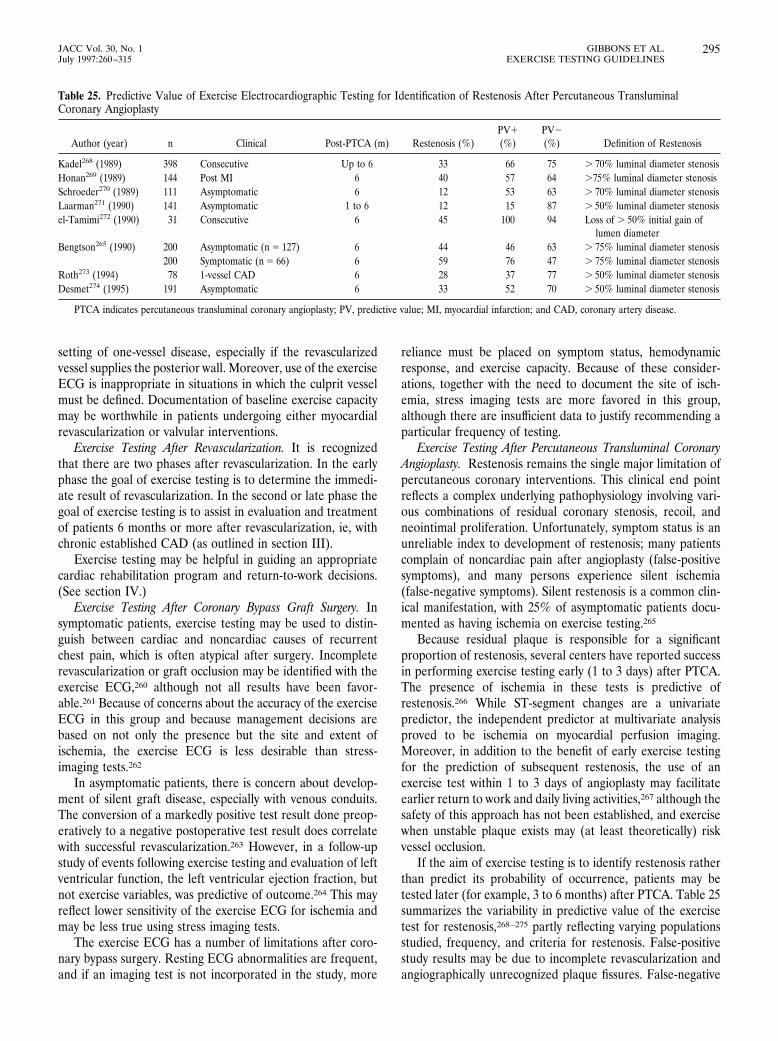
setting of one-vessel disease, especially if the revascularizedvessel supplies the posterior wall. Moreover, use of the exerciseECG is inappropriate in situations in which the culprit vesselmust be defined. Documentation of baseline exercise capacitymay be worthwhile in patients undergoing either myocardialrevascularization or valvular interventions.
Exercise Testing After Revascularization. It is recognizedthat there are two phases after revascularization. In the earlyphase the goal of exercise testing is to determine the immedi-ate result of revascularization. In the second or late phase thegoal of exercise testing is to assist in evaluation and treatmentof patients 6 months or more after revascularization, ie, withchronic established CAD (as outlined in section III).
Exercise testing may be helpful in guiding an appropriatecardiac rehabilitation program and return-to-work decisions.(See section IV.)
Exercise Testing After Coronary Bypass Graft Surgery. Insymptomatic patients, exercise testing may be used to distin-guish between cardiac and noncardiac causes of recurrentchest pain, which is often atypical after surgery. Incompleterevascularization or graft occlusion may be identified with theexercise ECG,260 although not all results have been favor-able.261 Because of concerns about the accuracy of the exerciseECG in this group and because management decisions arebased on not only the presence but the site and extent ofischemia, the exercise ECG is less desirable than stress-imaging tests.262
In asymptomatic patients, there is concern about develop-ment of silent graft disease, especially with venous conduits.The conversion of a markedly positive test result done preop-eratively to a negative postoperative test result does correlatewith successful revascularization.263 However, in a follow-upstudy of events following exercise testing and evaluation of leftventricular function, the left ventricular ejection fraction, butnot exercise variables, was predictive of outcome.264 This mayreflect lower sensitivity of the exercise ECG for ischemia andmay be less true using stress imaging tests.
The exercise ECG has a number of limitations after coro-nary bypass surgery. Resting ECG abnormalities are frequent,and if an imaging test is not incorporated in the study, more
reliance must be placed on symptom status, hemodynamicresponse, and exercise capacity. Because of these consider-ations, together with the need to document the site of isch-emia, stress imaging tests are more favored in this group,although there are insufficient data to justify recommending aparticular frequency of testing.
Exercise Testing After Percutaneous Transluminal CoronaryAngioplasty. Restenosis remains the single major limitation ofpercutaneous coronary interventions. This clinical end pointreflects a complex underlying pathophysiology involving vari-ous combinations of residual coronary stenosis, recoil, andneointimal proliferation. Unfortunately, symptom status is anunreliable index to development of restenosis; many patientscomplain of noncardiac pain after angioplasty (false-positivesymptoms), and many persons experience silent ischemia(false-negative symptoms). Silent restenosis is a common clin-ical manifestation, with 25% of asymptomatic patients docu-mented as having ischemia on exercise testing.265
Because residual plaque is responsible for a significantproportion of restenosis, several centers have reported successin performing exercise testing early (1 to 3 days) after PTCA.The presence of ischemia in these tests is predictive ofrestenosis.266 While ST-segment changes are a univariatepredictor, the independent predictor at multivariate analysisproved to be ischemia on myocardial perfusion imaging.Moreover, in addition to the benefit of early exercise testingfor the prediction of subsequent restenosis, the use of anexercise test within 1 to 3 days of angioplasty may facilitateearlier return to work and daily living activities,267 although thesafety of this approach has not been established, and exercisewhen unstable plaque exists may (at least theoretically) riskvessel occlusion.
If the aim of exercise testing is to identify restenosis ratherthan predict its probability of occurrence, patients may betested later (for example, 3 to 6 months) after PTCA. Table 25summarizes the variability in predictive value of the exercisetest for restenosis,268–275 partly reflecting varying populationsstudied, frequency, and criteria for restenosis. False-positivestudy results may be due to incomplete revascularization andangiographically unrecognized plaque fissures. False-negative
Table 25. Predictive Value of Exercise Electrocardiographic Testing for Identification of Restenosis After Percutaneous TransluminalCoronary Angioplasty
Author (year) n Clinical Post-PTCA (m) Restenosis (%)PV1(%)
PV2(%) Definition of Restenosis
Kadel268 (1989) 398 Consecutive Up to 6 33 66 75 . 70% luminal diameter stenosisHonan269 (1989) 144 Post MI 6 40 57 64 .75% luminal diameter stenosisSchroeder270 (1989) 111 Asymptomatic 6 12 53 63 . 70% luminal diameter stenosisLaarman271 (1990) 141 Asymptomatic 1 to 6 12 15 87 . 50% luminal diameter stenosisel-Tamimi272 (1990) 31 Consecutive 6 45 100 94 Loss of . 50% initial gain of
lumen diameterBengtson265 (1990) 200 Asymptomatic (n 5 127) 6 44 46 63 . 75% luminal diameter stenosis
200 Symptomatic (n 5 66) 6 59 76 47 . 75% luminal diameter stenosisRoth273 (1994) 78 1-vessel CAD 6 28 37 77 . 50% luminal diameter stenosisDesmet274 (1995) 191 Asymptomatic 6 33 52 70 . 50% luminal diameter stenosis
PTCA indicates percutaneous transluminal coronary angioplasty; PV, predictive value; MI, myocardial infarction; and CAD, coronary artery disease.
295JACC Vol. 30, No. 1 GIBBONS ET AL.July 1997:260–315 EXERCISE TESTING GUIDELINES
results may be caused by the failure of moderate (angiographi-cally but not functionally significant) one-vessel stenoses tolead to significant ischemia. Some authorities have advocatedroutine testing, because restenosis is frequent and commonlyinduces silent ischemia. The rationale of this approach is thatischemia, whether painful or silent, worsens prognosis.276,277
The alternative approach, which the committee favors, is to usea selective evaluation in patients considered to be at particu-larly high risk, because the prognostic benefit of controllingsilent ischemia needs to be proved. Examples of patients whoare likely to be at high risk include those with decreased leftventricular function, multivessel CAD, proximal left anteriordescending disease, previous sudden death, diabetes mellitus,hazardous occupations, and suboptimal PTCA results. Which-ever policy is followed, the exercise ECG is an insensitivepredictor of restenosis, with sensitivities ranging from 40% to55%, significantly less than those obtainable with SPECT5,278
or exercise echocardiography.6,279 The insensitivity of theexercise ECG probably reflects the high prevalence of one-vessel disease in this population.
In conclusion, the lower sensitivity of the exercise ECG(compared with imaging techniques) as well as its inability tolocalize disease limits its usefulness in patient managementboth before and after intervention. Despite the large numbersof procedures performed and widespread variation in use ofexercise testing in this context, there are insufficient data tojustify a particular testing regimen after intervention.
Investigation of Heart Rhythm Disorders
Class I
1. Identification of appropriate settings in patients with rate-adaptive pacemakers.
Class IIa
1. Evaluation of patients with known or suspected exercise-induced arrhythmias.
2. Evaluation of medical, surgical, or ablative therapy inpatients with exercise-induced arrhythmias (including atrialfibrillation).
Class IIb
1. Investigation of isolated ventricular ectopic beats in middle-aged patients without other evidence of CAD.
Class III
1. Investigation of isolated ectopic beats in young patients.
Evaluation of Patients With Known or Suspected Exercise-Induced Arrhythmias Use of exercise testing in patients withsyncope may identify those with CAD, although this is notusually the cause of syncope. Syncope due to sinus nodedysfunction, atrioventricular block, and tachycardias may alsobe identified.
Ventricular Arrhythmias. Exertional syncope due to tachy-cardias may reflect the presence of ischemia, other structural
abnormalities that induce an abnormal cardiac response tostress, and increased circulating catecholamines. The useful-ness of exercise testing in patients with VT is variable, accord-ing to the cause of the tachycardia. In some syndromes, such asright ventricular outflow tract tachycardia in a normal heart,VT may be reproducibly induced during exercise testing. Inadrenergic-dependent rhythm disturbances (including mono-morphic VT as well as polymorphic VT related to long QTsyndromes), ambulatory ECG monitoring may fail to supplythe circumstances necessary for induction of VT, particularly ifthe patient is sedentary and the arrhythmia is infrequent. Useof exercise testing is therefore a useful prelude to electrophys-iological study. Moreover, exercise testing may be of prognos-tic value in these patients: 12-month mortality is three timesgreater in persons exhibiting exercise-induced ectopy thanthose with ectopy at rest only,280 and in patients with exercise-induced ectopy, mortality of those with complex ectopy ex-ceeds that of those with simple ectopy.281 In patients onantiarrhythmic therapy, sustained exercise-induced VT is as-sociated with a high risk of sudden death,282 and exercisetesting has been used to unmask proarrhythmic responses.
While serious arrhythmias are uncommon in unselectedpopulations undergoing exercise testing,283 the use of maximalexercise testing in patients at risk of ventricular arrhythmia isassociated with a 2.3% incidence of arrhythmias requiringcardioversion, intravenous drugs, or resuscitation.284 Nonethe-less, even in this population, testing can be performed with lowmortality and few lasting morbid events. The main limitation ofexercise testing in patients with malignant ventricular arrhyth-mias is related to its limited reproducibility. While it issufficiently reproducible to serve as an adjunct in the evalua-tion and treatment of these patients,285 other testing is alsorequired.
Supraventricular Arrhythmias. Patients developing su-praventricular arrhythmias during exercise often displaymarked tachycardia due to their heightened adrenergic state.In patients with Wolff-Parkinson-White syndrome, exercisetesting may be used to help evaluate the risk of developingrapid ventricular response during atrial arrhythmias. Abruptloss of preexcitation during exercise infers a longer antegraderefractory period in the accessory pathway than the atrioven-tricular node. It is unlikely that a rapid ventricular responsewill occur at heart rates above this rate. However, this responseto exercise may be difficult to recognize because the adrenergicstate speeds conduction in the atrioventricular node andtherefore reduces the area of myocardium that is stimulatedprematurely from the accessory pathway.
In patients with atrial fibrillation, the ventricular response isgoverned by the atrioventricular node, and the heart rate istherefore dependent on the rate of repolarization and theeffective refractory period, both of which may be influenced byantiarrhythmic drugs used for rate control in patients withatrial fibrillation. Effective rate control at rest does not neces-sarily signify adequate rate control during exercise, and thetitration of additional drugs for this purpose may be facilitatedby exercise testing. The heart rate response to exercise in atrial
296 GIBBONS ET AL. JACC Vol. 30, No. 1EXERCISE TESTING GUIDELINES July 1997:260–315
fibrillation comprises an initial reduction of heart rate, fol-lowed by delayed acceleration in very early exercise and anexaggerated heart rate response. Prolonged tachycardia oftenpersists into the recovery period. In patients on medication,95% demonstrate an abnormal chronotropic response earlyduring exercise (74% being fast), and 84% demonstrate anabnormal chronotropic response during late exercise (53%being slow). Thus, the majority of patients with atrial fibrilla-tion demonstrate an abnormal chronotropic response to exer-cise.286
Sinus Node Dysfunction. Exercise testing may distinguishresting bradycardia with a normal exercise heart rate response(which is seen in well-trained subjects with predominantparasympathetic tone) from sinus node dysfunction with rest-ing bradycardia and in patients who fail to make an exerciseresponse. Chronotropic incompetence has been variously de-fined, the most common definition being failure to achieve85% of age-predicted maximum heart rate (ie, more than twostandard deviations below age-predicted maximum).287 Theuse of a heart rate response less than 100 bpm with maximalexercise288 is specific but insensitive. A more complicateddefinition, recently shown to be prognostically significant,289 isthe ratio between heart rate and metabolic reserve used bystage II of the Bruce protocol.290 Using various definitions,some authors have reported chronotropic incompetence inpatients with sinus node dysfunction, whereas others haveidentified the sensitivity and specificity of this marker for sinusnode dysfunction as being suboptimal. Moreover, exercisetesting has limited reproducibility in this respect, and a normaltest result does not negate the possibility of sinus nodedysfunction. The use of exercise testing may, however, beparticularly useful in showing the benefits of sensor-triggeredrate-adaptive pacing, both in terms of absolute heart rateattained and the rate of increase of heart rate.
Cardiac Pacemakers The previous edition of the ACC/AHA guidelines for exercise testing291 suggested that exercisetesting was inappropriate in most patients with a permanentpacemaker. Indeed, this remains true from a diagnostic stand-point, and even the combination of exercise testing withimaging may be problematic for the diagnosis of coronarydisease. However, the development of adaptive rate pacingusing various physiological sensors has led to study of theexercise response being an important constituent in fine-tuningthese devices.292,293 Additionally, a number of studies havecompared different pacing modes with respect to their influ-ence on exercise capacity. In all of these situations, however, aformal exercise test may not be necessary, and the requireddata could be obtained during a 6-minute walk.294
Exercise testing in patients with implantable cardiac defi-brillators (ICDs) may provoke arrhythmias or ICD discharge.Before testing, the programmed detection interval of thedevice should be known. If the device has been implanted forventricular fibrillation (VF) or fast VT, this rate will normallyexceed that attainable during sinus tachycardia, and the testcan be terminated as the heart rate approaches 10 bpm belowthe detection interval of the device. Indeed, this approach is
informative if the test is being performed to assess the risk ofsinus rate crossover.295 In patients with slower programmeddetection rates, the ICD may be reprogrammed to a fasterrate for the test, or temporarily deactivated (usually bysuperimposition of a magnet). Care should be taken to avoidunnecessary shocks as they are both unpleasant and poten-tially hazardous.296
VII. Pediatric Testing: Exercise Testing inChildren and Adolescents
Class I
1. Evaluation of exercise capacity in children or adolescentswith congenital heart disease, those who have had surgeryfor congenital heart disease, and children who have ac-quired valvular or myocardial disease.
2. Evaluation of the rare child with a description of anginalchest pain.
3. Assessment of the response of an artificial pacing system toexertion.
4. Evaluation of exercise-related symptoms in young athletes.
Class IIa
1. Evaluation of the adequacy of response to medical, surgical,or radiofrequency ablation treatment for children with atachyarrhythmia that was found during exercise testingbefore therapy.
2. As an adjunct in assessment of the severity of congenital oracquired valvular lesions, especially aortic valve stenosis.
3. Evaluation of the rhythm during exercise in patients withknown or suspected exercise-induced arrhythmia.
Class IIb
1. As a component of the evaluation of children or adolescentswho have a family history of unexplained sudden deathrelated to exercise in young individuals.
2. Follow-up of cardiac abnormalities with possible late coro-nary involvement such as Kawasaki disease and systemiclupus erythematosus.
3. Assessment of ventricular rate response and developmentof ventricular arrhythmia in children and adolescents withcongenital complete atrioventricular block.
4. Quantitation of the heart rate response to exercise inchildren and adolescents treated with b-blocker therapy toestimate the adequacy of b-blockade.
5. Measurement of response of shortening or prolongation ofthe corrected QT interval to exercise as an adjunct in thediagnosis of hereditary syndromes of prolongation of theQT interval.
6. Evaluation of blood pressure response and/or arm-to-leggradient after repair of coarctation of the aorta.
7. Assessment of degree of desaturation with exercise inpatients with relatively well-balanced or palliated cyanoticcongenital cardiac defects.
297JACC Vol. 30, No. 1 GIBBONS ET AL.July 1997:260–315 EXERCISE TESTING GUIDELINES
Class III
1. Screening before athletic participation by healthy childrenand adolescents.
2. Routine use of exercise testing for evaluating the usualnonanginal chest pain common in children and adolescents.
3. Evaluation of premature atrial and ventricular contractionsin otherwise healthy children and adolescents.
Differences Between Pediatric and Adult TestingIschemic heart disease is rare in a young population. This
results in a much lower risk of routine testing as well asdifferences in indications, usefulness, and interpretation ofexercise laboratory data in the pediatric population. Exercisetesting of children and adolescents has a very low risk com-pared with testing of adults.297 Complications of pediatricexercise testing are extremely infrequent, even when testing isdone in populations of children with congenital cardiac defectsand arrhythmias.9,297–300 Applications of exercise testing in theyoung are most often related to measurement of exercisecapacity, evaluation of known or possible abnormalities ofcardiac rhythm, and evaluation of symptoms elicited by exer-tion. Exercise testing is rarely needed to look for occultcoronary obstructions in the pediatric population. Exercisecapacity is diminished in some children or adolescents withheart disease, and measurement is often useful in evaluatingsubjective limitations.
Exercise testing in children provides a number of technicalchallenges. Equipment must be modified for use by smallchildren. Testing protocols may need to be normalized by sizeand weight. Norms for age and size on protocols being used arerequired for proper test interpretation.9,300 Developmentalfactors as well as interpretation of data figure into the conductof the test. Young children are less cooperative than olderpersons with maximum exercise testing. Experience at encour-aging the reluctant youngster to maximal effort is needed forinterpretable tests. It is usually difficult to differentiate limita-tions of exercise capacity from a lack of cooperation inpediatric testing. Because of this difficulty, ventilatory mea-surements are used in many pediatric exercise testing labora-tories to measure respiratory exchange ratio and ventilatoryanaerobic threshold.9,301
Exercise Testing for Specific Pediatric andCongenital Cardiac Problems (Table 26)
Exercise Testing of Children and Adolescents With ChestPain Chest pain is common in children and adolescents.302,303
History and physical examination are generally adequate toconfidently provide reassurance to patient and family. Theseepisodes of benign chest pain are usually described as a briefstabbing or shooting pain that occurs with or without exercise.Pleuritic pain is common. Typical anginal symptoms are veryuncommon. Routine use of exercise testing for evaluation ofchest pain in children and adolescents is not required. Exercisetesting is appropriate for evaluation of the uncommon child
with chest pain that is typical angina by description andconsistently related to exercise. Exercise-induced broncho-spasm is a cause of chest pain in children that is identified bypulmonary function measurement test protocols.304
Exercise Testing of Patients With Unoperated Left-to-RightShunts Results of exercise testing in young persons with aleft-to-right intracardiac shunt such as an atrial septal defect, aventricular septal defect, or patent ductus arteriosus willgenerally be normal if the magnitude of the shunt is moderateor small. These persons are ordinarily asymptomatic, andexercise testing adds little to routine evaluation of thesedefects. Most of those with large shunts will be treatedsurgically before they reach an age at which exercise testing ispractical. Older persons with a large left-to-right shunt oftenhave a low exercise capacity. Exercise testing may help confirmor document subjective exercise impairment in these persons.Testing is not routinely required to make decisions aboutoperative intervention in these older patients but can be ofconsiderable value if the history of limitation is unclear.
Patients with substantial diastolic runoff from a large left-to-right shunt distal to the aortic valve (such as a large patentductus arteriosus with low pulmonary vascular resistance) willoccasionally have resting ST-segment depression due to arelative insufficiency of coronary perfusion during diastole.Left-to-right shunts beyond the tricuspid valve characteristi-cally result in left ventricular volume overload, and largeshunts result in LVH. ST-segment depression on the exerciseECG can be seen in these circumstances but provides littlebenefit in decisions about surgical intervention. False-positiveexercise ECG changes are seen in some patients with left-to-right intracardiac shunts who are treated with digoxin.
Limited exercise capacity, arrhythmias, ST-segment changeson the exercise ECG, and/or decrease in oxygen saturation canbe seen in exercise testing of patients with left-to-right shuntlesions who have developed severe elevations of pulmonaryvascular resistance (Eisenmenger syndrome).305,306 Exercisetesting is potentially hazardous in this group of patients andgenerally should not be performed. Exercise testing has beenused to evaluate response to specific therapies intended todiminish pulmonary resistance and improve pulmonary bloodflow.
Exercise Testing in Patients With Postoperative Left-to-Right Shunts The great majority of patients in this group dovery well, and long-term follow-up studies have shown goodexercise performance in this group of patients many yearslater.307–309 Routine exercise testing of these patients is notrequired. Indications for use of exercise testing in this popu-lation include symptoms related to exercise, known or sus-pected arrhythmias related to exercise, and subjective limita-tions of exertional capability. Atrial arrhythmias may developyears after repair of atrial defects in some patients and areoften identified by exercise testing.310 Exercise testing is ofvalue as a part of evaluation of some patients with ventriculararrhythmias with a ventricular septal defect that was repairedin childhood.311 Exercise testing is also of value in the groupwith an operated left-to-right shunt in whom pulmonary vas-
298 GIBBONS ET AL. JACC Vol. 30, No. 1EXERCISE TESTING GUIDELINES July 1997:260–315
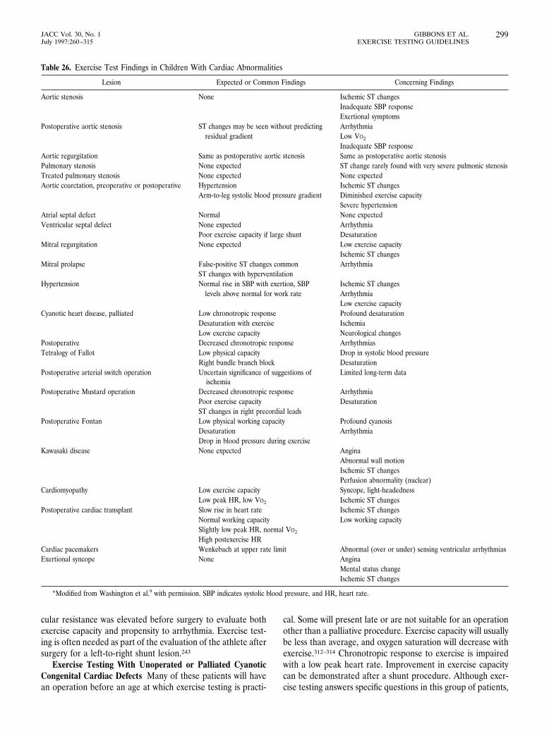
cular resistance was elevated before surgery to evaluate bothexercise capacity and propensity to arrhythmia. Exercise test-ing is often needed as part of the evaluation of the athlete aftersurgery for a left-to-right shunt lesion.243
Exercise Testing With Unoperated or Palliated CyanoticCongenital Cardiac Defects Many of these patients will havean operation before an age at which exercise testing is practi-
cal. Some will present late or are not suitable for an operationother than a palliative procedure. Exercise capacity will usuallybe less than average, and oxygen saturation will decrease withexercise.312–314 Chronotropic response to exercise is impairedwith a low peak heart rate. Improvement in exercise capacitycan be demonstrated after a shunt procedure. Although exer-cise testing answers specific questions in this group of patients,
Table 26. Exercise Test Findings in Children With Cardiac Abnormalities
Lesion Expected or Common Findings Concerning Findings
Aortic stenosis None Ischemic ST changesInadequate SBP responseExertional symptoms
Postoperative aortic stenosis ST changes may be seen without predicting Arrhythmiaresidual gradient Low VO2
Inadequate SBP responseAortic regurgitation Same as postoperative aortic stenosis Same as postoperative aortic stenosisPulmonary stenosis None expected ST change rarely found with very severe pulmonic stenosisTreated pulmonary stenosis None expected None expectedAortic coarctation, preoperative or postoperative Hypertension Ischemic ST changes
Arm-to-leg systolic blood pressure gradient Diminished exercise capacitySevere hypertension
Atrial septal defect Normal None expectedVentricular septal defect None expected Arrhythmia
Poor exercise capacity if large shunt DesaturationMitral regurgitation None expected Low exercise capacity
Ischemic ST changesMitral prolapse False-positive ST changes common Arrhythmia
ST changes with hyperventilationHypertension Normal rise in SBP with exertion, SBP Ischemic ST changes
levels above normal for work rate ArrhythmiaLow exercise capacity
Cyanotic heart disease, palliated Low chronotropic response Profound desaturationDesaturation with exercise IschemiaLow exercise capacity Neurological changes
Postoperative Decreased chronotropic response ArrhythmiasTetralogy of Fallot Low physical capacity Drop in systolic blood pressure
Right bundle branch block DesaturationPostoperative arterial switch operation Uncertain significance of suggestions of
ischemiaLimited long-term data
Postoperative Mustard operation Decreased chronotropic response ArrhythmiaPoor exercise capacity DesaturationST changes in right precordial leads
Postoperative Fontan Low physical working capacity Profound cyanosisDesaturation ArrhythmiaDrop in blood pressure during exercise
Kawasaki disease None expected AnginaAbnormal wall motionIschemic ST changesPerfusion abnormality (nuclear)
Cardiomyopathy Low exercise capacity Syncope, light-headednessLow peak HR, low VO2 Ischemic ST changes
Postoperative cardiac transplant Slow rise in heart rate Ischemic ST changesNormal working capacity Low working capacitySlightly low peak HR, normal VO2
High postexercise HRCardiac pacemakers Wenkebach at upper rate limit Abnormal (over or under) sensing ventricular arrhythmiasExertional syncope None Angina
Mental status changeIschemic ST changes
*Modified from Washington et al.9 with permission. SBP indicates systolic blood pressure, and HR, heart rate.
299JACC Vol. 30, No. 1 GIBBONS ET AL.July 1997:260–315 EXERCISE TESTING GUIDELINES
there is not often a specific need for exercise testing in theplanning of surgical intervention for these children.
Exercise Testing for Patients With Coarctation of the AortaBefore school age, exercise capacity and cardiopulmonaryfunction are usually normal after coarctation repair.315 Systolicblood pressure during exercise and the postexercise arm-to-leggradient potentially is of use to the clinician in the attempt todefine whether a residual or recoarctation is present.316 Whenpatients with operated coarctation are tested, a greater arm-to-leg blood pressure gradient is found immediately afterexercise than is found at rest. This has been thought to reflectresidual obstruction317 and has been used to compare results ofdifferent surgical approaches.318,319 Some have used the post-exercise arm-to-leg gradient as an indication for repeat cathe-terization. Some observers have found that resting data aresufficient to select those who require further intervention.Factors other than residual obstruction may contribute to thepostexercise arm-to-leg gradient.320,321 Residual gradientsduring exercise may ultimately lead to hyperdynamic ven-tricular function and resting upper extremity hypertension.322
Exercise-induced systolic blood pressure elevation has beenthought to indicate residual obstruction,323,324 but the widerange of variability in blood pressure response of normalchildren makes it difficult to use systolic blood pressureresponse alone in assessment of severity of residual coarcta-tion.316 Among pediatric cardiologists, there has been greatvariability in the use of exercise blood pressure data inplanning further interventions in coarctation of the aorta.325
Exercise testing may play an important role in evaluation ofsuitability for high-intensity sports participation after success-ful treatment of coarctation of the aorta.243 Exercise testing isoccasionally useful with coarctation of the aorta in assessingthe hemodynamic significance of other associated cardiacdefects (such as aortic valve disease).
Exercise Testing for the Child or Adolescent With Pulmo-nary Stenosis Although the transvalvular pressure gradient inpulmonary stenosis increases with exercise,326,327 natural his-tory data and strategies for intervention are based on restingmeasurements. Ventricular arrhythmia, cyanosis (from right-to-left shunting through a patent foramen ovale), or ischemicST-segment changes develop with exercise in persons withsevere pulmonary stenosis. Abnormalities in right ventricularfunction identified with exercise improve following relief of theobstruction.328 Exercise testing is not required for manage-ment decisions in the majority of cases with pulmonary steno-sis. Although exercise testing is not commonly needed fordecisions about advisability for participation in high physicalintensity sports after treatment of pulmonary stenosis,243 ath-letes with severe pulmonary valve incompetence and a dilatedright ventricle may require assessment before participation insports.
Exercise Testing for the Child or Adolescent With AorticStenosis or Regurgitation Exercise testing has been used tohelp assess the severity of obstruction in children with aorticstenosis.248–250 Before the development of reliable echocardio-graphic and Doppler methods, there was a greater need to rely
on exercise testing for children with aortic stenosis for assess-ment of the severity of aortic obstruction. Abnormalities foundon exercise testing of children with aortic stenosis includeST-segment changes with exercise,250,329 a drop in bloodpressure or failure of appropriate increase in blood pressurewith exercise,330 and development of arrhythmias during exer-cise testing.331 Although exercise-induced ST-segment changeshave been correlated with the severity of obstruction, theresting gradient is not the sole determinant of these ST-segment changes. The major hemodynamic determinant of STdepression during exercise in patients with aortic stenosisseems to be the left ventricular oxygen demand supply ratio.329
ST depression with exercise testing improves when the gradi-ent has been relieved surgically,331 although abnormalitiesafter treatment seem to be of less predictive value than inunoperated patients.332 Blood pressure response to exercise isextremely variable in children without heart disease, and lackof a rise in blood pressure with exercise is often seen in normalchildren. Although blood pressure rises less in children withsevere aortic stenosis than it does in control subjects, bloodpressure response is not diagnostic in individual patients.
Critical aortic stenosis can be identified by findings onexamination and confirmed by echocardiography. Althoughthe exercise test is likely to be abnormal in this setting, testingcarries some risk, and the decision to recommend interventionis made without requiring exercise testing. Another group ofpatients who have very mild aortic stenosis can also beconfidently identified by expert examination, confirmed, ifnecessary, by echocardiography. The exercise test adds little toassessment of severity in this group of patients, and anabnormal test result is likely to be a false-positive result.Exercise testing can provide useful information in combinationwith other data for management decisions about patients withaortic stenosis that is neither obviously quite severe or verymild. There are no criteria for operative intervention based onresults of exercise testing alone.
Exercise testing is often used for issues of the advisability ofsports participation by children with aortic stenosis and forchildren who have had treatment of aortic stenosis.243
Aortic regurgitation causes left ventricular dilation withstatic or dynamic exercise, and ST-segment changes duringexercise are expected with hemodynamically significant aorticregurgitation. ST-segment changes in children with aorticstenosis and regurgitation are inaccurate in predicting gradi-ent.333 Exercise testing is often useful in evaluation of exercisecapacity of children with aortic regurgitation and evaluationbefore participation in sports.243
Exercise Testing After Surgery for Tetralogy of FallotExercise capacity improves after successful repair of tetralogyof Fallot. Low exercise capacity is found in many persons withsignificant residual hemodynamic abnormalities. Exercise test-ing has been used to identify children with arrhythmias.Complex ventricular arrhythmias observed during exercisetesting are correlated with a risk of sudden cardiac death.Serial testing has been used to demonstrate the effectiveness ofan antiarrhythmic therapeutic regimen. The possibility of late
300 GIBBONS ET AL. JACC Vol. 30, No. 1EXERCISE TESTING GUIDELINES July 1997:260–315
development of arrhythmia after repair of tetralogy of Fallotled to a recommendation for periodic exercise testing (every 3or 4 years) in postoperative tetralogy patients. This recommen-dation is most applicable to older patients who underwentsurgery sometime ago. Diastolic restrictive physiology withexercise has recently been shown to predict a better long-termfunctional situation for patients with tetralogy of Fallot.334
Exercise testing is often needed as part of the evaluationbefore intense athletic training and competition for thesepatients, primarily to assess their propensity to arrhythmias.
Exercise Testing After the Fontan Operation (Total Sys-temic Venous to Pulmonary Connection) Low exercise capac-ity, low cardiac output and low oxygen uptake, development ofatrial or ventricular arrhythmias with exercise, limitation ofpeak exercise heart rate response, and development of cyano-sis is usual with exercise testing in this group of patients.Improvement in exercise capacity compared with preoperativestatus can be demonstrated.314,335,336 Exercise testing is helpfulin evaluation of exercise capacity and screening for arrhythmiain the postoperative Fontan patient.
Exercise Testing of Patients With Cardiomyopathy Exer-cise testing of children with hypertrophic cardiomyopathy hasnot been a routine part of evaluation. Children with severehypertrophy or obstruction are at risk for complications withexercise. Arrhythmias, ST-segment changes, drop in bloodpressure, and low exercise cardiac output occur with test-ing.337,338 Exercise testing is sometimes useful to help evaluateresponse to a specific therapy in patients with hypertrophiccardiomyopathy.
Children with dilated cardiomyopathy are likely to have alimited exercise capacity. ST-segment changes are often seen.The exercise test can be of value in identifying arrhythmias.Survival correlates with impaired exercise capacity. Informa-tion about exercise limitation influences decisions about thetiming of cardiac transplantation.
Restrictive cardiomyopathy is less common than dilated orhypertrophic cardiomyopathy. These children tend to be iden-tified later and seem less ill than those with dilated cardiomy-opathy, although the time interval from diagnosis to death isshorter.339 Near-normal exercise capacity is falsely reassuringin this setting.
Exercise Testing of Children or Adolescents With SyncopeFeatures of syncope that suggest a potentially life-threateningcardiac event include an abrupt loss of consciousness (asopposed to a prodrome or aura), injury on impact (as opposedto a gradual “slump”), and syncope related to physical activity.Evaluation for possible arrhythmia, left ventricular outflowobstruction, and cardiomyopathy is appropriate. Exercise test-ing is an important component of evaluation of children oradolescents who have unexplained syncope related to physicalexertion.
Exercise Testing of Children or Adolescents With AtrialArrhythmias Premature atrial contractions are common andbenign in young persons. Exercise test evaluation of prematureatrial contractions in pediatric patients is not required.
The majority of children with supraventricular tachycardia
will not have exercise-induced tachycardia. Exercise testinghelps in evaluation of selected cases in which the historysuggests an exercise-related tachycardia. Serial exercise testinghas been used to follow the response to therapy in cases ofexercise-induced atrial tachycardias.
Exercise Testing of Children or Adolescents With Ventric-ular Arrhythmias Isolated premature ventricular depolariza-tions in asymptomatic children and adolescents usually disap-pear with the higher heart rates associated with exercise. It isnot necessary to perform formal laboratory testing to demon-strate this.
Exercise testing is often of value in diagnosing VT andassessing the efficacy of treatment. VT with exercise is oftenobserved in children with no structural heart abnormality aswell as in children with myocarditis, cardiomyopathy, or acongenital cardiac malformation. Arrhythmogenic right ven-tricular dysplasia is particularly related to development of VTwith exercise.
Measurement of the response of the shortening or prolon-gation of the corrected QT interval to exercise has been usedas an adjunct in the diagnosis of hereditary syndromes ofprolongation of the QT interval. Accuracy of measurement issuspect, and its usefulness for decision making in borderlinecases has been questioned. T-wave alternans is a relativelyspecific but uncommon finding with exercise testing in patientswith long QT-interval syndromes. Exercise testing occasionallyidentifies VT.
Exercise Testing of Children or Adolescents With Conduc-tion Abnormalities and in Pacemaker Follow-up Ventricularrate response to exercise and development of ventriculararrhythmia with congenital complete atrioventricular block isuseful in determining the need for artificial pacing. Assessmentof sensing and capture of an artificial pacing system duringexercise is useful in some cases. Exercise testing to evaluate theadequacy of rate responsiveness to exercise is useful in man-agement.
Exercise Testing of Children or Adolescents With Knownor Suspected Coronary Artery Disease Some cardiac abnor-malities in childhood are predicted to be associated withdevelopment of coronary artery involvement later in life.Examples include persons who had surgery for anomalous leftcoronary artery from the pulmonary trunk (Bland-Garland-White syndrome). Some cases of tetralogy of Fallot include ananomalous right coronary artery arising from the left coronaryartery or from the left sinus of Valsalva and crossing the rightventricular outflow tract in proximity to the infundibularresection. One concern has been possible coronary anasto-motic obstruction late after neonatal arterial switch operationfor transposition of the great arteries in which the coronaryostia must be translocated. Kawasaki disease with coronaryaneurysms in early life is another possible source of develop-ment of coronary artery insufficiency. Exercise testing ofasymptomatic youths in these categories before intense athleticcompetition and testing if symptoms of possible ischemic origindevelop is indicated.
301JACC Vol. 30, No. 1 GIBBONS ET AL.July 1997:260–315 EXERCISE TESTING GUIDELINES
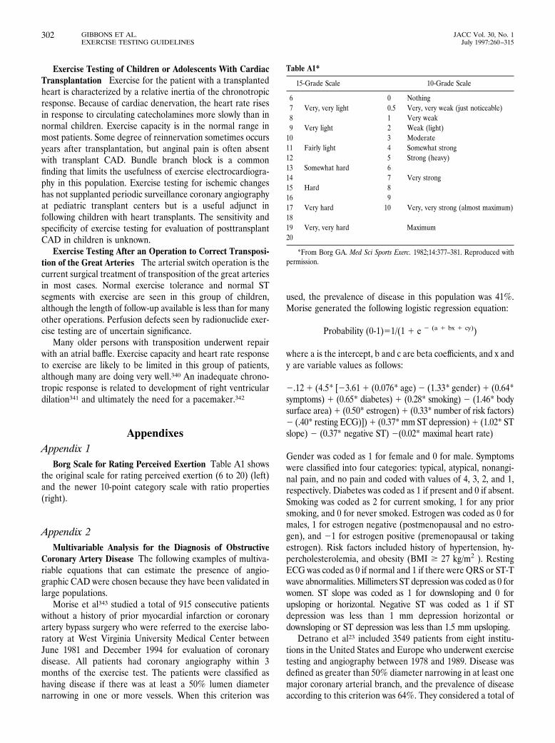
Exercise Testing of Children or Adolescents With CardiacTransplantation Exercise for the patient with a transplantedheart is characterized by a relative inertia of the chronotropicresponse. Because of cardiac denervation, the heart rate risesin response to circulating catecholamines more slowly than innormal children. Exercise capacity is in the normal range inmost patients. Some degree of reinnervation sometimes occursyears after transplantation, but anginal pain is often absentwith transplant CAD. Bundle branch block is a commonfinding that limits the usefulness of exercise electrocardiogra-phy in this population. Exercise testing for ischemic changeshas not supplanted periodic surveillance coronary angiographyat pediatric transplant centers but is a useful adjunct infollowing children with heart transplants. The sensitivity andspecificity of exercise testing for evaluation of posttransplantCAD in children is unknown.
Exercise Testing After an Operation to Correct Transposi-tion of the Great Arteries The arterial switch operation is thecurrent surgical treatment of transposition of the great arteriesin most cases. Normal exercise tolerance and normal STsegments with exercise are seen in this group of children,although the length of follow-up available is less than for manyother operations. Perfusion defects seen by radionuclide exer-cise testing are of uncertain significance.
Many older persons with transposition underwent repairwith an atrial baffle. Exercise capacity and heart rate responseto exercise are likely to be limited in this group of patients,although many are doing very well.340 An inadequate chrono-tropic response is related to development of right ventriculardilation341 and ultimately the need for a pacemaker.342
AppendixesAppendix 1
Borg Scale for Rating Perceived Exertion Table A1 showsthe original scale for rating perceived exertion (6 to 20) (left)and the newer 10-point category scale with ratio properties(right).
Appendix 2Multivariable Analysis for the Diagnosis of Obstructive
Coronary Artery Disease The following examples of multiva-riable equations that can estimate the presence of angio-graphic CAD were chosen because they have been validated inlarge populations.
Morise et al343 studied a total of 915 consecutive patientswithout a history of prior myocardial infarction or coronaryartery bypass surgery who were referred to the exercise labo-ratory at West Virginia University Medical Center betweenJune 1981 and December 1994 for evaluation of coronarydisease. All patients had coronary angiography within 3months of the exercise test. The patients were classified ashaving disease if there was at least a 50% lumen diameternarrowing in one or more vessels. When this criterion was
used, the prevalence of disease in this population was 41%.Morise generated the following logistic regression equation:
Probability (0-1)51/(1 1 e 2 (a 1 bx 1 cy))
where a is the intercept, b and c are beta coefficients, and x andy are variable values as follows:
2.12 1 (4.5* [23.61 1 (0.076* age) 2 (1.33* gender) 1 (0.64*symptoms) 1 (0.65* diabetes) 1 (0.28* smoking) 2 (1.46* bodysurface area) 1 (0.50* estrogen) 1 (0.33* number of risk factors)2 (.40* resting ECG)]) 1 (0.37* mm ST depression) 1 (1.02* STslope) 2 (0.37* negative ST) 2(0.02* maximal heart rate)
Gender was coded as 1 for female and 0 for male. Symptomswere classified into four categories: typical, atypical, nonangi-nal pain, and no pain and coded with values of 4, 3, 2, and 1,respectively. Diabetes was coded as 1 if present and 0 if absent.Smoking was coded as 2 for current smoking, 1 for any priorsmoking, and 0 for never smoked. Estrogen was coded as 0 formales, 1 for estrogen negative (postmenopausal and no estro-gen), and 21 for estrogen positive (premenopausal or takingestrogen). Risk factors included history of hypertension, hy-percholesterolemia, and obesity (BMI $ 27 kg/m2 ). RestingECG was coded as 0 if normal and 1 if there were QRS or ST-Twave abnormalities. Millimeters ST depression was coded as 0 forwomen. ST slope was coded as 1 for downsloping and 0 forupsloping or horizontal. Negative ST was coded as 1 if STdepression was less than 1 mm depression horizontal ordownsloping or ST depression was less than 1.5 mm upsloping.
Detrano et al23 included 3549 patients from eight institu-tions in the United States and Europe who underwent exercisetesting and angiography between 1978 and 1989. Disease wasdefined as greater than 50% diameter narrowing in at least onemajor coronary arterial branch, and the prevalence of diseaseaccording to this criterion was 64%. They considered a total of
Table A1*
15-Grade Scale 10-Grade Scale
6 0 Nothing7 Very, very light 0.5 Very, very weak (just noticeable)8 1 Very weak9 Very light 2 Weak (light)
10 3 Moderate11 Fairly light 4 Somewhat strong12 5 Strong (heavy)13 Somewhat hard 614 7 Very strong15 Hard 816 917 Very hard 10 Very, very strong (almost maximum)1819 Very, very hard Maximum20
*From Borg GA. Med Sci Sports Exerc. 1982;14:377–381. Reproduced withpermission.
302 GIBBONS ET AL. JACC Vol. 30, No. 1EXERCISE TESTING GUIDELINES July 1997:260–315
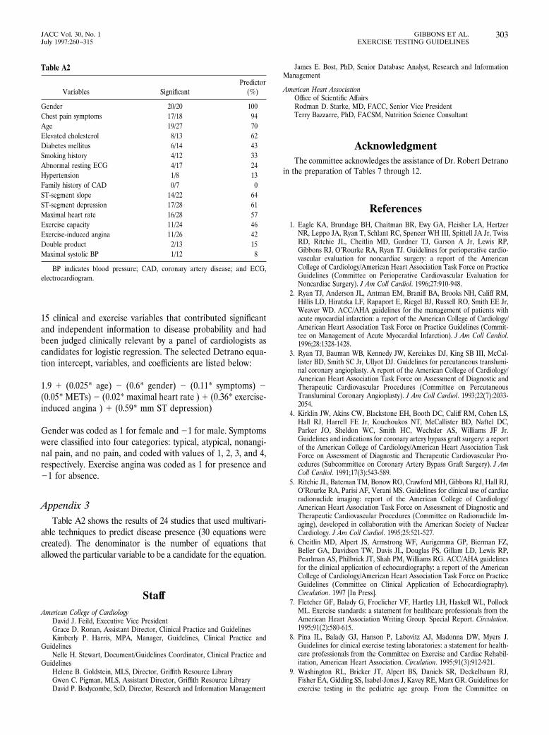
15 clinical and exercise variables that contributed significantand independent information to disease probability and hadbeen judged clinically relevant by a panel of cardiologists ascandidates for logistic regression. The selected Detrano equa-tion intercept, variables, and coefficients are listed below:
1.9 1 (0.025* age) 2 (0.6* gender) 2 (0.11* symptoms) 2(0.05* METs) 2 (0.02* maximal heart rate ) 1 (0.36* exercise-induced angina ) 1 (0.59* mm ST depression)
Gender was coded as 1 for female and 21 for male. Symptomswere classified into four categories: typical, atypical, nonangi-nal pain, and no pain, and coded with values of 1, 2, 3, and 4,respectively. Exercise angina was coded as 1 for presence and21 for absence.
Appendix 3Table A2 shows the results of 24 studies that used multivari-
able techniques to predict disease presence (30 equations werecreated). The denominator is the number of equations thatallowed the particular variable to be a candidate for the equation.
StaffAmerican College of Cardiology
David J. Feild, Executive Vice PresidentGrace D. Ronan, Assistant Director, Clinical Practice and GuidelinesKimberly P. Harris, MPA, Manager, Guidelines, Clinical Practice and
GuidelinesNelle H. Stewart, Document/Guidelines Coordinator, Clinical Practice and
GuidelinesHelene B. Goldstein, MLS, Director, Griffith Resource LibraryGwen C. Pigman, MLS, Assistant Director, Griffith Resource LibraryDavid P. Bodycombe, ScD, Director, Research and Information Management
James E. Bost, PhD, Senior Database Analyst, Research and InformationManagement
American Heart AssociationOffice of Scientific AffairsRodman D. Starke, MD, FACC, Senior Vice PresidentTerry Bazzarre, PhD, FACSM, Nutrition Science Consultant
AcknowledgmentThe committee acknowledges the assistance of Dr. Robert Detrano
in the preparation of Tables 7 through 12.
References1. Eagle KA, Brundage BH, Chaitman BR, Ewy GA, Fleisher LA, Hertzer
NR, Leppo JA, Ryan T, Schlant RC, Spencer WH III, Spittell JA Jr, TwissRD, Ritchie JL, Cheitlin MD, Gardner TJ, Garson A Jr, Lewis RP,Gibbons RJ, O’Rourke RA, Ryan TJ. Guidelines for perioperative cardio-vascular evaluation for noncardiac surgery: a report of the AmericanCollege of Cardiology/American Heart Association Task Force on PracticeGuidelines (Committee on Perioperative Cardiovascular Evaluation forNoncardiac Surgery). J Am Coll Cardiol. 1996;27:910-948.
2. Ryan TJ, Anderson JL, Antman EM, Braniff BA, Brooks NH, Califf RM,Hillis LD, Hiratzka LF, Rapaport E, Riegel BJ, Russell RO, Smith EE Jr,Weaver WD. ACC/AHA guidelines for the management of patients withacute myocardial infarction: a report of the American College of Cardiology/American Heart Association Task Force on Practice Guidelines (Commit-tee on Management of Acute Myocardial Infarction). J Am Coll Cardiol.1996;28:1328-1428.
3. Ryan TJ, Bauman WB, Kennedy JW, Kereiakes DJ, King SB III, McCal-lister BD, Smith SC Jr, Ullyot DJ. Guidelines for percutaneous translumi-nal coronary angioplasty. A report of the American College of Cardiology/American Heart Association Task Force on Assessment of Diagnostic andTherapeutic Cardiovascular Procedures (Committee on PercutaneousTransluminal Coronary Angioplasty). J Am Coll Cardiol. 1993;22(7):2033-2054.
4. Kirklin JW, Akins CW, Blackstone EH, Booth DC, Califf RM, Cohen LS,Hall RJ, Harrell FE Jr, Kouchoukos NT, McCallister BD, Naftel DC,Parker JO, Sheldon WC, Smith HC, Wechsler AS, Williams JF Jr.Guidelines and indications for coronary artery bypass graft surgery: a reportof the American College of Cardiology/American Heart Association TaskForce on Assessment of Diagnostic and Therapeutic Cardiovascular Pro-cedures (Subcommittee on Coronary Artery Bypass Graft Surgery). J AmColl Cardiol. 1991;17(3):543-589.
5. Ritchie JL, Bateman TM, Bonow RO, Crawford MH, Gibbons RJ, Hall RJ,O’Rourke RA, Parisi AF, Verani MS. Guidelines for clinical use of cardiacradionuclide imaging: report of the American College of Cardiology/American Heart Association Task Force on Assessment of Diagnostic andTherapeutic Cardiovascular Procedures (Committee on Radionuclide Im-aging), developed in collaboration with the American Society of NuclearCardiology. J Am Coll Cardiol. 1995;25:521-527.
6. Cheitlin MD, Alpert JS, Armstrong WF, Aurigemma GP, Bierman FZ,Beller GA, Davidson TW, Davis JL, Douglas PS, Gillam LD, Lewis RP,Pearlman AS, Philbrick JT, Shah PM, Williams RG. ACC/AHA guidelinesfor the clinical application of echocardiography: a report of the AmericanCollege of Cardiology/American Heart Association Task Force on PracticeGuidelines (Committee on Clinical Application of Echocardiography).Circulation. 1997 [In Press].
7. Fletcher GF, Balady G, Froelicher VF, Hartley LH, Haskell WL, PollockML. Exercise standards: a statement for healthcare professionals from theAmerican Heart Association Writing Group. Special Report. Circulation.1995;91(2):580-615.
8. Pina IL, Balady GJ, Hanson P, Labovitz AJ, Madonna DW, Myers J.Guidelines for clinical exercise testing laboratories: a statement for health-care professionals from the Committee on Exercise and Cardiac Rehabil-itation, American Heart Association. Circulation. 1995;91(3):912-921.
9. Washington RL, Bricker JT, Alpert BS, Daniels SR, Deckelbaum RJ,Fisher EA, Gidding SS, Isabel-Jones J, Kavey RE, Marx GR. Guidelines forexercise testing in the pediatric age group. From the Committee on
Table A2
Variables SignificantPredictor
(%)
Gender 20/20 100Chest pain symptoms 17/18 94Age 19/27 70Elevated cholesterol 8/13 62Diabetes mellitus 6/14 43Smoking history 4/12 33Abnormal resting ECG 4/17 24Hypertension 1/8 13Family history of CAD 0/7 0ST-segment slope 14/22 64ST-segment depression 17/28 61Maximal heart rate 16/28 57Exercise capacity 11/24 46Exercise-induced angina 11/26 42Double product 2/13 15Maximal systolic BP 1/12 8
BP indicates blood pressure; CAD, coronary artery disease; and ECG,electrocardiogram.
303JACC Vol. 30, No. 1 GIBBONS ET AL.July 1997:260–315 EXERCISE TESTING GUIDELINES
Atherosclerosis and Hypertension in Children, Council on CardiovascularDisease in the Young, the American Heart Association. Circulation.1994;90(4):2166-2179.
10. Stuart RJ Jr, Ellestad MH. National survey of exercise stress testingfacilities. Chest. 1980;77(1):94-97.
11. Schlant RC, Friesinger GC II, Leonard JJ. Clinical competence in exercisetesting: a statement for physicians from the ACP/ACC/AHA Task Force onClinical Privileges in Cardiology. J Am Coll Cardiol. 1990;16(5):1061-1065.
12. Myers J, Froelicher VF. Optimizing the exercise test for pharmacologicalinvestigations. Circulation. 1990;82(5):1839-1846.
13. Borg GA. Psychophysical bases of perceived exertion. Med Sci Sports Exerc.1982;14(5):377-381.
14. Braunwald E, Mark DB, Jones RH, et al. Unstable Angina: Diagnosis andManagement. Rockville, Md: Clinical Practice Guideline No. 10. AHCPRPub No. 94-0602; 1994.
15. Logan SE. On the fluid mechanics of human coronary artery stenosis. IEEETrans Biomed Eng. 1975;22(4):327-334.
16. Marcus ML, Wilson RF, White CW. Methods of measurement of myocar-dial blood flow in patients: a critical review. Circulation. 1987;76(2):245-253.
17. Diamond GA, Forrester JS. Analysis of probability as an aid in the clinicaldiagnosis of coronary-artery disease. N Engl J Med. 1979;300(24):1350-1358.
18. Diamond GA. A clinical relevant classification of chest discomfort. J AmColl Cardiol. 1983;1:574-575. Letter.
19. Pryor DB, Harrell FE Jr, Lee KL, Califf RM, Rosati RA. Estimating thelikelihood of significant coronary artery disease. Am J Med. 1983;75(5):771-780.
20. Ellestad MH, Savitz S, Bergdall D, Teske J. The false positive stress test:multivariate analysis of 215 subjects with hemodynamic, angiographic andclinical data. Am J Cardiol. 1977;40(5):681-685.
21. Yamada H, Do D, Morise A, Froelicher VF. Multivariable equations topredict CAD. Prog Cardiovasc Dis. 1997 [In press].
22. Lee KL, Pryor DB, Harrell FE Jr, Califf RM, Behar VS, Floyd WL, MorrisJJ, Waugh RA, Whalen RE, Rosati RA. Predicting outcome in coronarydisease: statistical models versus expert clinicians. Am J Med. 1986;80(4):553-560.
23. Detrano R, Bobbio M, Olson H, Shandling A, Ellestad MH, Alegria E,Martinez-Caro D, Righetti A, Janosi A, Steinbrunn W. Computer proba-bility estimates of angiographic coronary artery disease: transportability andcomparison with cardiologists’ estimates. Comput Biomed Res. 1992;25(5):468-485.
24. Guyatt GH. Readers’ guide for articles evaluating diagnostic tests: whatACP Journal Club does for you and what you must do yourself. ACP J Club.1996;115:A-16.
25. Gianrossi R, Detrano R, Mulvihill D, Lehmann K, Dubach P, Colombo A,McArthur D, Froelicher V. Exercise-induced ST depression in the diagno-sis of coronary artery disease: a meta-analysis. Circulation. 1989;80(1):87-98.
26. Detrano R, Gianrossi R, Froelicher V. The diagnostic accuracy of theexercise electrocardiogram: a meta-analysis of 22 years of research. ProgCardiovasc Dis. 1989;32(3):173-206.
27. Reid MC, Lachs MS, Feinstein AR. Use of methodological standards indiagnostic test research: getting better but still not good. JAMA. 1995;274(8):645-651.
28. Morise AP, Diamond GA. Comparison of the sensitivity and specificity ofexercise electrocardiography in biased and unbiased populations of menand women. Am Heart J. 1995;130(4):741-747.
29. DelCampo J, Do D, Umann T, McGowan V, Froning J, Froelicher VF.Comparison of computerized and standard visual criteria of exercise ECGfor diagnosis of coronary artery disease. Ann Non-Invasive Electrocardiog-raphy. 1996;1(4):430-442.
30. Sketch MH, Mooss AN, Butler ML, Nair CK, Mohiuddin SM. Digoxin-induced positive exercise tests: their clinical and prognostic significance.Am J Cardiol. 1981;48:655-659.
31. LeWinter MM, Crawford MH, O’Rourke RA, Karliner JS. The effects oforal propranolol, digoxin and combination therapy on the resting andexercise electrocardiogram. Am Heart J. 1977;93:202-209.
32. Sundqvist K, Atterhog JH, Jogestrand T. Effect of digoxin on the electro-cardiogram at rest and during exercise in healthy subjects. Am J Cardiol.1986;57(8):661-665.
33. Herbert WG, Dubach P, Lehmann KG, Froelicher VF. Effect of beta-
blockade on the interpretation of the exercise ECG: ST level versus deltaST/HR index. Am Heart J. 1991;122(4 Pt 1):993-1000.
34. Cantwell JD, Murray PM, Thomas RJ. Current management of severeexercise-related cardiac events. Chest. 1988;93(6):1264-1269.
35. Anastasiou-Nana MI, Anderson JL, Stewart JR, Crevey BJ, Yanowitz FG,Lutz JR, Johnson TA. Occurrence of exercise-induced and spontaneouswide complex tachycardia during therapy with flecainide for complexventricular arrhythmias: a probable proarrhythmic effect. Am Heart J.1987;113(5):1071-1077.
36. Whinnery JE, Froelicher VF, Stuart AJ. The electrocardiographic responseto maximal treadmill exercise in asymptomatic men with left bundle branchblock. Am Heart J. 1997;94:316.
37. Whinnery JE, Froelicher VF Jr, Longo MR Jr, Triebwasser JH. Theelectrocardiographic response to maximal treadmill exercise in asymptom-atic men with right branch bundle block. Chest. 1977;71:335-340.
38. Blackburn H. Canadian colloquium on computer-assisted interpretation ofelectrocardiograms, VI: importance of the electrocardiogram in popula-tions outside the hospital. Can Med Assoc J. 1973;108(10):1262-1265.
39. Cullen K, Stenhouse NS, Wearne KL, Cumpston GN. Electrocardiogramsand 13 year cardiovascular mortality in Busselton study. Br Heart J.1982;47(3):209-212.
40. Aronow WS. Correlation of ischemic ST-segment depression on the restingelectrocardiogram with new cardiac events in 1,106 patients over 62 years ofage. Am J Cardiol. 1989;64(3):232-233.
41. Califf RM, Mark DB, Harrell FE Jr, Hlatky MA, Lee KL, Rosati RA, PryorDB. Importance of clinical measures of ischemia in the prognosis ofpatients with documented coronary artery disease. J Am Coll Cardiol.1988;11(1):20-26.
42. Harris PJ, Harrell FE Jr, Lee KL, Behar VS, Rosati RA. Survival inmedically treated coronary artery disease. Circulation. 1979;60(6):1259-1269.
43. Miranda CP, Lehmann KG, Froelicher VF. Correlation between resting STsegment depression, exercise testing, coronary angiography, and long-termprognosis. Am Heart J. 1991;122(6):1617-1628.
44. Kansal S, Roitman D, Sheffield LT. Stress testing with ST-segment depres-sion at rest: an angiographic correlation. Circulation. 1976;54(4):636-639.
45. Harris FJ, DeMaria AN, Lee G, Miller RR, Amsterdam EA, Mason DT.Value and limitations of exercise testing in detecting coronary disease withnormal and abnormal resting electrocardiograms. Adv Cardiol. 1978;22:11-15.
46. Roitman D, Jones WB, Sheffield LT. Comparison of submaximal exerciseECG test with coronary cineangiocardiogram. Ann Intern Med. 1970;72(5):641-647.
47. Meyers DG, Bendon KA, Hankins JH, Stratbucker RA. The effect ofbaseline electrocardiographic abnormalities on the diagnostic accuracy ofexercise-induced ST segment changes. Am Heart J. 1990;119(2 Pt 1):272-276.
48. Miranda CP, Liu J, Kadar A, Janosi A, Froning J, Lehmann KG, FroelicherVF. Usefulness of exercise-induced ST-segment depression in the inferiorleads during exercise testing as a marker for coronary artery disease. Am JCardiol. 1992;69(4):303-307.
49. Rijneke RD, Ascoop CA, Talmon JL. Clinical significance of upsloping STsegments in exercise electrocardiography. Circulation. 1980;61:671-678.
50. Stuart RJ, Ellestad MH. Upsloping S-T segments in exercise stress testing:six-year follow-up study of 438 patients and correlation with 248 angio-grams. Am J Cardiol. 1976;37:19-22.
51. Fortuin NJ, Friesinger GC. Exercise-induced S-T segment elevation: clin-ical, electrocardiographic and arteriographic studies in twelve patients.Am J Med. 1970;49(4):459-464.
52. Hegge FN, Tuna N, Burchell HB. Coronary arteriographic findings inpatients with axis shifts or S-T-segment elevations on exercise-stress testing.Am Heart J. 1973;86(5):603-615.
53. Chahine RA, Raizner AE, Ishimori T. The clinical significance of exercise-induced ST-segment elevation. Circulation. 1976;54(2):209-213.
54. Longhurst JC, Kraus WL. Exercise-induced ST elevation in patientswithout myocardial infarction. Circulation. 1979;60(3):616-629.
55. Mark DB, Hlatky MA, Lee KL, Harrell FE, Califf RM, Pryor DB.Localizing coronary artery obstructions with the exercise treadmill test. AnnIntern Med. 1987;106(1):53-55.
56. Bruce RA, Fisher LD. Unusual prognostic significance of exercise-inducedST elevation in coronary patients. J Electrocardiol. 1987;20(suppl):84-88.
304 GIBBONS ET AL. JACC Vol. 30, No. 1EXERCISE TESTING GUIDELINES July 1997:260–315
57. de Feyter PJ, Majid PA, Van Eenige MJ, Wardeh R, Wempe FN, Ross JP.Clinical significance of exercise-induced ST segment elevation: correlativeangiographic study in patients with ischaemic heart disease. Br Heart J.1981;46:84-92.
58. Manvi KN, Ellestad MH. Elevated ST segments with exercise in ventricularaneurysm. J Electrocardiol. 1972;5(4):317-323.
59. Haines DE, Beller GA, Watson DD, Kaiser DL, Sayre SL, Gibson RS.Exercise-induced ST segment elevation 2 weeks after uncomplicated myo-cardial infarction: contributing factors and prognostic significance. J AmColl Cardiol. 1987;9(5):996-1003.
60. Margonato A, Ballarotto C, Bonetti F, Cappelletti A, Sciammarella M,Cianflone D, Chierchia SL. Assessment of residual tissue viability byexercise testing in recent myocardial infarction: comparison of the electro-cardiogram and myocardial perfusion scintigraphy. J Am Coll Cardiol.1992;19(5):948-952.
61. Margonato A, Chierchia SL, Xuereb RG, Xuereb M, Fragasso G, Cappel-letti A, Landoni C, Lucignani G, Fazio F. Specificity and sensitivity ofexercise-induced ST segment elevation for detection of residual viability:comparison with fluorodeoxyglucose and positron emission tomography.J Am Coll Cardiol. 1995;25(5):1032-1038.
62. Lombardo A, Loperfido F, Pennestri F, Rossi E, Patrizi R, Cristinziani G,Catapano G, Maseri A. Significance of transient ST-T segment changesduring dobutamine testing in Q wave myocardial infarction. J Am CollCardiol. 1996;27(3):599-605.
63. Kentala E, Luurila O. Response of R wave amplitude to postural changesand to exercise: a study of healthy subjects and patients surviving acutemyocardial infarction. Ann Clin Res. 1975;7(4):258-263.
64. Bonoris PE, Greenberg PS, Christison GW, Castellanet MJ, Ellestad MH.Evaluation of R wave amplitude changes versus ST-segment depression instress testing. Circulation. 1978;57(5):904-910.
65. De Feyter PJ, de Jong JP, Roos JP, van Eenige MJ. Diagnostic incapacityof exercise-induced QRS wave amplitude changes to detect coronary arterydisease and left ventricular dysfunction. Eur Heart J. 1982;3:9-16.
66. Myers J, Ahnve S, Froelicher V, Sullivan M. Spatial R wave amplitudechanges during exercise: relation with left ventricular ischemia and func-tion. J Am Coll Cardiol. 1985;6(3):603-608.
67. Elamin MS, Boyle R, Kardash MM, Smith DR, Stoker JB, Whitaker W,Mary DA, Linden RJ. Accurate detection of coronary heart disease by newexercise test. Br Heart J. 1982;48(4):311-320.
68. Okin PM, Kligfield P. Computer-based implementation of the ST-segment/heart rate slope. Am J Cardiol. 1989;64(14):926-930.
69. Detrano R, Salcedo E, Passalacqua M, Friis R. Exercise electrocardio-graphic variables: a critical appraisal. J Am Coll Cardiol. 1986;8(4):836-847.
70. Kligfield P, Ameisen O, Okin PM. Heart rate adjustment of ST segmentdepression for improved detection of coronary artery disease. Circulation.1989;79(2):245-255.
71. Lachterman B, Lehmann KG, Detrano R, Neutel J, Froelicher VF.Comparison of ST segment/heart rate index to standard ST criteria foranalysis of exercise electrocardiogram. Circulation. 1990;82(1):44-50.
72. Pryor DB. The academic life cycle of a noninvasive test. Circulation.1990;82(1):302-304.
73. Milliken JA, Abdollah H, Burggraf GW. False-positive treadmill exercisetests due to computer signal averaging. Am J Cardiol. 1990;65(13):946-948.
74. Erikssen J, Rasmussen K, Forfang K, Storstein O. Exercise ECG and casehistory in the diagnosis of latent coronary heart disease among presumablyhealthy middle-aged men. Eur J Cardiol. 1977;5(6):463-476.
75. Silber S, Fleck E, Klein U, Rudolph W. [The value of the thallium-201scintigram as compared with the exercise electrocardiogram in patients withcoronary artery disease but no myocardial infarction (author’s transl)].Herz. 1979;4(4):359-369.
76. Dunn RF, Kelly DT, Bailey IK, Uren R, McLaughlin A. Serial exercisethallium myocardial perfusion scanning and exercise electrocardiography inthe diagnosis of coronary artery disease. Aust N Z J Med. 1979;9(5):547-553.
77. Weiner DA, Ryan TJ, McCabe CH, Kennedy JW, Schloss M, Tristani F,Chaitman BR, Fisher LD. Exercise stress testing: correlations amonghistory of angina, ST-segment response and prevalence of coronary-arterydisease in the coronary artery surgery study (CASS). N Engl J Med.1979;301(5):230-235.
78. Marcomichelakis J, Donaldson R, Green J, Joseph S, Kelly HB, Taggart P,Somerville W. Exercise testing after beta-blockade: improved specificityand predictive value in detecting coronary heart disease. Br Heart J.1980;43(3):252-261.
79. Morales-Ballejo H, Greenberg PS, Ellestad MH, Bible M. Septal Q wave inexercise testing: angiographic correlation. Am J Cardiol. 1981;48(2):247-251.
80. Machecourt J, Denis B, Comet M, Wolf JE, Pellet J, Martin-Noel P.Comparison between the predictive value of thallium 201 myocardialscintigraphy during effort, clinical findings and the effort electrocardiogram:study in 112 patients undergoing coronary angiography, without previousmyocardial infarction. Arch Mal Coeur Vaiss. 1981;74(1):11-20.
81. Guiteras P, Chaitman BR, Waters DD, Bourassa MG, Scholl JM, FergusonRJ, Wagniart P. Diagnostic accuracy of exercise ECG lead systems inclinical subsets of women. Circulation. 1982;65(7):1465-1474.
82. Santinga JT, Flora J, Maple R, Brymer JF, Pitt B. The determination of thepost-test likelihood for coronary disease using Bayes Theorem. J Electro-cardiol. 1982;15(1):61-68.
83. Currie PJ, Kelly MJ, Harper RW, Federman J, Kalff V, Anderson ST, PittA. Incremental value of clinical assessment, supine exercise electrocardi-ography, and biplane exercise radionuclide ventriculography in the predic-tion of coronary artery disease in men with chest pain. Am J Cardiol.1983;52(8):927-935.
84. Hlatky MA, Pryor DB, Harrell FE Jr, Califf RM, Mark DB, Rosati RA.Factors affecting sensitivity and specificity of exercise electrocardiography:multivariable analysis. Am J Med. 1984;77(1):64-71.
85. O’Hara MJ, Lahiri A, Whittington JR, Crawley JC, Raftery EB. Detectionof high risk coronary artery disease by thallium imaging. Br Heart J.1985;53(6):616-623.
86. Machecourt J, Reboud JP, Comet M, Wolf JE, Fagret D, Bourlard P, DenisB. Cost/efficacy ratio in the diagnosis of coronary disease: Bayes’ analysis bycomputer: respective role of the exercise test, isotopic methods andcoronarography. Arch Mal Coeur Vaiss. 1985;78(12):1769-1778.
87. Huerta EM, Padial LR, Pey J, Palomeque CF, Aviles FF, Asin E. Utilidadde la prueba de esfuerzo para identificar pacientes de alto riesgo. Corre-lacion con la coronariografia. Rev Esp Cardiol. 1985;38:84-92.
88. Melin JA, Wijns W, Vanbutsele RJ, Robert A, De Coster P, Brasseur LA,Beckers C, Detry JM. Alternative diagnostic strategies for coronary arterydisease in women: demonstration of the usefulness and efficiency ofprobability analysis. Circulation. 1985;71(3):535-542.
89. Hung J, Chaitman BR, Lam J, Lesperance J, Dupras G, Fines P, CherkaouiO, Robert P, Bourassa MG. A logistic regression analysis of multiplenoninvasive tests for the prediction of the presence and extent of coronaryartery disease in men. Am Heart J. 1985;110(2):460-469.
90. Detry JM, Robert A, Luwaert RJ, Rousseau MF, Brasseur LA, Melin JA,Brohet CR. Diagnostic value of computerized exercise testing in menwithout previous myocardial infarction: a multivariate, compartmental andprobabilistic approach. Eur Heart J. 1985;6(3):227-238.
91. Weiner DA. Accuracy of cardiokymography during exercise testing: resultsof a multicenter study. J Am Coll Cardiol. 1985;6(3):502-509.
92. Ananich VA, Karasev AV, Kalinkina OM, Vladimirov SS. Initial occur-rence of angina: diagnostic value of bicycle ergometry and Holter ECGmonitoring. Kardiologiia. 1986;26(9):49-53.
93. Vincent NR, Denis L. Exercise thallium stress testing compared withcoronary angiography in patients without exclusions for suboptimal exerciseor cardioactive medications. Clin Nucl Med. 1986;11(10):688-691.
94. Detrano R, Salcedo E, Passalacqua M, Friis R. Exercise electrocardio-graphic variables: a critical appraisal. J Am Coll Cardiol. 1986;8(4):836-847.
95. Sketch MH, Mohiuddin SM, Nair CK, Mooss AN, Runco V. Automatedand nomographic analysis of exercise tests. JAMA. 1980;243(10):1052-1055.
96. Nair CK, Aronow WS, Sketch MH, Pagano T, Lynch JD, Mooss AN,Esterbrooks D, Runco V, Ryschon K. Diagnostic and prognostic signifi-cance of exercise-induced premature ventricular complexes in men andwomen: a four year follow-up. J Am Coll Cardiol. 1983;1(5):1201-1206.
97. Furuse T, Mashiba H, Jordan JW, O’Donnell J, Morris SN, McHenry PL.Usefulness of Q-wave response to exercise as a predictor of coronary arterydisease. Am J Cardiol. 1987;59(1):57-60.
98. Pryor DB, Shaw L, McCants CB, Lee KL, Mark DB, Harrell FE Jr,Muhlbaier LH, Califf RM. Value of the history and physical in identifyingpatients at increased risk for coronary artery disease. Ann Intern Med.1993;118(2):81-90.
99. Christian TF, Miller TD, Bailey KR, Gibbons RJ. Exercise tomographicthallium-201 imaging in patients with severe coronary artery disease andnormal electrocardiograms. Ann Intern Med. 1994;121:825-832.
100. Gibbons RJ, Zinsmeister AR, Miller TD, Clements IP. Supine exerciseelectrocardiography compared with exercise radionuclide angiography in
305JACC Vol. 30, No. 1 GIBBONS ET AL.July 1997:260–315 EXERCISE TESTING GUIDELINES

noninvasive identification of severe coronary artery disease. Ann InternMed. 1990;112:743-749.
101. Ladenheim ML, Kotler TS, Pollock BH, Berman DS, Diamond GA.Incremental prognostic power of clinical history, exercise electrocardiogra-phy and myocardial perfusion scintigraphy in suspected coronary arterydisease. Am J Cardiol. 1987;59(4):270-277.
102. Weiner DA, Ryan TJ, McCabe CH, Chaitman BR, Sheffield LT, FergusonJC, Fisher LD, Tristani F. Prognostic importance of a clinical profile andexercise test in medically treated patients with coronary artery disease.J Am Coll Cardiol. 1984;3(3):772-779.
103. Mark DB, Hlatky MA, Harrell FE Jr, Lee KL, Califf RM, Pryor DB.Exercise treadmill score for predicting prognosis in coronary artery disease.Ann Intern Med. 1987;106(6):793-800.
104. Morrow K, Morris CK, Froelicher VF, Hideg A, Hunter D, Johnson E,Kawaguchi T, Lehmann K, Ribisl PM, Thomas R. Prediction of cardiovas-cular death in men undergoing noninvasive evaluation for coronary arterydisease. Ann Intern Med. 1993;118(9):689-695.
105. Brunelli C, Cristofani R, L’Abbate A. Long-term survival in medicallytreated patients with ischaemic heart disease and prognostic importance ofclinical and electrocardiographic data (the Italian CNR Multicentre Pro-spective Study OD1). Eur Heart J. 1989;10(4):292-303.
106. Luwaert RJ, Melin JA, Brohet CR, Rousseau MF, Ponlot R, D’Hondt AM,Vanbutsele R, Robert A, Detry JM. Non-invasive data provide independentprognostic information in patients with chest pain without previous myo-cardial infarction: findings in male patients who have had cardiac catheter-ization. Eur Heart J. 1988;9(4):418-426.
107. Gohlke H, Samek L, Betz P, Roskamm H. Exercise testing providesadditional prognostic information in angiographically defined subgroups ofpatients with coronary artery disease. Circulation. 1983;68(5):979-985.
108. Hammermeister KE, DeRouen TA, Dodge HT. Variables predictive ofsurvival in patients with coronary disease: selection by univariate andmultivariate analyses from the clinical, electrocardiographic, exercise, arte-riographic, and quantitative angiographic evaluations. Circulation. 1979;59(3):421-430.
109. Froelicher VF. Prognostic applications of the exercise test. In: Myers J,Follansbee WP, Labovitz AJ, eds. Exercise and the Heart. 3rd ed. St Louis,Mo: Mosby-Year Book, Inc; 1993:148-174.
110. Smith RF, Johnson G, Ziesche S, Bhat G, Blankenship K, Cohn JN.Functional capacity in heart failure: comparison of methods for assessmentand their relation to other indexes of heart failure: the V-HeFT VACooperative Studies Group. Circulation. 1993;87(suppl VI):VI-88-VI-93.
111. McNeer JF, Margolis JR, Lee KL, Kisslo JA, Peter RH, Kong Y, Behar VS,Wallace AG, McCants CB, Rosati RA. The role of the exercise test in theevaluation of patients for ischemic heart disease. Circulation. 1978;57(1):64-70.
112. Mark DB, Shaw L, Harrell FE Jr, Hlatky MA, Lee KL, Bengtson JR,McCants CB, Califf RM, Pryor DB. Prognostic value of a treadmill exercisescore in outpatients with suspected coronary artery disease. N Engl J Med.1991;325(12):849-853.
113. Bruce RA, DeRouen TA, Hossack KF. Pilot study examining the motiva-tional effects of maximal exercise testing to modify risk factors and healthhabits. Cardiology. 1980;66(2):111-119.
114. Hachamovitch R, Berman DS, Kiat H, Cohen I, Cabico JA, Friedman J,Diamond GA. Exercise myocardial perfusion SPECT in patients withoutknown coronary artery disease: incremental prognostic value and use in riskstratification. Circulation. 1996;93(5):905-914.
115. Kesler KL, O’Brien JE, Peterson ED, Shaw LJ, DeLong ER, Mark DB.Examining the prognostic accuracy of exercise treadmill testing in 1,617symptomatic women. Circulation. 1994;1:I-565.
116. Bogaty P, Dagenais GR, Cantin B, Alain P, Rouleau JR. Prognosis inpatients with a strongly positive exercise electrocardiogram. Am J Cardiol.1989;64(19):1284-1288.
117. Butman SM, Olson HG, Gardin JM, Piters KM, Hullett M, Butman LK.Submaximal exercise testing after stabilization of unstable angina pectoris.J Am Coll Cardiol. 1984;4(4):667-673.
118. Larsson H, Areskog M, Areskog NH, Nylander E, Nyman I, Swahn E,Wallentin L. Should the exercise test (ET) be performed at discharge orone month later after an episode of unstable angina or non-Q-wavemyocardial infarction? Int J Card Imaging. 1991;7(1):7-14.
119. Nyman I, Larsson H, Areskog M, Areskog NH, Wallentin L. The predictivevalue of silent ischemia at an exercise test before discharge after an episode
of unstable coronary artery disease. RISC Study Group. Am Heart J.1992;123(2):324-331.
120. Pitt B. Evaluation of the postinfarct patient. Circulation. 1995;91(6):1855-1860.
121. Rouleau JL, Talajic M, Sussex B, Potvin L, Warnica W, Davies RF,Gardner M, Stewart D, Plante S, Dupuis R, Lauzon C, Ferguson J, MikesE, Balnozan V, Savard P. Myocardial infarction patients in the 1990s—theirrisk factors, stratification and survival in Canada: the Canadian Assessmentof Myocardial Infarction (CAMI) Study. J Am Coll Cardiol. 1996;27(5):1119-1127.
122. Lavie CJ, Gibbons RJ, Zinsmeister AR, Gersh BJ. Interpreting results ofexercise studies after acute myocardial infarction altered by thrombolytictherapy, coronary angioplasty or bypass. Am J Cardiol. 1991;67(2):116-120.
123. Villella A, Maggioni AP, Villella M, Giordano A, Turazza FM, Santoro E,Franzosi MG. Prognostic significance of maximal exercise testing aftermyocardial infarction treated with thrombolytic agents: the GISSI-2 data-base. Gruppo Italiano per lo Studio della Sopravvivenza Nell’Infarto.Lancet. 1995;346(8974):523-529.
124. Topol EJ, Burek K, O’Neill WW, Kewman DG, Kander NH, Shea MJ,Schork MA, Kirscht J, Juni JE, Pitt B. A randomized controlled trial ofhospital discharge three days after myocardial infarction in the era ofreperfusion. N Engl J Med. 1988;318(17):1083-1088.
125. Volpi A, de Vita C, Franzosi MG, Geraci E, Maggioni AP, Mauri F, NegriE, Sontoro E, Tavazzi L, Tognoni G. Predictors of nonfatal reinfarction insurvivors of myocardial infarction after thrombolysis: results of the GruppoItaliano per lo Studio della Sopravvivenza nell’Infarto Miocardico(GISSI-2) Data Base. J Am Coll Cardiol. 1994;24(3):608-615.
126. Chaitman BR, McMahon RP, Terrin M, Younis LT, Shaw LJ, Weiner DA,Frederick MM, Knatterud GL, Sopko G, Braunwald E. Impact of treat-ment strategy on predischarge exercise test in the Thrombolysis in Myo-cardial Infarction (TIMI) II Trial. Am J Cardiol. 1993;71(2):131-138.
127. Krone RJ, Dwyer EM, Greenberg H, Miller JP, Gillespie JA. Riskstratification in patients with first non-Q wave infarction: limited value ofthe early low level exercise test after uncomplicated infarcts: the Multi-center Post-Infarction Research Group. J Am Coll Cardiol. 1989;14(1):31-37.
128. Ronnevik PK, von der Lippe G. Prognostic importance of predischargeexercise capacity for long-term mortality and non-fatal myocardial infarc-tion in patients admitted for suspected acute myocardial infarction andtreated with metoprolol. Eur Heart J. 1992;13(11):1468-1472.
129. Krone RJ, Gillespie JA, Weld FM, Miller JP, Moss AJ. Low-level exercisetesting after myocardial infarction: usefulness in enhancing clinical riskstratification. Circulation. 1985;71(1):80-89.
130. Juneau M, Colles P, Theroux P, de Guise P, Pelletier G, Lam J, Waters D.Symptom-limited versus low level exercise testing before hospital dischargeafter myocardial infarction. J Am Coll Cardiol. 1992;20(4):927-933.
131. Hamm LF, Crow RS, Stull GA, Hannan P. Safety and characteristics ofexercise testing early after acute myocardial infarction. Am J Cardiol.1989;63(17):1193-1197.
132. Nielsen JR, Mickley H, Damsgaard EM, Froland A. Predischarge maximalexercise test identifies risk for cardiac death in patients with acute myocar-dial infarction. Am J Cardiol. 1990;65(3):149-153.
133. Senaratne MP, Hsu LA, Rossall RE, Kappagoda CT. Exercise testing aftermyocardial infarction: relative values of the low level predischarge and thepostdischarge exercise test. J Am Coll Cardiol. 1988;12(6):1416-1422.
134. Stone PH, Turi ZG, Muller JE, Parker C, Hartwell T, Rutherford JD, JaffeAS, Raabe DS, Passamani ER, Willerson JT. Prognostic significance of thetreadmill exercise test performance 6 months after myocardial infarction.J Am Coll Cardiol. 1986;8(5):1007-1017.
135. Jain A, Myers GH, Sapin PM, O’Rourke RA. Comparison of symptom-limited and low level exercise tolerance tests early after myocardialinfarction. J Am Coll Cardiol. 1993;22(7):1816-1820.
136. Myers J, Buchanan N, Walsh D, Kraemer M, McAuley P, Hamilton-Wessler M, Froelicher VF. Comparison of the ramp versus standardexercise protocols. J Am Coll Cardiol. 1991;17(6):1334-1342.
137. Jespersen CM, Hagerup L, Hollander N, Launbjerg J, Linde NC, SteinmetzE. Exercise-provoked ST-segment depression and prognosis in patientsrecovering from acute myocardial infarction: significance and pitfalls.J Intern Med. 1993;233(1):27-32.
138. Newby LK, Califf RM, Guerci A, Weaver WD, Col J, Horgan JH, MarkDB, Stebbins A, Van de Werf F, Gore JM, Topol EJ. Early discharge in thethrombolytic era: an analysis of criteria for uncomplicated infarction from
306 GIBBONS ET AL. JACC Vol. 30, No. 1EXERCISE TESTING GUIDELINES July 1997:260–315

the Global Utilization of Streptokinase and t-PA for Occluded CoronaryArteries (GUSTO) trial. J Am Coll Cardiol. 1996;27(3):625-632.
139. Theroux P, Waters DD, Halphen C, Debaisieux JC, Mizgala HF. Prognosticvalue of exercise testing soon after myocardial infarction. N Engl J Med.1979;301(7):341-345.
140. DeBusk RF, Haskell W. Symptom-limited vs heart-rate-limited exercisetesting soon after myocardial infarction. Circulation. 1980;61(4):738-743.
141. Froelicher VF, Perdue S, Pewen W, Risch M. Application of meta-analysisusing an electronic spread sheet for exercise testing in patients aftermyocardial infarction. Am J Med. 1987;83:1045-1054.
142. Abboud L, Hir J, Eisen I, Markiewicz W. Angina pectoris and ST-segmentdepression during exercise testing early following acute myocardial infarc-tion. Cardiology. 1994;84(4-5):268-273.
143. Ciaroni S, Delonca J, Righetti A. Early exercise testing after acutemyocardial infarction in the elderly: clinical evaluation and prognosticsignificance. Am Heart J. 1993;126(2):304-311.
144. Miranda CP, Herbert WG, Dubach P, Lehmann KG, Froelicher VF.Post-myocardial infarction exercise testing: non–Q-wave versus Q-wavecorrelation with coronary angiography and long-term prognosis. Circula-tion. 1991;84(6):2357-2365.
145. Griffith LS, Varnauskas E, Wallin J, Bjuro T, Ejdeback J. Correlation ofcoronary arteriography after acute myocardial infarction with predischargelimited exercise test response. Am J Cardiol. 1988;61(4):201-207.
146. Hamouratidis N, Katsaliakis N, Manoudis F, Lazaridis K, Tselegaridis T,Stravelas V, Simeonidou E, Roussis S. Early exercise test in acute myocar-dial infarction treated with intravenous streptokinase. Angiology. 1991;42(9):696-702.
147. Krone RJ, Miller JP, Gillespie JA, Weld FM. Usefulness of low-levelexercise testing early after acute myocardial infarction in patients takingbeta-blocking agents. Am J Cardiol. 1987;60(1):23-27.
148. Stevenson R, Umachandran V, Ranjadayalan K, Wilkinson P, Marchant B,Timmis AD. Reassessment of treadmill stress testing for risk stratificationin patients with acute myocardial infarction treated by thrombolysis. BrHeart J. 1993;70(5):415-420.
149. Kishida H, Hata N, Kanazawa M. Prognostic value of low-level exercisetesting in patients with myocardial infarction. Jpn Heart J. 1989;30(3):275-285.
150. Shaw LJ, Peterson ED, Kesler K, Hasselblad V, Califf RM. A meta-analysisof predischarge risk stratification after acute myocardial infarction withstress electrocardiographic, myocardial perfusion, and ventricular functionimaging. Am J Cardiol. 1996;78(12):1327-1337.
151. Moss AJ, Goldstein RE, Hall WJ, Bigger JT Jr, Fleiss JL, Greenberg H,Bodenheimer M, Krone RJ, Marcus FI, Wackers FJ. Detection andsignificance of myocardial ischemia in stable patients after recovery from anacute coronary event: Multicenter Myocardial Ischemia Research Group.JAMA. 1993;269(18):2379-2385.
152. Fioretti P, Brower RW, Simoons ML, ten Katen H, Beelen A, Baardman T,Lubsen J, Hugenholtz PG. Relative value of clinical variables, bicycleergometry, rest radionuclide ventriculography and 24 hour ambulatoryelectrocardiographic monitoring at discharge to predict 1 year survival aftermyocardial infarction. J Am Coll Cardiol. 1986;8(1):40-49.
153. Arnold AE, Simoons ML, Detry JM, vonEssen R, Van de Werf F, DeckersJW, Lubsen J, Verstraete M. Prediction of mortality following hospitaldischarge after thrombolysis for acute myocardial infarction: is there a needfor coronary angiography? Eur Heart J. 1993;14:306-315.
154. Sia ST, Macdonald PS, Horowitz JD, Goble AJ, Doyle AE. Usefulness ofearly exercise testing after non-Q-wave myocardial infarction in predictingprognosis. Am J Cardiol. 1986;57(10):738-744.
155. Gibson RS, Beller GA, Gheorghiade M, Nygaard TW, Watson DD, HueyBL, Sayre SL, Kaiser DL. The prevalence and clinical significance ofresidual myocardial ischemia 2 weeks after uncomplicated non-Q waveinfarction: a prospective natural history study. Circulation. 1986;73(6):1186-1198.
156. Fox JP, Beattie JM, Salih MS, Davies MK, Littler WA, Murray RG. Non Qwave infarction: exercise test characteristics, coronary anatomy, and prog-nosis. Br Heart J. 1990;63(3):151-153.
157. Mickley H, Pless P, Nielson JR, Moller M. Residual myocardial ischaemiain first non-Q versus Q wave infarction: maximal exercise testing andambulatory ST-segment monitoring. Eur Heart J. 1993;14:18-25.
158. Klein J, Froelicher VF, Detrano R, Dubach P, Yen R. Does the restelectrocardiogram after myocardial infarction determine the predictive
value of exercise-induced ST depression? A 2 year follow-up study in aveteran population. J Am Coll Cardiol. 1989;14:305-311.
159. Ades PA, Thomas JD, Hanson JS, Shapiro SM, LaMountain J. Effect ofmetoprolol on the submaximal stress test performed early after acutemyocardial infarction. Am J Cardiol. 1987;60(13):963-966.
160. Curtis JL, Houghton JL, Patterson JH, Koch G, Bradley DA, Adams KF Jr.Propranolol therapy alters estimation of potential cardiovascular riskderived from submaximal postinfarction exercise testing. Am Heart J.1991;121(6 Pt 1):1655-1664.
161. Murray DP, Tan LB, Salih M, Weissberg P, Murray RG, Littler WA. Doesbeta adrenergic blockade influence the prognostic implications of post-myocardial infarction exercise testing? Br Heart J. 1988;60(6):474-479.
162. Wenger NK, Froelicher ES, Smith LK, et al. Cardiac Rehabilitation. ClinicalPractice Guideline No. 17. Rockville, Md: US Department of Health andHuman Services, Public Health Service, Agency for Health Care Policy andResearch and the National Heart, Lung, and Blood Institute. AHCPRPublication No. 96-0672; 1995.
163. Wilke NA, Sheldahl LM, Dougherty SM, Levandoski SG, Tristani FE.Baltimore Therapeutic Equipment work simulator: energy expenditure ofwork activities in cardiac patients. Arch Phys Med Rehabil. 1993;74(4):419-424.
164. American Association of Cardiovascular and Pulmonary Rehabilitation.Guidelines for Cardiac Rehabilitation Programs. Champaign, Ill: HumanKinetics Publications; 1991:68.
165. Sheldahl LM, Wilke NA, Tristani FE. Exercise prescription for return towork. J Cardiopulm Rehabil. 1985;5:567-575.
166. Balady GJ, Fletcher BJ, Froelicher ES, Hartley LH, Krauss RM, ObermanA, Pollock ML, Taylor CB. Cardiac rehabilitation programs. Scientificstatement. Circulation. 1994;90:1602-1610.
167. O’Connor GT, Buring JE, Yusuf S, Goldhaber SZ, Olmstead EM, Paffen-barger RS, Hennekens CH. An overview of randomized trials of rehabili-tation with exercise after myocardial infarction. Circulation. 1989;80(2):234-244.
168. Mahler DA, Froelicher VF, Miller NH, York T, Kenney WL, HumphreyRH, Bryant CX, eds. American College of Sports Medicine’s Guidelines forExercise Testing and Prescription. 5th ed. Baltimore, Md: Williams &Wilkins; 1995.
169. Piccalo G, Pirelli S, Massa D, Cipriani M, Sarullo FM, de Vita C. Value ofnegative predischarge exercise testing in identifying patients at low riskafter acute myocardial infarction treated by systemic thrombolysis. Am JCardiol. 1992;70(1):31-33.
170. McConnell TR, Clark PA. Prediction of maximal oxygen consumptionduring handrail-supported treadmill exercise. J Cardiopulm Rehabil. 1987;7:324-331.
171. Milani RV, Lavie CJ, Spiva H. Limitations of estimating metabolicequivalents in exercise assessment in patients with coronary artery disease.Am J Cardiol. 1995;75(14):940-942.
172. Sullivan M, Genter F, Savvides M, Roberts M, Myers J, Froelicher V. Thereproducibility of hemodynamic, electrocardiographic, and gas exchangedata during treadmill exercise in patients with stable angina pectoris. Chest.1984;86(3):375-382.
173. Wasserman K. Determinants and detection of anaerobic threshold andconsequences of exercise above it. Circulation. 1987;76(suppl VI):VI-29-VI-39.
174. Froelicher VF, Myers J, Follansbee WP, Labovitz AJ. Exercise and theHeart. Boston, Mass: Mosby Publishers; 1993:32.
175. Wasserman K, Hansen JE, Sue DY, Whipp BJ. Principles of Exercise Testingand Interpretation. Philadelphia, Pa: Lea & Febiger; 1987.
176. McConnell TR, Laubach CA, Clark BA. Value of gas exchange analysis inheart disease. J Cardiopulm Rehabil. 1995;15:257-261.
177. Surgeon General’s report on physical activity and health. From the Centersfor Disease Control and Prevention. JAMA. 1996;276(7):522.
178. Stelken AM, Younis LT, Jennison SH, Miller DD, Miller LW, Shaw LJ,Kargl D, Chaitman BR. Prognostic value of cardiopulmonary exercisetesting using percent achieved of predicted peak oxygen uptake for patientswith ischemic and dilated cardiomyopathy. J Am Coll Cardiol. 1996;27(2):345-352.
179. Mancini DM, Eisen H, Kussmaul W, Mull R, Edmunds LH Jr, Wilson JR.Value of peak exercise oxygen consumption for optimal timing of cardiactransplantation in ambulatory patients with heart failure. Circulation.1991;83(3):778-786.
180. Likoff MJ, Chandler SL, Kay HR. Clinical determinants of mortality in
307JACC Vol. 30, No. 1 GIBBONS ET AL.July 1997:260–315 EXERCISE TESTING GUIDELINES
chronic congestive heart failure secondary to idiopathic dilated or toischemic cardiomyopathy. Am J Cardiol. 1987;59(6):634-638.
181. Williams JF Jr, Bristow MR, Fowler MB, Francis GS, Garson A Jr, GershBJ, Hammer DF, Hlatky MA, Leier CV, Packer M, Pitt B, Ullyot DJ,Wexler LF, Winters WL Jr. Guidelines for the evaluation and managementof heart failure: report of the American College of Cardiology/AmericanHeart Association Task Force on Practice Guidelines (Committee onEvaluation and Management of Heart Failure). J Am Coll Cardiol. 1995;26(5):1376-1398.
182. Weber KT, Janicki JS, McElroy PA. Determination of aerobic capacity andthe severity of chronic cardiac and circulatory failure. Circulation. 1987;76(suppl VI):VI-40-VI-45.
183. Costanzo MR, Augustine S, Bourge R, Bristow M, O’Connell JB, DriscollD, Rose E. Selection and treatment of candidates for heart transplantation:a statement for health professionals from the Committee on Heart Failureand Cardiac Transplantation of the Council on Clinical Cardiology, Amer-ican Heart Association. Circulation. 1995;92(12):3593-3612.
184. Mudge GH, Goldstein S, Addonizio LJ, Caplan A, Mancini D, Levine TB,Ritsch ME Jr, Stevenson LW. 24th Bethesda Conference: Cardiac trans-plantation. Task Force 3: Recipient guidelines/prioritization. J Am CollCardiol. 1993;22(1):21-31.
185. AHA Heart and Stroke Facts 1996. Dallas, Tex: American Heart Associa-tion; 1996.
186. Weiner D, McCabe C, Fisher L, et al. Similar rates of false-positive andfalse-negative exercise tests in matched males and females (CASS). Circu-lation. 1978;58:140-147.
187. Kennedy H, Killip T, Fischer L, et al. The clinical spectrum of coronaryartery disease and its surgical and medical management: 1974-1979, TheCoronary Artery Surgery Study. Circulation. 1977;56:756-761.
188. James FW. Chung EK, ed. Exercise ECG test in children. In: ExerciseElectrocardiography: A Practical Approach. 2nd ed. Baltimore, Md: Williams& Wilkins; 1983:132.
189. Guiteras VP, Chaitman BR, Waters DD, Bourassa MG, Scholl JM,Ferguson RJ, Wagniart P. Diagnostic accuracy of exercise ECG leadsystems in clinical subsets of women. Circulation. 1972;65:1465-1473.
190. Linhart JW, Laws JG, Satinsky JD. Maximum treadmill exercise electro-cardiography in female patients. Circulation. 1974;50:1173-1178.
191. Sketch MH, Mohiuddin SM, Lynch JD, Zencka AE, Runco V. Significantsex differences in the correlation of electrocardiographic exercise testingand coronary arteriograms. Am J Cardiol. 1975;36(2):169-173.
192. Barolsky SM, Gilbert CA, Faruqui A, Nutter DO, Schlant RC. Differencesin electrocardiographic response to exercise of women and men: a non-Bayesian factor. Circulation. 1979;60(5):1021-1027.
193. Weiner DA, Ryan TJ, McCabe CH, Kennedy JW, Schloss M, Tristani F,Chaitman BR, Fisher LD. Exercise stress testing: correlations amonghistory of angina, ST-segment response and prevalence of coronary-arterydisease in the Coronary Artery Surgery Study (CASS). N Engl J Med.1979;301:230-235.
194. Ilsley C, Canepa-Anson R, Westgate C, Webb S, Rickards A, Poole-WilsonP. Influence of R wave analysis upon diagnostic accuracy of exercise testingin women. Br Heart J. 1982;48(2):161-168.
195. Hung J, Chaitman BR, Lam J, Lesperance J, Dupras G, Fines P, BourassaMG. Noninvasive diagnostic test choices for the evaluation of coronaryartery disease in women: a multivariate comparison of cardiac fluoroscopy,exercise electrocardiography and exercise thallium myocardial perfusionscintigraphy. J Am Coll Cardiol. 1984;4(1):8-16.
196. Robert AR, Melin JA, Detry JM. Logistic discriminant analysis improvesdiagnostic accuracy of exercise testing for coronary artery disease inwomen. Circulation. 1991;83(4):1202-1209.
197. Chae SC, Heo J, Iskandrian AS, Wasserleben V, Cave V. Identification ofextensive coronary artery disease in women by exercise single-photonemission computed tomographic (SPECT) thallium imaging. J Am CollCardiol. 1993;21(6):1305-1311.
198. Williams MJ, Marwick TH, O’Gorman D, Foale RA. Comparison ofexercise echocardiography with an exercise score to diagnose coronaryartery disease in women. Am J Cardiol. 1994;74(5):435-438.
199. Marwick TH, Anderson T, Williams MJ, Haluska B, Melin JA, Pashkow F,Thomas JD. Exercise echocardiography is an accurate and cost-efficienttechnique for detection of coronary artery disease in women. J Am CollCardiol. 1995;26(2):335-341.
200. Morise AP, Diamond GA, Detrano R, Bobbio M. Incremental value ofexercise electrocardiography and thallium-201 testing in men and women
for the presence and extent of coronary artery disease. Am Heart J.1995;130(2):267-276.
201. Pryor DB, Shaw L, Harrell FE, Lee KL, Hlatky MA, Mark DB, MuhlbaierLH, Califf RM. Estimating the likelihood of severe coronary artery disease.Am J Med. 1991;90(5):553-562.
202. Hubbard BL, Gibbons RJ, Lapeyre AC III, Zinsmeister AR, Clements IP.Identification of severe coronary artery disease using simple clinical param-eters. Arch Intern Med. 1992;152(2):309-312.
203. Okin PM, Kligfield P. Gender-specific criteria and performance of theexercise electrocardiogram. Circulation. 1995;92(5):1209-1216.
204. Pratt CM, Francis MJ, Divine GW, Young JB. Exercise testing in womenwith chest pain: are there additional exercise characteristics that predicttrue positive test results? Chest. 1989;95(1):139-144.
205. Hazzard WR, Petersdorf RG, eds. Harrison’s Principles of Internal Medicine.10th ed. New York, NY: McGraw-Hill; 1983.
206. DeStefano F, Merritt RK, Anda RF, Casper ML, Eaker ED. Trends innonfatal coronary heart disease in the United States, 1980 through 1989.Arch Intern Med. 1993;153(21):2489-2494.
207. Fleg JL, Gerstenblith G, Zsonderman AB, Becker LC, Weisfeldt ML, CostaPT Jr, Lakatta EG. Prevalence and prognostic significance of exercise-induced silent myocardial ischemia detected by thallium scintigraphy andelectrocardiography in asymptomatic volunteers. Circulation. 1990;81(2):428-436.
208. Martinez-Caro D, Alegrıa E, Lorente D, Azpilicueta J, Calabuig J, AncınR. Diagnostic value of stress testing in the elderly. Eur Heart J. 1984;5(supplE):63-67.
209. Vasilomanolakis EC. Geriatric cardiology: when exercise stress testing isjustified. Geriatrics. 1985;40:47-57.
210. Kasser IS, Bruce RA. Comparative effects of aging and coronary heartdisease on submaximal and maximal exercise. Circulation. 1969;39(6):759-774.
211. Vaitevicius PV, Fleg JL. An abnormal exercise treadmill test in anasymptomatic older patient. J Am Geriatr Soc. 1996;44:83-88.
212. Bruce RA, DeRouen TA, Hossack KF. Value of maximal exercise tests inrisk assessment of primary coronary heart disease events in healthy men:five years’ experience of the Seattle heart watch study. Am J Cardiol.1980;46(3):371-378.
213. Anderson KM, Wilson PWF, Odell PM, Kannel WB. An updated coronaryrisk profile: a statement for health professionals. Circulation. 1991;83:356-362.
214. Relationship of atherosclerosis in young men to serum lipoprotein choles-terol concentrations and smoking: a preliminary report from the Pathobio-logical Determinants of Atherosclerosis in Youth (PDAY) ResearchGroup. JAMA. 1990;264(23):3018-3024.
215. Sox HC, Littenberg B, Garber AM. The role of exercise testing in screeningfor coronary artery disease. Ann Intern Med. 1989;110(6):456-469.
216. Bourassa MG, Pepine CJ, Forman SA, Rogers WJ, Dyrda I, Stone PH,Chaitman BR, Sharaf B, Mahmarian J, Davies RF. Asymptomatic CardiacIschemia Pilot (ACIP) study: effects of coronary angioplasty and coronaryartery bypass graft surgery on recurrent angina and ischemia: the ACIPinvestigators. J Am Coll Cardiol. 1995;26(3):606-614.
217. Rogers WJ, Bourassa MG, Andrews TC, Bertolet BD, Blumenthal RS,Chaitman BR, Forman SA, Geller NL, Goldberg AD, Habib GB. Asymp-tomatic Cardiac Ischemia Pilot (ACIP) study: outcome at 1 year forpatients with asymptomatic cardiac ischemia randomized to medical ther-apy or revascularization: the ACIP Investigators. J Am Coll Cardiol.1995;26(3):594-605.
218. Mulcahy D, Husain S, Zalos G, Rehman A, Andrews NP, Schenke WH,Geller NL, Quyyumi AA. Ischemia during ambulatory monitoring as aprognostic indicator in patients with stable coronary artery disease. JAMA.1997;277(4):318-324.
219. Califf RM, Armstrong PW, Carver JR, D’Agostino RB, Strauss WE. 27thBethesda Conference: matching the intensity of risk factor managementwith the hazard for coronary disease events: Task Force 5: stratification ofpatients into high, medium and low risk subgroups for purposes of riskfactor management. J Am Coll Cardiol. 1996;27:1007-1019.
220. Second report of the National Cholesterol Education Program (NCEP)expert panel on detection, evaluation and treatment of high cholesterol inadults. JAMA. 1991;269:3015-3023.
221. Hammond HK, Froelicher VF. Normal and abnormal heart rate responsesto exercise. Prog Cardiovasc Dis. 1985;27(4):271-296.
222. Erikssen J, Enge I, Forfang K, Storstein O. False positive diagnostic tests
308 GIBBONS ET AL. JACC Vol. 30, No. 1EXERCISE TESTING GUIDELINES July 1997:260–315
and coronary angiographic findings in 105 presumably healthy males.Circulation. 1976;54(3):371-376.
223. Hopkirk JA, Uhl GS, Hickman JR, Fischer J, Medina A. Discriminantvalue of clinical and exercise variables in detecting significant coronaryartery disease in asymptomatic men. J Am Coll Cardiol. 1984;3(4):887-894.
224. Blumenthal DS, Weiss JL, Mellits ED, Gerstenblith G. The predictive valueof a strongly positive stress test in patients with minimal symptoms. Am JMed. 1981;70(5):1005-1010.
225. Rautaharju PM, Prineas RJ, Eifler WJ, Furberg CD, Neaton JD, Crow RS,Stamler J, Cutler JA. Prognostic value of exercise electrocardiogram in menat high risk of future coronary heart disease: Multiple Risk FactorIntervention Trial experience. J Am Coll Cardiol. 1986;8(1):1-10.
226. Giagnoni E, Secchi MB, Wu SC, Morabito A, Oltrona L, Mancarella S,Volpin N, Fossa L, Bettazzi L, Arangio G. Prognostic value of exerciseEKG testing in asymptomatic normotensive subjects: a prospectivematched study. N Engl J Med. 1983;309(18):1085-1089.
227. Froelicher VF Jr, Thomas MM, Pillow C, Lancaster MC. Epidemiologicstudy of asymptomatic men screened by maximal treadmill testing for latentcoronary artery disease. Am J Cardiol. 1974;34(7):770-776.
228. McHenry PL, O’Donnell J, Morris SN, Jordan JJ. The abnormal exerciseECG in apparently healthy men: a predictor of angina pectoris as an initialcoronary event during long-term follow-up. Circulation. 1984;70:547-551.
229. Bruce RA, Hossack KF, DeRouen TA, Hofer V. Enhanced risk assessmentfor primary coronary heart disease events by maximal exercise testing: tenyears’ experience of Seattle Heart Watch. J Am Coll Cardiol. 1983;2:565-569.
230. Allen WH, Aronow WS, Goodman P, Stinson P. Five-year follow-up ofmaximal treadmill stress test in asymptomatic men and women. Circulation.1980;62(3):522-527.
231. Gordon DJ, Ekelund LG, Karon JM, Probstfield JL, Rubenstein C,Sheffield LT, Weissfeld L. Predictive value of the exercise tolerance test formortality in North American men: the Lipid Research Clinics MortalityFollow-up Study. Circulation. 1986;74:252-261.
232. Okin PM, Anderson KM, Levy D, Kligfield P. Heart rate adjustment ofexercise-induced ST segment depression: improved risk stratification in theFramingham Offspring Study. Circulation. 1991;83(3):866-874.
233. Bruce RA, Fisher LD, Pettinger M, Weiner DA, Chaitman BR. ST segmentelevation with exercise: a marker for poor ventricular function and poorprognosis. Coronary Artery Surgery Study (CASS) confirmation of SeattleHeart Watch results. Circulation. 1988;77(4):897-905.
234. Blumenthal RS, Becker DM, Moy TF, Coresh J, Wilder LB, Becker LC.Exercise thallium tomography predicts future clinically manifest coronaryheart disease in a high-risk asymptomatic population. Circulation. 1996;93(5):915-923.
235. Frame PS. A critical review of adult health maintenance, I: prevention ofatherosclerotic diseases. J Fam Pract. 1986;22(4):341-346.
236. Breslow L, Somers AR. The lifetime health-monitoring program: a practi-cal approach to preventive medicine. N Engl J Med. 1977;296(11):601-608.
237. Okin PM, Grandits G, Rautaharju PM, Prineas RJ, Cohen JD, Crow RS,Kligfield P. Prognostic value of heart rate adjustment of exercise-inducedST segment depression in the multiple risk factor intervention trial. J AmColl Cardiol. 1996;27(6):1437-1443.
238. Hertzer NR. Basic data concerning associated coronary disease in periph-eral vascular patients. Ann Vasc Surg. 1987;1(5):616-620.
239. Miller LW, Schlant RC, Kobashigawa J, Kubo S, Renlund DG. 24thBethesda Conference: cardiac transplantation: Task Force 5: complica-tions. J Am Coll Cardiol. 1993;22(1):41-54.
240. Braun WE, Phillips DF, Vidt DG, Novick AC, Nakamoto S, PopowniakKL, Paganini E, Magnusson M, Pohl M, Steinmuller DR. Coronary arterydisease in 100 diabetics with end-stage renal failure. Transplant Proc.1984;16(3):603-607.
241. Smart FW, Ballantyne CM, Cocanougher B, Farmer JA, Sekela ME, NoonGP, Young JB. Insensitivity of noninvasive tests to detect coronary arteryvasculopathy after heart transplant. Am J Cardiol. 1991;67(4):243-247.
242. Van Camp SP, Peterson RA. Cardiovascular complications of outpatientcardiac rehabilitation programs. JAMA. 1986;256(9):1160-1163.
243. 26th Bethesda Conference: recommendations for determining eligibility forcompetition in athletes with cardiovascular abnormalities. January 6-7,1994. J Am Coll Cardiol. 1994;24(4):845-899.
244. Siscovick DS, Weiss NS, Fletcher RH, Lasky T. The incidence of primarycardiac arrest during vigorous exercise. N Engl J Med. 1984;311(14):874-877.
245. Thompson PD, Funk EJ, Carleton RA, Sturner WQ. Incidence of deathduring jogging in Rhode Island from 1975 through 1980. JAMA. 1982;247(18):2535-2538.
246. Siscovick DS, Ekelund LG, Johnson JL, Truong Y, Adler A. Sensitivity ofexercise electrocardiography for acute cardiac events during moderate andstrenuous physical activity: the Lipid Research Clinics Coronary PrimaryPrevention Trial. Arch Intern Med. 1991;151:325-330.
247. Roger VL, Ballard DJ, Hallett JW Jr, Osmundson PJ, Puetz PA, GershBJ. Influence of coronary artery disease on morbidity and mortality afterabdominal aortic aneurysmectomy: a population-based study, 1971-1987.J Am Coll Cardiol. 1989;14(5):1245-1252.
248. James FW, Schwartz DC, Kaplan S, Spilkin SP. Exercise electrocardiogram,blood pressure, and working capacity in young patients with valvular ordiscrete subvalvular aortic stenosis. Am J Cardiol. 1982;50(4):769-775.
249. Halloran KH. The telemetered exercise electrocardiogram in congenitalaortic stenosis. Pediatrics. 1971;47(1):31-39.
250. Chandramouli B, Ehmke DA, Lauer RM. Exercise-induced electrocardio-graphic changes in children with congenital aortic stenosis. J Pediatr.1975;87(5):725-730.
251. Areskog NH. Exercise testing in the evaluation of patients with valvularaortic stenosis. Clin Physiol. 1984;4(3):201-208.
252. Linderholm H, Osterman G, Teien D. Detection of coronary artery diseaseby means of exercise ECG in patients with aortic stenosis. Acta Med Scand.1985;218(2):181-188.
253. Nylander E, Ekman I, Marklund T, Sinnerstad B, Karlsson E, Wranne B.Severe aortic stenosis in elderly patients. Br Heart J. 1986;55(5):480-487.
254. Archer SL, Mike DK, Hetland MB, Kostamo KL, Shafer RB, Chesler E.Usefulness of mean aortic valve gradient and left ventricular diastolic fillingpattern for distinguishing symptomatic from asymptomatic patients. Am JCardiol. 1994;73(4):275-281.
255. Bonow RO. Management of chronic aortic regurgitation. N Engl J Med.1994;331(11):736-737.
256. Schon HR, Dorn R, Barthel P, Schomig A. Effects of 12 months quinapriltherapy in asymptomatic patients with chronic aortic regurgitation. J HeartValve Dis. 1994;3(5):500-509.
257. Hochreiter C, Borer JS. Exercise testing in patients with aortic and mitralvalve disease: current applications. Cardiovasc Clin. 1983;13(3):291-300.
258. Stoddard MF, Prince CR, Dillon S, Longaker RA, Morris GT, Liddell NE.Exercise-induced mitral regurgitation is a predictor of morbid events insubjects with mitral valve prolapse. J Am Coll Cardiol. 1995;25(3):693-699.
259. Topol EJ, Ellis SG, Cosgrove DM, Bates ER, Muller DWM, Schork NJ,Schork MA, Loop FD. Analysis of coronary angioplasty practice in theUnited States with an insurance-claims data base. Circulation. 1993;87(5):1489-1497.
260. Assad-Morell JL, Frye RL, Connolly DC, Davis GD, Pluth JR, Wallace RB,Barnhorst DA, Elveback LR, Danielson GK. Aorta-coronary artery saphe-nous vein bypass surgery: clinical and angiographic results. Mayo Clin Proc.1975;50(7):379-386.
261. Visser FC, van Campen L, de Feyter PJ. Value and limitations of exercisestress testing to predict the functional results of coronary artery bypassgrafting. Int J Card Imaging. 1993;9(suppl 1):41-47.
262. Kafka H, Leach AJ, Fitzgibbon GM. Exercise echocardiography aftercoronary artery bypass surgery: correlation with coronary angiography.J Am Coll Cardiol. 1995;25(5):1019-1023.
263. McConahay DR, Valdes M, McCallister BD, Crockett JE, Conn RD, ReedWA. Accuracy of treadmill testing in assessment of direct myocardialrevascularization. Circulation. 1977;56(4):548-552.
264. Yli-Mayry S, Huikuri HV, Airaksinen KE, Ikaheimo MJ, Linnaluoto MK,Takkunen JT. Usefulness of a postoperative exercise test for predictingcardiac events after coronary artery bypass grafting. Am J Cardiol. 1992;70(1):56-59.
265. Bengtson JR, Mark DB, Honan MB, Rendall DS, Hinohara T, Stack RS,Hlatky MA, Califf RM, Lee KL, Pryor DB. Detection of restenosis afterelective percutaneous transluminal coronary angioplasty using the exercisetreadmill test. Am J Cardiol. 1990;65(1):28-34.
266. Renkin J, Melin J, Robert A, Richelle F, Bachy JL, Col J, Detry JM, WijnsW. Detection of restenosis after successful coronary angioplasty: improvedclinical decision making with use of a logistic model combining proceduraland follow-up variables. J Am Coll Cardiol. 1990;16(6):1333-1340.
267. Balady GJ, Leitschuh ML, Jacobs AK, Merrell D, Weiner DA, Ryan TJ.Safety and clinical use of exercise testing one to three days after percuta-
309JACC Vol. 30, No. 1 GIBBONS ET AL.July 1997:260–315 EXERCISE TESTING GUIDELINES
neous transluminal coronary angioplasty. Am J Cardiol. 1992;69(16):1259-1264.
268. Kadel C, Strecker T, Kaltenbach M, Kober G. Recognition of restenosis:can patients be defined in whom the exercise-ECG result makes angio-graphic restudy unnecessary? Eur Heart J. 1989;10(suppl G):22-26.
269. Honan MB, Bengtson JR, Pryor DB, Rendall DS, Stack RS, Hinohara T,Skelton TN, Califf RM, Hlatky MA, Mark DB. Exercise treadmill testing isa poor predictor of anatomic restenosis after angioplasty for acute myocar-dial infarction. Circulation. 1989;80(6):1585-1594.
270. Schroeder E, Marchandise B, DeCoster P, Brichant C, Mitri K, Pieters D,Kremer R. Detection of restenosis after coronary angioplasty for singlevessel disease: how reliable are exercise electrocardiography and scintigra-phy in asymptomatic patients? Eur Heart J. 1989;10:18-21.
271. Laarman G, Luijten HE, vanZeyl LG, et al. Assessment of “silent”restenosis and long-term follow-up after successful angioplasty in singlevessel coronary artery disease: the value of quantitative exercise electro-cardiography and quantitative coronary angiography. J Am Coll Cardiol.1990;16:578-585.
272. el-Tamimi H, Davies GJ, Hackett D, Fragasso G, Crea F, Maseri A. Veryearly prediction of restenosis after successful coronary angioplasty: ana-tomic and functional assessment. J Am Coll Cardiol. 1990;15(2):259-264.
273. Roth A, Miller HI, Keren G, Soffer B, Kerbel S, Sheps D, Laniado S, EsharY. Detection of restenosis following PTCA in single vessel coronarydisease: the value of clinical assessment and exercise tolerance testing.Cardiology. 1994;84:106-113.
274. Desmet W, De Scheerder I, Piessens J. Limited value of exercise testing inthe detection of silent restenosis after successful coronary angioplasty. AmHeart J. 1995;129:452-459.
275. Vlay SC, Chernilas J, Lawson WE, Dervan JP. Restenosis after angioplasty:don’t rely on the exercise test. Am Heart J. 1989;117(4):980-986.
276. Pepine CJ, Cohn PF, Deedwania PC, Gibson RS, Handberg E, Hill JA,Miller E, Marks RG, Thadani U, ASIST Group. Effects of treatment onoutcome in mildly symptomatic patients with ischemia during daily life: theatenolol silent ischemia study (ASIST). Circulation. 1994;90:762-768.
277. Knatterud G, Pepine C, Geller NL, Sopko G, Chaitman BR, Pratt C, StonePH, Davies RF, Rogers WJ. Effects of treatment strategies to suppressischemia in patients with coronary artery disease: 12-week results of theasymptomatic cardiac ischemia pilot (ACIP) study. J Am Coll Cardiol.1994;24:11-20.
278. Hecht HS, Shaw RE, Chin HL, Ryan C, Stertzer SH, Myler RK. Silentischemia after coronary angioplasty: evaluation of restenosis and extent ofischemia in asymptomatic patients by tomographic thallium-201 exerciseimaging and comparison with symptomatic patients. J Am Coll Cardiol.1991;17(3):670-677.
279. Hecht HS, DeBord L, Shaw R, Dunlap R, Ryan C, Stertzer SH, Myler RK.Usefulness of supine bicycle stress echocardiography for detection ofrestenosis after percutaneous transluminal coronary angioplasty. Am JCardiol. 1993;71(4):293-296.
280. Podrid PJ, Graboys TB. Exercise stress testing in the management ofcardiac rhythm disorders. Med Clin North Am. 1984;68(5):1139-1152.
281. Califf RM, McKinnis RA, McNeer JF, Harrell FE Jr, Lee KL, Pryor DB,Waugh RA, Harris PJ, Rosati RA, Wagner GS. Prognostic value ofventricular arrhythmias associated with treadmill exercise testing in patientsstudied with cardiac catheterization for suspected ischemic heart disease.J Am Coll Cardiol. 1983;2(6):1060-1067.
282. Graboys TB, Lown B, Podrid PJ, DeSilva R. Long-term survival of patientswith malignant ventricular arrhythmia treated with antiarrhythmic drugs.Am J Cardiol. 1982;50(3):437-443.
283. Irving JB, Bruce RA, DeRouen TA. Variations in and significance ofsystolic pressure during maximal exercise (treadmill) testing. Am J Cardiol.1977;39(6):841-848.
284. Young DZ, Lampert S, Graboys TB, Lown B. Safety of maximal exercisetesting in patients at high risk for ventricular arrhythmia. Circulation.1984;70(2):184-191.
285. Saini V, Graboys TB, Towne V, Lown B. Reproducibility of exercise-induced ventricular arrhythmia in patients undergoing evaluation formalignant ventricular arrhythmia. Am J Cardiol. 1989;63:697-701.
286. Corbelli R, Masterson M, Wilkoff BL. Chronotropic response to exercise inpatients with atrial fibrillation. Pacing Clin Electrophysiol. 1990;13(2):179-187.
287. Ellestad MH, Wan MK. Predictive implications of stress testing. Follow-up
of 2700 subjects after maximum treadmill stress testing. Circulation. 1975;51(2):363-369.
288. Dreifus LS, Fisch C, Griffin JC, Gillette PC, Mason JW, Parsonnet V.Guidelines for implantation of cardiac pacemakers and antiarrhythmiadevices: a report of the American College of Cardiology/American HeartAssociation Task Force on Assessment of Diagnostic and TherapeuticCardiovascular Procedures. (Committee on Pacemaker Implantation). Cir-culation. 1991;84(1):455-467.
289. Lauer MS, Okin PM, Larson MG, Evans JC, Levy D. Impaired heart rateresponse to graded exercise: prognostic implications of chronotropic in-competence in the Framingham Heart Study. Circulation. 1996;93(8):1520-1526.
290. Wilkoff BL, Miller RE. Exercise testing for chronotropic assessment.Cardiol Clin. 1992;10(4):705-717.
291. Schlant RC, Blomqvist CG, Brandenburg RO, DeBusk RF, Ellestad MH,Fletcher GF, Froelicher VF Jr, Hall RJ, McCallister BD, McHenry PL,Ryan TJ, Sheffield LT. Guidelines for exercise testing. A report of theAmerican College of Cardiology/American Heart Association Task Forceon Assessment of Cardiovascular Procedures (Subcommittee on ExerciseTesting). J Am Coll Cardiol. 1986;8(3):725-738.
292. Charles RG, Heemels JP, Westrum BL. Accelerometer-based adaptive-ratepacing: a multicenter study. European EXCEL Study Group. Pacing ClinElectrophysiol. 1993;16(3 Pt 1):418-425.
293. Leung SK, Lau CP, Wu CW, Leung WH, Tai YT, Lee I, Chow YH.Quantitative comparison of rate response and oxygen uptake kineticsbetween different sensor modes in multisensor rate adaptive pacing. PacingClin Electrophysiol. 1994;17(11 Pt 2):1920-1927.
294. Provenier F, Jordaens L. Evaluation of six minute walking test in patientswith single chamber rate responsive pacemakers. Br Heart J. 1994;72(2):192-196.
295. Martin D, Venditti FJ Jr. Use of event markers during exercise testing tooptimize morphology criterion programming of implantable defibrillator.Pacing Clin Electrophysiol. 1992;15(7):1025-1032.
296. Pinski SL, Fahy GJ. The proarrhythmic potential of implantablecardioverter-defibrillators. Circulation. 1995;92(6):1651-1664.
297. Freed MD. Exercise testing in children: a survey of techniques and safety.Circulation. 1981;64(suppl IV):IV-278.
298. Alpert BS, Verrill DE, Flood NL, Boineau JP, Strong WB. Complicationsof ergometer exercise in children. Pediatr Cardiol. 1983;4(2):91-96.
299. Cumming GR. Exercise studies in clinical pediatric cardiology. In: LavalleeH, ed. Frontiers of Activity and Child Health. Quebec: Pelican; 1977:17-45.
300. Tomassoni TL. Conducting the pediatric exercise test. In: Rowland TW, ed.Pediatric Laboratory Exercise Testing: Clinical Guidelines. Champaign, Ill:Human Kinetics Publishers; 1993:1-17.
301. Washington RL. Anaerobic threshhold. In: Rowland TW, ed. PediatricLaboratory Exercise Testing: Clinical Guidelines. Champaign, Ill: HumanKinetics Publishers; 1993:115-130.
302. Driscoll DJ, Glicklich LB, Gallen WJ. Chest pain in children: a prospectivestudy. Pediatrics. 1976;57:648-651.
303. Duster MC. Chest pain. In: Garson A, Bricker JT, McNamara DG, eds. TheScience and Practice of Pediatric Cardiology. Philadelphia, Pa: Lea &Febiger; 1990:1947-1950.
304. Orenstein DM. Assessment and exercise pulmonary function. In: RowlandTW, ed. Pediatric Laboratory Exercise Testing: Clinical Guidelines. Cham-paign, Ill: Human Kinetics Publishers; 1993:141-163.
305. Jablonsky G, Hilton JD, Liu PP, Morch JE, Druck MN, Bar-Shlomo BZ,McLaughlin PR. Rest and exercise ventricular function in adults withcongenital ventricular septal defects. Am J Cardiol. 1983;51(2):293-298.
306. Gazetopoulos N, Salonikides N, Davies H. Cardiopulmonary function inpatients with pulmonary hypertension. Br Heart J. 1974;36(1):19-28.
307. O’Fallon MW, Weidman WH. Long-term follow-up of congenital aorticstenosis, pulmonary stenosis, and ventricular septal defect: report from theSecond Joint Study on the Natural History of Congenital Heart Defects(NHS-2). Circulation. 1993;87(suppl 2):I1-I123.
308. Reybrouck T, Bisschop A, Dumoulin M, van der Hauwaert LG. Cardiore-spiratory exercise capacity after surgical closure of atrial septal defect isinfluenced by the age at surgery. Am Heart J. 1991;122(4 Pt 1):1073-1078.
309. Meijboom F, Szatmari A, Utens E, Deckers JW, Roelandt JR, Bos E, HessJ. Long-term follow-up after surgical closure of ventricular septal defect ininfancy and childhood. J Am Coll Cardiol. 1994;24(5):1358-1364.
310. Meijboom F, Hess J, Szatmari A, Utens EM, McGhie J, Deckers JW,Roelandt JR, Bos E. Long-term follow-up (9 to 20 years) after surgical
310 GIBBONS ET AL. JACC Vol. 30, No. 1EXERCISE TESTING GUIDELINES July 1997:260–315
closure of atrial septal defect at a young age. Am J Cardiol. 1993;72(18):1431-1434.
311. Houyel L, Vaksmann G, Fournier A, Davignon A. Ventricular arrhythmiasafter correction of ventricular septal defects: importance of surgical ap-proach. J Am Coll Cardiol. 1990;16(5):1224-1228.
312. Barber G, Danielson GK, Heise CT, Driscoll DJ. Cardiorespiratory re-sponse to exercise in Ebstein’s anomaly. Am J Cardiol. 1985;56(8):509-514.
313. Barber G, Danielson GK, Puga FJ, Heise CT, Driscoll DJ. Pulmonaryatresia with ventricular septal defect: preoperative and postoperativeresponses to exercise. J Am Coll Cardiol. 1986;7(3):630-638.
314. Zellers TM, Driscoll DJ, Mottram CD, Puga FJ, Schaff HV, Danielson GK.Exercise tolerance and cardiorespiratory response to exercise before andafter the Fontan operation. Mayo Clin Proc. 1989;64(12):1489-1497.
315. Balderston SM, Daberkow E, Clarke DR, Wolfe RR. Maximal voluntaryexercise variables in children with postoperative coarctation of the aorta.J Am Coll Cardiol. 1992;19(1):154-158.
316. Alpert BS, Fox ME. Blood pressure response to dynamic exercise. In:Rowland TW, ed. Pediatric Laboratory Exercise Testing: Clinical Guidelines.Champaign, Ill: Human Kinetics Publishers; 1993:67-90.
317. Connor TM. Evaluation of persistent coarctation of the aorta after surgerywith blood pressure measurement and exercise testing. Am J Cardiol.1978;43:74-78.
318. Connor TM, Baker WP. A comparision of coarctation resection and patchangioplasty using postexercise measurements. Circulation. 1981;64:567-572.
319. Smith RT Jr, Sade RM, Riopel DA, Taylor AB, Crawford FA, Hohn AR.Stress testing for comparison of synthetic patch aortoplasty with resectionand end to end anastomosis for repair of coarctation in childhood. J AmColl Cardiol. 1984;4(4):765-770.
320. Gidding SS, Rocchini AP, Moorehead C. Increased forearm vascularreactivity in patients with hypertension after coarctation repair. Circulation.1983;68(suppl III):III-258.
321. Guenthard J, Wyler F. Exercise-induced hypertension in the arms due toimpaired arterial reactivity after successful coarctation resection. Am JCardiol. 1995;75(12):814-817.
322. Kimball TR, Reynolds JM, Mays WA, Khoury P, Claytor RP, Daniels SR.Persistent hyperdynamic cardiovascular state at rest and during exercise inchildren after successful repair of coarctation of the aorta. J Am CollCardiol. 1994;24(1):194-200.
323. Freed MD, Rocchini A, Rosenthal A, Nadas AS, Castaneda AR. Exercise-induced hypertension after surgical repair of coarctation of the aorta. Am JCardiol. 1979;43(2):253-258.
324. Markel H, Rocchini AP, Beekman RH, Martin J, Palmisano J, MooreheadC, Rosenthal A. Exercise-induced hypertension after repair of coarctationof the aorta: arm versus leg exercise. J Am Coll Cardiol. 1986;8(1):165-171.
325. Zellers TM, Driscoll DJ. Utility of exercise testing to assess aortic recoarc-tation. Pediatric Exercise Science. 1989;1:163-170.
326. Moller JH, Rao S, Lucas RV Jr. Exercise hemodynamics of pulmonaryvalvular stenosis: Study of 64 children. Circulation. 1972;46:1018-1026.
327. Finnegan P, Ihenacho HN, Singh SP, Abrams LD. Haemodynamic studiesat rest and during exercise in pulmonary stenosis after surgery. Br Heart J.1974;36(9):913-918.
328. Stone FM, Bessinger FB Jr, Lucas RV Jr, Moller JH. Pre- and postoper-ative rest and exercise hemodynamics in children with pulmonary stenosis.Circulation. 1974;49(6):1102-1106.
329. Kveselis DA, Rocchini AP, Rosenthal A, Crowley DC, Dick M, Snider AR,Moorehead C. Hemodynamic determinants of exercise-induced ST-segment depression in children with valvar aortic stenosis. Am J Cardiol.1985;55(9):1133-1139.
330. Alpert BS, Kartodihardjo W, Harp R, Izukawa T, Strong WB. Exerciseblood pressure response: a predictor of severity of aortic stenosis inchildren. J Pediatr. 1981;98(5):763-765.
331. Whitmer JT, James FW, Kaplan S, Schwartz DC, Knight MJ. Exercisetesting in children before and after surgical treatment of aortic stenosis.Circulation. 1981;63(2):254-263.
332. Barton CW, Katz B, Schork MA, Rosenthal A. Value of treadmill exercisetest in pre- and postoperative children with valvular aortic stenosis. ClinCardiol. 1983;6(10):473-477.
333. Goforth D, James FW, Kaplan S, Donner R, Mays W. Maximal exercise inchildren with aortic regurgitation: an adjunct to noninvasive assessment ofdisease severity. Am Heart J. 1984;108(5):1306-1311.
334. Gatzoulis MA, Clark AL, Cullen S, Newman CG, Redington AN. Rightventricular diastolic function 15 to 35 years after repair of tetralogy ofFallot: restrictive physiology predicts superior exercise performance. Cir-culation. 1995;91(6):1775-1781.
335. Driscoll DJ, Danielson GK, Puga FJ, Schaff HV, Heise CT, Staats BA.Exercise tolerance and cardiorespiratory response to exercise after theFontan operation for tricuspid atresia or functional single ventricle. J AmColl Cardiol. 1986;7(5):1087-1094.
336. Nir A, Driscoll DJ, Mottram CD, Offord KP, Puga FJ, Schaff HV,Danielson GK. Cardiorespiratory response to exercise after the Fontanoperation: a serial study. J Am Coll Cardiol. 1993;22(1):216-220.
337. Nihoyannopoulos P, Karatasakis G, Frenneaux M, McKenna WJ, OakleyCM. Diastolic function in hypertrophic cardiomyopathy: relation to exer-cise capacity. J Am Coll Cardiol. 1992;19(3):536-540.
338. Counihan PJ, Frenneaux MP, Webb DJ, McKenna WJ. Abnormal vascularresponses to supine exercise in hypertrophic cardiomyopathy. Circulation.1991;84(2):686-696.
339. Denfield SW, Bricker JT, Gajarski RJ, Schowengerdt KO, Price JK, TowbinJA. Restrictive cardiomyopathies in childhood: etiologies and naturalhistory. Pediatr Cardiol. 1994;15:285. Abstract.
340. Myridakis DJ, Ehlers KH, Engle MA. Late follow-up after venous switchoperation (Mustard procedure) for simple and complex transposition of thegreat arteries. Am J Cardiol. 1994;74(10):1030-1036.
341. Paridon SM, Humes RA, Pinsky WW. The role of chronotropic impairmentduring exercise after the Mustard operation. J Am Coll Cardiol. 1991;17(3):729-732.
342. Matthys D, Verhaaren H. Exercise-induced bradycardia after Mustardrepair for complete transposition. Int J Cardiol. 1992;36(1):126-128.
343. Morise AP, Detrano R, Bobbio M, Diamond GA. Development andvalidation of a logistic regression-derived algorithm for estimating theincremental probability of coronary artery disease before and after exercisetesting. J Am Coll Cardiol. 1992;20(5):1187-1196.
311JACC Vol. 30, No. 1 GIBBONS ET AL.July 1997:260–315 EXERCISE TESTING GUIDELINES
Subject Index
AAbnormal test, positive predictive value of, 269ACC Staff, 303ACC/AHA
classifications, 264Guidelines for Exercise Testing, 260
ACC/AHA Task Force on Practice Guidelines, 263Accuracy of exercise testing, in asymptomatic
persons, 290ACIP. See Asymptomatic Cardiac Ischemia Pilot
studyActivity counseling, exercise testing for, after
myocardial infarction, 284–285Age. See also Elderly
pretest probability of coronary artery disease by,266, 268t
Agency for Health Care Policy and Research, 279AHA. See also ACC/AHA guidelines
Staff, 303American Academy of Family Physicians, 264American College of Cardiology. See ACCAmerican College of Physicians, 264American College of Sports Medicine, 263, 264American Heart Association. See AHAAmerican Society of Echocardiography, 263American Society of Nuclear Cardiology, 263Anaerobic threshold. See Ventilatory/anaerobic
thresholdAngina pectoris. See also Chest pain
atypical or probable, 268end point, 290–291during exercise, degree of, 278stable, 279typical or definite, 268unstable, 275, 280
short-term risk of death or nonfatal infarctionin patients with, 279t, 279–280
vasospastic, 266Angiography, 267f, 268
costs and availability of, 266, 266trisks, in elderly, 287
Angioplasty, 280percutaneous transluminal coronary, exercise
testing after, 295t, 295–296postdischarge mortality related to, 282
Angiotensin-converting enzyme inhibitors, 280Antihypertensive agents, influence on test
performance, 272Aortic coarctation, exercise testing in pediatric
patients with, 299t, 300Aortic regurgitation, exercise testing in, 294
in pediatric patients with, 299t, 300Aortic stenosis, exercise testing in, 293
in pediatric patients with, 299tAortic valve, 298Arrhythmias. See also specific type of arrhythmia
exercise-induced, known or suspected, evaluationof patients with, 296–298
provoking of, 297Arterial switch operation, postoperative exercise
testing in pediatric patients, 299tAspirin, 280Asymptomatic Cardiac Ischemia Pilot (ACIP)
study, 290Asymptomatic persons without known coronary
artery disease, exercise testing in, 290–293
Atherosclerosis, 279aggressiveness of, 275coronary tree, 275early changes of, 290
Atrial arrhythmias, exercise testing in pediatricpatients with, 301
Atrial fibrillation, heart rate response to exercisein, 296
Atrial septal defect, exercise testing in pediatricpatients with, 299t
Availability of exercise test, 265, 266
BBayes theorem, 269Believability criteria for diagnostic tests, 270b-Adrenergic blocking agents, 265, 280
influence on test performance, 272after myocardial infarction, exercise test
response and, 283, 284in pediatric patients, estimation of adequacy of,
297Bias, work-up, avoiding of, 271Bicycle exercise test, 263, 282. See also Cycle
egonometerin elderly, 289
Bland-Garland-White syndrome, 301Blood pressure. See also Hypertension
abnormal response to exercise testing, 294monitoring of, 263, 264after myocardial infarction, prognostic
implications, 283systolic, 291
Borg scalefor rating of perceived exertion, 302, 302tuse of, 265
Bruce protocol, 277modified, 282standard, 282
Bundle branch blockleft, 276, 283
electrocardiographic abnormalities associatedwith, 272
rightelectrocardiographic abnormalities associated
with, 272
CCAMI study. See Canadian Assessment of
Myocardial Infarction studyCanadian Assessment of Myocardial Infarction
(CAMI) study, 280Candidates for exercise testing, 266Carbon dioxide (CO2)
analysis, 286in expired air, 287output (Vco2), 286
peak, 286Cardiac arrest, during exercise testing, 293Cardiac death
exercise-testing-related, after myocardial infarc-tion, 283
prediction of, exercise testing for, 278, 279t,279–280
Cardiac events, 274prediction of, in asymptomatic patients, 292t
Cardiac function, parameters of, 286Cardiac rehabilitation, after myocardial infarction,
exercise testing in, 285Cardiomyopathy. See also Hypertrophy
exercise testing in, in pediatric patients with,299t, 301
Cardioverter-defibrillator, implantable, discharge,297
Chest pain, 264. See also Angina pectoriscauses of, 268exercise testing in, 267f
in infants and children with, 297ischemic, 265
Chronic coronary artery disease, 263Chronotropic incompetence, in sinus node
dysfunction, 296Chronotropic response to exercise
in elderly, 290in supraventricular arrhythmias, 296
Class I conditions, exercise testing inin asymptomatic patients without known
coronary artery disease, 290in diagnosis of obstructive coronary artery
disease, 266indications for, 264for investigation of heart rhythm disorders, 296after myocardial infarction in, 280in pediatric patients, 297before and after revascularization, 294for risk assessment and prognosis in patients
with symptoms or prior history of coronaryartery disease, 274
using ventilatory gas analysis, 285Class II conditions, exercise testing in
indications for, 264after myocardial infarction in, 280
Class IIa conditions, exercise testing inin diagnosis of obstructive coronary artery
disease, 266indications for, 264for investigation of heart rhythm disorders, 296after myocardial infarction, 280in pediatric patients, 297before and after revascularization, 294using ventilatory gas analysis, 285
Class IIb conditions, exercise testing inin asymptomatic patients without known
coronary artery disease, 290in diagnosis of obstructive coronary artery
disease, 266indications for, 264for investigation of heart rhythm disorders, 296after myocardial infarction in, 280in pediatric patients, 297before and after revascularization, 294for risk assessment and prognosis in patients
with symptoms or prior history of coronaryartery disease, 274
using ventilatory gas analysis, 285for valvular heart disease, 293
Class III conditions, exercise testing inin asymptomatic patients without known
coronary artery disease, 290, 293for diagnosis of obstructive coronary artery
disease, 266, 268indications for, 264
JACC Vol. 30, No. 1July 1997:260–315
312
for investigation of heart rhythm disorders, 296in pediatric patients, 297before and after revascularization, 294for risk assessment and prognosis in patients
with symptoms or prior history of coronaryartery disease, 274
using ventilatory gas analysis, 286for valvular heart disease, 293
Clinical practice, exercise testing in asymptomaticpatients and, 293
Computer processing, of exercise ECG, 274Conduction abnormalities, exercise testing in, of
pediatric patients with, 301Conflict of interest, 263Congenital heart defects. See also specific defect
exercise testing for, 298–302Contraindications to exercise testing, 264, 265t,
267fCool-down period, 294Coordination, problems of in elderly, 289Coronary artery bypass surgery. See Surgery,
coronary artery bypassCoronary artery disease
chronic, 263diagnosis of, 266
early, 290in elderly, 289–290
exercise testing inof pediatric patients with known or suspected,
301for screening, 290
nonacute, symptomatic patients with, riskassessment of, 276
obstructive. See Obstructive coronary arterydisease
prognosis of, general considerations, 274–275risk assessment and prognosis in patients with
symptoms or prior history of, 274–280. Seealso Risk assessment
three-vessel, 282Coronary Artery Surgery Registry, 276Coronary tree, atherosclerotic involvement of, 275Cost
of exercise test, 265–266of gas exchange measurement, 287
Cost-effectiveness of exercise testing, factors in, 263Cox regression analysis, 277Cut point, for obstructive coronary artery disease
diagnosis, 268, 269Cyanosis, 300
congenital, exercise testing in patients with, 298,299, 299t
Cycle ergometer, 264, 265protocols, after myocardial infarction, 282
DDeath, cardiovascular
negative test results and, 291probability of, 274
Defibrillators, implantable, exercise testing inpatients with, 297
Depolarization, premature ventricular, 301Diabetes, 275, 291
multivariate analysis for diagnosis of obstructivecoronary artery disease and, 302
Diagnostic accuracy of standard exercise test, 270–272
Diastolic pathology, in tetralogy of Fallot, 300Diastolic runoff, 298Digoxin, 282
influence on test performance, 272, 273patients receiving, studies including, 271, 272
Dilation, left ventricular, 300
Discriminant value, for obstructive coronary arterydisease diagnosis, 268, 269
Disease prevalence, of obstructive coronary arterydisease, effect on predictive value ofpositive test, 269, 269t
Doppler echocardiography, 293Drugs, influence on test performance, 272Duke Cardiovascular Disease Databank, 276Duke treadmill score, 277, 278f
EEchocardiography
of aortic stenosis, 300stress, costs and availability of, 266, 266t
Ectopy, exercise-induced, 296Egonometer. See Cycle egonometerEisenmenger syndrome, 298Ejection fraction, 275, 295Elderly
diagnosis of coronary artery disease in, 289–290exercise testing in
sensitivity of, 269for valvular heart disease, 293
Electrocardiography (ECG), 264abnormalities, 272–273
after myocardial infarction, exercise testingfor, 280
exercise, 263after coronary artery bypass, limitations, 295diagnostic accuracy of, 270diagnostic value of, 271after myocardial infarction, meta-analysis of,
282, 283tin women, accuracy of, 287–288
restingabnormal, after myocardial infarction,
exclusion from exercise testing, 281–282abnormalities, in elderly, 289inability of interpretation, 267f
End pointsin angina, 290–291exercise, 265, 265tnon-ECG, in women, 288
Equipment, for exercise testing, 264, 265Estrogen, multivariate analysis for diagnosis of
obstructive coronary artery disease and, 302Exclusions from exercise testing, after myocardial
infarction, 281–282Exercise, inability in, after myocardial infarction,
risk and prognosis related to, 282Exercise capacity, 265
in asymptomatic patients without knowncoronary artery disease, 291
markers of, 276, 276tmaximum, 276
left ventricular function and, 276after myocardial infarction, prognostic
implications, 283Exercise testing, 263
contraindications to, 264, 265t, 267fpersons performing, 264procedure
clinical context, 266cost and availability, 265–266equipment and protocols, 264, 265exercise end points, 265, 265tgeneral overview, 264indications and safety, 264, 265tinterpretation of exercise test, 265
termination of, indications for, 265, 265tExercise testing modality, initial in symptomatic
patients with nonacute coronary arterydisease, 276
Exercise training. See Cardiac rehabilitation
Exertion, perceived, Borg scale for, 302. 302t
FFalse-negative results, in exercise testing for resten-
osis, 295False-positive results, 271, 291
in elderly, 289in ST-segment evaluation, 288
Fatigue, assess of, 265FEO2, 286Fitness program, exercise testing of asymptomatic
patients before, 293Flecainide, 272Fontan procedure, exercise testing after, in
pediatric patients, 299t, 300Foramen ovale, patent, 300Functional capacity, assessment of, 265
GGait, problems of, in elderly, 289Gas
expiratory, measurement of, during exercisetesting, 287
ventilatory. See Ventilatory gas measurementGas analyzers, rapid, 286Gender. See also Males; Women
multivariate analysis for diagnosis of obstructivecoronary artery disease and, 302
pretest probability of coronary artery disease by,266, 268t
GISSI-2 trial, 283Global Utilization of Streptokinase and TPA for
Occluded Arteries (GUSTO) trial, 282Graft disease, silent, 295GUSTO trial. See Global Utilization of
Streptokinase and TPA for OccludedArteries trial
HHandrail support, 286Heart failure, exercise capacity in patients with,
287, 288tHeart rate
adjustment, 273–274excessive, 265impairments, 265maximum
after myocardial infarction, 282regression line for, 265
monitoring of, 264ST segment and, in women, 287, 288target, 265
Heart rhythm disorders. See also Arrhythmiasinvestigation of, 296–297
Heart transplantationcandidates for, identification of, 263exercise testing after, in pediatric patients, 299t,
301High-risk exercise test results, patients with, 280High-risk population, identification of, 276History, pretest probability of obstructive coronary
artery disease based on, 268Hypercholesterolemia, 275, 291Hypertension, 275, 291
exercise testing in pediatric patients with, 299tHypertrophy. See also CardiomyopathyHypertrophy, left ventricular, 266, 273, 282, 298
electrocardiographic abnormalities associatedwith, 272
studies excluding, 272, 272tstudies including, 271, 271t
313JACC Vol. 30, No. 1 GIBBONS ET AL.July 1997:260–315 EXERCISE TESTING GUIDELINES
Hyperventilation, 272preexercise, 271
Hypotensive response to exercise, in elderly, 290
IImaging, 263
for symptomatic patients with nonacute coronaryartery disease, 276
Indications for exercise testing, 264, 265tInotropic response to exercise, in elderly, 290Interpretation of exercise test, 265Ischemia, 295. See also Myocardial ischemia
diagnosis of, 290exercise- or stress-induced, 276
after myocardial infarction, 282, 283localization of, 273in pediatric patients, 297
KKawasaki disease, exercise testing in pediatric
patients with, 299t, 301
LLead, selection, 273Left coronary artery, anomalous, 301Left ventricular dysfunction
extent of, 274, 275Left ventricular function, 274, 275t
in aortic regurgitation, 294maximum exercise capacity and, 276
“Limited challenge,” 270Lipid Research Clinics (LRC) studies, 290Low-risk exercise test results, patients with, 280
MMales. See also Gender
symptomatic, exercise testing in obtainingprognostic information from, 291
Management strategy, election of, 274Measurement values of test, overlap of, 268Medicare fees and volumes for exercise test, 265–
266MET level, peak, 282Meta-analysis of exercise testing
in obstructive coronary artery disease diagnosis,270t, 270–271, 272t
sensitivity of test from, 271specificity of exercise from, 271
Metabolic equivalents (METS), of exercise, 265Minute ventilation (VE), 286
increase in, 286linearity of, departure of, 286lowest value, 287, 288f
Mitral regurgitationexercise testing in, 294
in pediatric patients with, 299texercise-induced, 294
Mitral stenosis, exercise testing in, 294Mitral valve prolapse, 294
exercise testing in, in pediatric patients with,299t
Mortality. See also Death, cardiovascularexercise testing-induced, 264
Mortality rates, 277after myocardial infarction, postdischarge, 282
MRFIT. See Multiple Risk Factor InterventionTrial
Multiple Risk Factor Intervention Trial (MRFIT),290, 291
Mustard operation, exercise testing in pediatricpatients after, 299t
Myocardial infarction, 273, 275exercise testing after, 266, 280–281, 285
activity counseling, 284–285for cardiac rehabilitation, 285exercise test logistics, 281–282risk stratification and prognosis, 282–284safety in, 282for therapies and treatment strategies, 280
exercise testing-induced, 264fatal, 282inferior, 281nonfatal, 282non-Q-wave, 279, 280, 283prediction of, exercise testing for, 278, 279t,
279–280prediction of disease severity after, 270previous, exercise testing for obstructive
coronary artery disease in, 267fQ-wave, 273, 281, 283risk assessment after, 263
Myocardial ischemia, 268, 272Myocardial perfusion imaging, 278, 295Myocardial revascularization. See Revascularization
NNational Health Interview Survey, 289Naughton protocol, modified, 282Negative test results, subsequent cardiovascular
death and, 291NHLBI, 290Nitrates, 272Nodal points, 266
OObstructive coronary artery disease
diagnosis of, multivariate analysis for, 302, 302texercise testing in diagnosis of, 266, 267f, 268
believability criteria for diagnostic tests, 270diagnostic accuracy of standard exercise test,
270–272diagnostic characteristics and test
performance, 268–270electrocardiographic abnormalities, 272–273factors influencing test performance, 272overview of confounders: digoxin, resting ST
depression, left ventricular hypertrophy,273
pretest probability, 268, 268trationale, 268ST-segment interpretation issues, 273–274
Occupation, exercise testing and, 293Oxygen (O2)
analysis, 286consumption, 286
measurement, usefulness of, 263peak, measurement of, 287, 288
in expired air (FEO2), 286, 287uptake (VO2), 286
increase in, 286lowest value, 287, 288fmaximal (VO2 max), 276, 286, 287
PPacemaker, exercise testing in patients with, 297
in pediatric patients, 299tfor follow-up, 301
Pediatric patientsexercise testing in, 297
in aortic stenosis, 293–294differences between pediatric and adult
testing, 297–298for specific pediatric and congenital cardiac
problems, 298–302
normal values for VAT in, 287Perceived exertion scales, 265Perfusion imaging. See Myocardial perfusion
imagingPharmacological stress study, 263, 276
for obstructive coronary artery disease, 267fPlaque
coronary event, 275tresidual, after PTCA, 295t, 295–296ruptured, 279
Population effect, for obstructive coronary arterydisease diagnosis, 269
Population screening, in asymptomatic patientswithout known coronary artery disease, 291
Positive exercise test, 291for obstructive coronary artery disease, effect of
disease prevalence on, 269, 269tresponse to, 293
Positive predictive value, of abnormal test, 269Posttest probability, of coronary artery disease, 290Predischarge, exercise test in, 279, 280
after myocardial infarction, 282clinical indications of high risk patients, 281f
Preexcitation, 266, 274, 283. See also Wolff-Parkinson-White syndrome
Pretest probability, of coronary artery disease, 266,290
obstructive, 268, 268tProbability analysis, in obstructive coronary artery
disease, 269Prognosis. See also Risk assessmentPrognostic evaluation, use of exercise testing for
in asymptomatic patients, 290–291after myocardial infarction, 280
Protocol of exercise test, 264, 265after myocardial infarction, 283
Pulmonary function, parameters of, 286Pulmonary stenosis, exercise testing in, in pediatric
patients with, 299t, 300Pulmonary vascular resistance, elevations of, 298
QQRS complex, 265QT interval
corrected, to exercise, in ventricular arrhythmiasin pediatric patients, 301
long QT syndrome, ventricular tachycardia-related to, 296
RR-wave, changes, 273Regression formula, 286Regurgitation, valvular
exercise testing for, 293, 294. See also specificvalve
Rehabilitation. See Cardiac rehabilitationRepolarization
abnormalities, left ventricular hypertrophy with,272
early, 273Research on Instability in Coronary artery Disease
(RISC) study group, 279Respiratory exchange ratio (RER), 286
increase in, 286Restenosis, after PTCA, exercise testing for, 295t,
295–296Revascularization, 290
exercise testing before and after, 294–296exercise testing for obstructive coronary artery
disease after, 267fRight coronary artery, anomalous, 301Right ventricular function, abnormalities, 300
314 GIBBONS ET AL. JACC Vol. 30, No. 1EXERCISE TESTING GUIDELINES July 1997:260–315
Risk, of exercise testing, 274, 291in pediatric patients, 297
Risk assessment and prognosis, 263in patients with symptoms or prior history of
coronary artery disease, 274. See also Riskstratification
general considerations, 274, 275tRisk factors, 275
for coronary artery diseasein asymptomatic patients, 291screening of patients with, 291
modification, 290Risk stratification
exercise testing for, 293after myocardial infarction, 282–284
in patients with symptoms or prior history ofcoronary artery disease, 275–280
general considerations, 274Rupture, cardiac, 282
SSafety, of exercise testing, 264, 265t
after myocardial infarction, 282, 285Screening
general, 291of patients with coronary artery disease risk,
291–293Seattle Heart Watch Study, 291Sensitivity, of exercise test
in elderly, 289, 289tfrom meta-analysis, 271for obstructive coronary artery disease diagnosis,
268–270variability of, 270
Shunt, left-to-right, unoperated, exercise testing ofpediatric patients with, 298
Silent defectsgraft, 295ischemia, in elderly, 289
Smoking, 275Specificity, of exercise test
in elderly, 289, 289tfrom meta-analysis, 271for obstructive coronary artery disease diagnosis,
268–270variability of, 270
SPECT myocardial imaging, costs and availabilityof, 266, 266t
Sports participation, by children with aorticstenosis, 300
ST-segmentabnormalities, 264, 271
in women, 287, 288in asymptomatic patients without known
coronary artery disease, 291changes, during exercise, in children with aortic
stenosis, 300depression, 265, 268, 298
causes of, 272–273digoxin-related, 272downsloping, 272, 273
in elderly, 290multivariate analysis for diagnosis of
obstructive coronary artery disease and, 302after myocardial infarction, 282, 283, 285in obstructive coronary artery disease, 266at peak exercise, 273resting, 271, 272, 273, 276upsloping, 272, 273
deviation, exercise-induced, 278elevation, 265, 273false-positive responses, 288interpretation issues, 273–274in obstructive coronary artery disease diagnosis,
269prognostic variables, 276response criteria, in elderly, 289resting, depression
studies excluding, 271tstudies including, 270t, 271
in women, 288Statistical techniques, 276Stenosis
in elderly, 289valvular, exercise testing in, 293–294. See also
specific valveSubmaximal protocols, after myocardial infarction,
282Sudden death, 275, 291
during exercise testing, risk of, 293Supraventricular arrhythmias during exercise, 296Surgery
for aortic regurgitation, 294coronary artery bypass, 280
exercise testing after, 294, 295postdischarge mortality related to, 282
Symptom(s), pretest probability of coronary arterydisease by, 266, 268t
Symptom-limited testing, 265after myocardial infarction, 282
event rate, 282Syncope
exercise testing in, in pediatric patients with,299t, 300
in tachycardia, 296
TT-wave, changes, 273Tachycardia. See also specific type of tachycardia
prolonged, 296Technical challenges, in exercise testing of pediatric
patients, 298Termination of exercise testing, indications for,
265, 265tTests, choice of, 263Tetralogy of Fallot, exercise testing in pediatric
patients with, 299t, 300Thrombolytic therapy, postdischarge mortality
related to, 282Timing of exercise test, after myocardial infarction,
283Transposition of great arteries, exercise testing in,
in pediatric patients with, 301–302
Treadmill scoreDuke, 277, 278fintermediate-risk, 278
Treadmill testing, 263, 264, 266, 279clinical context, 266costs and availability of, 265, 266tin elderly, 289after myocardial infarction, 282prognosis from, 276protocols, 264, 265safety, 286
Treatmentchoice of, 263use of exercise test results in, 280
Tricuspid valve, 298
UUsefulness of exercise testing, 263
VValvular heart disease. See also specific disease;
specific valveexercise testing in, 293–294
Vco2. See Carbon dioxide outputsVentilatory/anaerobic threshold (VAT), 286
determination of, 286–287Ventilatory gas measurement
data derived from, 287exercise testing using, 285–287limitations of, 287
Ventricular arrhythmias, 300in children and adolescents, 297exercise testing in, 296
of pediatric patients with, 301Ventricular function. See Left ventricular function;
Right ventricular functionVentricular rhythm, paced, 266, 274, 276Ventricular septal defect, exercise testing in
patients within pediatric patients, 299t
Ventricular tachycardia, exercise testing in patientswith, 296
in pediatric patients with, 301Vo2. See Oxygen, uptakeVolume overload, 294, 298
WWolff-Parkinson-White syndrome, 266, 274Women. See also Gender
cardiovascular disease in, 287, 288electrocardiographic analysis in, accuracy of,
287–288exercise testing in
for risk stratification, 278non-ECG end points in, 288
Work effort, relative, 286Writing groups, 263
315JACC Vol. 30, No. 1 GIBBONS ET AL.July 1997:260–315 EXERCISE TESTING GUIDELINES
Recommended