I 11111 11111111 111 11111 11111 11111 11111 11111 11111 11111 lilll111111 til 11111 1111 US005466676A
United States Patent [191 [ i l l Patent Number: 5,466,676 Booth et al. [45] Date of Patent: Nov. 14, 1995
SATELLITE CELL PROLIFERATION IN ADULT SKELETAL MUSCLE
Inventors: Frank W. Booth, Houston, Tex.; Donald B. Thomason, Memphis, Tenn.; Paul R. Morrison, Indianapolis, Ind.; George M. Stancel, Houston, Tex.
Assignee: Board of Regents, The University of Texas at Austin
Appl. No.: 823,783
Filed: Jan. 23, 1992
Related U.S. Application Data
Continuation of Ser. No. 479,065, Feb. 12,1990, abandoned.
Int. C1.6 ..................................................... C12N 15/00 U.S. C1. ............................................ 51444; 4351172.3 Field of Search ................................ 435169.1, 172.3,
4351320.1; 935157, 62; 514144, 424193
References Cited
FOREIGN PATENT DOCUMENTS
W089/02468 3/1989 WIPO . W089/04663 6/1989 WIF'O. W090/06757 6/1990 WIPO. W090/15863 12/1990 WIPO .
OTHER PUBLICATIONS
Karpati et al. (1989), Dystrophin is Express in mdx Skeletal Muscle Fibers After Normal Myoblast Implantation., Am. J. Pathol. 135:27-32. Schultz, E. and Jaryszak, D. (1985), Effects of Skeletal Muscle Regeneration on the Proliferation Potential of Sat- ellite Cells., Mechanisms of Aging and Development,
McGeachie J. K. and Grounds, M. D. (1987), Initiation and duration of muscle precursor replication after mild and severe injury to skeletal muscle of mice., Cell Tissue Res.,
England et al. (1990), Very mild muscular dystrophy asso- ciated with the deletion of 46% of dystrophin., Nature, 343:180-182. Gilboa et al. (1986), Transfer and Expression of Cloned Genes Using Retroviral Vectors., Biotechniques, 4(6):5O4-5 12. Allbrook, D. (1981), Skeletal Muscle Regeneration, Muscle and Nerve, 4:234-245. Ishiura et al. (1986), Biochemical Aspects of Bupivacaine-Induced Acute Muscle Degradation., J. Cell Sci., 83:197-212. Mandel, J. L. (1989), Dystrophia The gene and its product., Nature, 339:584-586. Fabrikant (1987), Adaptation of Cell Renewal Systems Under Continuous Irradiation, Health Physics,
Kantoff et al. (1986), Retroviral-Mediated Gene Transfer Into Hematopoietic Cells., Tram. Assoc. Am. Phy.,
Williams et al., eds. (1989), Booth, F. W. (author), Physical Activity as a Stimulus to Changes in Gene Expression in Skeletal Muscle., In: Biological Effects of Physical Activify,
30:63-72.
248:125-130.
52(5):561-570.
99:92-102.
(2):91-104. Wolfe et al. (1 990), Direct Gene Transfer into Mouse Muscle in Vivo., Science, 247: 1465-8. Benoit, P. W. and Belt, W. D. (1970), Destruction and regeneration of skeletal muscle after treatment with a local anaesthetic, bupivacaine (Marcaine), J. Anat., 107547-556. Hall-Craggs (1974), Rapid Degeneration and Regeneration of a Whole Skeletal Muscle Following Treatment with Bupivacaine (Marcain), Experimental Neurology,
Hwang et al. (1984), Role of Intron-Contained Sequences in Formation of Moloney Murine Leukemia Virus env mRNA., Moleculrrr and Cellular Biology, 4( 11):2289-2297. Williams et al. (1984), Introduction of new genetic material into pluripotent haematopoietic stem cells of the mouse., Nature, 3 10(9):476-480. Anderson, W. F. (1984), Prospects for Human Gene Therapy., Science, 226:401-409. Nonaka et al. (1984), Regenerative Capability of Skeletal Muscle in Chicken Muscular Dystrophy., Muscle & Nerve, 7:400-407. Love et al. (1989), An autosomal transcript in skeletal muscle with homology to dystrophin., Nature, 339:55-58. Darr, K. C. and Schultz, E. (1987), Exercise-induced satel- lite cell activation in growing and mature skeletal muscle., J. Appl. Physiol., 63(5):1816-1821. Edwall et al. (1989), Induction of Insulin-Like Growth Factor I Messenger Ribonucleic Acid during Regeneration of Rat Skeletal Muscle., Endocrinology, 124(2):820-825.
(List continued on next page.)
43 ;349-35 8.
Primary Examiner-James Martinell Attorney, Agent, or Firm-Denise L. Mayfield
[571 ABSTRACT
Novel methods of retroviral-mediated gene transfer for the in vivo corporation and stable expression of eukaryotic or prokaryotic foreign genes in tissues of living animals is described. More specifically, methods of incorporating for- eign genes into mitotically active cells are disclosed. The constitutive and stable expression of E. coli P-galactosidase gene under the promoter control of the Moloney murine leukemia virus long terminal repeat is employed as a par- ticularly preferred embodiment, by way of example, estab- lishes the model upon which the incorporation of a foreign gene into a mitotically-active living eukaryotic tissue is based.
Use of the described methods in therapeutic treatments for genetic diseases, such as those muscular degenerative dis- eases, is also presented. In muscle tissue, the described processes result in genetically-altered satellite cells which proliferate daughter myoblasts which preferentially fuse to form a single undamaged muscle fiber replacing damaged muscle tissue in a treated animal. The retroviral vector, by way of example, includes a dystrophin gene construct for use in treating muscular dystrophy.
The present invention also comprises an experimental model utilizable in the study of the physiological regulation of skeletal muscle gene expression in intact animals.
14 Claims, 2 Drawing Sheets
https://ntrs.nasa.gov/search.jsp?R=20080004741 2020-07-08T17:36:47+00:00Z
5,466,676 Page 2
OTHER PUBLICATIONS
Partridge et al. (1989), Conversion of mdx myofibres from dystrophin-negative to -positive by injection of normal myoblasts., Nature, 337(12):176-179. Grounds, M. D. and McGeachie J. K. (1987), A model of myogenesis in vivo, derived from detailed autoradiographic studies of regenerating skeletal muscle, challenges the con- cept of quantal mitosis., Cell %sue Res., 250563-569. Wigler et al. (1977), Transfer of Purified Herpes Virus Thymidine Kinase Gene to Cultured Mouse Cells., Cell,
Moore, M. A. S. (1979), Stem Cell Concepts In: Muscle Regeneration, pp. 1-7. Dannenberg (198 l), Histochemical Stains for Macrophages in Cell Smears and Tissue Sections: PGalactosidase, Acid Phosphates, Nonspecific Esterase, Succinic Dehydrogenase, and Cytochrome Oxidase., In: Methods for Studying Mono- nuclear Phagocytes, pp. 375-396. Florini, J. R. and Magri, K. A. (1989), Effects of growth factors on myogenic differentiation., American Journal of Physiology, C701-C703. Wilson et al. (1990), Correction of CD18-Deficient Lym- phocytes by Retrovirus-Mediated Gene Transfer., Science,
Wintrobe, et al., eds. (1974) Diabetes Melitis In: Harrison’s Principles of Internal Medicine, p. 543. Bigsby et al. (1988), Progesterone and dexamethasone inhi- bition of uterine epithelial proliferation in two models of estrogen-independent growth., Am. J. Obstet. Gynecol., 158:646-650. B. N. Fields, eds., Wrology (1990) Raven Press, pp. 342, 1439. Turner et al. (1988), Induction of mRNA for IGF-I and -II during growth hormone-stimulated muscle hypertrophy., Am. J. Physiol., 255 (Endocrinol. Metab.) lttE513-517. Bell et al. (1980), Sequence of the human insulin gene., Nature, 284:26-32. Price et al. (1987), Lineage analysis in the vertebrate ner- vous system by retrovims-mediated gene transfer., Proc. Natl. Acad. Sci. USA, 84:156-160. Rudman et al. (1990), Effects of Human Growth Hormone in Men Over 60 Years Old., The New England Journal of Medicine, 323( 1): 1-6.
1:223-232.
248~1413-1416.
Mary Lee Vance (1990), Growth Hormone for the Elderly?, The New England Journal of Medicine, 323(1):52-54. Inder M. Verma (Nov. 1990), Gene Therapy., Scient$c American, 68-72. Patent Cooperation Treaty International Search Report (1991) PCTAJS91/00941. Thomason, D. B. and Booth, F. W. (1989), Twenty-ninth Annual Meeting of the American Society of Cellular Biol- ogy: In: Cell Biology, 109 (No. 4, part 2):263, Abstract No. 1452. Friedmann, T. (1989), Progress Toward Human Gene Therapy., Science, 244(4910): 1275-1281. Reimann et al. (1986), Introduction of a selectable gene into murine T-lymphoblasts by a retroviral vector., Journal of Immunological Methods, 89:93-101. Palmer et al. (1987), Efficient retrovirus-mediated transfer and expression of a human adenosine deaminase gene in diploid skin fibroblasts from an adenosine deaminase-deficient human., Proc. Natl. Acad. Sci, USA,
Ewen, C. and J. H. Hendry, (1989) Radiation Research, 118:169-179, ‘The Radiosensitivity of Kidney Colony-Forming Cells: A Short-Term Assay in Situ in the Mouse”. Fisher, D. R. et al., (1988) Radiation Research 113:40-50, “Long-Term Repair in vivo of Colony-Forming Ability and Chromosomal Injury in X-Irradiated Mouse Hepatocytes”. Greenberger, Joel S. et al., (1988) Int. J. Radiation Oncology Biol. Phys. 14%-94, “Alternation in Hematopoietic Stem Cell Seeding and Proliferation by Both High and Low Dose Rate Irradiation of Bone Marrow Stromal Cells in Vitro”. Thomason, Donald B. and Booth, Frank W., (1990) Am. J. Physiol. 258, Cell Physiol. 27:C578-C581, “Stable Incor- poration of a bacterial gene into adult rat skeletal muscle in vivo”. Kunkel, L. M., (1988) Proc. R. Soc. Lond. B 237:l-9, “Muscular Dystrophy: a Time of Hope”. vanBeek, M. E. A. B. et al., (1986) Radiation Research 108:282-295, “Variation in the Sensitivity of the Mouse Spermatogonial Stem Cell Population to Fission Neutron Irradiation during the Cycle of the Seminiferous Epithe- lium”.
84: 1055-1059.
US. Patent Nov. 14,1995 Sheet 1 of 2 5,466,676
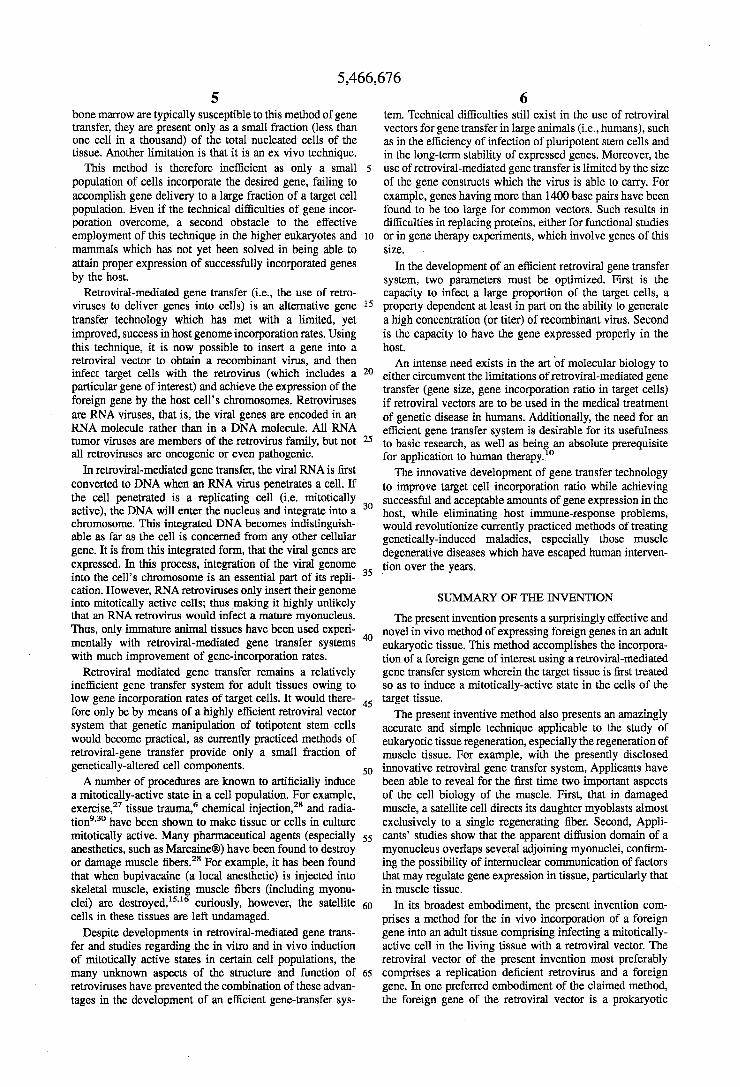
FIG. 1 A
US. Patent Nov. 14,1995 Sheet 2 of 2 5,466,676
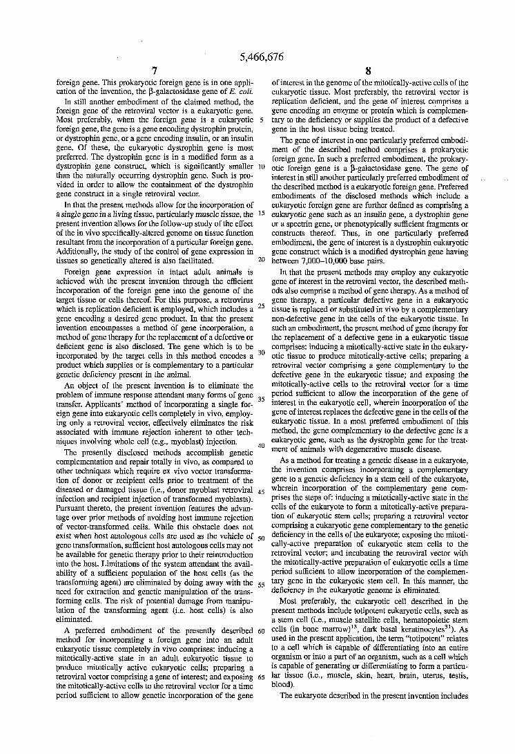
FIG, 1 C
FIG, I D
5,466,676 1 2
SATELLITE CELL PROLIFERATION IN ADULT SKELETAL MUSCLE
The insertion of foreign genes or DNA into an organism’s genome has become a powerful tool in experimental biology during the past decade. This modification of genetic infor-
The United States government may own rights in the mation has been attempted via the development of a variety present invention pursuant to NASA grant NAG2-239 5 of experimental protocols. Many experiments have trans- (F.W.B.), PHS grant AB19393 (F.W.B.1, and postdoctoral ferred genes into mammalian cells in culture and into newly training grant DK07520 (D.B.T.). fertilized mammalian eggs that then develop into an intact
The present application is a continuation of U.S. Ser. No. animal. L~~~ commo,.,ly, have been performed 07/479,065, filed Feb. 12, 1990, now abandoned. .^ wherein foreign DNA is inserted into the genome of cells of
young or adult animals in situ. The insertion of foreign DNA into the genome of a cell or embryonic tissue has been
I U
BACKGROUND OF THE INVENTION
1. Field of the Invention postulated to be applicable in the treatment of genetic The present invention relates to the field of molecular diseases in higher e*Wotes, PmiCularlY in h n a n s . kkth-
genetics and its use in the preparation of therapeutic agents 15 ods currently under extensive study in the manipulation of effective in vivo in adult tissue. More particularly, stem cells genes include in vitro DNA transfection techniques, in vivo are genetically modified in vivo and then function to induce whole cell injection (i.e., hematopoetic and myoblast cells), regeneration and repair of damaged tissue throughout the and in vivo as well as in vitro retroviral-mediated gene life span of an animal. More specifically, the present inven- transfer. ~
tion relates to the retroviral-mediated transfer of marker 20 genes into adult tissue in which a mitotically-active state of
The insertion of foreign genes into an organism’s genome thus presents a potentially revolutionary method for treating
satellite cells has been induced. Additionally, the present invention relates to methods of
treating genetically-transmitted diseases. The invention also relates to methods and therapeutic treatInentS for regener- 25 sting damaged ~ ~ c l e tissuey as a PmiCUlar method for muscle regeneration in Persons With r~~ScUlar dystrophy is disclosed.
genetic-defect tractable diseases in man. However, many of these genetic diseases remain to be more fully characterized before gene therapy can be used in their treatment.
genetic-defect diseases identified in the literature include, by way of example, the various forms of muscular dystrophy (see Table l), diabetes, hemophilia and albinism. The vari- ous forms of muscular dystrophic diseases appear below.
Some of the more fully characterized ecamples of
2. Description of Related Art
TABLE 1
The Progressive Muscular Dystrophies
Pattern of Muscular
Type of Dystrophy ~ Age of Onset Involvement Special Features CELevel Inheritance
Duchenne
Becker
Emery-Dreifuss
Landouzy-Dejerine
Scapulohumeral of Seitz
Limb-girdle (Erb)
von Graefe- Fuchs
Oculophqngeal
Infancy or early childhood
Childhood, adolescence, or early adult
Childhood, adolescence
Late Childhood, adolescence girdle-late Childhood, or adult
Childhood, adolescence, sometimes adult Childhood, adolescence
Middle or late adult
Pelvifemod, later pectoral girdle
Pelvifemoral, later pectoral girdle
Humeroperoneal
Facioscapulo- humera pelvic normal Spinal and humeral, later pelvic girdle girdle-late Pectoral or pelvic or both
ocular (sparing pupils); later facial and other muscles (slight)
Levators of lids; other ocular-
~
Hypemophy- pseudohyper- trophy; cardiac involvement; mental retardation cardiac involvement, sight; mentation normal Contractures posterior neck and biceps muscles Heart normal, mentation
Cardiomyopathy
Heart usually normal, mentation normal Kearns-Sap group have retinitis pigmentosa heart block, stunting of growth, and ovarian dysgenesis
High
~
Moderate
Moderate ~
Slight to moderate
Slight to moderate
Slight to moderate
Slight to moderate
Slight to normal
X-linked recessive
X-linked recessive
X-linked recessive
Dominant
Dominant
Variable, recessive, or dominant
Dominant; Kearne-Sayre recessive
Dominant
5,466,676 3 4
TABLE I -continued
The Progressive Muscular Dystrophies
Pattern of Muscular
Type of Dystrophy Age of Onset Involvement Special Features CK Level Inheritance
P h W W a l muscles later
Myotonic Infancy, Ocular, facial, Cataracts, Slight to Dominant (Steinert) childhood, Stemomastoid, testicular normal
adult forearm, aaophy peroneal
pelvic girdles, retardation, moderate or recessive or difFuse arthrogryposis
Congenital Birth, Infancy Pectoral and Mental Slight to Dominant
Among the many genetic diseases described, the muscle degenerative, or muscular dystrophy-causing diseases, have experienced significant recent technological advances. For example, the muscular dystrophies have been traced to particular gene defects, the elucidation of which has expe- rienced particularly significant scientific breakthroughs in terms of genetic molecular characterization. The most widely known of the muscular dystrophies are Duchenne’s muscular dystrophy and the less severe Becker muscular dystrophy. Both of these genetic diseases are characterized by an inability of the muscle to produce dystrophin, which is a muscle protein. This defect is an x-linked recessive disease potentially caused by a defect in the dystrophin gene. The dystrophin gene has most recently been established as having a close relative gene on human chromosome 624.
Prior studies have attempted the use of foreign normal myoblast injection as a form of gene product replacement. For example, foreign myoblasts containing a normal dys- trophin gene have been injected into dystrophic tissue to invoke the expression of the dystrophin protein in the muscle tissue. However, this method presents the inherent risk of immune rejection, as well as the necessity of injection at multiple, probably closely-spaced sitesz3. Additionally, injection at multiple sites is necessary with such a therapy because dystrophin, like other muscle proteins, tends to remain localized within a single fiber, close to the nuclei from which it was derived. An additional limitation of currently practiced myoblast-injection techniques is the low fusion rate of implanted myoblasts into normal host muscle tissues.
Muscle tissue is characterized by the presence of satellite cells, which are located between the basal lamina and the sarcolemma of the skeletal muscle fibers. Satellite cells in skeletal muscle are dormant stem cells which are present outside the muscle fibers in an adult skeletal muscle. Each of these inactive stem cells can either proliferate into many satellite cells or mature into an embryonic myoblast upon the appropriate stimulation.
By way of example, satellite cells of the muscle have been shown to become stimulated to replicate by muscle damage (Allbrook, 1981; Carlson et al., 1983). It is also possible that strenuous exercise may stimulate cells to become mitotically active and to repli~ate.’~ Chemical damage via bupivacaine injection into mammalian skeletal muscle cells has been shown to result in rapid recovery of the tissue showing maximum proliferative capacity of satellite cells 36-48 hours following damage6.14. As stem cells, satellite cells function to supply myonuclei to growing fibers in immature animals as well as to provide myogenic cells for muscle regeneration and repair throughout the life of the animal.
Several laboratories have published protocols by which the hematopoietic cells of mice have had new genes intro- duced with retroviral vectors ex vivo, with subsequent
2o reintroduction of the treated cells into the bone marrow in vivozz. In this system, marrow cells are obtained from a donor and incubated with a monolayer of vector-generating producer cells. Since marrow cells, unlike the producer cells, do not attach to the culture disk, they are easily recovered
25 after co-cultivation. The hematopoietic cells are then intro- duced into a recipient animal by intravenous injection. Space in the hematopoietic system to receive the vector- treated marrow must be made usually by lethally irradiating the recipient. While this technique has been widely used
30 experimentally, its use in humans is limited by the recurrent risk of host immune rejection, as current studies on muscle were performed on mdx (immunosuppressed) animalsz3. Additionally, low proportion of cells incorporate the normal myoblast dystrophin gene in myoblast transfer procedures.
35 Moreover, multiple injections of myoblasts are typically required to achieve proper dispersion of the myoblasts in situ.
A need thus remains in the art for the development of a 4o therapeutic system which minimizes or eliminates host
immune response, perhaps through the development of an entirely in vivo vector-induced, host cell gene incorporation process. However, technical difficulties in the manipulation of genomes from organisms both in achieving the initial
45 incorporation of the desired gene by the host cells as well known as achieving expression of those genes in the host have limited techniques of direct incorporation of a gene without a “carrier” cell into damaged tissue in vivo. DNA transfection and retroviral mediated gene incorporation are
5o two methods of such a “direct gene” incorporation which eliminates the need for a “carrier” cell.
The treatment of genetically-related diseases with tech- niques as DNA transfection has thus far, unfortunately, not met with great success. In DNA transfection, DNA (presum-
55 ably which includes a non-defective counterpart of the defective gene) is introduced into cells in culture as part of a coprecipitate with calcium phosphate or dextran sulfate.’ A successful result is a viable cell containing one to many copies of the new gene which continuously expresses the
While several limitations exist in this system, the most significant limitation is that it is a very inefficient means of transferring genes into mammalian cells. For example, only one in a thousand cells (more typically, one cell in a million)
65 will incorporate the newly transformed gene. Additionally, not all cultured cell lines are susceptible to this method of gene transfer. For example, while the stem cells located in
60 new genetic information.
5,466,676 5 6
bone marrow are typically susceptible to this method of gene tem. Technical difficulties still exist in the use of retroviral transfer, they are present only as a small fraction (less than vectors for gene transfer in large animals (i.e., humans), such one cell in a thousand) of the total nucleated cells of the as in the efficiency of infection of pluripotent stem cells and tissue. Another limitation is that it is an ex vivo technique. in the long-term stability of expressed genes. Moreover, the
This method is therefore inefficient as only a small 5 use of retroviral-mediated gene transfer is limited by the size population of cells incorporate the desired gene, failing to of the gene constructs which the virus is able to carry. For accomplish gene delivery to a large fraction of a target cell example, genes having more than 1400 base pairs have been population. Even if the technical difficulties of gene incor- found to be too large for common vectors. Such results in poration overcome, a second obstacle to the effective difficulties in replacing proteins, either for functional studies employment of this technique in the higher eukaryotes and 10 or in gene therapy experiments, which involve genes of this mammals which has not yet been solved in being able to size. attain proper expression of successfully incorporated genes In the development of an efficient retroviral gene transfer by the host. system, two parameters must be optimized. First is the
Retroviral-mediated gene transfer (ix., the use of retro- capacity to infect a large proportion of the target cells, a viruses to deliver genes into cells) is an alternative gene 15 property dependent at least in part on the ability to generate transfer technology which has met with a limited, yet a high concentration (or titer) of recombinant virus. Second improved, success in host genome incorporation rates. Using is the capacity to have the gene expressed properly in the this technique, it is now possible to insert a gene into a host. retroviral vector to obtain a recombinant virus, and then An intense need exists in the art of molecular biology to infect target cells with the retrovirus (Which includes a *O either circumvent the limitations ofretroviral-mediated gene particular gene of interest) and achieve the expression ofthe transfer (gene size, gene incorporation ratio in target cells) foreign gene by the host cell's chromosomes. Retroviruses if retroviral vectors are to used in the medical treatment are RNA v h s e s , that is, the viral genes are encoded in an of genetic disease in humans. Additionally, the need for an RNA ~ o l e c u l e rather than in a DNA molecule. All RNA efficient gene transfer system is desirable for its usefulness hmor viruses are n ~ m b e r s of the retrovirus family, but not 25 to basic research, as well as being an absolute prerequisite all retroviruses are oncogenic or even pathogenic. for application to human therapy. lo
In retroviral-mediated gene transfer, the viral RNA is first The innovative development of gene transfer technology converted to DNA when an RNA virus penetrates a cell. If to improve target cell incorporation ratio while achieving the Cell penetrated is a replicating cell mitotically successful and acceptable amounts of gene expression in the active), the DNA will enter the nucleus and integrate into a 30 host, while eliminating host immme-response problems, chromosome. This integrated DNA becomes indistinguish- would revolutionize currently practiced methods of treating able as far as the cell is concerned from any other cellular genetically-induced maladies, especially those muscle gene. It is from this integrated form, that the Viral genes *e degenerative diseases which have escaped human interven- expressed. In this process, integration of the viral genome tion over the years. into the cell's chromosome is an essential part of its repli- 35 cation. However, RNA retroviruses only insert their genome into mitotically active cells; thus making it highly unlikely that an RNA retrovirus would infect a mature myonucleus. The present invention presents a surprisingly effective and Thus7 O d Y immature animal tissues have been used eXPeri- novel in vivo method of expressing foreign genes in an adult m e n a y With n%rOViral-m?&ited gene transfer systems 40 eukaryotic tissue. This method accomplishes the incorpora- with much improvement of gene-incorporation rates. tion of a foreign gene of interest using a retroviral-mediated
Retroviral mediated gene transfer remains a relatively gene transfer system wherein the target tissue is first treated inefficient gene transfer system for adult tissues owing to so as to induce a mitotically-active state in the cells of the low gene incorporation rates of target cells. It would there- 45 target tissue. fore only be by means of a highly efficient retroviral vector The present inventive method also presents an amazingly system that genetic maniPUlatiOn Of totipotent Stem Cells accurate and simple technique applicable to the study of would b ~ o m e practical, as currently practiced methods of eukaryotic tissue regeneration, especially the regeneration of retroviral-gene transfer Provide only a Small fraction of muscle tissue. For example, with the presently disclosed genetically-altered cell components. 50 innovative retroviral gene transfer system, Applicants have
A number of procedures are known to artificially induce been able to reveal for the first time two important aspects a mitotically-active state in a cell population. For example, of the cell biology of the muscle. First, that in damaged e~ercise, '~ tissue traumq6 chemical injection:' and radia- muscle, a satellite cell directs its daughter myoblasts almost tiongS3' have been shown to make tissue or cells in culture exclusively to a single regenerating fiber. Second, Appli- mitotically active. Many pharmaceutical agents (especially 55 cants' studies show that the apparent diffusion domain of a anesthetics, such as MarcaineB) have been found to destroy myonucleus overlaps several adjoining myonuclei, confirm- or damage muscle fibers." For example, it has been found ing the possibility of internuclear communication of factors that when bupivacaine (a local anesthetic) is injected into that may regulate gene expression in tissue, particularly that skeletal muscle, existing muscle fibers (including myonu- in muscle tissue. Clei) are In its broadest embodiment, the present invention com- cells in these tissues are left undamaged. prises a method for the in vivo incorporation of a foreign
Despite developments in retroviral-mediated gene trans- gene into an adult tissue comprising infecting a mitotically- fer and studies regarding the in vitro and in vivo induction active cell in the living tissue with a retroviral vector. The of mitotically active states in certain cell populations, the retroviral vector of the present invention most preferably many unknown aspects of the structure and function of 65 comprises a replication deficient retrovirus and a foreign retroviruses have prevented the combination of these advan- gene. In one preferred embodiment of the claimed method, tages in the development of an efficient gene-transfer sys- the foreign gene of the retroviral vector is a prokaryotic
SUMMARY OF THE INVENTION
CuriOuSlY, however, the satellite 60
5,466,676 7 8
foreign gene. This prokaryotic foreign gene is in one appli- of interest in the genome of the mitotically-active cells of the cation of the invention, the P-galactosidase gene of E. coli. eukaryotic tissue. Most preferably, the retroviral vector is
In still another embodiment of the claimed method, the replication deficient, and the gene of interest comprises a foreign gene of the retroviral vector is a eukaryotic gene. gene encoding an enzyme or protein which is complemen- Most preferably, when the foreign gene is a eukaryotic 5 tary to the deficiency or supplies the product of a defective foreign gene, the gene is a gene encoding dystrophin protein, gene in the host tissue being treated. or dystrophin gene, or a gene encoding insulin, or an insulin The gene of interest in one particularly preferred embodi- gene. Of these, the eukaryotic dYstroPhin gene is most ment of the described method comprises a prokaryotic Preferred. The dYstroPhin gene is in a nm3fied form as a foreign gene. In such a preferred embodiment, the prokary- dYstroPhin gene construct, which is Significantly smaller 10 otic foreign gene is a P-galactosidase gene. The gene of than the naturally occurring dYstroPhin gene. such is Pro- interest in still another particularly preferred embodiment of vided in order to allow the containment of the dystrophin the described method is a eukaryotic foreign gene. Preferred gene construct in a single retroviral vector. embodiments of the disclosed methods which include a
In that the present methods allow for the incorporation of eukaryotic foreign gene are further defined as comprising a a single gene in a living tissue, particularly muscle tissue, the 15 eukaryotic gene such as an insulin gene, a dystrophin gene present invention allows for the follow-up study of the effect or a spectrin gene, or phenotypically sufficient fragments or of the in vivo specifically-altered genome on tissue function constructs thereof. Thus, in one particularly preferred resultant from the incorporation of a particular foreign gene. embodiment, the gene of interest is a dystrophin eukaryotic Additionally, the study of the control of gene expression in gene construct which is a modified dystrophin gene having tissues so genetically altered is also facilitated.
Foreign gene expression in intact adult animals is In that the present methods may employ any eukaryotic achieved with the present invention through the efficient gene of interest in the retroviral vector, the described meth- incorporation of the foreign gene into the genome of the ods also comprise a method of gene therapy. As a method of target tissue or cells thereof. For this purpose, a retrovirus gene therapy, a particular defective gene in a eukaryotic which is replication deficient is employed, which includes a 25 tissue is replaced or substituted in vivo by a complementary gene encoding a desired gene product. In that the present non-defective gene in the cells of the eukaryotic tissue. In invention encompasses a method of gene incorporation, a such an embodiment, the present method of gene therapy for method of gene therapy for the replacement of a defective or the replacement of a defective gene in a eukaryotic tissue deficient gene is also disclosed. The gene which is to be Comprises: inducing a mitotically-active state in the eukary- incorporated by the target cells in this method encodes a 30 otic tissue to produce mitotically-active cells; preparing a product which supplies or is complementary to a particular retroviral vector comprising a gene complementary to the genetic deficiency present in the animal. defective gene in the eukaryotic tissue; and exposing the
An object of the present invention is to eliminate the IllkOtiCally-aCtiVe Cells to the retrOVkd Vector for a time problem of immune response attendant many forms of gene 35 Period sufficient to allow the incorporation of the gene of transfer. Appficantsi method of incorporating a single for- interest in the eukaryotic cell, wherein incorporation Of the e i p gene into eukaryotic cells completely in vivo, employ- gene of interest replaces the defective gene in the cells of the ing only a retrovkal vector, effectively eliminates the risk eukaryotic tissue. In a most preferred embodiment of this associated with immune rejection inherent to other tech- method, the gene complementary to the defective gene is a niques involving whole cell (e.g., myoblast) injection. eukaryotic gene, such as the dystrophin gene for the treat-
complementation and repair totally in vivo, as compared to As a method for treating a genetic disease in a eukaryote, other techniques which require ex vivo vector transforma- the invention comprises incorporating a complementary tion of donor or recipient cells prior to treatment of the gene to a genetic deficiency in a stem Cell of the eukaryote, diseased or damaged tissue (i.e., donor myoblast retroviral 45 wherein incorporation of the complementary gene corn- infection and recipient injection of transformed myoblasts). PriSeS the Steps Of: inducing a mitotically-active state in the Pursuant thereto, the present invention features the advan- cells of the eukaryote to form a mitoticallY-active PrePara- tage over prior methods of avoiding host immune rejection tion of eukaryotic stem Cells; Preparing a retroviral vector of vector-transformed cells. While this obstacle does not comprising a eukaryotic gene complementary to the genetic exist when host autologous cells are used as the vehicle of 50 deficiency in the Cells ofthe eukaryote; exposing the mitoti- gene transformation, sufficient host autologous cells may not CallY-active Preparation of eukaryotic Stem cells to the be available for genetic therapy prior to their reintroduction retrOViral Vector; and incubating the retroviral vector with into the host. Limitations of the system attendant the avail- the mitotically-active preparation Of eUkaryOtiC Cells a time ability of a sufficient population of the host cells (as the Period sufficient to allow incoWration of the ComPlemen- transforming agent) are eliminated by doing away with the 55 t W gene in the eukaryotic Stem cell. In this manner, the need for extraction and genetic manipulation of the trans- deficiency in the eukaryotic genome is eliminated. forming cells. The risk of potential damage from manipu- Most preferably, the eukaryotic cell described in the lation of the transforming agent (i.e. host cells) is also present methods include totipotent eukaryotic cells, such as eliminated. a stem cell (Le., muscle satellite cells, hematopoietic stem
A preferred embodiment of the presently described 60 cells (in bone mm0w)’3, dark basal keratinocYtes31). As method for incorporating a foreign gene into an adult used in the present application, the term “totipotent” relates eukaryotic tissue completely in vivo comprises: inducing a to a cell which is capable of differentiating into an entire mitotically-active state in an adult eukaryotic tissue to Organism or into a part of an organism, such as a cell which produce mitotically active eukaryotic cells; preparing a is capable of generating or differentiating to form a particu- retroviral vector comprising a gene of interest; and exposing 65 lar tissue (i.e., muscle, skin, heart, brain, uterus, testis, the mitotically-active cells to the retroviral vector for a time blood). period sufficient to allow genetic incorporation of the gene The eukaryote described in the present invention includes
20 between 7,000-10,000 base pairs.
The presently disclosed accomplish genetic 40 ment of animals with degenerative muscle disease.
5,466,676 9
all organisms comprising cells which contain a membrane- bound nucleus. In particularly preferred embodiments of the described methods, the eukaryotic tissue described in rela- tion to the disclosed methods is human tissue, rat tissue or mouse tissue.
In the more preferred embodiments of the described methods, a mitotically-active state in a living eukaryotic cell or tissue comprises inducing cellular-repair mechanisms in the cell or tissue. Thus, the induction of a mitotically-active state comprises discomposing the tissue, exposing the cell to radiation, or administering a pharmaceutical chemical agent. As used in this application, the term discompose relates to any change in the normal resting state or non-mitotically active state of a eukaryotic cell culture or tissue, particularly that of a eukaryotic tissue. By way of example, discompo- sition of a tissue may be accomplished through vigorous exercise stimulation’, agitation, surgical intrusion or other trauma of the tissue or cell culture.
The use of chemicals to induce a mitotically-active state in the cell (Le., in cell culture) or tissue described includes the administration of any of a variety of pharmaceutical agents. By way of example, the pharmaceutical agents capable of inducing a mitotically-active state in vitro or in vivo include bupivacaine, collagenase, dexamethasone, fibroblast growth factor, and any other reagent capable of inducing a mitotically-active state in a eukaryotic tissue or a eukaryotic cell.
The retroviral vector is a retrovirus (RNA virus) which is replication deficient or replication incompetent. That is to say, the retrovirus lacks one or more of the replication genes, gag (group-specific antigen), pol (polymerase) or env (enve- lope) protein encoding genes.
The gene therapy of the present invention comprises a method by which any of a variety of genetic diseases may be eliminated. The invention thus comprises a method of treat- ing genetic disease. Such is accomplished through the suc- cessful incorporation of a particular complementary gene or part of a complimentary gene in the genome of totipotent cells of the diseased organism, thereby facilitating the pro- duction of cells in the diseased host with similarly corrected genomes. By way of example, genetic diseases which may be treated employing the described methods include diabe- tes, albinism, and the various forms of muscular dystrophy. However, any recessive gene is hypothesized to be treatable employing the described methods of gene transfer and gene replacement.
In a most preferred embodiment, a method for treating the genetic disease of muscular dystrophy is described. Muscu- lar dystrophy is a genetic disease characterized by a defec- tive gene encoding the muscle protein, dystrophin. In this particular embodiment of the invention, the gene of interest would be a part of the dystrophin gene (i.e., a dystrophin gene construct) which encodes a part, segment or fragment of the dystrophin protein, this particular protein segment supplying a sufficient part of the native dystrophin protein to protect the eukaryotic tissue from all but only the mildest phenotypic manifestation of muscle degeneration. This par- ticular dystrophin gene fragment is described by England et aLZ0
The spectrin gene, which encodes a muscle protein simi- lar to dystrophin, is also hypothesized to be efective in the methods described herein for the treatment of muscular dystrophy. More particularly, a retroviral vector which includes a spectrin gene or a fragment of the spectrin gene may be effective in treating muscular dystrophy through the injection of such a retroviral vector into a eukaryotic tissue,
5
10
15
20
25
30
35
40
45
50
55
60
65
10 the eukaryotic tissue being treated so as to induce a mitoti- cally active state therein. Various methods for inducing such a mitotically active state are as otherwise described herein (radiation, trauma, exercise, chemical injection].
The retroviral vector in one preferred embodiment of the present invention would include a dystrophin gene construct which encodes a particular dystrophin protein fragment which is effective to halt massive or significant muscle degeneration, as well as to regenerate already damaged muscle tissue. In a most preferred embodiment of the invention for the treatment of muscular dystrophy, the retroviral vector includes a dystrophin gene construct com- prising between 7,000-10,OOO base pairs. In an even more preferred embodiment, the dystrophin gene construct com- prises about 9,000 base pairs of the native dystrophic gene. This dystrophin gene construct encodes a protein having a relative molecular weight of about 200,000 K.
As a method of treating muscular dystrophy in a human or other eukaryotic organism, the present invention in a most preferred embodiment comprises the initial induction of a mitotically-active state in the tissue of the eukaryote through the intramuscular administration of bupivacaine to the skel- etal muscle tissue. The retroviral vector of this particular embodiment of the invention comprises a replication defec- tive murine leukemia virus. Most preferably, the murine leukemia virus comprises AKR, Moloney or Friend leuke- mia virus. However, most preferably, the retroviral vector comprises the replication defective Moloney murine leuke- mia virus.
In still another embodiment of the present invention, the retroviral vector comprises a prokaryotic gene, such as a P-galactosidase gene, wherein the P-galactosidase gene is under the control of the retroviral 5’ long terminal repeat promoter region. More particularly, the P-galactosidase gene is under the control of a constitutive promote?. A method for monitoring gene expression in tissue is thereby provided (i.e., focal and diffuse expression). For example, as employed to infect muscle tissue in eukaryotes, Applicants propose that the use of such a vector system would track regenerating and fully recovered muscle fibers, in that regenerating and fully recovered muscle fibers would express the P-galactosidase gene product if the infected satellite cells gave rise to viable myoblasts.
The disclosed methods are not to be limited to treatment of muscular degenerative diseases. For example, the gene of interest in the present invention may comprise any gene or part of any gene which complements a deficiency in the animal being treated. However, length of the gene of inter- est, or the coding region thereof, is preferably less than 14,000 base pairs so as to facilitate the inclusion of the particular gene in a single retrovirus.
Use of the presently described methods to stimulate stem cell proliferation allows the transformation of the genome of, in one particular embodiment, skeletal muscle satellite cells, resulting in the production of genetically-corrected myoblasts. These myoblasts then become the non-defective myonuclei of newly formed and non-defective muscle fibers. As will be appreciated, the invention in such an application comprises a method for regenerating muscle tissue. I n a most preferred embodiment, this technique comprises a method of treating degenerated dystrophic muscle tissue. In this preferred embodiment, the regenera- tion of dystrophic muscular tissue, characteristic of such diseases as Duchenne’s muscular dystrophy and Becker muscular dystrophy, is specifically envisioned.
As a therapeutic treatment for muscular dystrophy, Appli-
5,466,676 11
cants have found that the technique of stimulating stem cell proliferation to allow retroviral-mediated gene transfer is generally applicable to all eukaryotic tissues. Potential target cell populations for use with the present methods are char- acterized by the presence of stem cells. By way of example, tissues expected to be susceptible to the described treatments and methods are those tissues which include stem cells. By way of example, these stem-cell containing tissues include those tissues of the uterus, skeletal muscle, liver, kidney, blood, alimentary epithelium, testes, skin and gastrointesti- nal tissue. The stem cells of muscle tissue are the satellite cells. In the described methods, the stem cells &e., satellite cells) in muscle tissue are the cells which are infected with the retroviral vector to facilitate incorporation of the eukary- otic gene of interest in the satellite cell itself. These satellite cells may then function to supply myonuclei to growing fibers in immature animals and to provide myogenic cells for muscle regeneration and repair throughout the life of an animal.
In that any of the methods described in the present application may be used in vivo, the retroviral vector may be further defined as comprising a retroviral vector conjugated to a protein. Such may be accomplished by methods well known to those of skill in the art, and will function to enhance the targeting of the vector to particular populations of target cells.
The following abbreviations are used throughout the Specification:
CFW=colony forming units ug=microgram EGTA=methylene glycol tetraacetic acid k b e t a um=micromolar P-galactosidasebeta-galactosidase (EC 3.2.1.23) MoLV=Moloney murine leukemia virus
BRIEF DESCRIPTION OF THE DRAWINGS
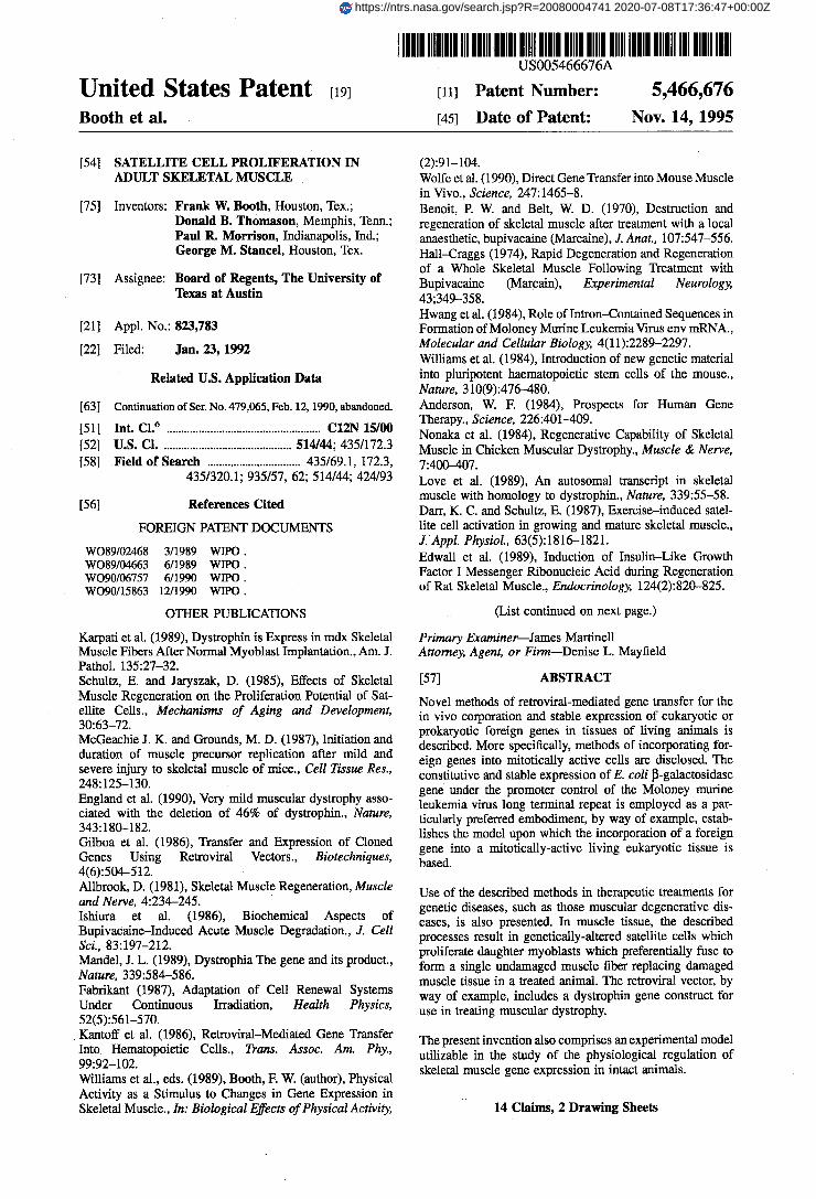
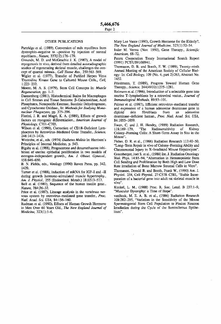
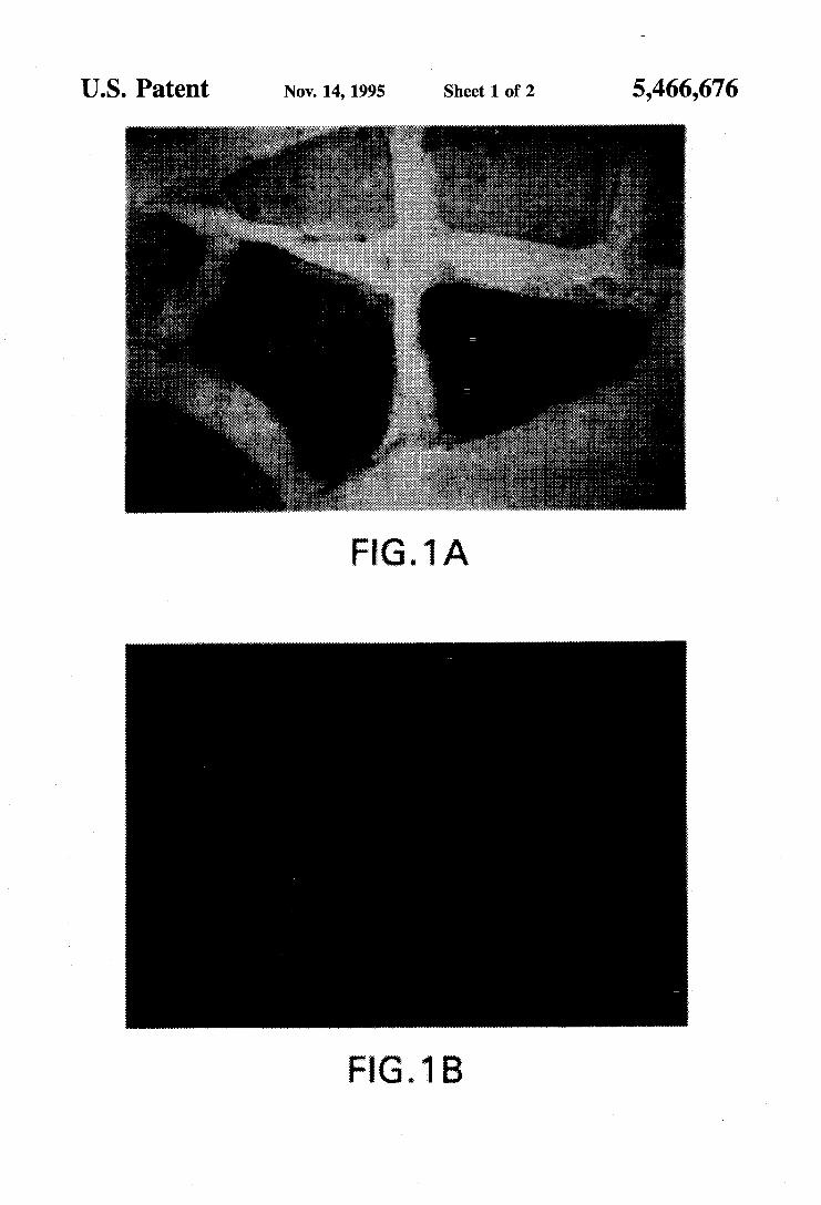
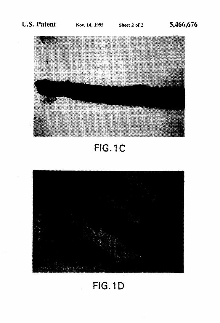
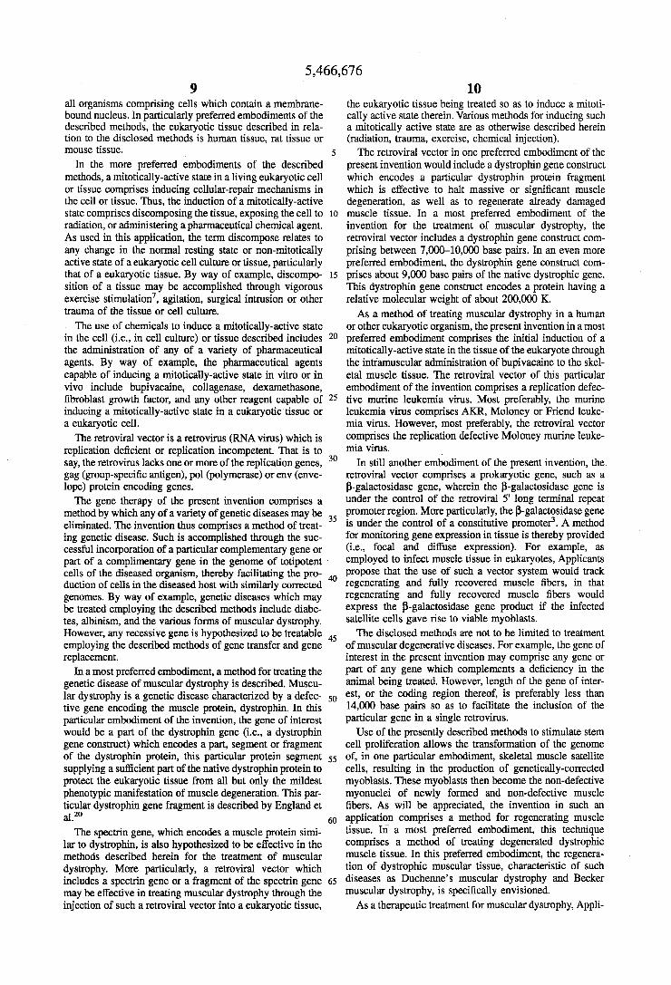
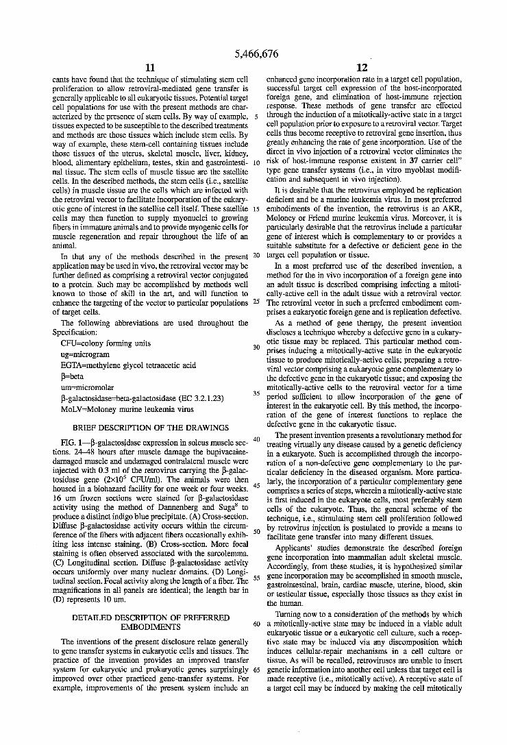
FIG. 1-P-galactosidase expression in soleus muscle sec- tions. 24-48 hours after muscle damage the bupivacaine- damaged muscle and undamaged contralateral muscle were injected with 0.3 ml of the retrovirus carrying the p-galac- tosidase gene (2x10⁵ CFU/ml). The animals were then housed in a biohazard facility for one week or four weeks. 16 um frozen sections were stained for P-galactosidase activity using the method of Dannenberg and Suga’ to produce a distinct indigo blue precipitate. (A) Cross-section. Diffuse P-galactosidase activity occurs within the circum- ference of the fibers with adjacent fibers occasionally exhib- iting less intense staining. (B) Cross-section. More focal staining is often observed associated with the sarcolemma. (C) Longitudinal section. Diffuse P-galactosidase activity occurs uniformly over many nuclear domains. (D) Longi- tudinal section. Focal activity along the length of a fiber. The magnifications in all panels are identical; the length bar in (D) represents 10 um.
DETAILED DESCRIPTION OF PREFERRED EMBODIMENTS
The inventions of the present disclosure relate generally to gene transfer systems in eukaryotic cells and tissues. The practice of the invention provides an improved transfer system for eukaryotic and prokaryotic genes surprisingly improved over other practiced gene-transfer systems. For example, improvements of the present system include an
12 enhanced gene incorporation rate in a target cell population, successful target cell expression of the host-incorporated foreign gene, and elimination of host-immune rejection response. These methods of gene transfer are effected through the induction of a mitotically-active state in a target cell population prior to exposure to a retroviral vector. Target cells thus become receptive to retroviral gene insertion, thus greatly enhancing the rate of gene incorporation. Use of the direct in vivo injection of a retroviral vector eliminates the risk of host-immune response existent in 37 carrier cell” type gene transfer systems (ix., in vitro myoblast modifi- cation and subsequent in vivo injection).
It is desirable that the retrovirus employed be replication deficient and be a murine leukemia virus. In most preferred
15 embodiments of the invention, the retrovirus is an AKR, Moloney or Friend murine leukemia virus. Moreover, it is particularly desirable that the retrovirus include a particular gene of interest which is complementary to or provides a suitable substitute for a defective or deficient gene in the
20 target cell population or tissue. In a most preferred use of the described invention, a
method for the in vivo incorporation of a foreign gene into an adult tissue is described comprising infecting a mitoti- cally-active cell in the adult tissue with a retroviral vector.
25 The retroviral vector in such a preferred embodiment com- prises a eukaryotic foreign gene and is replication defective.
As a method of gene therapy, the present invention discloses a technique whereby a defective gene in a eukary- otic tissue may be replaced. This particular method com-
30 prises inducing a mitotically-active state in the eukaryotic tissue to produce mitotically-active cells; preparing a retro- viral vector comprising a eukaryotic gene complementary to the defective gene in the eukaryotic tissue; and exposing the mitotically-active cells to the retroviral vector for a time
35 period sufficient to allow incorporation of the gene of interest in the eukaryotic cell. By this method, the incorpo- ration of the gene of interest functions to replace the defective gene in the eukaryotic tissue.
The present invention presents a revolutionary method for treating virtually any disease caused by a genetic deficiency in a eukaryote. Such is accomplished through the incorpo- ration of a non-defective gene complementary to the par- ticular deficiency in the diseased organism. More particu- larly, the incorporation of a particular complementary gene comprises a series of steps, wherein a mitotically-active state is first induced in the eukaryote cells, most preferably stem cells of the eukaryote. Thus, the general scheme of the technique, i.e., stimulating stem cell proliferation followed
5o by retrovirus injection is postulated to provide a means to facilitate gene transfer into many different tissues.
Applicants’ studies demonstrate the described foreign gene incorporation into mammalian adult skeletal muscle. Accordingly, from these studies, it is hypothesized similar
55 gene incorporation may be accomplished in smooth muscle, gastrointestinal, brain, cardiac muscle, uterine, blood, skin or testicular tissue, especially those tissues as they exist in the human.
W i n g now to a consideration of the methods by which 60 a mitotically-active state may be induced in a viable adult
eukaryotic tissue or a eukaryotic cell culture, such a recep- tive state may be induced via any discomposition which induces cellular-repair mechanisms in a cell culture or tissue. As will be recalled, retroviruses are unable to insert
65 genetic information into another cell unless that target cell is made receptive (i.e., mitotically active). A receptive state of a target cell may be induced by making the cell mitotically
5
40
45
5,466,676 13
active. A variety of techniques are available which induce such a mitotically-active state. By way of specific example, such includes the irradiation of the tissue or cell culture, the exposure of the cells or tissue to particular pharmaceutical agents, the physical agitation of a cell culture, or the surgical intrusion of a tissue, as well as through the vigorous exercise of a tissue.
Among those pharmaceutical agents expected to be effec- tive in inducing the described mitotic or active state in cell culture or in vivo, the following list presents those agents most particularly preferred: collagenase, fibroblast growth factor, bupivacaine, estrogen and dexamethasone.
The following tissue sectioning and staining protocol represents a method which may generally be used in the fixation of any type of tissue from any of a variety of animal species. Several experimental examples follow thereafter which are designed to illustrate particularly preferred embodiments of the inventions, both as they actually exist and as they are proposed to exist in the future (Le., prophetic exemplary use in treating genetic disease in humans).
It should be appreciated that many modifications and changes can be made in the particular stimulatory agents and their doses, the particular retroviral construct (i.e., the pro- moter, the gene of interest, and the particular retrovirus) and the particular conditions under which the retrovirus intro- duced to a culture of cells or into a living adult tissue without departing from the spirit and scope of the invention.
Tissue Sectioning and Staining Muscle sections were stained by the method of Dannen-
berg and Suga.' Briefly, 16 um sections were mounted on 0.5% gelatin slides and fixed 5-10 minutes at 4" C. with the same fixative as used in the dissection. The sections were washed briefly with PBS containing 2 mM MgCl, at 4" C. and once again for 10 minutes with the same solution. The sections were then washed for 10 minutes in PBS containing 2 mM MgCl,, 0.01% sodium deoxycholate, and 0.02% Nonidet P40 at 4" C. The slides were briefly dried and stained for 18 hours at 37" C. in a PBS solution containing 35 mM K,Fe(CN),, 35 mM K4Fe(CN),.3H,0, 2 mM MgCl,, 0.01% sodium deoxycholate, 0.02% Nonidet P4, and 1 mg/ml 5-bromo-4-chloro-3-indoyl-b-D-galactopyra- noside (Xgal). Following staining the slides were rinsed three times for 5 minutes each with PBS at room tempera- ture, blotted dry, and stained with 5% eosin.
Applicants are in the process of replacing the P-galac- tosidase gene with the rat insulin-like growth factor I coding region, using the following strategy. The entire P-galactosi- dase gene was removed from the gag region of the retroviral "BAG" construct of Price, et al.? by restriction with Pvu I and BamHI. The insulin-like growth factor I coding region will be added to the gag site where P-galactosidase was removed as follows. Applicants were supplied with prIGF- Ib-42-a, a 422 bp insert in pUC-19, which included a partial 5' untranslated region with 2 putative ATG translation start sites, the Pre, B, C , A, D and part of the E domains of the rat insulin like growth factor 1 coding region, without introns. The processed mature insulin-like growth factor I peptide did not contain the E domain. The 422-bp insulin- like growth factor I coding region was flanked by EcoRI restriction sites.
BL-2 tissue culture facility An 80 ftz room was devoted solely to culture of the Psi2
packing cell line and 3T3 cells, which were used to prove that the produced retrovirus is replication-incompetent, that is no recombinant wild-type retrovirus is produced. A BL-2 rated laminar flow hood (Nuaire 425400) and a CO,
5
10
15
20
25
30
35
40
45
50
55
60
65
14 H,O-jacketed incubator (Nuaire) was also used in the . following studies. An Olympus CK2 inverted scope with phase contrast was available outside the culture facility.
Laboratory facilities Two cryostats (IEC minotome) were available for pro-
duction of tissue sections. An automatic microtome knife sharpener was also available for usage. Applicants' labora- tory has an Olympus BH-2 microscope with attached mOS color video camera so that a slide may be viewed simulta- neously through the binocular observation tube and on a video color monitor. This system is attached to a PC Vision Plus Frame Grabber Board in an AT-clone computer. Appli- cants have a JAVA image-processing program which permits the determination of relative area of indigo blue on a tissue section.
Applicants' laboratory also contains apparatus for elec- trophoresis of agarose, sequencing and polyacrylamide gels, power supplies, microfuges, water baths, tissue homogeniz- ers, vacuum oven, sterilizer, freezers, gel dryers, pH meters, stirrers, IEC-5000 centrifuge, DNA sequencing apparatus, Geiger counters, recorders, pumps and electrical stimulators. Applicants have access to Departmental low and high cen- trifuges (Sorvall RC series to Beckman L5-50), gamma and beta scintillation counters, and Gilford spectrophotometer.
EXAMPLES
The following examples further illustrate certain features of particularly preferred methods as provided by the present invention.
Example 1-Preparation of a Retroviral Construct
The replication-defective retroviral construct is that of Price et a13 Psi2 cells producing the retrovirus containing the P-galactosidase and neomycin phosphotransferase genes (American Type Culture Collection CRL 9560) were grown to confluence in Dulbecos Modified Eagles Medium con- taining 10% fetal calf serum. The E. coli P-galactosidase gene used was under the promoter control of the retroviral Moloney murine leukemia virus 5' long-terminal repeat promoter region, and thus is constitutively expressed. The medium was withdrawn and replaced with fresh medium. Following three days of incubation, the retrovirus-contain- ing medium was collected, filtered through 0.45 um filters, brought to 8 ug/ml polybrene, and stored at -80" C. A titer of 2x10~ colony forming units (CFU) per milliliter was obtained, as determined by infection of NIH 3T3 cells.
The retrovirus was tested for wild-type (replication-com- petent) viral particles by testing the ability of the condi- tioned media from infected NIH 3T3 cultures to produce a secondary infection in uninfected cells. No wild-type viral particles were detected, but as a precaution the retrovirus was handled in type IIB culture facilities.
The retroviral construct was used in the experiments described in Examples 2 and 3.
Example 2-In Vivo Foreign Gene Incorporation and Expression in Adult Female Rat Muscle
The following experiment was designed to determine if a foreign gene could be successfully inserted and achieve expression in the tissue of a living animal employing a totally in vivo system. The experiment tests a method for inserting a vector which includes a foreign gene, in this case, the E. coli gene coding for P-galactosidase, into the existing DNA of skeletal muscle in adult rats. This system constitutes
5,466,676 15
an effective marker system to follow the incorporation of a gene of interest into a tissue.
To maximize the probability of retroviral gene insertion, the retrovirus containing E. coli P-galactosidase gene was injected into the bupivacaine-damaged soleus muscle of the rat 24-48 hours following damage.
In order to produce bupivacaine damage, the soleus muscles of 125 g female rats were exposed by lateral incisions. The left soleus muscle was injected bilaterally in each quarter with 0.3-0.5 ml 0.5% bupivacaine HCl, 0.1% methylparaben in isotonic NaCl (0.5% MarcaineQ Win- throp-Breon Labs) using a 27 g needle. There was consid- erable leakage of the liquid from the injection sites. The right soleus muscle served as the contralateral control and was similarly injected with 0.9% NaC1, 0.9% benzyl alcohol (bacteriostatic NaCl). 24-48 hours following bupivacaine damage, both left and right soleus muscles were again exposed and 0.3 ml of the retrovirus-containing medium was administered by injection in the same pattern as the bupiv- acaine and saline injections. The animals were then allowed to recover either 6 days or one month in a biohazard suite.
Upon dissection, the soleus muscles were placed in a fresh solution of ice-cold 2% paraformaldehyde, 100 mM PIPES, 2 mM MgCl,, 1.25 mM EGTA, pH 6.9, for 15 min. The tissue was then placed in an ice-cold PBS solution contain- ing 2 mM MgCl, and 30% sucrose for 3 hours. The muscles were blotted dry and embedded in OCT medium (Miles Laboratories) on dry ice. Embedded muscle was stored at -20” c.
The contralateral soleus muscle received saline injections instead of bupivacaine, but otherwise was treated identically to the bupivacaine-damaged muscle.
In all cases, injection of the retrovirus into the damaged soleus muscle resulted in the expression of histochemically detectable P-galactosidase activity in satellite cells and muscle fibers within the week following injection (FIG. 1). Furthermore, the expression was stable for at least one month following the injection of the retrovirus. Several tens of fibers were routinely found to express P-galactosidase activity in serial sections, though only a few fibers expressed the enzyme activity densely throughout the entire circum- ference of the fiber. In contrast, the contralateral control soleus muscle (which received saline instead of bupivacaine injections and was also injected with retrovirus) exhibited far less expression of P-galactosidase activity. (Table 1). Bupivacaine-damaged muscle that did not receive injections of retrovirus did not express P-galactosidase activity.
Diffuse and Focal Foreign Gene Expression In Vivo Two patterns of P-galactosidase expression, diffuse and
focal, were evident. Diffuse expression occurred within the circumference of a single fiber (FIG. 1, A and C). Often an adjacent fiber has a less dense expression of P-galactosidase activity within the fiber circumference; this apparent exclu- sivity of lineage was not a result of undamaged neighboring fibers because the phenomenon also occurred when the neighboring fibers were regenerating myotubes. Focal P-ga- lactosidase activity occurred immediately adjacent to the sarcolemma (FIG. 1 B and D) and occasionally in the intracellular matrix between fibers. Because this focal expression was maintained even after 1 month of regenera- tion, the structures associated with this expression might have been satellite cells or their daughter myonuclei. How- ever, because the apparent diffusible domain of a myo- nucleus overlapped several adjoining myonuclei, Applicants did not expect the expression of the P-galactosidase to be limited to within the myonuclear envelope. Nonetheless, at
5
10
15
20
25
30
35
40
45
50
55
60
65
16 the present time Applicants do not have an explanation for the mechanism underlying the specific patterns and variabil- ity of P-galactosidase expression in the regenerating skeletal muscle, and further do not wish to be limited to any such theory of gene expression in the presently described inven- tion.
Applicants outlined a method to incorporate a single foreign gene into mammalian adult skeletal muscle that does not risk the possibility of immune rejection inherent in techniques involving the injection of infected myoblasts into an animal.” The technique introduced here opens the future possibility for the introduction of muscle-specific genes into muscle in vivo, followed by studies of altered muscle function and the control of muscle gene expression.
The general scheme of the technique, i.e., stimulating stem cell proliferation followed by retrovirus injection, may provide a means to facilitate gene transfer into tissues other than muscle tissue. By way of example and not limitation, it is envisioned that the present retroviral mediated gene transfer system would be effective in such tissues as brain, intestinal tissue, cardiac tissue, skin, blood, testicular tissue and uterine tissue.
TABLE 2
P-galactosidase Expression
bupivacaine-damaged control
no. of muscles no. of samples
8 5 17 14
0 (no stain) 0 I 1 (light stain) 1 I 2 (dark stain in serial sections) 16 0 Fisher’s 1-tail test on proportions no. of successful trials 17* I
*p<0.05 Differential expression of retrovirally-transfected P-galactosidase activity in hupivacaine-damaged and undamaged soleus muscle. From each muscle at least one sample of ten to thirty 16 urn serial cross-sections were processed for the histochemical detection of P-galactosidase activity. The degree of P-galactosidase expression in each sample was scored according to the following system: 0, no activity; 1, occasional light diffuse or focal staining that does not necessarily appear in serial sections; 2, dark diffuse staining within the circumference of one or more fibers in serial sections. The Fisher’s 1-tail test on proportions was performed (where scores of 1 and 2 were considered successful trials) and indicates a significantly greater P-galactosi- dase expression in the bupivacaine-damaged soleus muscle.
Example 3-In Vivo Retroviral Gene Incorporation in Rat Uterine Tissue
The following experiment was designed to examine the effectiveness of the presently described retroviral-mediated gene transfer system in mitotically-active uterine tissue of adult ovariectomized rats. Adult Sprague-Dawly female rats were ovariectomized under light ether anesthesia. The ani- mals were then allowed to recover for a period of 7 days.
Following ovariectomy, the uterus of each animal was examined and found to have atrophied to the unstimulated state. The fully recovered ovariectomized rats with com- pletely atrophied uterine tissue were then injected with estrogen at a dose of about 40 micrograms per Kg body weight (40 ug/mg B.W.). Upon estrogenic stimulation, the uterine tissue began to regenerate, with the attendant increase in cell proliferation.
Retroviral vectors including the P-galactosidase gene under the promoter control of the Moloney murine leukemia
5,466,676 17 18
virus (MuLV) 5' long-terminal repeat were then injected into the uterine cavity and allowed to bath in the estrogen-treated uterus.
sacrificed. Uterine tissue samples were then fixed and exam- ined microscopically as described in Example 1.
Results from this study indicate that the mitotically-active
incorporated the P-galactosidase gene of the retroviral vec- 10 tor. This study also suggests the use of other hormones, such as testosterone, prostaglandins, prolactin, follicle stimulat- ing hormone (FSH), human chorionic gonadotrophin (hCG), luteonizing hormone (LH), insulin, and other stimulatory hormones well known to those of skill in the art would be 15 useful in the induction of a mitotically-active state in stimu- lating other cell types.
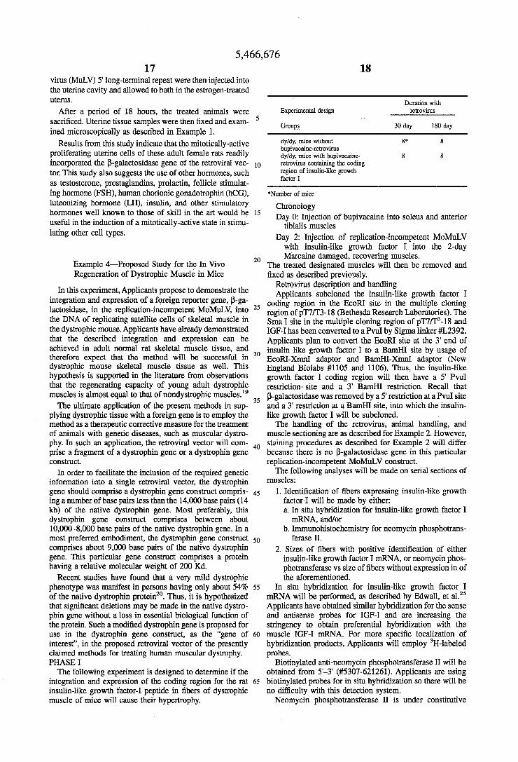
Duranon with retrovims After a period of 18 hours, the treated animals were Expenmental design
Groups 30day 180 day
dyldy, mice without 8* 8 bupivacaine-retrovirus
retrovirus containing the coding regon of insdin-like growth factor I
*Number of mice
Chonology Day o: Injection of bupivacaine into soleus and anterior
tibialis muscles Day 2: Injection of replication-incompetent MoMuLV
with insulin-like growth factor I into the 2-day
The treated designated muscles will then be removed and fixed as described previously.
proliferating uterine cells of these adult female rats readily dyldy, mce with bupivacane- 8 8
2o Marcaine damaged, recovering muscles.
Retrovirus description and handling
Example &Proposed Study for the In Vivo Regeneration of Dystrophic Muscle in Mice
integration and expression of a foreign reporter gene, p-ga- coding region in the site in the multiple clodng lactosidase, in the replication-incompetent MoMuLV, into 25 region of p ~ 7 ~ 3 - 1 8 (Bethesda Research Laboratories). ne the DNA of replicating satellite cells of skeletal muscle in sma I site in the multiple region of p ~ ~ 3 - 1 8 and the dystrophic mouse. Applicants have already demonstrated IGF-I has been converted to a PvuI by Sigma linker #L2392. that the described interntion and expression Can be Applicants plan to convert the EcoRI site at the 3' end of achieved in adult normal rat skeletal muscle tissue, and insulk like growth factor I to a B& site by usage of therefore expect that the method Will be Successful in 30 & o M - x d adaptor and B d - m adaptor (New dYStrOPhiC mouse skeletal muscle tissue as well. This England Biolabs #1105 and 1106). Thus, the insulin-like hypothesis is s~pported in the literature from observations growth factor I coding region will then have a 5' p d that the regenerating capacity Of young adult dystrophic restriction, site and a 3' BamHI restriction. Recall that muscles is almost equal to that of nondystrophic muscle^.'^ 35 P-galactosidase was removed by a 5' restriction at a PvuI site
The ultimate application of the present methods in sup- and a 3' restriction at a BamHI site, into which the insulin- plying dystrophic tissue with a foreign gene is to employ the like growth factor I will be subcloned. method as a therapeutic corrective measure for the treatment The handling of the retrovirus, animal handling, and of animals with genetic diseases, such as muscular dystro- muscle sectioning are as described for Example 2. However, phy. In such an application, the retroviral vector will com- 4o staining procedures as described for Example 2 will differ prise a fragment of a dystrophin gene or a dystrophin gene because there is no P-galactosidase gene in this particular construct. replication-incompetent MoMuLV construct.
In order to facilitate the inclusion of the required genetic The following analyses will be made on serial sections of information into a single retroviral vector, the dystrophin gene should comprise a dystrophin gene construct compris- 45 1. Identification of fibers expressing insulin-like growth ing a number of base pairs less than the 14,000 base pairs (14 factor 1 will be made by either: kb) of the native dystrophin gene. Most preferably, this a. In situ hybridization for insulin-like growth factor I dystrophin gene construct comprises between about mRNA, andor 1O,O00-8,000 base pairs of the native dystrophin gene. In a b. Immunohistochemistry for neomycin phosphotrans- most preferred embodiment, the dystrophin gene construct 50 comprises about 9,OOO base pairs of the native dystrophin 2. Sizes of fibers with positive identification of either gene. This particular gene construct comprises a protein insulin-like growth factor I -A, or neomycin phos- having a relative molecular weight of 200 Kd. photransferase vs size of fibers without expression in of
Recent studies have found that a very mild dystrophic the aforementioned. phenotype was manifest in persons having only about 54% 55 In situ hybridization for insulin-like growth factor I of the native dystrophin protein". Thus, it is hypothesized mRNA will be performed, as described by Edwall, et aLZ5 that significant deletions may be made in the native dystro- Applicants have obtained similar hybridization for the sense phin gene without a loss in essential biological function of and antisense probes for IGF-I and are increasing the the protein. Such a modified dystrophin gene is proposed for stringency to obtain preferential hybridization with the use in the dystrophin gene construct, as the "gene of 60 muscle IGF-I mRNA. For more specific localization of interest", in the proposed retroviral vector of the presently hybridization products, Applicants will employ 3H-labeled claimed methods for treating human muscular dystrophy. probes. PHASE I Biotinylated anti-neomycin phosphotransferase II will be
The following experiment is designed to determine if the obtained from 5'-3' (#5307-621261). Applicants are using integration and expression of the coding region for the rat 65 biotinylated probes for in situ hybridization so there will be insulin-like growth factor-I peptide in fibers of dystrophic no difficulty with this detection system. muscle of mice will cause their hypertrophy. Neomycin phosphotransferase II is under constitutive
In this experiment, propose to demonstrate the Applicants subcloned the insulin-like growth factor 1
mwles :
ferase II.
5,466,676 19
expression by SV 40 early promoter region. Applicants have a JAVA image-processing software system which they use with a PC Vision Plus Frame Grabber Board in an AT-clone computer. Applicants use this system to determine the quan- tities of in situ hybridization, immunohistochemistry, and size of muscle fibers within serial sections. PHASE I1
Retroviral vectors will be prepared which include a dys- trophin gene construct under the constitutive promoter con- trol of the MoLV 5’ long-terminal repeat. This dystrophin gene construct will comprise between about 10,000 and 8,000 base pairs of the native dystrophin gene, and encode a dystrophin protein having a relative molecular weight of 200,000. This particular gene fragment will be prepared as described by England et al?’ In a most preferred embodi- ment, the dystrophin gene construct will comprise about 9,000 base pairs.
Dystrophic muscle of mice will then be treated with bupivacaine, as described above. The tissue will then be exposed to the described dystrophin-construct containing retroviral vector. Excision, fixation and analysis of the treated tissue of the dystrophic animals will then be per- formed as described in the previous examples.
Example 5-Proposed In Vivo Use in Human Gene Therapy of Muscular Degenerative Disease
The gene encoding human dystrophin has been isolated as a “mammoth” gene, postulated to comprise 2.3 megabases. However, recent reports indicate the isolation of a segment of the native gene which has been demonstrated to impart to human subjects only mild dystrophic phenotype^'^. Appli- cants propose the use of this “mini gene” together with the presently described and claimed retroviral mediated gene transfer system as applied to satellite muscle cells for the gene therapy of humans with genetic muscular degenerative diseases. These diseases include, by way of example, Duch- enne’s and Becker muscular dystrophies.
Use of the retroviral-mediated gene transfer systems described by the Applicants will effectively eliminate cur- rently-recognized limitations in the art of gene therapy, such as the risk of immune rejection, diffusion, retroviral gene insert limitations and the requirement for multiple site injections (as in the case of myoblast injection to degenera- tive human muscle sites).
While the determination of exact treatment regimens for such a therapy await human in vivo clinical trials, it is hypothesized that the same pharmaceutical agents, radiation treatments, hormones, and other cell or tissue-discomposing events would elicit a mitotically-active state in human tissue and cells as they have in Applicants’ experimental rats and mice. Current reviews suggest the process of muscle regen- eration in humans shows few significant differences from the processes in mammalian experiments, saving those of scale due to the large size of human muscles, nor is there currently any reason to believe that the control mechanisms of muscle regeneration differ to any significant degree5. Thus, the successful gene incorporation accomplished with the tech- niques provided and described herein in rats are expected to be mimicked for the successful gene incorporation in humans.
The instant invention. has been disclosed in connection with specific embodiments. However, it will be apparent to those skilled in the art that variations from the illustrated embodiments may be undertaken without departing the spirit and scope of the invention.
5
10
15
20
25
30
35
40
45
50
55
60
65
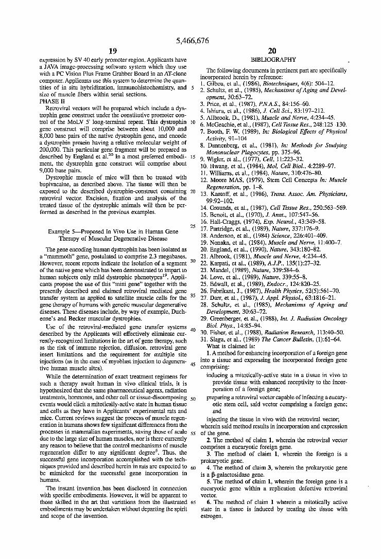
20 BIBLIOGRAPHY
The following documents in pertinent part are specifically incorporated herein by reference: 1. Gilboa, et al., (1986), Biotechniques, 4(6): 504-12. 2. Schultz, et al., (1985), Mechanisms of Aging and Devel-
3. Price, et al., (1987), RN.A.S., 84:156-60. 4. Ishiura, et al., (1986), J. Cell Sci., 83:197-212. 5. Allbrook, D., (1981), Muscle and Nerve, 4:234-45. 6. McGeachie, et al., (1987), Cell Tissue Res., 248:125-130. 7. Booth, F. W. (1989), In: Biological Effects of Physical
Activity, 91-104 8. Dannenberg, et al., (1981), In: Methods for Studying
Mononuclear Plagocytes, pp. 375-96. 9. Wigler, et al., (1977), Cell, 11:223-32. 10. Hwang, et al., (1984), Mol, Cell Biol., 4:2289-97. 11. Williams, et al., (1984), Nature, 310:476-80. 12. Moore MAS, (1979), Stem Cell Concepts In: Muscle
13. Kantoff, et al., (1986), Trans. Assoc. Am. Physicians,
14. Grounds, et al., (1987), Cell Tissue Res., 250563-569. 15. Benoit, et al., (1970), J. Anat., 107547-56. 16. Hall-Craggs, (1974), Exp. Neurol., 43:349-58. 17. Partridge, et al., (1989), Nature, 337:176-9. 18. Anderson, et al., (1984) Science, 226:401-409. 19. Nonaka, et al., (1984), Muscle and Nerve, 11:400-7. 20. England, et al., (1990), Nature, 343:180-82. 21. Albrook, (1981), Muscle and Nerve, 4234-45. 22. Karpati, et al., (1989), A.J.P., 135(1):27-32. 23. Mandel, (1989), Nature, 339584-6. 24. Love, et al., (1989), Nature, 33955-8. 25. Edwall, et al., (1989), Endocr., 12482G25. 26. Fabrikant, J., (1987), Health Physics, 52(5):561-70. 27. Dan, et al., (1987), J. Appl. Physiol., 63:1816-21. 28. Schultz, et al., (1985), Mechanisms of Ageing and
29. Greenberger, et al., (1988), Znt. J. Radiation Oncology
30. Fisher, et al., (1988), Radiation Research, 113:40-50. 31. Slaga, et al., (1989) The Cancer Bulletin, (1):61-64.
opment, 30:63-72.
Regeneration, pp. 1-8.
99:92-102.
Development, 30:63-72.
Biol. Phys., 14:85-94.
What is claimed is: 1. A method for enhancing incorporation of a foreign gene
into a tissue and expressing the incorporated foreign gene comprising:
inducing a mitotically-active state in a tissue in vivo to provide tissue with enhanced receptivity to the incor- poration of a foreign gene;
preparing a retroviral vector capable of infecting a eucary- otic stem cell, said vector comprising a foreign gene; and
injecting the tissue in vivo with the retroviral vector; wherein said method results in incorporation and expression of the gene.
2. The method of claim 1, wherein the retroviral vector comprises a eucaryotic foreign gene.
3. The method of claim 1, wherein the foreign is a prokaryotic gene.
4. The method of claim 3, wherein the prokaryotic gene is a P-galactosidase gene.
5. The method of claim 1, wherein the foreign gene is a eucaryotic gene within a replication defective retroviral vector.
6. The method of claim 1 wherein a mitotically active state in a tissue is induced by treating the tissue with estrogen.
5,466,676 21 22
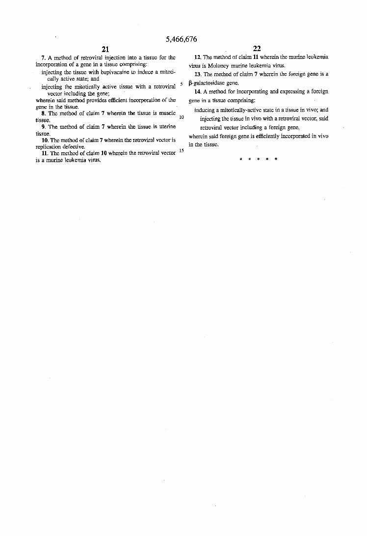
7. A method of retroviral injection into a tissue for the 12. The method of claim 11 wherein the murine leukemia incorporation of a gene in a tissue comprising: virus is Moloney murine leukemia virus.
injecting the tissue with bupivacaine to induce a mitoti-
injecting the mitotically active tissue with a retroviral
13. The method of claim 7 wherein the foreign gene is a
14. A method for incorporating and expressing a foreign
cally active state; and
vector including the gene:
5 P-galactosidase gene.
Y - I
wherein said method provides efficient incorporation of the gene in a tissue comprising: gene in the tissue.
tissue.
inducing a mitotically-active state in a tissue in vivo; and injecting the tissue in vivo with a retroviral vector, said
8. The method of claim 7 wherein the tissue is muscle lo
9. The method of claim 7 wherein the tissue is uterine
10. The method of claim 7 wherein the retroviral vector is
11. The method of claim 10 wherein the retroviral vector l5
retroviral vector including a foreign gene, wherein said foreign gene is efficiently incorporated in vivo tissue.
replication defective. in the tissue.
is a murine leukemia virus. * * * * *
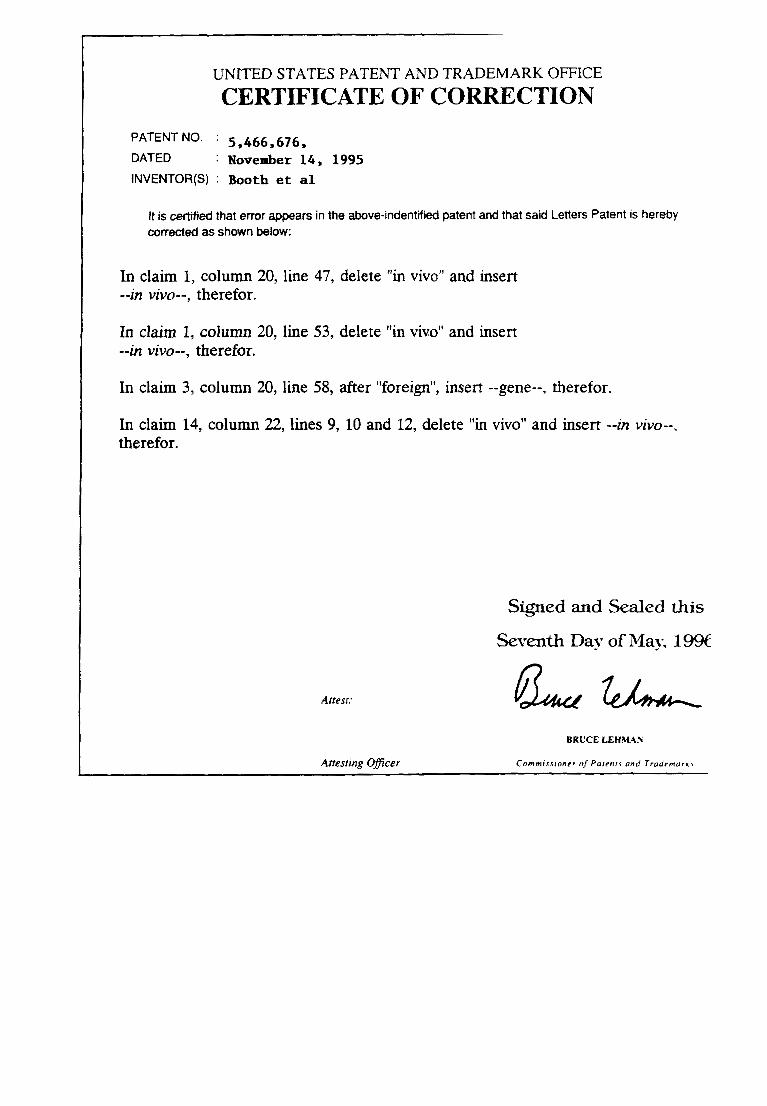
UNITED STATES PATENT AND TRADEMARK OFFICE CERTIFICATE OF CORRECTION
PATENT No. ' 5,466,676, DATED November 1 4 , 1995 INVENTOR(S) : Booth et a1
It is certified that error appears in the above-indentified patent and that said Letters Patent is hereby corrected as shown below:
In claim 1, column 20, line 47, delete "in vivo" and insert --in vivo--, therefor.
In claim 1, column 20, line 53, delete "in vivo" and insert --in vivo--, therefor.
In claim 3, column 20, line 58, after "foreign", insert --gene--, therefor.
In claim 14, column 22, lines 9, 10 and 12, delete "in vivo" and insert --in vivo--, therefor.
Signed and Sealed this
Seventh Day of May, 19% n
Atresr:
BRUCE LEHMAh
Cornmirsionrr 01 Parrnry and Trudrmur l t I Anesring qfficer
Recommended