Embed Size (px)
Citation preview
1170
Accepted by J. Brown: 11 Feb. 2006; published: 10 Apr. 2006 1
ZOOTAXAISSN 1175-5326 (print edition)
ISSN 1175-5334 (online edition)Copyright © 2006 Magnolia Press
Zootaxa 1170: 1–26 (2006) www.mapress.com/zootaxa/
DNA barcodes: Evaluating the potential of COI to diffentiate closely related species of Elachista (Lepidoptera: Gelechioidea: Elachistidae) from Australia
LAURI KAILA 1* & GUNILLA STÅHLS 2
Finnish Museum of Natural History, Dept. of Entomology, PO Box 17, FI-00014 University of Helsinki, Fin-land. E-mail: 1 [email protected], 2 [email protected]
*Corresponding author
Abstract
We compared DNA barcoding to “traditional” taxonomic tools in clarifying relationships incomplexes of closely related, putative “species” of Elachistinae moths (Gelechioidea: Elachistidae)occurring in Australia. A 705 bp fragment of the 3’-end of cytochrome c oxidase subunit I gene(COI) was used. This mtDNA fragment did not differentiate between all species-level taxa thatcould be defined by morphological and/or ecological differences. Different evolutionary rates ofCOI among closely related lineages were observed. Although our findings are based on thevariability of the 3’ end of the COI gene and not the 5’ end barcode fragment, we are convinced thatthorough exploration of traditional morphology and ecology is a prerequisite for exploringinsufficiently known taxonomies by the barcode approach. The sole use of COI barcoding, whetherconsidering COI-5’ or COI-3’ fragment, may fail to recognize closely related species. Our resultsdiscourage this approach for delimitation of closely related species, but its use is encouraged as anadditional tool for exploring little known taxonomies or as an identification tool for previouslythoroughly studied species complexes.
Key words: barcode, COI-3’, taxonomy, Elachista, identification, delimitation of species,morphology
Introduction
DNA barcoding proposes the use of DNA sequences to identify and classify an organism.The potential of a 650 bp fragment of the 5’-end of mitochondrial cytochrome c oxidasesubunit I (COI)-based species identification system was proposed and partiallydemonstrated by Hebert et al. (2003b). Among the benefits of this particular gene is itsease of acquisition and alignment, in addition to the fundamental criterion, a high level of
KAILA & STÅHLS2 © 2006 Magnolia Press
1170ZOOTAXA diversity. Hence, the COI-5’ fragment has been proposed to serve as the core of a global
bio-identification system for animals (Hebert et al. 2003a). The performance of thissystem remains unclear, however, when applied to species that by traditional taxonomy areclassified as very closely related (e.g. Prendini 2005). As Blaxter (2003) and Hebert et al.(2003a) have discussed, a major unresolved issue is how closely the molecular taxacorrespond to what traditional biologists recognize as species, i.e., species defined byDNA barcodes might not always correspond with species recognized by traditionalecological and morphological criteria. The question remains as how to delimit the speciesin question. The sole use of barcoding could incorrectly identify two members of a speciesas separate species, or two separate species as the same. Moreover, the conceptual issue ofdefinition of species remains intact: if a genomic integrity of a species is assumed, how isit defined or characterized (see Sperling 2003 for discussion)?
In this paper we present examples from Australian complexes of Elachistinae mothsthat presently are unresolved species, and demonstrate discrepancies in delimitation ofspecies using ’traditional’ criteria (i.e., ecological and morphological) versus themitochondrial COI-3’ fragment. We also demonstrate problems in applying the use of thisgenomic fragment as a DNA barcode of species.
Taxonomy
Our focal taxon is Elachista, a large genus of Lepidoptera (Gelechioidea: Elachistidae).Elachista comprises 550 named and two hundred discovered, yet unnamed speciesworldwide (L. Kaila, unpublished). The larvae of Elachista are leaf-miners specialising onmonocotyledenous plants, especially Poaceae and Cyperaceae. Their phylogeny wasexamined previously in a morphological analysis that used 131 characters with 171informative character states derived from adult and pupal morphology and larval ecology(Kaila 1999a), and later revisited by a more extensive morphology-based data set by Kaila& Sugisima (2003, and in preparation).
The taxonomic knowledge of Elachista has a long history in Europe where highlyskilled amateurs have contributed detailed knowledge on the life histories andidentification of species. Elachista species are generally morphologically rather uniformand thus difficult to identify based on external examination (Traugott-Olsen & SchmidtNielsen 1977 and references therein). During the past two decades the taxonomy of thegenus has been under revision worldwide by LK, resulting in nineteen revisionary works(Albrecht & Kaila 1997, Huemer & Kaila 2003, Kaila 1992, 1996, 1997, 1998ab, 1999abc,2005, Kaila et al. 2001, Kaila & Jalava 1994, Kaila & Junnilainen 2002, Kaila & Karsholt2002, Kaila et al. 2003, Kaila & Sugisima 2003, Kaila & Varalda 2004, Sugisima & Kaila2005). The taxa examined in the present paper are included in an ongoing revision of thefauna of Australia (L. Kaila, in preparation).
© 2006 Magnolia Press 3ELACHISTA
1170ZOOTAXAThe aim of the present study is to elucidate the taxonomic utility of a sequence
fragment, the 3’ end of the COI gene, for a group of very closely related, putative speciesof the genus Elachista. The taxa were selected for the present study from complexes ofclosely related species or populations which show differences in host plant selection,larval mine architecture, external appearance of the adults, and to some extent,morphology of the adult genitalia. The morphological differences, however, are slight,sometimes overlapping, and frequently only one sex can be identified by genitalcharacteristics. Based on this kind of evidence the specimens were preliminarily classifiedto “species,” which are assumed to be cohesive genealogical lineages. This practice is inaccordance with the current (traditional) concept of delineating species in the Elachistinae,which, however, should not be equated with the “correct” way in our opinion. It is hereused as a starting point towards a more integrative taxonomic approach (cf. Dayrat 2005).The “species” are grouped into three informal species complexes, referred below to as“yellow” (name derived from the characteristic yellowish wing colour), “Ficinia” (namederived from the host plant genus), and “zigzagger” (name derived from the peculiarzigzagging larval mine) complexes.
Material and methods
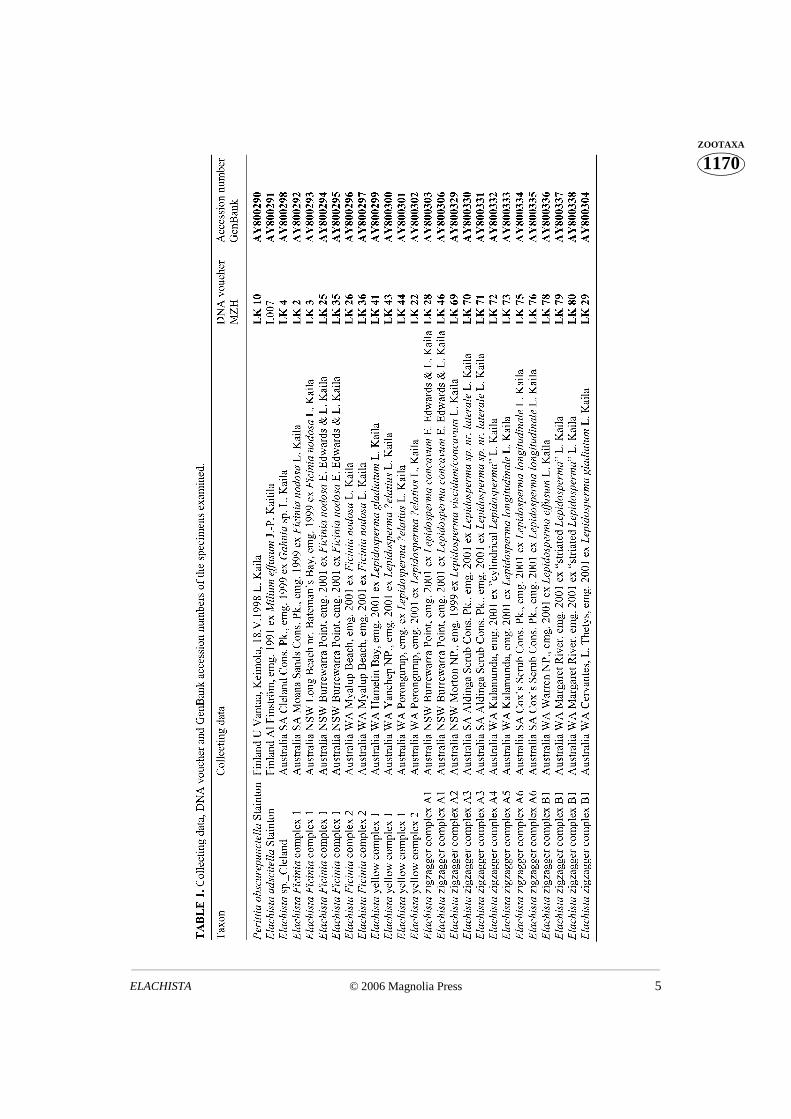
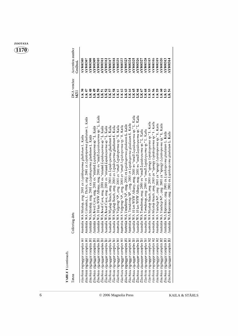
Molecular studyThe specimens used for molecular analysis are listed in Table 1. All of them were
collected as larvae by LK thus confirming the correct host plant association. The larvaewere reared in laboratory conditions using standard methods. The samples cover a modestsubset of the taxonomic diversity of Australian representatives of Elachista subgenusElachista that could be obtained for the molecular study. Presently at least 140 Elachistaspecies are recorded from Australia (L. Kaila, unpublished), of which fourteen are treatedhere.
DNA was extracted usually from legs or head+thorax of single individuals from dry,pinned specimens (Table 1). They are preserved as DNA voucher specimens in theZoological Museum of the Finnish Museum of Natural History (MZH) DNA voucherspecimen collection, and labeled as listed in Table 1. DNA was extracted using theNucleospin Tissue Kit (Machery-Nagel, Düren, Germany) according to manufacturer’sprotocols, and re-suspended in 50 µl of ultra-pure water.
PCR’s were carried out in 25 µl reactions containing 1–2 µl DNA extract, 1 µl of eachprimer (at 10 pmol/µl), 0.25 µl of Amplitaq DNA polymerase (5U / µl), 2 µl 2.5 mMMgCl2, 2.5 µl 10X Buffer II (Applied Biosystems) and 4 µl 200 mM dNTP (GeneAmp)
and water. Thermocycler conditions were initial denaturing at 95°C 2 min, 29 cycles of 30s denaturing at 94°C, 30 s annealing at 49°C, 2 min extension at 72°C, followed by a final
KAILA & STÅHLS4 © 2006 Magnolia Press
1170ZOOTAXA extension of 8 min at 72°C. PCR products were purified using the GFX PCR Purification
Kit (Amersham Biotech) and then sequenced (using the PCR primers) in both directionsusing the Big Dye Terminator Cycle Sequencing Kit vs. 1.1 (Applied Biosystems) at one-fourth of the recommended volumes on ABI PRISM 377 DNA sequencer. The primersused for amplifying and sequencing the COI-3’ were C1-J-2183 (alias “Jerry”, 5’CAACATTTATTTTGATTTTTTGG 3’) and tl2-n-3014 (alias “Pat”, 5’TCCAATGCACTAATCTGCCATATTA 3’) (Simon et al. 1994).
Sequences were assembled and edited using Sequence Navigator (version 1.01).The alignment of the protein-coding COI was straightforward. Phylogenetic relationshipsof included terminals were estimated (using equal weights) using the parsimony programNoNa vs. 2.0 (Goloboff 1999) using the command line “hold 100000; hold*; hold/50,mult*100; max*;”. Bremer (Bremer 1988, 1994) values were estimated using NoNa andJackknife support values using WinClada (Nixon 2002).
Morphological studyAdult specimens and their pupal exuviae were examined externally using a
stereomicroscope, in order to evaluate possible differences in their colouration and wingshape. Extensive series, whenever available, were dissected using standard procedure(Robinson 1976). The genital morphology was examined using a Wild M10stereomicroscope (maximum magnification 512x and Leitz Diaplan phase contrastcompound microscope (maximum magnification 1560x). The terminology of anatomyfollows Traugott-Olsen & Schmidt Nielsen (1977).
Results
Molecular studyWe obtained 705 nucleotides of the 3’ end of the COI gene spanning nucleotide
positions 2239 to 2944 in COI (numbering is based on Drosophila yakuba sequence; Claryand Wolstenholme 1985) for 47 ingroup specimens belonging to 14 putativemorphospecies and two outgroup taxa. The mean AT was 72.2 %.
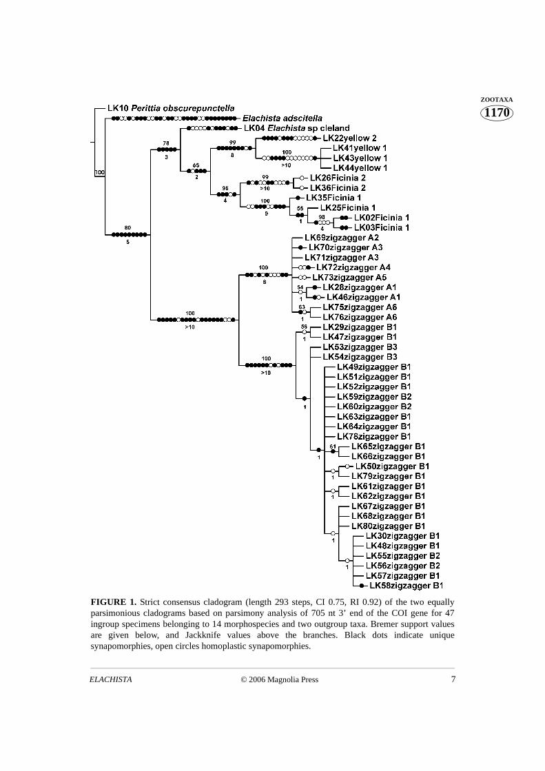
The number of parsimony informative sites was 120. Parsimony analysis using NoNafound two equally parsimonious trees (length 290 steps). Their strict consensus cladogramwith Bremer support values and Jackknife support values is shown in Fig. 1. COI resolvesspecies groups well, but is invariant in four cases of putative recently diverged specieswithin the ‘zigzagger ’complex, and within the two sections of “zigzaggers” at most threebp differences are observed. For species in the ‘yellow’ clade complex we obtainedintraspecifically identical sequences that support morphological taxonomy. On the otherhand, the amount of the intraspecific variation of included taxa varied considerablybetween the complexes.
© 2006 Magnolia Press 5ELACHISTA
1170ZOOTAXA
KAILA & STÅHLS6 © 2006 Magnolia Press
1170ZOOTAXA
© 2006 Magnolia Press 7ELACHISTA
1170ZOOTAXA
FIGURE 1. Strict consensus cladogram (length 293 steps, CI 0.75, RI 0.92) of the two equallyparsimonious cladograms based on parsimony analysis of 705 nt 3’ end of the COI gene for 47ingroup specimens belonging to 14 morphospecies and two outgroup taxa. Bremer support valuesare given below, and Jackknife values above the branches. Black dots indicate uniquesynapomorphies, open circles homoplastic synapomorphies.
KAILA & STÅHLS8 © 2006 Magnolia Press
1170ZOOTAXA Uncorrected interspecific pairwise divergence between yellow 1 and yellow 2 was
4.54%, and that between Ficinia 1 and Ficinia 2 samples ranged from 3.69% to 4.40%.Between the taxa of the A section of the zigzagger complex these value ranged from 0.0%to 0.85%. Between the taxa of the B section of the zigzagger complex they ranged from0.0% to 0.71%. The divergence between the taxa of A and B sections ranged from 3.55%to 4.11%. The divergence between the yellow and Ficinia complexes (yellow 1 vs. Ficinia1) ranged between 5.96%–6.95%, and divergences between yellow and Ficinia complextaxa vs. zigzagger complex’s taxa were of the same magnitude.
Uncorrected intraspecific pairwise divergences of the yellow 1 was 0.0%, withinFicinia-group complex taxa the values ranged from 0.28–1.56%. Within the A section ofthe zigzagger complex values ranged from 0.0% to 0.43%, and within the B section valueswere 0.0% to 0.85%, respectively.
The division of the zigzagger complex into A and B sections, based on the presence/absence of the female signum (for definition see Scoble 1992), was supported by thesequence data. Intra- and interspecific uncorrected pairwise divergences betweenspecimens and hypothetical taxa of the zigzagger complex were almost completelyoverlapping, with intraspecific divergences greater than interspecific in some cases.Therefore, within the zigzagger B complex of species the morphospecies B1 appearsparaphyletic with respect to two other species, and B2 appears as polyphyletic.
The intraspecific divergence ranges for zigzagger A and B complexes overlap with theinterspecific ranges between taxa of these complexes; hence, intraspecific divergences arebigger than interspecific values in some instances.
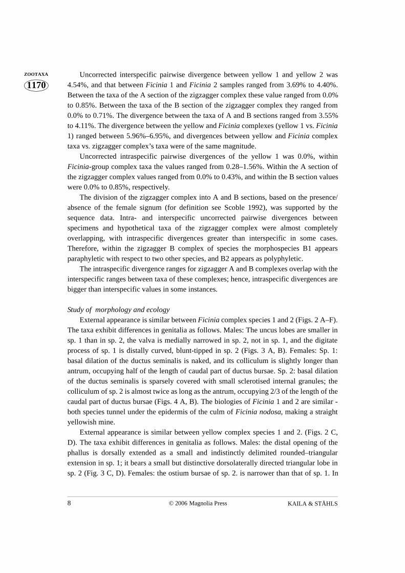
Study of morphology and ecology External appearance is similar between Ficinia complex species 1 and 2 (Figs. 2 A–F).
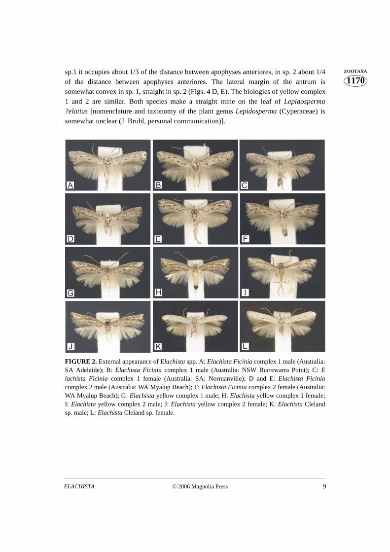
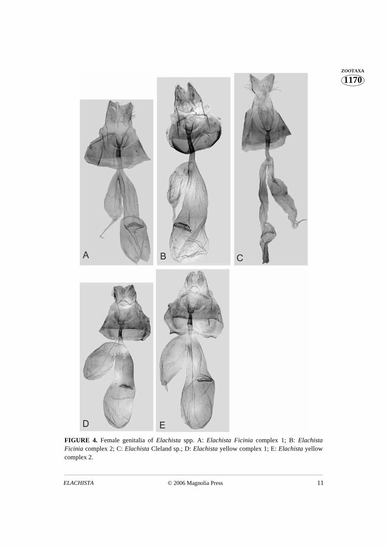
The taxa exhibit differences in genitalia as follows. Males: The uncus lobes are smaller insp. 1 than in sp. 2, the valva is medially narrowed in sp. 2, not in sp. 1, and the digitateprocess of sp. 1 is distally curved, blunt-tipped in sp. 2 (Figs. 3 A, B). Females: Sp. 1:basal dilation of the ductus seminalis is naked, and its colliculum is slightly longer thanantrum, occupying half of the length of caudal part of ductus bursae. Sp. 2: basal dilationof the ductus seminalis is sparsely covered with small sclerotised internal granules; thecolliculum of sp. 2 is almost twice as long as the antrum, occupying 2/3 of the length of thecaudal part of ductus bursae (Figs. 4 A, B). The biologies of Ficinia 1 and 2 are similar -both species tunnel under the epidermis of the culm of Ficinia nodosa, making a straightyellowish mine.
External appearance is similar between yellow complex species 1 and 2. (Figs. 2 C,D). The taxa exhibit differences in genitalia as follows. Males: the distal opening of thephallus is dorsally extended as a small and indistinctly delimited rounded–triangularextension in sp. 1; it bears a small but distinctive dorsolaterally directed triangular lobe insp. 2 (Fig. 3 C, D). Females: the ostium bursae of sp. 2. is narrower than that of sp. 1. In
© 2006 Magnolia Press 9ELACHISTA
1170ZOOTAXAsp.1 it occupies about 1/3 of the distance between apophyses anteriores, in sp. 2 about 1/4
of the distance between apophyses anteriores. The lateral margin of the antrum issomewhat convex in sp. 1, straight in sp. 2 (Figs. 4 D, E). The biologies of yellow complex1 and 2 are similar. Both species make a straight mine on the leaf of Lepidosperma?elatius [nomenclature and taxonomy of the plant genus Lepidosperma (Cyperaceae) issomewhat unclear (J. Bruhl, personal communication)].
FIGURE 2. External appearance of Elachista spp. A: Elachista Ficinia complex 1 male (Australia:SA Adelaide); B: Elachista Ficinia complex 1 male (Australia: NSW Burrewarra Point); C: Elachista Ficinia complex 1 female (Australia: SA: Normanville); D and E: Elachista Ficiniacomplex 2 male (Australia: WA Myalup Beach); F: Elachista Ficinia complex 2 female (Australia:WA Myalup Beach); G: Elachista yellow complex 1 male; H: Elachista yellow complex 1 female;I: Elachista yellow complex 2 male; J: Elachista yellow complex 2 female; K: Elachista Clelandsp. male; L: Elachista Cleland sp. female.
KAILA & STÅHLS10 © 2006 Magnolia Press
1170ZOOTAXA
FIGURE 3. Male genitalia of Elachista taxa, in ventral view. A: Elachista Ficinia complex 1; B:Elachista Ficinia complex 2; C: Elachista yellow complex 1; D: Elachista yellow complex 2.
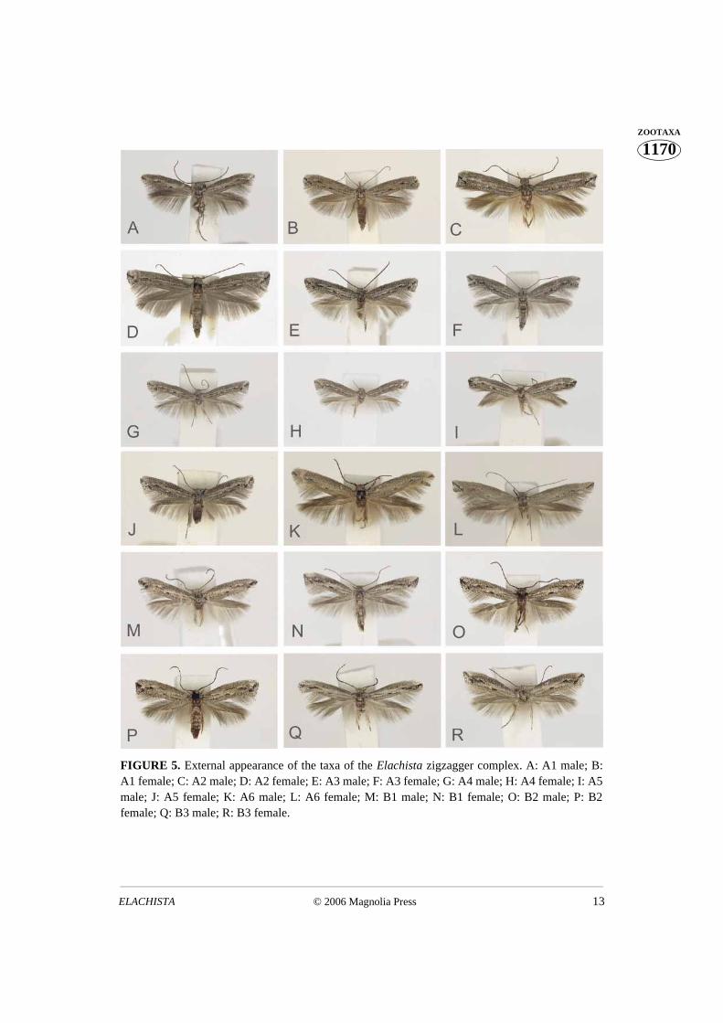
External appearance differs to some extent among the taxa of the zigzagger complex(Fig. 5). A2 and A6 are the largest, and A4 is the smallest and most narrow-winged. A1,A3, or A5 can hardly be distinguished from each other on the basis of their appearanceonly. All taxa of the A section are greyer than those of B section whose forewings arepowdered with brownish scales as well. B1, B2, and B3 are similar to each other.Examination of a large number of samples of the B1–B3 taxa implies, however, thefollowing trends: B2 tends to be the darkest, brownest, and most broad-winged of thesetaxa; and B3 has quite bright black markings in the distal part of its forewing.
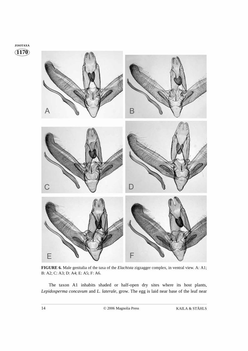
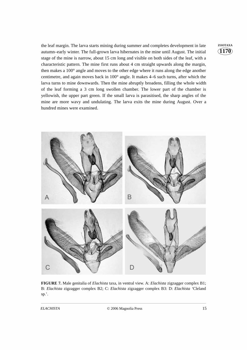
The male genitalia of taxa of the zigzagger complex are shown in Figs. 6 and 7. A2seems consistently distinguishable from others by the thin basal half of the phallus (Fig. 6B). The cucullus of the valva is more expanded in A6 than in the other species. The juxtalobes of A5 differ in their shape from the other species (Fig. 6 E). Although the shape ofthe spinose knob of the gnathos is somewhat variable within the taxa, it neverthelessdistinguishes A2 and B3 from others, as being basally broader in these taxa than in others.
© 2006 Magnolia Press 11ELACHISTA
1170ZOOTAXA
FIGURE 4. Female genitalia of Elachista spp. A: Elachista Ficinia complex 1; B: ElachistaFicinia complex 2; C: Elachista Cleland sp.; D: Elachista yellow complex 1; E: Elachista yellowcomplex 2.
KAILA & STÅHLS12 © 2006 Magnolia Press
1170ZOOTAXA A summary of the further male genital characters of the taxa of the zigzagger complex
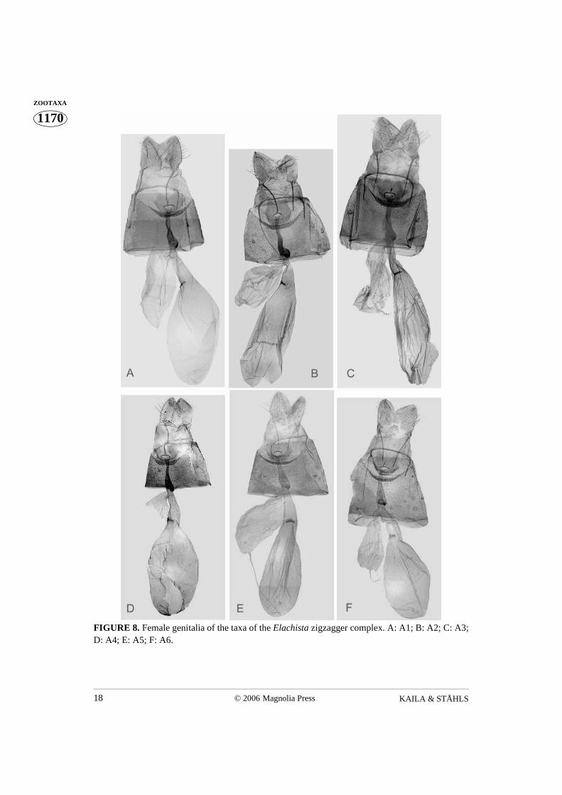
is given in Table 3. The females of this groupcomplex (Figs. 8–10) are keyed here:
1. Signum present (Fig. 8) ................................................................................................ 2- Signum absent (Fig. 9) ................................................................................................. 72. Between sclerotised spinose area that surrounds ostium and invagination of integu-
ment between sternum 7 and 8 a membranous area that is wider than invagination ofintegument between sternum 7 and 8 (Fig. 10 F)....................................................... A6
- Sclerotised spinose area that surrounds ostium and invagination of integumentbetween sternum 7 and 8 without membranous area, or membranous area narrowerthan invagination of integument between sternum 7 and 8 8 (Fig. 10 A–E) ................ 3
3. Width of ostium bursae half of width of invagination of integument between sternum 7and 8 (Fig. 10D) ......................................................................................................... A4
- Width of ostium bursae less than half of width of invagination of integument betweensternum 7 and 8 ............................................................................................................. 4
4. Caudal longitudinal, and cephalic transverse sclerotisations of colliculum separatefrom each other (Figs. 10 B, E)..................................................................................... 5
- Sclerotisations of colliculum fused to each other (Figs. 10 A, C) ................................ 65. Cephalic transverse sclerotisation of colliculum a simple evenly sclerotised bent band
(Fig. 10 E) .................................................................................................................. A5- Cephalic transverse sclerotisation of colliculum asymmetric, with one end strongly
sclerotised and sickle-shaped (Fig. 10 B)................................................................... A26. Ductus bursae constricted at caudal end of colliculum (Fig. 10 A) .......................... A1- Ductus bursae not constricted at caudal end of colliculum (Fig. 10 C) ..................... A3
7. Females of Elachista zigzagger complex B1, B2 and B3 only identifiable from imma-ture stages.
There appear to be no differences in the immature stages or the life histories amongyellow complex species or among the Ficinia complex species. The pupal exuviae of thetaxa of the zigzagger complex are characterised by the dark brownish grey mesial area ofthe ventral side (Fig. 11). There appear to be constant differences in this character amongsome taxa of this complex: A2 has most expanded dark area with forewing veins visible aspale only laterally; the A taxa have in general larger dark area than B taxa. The larvalmines of the zigzagger complex taxa A1, A2, and B2 are shown in Fig. 12. The biologicaltraits of the taxa of the zigzagger complex are summarised here. Some differentiating traitsare summarised in Table 3.
© 2006 Magnolia Press 13ELACHISTA
1170ZOOTAXA
FIGURE 5. External appearance of the taxa of the Elachista zigzagger complex. A: A1 male; B:A1 female; C: A2 male; D: A2 female; E: A3 male; F: A3 female; G: A4 male; H: A4 female; I: A5male; J: A5 female; K: A6 male; L: A6 female; M: B1 male; N: B1 female; O: B2 male; P: B2female; Q: B3 male; R: B3 female.
KAILA & STÅHLS14 © 2006 Magnolia Press
1170ZOOTAXA
FIGURE 6. Male genitalia of the taxa of the Elachista zigzagger complex, in ventral view. A: A1;B: A2; C: A3; D: A4; E: A5; F: A6.
The taxon A1 inhabits shaded or half-open dry sites where its host plants,Lepidosperma concavum and L. laterale, grow. The egg is laid near base of the leaf near
© 2006 Magnolia Press 15ELACHISTA
1170ZOOTAXAthe leaf margin. The larva starts mining during summer and completes development in late
autumn–early winter. The full-grown larva hibernates in the mine until August. The initialstage of the mine is narrow, about 15 cm long and visible on both sides of the leaf, with acharacteristic pattern. The mine first runs about 4 cm straight upwards along the margin,then makes a 100° angle and moves to the other edge where it runs along the edge anothercentimetre, and again moves back in 100° angle. It makes 4–6 such turns, after which thelarva turns to mine downwards. Then the mine abruptly broadens, filling the whole widthof the leaf forming a 3 cm long swollen chamber. The lower part of the chamber isyellowish, the upper part green. If the small larva is parasitised, the sharp angles of themine are more wavy and undulating. The larva exits the mine during August. Over ahundred mines were examined.
FIGURE 7. Male genitalia of Elachista taxa, in ventral view. A: Elachista zigzagger complex B1;B: Elachista zigzagger complex B2; C: Elachista zigzagger complex B3: D: Elachista ‘Clelandsp.’.
KAILA & STÅHLS16 © 2006 Magnolia Press
1170ZOOTAXA Larvae of A2 were found in moist sandstone sites with open stone surfaces surrounded
by dense bush vegetation and small eucalypts. Lepidosperma concavum and L. viscidumhave been recorded as the host plants. The egg is laid near base of the leaf near margin.The initial stage of the mine is narrow, about 20 cm long, otherwise similar to that of A3,but the intervals of the nearly right-angled turns are about 10–15 mm from each other,usually not quite reaching the margin of the leaf on one side. Every other across-leafsection of the mine is visible only on the upper side of the leaf, while the remaining arevisible only on the lower side. Then the mine abruptly broadens encompassing nearly theentire width of the leaf, and the larva mines downwards making a 5–7 cm long swollenchamber. The larva feeds during autumn and early winter, and appears to hibernate as afull-grown larva within the mine. The larva exits the mine during August. The initialmines of two larvae have been observed in the same leaf, but under such conditionsapparently only one of them is able to survive. About 50 mines were examined.
TABLE 2 . Male genital traits of the taxa of the Elachista zigzagger group complex..
Larvae of A3 were found in shaded and open forest in rather dry sites where its hostplants, Lepidosperma curtisiae and Lepidosperma sp. nr. laterale, grow. The egg is laidnear the base of the leaf near the margin. The initial mine is narrow, 5–10 cm long in L.curtisiae, 15 cm in L. sp., similar to that of A2. The mine runs, however, along margins ofthe leaf. The intervals of the nearly right-angled turns are about 5 mm from each other, andevery second across-leaf section of the mine is visible only on the upper, every second onthe lower side of the leaf. Then the mine abruptly broadens filling the whole width of theleaf, and the larva mines downwards making a 2–5 cm long swollen chamber. In L.
Scales of juxta lobes Length of aedeagus as% of length of valva
Length of valva inrelation to width ofvalva
complex A1 as group on truncate lobe 80 % 4.5 x length of v.
complex A2 in row along distal margin 85–92 % 4.5 x length of v.
complex A3 as group on truncate lobe 85 % 4.5 x length of v.
complex A4 as group on truncate lobe 75 % 4.5 x length of v.
complex A5 as group near convex dist. margin 78–80 % 4.5 x length of v.
complex A6 as group near convex dist. margin 85 % 4 x length of v.
complex B1 as group in convex dist. margin 82–84 % 4.5 x length of v.
complex B2 as group in convex dist. margin 82 % 4.5 x length of v.
complex B3 as group in convex dist. margin 80–83 % 4.5 x length of v.
© 2006 Magnolia Press 17ELACHISTA
1170ZOOTAXAcurtisiae the leaf soon withers above the mine. The larva feeds during autumn and early
winter, and appears to hibernate as a full-grown larva within the mine. It exits the mineduring August. About 50 mines were examined.
TABLE 3. Mine traits of the taxa of the Elachista zigzagger-group complex.
Larvae of A4 were found in an exposed site where its host plant, Lepidosperma sp.,grows. The unidentified host plant species is characterised by its cylindrical culm (flowerstem) transsection. The initial mine is narrow, about 10 cm long. The initial mineresembles that of the other species in the zigzagger complex, making turns at right angle atintervals of 5 mm on its way upwards the culm. Finally the mine abruptly broadens, andthe larva mines downwards making a 4 cm long chamber which occupies the entire spaceof the culm. The larva feeds during winter and exits the mine during July. Fifteen mineswere examined.
Larvae of A5 were found in a shaded site where its host plant, Lepidospermalongitudinale, grows. The egg is laid near base of the leaf near margin. The initial mine isnarrow, about 15 cm long. The mine first runs 1 cm straight upwards along the margin,then makes a right angle and moves to the other edge where it runs along the edge 7 mm,and again moves back in a right angle. The intervals of the right-angled turns are regular, 7mm from each other, and every other across-leaf section of the mine is visible only on theupper side of the leaf, the remaining only on lower side. Then the mine abruptly broadens,and the larva mines downwards making a 4 cm long swollen chamber, which occupiestwo-thirds of the width of the leaf. The larva feeds during winter and larva exits the mineduring July. Thirty mines were examined.
Angle of turns reaching of leaf edge Visibility on one/both sides of the leaf
complex A1 100° y both
complex A2 90° n one
complex A3 90° y one
complex A4 90° ? ?
complex A5 90° y one
complex A6 90° y one
complex B1 100° y/n one
complex B2 110° y (both)
complex B3 100° n one
KAILA & STÅHLS18 © 2006 Magnolia Press
1170ZOOTAXA
FIGURE 8. Female genitalia of the taxa of the Elachista zigzagger complex. A: A1; B: A2; C: A3;D: A4; E: A5; F: A6.
© 2006 Magnolia Press 19ELACHISTA
1170ZOOTAXA
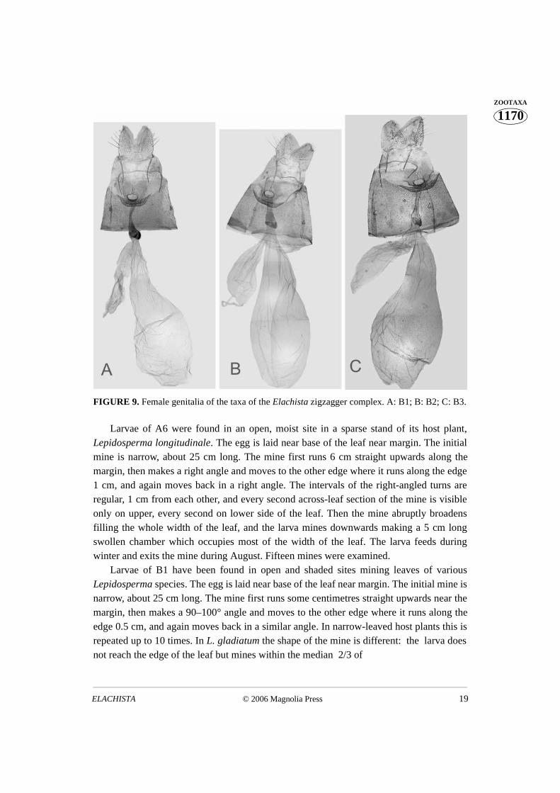
FIGURE 9. Female genitalia of the taxa of the Elachista zigzagger complex. A: B1; B: B2; C: B3.
Larvae of A6 were found in an open, moist site in a sparse stand of its host plant,Lepidosperma longitudinale. The egg is laid near base of the leaf near margin. The initialmine is narrow, about 25 cm long. The mine first runs 6 cm straight upwards along themargin, then makes a right angle and moves to the other edge where it runs along the edge1 cm, and again moves back in a right angle. The intervals of the right-angled turns areregular, 1 cm from each other, and every second across-leaf section of the mine is visibleonly on upper, every second on lower side of the leaf. Then the mine abruptly broadensfilling the whole width of the leaf, and the larva mines downwards making a 5 cm longswollen chamber which occupies most of the width of the leaf. The larva feeds duringwinter and exits the mine during August. Fifteen mines were examined.
Larvae of B1 have been found in open and shaded sites mining leaves of variousLepidosperma species. The egg is laid near base of the leaf near margin. The initial mine isnarrow, about 25 cm long. The mine first runs some centimetres straight upwards near themargin, then makes a 90–100° angle and moves to the other edge where it runs along theedge 0.5 cm, and again moves back in a similar angle. In narrow-leaved host plants this isrepeated up to 10 times. In L. gladiatum the shape of the mine is different: the larva doesnot reach the edge of the leaf but mines within the median 2/3 of

KAILA & STÅHLS20 © 2006 Magnolia Press
1170ZOOTAXA
FIGURE 10. Ostium bursae of the female of the taxa of the Elachista zigzagger complex. A: ‘ A1;B: ‘ A2; C: A3; D: A4; E: A5; F: A6; G: B1; H: B2; I: B3.
the leaf width. It makes a few turns of 100°, after which the turns become more frequent.The larva makes 100° angle turns in every 2–4 mm, after which the mine continues asirregularly undulating for some centimetres. Every second across-leaf section of the mineis visible only on upper, every second on lower side of the leaf. Finally the mine abruptlybroadens and the larva mines downwards making a 5 cm long swollen chamber whichoccupies half of the width of the leaf. The larva feeds during winter and exits the mineduring July-August. Over two hundred mines were examined.
Larvae of B2 were found in shady lakeshore sites mining leaves of an unidentifiedvery large Lepidosperma species characterised by the spongy matrix of its leaves. Theinitial mine is narrow, about 25 cm long. The mine first runs 5 cm straight upwards nearthe margin, then makes a 100° angle and moves to the other edge where it runs along theedge 5 millimetres, and again moves back in a 100° angle. The larva makes a few suchturns, after which the turns become more frequent, the larva mining parallel to the leafedge 2 mm, and makes 110° angle turns whenever it encounters the leaf margin. This isrepeated about five times. The mine runs within the epidermis, and is equally faintlyvisible in both sides of the leaf as a reddish brown line. Finally the mine abruptly broadensand the larva mines downwards making a 5 cm long swollen chamber which occupies halfof the width of the leaf. The larva feeds during winter and exits the mine during July-August. Sixty mines were examined.

© 2006 Magnolia Press 21ELACHISTA
1170ZOOTAXA
FIGURE 11. Pupal exuviae of the taxa of the Elachista zigzagger complex, in ventral view. A: A1;B: A2; C: A3; D: A4; E: A5; F: A6; G: B1; H: B2; I: B3.
Larvae of B3 were found in open sandy site mining leaves of Lepidospermagladiatum. The egg is laid near base of the leaf near margin. The initial mine is narrow,about 25 cm long. The mine first runs 10 cm straight upwards near the margin, then makesa 100° angle and moves to the other edge where it runs along the edge 1 cm, and againmoves back in a 100° angle. The larva makes a few such turns, after which the turnsbecome more frequent, the larva not mining parallel to the leaf edge at all, but makes 100°angle turns whenever it encounters the leaf margin. This is repeated about five times. Thenthe larva again changes the style as continuing near midrib of the leaf, the mine continuingas irregularly undulating for about five centimetres. Every other across-leaf section of the

KAILA & STÅHLS22 © 2006 Magnolia Press
1170ZOOTAXA mine is visible only on the upper surface of the leaf, the remainder only on lower surface.
Finally the mine abruptly broadens and the larva mines downwards making a 5 cm longswollen chamber which occupies half of the width of the leaf. The larva feeds duringwinter and exits the mine during July-August. Thirty mines were examined.
Taxonomic conclusions Integrating the evidence from the 3’ end of CO1 mitochondrial DNA sequences,
biological traits of larvae, and pupal and adult morphology, we conclude the following.Yellow 1 and yellow 2 are distinct species supported by MtDNA and genital morphology.Ficinia 1 and 2 are distinct species based on similar reasoning. The A and B sections of thezigzagger complex are distinct, monophyletic entities.
A1 merits species rank based on its characteristic larval mine and its female genitalmorphology; the genomic data neither strongly support nor contradict this conclusion.
A2 and A3 are distinct from other taxa based on their characteristic larval mines andtheir male and female genital morphology; the mitochondrial data neither strongly supportnor contradict this conclusion. The two species cannot be distinguished from each otherbased on mtDNA data (uncorrected pairwise divergence 0.0%). Characteristics of theiradult external appearance, as well as both their male and female genitalia, suggest thatthey belong to separate cohesive genealogical units, i.e., are distinct species.
A4 merits species rank owing to its life history, adult external appearance, and femalegenitalia; the mtDNA data neither strongly support nor contradict this conclusion.
A5 merits species rank owing to its life history and male and female genitalia; themtDNA data neither strongly support nor contradict this conclusion.
A6 merits species rank owing to its life history, adult external appearance and maleand female genitalia; the mtDNA data neither strongly support nor contradict thisconclusion.
As here delimited, B1 appears paraphyletic with respect to B2 and B3 on the basis ofthe mtDNA data. B2 appears polyphyletic. The samples LK 29 and 47, resolved as aseparate clade and as sister clade of the remaining B section, are allopatric to the other B1samples and also somewhat externally different. Their status as representing B1 could (andperhaps should) be questioned. If they were excluded, B3 would be pulled off from theremaining B clade, though without molecular synapomorphies. B3 is distinct from othersaccording to the male genitalia. B1 cannot be differentiated from B2 by morphologyeither. The sole difference among these taxa seems to be the characteristic mine structureof B2. It is, however, noteworthy that among B1 samples (as here delimited) there is morevariation in this trait than in the other taxa. Therefore, the present evidence is not sufficientto delineate B1; the status of B2 is unclear. B3 is distinct, allopatric taxon in relation of theother B samples; its rank is a matter of opinion and should be consistent with the rankingof other similar taxa of Elachista.
© 2006 Magnolia Press 23ELACHISTA
1170ZOOTAXA
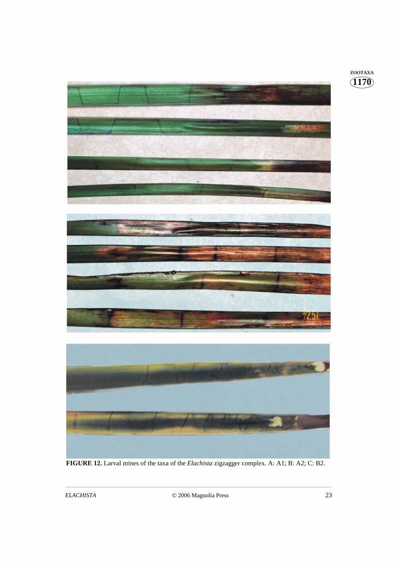
FIGURE 12. Larval mines of the taxa of the Elachista zigzagger complex. A: A1; B: A2; C: B2.
KAILA & STÅHLS24 © 2006 Magnolia Press
1170ZOOTAXA Discussion
Barcoding as a method for recognizing taxa is independent of questions as to whetherindividual taxa are species, what species are or should be, and where they fit in a unifiedtree of life (Besansky et al. 2003, Sperling 2003). We agree with this. The delimitation ofspecies in problematic situations should be based on maximal evidence derived fromdifferent sources, including morphological and ecological data and molecular evidencefrom more than one genomic fragment as already suggested by Sperling (2003, see alsoDayrat 2005). We suggest that the use of COI barcoding in species identification should berestricted to previously thoroughly studied specific cases, such as the pest ermine moths ofthe genus Yponomeuta (Yponomeutidae) (e.g. Sperling et al. 1995).
Our results support previous studies on the uninformativeness of the barcoding gene inproviding information in cases with recently diverged species. Of the nine putative speciesin the complex of Australian zigzagger species, only two groups (A and B) were recoveredwith consistent robust molecular support. Based on morphological and biologicalevidence, both of these groups segregate into several species. The taxon zigzagger B2displays a distinctive shape in its larval mine architecture, which supports its status as adistinct species; however, samples representing it (LK 55, 56, 59 and 60) were not nestedtogether in the COI tree (Fig. 1). Another species group, the Ficinia-complex of taxa,showed more intraspecific variation in their COI sequence within one putativelyconspecific population (all larvae sampled from a single tussock of the host plant) than didputatively different species of the zigzagger species complex. These complexes are (very)closely related to each other, yet they show distinctly different rates of differentiation. Thisfinding argues against generalisations about the utility of this single gene fragment inspecies delimitation, unless supported by other kinds of evidence.
Besansky et al. (2003) argued that DNA barcoding is not an end in itself, but may aidresearch and boost the rate of discovery. Our experience gives credit to the potential utilityof routine barcoding as a one source of taxonomic information, as even our small sampledetected one species (yellow complex 2) morphologically so close to others that it hadremained unrecognised. After characterised by the COI sequence, diagnosticmorphological features subsequently were recognised. Barcoding of a COI fragment iscertainly a useful source of information, among others sources, to explore the taxonomy ofinsufficiently known organisms. Nevertheless, caution should be taken when interpretingthe patterns observed from a single gene sequence. We emphasize that DNA taxonomyshould be firmly anchored with morphological taxonomy and take into account biologicaltraits of the organisms under study. Only in this way can pitfalls caused by features such asgenetic polymorphisms older than the divergences of the species in question (Wahlberg etal. 2003, Hebert et al. 2004) be avoided.
© 2006 Magnolia Press 25ELACHISTA
1170ZOOTAXAAcknowledgements
We are grateful to Elvira Rättel for technical help, Marianne Horak and Ted Edwards(ANIC, CSIRO Entomology, Canberra) for support during LK’s visits in Australia. KauriMikkola and Niklas Wahlberg kindly commented an earlier version. Two anonymousreviewers provided useful comments.
References
Albrecht, A. & Kaila, L. (1997) Variation of wing venation in Elachistidae (Lepidoptera, Gelechio-idea): methodology and implications to systematics. Systematic Entomology, 22, 185–198.
Besansky, N.J., Severson, D.W. & Ferdig, M.T. (2003) DNA barcoding of parasites and inverte-brate disease vectors: what you don’t know can hurt you. Trends in Parasitology, 19, 545–546.
Blaxter, M. (2003) Molecular systematics: Counting angels with DNA. Nature, 421, 122–124.Bremer, K. (1988) The limits of amino acid sequence data in angiosperms phylogenetic reconstruc-
tion. Evolution, 42, 795–803.Bremer, K. (1994) Branch support and tree stability. Cladistics, 10, 295–304.Clary, D.O. & Wolstenholme, D.R. (1985) The mitochondrial DNA molecule of Drosophila
yakuba. Nucleotide sequence, gene organization and geneticcode. Journal of Molecular Evolu-tion, 22, 252–271.
Dayrat, B. (2005) Towards integrative taxonomy. Biological Journal of the Linnean Society, 85,407–415.
Goloboff, P. (1999) NONA, vs. 2.0, parsimony program and documentation.Hebert, P.D.N., Cywinska, A., Ball, S.L. & deWaard, J.R. (2003a) Biological identifications
through DNA barcodes. Proceedings of the Royal Society of London, series B, 270, 313–321.Hebert, P.D.N., Ratnasingham, D. & de Waard, J.R. (2003b) Barcoding animal life: cytochrome c
oxidase subunit I divergences among closely related species. Proceedings of the Royal Societyof London, series B (Suppl.) DOI 10.1098/rsbl.2003.0025.
Hebert, P.D.N., Penton, E.H., Burns, J.M., Janzen, D.H. & Hallwachs, W. (2004) Ten species inone: DNA barcoding reveals cryptic species in the neotropical skipper butterfly Astraptesfulgerator. Proceedings of the National Academy of Sciences of the United States of America,101, 14812–14827.
Huemer, P. & Kaila, L. (2003) Elachista (Elachista) morandinii sp. n., a new species from CentralEurope (Lepidoptera, Elachistidae). Gortania, 24 (2002), 211–220.
Kaila, L. (1992) The Elachistidae of southern Siberia and Central Asia, with descriptions of fivenew species (Lepidoptera). Entomologica Fennica, 3, 177–194.
Kaila, L. (1996) A revision of the Nearctic Elachista s. l. I. The tetragonella group (Lepidoptera, Elachistidae). Entomologica scandinavica, 27, 217–238.
Kaila, L. (1997) A revision of the Nearctic Elachista s. l. II. The argentella group (Lepidoptera, Elachistidae). Acta Zoologica Fennica, 206, 1–93.
Kaila, L. (1998a) Redescriptions of three Meyrick’s Asiatic elachistid species, with descriptions ofthree new species (Lepidoptera, Gelechioidea). Entomologica Fennica, 9, 53–63.
Kaila, L. (1998b) Two new Elachista species (Lepidoptera, Elachistidae) from the Polar Uralsregion, Russia. Entomologica Fennica, 8, 219–223.
Kaila, L., (1999a) Phylogeny and classification of the Elachistidae s. s. (Lepidoptera, Gelechio-idea). Systematic Entomology, 24, 139–169.
Kaila, L., (1999b) A revision of the Nearctic species of the Elachista s. l. III. The bifasciella, prae-
KAILA & STÅHLS26 © 2006 Magnolia Press
1170ZOOTAXA lineata, saccharella and freyerella groups (Lepidoptera, Elachistidae). Acta Zoologica Fen-
nica, 211, 1–235.Kaila, L., (1999c) A review of the South-American Elachistidae s. str. (Lepidoptera, Gelechioidea)
with descriptions of 15 new species. Steenstrupia, 25, 159–193.Kaila, L. (2005) A review of Dibrachia Sinev & Sruoga, 1992, a subgenus of Elachista (E lachis-
tidae: Elachistinae). Nota lepidopterologica, 28, 139–155.Kaila, L., Bengtsson, B.Å., Šulcs, I. & Junnilainen, J. (2001) A revision of the Elachista regificella
Sircom –complex (Lepidoptera, Elachistidae). Entomologica Fennica, 12, 153–168.Kaila, L. & Jalava, J. (1994) Elachista adelpha sp. n., E. coeneni titanella ssp. n., and other Elachis-
tidae (Lepidoptera) from North Caucasus. Entomologica Fennica, 5, 97–102.Kaila, L. & Junnilainen, J. (2002) Taxonomy and identification of Elachista cingillella (H. S.) and
its close relatives (Lepidoptera, Elachistidae), with descriptions of two new species. Entomo-logica Fennica, 13, 167–188.
Kaila, L. & Karsholt, O. (2002) Contribution to the Lepidoptera fauna of the Madeira Islands 3. Elachistidae. Beiträge zur Entomologie, 52, 225–233.
Kaila, L., Nupponen, K., Junnilainen, J., Nupponen, T., Kaitila, J.-P. & Olschwang, V. (2003) Con-tribution to the fauna of Elachistidae (Lepidoptera) of the Southern Ural Mountains. Entomo-logica Fennica, 14, 65–90.
Kaila, L. & Sugisima, K. (2003) Phylogeny of Elachistinae (Lepidoptera: Gelechioidea) revisited.Cladistics, 19, 154–155.
Kaila, L. & Varalda, P. (2004) The Elachista juliensis complex revisited (Elachistidae). Nota lepi-dopterologica, 27, 217–237.
Nixon, K. (2002) WinClada., Version 1.00.08. Published by the author, Ithaca, New York.Prendini, L. (205) Comment on “identifying spiders through DNA barcodes”. Canadian Journal of
Zoology, 83. 498–504.Robinson, G.S. (1976) The preparation of slides of Lepidoptera genitalia with special reference to
the Microlepidoptera. Entomologist’s Gazette, 27, 127–132.Scoble, M.J. (1992) The Lepidoptera. Form, function and diversity. Oxford University Press, 404
pp.Simon, C., Frati, F., Beckenbach, A., Crespi, B., Liu, H. & Flook, P. (1994) Evolution, weighting
and phylogenetic utility of mitochondrial gene-sequences and a compilation of conserved poly-merase chain-reaction primers. Annals of the Entomological Society of America, 87, 651–701.
Sperling, F.A.H. (2003) Butterfly molecular systematics: From species definitions to higher-levelphylogenies. In: Boggs, C. L., Ehlrich, P. R. & Watt, W. B., (E d.), Butterflies: Ecology and Evolution Taking Flight: Butterflies as Model Study Systems. University of Chicago Press,Chapter 20, pp. 431–458.
Sperling, F.A, Landry, J.-F. & Hickey, D.A. (1995) DNA-based identification of introduced erminemoth species in North America (Lepidoptera: Yponomeutidae). Annals of the EntomologicalSociety of America, 88, 155–162.
Sugisima K. & Kaila, L. (2005) Japanese Elachista (Lepidoptera, Elachistidae s. str.) mining on theleaf of woody Poaceae. Entomologica Fennica, 16, 83–102.
Traugott-Olsen, E. & Schmidt Nielsen, E. (1977) The Elachistidae (Lepidoptera) of Fennoscandiaand Denmark. Fauna Enomologica Scandinavica, 6, 1–299.
Wahlberg, N., Oliveira, R. & Scott, J.A. (2003) Phylogenetic relationships of Phyciodes butterflyspecies (Lepidoptera: Nymphalidae): complex mtDNA variation and species delimitations.Systematic Entomology, 28, 257–273.
![OHBR Checklist: Butterflies & Moths (Lepidoptera) · 1 OHBR Checklist: Butterflies & Moths (Lepidoptera) ... 2 OHBR Checklist: Butterflies & Moths (Lepidoptera) ... (Hübner, [1817])](https://img.dokumen.tips/doc/110x75/5b86a2117f8b9a3a608d2f05/ohbr-checklist-butterflies-moths-lepidoptera-1-ohbr-checklist-butterflies.jpg)