Embed Size (px)
Citation preview

b r a z i l i a n j o u r n a l o f m i c r o b i o l o g y 4 9 (2 0 1 8) 128–137
ht t p: / /www.bjmicrobio l .com.br /
Clinical Microbiology
Yersinia pestis detection by loop-mediatedisothermal amplification combined with magneticbead capture of DNA
Na Fenga,b, Yazhou Zhoub, Yanxiao Fana,b, Yujing Bib, Ruifu Yangb, Yusen Zhoua,b,∗,Xiaoyi Wanga,b,∗
a Anhui Medical University, Anhui, People’s Republic of Chinab Beijing Institute of Microbiology and Epidemiology, State Key Laboratory of Pathogen and Biosecurity, Laboratory of AnalyticalMicrobiology, Beijing, China
a r t i c l e i n f o
Article history:
Received 22 June 2016
Accepted 17 March 2017
Available online 26 August 2017
Associate Editor: Roxane Piazza
Keywords:
Plague
Yersinia pestis
Loop-mediated isothermal
amplification
Magnetic beads
a b s t r a c t
We developed a loop-mediated isothermal amplification (LAMP) assay for the detection of Y.
pestis by targeting the 3a sequence on chromosome. All 11 species of the genus Yersinia were
used to evaluate the specificity of LAMP and PCR, demonstrating that the primers had a high
level of specificity. The sensitivity of LAMP or PCR was 2.3 or 23 CFU for pure culture, whereas
2.3 × 10⁴ or 2.3 × 10⁶ CFU for simulated spleen and lung samples. For simulated liver samples,
the sensitivity of LAMP was 2.3 × 10⁶ CFU, but PCR was negative at the level of 2.3 × 10⁷ CFU.
After simulated spleen and lung samples were treated with magnetic beads, the sensitivity
of LAMP or PCR was 2.3 × 10³ or 2.3 × 10⁶ CFU, whereas 2.3 × 10⁵ or 2.3 × 10⁷ CFU for magnetic
bead-treated liver samples. These results indicated that some components in the tissues
could inhibit LAMP and PCR, and liver tissue samples had a stronger inhibition to LAMP
and PCR than spleen and lung tissue samples. LAMP has a higher sensitivity than PCR, and
magnetic bead capture of DNAs could remarkably increase the sensitivity of LAMP. LAMP is
a simple, rapid and sensitive assay suitable for application in the field or poverty areas.
© 2017 Published by Elsevier Editora Ltda. on behalf of Sociedade Brasileira de
Microbiologia. This is an open access article under the CC BY-NC-ND license (http://
the U. S. Centers for Disease Control and Prevention. Histori-
Introduction
Plague is a zoonotic disease caused by Gram-negative bac-terium Yersinia pestis, which is occasionally transmitted to
humans from Y. pestis-infected rodents via the bites of infectedfleas.1 Y. pestis is thought to have evolved from a serotypeO: 1b strain of Y. pseudotuberculosis about 6000–10,000 years∗ Corresponding authors.E-mails: [email protected] (Y. Zhou), [email protected] (X. Wang
https://doi.org/10.1016/j.bjm.2017.03.0141517-8382/© 2017 Published by Elsevier Editora Ltda. on behalf of Socunder the CC BY-NC-ND license (http://creativecommons.org/licenses/
creativecommons.org/licenses/by-nc-nd/4.0/).
ago although these two species cause remarkably differ-ent diseases.2–3 Y. pestis is a highly virulent and infectiouspathogen, and was classified as a Category A pathogen by
4
).
cally, Y. pestis has given rise to three major plague pandemics,leading to millions of human deaths. Recently, the increasedoutbreak of plague around the world is reported annually to
iedade Brasileira de Microbiologia. This is an open access articleby-nc-nd/4.0/).

b r a z i l i a n j o u r n a l o f m i c r o b i o l o g y 4 9 (2 0 1 8) 128–137 129
Table 1 – Sequence of the primers for LAMP and PCR reactions.
Primer names Type Sequence (5′–3′) Primer length (bp)
3a-F3 Forward outer primer ACTACCATCCCCTCAAGGTT 203a-B3 Backward outer primer GAGGGCGTTTTGGTAGAGAA 203a-FIP Forward inner primer CACCCGCGTTATCTCATCCCG-TTTTCGAGTAGGGTTAGGTGGGC 44
GGCGATGG
tsabomt
mbYtumtimenpisppYLainp
oesrasfdaitriuspwosp
3a-BIP Backward inner primer CATGGACGTAT3a-LF Forward loop primer ACCGCCATGAA
he World Health Organization (WHO), and plague was clas-ified as a re-emerging infectious disease by the WHO.5–6 Inddition, plague has been attracting a considerable attentionecause its causative agent has always been recognized as onef the classical biological warfare or bioterrorism agents.7–8 Toinimize these threats, development of a rapid method for
he detection of Y. pestis is essential.Y. pestis was often detected by bacterial isolation and
icroscopy observation,9 phage lysis assay,10–11 ELISA assaysased on the detection of F1 antigen and antibodies against. pestis,12–15 conventional PCR assays,16–18 real-time quan-itative PCR assays,19–28 biosensors based on fiber-optic orpconverting phosphor technology,29–31 solid-phase radioim-unoassay based on radiolabeled monoclonal antibody for
he detection of plague antigen.32 All these methods are play-ng an important role in the diagnosis of plague, but these
ethods either are time-consuming and laborious, or requirexpensive equipment and personnel with a high level of tech-ical expertise. However, permanent surveillance of the foci oflague located in the poor regions to predict future epizootics
n rodents and exposure risk for humans or investigation ofamples suspected of bioterrorism on site requires a sim-le, rapid and efficient diagnostic method. A colloidal goldarticles-based lateral-flow (LF) strip detection method for. pestis has been developed based on antibodies to F1 andcrV proteins.33–34 This dipstick test is a low-cost, easy-to-usend rapid screening method in the surveillance of plague ornvestigation of samples suspected of bioterrorism on site, butucleic acid-based rapid detection technology could be a moreowerful alternative for detecting Y. pestis.
Loop-mediated isothermal amplification (LAMP) technol-gy has received considerable attention because it allowsfficiently amplification of DNA with high specificity and sen-itivity under isothermal conditions of 60–65 ◦C. The LAMPeaction can be accomplished within less than 1 h based on
set of four to six primers and the Bst DNA polymerase withtrand displacement activity.35 The method is more suitableor field applications, especially in poverty areas, because itoes not require specialized or expensive equipment, and only
simple and inexpensive water bath or heating block can sat-sfy LAMP assay.36–37 In addition, LAMP results can be read byhe naked eye, or the lateral flow dipstick (LFD) under natu-al light, which makes the detection results easy to be judgedn the field. Currently, the LAMP technique has been widelysed in the diagnosis of infectious diseases,38 and it is moreensitive in detecting bacteria compared to the conventionalolymerase chain reaction (PCR) method.39–40 In this study,
e construct a simple and rapid LAMP method for detectionf Y. pestis based on the specific sequence 3a (GenBank acces-ion no. AF350075) that is a specific fragment located on Y.estis chromosome found by using a comparative genomicGGTCA-TTTTGTGATGCCGTCCAATGCA 42ACAATG 21
method.41 The specificity of the method was evaluated byusing all 11 species of the genus Yersinia. The sensitivity wasevaluated by using Y. pestis pure culture, and simulated tissuesamples, and magnetic bead-treated simulated tissue sam-ples.
Materials and methods
Bacterial strains, reagents, instruments and animals
Y. pestis EV vaccine strain, Y. pseudotuberculosis, Y. enterocolit-ica, Y. frederiksenii, Y. intermedia, Y. kristensenii, Y. bercovieri, Y.mollaretii, Y. rohdei, Y. ruckeri, and Y. aldovae were used to evalu-ate the specificity of LAMP and PCR; 10× thermopol buffer,MgSO4 (100 mM), and Bst DNA Polymerase, Large Fragment(8000 U/ml) were purchased from NEB (Beijing, China); Cal-cein and real-time turbidity meter LA-320c were purchasedfrom Eiken China CO., LTD.; Taq DNA polymerase (5 U) anddNTPs (2.5 mM) were purchased from TaKaRa (Dalian, China);SM3-P100, amino-modified silica-coated magnetic beads, waspurchased form Shanghai Allrun Nano Science & Technol-ogy CO., LTD. BALB/c were obtained from Laboratory AnimalResearch Center, Academy of Military Medical Science, China(licensed from the Ministry of Health in General LogisticsDepartment of Chinese People’s Liberation Army, Permit No.SCXK-2007-004). The protocols were approved by Committeeof the Welfare and Ethics of Laboratory Animals, Beijing Insti-tute of Microbiology and Epidemiology. The experiments wereconducted strictly in compliance with the Regulations of GoodLaboratory Practice for nonclinical laboratory studies of drugissued by the National Scientific and Technologic Committeeof People’s Republic of China.
LAMP and PCR assays
For LAMP reaction, a set of five primers was designed accord-ing to the published sequence of 3a in Y. pestis KIM D46(GenBank accession no.: AF350075) using Primer Explorerversion 4. A forward inner primer (FIP), a backward innerprimer (BIP), two outer primers (F3 and B3), and a loop for-ward primer (LF) were used for LAMP amplification. Thesequences of the primers are shown in Table 1. LAMP reactionwas performed in a total volume of 25 �L mixture contain-ing 10× Thermopol buffer (2.5 �L), 100 mM MgSO4 (1.5 �L),2.5 mM dNTP Mix (14 �L), 100 mM 3a-FIP (0.4 �L), 100 mM 3a-BIP (0.4 �L), 10 mM 3a-F3 (0.5 �L), 10 mM 3a-B3 (0.5 �L), 10 mM
3a-LF (0.2 �L), 8000 U/mL of Bst DNA Polymerase, Large Frag-ment (1 �L), water (3 �L), template DNA (1 �L). The LAMPreaction was carried out at 65 ◦C for 60 min and inactivated at80 ◦C for 5 min in water bath. For direct visual detection of DNA
130 b r a z i l i a n j o u r n a l o f m i c r o b i o l o g y 4 9 (2 0 1 8) 128–137
A E
B F
GC
D H
1.21.21.4
110.8
0.80.60.60.4 0.40.2 0.2
0 0-0.2 -0.2
0 010 1020 2030 3040 4050 5060 6070 70
Time (min) Time (min)
Turb
idity
(40
0nm
)
Turb
idity
(40
0nm
)
1 1
1 1
2 2
2 2
3 3
3 3
44
4 4
55
5 5
66
6 6
7
7
7 7
8
8
8 8
9
9 9
10
10
1 2 3 5
5
6
6
7
7
8
8
9
9
10
101 2 43
1 2 3 4 45 6 7 8 9 10
10
11
11
11
1111
12
12
12
12
13
13 145 6 7 8 9 101 2 43 11 12
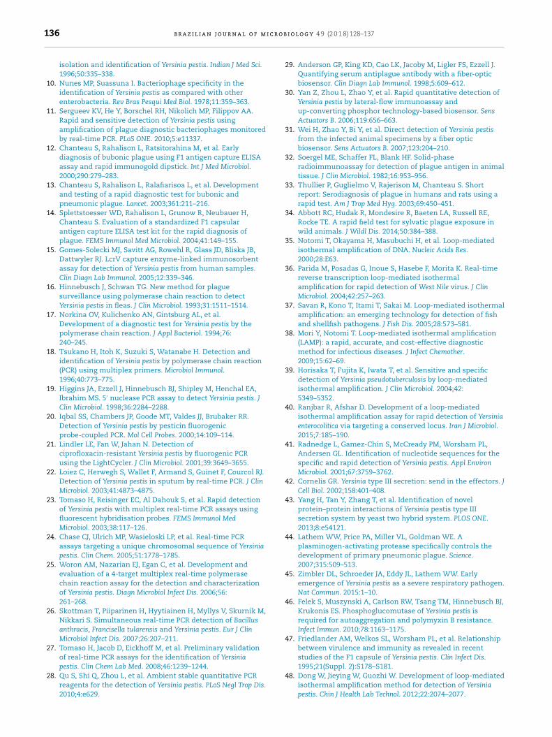
Fig. 1 – The sensitivity (A–D) of the LAMP or PCR for the detection of Y. pestis using serial dilutions of extracted DNA, and thespecificity (E–H) of the LAMP or PCR for the detection of Y. pestis using 20 ng DNA from each bacterium of the genus Yersinia.(A) Detection of LAMP products with a real-time turbidity meter. 1–8: 20 ng–0.002 pg; 10: negative control. (B) Visualization ofLAMP products stained with calcein and inspected under natural light. Tube 1–9: 20 ng–0.0002 pg; 10: negative control. (C)Detection of LAMP products with agarose gel electrophoresis. Lane 1: marker; 2–10: 20 ng–0.0002 pg; 11: negative control. (D)Electrophoretic analysis of PCR products with agarose gel. Lane 1: marker; 2–10: 20 ng–0.0002 pg; 11: negative control; 12:marker. (E) Detection of the LAMP products with a real-time turbidity meter. 1: Y. pestis; 2: Y. pseudotuberculosis; 3: Y.enterocolitica; 4: Y. frederiksenii; 5: Y. intermedia; 6: Y. kristensenii; 7: Y. bercovieri; 8: Y. mollaretii; 9: Y. rohdei; 10: Y. ruckeri; 11: Y.aldovae; 12: negative control. (F) Visualization of LAMP products stained with calcein and inspected under natural light. Tube1: Y. pestis; 2: Y. pseudotuberculosis; 3: Y. enterocolitica; 4: Y. frederiksenii; 5: Y. intermedia; 6: Y. kristensenii; 7: Y. bercovieri; 8: Y.mollaretii; 9: Y. rohdei; 10: Y. ruckeri; 11: Y. aldovae; 12: negative control. (G) Detection of LAMP products with agarose gelelectrophoresis. Lane 1: marker; 2: Y. pestis; 3: Y. pseudotuberculosis; 4: Y. enterocolitica; 5: Y. frederiksenii; 6: Y. intermedia; 7: Y.kristensenii; 8: Y. bercovieri; 9: Y. mollaretii; 10: Y. rohdei; 11: Y. ruckeri; 12: Y. aldovae; 13: negative control. (H) Electrophoreticanalysis of PCR products with 1.5% agarose gel. Lane 1: marker; 2: Y. pestis; 3: Y. pseudotuberculosis; 4: Y. enterocolitica; 5: Y.frederiksenii; 6: Y. intermedia; 7: Y. kristensenii; 8: Y. bercovieri; 9: Y. mollaretii; 10: Y. rohdei; 11: Y. ruckeri; 12: Y. aldovae; 13:negative control; 14: marker.
amplification, 1 �L of Fluorescent Detection Reagent contain-ing calcein (EIKEN CHINA CO., LTD.) was added to the reactiontube before the LAMP reaction. For a positive reaction, a colorchange from orange to green was observed through the nakedeye under natural light, whereas a negative reaction mixtureremained orange. Alternatively, specific DNA amplificationwas monitored spectrophotometrically by a real-time turbid-ity meter. To confirm that color change in reaction tubes is aresult of DNA amplification, LAMP product was also detectedby 0.8% agarose gel electrophoresis and ethidium bromidestaining.
Conventional PCR reaction was performed in a total vol-ume of 25 �L mixture containing 10 × buffer (3 �L), 2.5 mMdNTPs (0.6 �L), 10 mM 3a-F3 (0.5 �L), 10 mM 3a-B3 (0.5 �L), 5 U
of Taq DNA polymerase (0.1 �L), deionized water (15.3 �L) andtemplate DNA (5 �L). F3 and B3 primers were used for theconventional PCR amplification (Table 1). The reaction wassubjected to an initial denaturation at 95 ◦C for 5 min, followedby 35 cycles of denaturation at 95 ◦C for 30 s, annealing at56 ◦C for 30 s, extension at 72 ◦C for 1 min and a final exten-sion at 72 ◦C for 5 min. The PCR products were analyzed by1.5% agarose gel electrophoresis and stained with ethidiumbromide.
Extraction of bacterial genomic DNA
For preparation of high-quality genomic DNA, Y. pestis EV vac-cine strain and other bacteria were cultured in Luria broth

b r a z i l i a n j o u r n a l o f m i c r o b i o l o g y 4 9 (2 0 1 8) 128–137 131
1.21
0.80.60.40.2
0-0.2
1.21.41.6
10.80.60.40.2
0-0.2
0 10 20 30 40 50 60 70 0 10 20 30 40 50 60 70
1 123 345 567 78
2468
9 9 1010 11 12
Time (min) Time (min)Tu
rbid
ity (
400n
m)
Turb
idity
(40
0nm
)
A
EB
FC
D
1 2 3 4 5 6 7 8 9 10 1 2 3 4 5 6 7 8 9 10 11 12
1 2 3 4 5 6 7 8 9 10 11 12 5 6 7 8 9 101 2 43 11 12 13 14
Fig. 2 – The sensitivity (A–C) of the LAMP and PCR for the detection of Y. pestis by using the DNA extracted crudely fromserial dilutions of bacterial solutions, and the specificity (D–F) of the LAMP and PCR for the detection of Y. pestis usingcrudely extracted DNA from each bacterium (2.3 × 10⁷ CFU) of the genus Yersinia. (A) Detection of the LAMP products with areal-time turbidity meter. 1–9: 2.3 × 10⁷–2.3 × 10⁻¹ CFU of Y. pestis; 10: negative control. (B) Visualization of LAMP productsstained with calcein and inspected under natural light. Tube 1–9: 2.3 × 10⁷–2.3 × 10⁻¹ CFU of Y. pestis; 10: negative control. (C)Electrophoretic analysis of PCR products with agarose gel. Lane 1: marker; Lane 2–10: 2.3 × 10⁷–2.3 × 10⁻¹ CFU of Y. pestis; 11:negative control; 12: marker. (D) Detection of LAMP products with a real-time turbidity meter. 1: Y. pestis; 2: Y.pseudotuberculosis; 3: Y. enterocolitica; 4: Y. frederiksenii; 5: Y. intermedia; 6: Y. kristensenii; 7: Y. bercovieri; 8: Y. mollaretii; 9: Y.rohdei; 10: Y. ruckeri; 11: Y. aldovae; 12: negative control. (E) Visualization of LAMP products stained with calcein andinspected under natural light. Tube 1: Y. pestis; 2: Y. pseudotuberculosis; 3: Y. enterocolitica; 4: Y. frederiksenii; 5: Y. intermedia; 6:Y. kristensenii; 7: Y. bercovieri; 8: Y. mollaretii; 9: Y. rohdei; 10: Y. ruckeri; 11: Y. aldovae; 12: negative control. (F) Electrophoreticanalysis of PCR products with agarose gel. Lane 1: marker; 2: Y. pestis; 3: Y. pseudotuberculosis; 4: Y. enterocolitica; 5: Y.frederiksenii; 6: Y. intermedia; 7: Y. kristensenii; 8: Y. bercovieri; 9: Y. mollaretii; 10: Y. rohdei; 11: Y. ruckeri; 12: Y. aldovae; 13:n
(tS8aetipt3stoF1f
tgsur
egative control; 14: marker.
LB) at 26 ◦C and 37 ◦C, respectively, and then collected by cen-rifugation. The collected bacterial pellet was resuspended inE buffer (0.15 M NaCl, 0.1 M EDTA-2Na·2H2O, adjusted to pH.0 by 10 M HCl), added to a final concentration of 2% SDS,nd then placed in a water bath at 60 ◦C for 10 min. Then, anqual volume of phenol, chloroform and isoamyl alcohol mix-ure (25:4:1) was added and the solution was mixed by slowlynverting or rotating to denature proteins, then to removeroteins. The aqueous phase was added a final concentra-ion of 50–100 �g/ml of RNase to degrade RNAs at 37 ◦C for0–60 min. Then, an equal volume of chloroform was added,haking vigorously. The aqueous phase was precipitated withwo times volume of ethanol, and then the DNA was spooledut with a glass rod, rinsed with 70% ethanol, and dried in air.inally, the DNA was dissolved into TE buffer (10 mM Tris–HCl,
mM EDTA-2Na·2H2O, adjusted to pH 8.0 by 10 M HCl)or use.
For simple and rapid isolation of bacterial DNA, the bac-erial cells were resuspended in deionized water, and then
enomic DNA was released by boiling the bacterial suspen-ion at 100 ◦C for 10 min. The supernatant was collected andsed as the DNA template in both LAMP and conventional PCReactions.Capture of bacterial genomic DNA by magnetic beads
Bacterial genomic DNA was released by boiling bacterial sus-pension at 100 ◦C for 10 min, 300 �L of the supernatant wasmixed with the same volume of binding buffer [4 M NaCland 20% polyethylene glycol (PEG) 8000], followed by additionof 0.2 mg magnetic nanoparticles SM3-P100. The suspensionwas vibrated vigorously for 15 s, followed by standing at roomtemperature for 5 min. The magnetic beads were collected byusing an external magnet, and the supernatant was discarded.The magnetic pellet was washed two times with 200 �L of70% ethanol, and then dried completely at room tempera-ture. Finally, the magnetic pellet was resuspended in 100 �Lof deionized water to elute bound DNA at room temperaturefor 5 min with continuous agitation. The supernatant contain-ing the genomic DNA was used as the DNA template in bothLAMP and conventional PCR assays.
Preparation of simulated samples and magnetic bead
capture of DNAThe spleen, liver and lungs were collected aseptically frompathogen-free BALB/c mice, and then blended with the

132 b r a z i l i a n j o u r n a l o f m i c r o b i o l o g y 4 9 (2 0 1 8) 128–137
1.21
0.80.60.40.2
0-0.2
1.21
0.80.60.40.2
0-0.2
0 10 20 30 40 50 60 70 0 10 20 30 40 50 60 70
1 12 23 34 45 56 67 78 89 910 10
5 6 7 8 9 101 2 43 1 2 3 5 6 7 8 9 104
5 6 7 8 9 101 2 43 11 125 6 7 8 9 101 2 43 11 12
A
EB
FC
D
Time (min)Time (min)Tu
rbid
ity (
400n
m)
Turb
idity
(40
0nm
)Fig. 3 – Detection of Y. pestis in simulated spleen samples by LAMP and PCR. The sensitivity of LAMP or PCR wasdetermined by using the DNA extracted by boiling simulated spleen sample for 10 min (A–C) or by using the DNA capturedby magnetic beads (D–F). (A) Detection of LAMP products with a real-time turbidity meter. 1–8: 2.3 × 100–2.3 × 10⁷ CFU of Y.pestis; 9: positive control; 10: negative control. (B) Visualization of LAMP products stained with calcein and inspected undernatural light. Tube 1–8: 2.3 × 100–2.3 × 10⁷ CFU of Y. pestis; 9: positive control; 10: negative control. (C) Electrophoreticanalysis of PCR products with agarose gel. Lane 1: marker; Lane 2–9: 2.3 × 10⁷–2.3 × 100 CFU of Y. pestis; 10: positive control;11: negative control; 12: marker. (D) Detection of LAMP products with a real-time turbidity meter. 1–8: 2.3 × 100–2.3 × 10⁷ CFUof Y. pestis; 9: positive control; 10: negative control. (E) Visualization of LAMP products stained with calcein and inspectedunder natural light. Tube 1–8: 2.3 × 100–2.3 × 10⁷ CFU of Y. pestis; 9: positive control; 10: negative control. (F) Electrophoreticanalysis of PCR products with agarose gel. Lane 1: marker; Lane 2–9: 2.3 × 10⁷–2.3 × 100 CFU of Y. pestis; 10: positive control(20 ng DNA); 11: negative control; 12: marker.
of the LAMP or PCR was also evaluated with pure cultures from
appropriate volume of PBS buffer to prepare tissuehomogenates. Y. pestis was cultured in LB, collected bycentrifugation, and then resuspended in deionized water toprepare serial 10-fold dilutions of bacterial suspension. Thespleen, liver and lung tissue homogenates were respectivelyinoculated with the same volume of serial 10-fold dilutionsof bacterial suspension to obtain different concentrations ofartificially contaminated samples (2.3 × 10⁷–2.3 × 10⁻¹ CFU).Noninoculated tissue homogenates were mixed with thesame volume of deionized water to be used as a negativecontrol. Artificially contaminated samples for each tissuewere boiled at 100 ◦C for 10 min, and then centrifugated at10,000 × g for 2 min. The supernatants can be directly used asthe DNA template in both LAMP and conventional PCR assays.The supernatants can be further used to isolate bacterialDNA by magnetic beads according to the protocol describedabove, and then used in both LAMP and conventional PCRassays.
Determination of LAMP or PCR sensitivity
The sensitivity of LAMP or PCR for the detection of Y. pestis wasevaluated with the high-quality genomic DNA. Serial 10-folddilutions of DNA were made in sterile deionized water, andused as the DNA templates to perform LAMP and PCR assays.
The sensitivity of LAMP or PCR was evaluated with Y. pestispure culture. Serial 10-fold dilutions of Y. pestis cells were madein sterile deionized water, boiled at 100 ◦C for 10 min, and thesupernatants were used for LAMP and conventional PCR reac-tions. The sensitivity of LAMP or PCR was evaluated with thesimulated samples prepared by using mouse spleen, liver andlungs. Serial 10-fold dilutions of Y. pestis cells were respec-tively spiked into the same volume of tissue homogenates,and boiled at 100 ◦C for 10 min. The supernatants were directlyused for LAMP and PCR assays. In addition, the bacterialDNA was captured from the supernatants by magnetic beads,and then used to determine the sensitivity of LAMP and PCRassays.
Evaluation of specificity of LAMP or PCR
The specificity of LAMP or PCR for the detection of Y. pestiswas evaluated by using the high-quality bacterial DNAs (20 ng)from 11 species of bacteria in the genus Yersinia. The specificity
11 species of bacteria in the genus Yersinia. Each bacterium wasdiluted to 107 CFU in sterile deionized water, boiled at 100 ◦Cfor 10 min, and the supernatants were used for LAMP and PCRreactions.

b r a z i l i a n j o u r n a l o f m i c r o b i o l o g y 4 9 (2 0 1 8) 128–137 133
Turb
idity
(40
0nm
)
Turb
idity
(40
0nm
)
Time (min) Time (min)
13579
24
6810
0 10 20 30 40 50 60 70 0 10 20 30 40 50 60 70
1.21.21
0.80.60.40.2
0-0.2
10.80.60.40.2
0-0.2
135
79
246
810
5 6 7 8 9 101 2 43
5 6 7 8 9 101 2 43 11 125 6 7 8 9 101 2 43 11 12
5 6 7 8 9 101 2 43
A
EB
FC
D
Fig. 4 – Detection of Y. pestis in simulated liver samples by LAMP and PCR. The sensitivity of LAMP or PCR was determinedby using the DNA extracted by boiling simulated liver sample for 10 min (A-C) or by using the DNA captured by magneticbeads (D–F). (A) Detection of LAMP products with a real-time turbidity meter. 1–8: 2.3 × 100–2.3 × 10⁷ CFU of Y. pestis; 9:positive control; 10: negative control. (B) Visualization of LAMP products stained with calcein and inspected under naturallight. Tube 1–8: 2.3 × 100–2.3 × 10⁷ CFU of Y. pestis; 9: positive control; 10: negative control. (C) Electrophoretic analysis of PCRproducts with agarose gel. Lane 1: marker; Lane 2–9: 2.3 × 10⁷–2.3 × 100 CFU of Y. pestis; 10: positive control (20 ng DNA); 11:negative control; 12: marker. (D) Detection of LAMP products with a real-time turbidity meter. 1–8: 2.3 × 100–2.3 × 10⁷ CFU ofY. pestis; 9: positive control; 10: negative control. (E) Visualization of LAMP products stained with calcein and inspectedunder natural light. Tube 1–8: 2.3 × 100–2.3 × 10⁷ CFU of Y. pestis; 9: positive control; 10: negative control. (F) Electrophoretica ane
(
R
Ta
T1TwbesrfmmtuusLh
Ta
TL
nalysis of PCR products with agarose gel. Lane 1: marker; L20 ng DNA); 11: negative control; 12: marker.
esults
he sensitivity of detection of Y. pestis by LAMP and PCRssays
o determine the sensitivity of LAMP or PCR assay, serial0-fold dilutions of high-purity Y. pestis DNAs were tested.he results showed that the lowest detection limit for LAMPas 0.02 ng of Y. pestis DNA when the product was detected
y the naked eye, a real-time turbidity meter or agarose gellectrophoresis and ethidium bromide staining, whereas theensitivity for PCR was 0.2 ng of Y. pestis DNA (Fig. 1A–D). Theseesults indicated that LAMP had a higher sensitivity than PCRor the detection of Y. pestis DNA, and two LAMP detection
ethods had the same sensitivity. When Y. pestis was deter-ined by using serial 10-fold dilutions of bacterial solution,
he sensitivity for LAMP was 2.3 CFU of Y. pestis when the prod-ct was detected by a real-time turbidity meter, the naked eyender natural light or agarose gel electrophoresis, whereas theensitivity for PCR was 23 CFU of Y. pestis (Fig. 2A–D). ThreeAMP detection methods also had the same sensitivity, andad a higher sensitivity than PCR.
he specificity of detection of Y. pestis by LAMP and PCR
ssayso determine the specificity for the detection of Y. pestis byAMP or PCR assay, 11 species of the genus Yersinia were tested.
2–9: 2.3 × 10⁷–2.3 × 100 CFU of Y. pestis; 10: positive control
When the high-purity DNAs extracted from 11 species of thegenus Yersinia were used for the detection of Y. pestis, respec-tively, the LAMP product was only detected in the reaction tubeof Y. pestis, and no LAMP amplicons appeared in the reactiontubes of other Yersinia strains. Similarly, when the high-purityDNAs extracted from 11 species of the genus Yersinia wererespectively used to detect Y. pestis by PCR, specific band wasonly observed in Y. pestis EV vaccine strain (Fig. 1D–F). WhenY. pestis was determined by using 107 CFU of pure culturefrom 11 species of the genus Yersinia, LAMP product was onlydetected in the reaction tube of Y. pestis, and no LAMP ampli-cons appeared in the reaction tubes of other Yersinia strains.Similarly, when 11 species of the genus Yersinia were used todetect Y. pestis by PCR, specific band was only observed in Y.pestis EV vaccine strain (Fig. 2D–F).
Detection of Y. pestis in simulated samples by LAMP andPCR assays
To determine the sensitivity for the detection of Y. pestis insimulated spleen, liver and lung samples by LAMP or PCR.Artificially contaminated spleen, liver and lung samples con-taining different concentrations of Y. pestis were prepared by
mixing spleen, liver and lung tissue homogenates with thesame volume of serial 10-fold dilutions of Y. pestis suspension.Artificially contaminated spleen, liver and lung samples wereboiled for 10 min, and then the supernatants were directly
134 b r a z i l i a n j o u r n a l o f m i c r o b i o l o g y 4 9 (2 0 1 8) 128–137
Turb
idity
(40
0nm
)
Turb
idity
(40
0nm
)
Time (min) Time (min)
1.21
0.80.60.40.2
0-0.2
1.21
0.80.60.40.2
0-0.20 10 20 30 40 50 60 70 0 10 20 30 40 50 60 70
5 6 7 8 9 101 2 435 6 7 8 9 101 2 43
5 6 7 8 9 101 2 43 11 125 6 7 8 9 101 2 43 11 12
2
46810
246810
13579
13579
A
EB
FC
D
Fig. 5 – Detection of Y. pestis in simulated lung samples by LAMP and PCR. The sensitivity of LAMP or PCR was determinedby using the DNA extracted by boiling simulated lung samples for 10 min (A–C) or by using the DNA captured by magneticbeads (D–F). (A) Detection of LAMP products with a real-time turbidity meter. 1–8: 2.3 × 100–2.3 × 10⁷ CFU of Y. pestis; 9:positive control; 10: negative control. (B) Visualization of LAMP products stained with calcein and inspected under naturallight. Tube 1–8: 2.3 × 100–2.3 × 10⁷ CFU of Y. pestis; 9: positive control; 10: negative control. (C) Electrophoretic analysis of PCRproducts with agarose gel. Lane 1: marker; Lane 2–9: 2.3 × 10⁷–2.3 × 100 CFU of Y. pestis; 10: positive control (20 ng DNA); 11:negative control; 12: marker. (D) Detection of LAMP products with a real-time turbidity meter. 1–8: 2.3 × 100–2.3 × 10⁷ CFU ofY. pestis; 9: positive control; 10: negative control. (E) Visualization of LAMP products stained with calcein and inspectedunder natural light. Tube 1–8: 2.3 × 100–2.3 × 10⁷ CFU of Y. pestis; 9: positive control; 10: negative control. (F) Electrophoreticanalysis of PCR products with agarose gel. Lane 1: marker; Lane 2–9: 2.3 × 10⁷–2.3 × 100 CFU of Y. pestis; 10: positive control
(20 ng DNA); 11: negative control; 12: marker.determined by LAMP and PCR assays, respectively. The sensi-tivity for the artificially contaminated spleen by LAMP or PCRassay was 2.3 × 10⁴ or 2.3 × 10⁶ CFU of Y. pestis, respectively.The LAMP assay was 100-fold more sensitive than PCR assay(Fig. 3A–C). The lowest detection limit for the artificially con-taminated liver samples by LAMP was 2.3 × 10⁶ CFU of Y. pestis,but PCR assay didn’t detect 2.3 × 10⁷ CFU of Y. pestis in the sim-ulated liver samples (Fig. 4A–C). The lowest detection limit forthe artificially contaminated lung samples by LAMP and PCRassay was 2.3 × 10⁴ and 2.3 × 10⁶ CFU of Y. pestis, respectively.The LAMP assay was 100-fold more sensitive than PCR assay(Fig. 5A–C). To increase the sensitivity for the detection of Y.pestis in the simulated samples, the supernatants obtainedby boiling the artificially contaminated spleen, liver and lungsamples were respectively mixed with the magnetic beads tocapture the DNAs for LAMP and PCR assays. After the DNAsin the simulated spleen samples were captured by magneticbeads, the sensitivity for LAMP or PCR assay was 103 or 106 CFUof Y. pestis, respectively. LAMP assay was 1000-fold more sen-sitive than PCR assay (Fig. 3E and F). The lowest detectionlimit for the artificially contaminated liver by LAMP or PCR was2.3 × 105 or 2.3 × 10⁷ CFU of Y. pestis. LAMP assay was 100-foldmore sensitive than PCR assay (Fig. 4E and F). The sensitivity
for the artificially contaminated lungs by LAMP or PCR assaywas 103 or 106 CFU of Y. pestis. LAMP assay was 1000-fold moresensitive than PCR assay (Fig. 5E and F).Discussion
Y. pestis strains usually contain three virulence plasmids pCD1,pMT1 and pPCP1, of which pCD1 was inherited from its ances-tor Y. pseudotuberculosis. The plasmid pCD1 mainly encodestype three secretion system (T3SS), which plays an essentialrole in the pathogenesis of Y. pestis.42–43 The newly acquiredpPCP1 encodes a surface proteinase, plasminogen activator(Pla), that is responsible for the increase of the pathogen’sinvasive ability in bubonic plague and the replication of Y.pestis rapidly in the lungs.44–45 Another plasmid pMT1 encodesF1 antigen, one of the major protective antigens in Y. pestis,and the murine toxin (Ymt) required for survival of Y. pestisin the midgut of flea.46 Immunological diagnosis of Y. pestiswas mainly based on F1 antigen or its antibodies, but thismethod could not detect the virulent Y. pestis strains lackingF1 antigen.47 In addition, our recent studies found some nat-ural isolates of Y. pestis with one or two plasmids. By contrast,the target genes on chromosome might be more stable. In thisstudy, we constructed a LAMP method to detect Y. pestis basedon the 3a sequence on chromosome, which might recognize agreater amount of samples than previous studies. Currently,
there have been two reports that described the detection ofY. pestis by LAMP.48–49 Compared with these two reports, ourmethod had a similar sensitivity with Nune’s method for the
r o b i
dDtpeacmiitp
epsfbYtb1wpfnCLpsi
fiLtDtYitptdabotp
bYrpDlwsLtt
r
b r a z i l i a n j o u r n a l o f m i c
etection of Y. pestis DNA, but showed a higher sensitivity thanong’s method for the detection of Y. pestis pure culture. All
hese three methods had a higher specificity. However, the tworevious reports are based on the target of F1 antigen genencoded on the pMT1 for the detection of Y. pestis pure culturend high-quality genomic DNA. Evidently, these two methodsannot detect Y. pestis strains lacking F1 antigen on the plas-id pMT1, and a LAMP method is also needed to detect Y. pestis
n actual samples. Based on the LAMP method we constructedn this study, we determined simulated spleen, liver and lungissue samples combined with magnetic bead capture of Y.estis DNA.
The genus Yersinia contains 11 species, of which Yersinianterocolitica, Yersinia pseudotuberculosis and Y. pestis areathogenic for humans and animals, whereas other eightpecies, formerly called Y. enterocolitica-like isolates, Yersiniarederiksenii, Yersinia intermedia, Yersinia kristensenii, Yersiniaercovieri, Yersinia mollaretii, Yersinia rohdei, Yersinia ruckeri, andersinia aldovae are opportunistic pathogens mostly found inhe environment.1 To evaluate the specificity of the primersased on the 3a sequence for the detection of Y. pestis, all1 species of the genus Yersinia, closely related organisms,ere tested in the experiments. The results showed that therimers designed in this study had a high level of specificityor Y. pestis by LAMP or PCR. However, in our recent study, 3a-egative natural isolates of Y. pestis were found in Dingbianounty, Shaanxi Province, China,50 indicating that a 3a-basedAMP or PCR method is also not reliable for the detection of Y.estis. Therefore, a multi-target LAMP or PCR detection methodhould be developed for the reliable identification of Y. pestisn the future.
To determine the sensitivity for the detection of Y. pestis, werst use serial 10-fold dilutions of high-quality DNA to performAMP or PCR assay. The results showed that the lowest detec-ion limit of LAMP or PCR assay was 0.02 or 0.2 ng of Y. pestisNA, indicating that LAMP assay was 10-fold more sensitive
han conventional PCR assay. Then, serial 10-fold dilutions of. pestis pure culture were used to crudely extract DNA by boil-ng, and then detected by LAMP and PCR. The results showedhat the sensitivity of LAMP or PCR assay was 2.3 or 23 CFU of Y.estis, indicating that LAMP assay was 10-fold more sensitivehan PCR assay. In addition, the results also showed that theetection of LAMP products by the naked eye was as sensitives that by a real-time turbidity meter. LAMP amplification cane accomplished within 60 min at an isothermal temperaturef 65 ◦C. Therefore, LAMP assay is a simple, rapid and sensi-ive method suitable for application in the field, especially inoverty areas.
To evaluate the reliability for the determination of Y. pestisy LAMP and PCR assays in actual samples, we determined. pestis in the simulated spleen, liver and lung samples. Theesults showed that the sensitivity of three simulated sam-les were much lower than that of Y. pestis pure culture andNA, indicating that some components in the spleen, liver and
ung tissues might inhibit LAMP and PCR reactions. This resultas similar to the previous studies in which clinical samples,
uch as serum, plasma and urine, had an inhibition to bothAMP and PCR assay.51–52 In addition, our results also showedhat the simulated liver sample had a lower sensitivity thanhe simulated spleen and lung samples for the detection of Y.
o l o g y 4 9 (2 0 1 8) 128–137 135
pestis by LAMP and PCR. This result indicated that there mightbe more bioactive components in liver tissue that could inhibitLAMP and PCR reactions. Compared with PCR for the detec-tion of three simulated samples, LAMP assay was 100-foldmore sensitive than PCR assay, indicating that LAMP was lessaffected by inhibitory substances present in mouse spleen,liver and lung tissue samples than PCR. LAMP is not only moresimple, rapid and sensitive than PCR, but also more suitableto detect spleen, liver and lung tissue samples than PCR.
To further enhance the sensitivity for the detection ofactual samples, magnetic beads were used to capture Y. pestisDNAs from three simulated samples. After the DNAs werecaptured from the simulated spleen and lung samples withmagnetic beads, the sensitivity of these two samples wereincreased for the detection of Y. pestis by LAMP, but the sensi-tivity of these two samples were unchanged for the detectionof Y. pestis by PCR, and LAMP was 1000-fold more sensitivethan PCR. After the DNAs were captured from simulated liversamples with magnetic beads, the sensitivity of LAMP andPCR were equally increased, and the sensitivity of LAMP was100-fold more sensitive than that of PCR. These results indi-cated that magnetic bead capture of DNAs could remarkablyincrease the sensitivity of LAMP for the detection of Y. pestisin spleen, liver and lung tissue samples.
Conflicts of interest
The authors declare that they have no competing interests.
Acknowledgement
Financial support for this study came from the National Nat-ural Science Foundation of China (31430006, 81171529).
e f e r e n c e s
1. Perry RD, Fetherston JD. Yersinia pestis-etiologic agent ofplague. Clin Microbiol Rev. 1997;10:35–66.
2. Cui Y, Yu C, Yan Y, et al. Historical variations in mutationrate in an epidemic pathogen, Yersinia pestis. Proc Natl AcadSci U S A. 2013;110:577–582.
3. Achtman M, Zurth K, Morelli G, Torrea G, Guiyoule A, CarnielE. Yersinia pestis, the cause of plague, is a recently emergedclone of Yersinia pseudotuberculosis. Proc Natl Acad Sci U S A.1999;96:14043–14048.
4. Greenfield RA, Bronze MS. Prevention and treatment ofbacterial diseases caused by bacterial bioterrorism threatagents. Drug Discov Today. 2003;8:881–888.
5. Human plague. Review of regional morbidity and mortality2004–2009. Wkly Epidemiol Rec. 2009;85:40–45.
6. Stenseth NC, Atshabar BB, Begon M, et al. Plague: past,present, and future. PLoS Med. 2008;5:e3.
7. Riedel S. Plague: from natural disease to bioterrorism. Proc(Bayl Univ Med Cent). 2005;18:116–124.
8. Inglesby TV, Dennis DT, Henderson DA, et al. Plague as abiological weapon: medical and public health management.
Working Group on Civilian Biodefense. JAMA.2000;283:2281–2290.9. Gaval SR, Shrikhande SN, Makhija SK, Tankhiwale NS,Pathak AA, Saoji AM. Study of suspected plague cases for
i c r o
136 b r a z i l i a n j o u r n a l o f misolation and identification of Yersinia pestis. Indian J Med Sci.1996;50:335–338.
10. Nunes MP, Suassuna I. Bacteriophage specificity in theidentification of Yersinia pestis as compared with otherenterobacteria. Rev Bras Pesqui Med Biol. 1978;11:359–363.
11. Sergueev KV, He Y, Borschel RH, Nikolich MP, Filippov AA.Rapid and sensitive detection of Yersinia pestis usingamplification of plague diagnostic bacteriophages monitoredby real-time PCR. PLoS ONE. 2010;5:e11337.
12. Chanteau S, Rahalison L, Ratsitorahina M, et al. Earlydiagnosis of bubonic plague using F1 antigen capture ELISAassay and rapid immunogold dipstick. Int J Med Microbiol.2000;290:279–283.
13. Chanteau S, Rahalison L, Ralafiarisoa L, et al. Developmentand testing of a rapid diagnostic test for bubonic andpneumonic plague. Lancet. 2003;361:211–216.
14. Splettstoesser WD, Rahalison L, Grunow R, Neubauer H,Chanteau S. Evaluation of a standardized F1 capsularantigen capture ELISA test kit for the rapid diagnosis ofplague. FEMS Immunol Med Microbiol. 2004;41:149–155.
15. Gomes-Solecki MJ, Savitt AG, Rowehl R, Glass JD, Bliska JB,Dattwyler RJ. LcrV capture enzyme-linked immunosorbentassay for detection of Yersinia pestis from human samples.Clin Diagn Lab Immunol. 2005;12:339–346.
16. Hinnebusch J, Schwan TG. New method for plaguesurveillance using polymerase chain reaction to detectYersinia pestis in fleas. J Clin Microbiol. 1993;31:1511–1514.
17. Norkina OV, Kulichenko AN, Gintsburg AL, et al.Development of a diagnostic test for Yersinia pestis by thepolymerase chain reaction. J Appl Bacteriol. 1994;76:240–245.
18. Tsukano H, Itoh K, Suzuki S, Watanabe H. Detection andidentification of Yersinia pestis by polymerase chain reaction(PCR) using multiplex primers. Microbiol Immunol.1996;40:773–775.
19. Higgins JA, Ezzell J, Hinnebusch BJ, Shipley M, Henchal EA,Ibrahim MS. 5′ nuclease PCR assay to detect Yersinia pestis. JClin Microbiol. 1998;36:2284–2288.
20. Iqbal SS, Chambers JP, Goode MT, Valdes JJ, Brubaker RR.Detection of Yersinia pestis by pesticin fluorogenicprobe-coupled PCR. Mol Cell Probes. 2000;14:109–114.
21. Lindler LE, Fan W, Jahan N. Detection ofciprofloxacin-resistant Yersinia pestis by fluorogenic PCRusing the LightCycler. J Clin Microbiol. 2001;39:3649–3655.
22. Loiez C, Herwegh S, Wallet F, Armand S, Guinet F, Courcol RJ.Detection of Yersinia pestis in sputum by real-time PCR. J ClinMicrobiol. 2003;41:4873–4875.
23. Tomaso H, Reisinger EC, Al Dahouk S, et al. Rapid detectionof Yersinia pestis with multiplex real-time PCR assays usingfluorescent hybridisation probes. FEMS Immunol MedMicrobiol. 2003;38:117–126.
24. Chase CJ, Ulrich MP, Wasieloski LP, et al. Real-time PCRassays targeting a unique chromosomal sequence of Yersiniapestis. Clin Chem. 2005;51:1778–1785.
25. Woron AM, Nazarian EJ, Egan C, et al. Development andevaluation of a 4-target multiplex real-time polymerasechain reaction assay for the detection and characterizationof Yersinia pestis. Diagn Microbiol Infect Dis. 2006;56:261–268.
26. Skottman T, Piiparinen H, Hyytiainen H, Myllys V, Skurnik M,Nikkari S. Simultaneous real-time PCR detection of Bacillusanthracis, Francisella tularensis and Yersinia pestis. Eur J ClinMicrobiol Infect Dis. 2007;26:207–211.
27. Tomaso H, Jacob D, Eickhoff M, et al. Preliminary validationof real-time PCR assays for the identification of Yersiniapestis. Clin Chem Lab Med. 2008;46:1239–1244.
28. Qu S, Shi Q, Zhou L, et al. Ambient stable quantitative PCRreagents for the detection of Yersinia pestis. PLoS Negl Trop Dis.2010;4:e629.
b i o l o g y 4 9 (2 0 1 8) 128–137
29. Anderson GP, King KD, Cao LK, Jacoby M, Ligler FS, Ezzell J.Quantifying serum antiplague antibody with a fiber-opticbiosensor. Clin Diagn Lab Immunol. 1998;5:609–612.
30. Yan Z, Zhou L, Zhao Y, et al. Rapid quantitative detection ofYersinia pestis by lateral-flow immunoassay andup-converting phosphor technology-based biosensor. SensActuators B. 2006;119:656–663.
31. Wei H, Zhao Y, Bi Y, et al. Direct detection of Yersinia pestisfrom the infected animal specimens by a fiber opticbiosensor. Sens Actuators B. 2007;123:204–210.
32. Soergel ME, Schaffer FL, Blank HF. Solid-phaseradioimmunoassay for detection of plague antigen in animaltissue. J Clin Microbiol. 1982;16:953–956.
33. Thullier P, Guglielmo V, Rajerison M, Chanteau S. Shortreport: Serodiagnosis of plague in humans and rats using arapid test. Am J Trop Med Hyg. 2003;69:450–451.
34. Abbott RC, Hudak R, Mondesire R, Baeten LA, Russell RE,Rocke TE. A rapid field test for sylvatic plague exposure inwild animals. J Wildl Dis. 2014;50:384–388.
35. Notomi T, Okayama H, Masubuchi H, et al. Loop-mediatedisothermal amplification of DNA. Nucleic Acids Res.2000;28:E63.
36. Parida M, Posadas G, Inoue S, Hasebe F, Morita K. Real-timereverse transcription loop-mediated isothermalamplification for rapid detection of West Nile virus. J ClinMicrobiol. 2004;42:257–263.
37. Savan R, Kono T, Itami T, Sakai M. Loop-mediated isothermalamplification: an emerging technology for detection of fishand shellfish pathogens. J Fish Dis. 2005;28:573–581.
38. Mori Y, Notomi T. Loop-mediated isothermal amplification(LAMP): a rapid, accurate, and cost-effective diagnosticmethod for infectious diseases. J Infect Chemother.2009;15:62–69.
39. Horisaka T, Fujita K, Iwata T, et al. Sensitive and specificdetection of Yersinia pseudotuberculosis by loop-mediatedisothermal amplification. J Clin Microbiol. 2004;42:5349–5352.
40. Ranjbar R, Afshar D. Development of a loop-mediatedisothermal amplification assay for rapid detection of Yersiniaenterocolitica via targeting a conserved locus. Iran J Microbiol.2015;7:185–190.
41. Radnedge L, Gamez-Chin S, McCready PM, Worsham PL,Andersen GL. Identification of nucleotide sequences for thespecific and rapid detection of Yersinia pestis. Appl EnvironMicrobiol. 2001;67:3759–3762.
42. Cornelis GR. Yersinia type III secretion: send in the effectors. JCell Biol. 2002;158:401–408.
43. Yang H, Tan Y, Zhang T, et al. Identification of novelprotein–protein interactions of Yersinia pestis type IIIsecretion system by yeast two hybrid system. PLOS ONE.2013;8:e54121.
44. Lathem WW, Price PA, Miller VL, Goldman WE. Aplasminogen-activating protease specifically controls thedevelopment of primary pneumonic plague. Science.2007;315:509–513.
45. Zimbler DL, Schroeder JA, Eddy JL, Lathem WW. Earlyemergence of Yersinia pestis as a severe respiratory pathogen.Nat Commun. 2015:1–10.
46. Felek S, Muszynski A, Carlson RW, Tsang TM, Hinnebusch BJ,Krukonis ES. Phosphoglucomutase of Yersinia pestis isrequired for autoaggregation and polymyxin B resistance.Infect Immun. 2010;78:1163–1175.
47. Friedlander AM, Welkos SL, Worsham PL, et al. Relationshipbetween virulence and immunity as revealed in recentstudies of the F1 capsule of Yersinia pestis. Clin Infect Dis.1995;21(Suppl. 2):S178–S181.
48. Dong W, Jieying W, Guozhi W. Development of loop-mediatedisothermal amplification method for detection of Yersiniapestis. Chin J Health Lab Technol. 2012;22:2074–2077.

r o b i
52. Nkouawa A, Sako Y, Li T, et al. Evaluation of a loop-mediatedisothermal amplification method using fecal specimens for
b r a z i l i a n j o u r n a l o f m i c
49. Nunes ML, Mendes-Marques CL, Almeida AMP, Leal NC. Thedevelopment of a loop-mediated isothermal amplification(LAMP) procedure for plague diagnostic. Am J Anal Chem.2014;5:1069–1077.
50. Qi Z, Wu Y, Li Y, et al. 3a-Negative Yersinia Pestis, China. InfectDis Transl Med. 2015;1:61–62.
51. Kaneko H, Kawana T, Fukushima E, Suzutani T. Tolerance ofloop-mediated isothermal amplification to a culture medium
o l o g y 4 9 (2 0 1 8) 128–137 137
and biological substances. J Biochem Biophys Methods.2007;70:499–501.
differential detection of Taenia species from humans. J ClinMicrobiol. 2010;48:3350–3352.


![Stem loop-mediated isothermal amplification test ... · loop-mediated isothermal amplification (LAMP) of DNA was developed [22]. The technique is a novel strategy for gene amplification](https://img.dokumen.tips/doc/110x75/5f3d69bda996087e420db876/stem-loop-mediated-isothermal-amplification-test-loop-mediated-isothermal-amplification.jpg)


