Embed Size (px)
Citation preview
IAP UG Teaching slides 2015-16
BRONCHIECTASIS
1
IAP UG Teaching slides 2015-16
BRONCHIECTASIS
• Age group 5‐15 Yrs.
• Causes – Congenital / Hereditary– Acquired
2
IAP UG Teaching slides 2015-16
DEFINITION
Abnormal permanent dilatation of bronchi or sub segmental bronchi with inflammatory destruction of peribronchial tissue [muscle, cartilage] with accumulation of secretions in the dependent bronchi
3
IAP UG Teaching slides 2015-16
CAUSES
• In developing countries Frequently sequelae of acute infection.
• In developed worldin association with underlying disorders such as cystic fibrosis
immune deficiencies (including HIV) primary ciliary dyskinesia recurrent aspiration syndromes.
4
IAP UG Teaching slides 2015-16
TYPES
Classic ‘Reid’ classification (gross histological appearance) ‐ three different patterns
1. Cylindrical bronchiectasis ‐ mildly uniform airway dilation
2. Varicose bronchiectasis ‐ focally dilated areas between narrowed segments 3. Saccular bronchiectasis ‐ balloon‐like airway dilation with more disruption of lung parenchyma
5
IAP UG Teaching slides 2015-16
PATHOGENESIS
• Chronic infection – recruitment of neutrophils, T‐lymphocytes, monocyte‐derived cytokines with release of inflammatory mediators ‐ elastases & collagenases
• Loss of ciliated columnar epithelium• Micro‐abscess in bronchial wall with peribronchial
inflammation• Destruction ‐elastic & muscle tissue, cartilage of bronchus.• Endarteritis of pulmonary vessels • Bronchial arterial proliferation(predisposes to hemoptysis)
6
IAP UG Teaching slides 2015-16
CONGENITAL CAUSES
• Structural –William Campbell Syndrome, Mounier Kuhn syndrome , airway malacia
• Abnormal immune function – agammaglobulinemia, combined immunodeficiency, neutrophil function abnormalities
• Cystic fibrosis (CF)
• Ciliary abnormalities ‐ Kartageners syndrome, Immotile cilia syndrome
• Others – yellow nail syndrome, alpha 1 anti‐trypsin deficiency, ataxia telangiectasia
7
IAP UG Teaching slides 2015-16
ACQUIRED CAUSES
Infection Obstruction Aspiration syndrome
BACTERIAL Tb ,Pertussis, MycoplasmaVIRALMeasles, Viral Pneumonia, HIVChronic lung allograft rejection
Foreign bodyLymph nodesCardiomegalyTumorsEndobronchial TBInspissated mucus‐[cystic fibrosis, Immotile cilia syndrome]
Near drowning Oropharyngeal surgery General anesthesiaDental extraction GERDHydrocarbon poisoning
8
IAP UG Teaching slides 2015-16
CLINICAL MANIFESTATIONS
• Most common symptom ‐ persistent cough, typically "wet" or productive
• Episodic exacerbations of infection‐ • Increased cough and sputum production• Fever• Pleuritic chest pain• Dyspnea
• Absence of sputum production does not exclude bronchiectasis (younger children may not be able to expectorate sputum)
9
IAP UG Teaching slides 2015-16
CLINICAL MANIFESTATIONS
• Hemoptysis ‐ uncommon in children• Occurs ‐ erosion of inflamed airway tissue adjacent
to pulmonary vessels. • Bleeding
• Mild, with blood streaked sputum• Profuse amounts of fresh bleeding if larger
pulmonary vessels rupture.
10
IAP UG Teaching slides 2015-16
CLINICAL MANIFESTATIONS
• Dyspnea and exercise intolerance
• Uncommon at presentation
• May develop as disease progresses
• May occur during acute exacerbation (intercurrent infection)
11
IAP UG Teaching slides 2015-16
CLINICAL MANIFESTATIONS
• Cyanosis‐ severe bronchiectatic lung disease
• Severe hypoxemia due to mismatched pulmonary ventilation and perfusion.
• If hypoxemia ‐ prolonged and profound‐ cause pulmonary hypertension & cor‐pulmonale.
12

IAP UG Teaching slides 2015-16
CLUES FOR AETIOLOGY
Failure to thrive‐ cystic fibrosis (CF) and immunodeficiency disorders.
Chronic sinusitis – cystic fibrosis, ciliary dysfunction disorders, immunodeficiencies
Chronic ear infection with or without otorrhea ‐ciliary dysfunction.
13
IAP UG Teaching slides 2015-16
CLUES FOR AETIOLOGY
Steatorrhea — suggests cystic fibrosis.
Choking history— foreign body aspiration or swallowing disorder with chronic aspiration of oropharyngeal contents.
Dextrocardia suggests primary ciliary dyskinesia.
14
IAP UG Teaching slides 2015-16
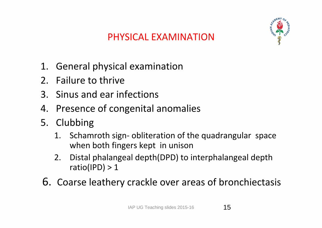
PHYSICAL EXAMINATION
1. General physical examination2. Failure to thrive3. Sinus and ear infections4. Presence of congenital anomalies5. Clubbing
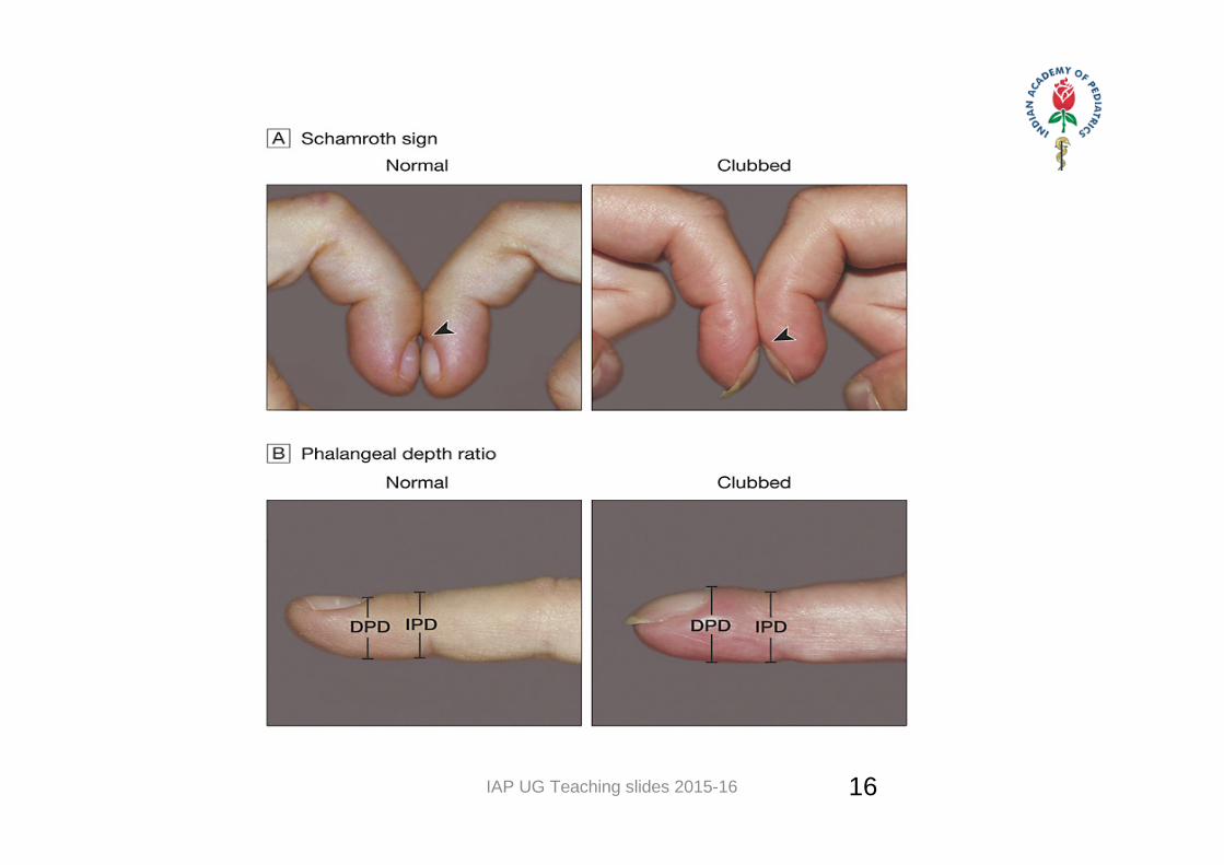
1. Schamroth sign‐ obliteration of the quadrangular space when both fingers kept in unison
2. Distal phalangeal depth(DPD) to interphalangeal depth ratio(IPD) > 1
6. Coarse leathery crackle over areas of bronchiectasis
15
IAP UG Teaching slides 2015-16 16
IAP UG Teaching slides 2015-16
DIAGNOSIS
• Diagnosis depends on radiographically or anatomically visualizing abnormal dilatation of airways
• Diagnostic procedure of choice ‐ High‐Resolution Computed Tomography (HRCT)scan
• Other tests‐ diagnose underlying conditions.
17
IAP UG Teaching slides 2015-16
LABORATORY INVESTIGATIONS
Complete blood count with differential Sputum exam‐ volume, gram stain, C & S , Immunodeficiency‐ Total IgM, IgA and IgG Tests for Tuberculosis –
• Mantoux• Chest X‐ray• Resting Gastric juice for AFB
Test for cystic fibrosis (sweat chloride and/or DNA testing)
18
IAP UG Teaching slides 2015-16
INVESTIGATIONS
Flexible bronchoscopy• shows structural alteration of bronchial tree • bronchoalveolar lavage ‐ remove mucus plugs/ to obtain lower airway cultures.
• If an airway foreign body is discovered‐rigid bronchoscopy ‐for removal
HIV screening Ciliary biopsy
19
IAP UG Teaching slides 2015-16
INVESTIGATIONS
• Test for GERD : 24 hour pH monitoring, upper gastrointestinal endoscopy and technetium milk scan scintigraphy.
• Test for aspiration due to inadequate airway protective mechanisms during swallowing or dysphagia ‐evaluated by video fluoroscopy.
20
IAP UG Teaching slides 2015-16 21
INVESTIGATIONS
• Pulmonary function tests ‐severity of lung disease • should be performed if the patient is able to do• useful tool to evaluate long‐term progression of
lung disease.
• Most patients with bronchiectasis have features of obstructive lung disease (low FEV1 & FEV1/FVC ratio)
21
IAP UG Teaching slides 2015-16
INVESTIGATIONS
• Test for allergic bronchopulmonary aspergillosis • Immunoglobulin E (IgE)
• Serum precipitins for Aspergillus species
• Sputum culture for fungus
• Aspergillus skin test
22
IAP UG Teaching slides 2015-16
CHEST RADIOGRAPHY
• Dilated and thickened airways (tram‐tracking or parallel lines)
• Irregular peripheral opacities that represent mucopurulent plugs
• Loss of lung volume and peribronchial fibrosis
23
IAP UG Teaching slides 2015-16
IMAGING ‐ DISTRIBUTION OF BRONCHIECTASIS
Cystic fibrosis ‐ upper lobes
Allergic bronchopulmonary aspergillosis‐Centrally located bronchiectasis
Bronchopulmonary sequestration‐lower lobe and usually unilateral
24
IAP UG Teaching slides 2015-16
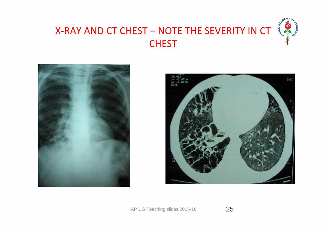
X‐RAY AND CT CHEST – NOTE THE SEVERITY IN CT CHEST
25
IAP UG Teaching slides 2015-16
IMAGING ‐ CT
• Most sensitive ‐to detect bronchiectasis ‐ high resolution computed tomography (HRCT)
• Internal diameter of airway ‐ larger than diameter of adjacent artery
• Airway wall thickening ("signet ring" shadows) with or without air fluid levels
• Volume loss, mucus plugging and air trapping
26
IAP UG Teaching slides 2015-16
MEDICAL TREATMENT
Control infection
Physiotherapy
Nutritional support
Identify etiology and treat accordingly
27
IAP UG Teaching slides 2015-16
MEDICAL MANAGEMENT
• Immediate– Medical treatment– Postural drainage– Relief of atelectasis– Treatment of associated problems
• Long term – Continuation of postural drainage– Follow up for reversal
28
IAP UG Teaching slides 2015-16
MANAGEMENT
Treatment –any identified underlying disorderTherapy
• reduce airway secretions• facilitating their removal ‐ with chest physiotherapy &
mucolytic agents
Pharmacotherapy• to improve mucociliary clearance.
Antibiotics • prevent & treat recurrent infections
Surgery‐ if localized disease ‐may be considered.
29
IAP UG Teaching slides 2015-16
CHEST PHYSIOTHERAPY
• Facilitate mucous expectoration ‐ manual and mechanical interventions
• Chest percussion• Vibration• Postural drainage• Cough‐assist devices & airway oscillation‐serve as
adjuncts to cough (most effective and efficient manner of clearing airway) .
30
IAP UG Teaching slides 2015-16
SURGICAL TREATMENT
Only in unilateral disease
Removal of affected segment/ lobe/lung
Surgery contraindicated if bilateral disease
Unilateral ‐ surgical resection ‐ good prognosis
31
IAP UG Teaching slides 2015-16
COMPLICATIONS
• Broncho‐ pneumonia• Empyema• Lung abscess• Hemoptysis• Metastatic abscess‐brain• Osteomyelitis• Hemoptysis• Cor pulmonale• Amyloidosis
32
IAP UG Teaching slides 2015-16
PREVENTION OF BRONCHIECTASIS
• Childhood immunization for measles and pertussis• Screening for tuberculosis and treatment wherever
needed• Aggressive appropriate therapy of lower respiratory
tract infections• Therapy of child with chronic or recurrent
respiratory problems due to recurrent aspiration and/or gastroesophageal reflux disease
33
IAP UG Teaching slides 2015-16
PROGNOSIS
• Overall ‐ prognosis – good• In absence of an underlying condition‐children with
isolated bronchiectasis‐ good prognosis• Progressive bronchiectasis from underlying disease
or ongoing pulmonary insult –causes progressive obstructive defect & ultimately, respiratory compromise
34
IAP UG Teaching slides 2015-16
Thank You
35