Embed Size (px)
Citation preview
Xanthophyll binding domains of CP29
1
XANTHOPHYLL BINDING SITES OF THE CP29 (Lhcb4) SUBUNIT OF HIGHER PLANT
PHOTOSYSTEM II INVESTIGATED BY DOMAIN SWAPPING AND MUTATION ANALYSIS.
Mirko Gastaldelli1, Giusy Canino1, Roberta Croce1,2 e Roberto Bassi 1,3
1 Dipartimento Scientifico e Tecnologico -Università di Verona, Strada Le Grazie,15-37134 Verona Italy 2 Istituto di Biofisica. CNR, sezione di Milano. Via Celoria 26. 20133-Milano Italy
3 Université Aix-Marseille II, LGBP (Laboratoire de Genetique et Biophysique des Plantes) Faculté des Sciences de Luminy - Département de Biologie - Case 901 - 163, Avenue de Luminy - 13288 Marseille Cedex 09.
RUNNING TITLE: XANTHOPHYLL BINDING DOMAINS OF CP29.
Address for correspondence: Roberto Bassi, Université Aix-Marseille II. e-mail: [email protected]
Abbreviations: AA, aminoacid; Car, carotenoid; Chl, chlorophyll; DCCD, dicyclohexylcarbodiimide; DM, β-D-dodecylmaltoside; EET, excitation energy transfer; Lhc, light-harvesting complex; L, lutein; LD, linear dichroism; N, neoxanthin; NPQ, non-photochemical quenching; rCP29, recombinant CP29; RT, room temperature; TMH, trans-membrane-helix; V, violaxanthin; VDE, violaxanthin deepoxydase; WT, wild type; PSII, Photosystem II;
Copyright 2003 by The American Society for Biochemistry and Molecular Biology, Inc.
JBC Papers in Press. Published on February 24, 2003 as Manuscript M212125200 by guest on February 4, 2018
http://ww
w.jbc.org/
Dow
nloaded from
Xanthophyll binding domains of CP29
2
Summary
The binding sites for xanthophylls in the CP29 antenna protein of higher plant Photosystem II have
been investigated using recombinant proteins refolded in vitro. Despite the presence of three
xanthophyll species CP29 binds 2 carotenoid per polypeptide. The localization of neoxanthin was
studied producing a chimeric protein constructed by swapping the C-helix domain from CP29 to
LHCII. The resulting holoprotein did not bind neoxanthin, confirming that the N1 site is not present
in CP29. Neoxanthin in CP29 was, instead, bound to the L2 site, which is thus shown to have a
wider specificity with respect to the homologous site L2 in LHCII. Lutein was found in the L1 site
of CP29. For each site the selectivity for individual xanthophyll species was studied as well as their
role in protein stabilization, energy transfer and photoprotection. Putative xanthophyll binding
sequences, identified by primary structure analysis as a stretch of hydrophobic residues including an
acidic term, were analyzed by site directed mutagenesis or, in one case, by deleting the entire
sequence. The mutant proteins were unaffected in their xanthophyll composition thus suggesting
that the target motifs had little influence in determining xanthophyll binding while hydrophobic
sequences in the membrane spanning helices are important.
by guest on February 4, 2018http://w
ww
.jbc.org/D
ownloaded from
Xanthophyll binding domains of CP29
3
Introduction
Carotenoids are involved in many aspects of higher plants photosynthesis. Reaction center
complexes bind β-carotene for light harvesting, Chl a triplet quenching and electron transport
between cytochrome b559 and P680+ (1;2). In the peripheral antenna, Lhc proteins bind a number
of xanthophyll species, namely lutein, neoxanthin and violaxanthin, acting in the harvesting of light
and transfer of excitation energy to chlorophyll (Chl) (3-6). The photoprotection function of
xanthophylls is accomplished through multiple mechanisms including the quenching of Chl a triplet
states, scavenging of singlet oxygen produced by reaction of Chl triplets with O2, and participation
to NPQ (Non-Photochemical Quenching): a mechanism in which violaxanthin is de-epoxidated to
zeaxanthin and excess energy is dissipated into heat (7). In addition, xanthophylls are essential for
Lhc protein folding (8) while their binding to a specific allosteric site controls the transition
between dissipative and conservative protein conformations (9) (10).
The reason why Lhc proteins require a number of xanthophyll species while reaction center
proteins only require β-carotene is not completely understood: these carotenoid species have very
similar physico-chemical properties, enabling, in each case, efficient light harvesting, triplet
quenching and singlet oxygen scavenging (11). Nonetheless, the pigment composition is one of the
most conserved traits in higher plants suggesting a specific function for each carotenoid species.
The best known xanthophyll binding protein is the major Light Harvesting Complex of
Photosystem II (LHCII) for which four distinct binding sites have been reported to be bound into
distinct domains of the protein. Sites L1 and L2 intersect the helix A/helix B cross domain in the
center of the Lhc structure (12). L1 is selective for lutein while L2 can also bind violaxanthin (13).
Site N1, is highly selective for neoxanthin and was located within the C helix domain of LHCII
(14). Finally, a low-affinity binding site has been named “V1” after its major ligand (violaxanthin)
in low light conditions (15;16). Each binding site was shown to play a distinct functional role:
structure stabilization and Chl a triplet quenching are provided by lutein in site L1 only (10;17) .
by guest on February 4, 2018http://w
ww
.jbc.org/D
ownloaded from
Xanthophyll binding domains of CP29
4
Site V1 is not involved in singlet nor in triplet energy transfer and was suggested to accommodate a
pool of readily available substrate molecules for the VDE enzyme (16) whose product, zeaxanthin,
can then be bound to the allosteric site L2 (18). Site N1 is active in light harvesting and singlet
oxygen scavenging (13) and stabilizes the long lifetime conformation of LHCII (19).
Despite the high homology in the transmembrane regions, which suggests a similar folding, the
number of xanthophyll binding sites, their selectivity and their occupancy in the structure seem to
be different in each Lhc gene product. Considering the different roles of individual sites and the
complexity of the Lhc multigene protein family, it is possible that the function of individual Lhc
gene products is largely determined by the presence/absence of individual xanthophyll sites and by
their selectivity and strength.
Primary sequence analysis of structural determinants for xanthophyll binding sites has been less
successful than in the case of Chl binding sites, possibly due to the contribution of neighbor Chls
rather than of aminoacid side chains, to the formation of binding pocket for xanthophylls (13;14).
Nevertheless, four short sequences, located in the hydrophilic domains and consisting into four
hydrophobic AA and a charged residue, could be involved in the binding by forming an
hydrophobic pocket hosting xanthophylls end-rings, while the charged side chain was supposed to
interact with oxygenated ring substituents (20). Alternatively, binding might be performed by
interactions between hydrophobic residues with the polyene chain located deeply in the membrane
as observed in the bacterial LH2 complex (21). In this study, we report the results of a detailed
analysis of the chlorophyll a/b/xanthophyll protein CP29 (Lhcb4) with respect to the xanthophyll
stoichiometry, location and function as well as of the sequence determinants involved in their
interaction with the polypeptide chain.
by guest on February 4, 2018http://w
ww
.jbc.org/D
ownloaded from
Xanthophyll binding domains of CP29
5
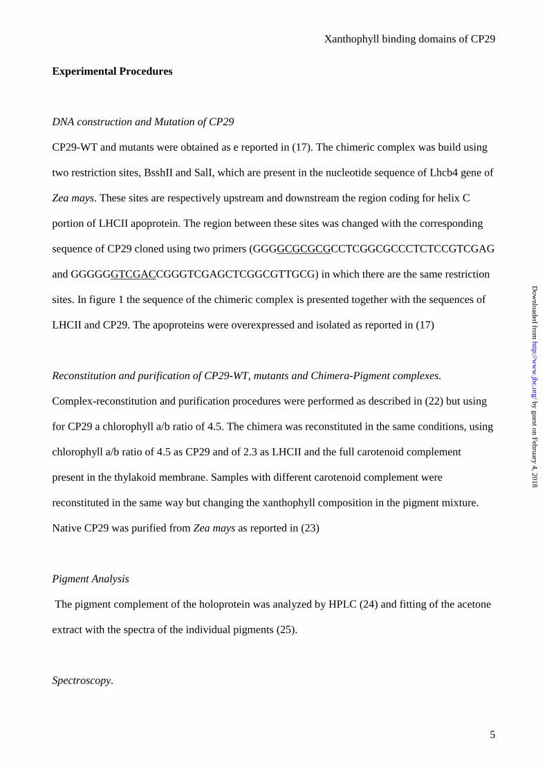
Experimental Procedures
DNA construction and Mutation of CP29
CP29-WT and mutants were obtained as e reported in (17). The chimeric complex was build using
two restriction sites, BsshII and SalI, which are present in the nucleotide sequence of Lhcb4 gene of
Zea mays. These sites are respectively upstream and downstream the region coding for helix C
portion of LHCII apoprotein. The region between these sites was changed with the corresponding
sequence of CP29 cloned using two primers (GGGGCGCGCGCCTCGGCGCCCTCTCCGTCGAG
and GGGGGGTCGACCGGGTCGAGCTCGGCGTTGCG) in which there are the same restriction
sites. In figure 1 the sequence of the chimeric complex is presented together with the sequences of
LHCII and CP29. The apoproteins were overexpressed and isolated as reported in (17)
Reconstitution and purification of CP29-WT, mutants and Chimera-Pigment complexes.
Complex-reconstitution and purification procedures were performed as described in (22) but using
for CP29 a chlorophyll a/b ratio of 4.5. The chimera was reconstituted in the same conditions, using
chlorophyll a/b ratio of 4.5 as CP29 and of 2.3 as LHCII and the full carotenoid complement
present in the thylakoid membrane. Samples with different carotenoid complement were
reconstituted in the same way but changing the xanthophyll composition in the pigment mixture.
Native CP29 was purified from Zea mays as reported in (23)
Pigment Analysis
The pigment complement of the holoprotein was analyzed by HPLC (24) and fitting of the acetone
extract with the spectra of the individual pigments (25).
Spectroscopy.
by guest on February 4, 2018http://w
ww
.jbc.org/D
ownloaded from
Xanthophyll binding domains of CP29
6
Absorption spectra were measured by an SLM-Aminco DW-2000 spectrophotometer at room
temperature. Fluorescence excitation and emission spectra were obtained using a Jasco-FP-777
spectrofluorimeter. Circular dichroism (CD) spectra were recorded at 10°C with a Jasco 600.
Samples were in 10 mM Hepes pH 7.6, 0.06% β-D-dodecylmaltoside (DM), 20% glycerol.
Chlorophyll concentration was ≈10 µg/ml for CD and absorption measurements and 0.01 µg/ml for
fluorescence measurements. LD spectra are the same reported in (26).
Photobleaching kinetic was measured as described in (10).
Stability measurements
The stability of the complexes was determined following the decrease of the CD signal at 490 nm
induced by the temperature. A temperature range between 20°C and 80°C was used. The
temperature was changing continuously by 1°C/min. The thermal stability of the protein was
determined by finding the t1/2 of the signal decay.
Data analysis
Deconvolution of spectra in the Soret (350-550 nm wavelength) range was performed as previously
described (27), using a home made program. Energy transfer efficiency from Cars to Chls was
estimated from the ratio of contributions in fluorescence excitation with respect to absorption of
individual pigment pools. In all the samples the energy transfer from Chl a was normalized to
100%.
Results
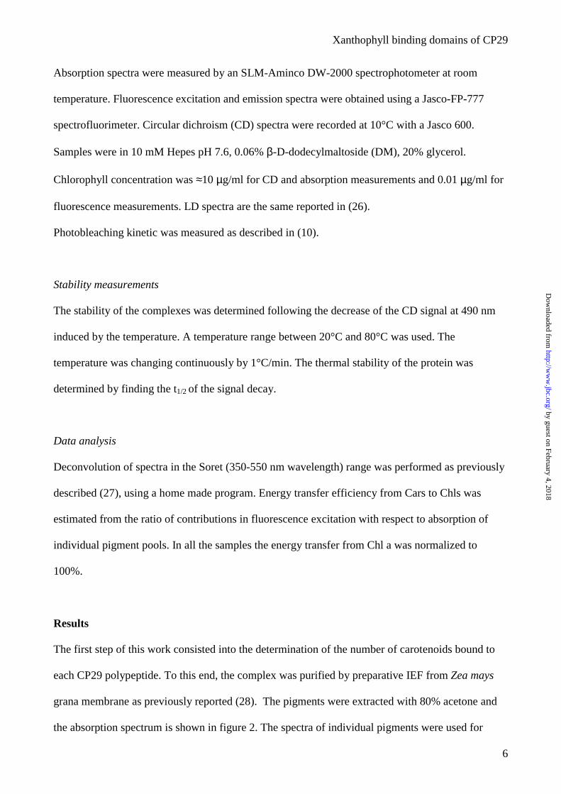
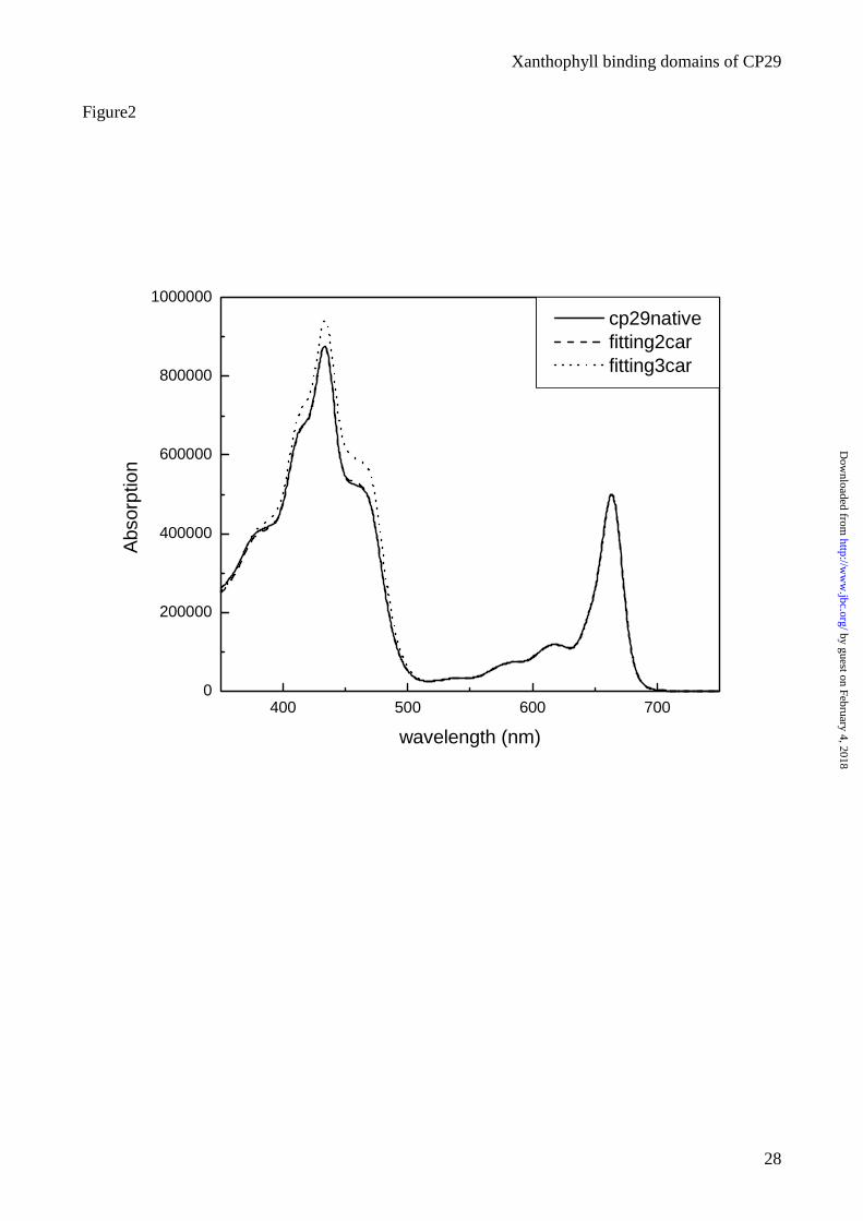
The first step of this work consisted into the determination of the number of carotenoids bound to
each CP29 polypeptide. To this end, the complex was purified by preparative IEF from Zea mays
grana membrane as previously reported (28). The pigments were extracted with 80% acetone and
the absorption spectrum is shown in figure 2. The spectra of individual pigments were used for
by guest on February 4, 2018http://w
ww
.jbc.org/D
ownloaded from
Xanthophyll binding domains of CP29
7
reconstructing the spectrum of the acetone extract from two hypothetical CP29 complexes binding 8
Chls (6 Chl a + 2 Chl b) (17;29) and either 2 or 3 xanthophylls in the ratio shown by HPLC
analysis: 0.85 lutein, 0.5 neoxanthin and 0.65 violaxanthin (30). The “synthetic” spectra are shown
in figure 2 together with the experimental spectrum. An almost perfect fit was obtained considering
two Cars per polypeptide, while the hypothesis of three Cars was discarded.
Reconstitution of recombinant CP29 with different xanthophyll species.
Although the Car to polypeptide stoichiometry in CP29 is 2:1, three xanthophyll species were found
in the pigment-protein complex, indicating promiscuity of at least one of the binding sites. In order
to investigate the affinity of the binding sites for the different xanthophylls, rCP29 apoprotein,
overexpressed in E. coli, was reconstituted in the presence of individual carotenoids or combination
of two. Stable complexes were obtained with all xanthophylls alone but neoxanthin, similar to what
observed in LHCII and CP26 (13;25;31).
The pigment composition of the reconstituted products was analyzed by HPLC and fitting of the
acetone extracts. The results are reported in table 1. All samples showed a Chl/Car ratio around 4.0
implying two xanthophylls per polypeptide. The complex reconstituted with zeaxanthin was the
only exception: although a complex with 2 zeaxanthin molecules per polypeptide was obtained
(CP29-Zb) in some experiments, in other preparations, a single Zea molecule was found (CP29-Za).
Neoxanthin in the complex never exceeded one molecule per polypeptide, suggesting that only one
site can accommodate this xanthophyll. Clearly, the occupancy of this site alone could not sustain
the folding of the pigment-protein complex.
Stability
It was previously shown that reconstitution in the absence of carotenoid does not yield into a folded
Chl-protein complex (8), implying that carotenoids play a primary role in stabilizing Lhc
holoproteins. In order to study the influence of each of the two Car binding sites, and of individual
by guest on February 4, 2018http://w
ww
.jbc.org/D
ownloaded from
Xanthophyll binding domains of CP29
8
xanthophyll species therein, in the stabilization of the structure, denaturation experiments were
performed. The data are reported in table 1. The control CP29 complex containing lutein,
violaxanthin and neoxanthin had a denaturation temperature of 64°C. Among the complexes
reconstituted in the presence of two xanthophyll species, CP29-LV and CP29-VN were slightly less
stable, while CP29-LN was somewhat more stable than CP29-Control thus showing that neoxanthin
has a stabilizing effect with respect to violaxanthin. Complexes containing only one carotenoid
species were the less stable, their denaturation temperature ranging between 53 and 49°C.
Light-harvesting
The absorption spectra of all complexes were recorded at room temperature (RT). The spectra were
almost identical in the Qy region, indicating that the binding of different xanthophyll species does
not strongly influence the Chl absorption (data not shown). Major differences were observed in the
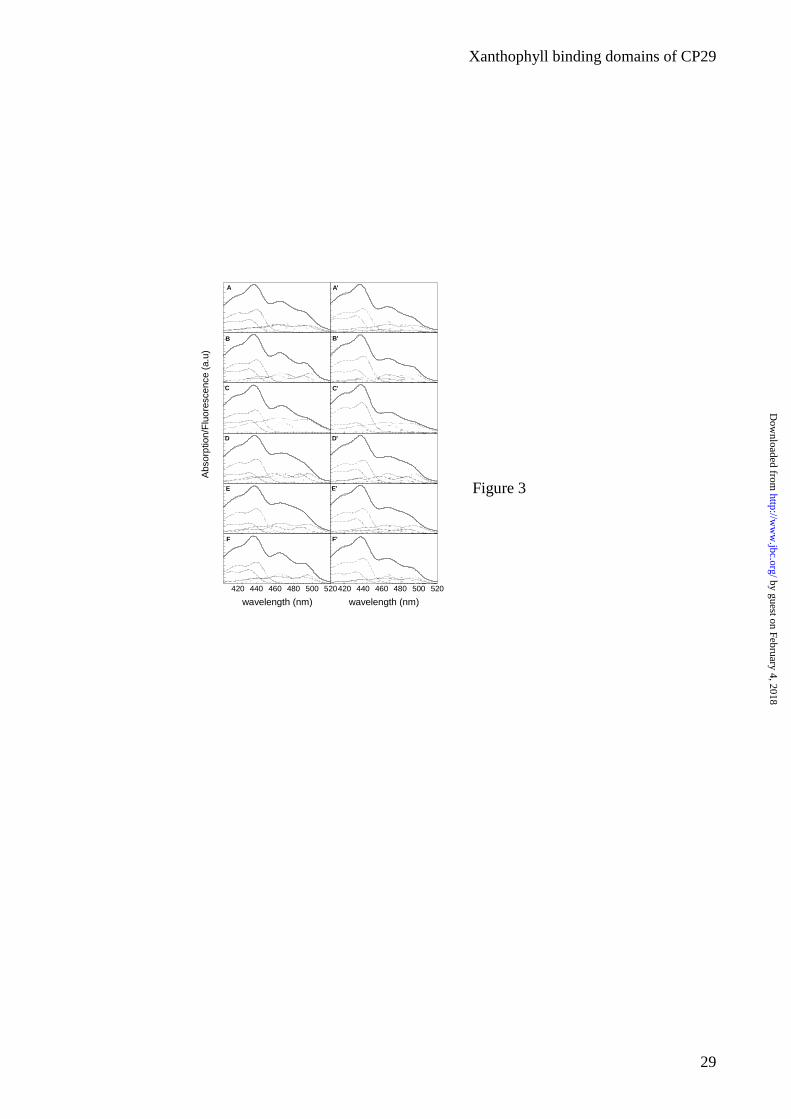
400-520 nm region where the Cars directly absorb by their S0-S2 transition (Fig 3, A-F).
In the antenna complexes of higher plants carotenoids have a light-harvesting function. In order to
determine the energy transfer efficiency of individual Cars in the two sites, the absorption spectra of
the complexes were described in term of absorption of individual pigments using 2 Chl a forms, 2
Chl b forms and 2 Cars forms. Once the best fit was chosen for the absorption spectrum, the same
set of data was used to describe the correspondent fluorescence excitation spectrum (fig.3 A’-F’).
The integrated areas of the pigment bands in the absorption and excitation spectra were used to
calculate the efficiencies of the energy transfer in the complex. The error is at around ∀ 5%
assuming for Chl a 100% transfer efficiency. The data are reported in table 2.
The overall Car to Chl a energy transfer depends on the xanthophyll composition. Among
complexes with a single xanthophyll species, CP29-L was the complex with lowest transfer
efficiency (60%), while CP29-V had the highest (73%). The two samples reconstituted with
zeaxanthin only had very different energy transfer efficiency: CP29-Za (with a single xanthophyll)
showed an efficiency of 62%, while for CP29-Zb (with two xanthophylls) this value dropped to
by guest on February 4, 2018http://w
ww
.jbc.org/D
ownloaded from
Xanthophyll binding domains of CP29
9
37%, thus indicating that the additional zeaxanthin molecule is unable to transfer energy to Chl a to
a significant extent (<5%). When two xanthophyll species were allowed, the highest efficiency was
obtained in the case of CP29-VN (81%), the lowest with CP29-LN (69%). In the control CP29
sample, three xanthophyll species were present, with the lutein transferring with 75% efficiency and
violaxanthin and neoxanthin with 60-65% (6).
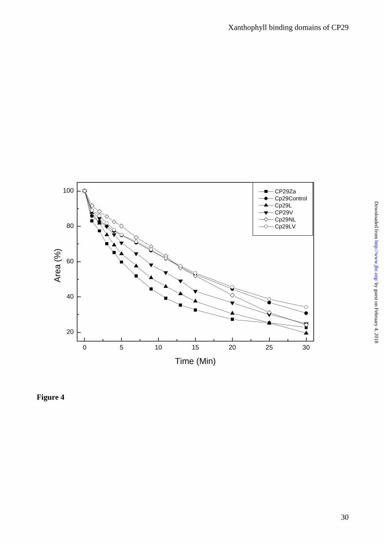
Photoprotection
Xanthophylls in Lhc complexes act in quenching 3Chl and scavenging of 1O2, thus providing
photoprotection. Photoprotection can be measured from the ability in preventing Chl
photobleaching under strong white light illumination in the presence of oxygen (13). The results of
this measurement for the rCP29 complexes are shown in figure 4. CP29-Control and the complexes
with two xanthophyll species show high resistance to photooxidation, while complexes with only
one xanthophyll species are more sensitive, thus yielding a behavior according to the following
series: CTR=LN=LV>V>L>Z. It is interesting to note that violaxanthin is more effective in
photoprotection than lutein; it should also be noted that CP29-Za contains a single carotenoid
molecule per polypeptide while the CP29-V sample has two.
Occupancy of the Xanthophyll binding sites: CP29 vs. LHCII
Three tightly bound xanthophyll binding sites have been described in LHCII: sites L1 and L2 (12),
accommodating mostly lutein in the native complex, and N1, which is selective for neoxanthin
(13;14). The data presented above shows that only two of these sites are conserved in CP29.
Previous work showed that site L1 is conserved in CP29 (17). However, it is not clear if either site
N1 or L2 is conserved. Alternatively, both sites could be present but only partially occupied. In
order to discriminate between the above hypotheses, a chimeric complex was produced, including
the central domain (helices A +B) of LHCII and the C helix domain of CP29. The helix A-helix C
lumenal loop and part of the helix C-helix B stromal loop were also included in the swapped
by guest on February 4, 2018http://w
ww
.jbc.org/D
ownloaded from
Xanthophyll binding domains of CP29
10
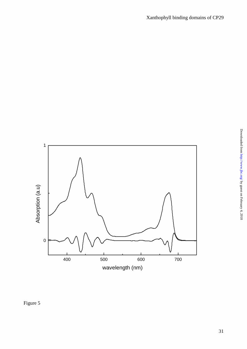
domain since the loops can affect Lhc protein folding (32) (Figs. 1A and 1B). The apoprotein was
over-expressed in E. coli and the complex was reconstituted in vitro with pigments. A pigment-
protein complex was obtained, which had the same mobility in a sucrose gradient of CP29 and
LHCII monomeric complexes and a denaturation temperature of 66°C, intermediate between that of
CP29 (64°C) and LHCII (73°C). The absorption spectrum of the complex along with its second
derivative is presented in figure 5. The maximum in the Qy region is at 676 nm, while a strong Chl a
contribution was detected at 666 nm. In the blue region the minima at 495, 468.4 and 437.2 nm in
the derivative spectrum were attributed respectively to Car, Chl b and Chl a. HPLC pigment
analysis showed that the chimeric complex has a Chl a/b ratio of 2.7, when reconstituted in the
same condition of LHCII-WT (Chl a/b 2.3 in the mixture), and 4.0 when reconstituted in the
condition of CP29-WT (Chl a/b 4.5 in the mixture). The Chl to Car ratio was 4.0 and the
xanthophyll species bound were lutein and small amount of violaxanthin. The chimeric
LHCII/CP29 complex never binds neoxanthin irrespectively from the xanthophyll availability in the
pigment mixture and other reconstitution conditions (table 3).
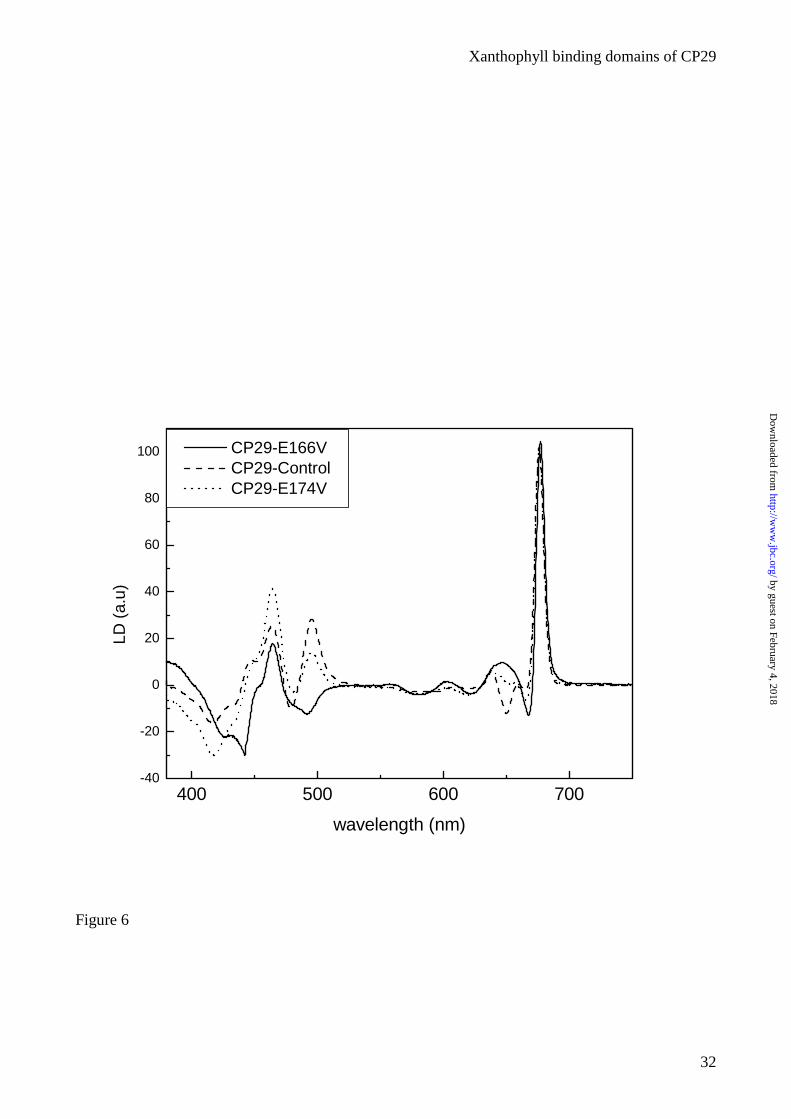
Orientation of xanthophyll transition moments in CP29.
Xanthophylls are bound to the L1 and L2 sites of LHCII, their polyene chain forming an angle of,
respectively, 56.4 and 59.4 ° with respect to the normal to the thylakoid membrane plane (12). We
have used LD in order to determine the orientation of xanthophyll transition moments, lying within
few degrees from the plane of the polyene chain (33), in CP29. In order to distinguish between the
contribution of xanthophylls in sites L1 and L2 to the LD signal, we have used the CP29 mutants
E166V and E174V previously shown to have either complete or partial emptiness of site L2 (17).
The major contribution of xanthophylls to the CP29-WT LD spectrum (Fig. 6) appears as a positive
signal at 495 nm, whose amplitude is reduced in the E174V mutant and becomes negative in the
E166V mutant. This suggests that the xanthophyll bound to the site L1 has its dipole moment
transition forming an angle smaller than 54.7° with the normal to the membrane plane.
by guest on February 4, 2018http://w
ww
.jbc.org/D
ownloaded from
Xanthophyll binding domains of CP29
11
The decreased intensity of the positive signal in CP29-E174V complex confirms that the positive
contribution is related to carotenoid in the L2 site. In this case, the angle between the dipole
moment transition axis and the normal to the membrane plane is larger than 54.7°. Using an internal
standard it is possible to determine the exact values for these angles. In CP29 the orientation of the
Chls has been determined from the LD signal after normalization. We therefore analyzed the
spectra previously presented in (26) and we calculated the orientation of the Cars transition
moments. The orientation of the xanthophyll located in the L1 site was calculated from the
spectrum of E166V mutant in which the only xanthophyll present is located in site L1. The analysis
of L2 was performed on the WT minus E166V difference spectrum. The spectra were fitted with the
absorption spectra of the Cars in order to calculate the carotenoid LD signal as reported in (14).
Than, using the FN (factor of normalization) value obtained from the analysis of the Chls transition
moments(26), the values for L1 and L2 were calculated. These values are respectively 50°and 70°.
L1 and L2 putative binding sequences
By primary structure analysis within the Lhc family, four consensus sequences have been identified
in the hydrophilic domains adjacent to TMH regions A and B, two on the stromal loops and two on
the lumenal loops, that could be involved in xanthophyll binding (20) (Figs. 1). These sequences are
characterized by an hydrophobic stretch of four residues with a polar residue insertion thus allowing
the formation of a hydrophobic pocket for hosting end-rings of xanthophylls, while the interaction
of the polar residue with the oxygenated ring substituents could have a stabilizing effect (20). In
order to check the involvement of these consensus sequences in xanthophyll binding, mutation
analysis was performed. In three cases the charged AA in the center of the hydrophobic sequence
was either substituted by a non-charged one or by a residue with an opposite charge; in one case the
putative binding sequence was deleted together with the whole N-terminal domain. In the case of
the putative lumenal ligand for the carotenoid in L1 site, the mutation at the central AA (Q230,
ligand for Chl A3) (17) has previously been performed with no effect on xanthophyll binding. Close
by guest on February 4, 2018http://w
ww
.jbc.org/D
ownloaded from
Xanthophyll binding domains of CP29
12
inspection of the LHCII structure (Kuhlbrandt et al. 1994) indicated that the end ring of the
xanthophyll in the L2 site appears to be closely spaced with respect to the carbonyl of Proline 238.
This residue could then interact with –OH xanthophyll ring substituents. This residue was mutated
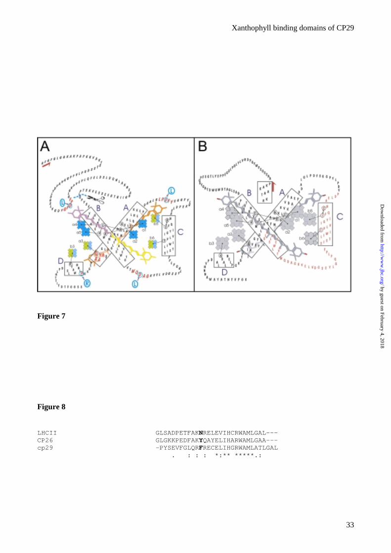
to arginine. In figure 7A a scheme representing the structure of CP29 with indication of the putative
carotenoid binding sequences and of the mutations performed is shown. The mutant sequences were
expressed in E. coli and the apoproteins were reconstituted into complexes using a pigment mixture
in which all xanthophylls were available together with Chl a and Chl b. The mutant Lhc complexes
were purified and characterized by biochemical and spectroscopic methods. All complexes showed
high yield of reconstitution, similar to WT complex. Heat denaturation experiments, accordingly,
show that all mutant proteins have the same stability of the WT (64°±1°C). The only exception was
the CP29-P238R mutant, which has a denaturation temperature of 57°C (see below).
In table 4 the pigment analysis of the reconstituted complexes is presented. The differences in the
pigment binding in the mutants with respect to the WT are rather small: CP29-D50L and CP29-N-
term showed Chl a/b and Chl/carotenoid ratio identical to the CP29-WT reconstituted in the same
conditions, while a small decrease in neoxanthin and violaxanthin was compensated by an increase
in lutein content. CP29-D138L was identical to the WT. CP29-D195L and CP29-P238R complexes
showed lower Chl a/b ratio than CP29-WT, especially in the case of the P238R mutant, but not
variation in the carotenoid composition, suggesting that these changes were due to the loss of a Chl
binding site. These data clearly indicates that protein motifs including the mutations here performed
do not play a major role in xanthophyll binding.
Discussion
CP29 is the simplest chlorophyll-binding protein of the Lhc superfamily since it binds 8 Chls per
polypeptide (29) each coordinated by a specific AA residue (17). The binding of xanthophylls to
CP29, however, is still a matter of discussion. Consensus results have been obtained on the binding
of three xanthophyll species: violaxanthin, lutein and neoxanthin (34) but different hypothesis have
by guest on February 4, 2018http://w
ww
.jbc.org/D
ownloaded from
Xanthophyll binding domains of CP29
13
been proposed for the location of each species to a particular binding site. While there is consensus
on the binding of lutein to site L1, violaxanthin and neoxanthin have either been proposed to bind to
different sites, like in LHCII (15) or to compete for site L2 only, thus implying that site N1 is absent
or empty in CP29 (17).
In this work, we performed an extensive analysis of xanthophyll binding in CP29. In particular, we
address four questions: (i) how many carotenoids are tightly bound to this complex, (ii) in which
sites they are located (iii) which is the role of individual sites and individual xanthophylls and (iv)
how the putative binding sequences can influence the binding. We approached the problem by using
recombinant pigment-protein complexes in which either the carotenoid complement was
biochemically modified or the apoprotein was engineered at the putative carotenoid binding sites.
How many carotenoids are bound to CP29?
The data presented clearly indicates that both native and recombinant CP29 bind tightly two
carotenoids per polypeptide. One mole of lutein and substechiometric amount of neoxanthin and
violaxanthin, together summing up to one per polypeptide, were found in all preparation thus
suggesting low selectivity of at least one of the binding sites. This is also supported by the analysis
of CP29 purified from different plant sources, showing variations in the ratio between the
xanthophylls (5;35;36).
Reconstitution experiments in the presence of either one xanthophyll species or combination of two
of them indicate that lutein, violaxanthin and zeaxanthin can enter both binding sites, while with
different affinity and binding strength. The neoxanthin, instead, can be accommodated in only one
of the two sites and its inability to drive the folding process alone testifies that the occupancy of this
site is not sufficient to stabilize the pigment-protein complex.
Which binding sites are present in CP29?
by guest on February 4, 2018http://w
ww
.jbc.org/D
ownloaded from
Xanthophyll binding domains of CP29
14
Four xanthophyll-binding sites have been described in LHCII: three tightly bound, L1, L2 and N1,
and a loose one (V1). In CP29 only two of the three tight sites are conserved. Previous results have
shown that the L1 site is present in CP29, where it accommodates mainly lutein (17). The second
carotenoid binding site present in CP29 shows high affinity for neoxanthin, but it does not
participate to the protein stability and it can be either L2 or N1 site. In order to discriminate
between these two sites, we constructed a chimeric LHCII/CP29 protein in which the two central
helices (A and B) derive from LHCII, while the C helix domain, previously shown to host the N1
binding site, derives from CP29 (Fig. 1). In figure 7B a schematic representation of the complex is
presented. The chimeric complex was stable and had spectral and pigment binding properties
intermediate between CP29 and LHCII. However, no neoxanthin was found in the complex despite
high amount of this carotenoid has been used during the reconstitution. We conclude that the N1
site is not present in CP29 and that the second carotenoid binding site in this complex is
homologous to the L2 site of LHCII.
The LD analysis indicates that the dipole moment transition axis of L1 and L2 forms respectively an
angle of 50° and 70° with the normal to the membrane plane. Despite it is not possible from these
data to determine the exact orientation of the polyene chain, it seems quite clear that the
xanthophylls are oriented differently compared to LHCII where a perfect symmetry was observed.
Although models of CP29 structure have been proposed based on LHCII the only available Lhc
structure (12), it appears that Lhc proteins may carry significant structural differences. New
structural data are of critical interest for the further understanding of Lhc protein structure and
function.
Stability
The results presented indicate that neoxanthin can enter only the L2 site, while all other Cars can be
accommodated in both sites. This implies that when a complex contains one molecule of neoxanthin
by guest on February 4, 2018http://w
ww
.jbc.org/D
ownloaded from
Xanthophyll binding domains of CP29
15
and one molecule of another xanthophyll, the latter has to be located on site L1. This allows
studying the role played by individual xanthophylls in the two sites.
We applied this model to the stability measurements in order to determine the influence of different
xanthophylls in the L1 and L2 sites. We considered the denaturation temperature of CP29-L, CP29-
V, CP29-NV, CP29-NL and CP29-VL and we resolved a system of five equations with five
variables. This calculation does not take in to account the contribution of Chls to the stability since
the Chl content of all the samples was identical.
The calculation yields a value of 50.3°C for the contribution of lutein in site L1 to the stability
of the protein, while the same xanthophyll contributed only for 3.2 °C when located in site L2. For
violaxanthin the corresponding values were respectively 42.9°C (For L1) and 6.2°C (for L2), while
neoxanthin in site L2 contributes for 14.5°C. As an example, we can calculate the stability of CP29-
Control (62.33°C) and verify that it matches with the measured denaturation temperature (64°C) of
the CP29-Control complex. The good match indicates the independence of all variables (e.g. lutein
in L1 site stabilizes the complex in the same way independently from which xanthophyll is present
in site L2). The results confirm that site L1 is the most important in Lhc protein stabilization as
previously assessed in Lhcb1 (10) and lutein in this site seems to confer the highest stability to the
complex. Lutein in the L2 site has almost no effect in the stability, while the occupancy of this site
by violaxanthin or, even better, by neoxanthin increases the temperature of denaturation.
Energy transfer
The excitation energy transfer efficiency from individual binding sites has been determined by
analyzing the absorption and excitation spectra in terms of spectral contribution of individual
pigments. The possibility of comparing samples, which differ only for the carotenoid composition,
allowed obtaining quantitative information from this analysis. It is worth mentioning that all
samples were fully equilibrated with the only partial exception of CP29-Za and CP29-Zb, which
by guest on February 4, 2018http://w
ww
.jbc.org/D
ownloaded from
Xanthophyll binding domains of CP29
16
showed a small emission contribution from Chl b fluorescence at 660 nm when the complex was
excited at 475 nm.
Again, we applied the model used for the stability analysis (see above). It has been demonstrated
that binding to either one or the other of the two sites yields into a different modulation of the S2
energy level of the xanthophylls (4). This indicates that the same xanthophyll in the two sites is
spectroscopically different. In agreement, two distinct values for the red-most peak (S0-S2,0
transition) were found for both lutein and violaxanthin, corresponding to the absorption in the L1
and L2 sites. In contrast, a single neoxanthin spectral form was needed for the best fit of both
absorption and fluorescence excitation spectra, consisting with its exclusive binding to the L2 site.
Considering that in the samples containing neoxanthin, violaxanthin and lutein can only be located
in the L1 site, it is possible to associate the two absorptions at site L1 or L2 based on the pigment
composition of the samples. Thus, lutein in the L1 site absorbs at 494 nm, while in the same site
violaxanthin absorbs at 496 nm. The same description found for the absorption spectra was applied
to the analysis of excitation spectra, thus allowing discriminating between the transfer from Car in
L1 and L2. It is therefore possible to read the data of table 2 with the understanding that notations
“1” and “2” actually correspond to xanthophyll binding sites L1 and L2. We can conclude that
lutein transfers with high efficiency (up to 91%) when in site L1, while its efficiency in exciting Chl
a fluorescence is very low (down to 28%) when it is located in the L2 site. Violaxanthin transfers
efficiently from the L1 site, while the efficiency is around 60% for both violaxanthin and
neoxanthin from the L2 site. Both violaxanthin and lutein show high (80-90%) efficiency from the
L1 site, while the decrease in the transfer efficiency from the L2 site is different: lutein decreases its
efficiency by a factor of 3.2 while viola of 1.3. This suggests that the arrangement of the two
xanthophylls in the L2 site is different. The case of zeaxanthin is even more extreme: its energy
transfer efficiency from the L1 site is around 60%, while from the L2 site is very low, if any. These
results suggest that the energy transfer from a single site is dependent from the site occupancy.
by guest on February 4, 2018http://w
ww
.jbc.org/D
ownloaded from
Xanthophyll binding domains of CP29
17
In general, the L2 site seems to be less active in the energy transfer as compared to the L1 site.
Moreover, the efficiency of excitation energy transfer to Chl is modulated by the xanthophyll
species bound there in the following gradient: V=N>L>Z. The lower transfer efficiency from the L2
site, common to all xanthophyll species, indicates that in this site the distance/orientation between
pigments is not optimal for EET. It has been recently observed that most of the transfer from Car to
Chl occurs via S2 Car state in less than 100 fs. Considering the short carotenoid S2 lifetime, a
perfect geometry between donor and acceptor is required for highly efficient energy transfer (3;4;6)
and small changes in this geometry can strongly influence the process. Moreover, the finding that
different xanthophylls behave differently when bound to the L2 site suggests that this protein
domain may change its folding in the presence of different xanthophylls, supporting the proposal
that the L2 site has allosteric nature (10). It is thus possible to suggest that when for example lutein
enters the site, which is usually occupied by neoxanthin and violaxanthin, a conformational change
occurs, changing the relative chromophore orientation and thus decreasing the transfer efficiency.
The figure in CP29 is thus different as compared to the case of LHCII where both L1 and L2 sites
showed high efficiency of EET to Chl a (4). This suggests the L2 site has a different function in the
two proteins. In the case of CP29 a Ca2+ and DCCD binding site in the loop between helix B and C
was suggested to work as a sensor for pH changes in the lumen (37;38). DCCD binding sites have
been also found in CP26 (39). At low lumenal pH the substitution of the Ca2+ with an H+ would
“open” the structure allowing violaxanthin to exit and zeaxanthin to enter the site (18). The high
flexibility, which is required for this process, can thus explain the low energy transfer efficiency
from this site. In the case of LHCII, zeaxanthin never enters the central sites and thus the structure
could be optimized for the function of EET.
Photoprotection
The photobleaching measurements show that samples with two or more xanthophyll species are
more resistant to photooxidation. Among the samples containing only one xanthophyll species,
by guest on February 4, 2018http://w
ww
.jbc.org/D
ownloaded from
Xanthophyll binding domains of CP29
18
CP29-L and CP29-Z are the most sensitive to photo-oxidation. It has been proposed above that
lutein in site L2 is organized differently compared to the other xanthophylls, and in effect does not
transfer energy efficiently. CP29-Z has only one xanthophyll molecule located in the L1 site. The
results thus indicate that the L2 site in CP29 can play an important role in photoprotection and again
that this role is modulated by the site occupancy. In this respect, CP29 differs strongly from LHCII
for which it has been clearly shown that L1 is the only site active in preventing photooxidation (10).
Do putative carotenoid binding sequences participate to actual xanthophyll binding?
In the last part of this work, the putative binding sequences for the carotenoids in the L1 and L2
sites have been studied by mutation analysis. Comparison between the primary structures of Lhc
multigenic family members has shown in the loops conserved sequences composed by four
hydrophobic residues with in between a polar aminoacid. Considering that Lhc proteins can not
bind β-carotene, it was argued that these sequences are involved in the xanthophyll binding by
establishing a H-bond between the polar residue in the polypeptide stretch and the ring -OH
substituent (20). However, our mutation of the four putative binding sequences not only did not
decrease pigment-protein stability but also did not change the affinity of binding sites for the
different xanthophyll species. The changes in Chl composition observed for the P238R mutant are
probably due to changes in the D helix domain and loss of the A3 site ligand as suggested by the
similar effects of mutation P238R (this work) and Q230L (17) detected by pigment analysis, CD
spectroscopy (data not shown) and the heat denaturation kinetics. The two mutations, which affect
the putative stromal binding sequences for xanthophylls in the L2 site (CP29-D50L and CP29-N-
term), support the view that pigment-protein stabilization by xanthophyll binding is not provided by
hydrophilic sequences. A small but reproducible decrease of violaxanthin and neoxanthin balanced
by an increase of lutein was however consistently found. This result may indicate that the sequence
encompassing the D50 residue plays a minor role in determining the selectivity of the L2 site.
by guest on February 4, 2018http://w
ww
.jbc.org/D
ownloaded from
Xanthophyll binding domains of CP29
19
The pigment-binding properties of chimeric LHCII/CP29 complex further support the conclusion
that lumenal sequences have little effect on site selectivity: despite the presence of the CP29
sequence in the lumenal region between B and C helices, it still accommodates mostly lutein in the
L2 site, as it is the case in LHCII (13) rather than violaxanthin and neoxanthin as in CP29.
Nevertheless, the absence of a clear phenotype for this chimeric complex can be useful in the search
of the structural determinants for the affinity of the Car binding sites for individual xanthophyll. In
fact, no effect in xanthophyll composition was observed by either deleting the N-terminal domain
down to residue 96 (CP29) or by substituting half of the B helix and the lumenal loop. We conclude
that the structural determinant for the selectivity of the L2 site has to be located in between these
two regions, namely within a 28 AA sequence (e.g. from residue 50 to residue 77 in the LHCII
sequence). It has been recently demonstrated that CP26 has the same L2 occupancy as in CP29 (25),
this means that “the determinant” has to be conserved in CP29 and CP26 and not in LHCII. The
three sequences are reported in figure 8. The only candidate is the N-61 in LHCII. An aromatic AA
in both CP29 and CP26 occupies this position. In the structure of LHCII this asparagine is within 5
Å from the carotenoid in L2. The presence in this position of a Phe or Tyr may change the sterical
interaction between pigments, possibly favoring the binding of violaxanthin and neoxanthin with
respect to lutein. This may be due to the difference in the ring orientation between lutein and
violaxanthin and neoxanthin: lutein is in its low energy conformation when the ε-cycle is oriented
parallel to the polyene chain, while the β-cycles of violaxanthin and neoxanthin are oriented
perpendicularly. This difference can play a role in the selectivity of the binding site: an aromatic
group in this site has higher sterical hindrance and than can select against lutein. However, the
stringency can not be very high being lutein able to enter the L2 site when the reconstitution is
performed in the absence of violaxanthin and neoxanthin. Consistent with this hypothesis is the
finding that CP29-L sample is less stable than the control CP29 and lutein in site L2 is differently
oriented in the complex as proposed above.
by guest on February 4, 2018http://w
ww
.jbc.org/D
ownloaded from
Xanthophyll binding domains of CP29
20
Although the putative carotenoid binding sequences on the stroma exposed side of the protein can
be somehow involved in the selectivity of the sites for the different xanthophylls, it is quite clear
that the stabilization of the carotenoid binding can not be attributed to these sequences. Similar
results have been obtained for the major light-harvesting complex, LHCII, where the deletion of the
N-terminal domain, which contains the putative binding sequence for L1 site, does not influence the
carotenoid composition of the complex. The dragging force for the carotenoid binding is most
probably to be searched in hydrophobic interaction in the portion of the complex inside the
membrane in a similar way to what observed in bacteria (40). Aromatic residues present in A and B
helices can be involved in this binding. The position of these residues is not conserved in different
members of the Lhc family but maintained in the same gene products from different species.
Mutation analysis on the Chl binding residues both in CP29 and LHCII have shown that the
interactions between Chls and carotenoids can play a major role in the xanthophylls binding. In
particular, the binding of neoxanthin in the N1 site of LHCII is stabilized by interaction with Chls
(14). This is consistent with CP29 lacking the N1 site: the number of Chls in the domain in between
helices A and C is lower than in LHCII, than critical Chl – neoxanthin interactions might be
lacking. The loop in between helix A and helix C of CP29 is shorter with respect to the
corresponding domain in LHCII; this may bring helix C closer to helix A and thus prevent Chl A6,
A7 and B1 binding in both CP29 and the chimeric LHCII / CP29 complex.
CP29 is a member of the Lhc family, which is conserved in higher plants and green algae
and is involved in light harvesting and protection from abiotic stress. Knowledge of the biochemical
and functional properties of the isolated proteins will contribute to the understanding of the
physiological roles of individual gene products in providing stress resistance. Recent work showed
that during light stress the xanthophyll ligand of site L2 (violaxanthin) is changed into zeaxanthin
(18) thus inducing a conformational change leading to fluorescence quenching while the binding of
neoxanthin in site N1 stabilizes the unquenched conformation (9,19). The absence of site N1 in
by guest on February 4, 2018http://w
ww
.jbc.org/D
ownloaded from
Xanthophyll binding domains of CP29
21
CP29 qualifies this antenna subunit for prompt response to light induced photoprotective
conformational change.
Acknowledgments
The authors thank Stefano Caffarri (University of Verona) for preparing figure 7. This work was
supported by “MIUR” progetto FIRB n. RBAu01ECX and by CNR “agenzia 2000”.
Reference List
1. Tracewell, C. A., Cua, A., Stewart, D. H., Bocian, D. F., and Brudvig, G. W.(2001) Biochemistry 40, 193-203
2. Faller, P., Pascal, A., and Rutherford, A. W. (2001) Biochemistry 40, 6431-6440
3. Gradinaru, C. C., van Stokkum, I. H. M., Pascal, A. A., van Grondelle, R., and Van Amerongen, H. (2000) J.Phys.Chem.B 104, 9330-9342
4. Croce, R., Muller, M. G., Bassi, R., and Holzwarth, A. R. (2001) Biophys.J. 80, 901-915
5. Das, S. K. and Frank, H. A. (2002) Biochemisty 41, 13087-13095
6. Croce, R., Müller, M. G., Caffarri, S., Bassi, R., and Holzwarth, A. R. (2003) Biophys.J. in press, 000
7. Demmig, B., Winter, K., Kruger, A., and Czygan, F.-C. (1987) Plant Physiol. 84, 218-224
8. Plumley, F. G. and Schmidt, G. W. (1987) Proc.Natl.Acad.Sci.USA 84, 146-150
9. Moya, I., Silvestri, M., Vallon, O., Cinque, G., and Bassi, R. (2001) Biochemistry 40, 12552-12561
10. Formaggio, E., Cinque, G., and Bassi, R. (2001) J.Mol.Biol. 314, 1157-1166
11. Bassi, R. and Caffarri, S. (2000) Photosynthesis Research 64, 243-256
12. Kühlbrandt, W., Wang, D. N., and Fujiyoshi, Y. (1994) Nature 367, 614-621
13. Croce, R., Weiss, S., and Bassi, R. (1999) J.Biol.Chem. 274, 29613-29623
14. Croce, R., Remelli, R., Varotto, C., Breton, J., and Bassi, R. (1999) FEBS Lett. 456, 1-6
15. Ruban, A. V., Lee, P. J., Wentworth, M., Young, A. J., and Horton, P. (1999) J.Biol.Chem. 274, 10458-10465
16. Caffarri, S., Croce, R., Breton, J., and Bassi, R. (2001) J.Biol.Chem. 276, 35924-35933
17. Bassi, R., Croce, R., Cugini, D., and Sandona, D. (1999) Proc.Natl.Acad.Sci.USA 96, 10056-10061
18. Morosinotto, T., Baronio, R., and Bassi, R. (2002) J.Biol.Chem. 277, 36913-36920
by guest on February 4, 2018http://w
ww
.jbc.org/D
ownloaded from
Xanthophyll binding domains of CP29
22
19. Polivka, T., Zigmantas, D., Sundström, V., Formaggio, E., Cinque, G., and Bassi, R. (2002) Biochemistry 41, 439-450
20. Pichersky, E. and Jansson, S. (1996) In Ort, D. R. and Yocum, C. F., editors. Oxygenic Photosynthesis: The Light Reactions, Kluwer Academic Publishers, Dordrecht
21. McDermott, G., Prince, S. M., Freer, A. A., Papiz, M., Hawthornthwaite-Lawless, A. M., Cogdell, R. J., and Isaacs, N. W. (1995) Nature 374, 517-521
22. Giuffra, E., Cugini, D., Croce, R., and Bassi, R. (1996) Eur.J.Biochem. 238, 112-120
23. Croce, R., Breton, J., and Bassi, R. (1996) Biochemistry 35, 11142-11148
24. Gilmore, A. M. and Yamamoto, H. Y. (1991) Plant Physiol. 96, 635-643
25. Croce, R., Canino, g., Ros, F., and Bassi, R. (2002) Biochemistry 41, 7343
26. Simonetto, R., Crimi, M., Sandona, D., Croce, R., Cinque, G., Breton, J., and Bassi, R. (1999) Biochemistry 38, 12974-12983
27. Croce, R., Cinque, G., Holzwarth, A. R., and Bassi, R. (2000) Photosynth.Res. 221-231
28. Dainese, P., Hoyer-hansen, G., and Bassi, R. (1990) Photochem.Photobiol. 51, 693-703
29. Dainese, P. and Bassi, R. (1991) J.Biol.Chem. 266, 8136-8142
30. Giuffra, E., Zucchelli, G., Sandona, D., Croce, R., Cugini, D., Garlaschi, F. M., Bassi, R., and Jennings, R. C. (1997) Biochemistry 36, 12984-12993
31. Frank, H. A., Das, S. K., Bautista, J. A., Bruce, D., Vasil'ev, S., Crimi, M., Croce, R., and Bassi, R. (2001) Biochemistry 40, 1220-1225
32. Heinemann, B. and Paulsen, H. (1999) Biochemistry 38, 14088-14093
33. Dolan, P. M., Miller, D., Cogdell, R. J., Birge, R. R., and Frank, H. A. (2001) J.Phys.Chem.B 105, 12134-12142
34. Bassi, R., Pineau, B., Dainese, P., and Marquardt, J. (1993) Eur.J.Biochem. 212, 297-303
35. Sandona, D., Croce, R., Pagano, A., Crimi, M., and Bassi, R. (1998) Biochim.Biophys.Acta 1365, 207-214
36. Pascal, A., Gradinaru, C., Wacker, U., Peterman, E., Calkoen, F., Irrgang, K.-D., Horton, P., Renger, G., van Grondelle, R., Robert, B., and Van Amerongen, H. (1999) Eur.J.Biochem. 262, 817-823
37. Jegerschöld, C., Rutherford, A. W., Mattioli, T. A., Crimi, M., and Bassi, R. (2000) J.Biol.Chem. 275, 12781-12788
38. Pesaresi, P., Sandona, D., Giuffra, E., and Bassi, R. (1997) FEBS Lett. 402, 151-156
39. Walters, R. G., Ruban, A. V., and Horton, P. (1994) Eur.J.Biochem. 226, 1063-1069
40. Prince, S. M., Papiz, M. Z., Freer, A. A., McDermott, G., Hawthornthwaite-Lawless, A. M., Cogdell, R. G., and Isaacs, N. W. (1997) J.Mol.Biol. 268, 412-423
by guest on February 4, 2018http://w
ww
.jbc.org/D
ownloaded from
Xanthophyll binding domains of CP29
23
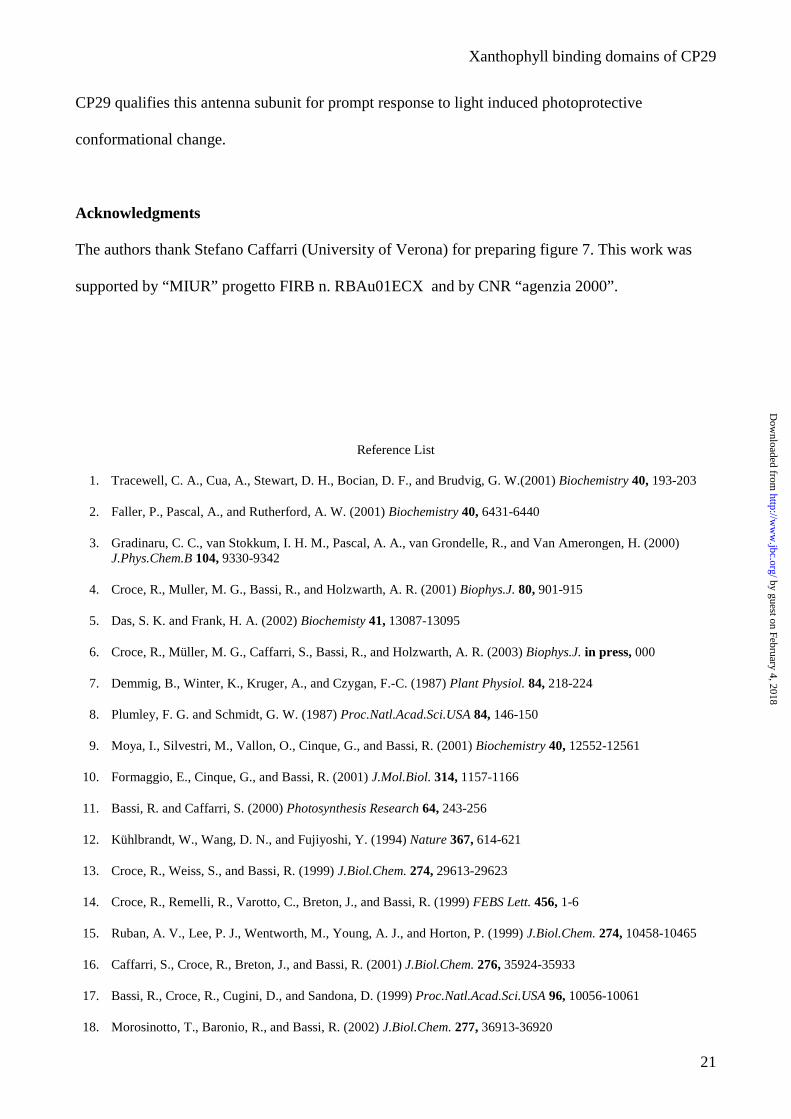
Figure legends
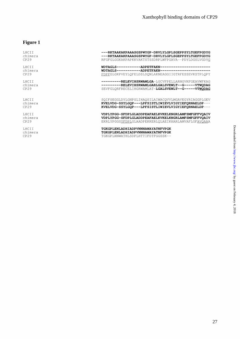
Figure 1. Sequence comparison between the chimeric complex, LHCII and CP29. The bold
indicates the region of LHCII and CP29, which are present in the chimera. In the CP29 sequence,
the putative carotenoid binding sequences are underlined.
Figure 2. Number of carotenoids present in CP29. Comparison between the absorption spectrum of
the acetone extract of CP29-WT (solid) and the reconstituted spectra with 2 (dotted) and 3 (dashed)
carotenoids.
Figure 3. Fitting of absorption and excitation spectra in the Soret region of samples with different
carotenoid composition: (A) CP29-L; (B) CP29-V; (C) CP29-Z; (D) CP29-NV; (E) CP29-LN and
(F) CP29-LV. Solid lines correspond to experimental absorption (left) and excitation (right). Short
dot line represents the fitting. The spectra were fitted with the spectra of individual pigments in
protein environment: Chl a (dash), Chl b (dot), lutein (dash-dot), violaxanthin (dash-dot-dot),
zeaxanthin (short dash) and neoxanthin (short dash-dot)
Figure 4. Photobleaching of recombinant CP29 proteins altered in the xanthophyll content. The
decay curves show the total Qy absorption relative to a 100% initial value. Points refer to
experimental data. Error was lower than 3% for each values as assessed from three independent
measurements.
Figure 5. Absorption spectrum of the chimeric complex. Absorption spectrum (solid) and second
derivative (dotted) at RT of LHCII-CP29-chimeric complex reconstituted with Chl a/b 4.5.
by guest on February 4, 2018http://w
ww
.jbc.org/D
ownloaded from
Xanthophyll binding domains of CP29
24
Figure 6. Linear dichroism. LD spectra at 100K of CP29-control (dash), CP29-E166V (solid) and
CP29-E174V (dotted).
Figure 7. Schematic representation of the structure of CP29 and the chimera. (A) Scheme for the
structure of CP29. The putative carotenoid binding sequences are indicated in red. The mutated
aminoacid is indicated by the blue circle and the substituted AA is indicated in blue. In the scheme
Chls a are indicated in blue, Chl b in green, lutein in orange, violaxanthin in magenta and
neoxanthin in yellow. Two color on the same pigment indicates mixed occupancy. (B) Schematic
representation of the chimerical complex: in black the domain of LHCII and in red the domain of
CP29.
Figure 8. Sequence comparison between CP29, CP26 and LHCII in the B helix.
by guest on February 4, 2018http://w
ww
.jbc.org/D
ownloaded from
Xanthophyll binding domains of CP29
25
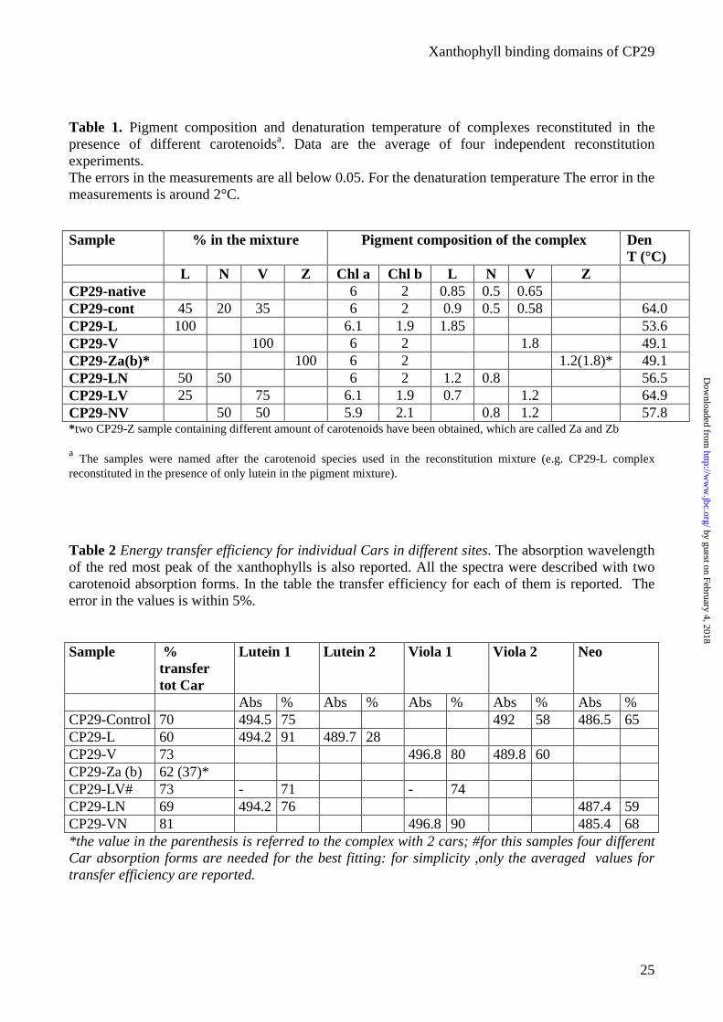
Table 1. Pigment composition and denaturation temperature of complexes reconstituted in the presence of different carotenoidsa. Data are the average of four independent reconstitution experiments. The errors in the measurements are all below 0.05. For the denaturation temperature The error in the measurements is around 2°C.
Sample % in the mixture Pigment composition of the complex Den T (°C)
L N V Z Chl a Chl b L N V Z CP29-native 6 2 0.85 0.5 0.65 CP29-cont 45 20 35 6 2 0.9 0.5 0.58 64.0 CP29-L 100 6.1 1.9 1.85 53.6 CP29-V 100 6 2 1.8 49.1 CP29-Za(b)* 100 6 2 1.2(1.8)* 49.1 CP29-LN 50 50 6 2 1.2 0.8 56.5 CP29-LV 25 75 6.1 1.9 0.7 1.2 64.9 CP29-NV 50 50 5.9 2.1 0.8 1.2 57.8 *two CP29-Z sample containing different amount of carotenoids have been obtained, which are called Za and Zb
a The samples were named after the carotenoid species used in the reconstitution mixture (e.g. CP29-L complex reconstituted in the presence of only lutein in the pigment mixture).
Table 2 Energy transfer efficiency for individual Cars in different sites. The absorption wavelength of the red most peak of the xanthophylls is also reported. All the spectra were described with two carotenoid absorption forms. In the table the transfer efficiency for each of them is reported. The error in the values is within 5%.
Sample % transfer tot Car
Lutein 1 Lutein 2 Viola 1 Viola 2 Neo
Abs % Abs % Abs % Abs % Abs % CP29-Control 70 494.5 75 492 58 486.5 65 CP29-L 60 494.2 91 489.7 28 CP29-V 73 496.8 80 489.8 60 CP29-Za (b) 62 (37)* CP29-LV# 73 - 71 - 74 CP29-LN 69 494.2 76 487.4 59 CP29-VN 81 496.8 90 485.4 68 *the value in the parenthesis is referred to the complex with 2 cars; #for this samples four different Car absorption forms are needed for the best fitting: for simplicity ,only the averaged values for transfer efficiency are reported.
by guest on February 4, 2018http://w
ww
.jbc.org/D
ownloaded from
Xanthophyll binding domains of CP29
26
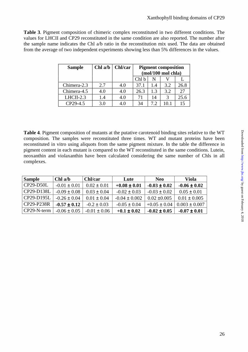
Table 3. Pigment composition of chimeric complex reconstituted in two different conditions. The values for LHCII and CP29 reconstituted in the same condition are also reported. The number after the sample name indicates the Chl a/b ratio in the reconstitution mix used. The data are obtained from the average of two independent experiments showing less than 5% differences in the values.
Pigment composition (mol/100 mol chla)
Sample Chl a/b Chl/car
Chl b N V L Chimera-2.3 2.7 4.0 37.1 1.4 3.2 26.8 Chimera-4.5 4.0 4.0 26.3 1.3 3.2 27 LHCII-2.3 1.4 4.0 71 14 3 25.6 CP29-4.5 3.0 4.0 34 7.2 10.1 15
Table 4. Pigment composition of mutants at the putative carotenoid binding sites relative to the WT composition. The samples were reconstituted three times. WT and mutant proteins have been reconstituted in vitro using aliquots from the same pigment mixture. In the table the difference in pigment content in each mutant is compared to the WT reconstituted in the same conditions. Lutein, neoxanthin and violaxanthin have been calculated considering the same number of Chls in all complexes.
Sample Chl a/b Chl/car Lute Neo Viola CP29-D50L -0.01 ± 0.01 0.02 ± 0.01 +0.08 ±±±± 0.01 -0.03 ±±±± 0.02 -0.06 ±±±± 0.02 CP29-D138L -0.09 ± 0.08 0.03 ± 0.04 -0.02 ± 0.03 -0.03 ± 0.02 0.05 ± 0.01 CP29-D195L -0.26 ± 0.04 0.01 ± 0.04 -0.04 ± 0.002 0.02 ±0.005 0.01 ± 0.005 CP29-P238R -0.57 ±±±± 0.12 -0.2 ± 0.03 -0.05 ± 0.04 +0.05 ± 0.04 0.003 ± 0.007 CP29-N-term -0.06 ± 0.05 -0.01 ± 0.06 +0.1 ±±±± 0.02 -0.02 ±±±± 0.05 -0.07 ±±±± 0.01
by guest on February 4, 2018http://w
ww
.jbc.org/D
ownloaded from
Xanthophyll binding domains of CP29
27
Figure 1
LHCII ---RKTAAKAKPAAASGSPWYGP-DRVLYLGPLSGEPPSYLTGEFPGDYGchimera ---RKTAAKAKPAAASGSPWYGP-DRVLYLGPLSGEPPSYLTGEFPGDYGCP29 RFGFGLGGKAKPAPKKVAKTSTSSDRPLWFPGAVA--PDYLDGSLVGDYG
LHCII WDTAGLS-----------ADPETFAKN-----------------------chimera WDTAGLS-----------ADPETFAKN-----------------------CP29 FDPFGLGKPVEYLQFELDSLDQNLAKNEAGGIIGTRFESSEVKSTPLQPY
LHCII ---------RELEVIHSRWAMLGA-LGCVFPELLARNGVKFGEAVWFKAGchimera ---------RELEVIHSRWAMLGARLGALSVEWLT--G-----VTWQDAGCP29 SEVFGLQRFRECELIHGRWAMLAT-LGALSVEWLT--G-----VTWQDAG
LHCII SQIFSEGGLDYLGNPSLIHAQSILAIWACQVVLMGAVEGYRIAGGPLGEVchimera KVELVDG-SSYLGQP---LPFSISTLIWIEVLVIGYIEFQRNAELDP---CP29 KVELVDG-SSYLGQP---LPFSISTLIWIEVLVIGYIEFQRNAELDP---
LHCII VDPLYPGG-SFDPLGLADDPEAFAELKVKELKNGRLAMFSMFGFFVQAIVchimera VDPLYPGG-SFDPLGLADDPEAFAELKVKELKNGRLAMFSMFGFFVQAIVCP29 EKRLYPGGSYFDPLGLAADPEKKERLQLAEIKHARLAMVAFLGFAVQAAA
LHCII TGKGPLENLADHIADPVNNNAWAYATNFVPGKchimera TGKGPLENLADHIADPVNNNAWAYATNFVPGKCP29 TGKGPLNNWATHLSDPLHTTIFDTFGGSSK--
by guest on February 4, 2018http://w
ww
.jbc.org/D
ownloaded from
Xanthophyll binding domains of CP29
28
Figure2
400 500 600 7000
200000
400000
600000
800000
1000000
Abso
rptio
n
wavelength (nm)
cp29native fitting2car fitting3car
by guest on February 4, 2018http://w
ww
.jbc.org/D
ownloaded from
Xanthophyll binding domains of CP29
29
Figure 3
420 440 460 480 500 520
420 440 460 480 500 520
A
A'
B
B'
C
Abs
orpt
ion/
Fluo
resc
ence
(a.u
)
C'
D
D'
E
E'
F
wavelength (nm)
F'
wavelength (nm)
by guest on February 4, 2018http://w
ww
.jbc.org/D
ownloaded from
Xanthophyll binding domains of CP29
30
0 5 10 15 20 25 30
20
40
60
80
100
CP29Za Cp29Control Cp29L CP29V Cp29NL Cp29LV
Area
(%)
Time (Min)
Figure 4
by guest on February 4, 2018http://w
ww
.jbc.org/D
ownloaded from
Xanthophyll binding domains of CP29
31
400 500 600 700
0
1
Abs
orpt
ion
(a.u
)
wavelength (nm)
Figure 5
by guest on February 4, 2018http://w
ww
.jbc.org/D
ownloaded from
Xanthophyll binding domains of CP29
32
400 500 600 700-40
-20
0
20
40
60
80
100
LD (a
.u)
wavelength (nm)
CP29-E166V CP29-Control CP29-E174V
Figure 6
by guest on February 4, 2018http://w
ww
.jbc.org/D
ownloaded from
Xanthophyll binding domains of CP29
33
Figure 7
Figure 8 LHCII GLSADPETFAKNRELEVIHCRWAMLGAL---CP26 GLGKKPEDFAKYQAYELIHARWAMLGAA---cp29 -PYSEVFGLQRFRECELIHGRWAMLATLGAL
. : : : *:** *****.:
by guest on February 4, 2018http://w
ww
.jbc.org/D
ownloaded from
Mirco Gastaldelli, Giusy Canino, Roberta Croce and Roberto BassiII investigated by domain swapping and mutation analysis
Xanthophll binding sites of the CP29 (Lhcb4) subunit of higher plant photosystem
published online February 24, 2003J. Biol. Chem.
10.1074/jbc.M212125200Access the most updated version of this article at doi:
Alerts:
When a correction for this article is posted•
When this article is cited•
to choose from all of JBC's e-mail alertsClick here
by guest on February 4, 2018http://w
ww
.jbc.org/D
ownloaded from