Embed Size (px)
Citation preview
Chemosensors 2015, 3, 70-117; doi:10.3390/chemosensors3020070
chemosensors ISSN 2227-9040
www.mdpi.com/journal/chemosensors
Review
X-Ray Photoelectron Spectroscopic Characterization of Chemically Modified Electrodes Used as Chemical Sensors and Biosensors: A Review
Elio Desimoni †,* and Barbara Brunetti †
DeFENS, University of Milan, via Celoria 2, Milan (I) 20133, Italy;
E-Mail: [email protected]
† These authors contributed equally to this work.
* Author to whom correspondence should be addressed; E-Mail: [email protected];
Tel.: +39-025-031-9223; Fax: +39-025-031-9227.
Academic Editors: Michael Ongaro and Paolo Ugo
Received: 3 December 2014 / Accepted: 27 March 2015 / Published: 10 April 2015
Abstract: The characterization of chemically modified sensors and biosensors is commonly
performed by cyclic voltammetry and electron microscopies, which allow verifying
electrode mechanisms and surface morphologies. Among other techniques, X-ray
photoelectron spectroscopy (XPS) plays a unique role in giving access to qualitative,
quantitative/semi-quantitative and speciation information concerning the sensor surface.
Nevertheless, XPS remains rather underused in this field. The aim of this paper is to review
selected articles which evidence the useful performances of XPS in characterizing the top
surface layers of chemically modified sensors and biosensors. A concise introduction to
X-ray Photoelectron Spectroscopy gives to the reader the essential background. The
application of XPS for characterizing sensors suitable for food and environmental analysis
is highlighted.
Keywords: chemically modified sensors; biosensors; X-ray photoelectron spectroscopy;
surface analysis of electrodes; electrocatalysis
OPEN ACCESS
Chemosensors 2015, 3 71
1. Introduction
According to IUPAC, chemically modified electrodes (CMEs) are electrodes made of a conducting
or semiconducting material that is coated with a selected monomolecular, multimolecular, ionic, or
polymeric film of a chemical modifier and that, by means of faradaic (charge-transfer) reactions or
interfacial potential differences (no net charge transfer), exhibit chemical, electrochemical, and/or
optical properties of the film [1]. Applications of CMEs span from solar energy conversion and storage,
to batteries, selective electro-organic synthesis, molecular electronics, electrochromic display devices,
corrosion protection, electrocatalysis and food or environmental analyses.
CMEs suitable as chemical sensors and biosensors (both named “sensors” from now on) have been
reviewed in a large series of papers describing their exciting performances. See references [2–15] for
some recent examples.
Investigations aimed at developing new sensors and evaluating their performances usually employ
combined experimental techniques. Cyclic voltammetry (CV) and electron microscopies are those most
generally used, since they allow a main access to electrode micro-mechanisms and surface
morphologies. Other techniques are more and more exploited for improving the characterization of
surface films, such as Atomic Force Microscopy (AFM), Fourier Transform Infrared (FTIR),
Electrochemical Impedance Spectroscopy (EIS), X-ray Diffraction (XRD), Scanning Electrochemical
Microscopy (SECM), X-ray Photoelectron Spectroscopy (XPS) and others (all acronyms used in this
review are also expanded in the Appendix). In particular, XPS (in the past also known as ESCA-Electron
Spectroscopy for Chemical Analysis) allows acquiring otherwise inaccessible information about the
qualitative, quantitative (or at-least semi-quantitative) and, most of all, speciation status of the CME
surface. However, XPS remains rather underused in this field. The literature information considered here
below spans from 2010 to about mid 2014. Inevitably, taking into account the huge number of relevant
articles, this review is not exhaustive. For this period, Scopus returns 4826 documents relevant to
“modified electrodes”, restricted to “only” 157 (about the 3%) if the search is extended to “modified
electrodes” and “ESCA” or “XPS”.
After an as much as possible concise introduction to XPS, in order to give the reader the essential
background, the attention focuses to those papers, arbitrarily selected within those above-mentioned, in
which XPS findings give a significant contribution to a better sensor characterization. To limit the length
of the review, the analytical performances of the sensors (linear ranges, limits of detection, selectivity,
repeatability etc.), as claimed by the various authors, are not considered here below. Sensor architectures
are represented by using the format surface layer/eventual middle layers/electrode material.
2. An Introduction to Photoelectron Spectroscopy
Several qualified books and reviews (see for example [16–22]) present principles and practices of
XPS. Nevertheless, the very basic principles of the technique are detailed below for allowing the
inexperienced reader a reasonable understanding of the reviewed results.
In XPS, a solid sample is introduced in a chamber maintained under ultra-high vacuum (UHV, below
10−8 mbar) where it is irradiated with soft X-rays, usually with Mg Kα or Al Kα radiations, whose
energies are 1253.6 eV or 1486.6 eV, respectively. Irradiation produces multiple ionizations from core
Chemosensors 2015, 3 72
and valence energy levels of the irradiated atoms or molecules. XPS is aimed at studying photoemission
from core level photoelectrons. The simplified equation describing the photoionization process is [16–22]
KE = h − BE (1)
in which KE (eV) is the kinetic energy of the photoemitted electron, BE (eV) its binding energy, hν is
the X-ray energy (eV). If a positive charge is left on the sample surface by the photoemission process,
photoelectrons appear at somewhat lower KE values. The correction of the KE scale is frequently made
by referencing to the BE of the 1s energy level of carbon atoms of hydrocarbons present in the sample
contamination layer (285.0 ± 0.2 eV). This contamination is usually ascribed to oils from the vacuum
system of the spectrometer.
Using UHV conditions is crucial, since (i) it ensures that almost no adsorbed gaseous contaminants
cover the solid surface since they could contribute to the XPS signal and (ii) under these conditions, the
inelastic mean free path of the electrons (the average distance travelled by electrons through a medium
before they are inelastically scattered) is sufficient to allow them leaving the sample and entering in the
spectrometer analyzer with unaltered KE). Only photoelectrons leaving the sample without inelastic
collision with other atoms or molecules (gaseous molecules adsorbed on the sample surface included)
retain their original KE value and give recognizable peak signals. The others, having suffered inelastic
collisions, only contribute to the background. In the KE range usually explored (from 500 to 1500 eV)
the sampling depth, the depth from which electrons can leave the sample surface without inelastic
collisions, ranges between 3 and 10 nm. This means few monolayers, and explains why XPS is an
ultimate surface analytical technique. The kinetic energy of the photoemitted electrons is measured by
the analyzer and, on knowing hν, it is possible measuring the relevant binding energy, specific of a given
core level of the photoemitting atom.
XPS investigations begin with measuring the intensity of photoemitted electrons at any available KE
value. The resulting “wide” or “survey” X-ray photoelectron (XP) spectrum displays a series of signals
relevant to all electrons photoemitted by energy levels having a BE lower than hν, overlapped to a
structured background.
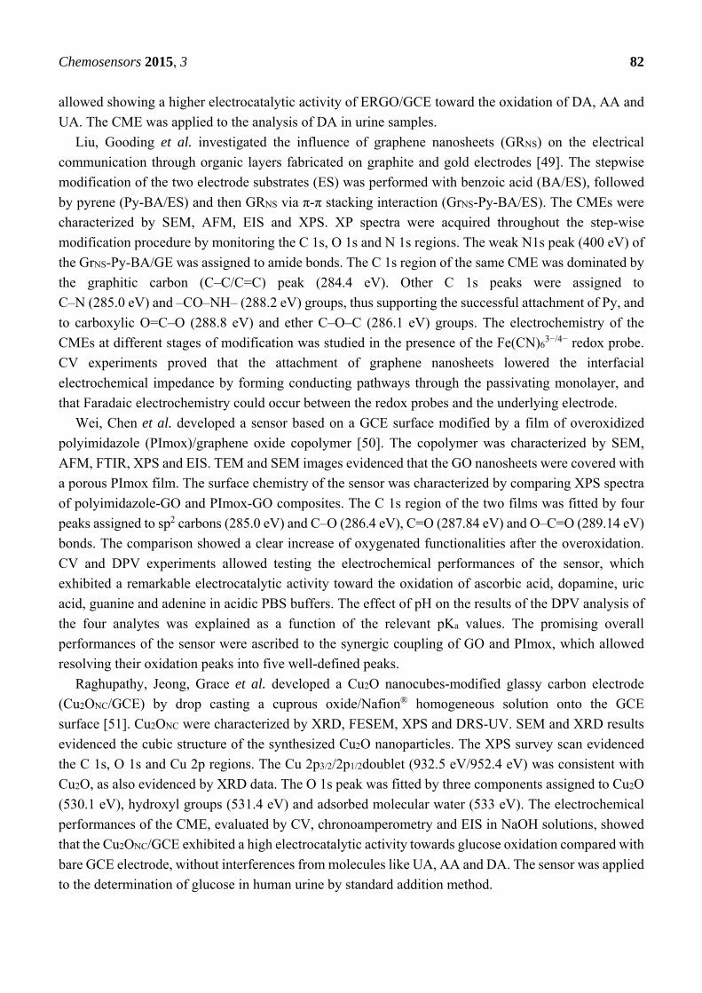
The survey scan in Figure 1, relevant to a glassy carbon electrode (GCE) surface-modified by
8-hydroxyquinoline-5-sulphonic acid (HQSA), evidences (from the right to the left) the signals relevant
to electrons photoemitted by the Si 2p, Si 2s, S 2p, Cl 2p, C 1s, N 1s and O 1s energy levels [23].
Survey scans give qualitative information about atoms/molecules present in the sampling depth.
The spectral features at about 950–1050 eV in Figure 1 originate from the decay of core holes left by
the photoelectrons. After photoemission from, atoms are left in an unstable excited state (z*+).
De-excitation can occur by X-ray fluorescence or by Auger electrons emission. In this last case
(see Figure 2), an electron of an outer shell (of energy level L1) fills the initial core hole (in the energy
level K), and this transition makes available sufficient energy for a second electron of an outer shell
(of energy level L2,3), the Auger one, to be emitted.
The Auger emission is the result of a three-electron process, and leaves the atom doubly-ionized (z2+).
Auger decays can continue until inner holes are available.
The Auger region (such as that around 1000 eV in Figure 1) gives access to useful information about
the sample composition. It was shown that the energy separation between the two major excursions in
the first derivative of the carbon KVV (this notation indicates the second and third electron of the Auger
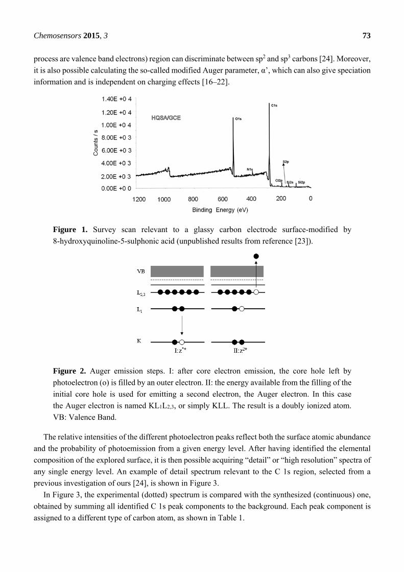
Chemosensors 2015, 3 73
process are valence band electrons) region can discriminate between sp2 and sp3 carbons [24]. Moreover,
it is also possible calculating the so-called modified Auger parameter, α’, which can also give speciation
information and is independent on charging effects [16–22].
Figure 1. Survey scan relevant to a glassy carbon electrode surface-modified by
8-hydroxyquinoline-5-sulphonic acid (unpublished results from reference [23]).
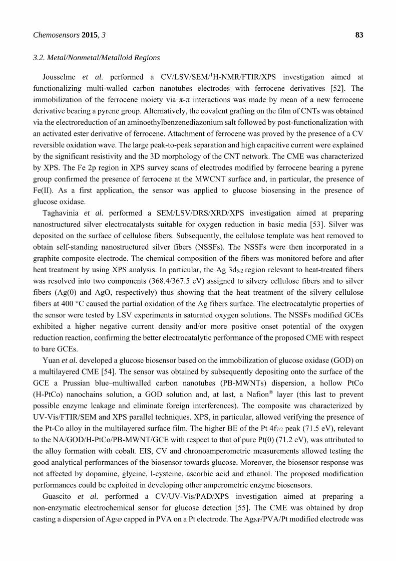
Figure 2. Auger emission steps. I: after core electron emission, the core hole left by
photoelectron (ο) is filled by an outer electron. II: the energy available from the filling of the
initial core hole is used for emitting a second electron, the Auger electron. In this case
the Auger electron is named KL1L2,3, or simply KLL. The result is a doubly ionized atom.
VB: Valence Band.
The relative intensities of the different photoelectron peaks reflect both the surface atomic abundance
and the probability of photoemission from a given energy level. After having identified the elemental
composition of the explored surface, it is then possible acquiring “detail” or “high resolution” spectra of
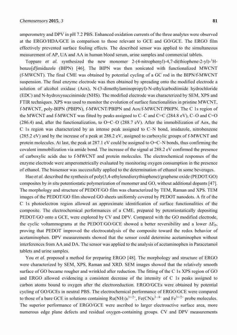
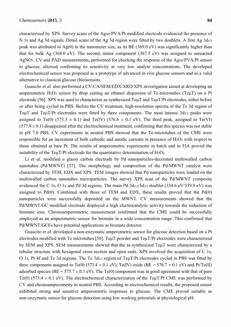
any single energy level. An example of detail spectrum relevant to the C 1s region, selected from a
previous investigation of ours [24], is shown in Figure 3.
In Figure 3, the experimental (dotted) spectrum is compared with the synthesized (continuous) one,
obtained by summing all identified C 1s peak components to the background. Each peak component is
assigned to a different type of carbon atom, as shown in Table 1.

Chemosensors 2015, 3 74
Figure 3. Curve fitting of a C 1s detail scan relevant to a sample of oxidized carbon
fibers [24]. Experimental spectrum: dotted curve; synthesized resultant (background + peaks):
continuous line. The residual window at the bottom of the figure shows the (properly
expanded) difference between experimental and synthesized spectra. The asymmetric
peak 1 is assigned to graphitic carbon atoms. Symmetrical peaks 3, 4 and 5 are assigned to
C–OH, C=O and O–C=O groups.
Table 1. C 1s binding energies (eV) of some types of carbon atoms [24].
Peak Carbon Type BE
1 Graphite, aromatics 284.6 2 Aliphatics 285.1 3 Alcohols, phenols 286.6
3/4 Cheto-enolic groups 287.1 4 Cheto groups 287.9 5 Carboxylic groups 289.3 6 Carbonate, CO2 290.6 7 Plasmon loss 291.3
Table 1 shows different BE values for the same energy level. This because atoms having a higher
positive oxidation state exhibit a higher binding energy due to the extra coulombic interaction between
the photoemitted electron and the ion core. Chemical shifts accounts for the difference in BE values of
electrons in one specific chemical state of the atom versus the value relevant to pure element, or a
convenient chemical state of that element. Several papers or handbooks report comprehensive
compilations of binding energies values/chemical shifts (see for example references [25,26]).
Photoelectron signals correspond, on a one-to-one basis, to different atomic energy levels. In the
simplest case (photoemission from energy levels having quantum number l = 0) the signal consists in
one peak, more often (if l > 0) in a doublet. This is known as spin-orbit splitting (SoS). The theoretical
peaks area ratio (PAR) is based on the degeneracy of each spin state of the doublet. In the case of energy
levels having quantum number l = 1, as in the case of the 2p3/2/2p1/2 doublet, the PAR is 2/1, while in
Chemosensors 2015, 3 75
the case of the 3d5/2/3d3/2 doublet the PAR is 3/2 and, in the case of the 4f5/2/4f7/2 doublet, the PAR is
4/3. SoSs are also important in identifying the correct chemical status of the photoemitting atom.
Each spectral feature can be the envelope of multiple contributions originated by some chemical
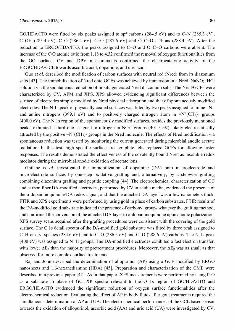
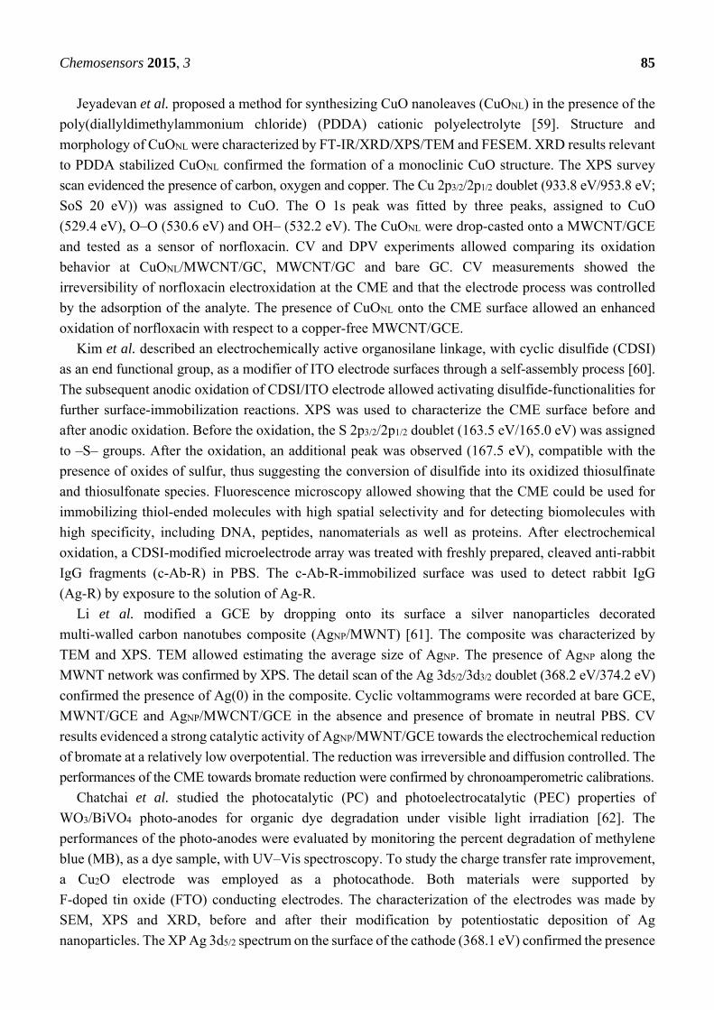
shifts, as shown in Figure 3. An example of multiple Cr 2p doublets is given in Figure 4 [27].
Figure 4. Mg Kα-excited spectrum of the Cr 2p region of a specimen prepared by
evaporating potassium dichromate onto a metallic chromium sheet. Doublet 1 + 2:
Cr metal; doublet 3 + 4: Cr(III) oxide; doublet 5 + 6: Cr(III) hydroxide; doublet 7 + 8:
potassium dichromate [27].
In this figure, the KE scale of the abscissa allows evidencing how the increasing formal positive
charge of chromium atoms (from the right to the left of the figure) produces decreasing KEs of
photoelectron peaks. Peak positions, full width al half Maximum (FWHM) and SoS data used in curve
fitting, reported in eV units, must be chosen in agreement with tabulated values.
Other spectral features are possible, such as shake-up peaks, plasmon loss peaks and satellite
peaks, but they are of minor importance in the present context, and are explained in the specific
literature [16–22].
Modern XP spectrometers are equipped with dedicated software facilities allowing almost automatic
acquisition/processing of XP spectra, and avoiding tedious manual routines. However, often a real good
interpretation of the acquired detail spectra requires some interactive adjustment/interpretation.
So, while survey scans allow performing a qualitative analysis, detail scans allow a sort of
speciation analysis, being capable of discriminating between atoms characterized by a different
charge/chemical surrounding. Moreover, a proper measurement of peak areas allows performing at least
a semi-quantitative analysis. On considering that this information is relevant to few surface monolayers,
XPS is invaluable in corrosion, adhesion and catalysis investigations.
From what above reported, XPS represent a favorable choice in CMEs investigations, since the very
surface of an electrode determines its electrochemical performances. However, some caution is
necessary, since the chemical status of the CME surface can undergo more or less heavy degradations,
when transferred to UHV working conditions and/or under X-ray irradiation. Moreover, it is
necessary considering that instruments are very expensive, so that accessibility cannot be very easy.

Chemosensors 2015, 3 76
These problems justify some hesitation towards exploiting XPS in CMEs investigations.
Nonetheless, according to our previous experience (see for example references [23,28–30]) XPS can
give precious or otherwise unavailable information in CMEs investigations.
3. Survey of Literature Information
The papers examined in this review can hardly be grouped according to some rigid criteria since,
often, their topics and experimental approaches are often more or less overlapped. For this reason, they
are here grouped in three sections according with the explored spectroscopic regions. The first section
includes papers in which XPS characterization was mainly focused at exploring C 1s and/or O 1s and/or
N 1s regions [31–52]. The second includes papers in which the spectroscopic regions of a specific
element were explored [53–70]. In the last one, a more comprehensive analysis of the spectroscopic
regions of multiple elements is involved [71–112]. In all cases, the BEs of some of the explored regions
are reported to allow a better understanding of XPS performances.
3.1. C 1s, N 1s and O 1s Regions
Lakard et al. performed a SEM/XPS/PM-IRRAS study aimed at developing functionalized
polypirrole (PPY) films suitable as sensitive layers for pH sensors [31]. The sensors were obtained by
potentiostatic electrodeposition of poly(11-N-pyrrolylundecanoic) acid (PPUA) or poly(N-
undecylpyrrole) (PUP) on Pt working electrodes. XPS survey spectra of the two CMEs evidenced the
presence of carbon, nitrogen, oxygen and chlorine. The absence of signals attributable to platinum
confirmed the presence of compact surface polymer layers. C 1s survey scan of PPUA/Pt modified
electrodes evidenced the presence of five contributions. They were assigned to carbon atoms of the
heteroaromatic ring (284.2 eV), aliphatic carbons from the ten-carbon alkyl chain grafted on the nitrogen
of pyrrole monomers (284.8 eV), carbon atoms of C–N+ (285.8 eV) and C=N+ (287.3 eV) groups and
carbon atoms of –COOH grafted on the alkyl chain (289.5 eV). Similar peak contributions, with the
exception of that assigned to carboxylic carbons, were evidenced in the case of the PUP/Pt electrode.
The characterization of the surface of the two sensors was completed by the analysis of the N 1s and
Cl 2p regions. The CMEs were tested as potentiometric pH sensors in aqueous media. The best
potentiometric response, observed at PPUA based sensors, were ascribed to the reversibly protonable
carboxylic acid groups.
Hu et al. prepared a carboxyl ion implantation-modified indium tin oxide (ITO) electrode suitable for
the determination of pirarubicin [32]. COOH+ ions to be implanted onto the ITO surface were obtained
by ionizing gaseous formic acid accelerated in an 80 keV electric field. The COOH/ITO electrode was
characterized by XPS. After the COOH+ ion implantation, the C 1s detail scan was fitted by four peaks.
They were assigned to carbon atoms of C–C/C–H bonds (284.80 eV), of C–OH groups (286.30 eV), of
C=O groups (287.00 eV) and carboxyl groups (289.38 eV), thus demonstrating that COOH+ ions were
successfully implanted onto the ITO surface and that they maintained the characteristics of the carboxyl
group. The electrochemical performances were explored by CV and Differential Pulsed Voltammetry
(DPV). Compared to the bare ITO electrode, the CME exhibited a marked enhancement in the current
response to pirarubicin. The COOH/ITO electrode was applied to its determination in water samples.

Chemosensors 2015, 3 77
Zhao et al. synthesized water-soluble poly(diallyldimethylammonium chloride)–graphene
nanosheets (PDDA–GRNS) [33]. PDDA–GRNS were characterized by UV–Vis absorption spectroscopy,
XRD and XPS. XP spectra of graphite oxide (GO), from which graphene was obtained by chemical
reduction, were compared to those of PDDA–GRNS. The C 1s region of GO was fitted by three peaks,
assigned to C–C (284.8 eV), C–O (286.8 eV) and C=O (288.6 eV) groups. After the reduction to GRNS,
the peak corresponding to C–C became predominant, while the others markedly decreased, indicating
the removal of oxygen-containing functional groups. PDDA-GRNS were used for constructing different
types of gold nanoparticles/graphene nanosheets hybrid nanocomposites by one-pot synthesis, in situ
reduction and adsorption methods. GRNS-based nanocomposites dispersions were dropped onto the
surface of GCEs. The electrocatalytic activity of the AuNP/PDDA–GRNS/GCE was evaluated in neutral
phosphate buffer solution (PBS) containing uric acid (UA). CV results showed that the anodic peak
current of UA at the CME was two-order of magnitude higher than that obtained at bare GCEs. DPV
was used to determine UA quantitatively in buffer solution and urine samples, even in the presence
of adrenaline.
Sundaram and Annamalai studied the electroxidation of phenol and its derivatives (o-cresol and
p-cresol) at several carbon nanotubes (CNT)-modified GCE in neutral PBS [34]. Hydroquinone (HQ), a
by-product of phenol oxidation, was selectively immobilized on the surface of a GCE modified with
purified multiwalled carbon nanotubes (p-MWCNT/GCE) as well as on screen-printed gold electrodes
(SPEAu). The TEM, FTIR and XPS characterization of the HQ/p-MWCNT hybrid material evidenced
the presence of unreacted phenol on its outer surface, and of clusters of HQ and of HQ-biphenol species
inside its walls. This was in agreement with the fitting of C 1s and O 1s regions, which evidenced the
existence of significant amount of oxygen functionalities on the p-MWCNT surface. This finding
suggested the formation of carbon rich organic compounds, phenol and HQ at the interface of the
p-MWCNT. CV and in-situ EQCM measurements at HQ/p-MWCNT/GCEs allowed evidencing a highly
selective electrocatalytic oxidation of hydrazine through the mediation of the immobilized HQ/quinone
redox species, without any interference from cysteine, ascorbic acid, uric acid, dopamine, nitrite, nitrate
and hydrogen peroxide. Most interestingly, the CME exhibited a twice-higher antibacterial activity
towards Escherichia coli bacteria over the native phenol, thus suggesting potential applications as
hydrazine sensor and thin-film based antibacterial agent.
Fu et al. performed a CV/SWV/DPV/SEM/TEM/XRD/FTIR/XPS investigation aimed at developing
electrocatalytic sensors based on C60 microspheres [35]. SEM pictures showed that microspheres were
hollow structures. TEM images showed that microspheres turned to solid microrods after ethanol
addition. Raman, XRD and FTIR spectra allowed determining the composition and crystalline nature of
hollow spheres and solid rods. XP spectra of the C 1s region of C60-film modified electrodes were fitted
by components assigned to non-oxidized carbon (284.7 eV), to C–OH (286.5 eV) and to C−O−
(289.0 eV). C60 microsphere-modified gold and glassy carbon electrodes (C60/Au and respectively
C60/GCE) were tested as DPV and SWV sensors for dopamine, ascorbic acid, L-cysteine, and uric acid.
Because of their porosity and hydroxylation, C60 films displayed a high affinity for the tested
biomolecules, so that C60 modified electrodes could be proposed as sensitive sensors for the highly
selective testing of the four analytes. These performances suggested the possibility of broad applications
in the fullerene nanotechnology.

Chemosensors 2015, 3 78
M. Carbone et al. performed a CV/CA/SEM/TEM/FTIR/XPS study aimed at modifying screen-printed
electrodes (SPEs) by graphene oxide (GO) nanoribbons/ionic liquid (IL) dispersions [36]. Explored ILs
were 1-butyl-3-methylimidazolium chloride (BMIM-Cl) or 1-butylpyridinium chloride (Bupy-Cl). XPS
was used to characterize GO powder before and after dispersion in BMIM-Cl and Bupy-Cl. The C 1s
and O 1s regions of these specimens were practically the same. The C 1s region of GO dispersed in
BMIM-Cl was fitted by three peaks assigned to aromatic carbon (284.7 eV), to C–O (286.3 eV) and to
carbon bonded twice to hydroxylic groups (288.1 eV). Nitrogen was absent. FTIR findings confirmed
the absence of carbonyl and/or carboxyl groups. The electrochemical detection of several molecules
(Fe(CN)63−, Na3IrCl6, Ru(NH3)6
3+, ascorbic and caffeic acid, dopamine, NADH and others) was tested by
comparing the CV results obtained at GO/IL-modified SPEs, GC and HOPG electrodes. Based on the
overall results, it was concluded that GO/ILs/SPE electrodes, characterized by the best electrochemical
performances, could be applied for assembling various biosensors.
Zhai et al. synthesized polyaniline (PANI) and copolymers of aniline with m-nitroaniline
(poly(aniline-co-m-nitroaniline)), in various molar ratios of co-monomers, by chemical and
electrochemical polymerization [37]. The UV-Vis spectra of PANI and of copolymers powders,
measured in DMF, allowed verifying the meta-coupling of m-nitroaniline with aniline. It was shown that
m-nitroaniline could polymerize by potential cycling on a PANI/GC electrode to generate the
polyaniline/poly(m-nitroaniline) composite. XPS survey scans relevant to polyaniline/
poly(m-nitroaniline)/GCE evidenced only the C 1s, N 1s and O 1s regions. The N 1s region was fitted
by four components assigned to nitrogen atoms of nitride –N= (398 eV), amine –NH– (400.0 eV), doped
imine –NH+– (402.00 eV) and nitro groups –NO2, (405.22 eV). Oxygen and carbon percents obtained
by XPS, allowed obtaining the proportion of m-nitroaniline in the polymers (22.7%) corresponding to
2.27 molecules m-nitroaniline polymerized on 7.73 molecules polyaniline after 50 cycles. The
possibility of polymerizing m-nitroaniline on PANI could be used to remove m-nitroaniline from
aqueous solutions.
Toppare et al. modified graphite electrodes with poly(2-(2,5-di(thiophene-2-yl)-1H-pyrrol-1-yl)
(SNS-acetic acid) [38]. The CME was subsequently functionalized with lysine (Lys) and with two
poly(amidoamine) (PAMAM) derivatives (PAMAM G2 and PAMAM G4). The XPS characterization
of the modified layers allowed determining the amide bond formation between carboxylic acid groups
of the polymer and amine groups of Lys, PAMAM G2 and PAMAM G4. The C 1s region of the Lys
immobilized surface was fitted by five peaks, one of which (287.84 eV) was assigned to amide bond
(–N–(C=O)) between polymer and Lys molecules. The relevant N 1s region was fitted by two peaks
assigned to –NH2 (398.43 eV) and –N–(C=O) (400.50 eV). Similar analyses were reported about the
other modified electrodes and the biomolecules immobilized their surfaces. The surface morphology of
the CMEs was explored by AFM. The amino groups of Lys, PAMAM G2 and PAMAM G4 favored the
subsequent immobilization of glucose oxidase (GOD) using glutaraldehyde (GA) as the crosslinker. The
GOD-modified CMEs were characterized by CV and amperometric studies in acetate buffers containing
variable glucose concentrations and in human blood serum samples.
Hong et al. described a Prussian blue (PB)-modified electrochemical sensor based on chitosan
(CS)-functionalized graphene nanosheets [39]. Graphene (rGO) was obtained by reduction of graphite
oxide (GO). The morphology and composition of the rGO-CS/PB nanocomposite sheets were
characterized by TEM, XPS and XRD. TEM images showed the high-loading and uniform distribution

Chemosensors 2015, 3 79
of the PB nanocubes on the nanocomposite. XPS was used for comparing the C 1s regions of GO and
rGO-CS/PB samples. The C 1s region of GO was fitted by four components assigned to C–C (284.6 eV),
C–O (286.7 eV), C=O (287.6 eV) and O–C=O (288.7 eV) carbons. After the chemical reduction to rGO,
all three peaks assigned to oxygenated functionalities significantly diminished, suggesting the occurred
deoxygenation, and a new C 1s peak was assigned to C–N bonds (285.7 eV), thus confirming the
combination of chitosan with graphene nanosheets. Hydrogen peroxide sensors were the prepared by
drop-casting, in sequence, rGO-CS/PB and Nafion© (NA) solutions on the surface of GCEs. The
electrochemical behavior of the resulting NA/rGO-CS/PB/GCE was investigated by CV and
amperometry. The CME showed a good electrocatalytic activity for the reduction of H2O2. The
amperometric detection was unaffected by the presence of ascorbic acid, cysteine, and citric acid.
Zen et al. described the electrochemical synthesis of electroactive poly(melamine) (PMEL) and its
application to prepare nanotubes–nanoparticles hybrid [40]. CV measurements allowed defining the best
polymerization parameters. The as synthesized PMEL was characterized by XPS. The N 1s region was
fitted by four peaks assigned to imine nitrogen at the ring (398.5 eV), neutral amine nitrogen (399.5 eV),
delocalized polaron-type nitrogen (400.8 eV) and positively charged protonated amine nitrogen (402.2 eV)
atoms. Screen-printed carbon electrodes (SPCE) were activated (SPCE*) by pre-anodization in PBS.
MWCNT were also activated in an acidic medium. The activated MWCNT were drop coated on SPCE*
(MWCNT-SPCE*). Melamine was then polymerized on the MWCNT-SPCE* by potential cycling. At
last, copper was electrodeposited on the PMEL-functionalized MWCNT-SPCE*. SEM images showed
that copper nanoparticles were present as flower-like clusters on the surface of the PMEL-modified
MWCNT. The described functionalization with PMEL was proposed as a methodology for preparing
new materials to be applied in different fields.
Jeon et al. described an electrochemical sensor for H2O2 based on electrochemically reduced
graphene oxide (ERGO) grafted with aminothiophenol (ATP) and covalently bonded to palladium
nanoparticles [41]. The ERGO-ATP-PdNP composite was characterized by TEM, XPS, EDS and EIS.
Survey and detail XPS scans were acquired of the ERGO–ATP–PdNP composite and of its GO,
GO-ATP, GO-ATP-PdNP precursors. The S 2p (168.92 eV) and N 1s (400.1 eV) signals in the survey
spectra proved the attachment of the ATP to ERGO. The Pd 3d5/2/3d3/2 doublet (336.3/341.6 eV) in the
same spectra confirmed the attachment of PdNP. The C 1s signals of the four samples were fitted by the
same three peaks (in different intensity ratios) assigned to C–C and C=C (285.0 eV), to C–O and to C=O
carbon atoms. O 1s spectra of the fabricated ERGO-ATP-PdNP evidenced a heavy deoxygenation,
indicating the efficient ATP combination with GO. The H2O2 sensor was obtained by coating GCEs with
the composite. CV and chronoamperometry (CA) results showed that ERGO-ATP-PdNP/GCEs were
characterized by a favorable catalytic activity. The sensor could be applied to the direct detection of
hydrogen peroxide and indirect detection of hydrogen peroxide generated from enzyme reactions, for
example for the determination of glucose.
Raj and John reported the fabrication of ERGO films on glassy carbon electrode by a self-assembly
method [42]. GCEs were immersed into a solution of 1,6-hexanediamine (HDA). GO was
self-assembled on HDA/GCEs via electrostatic interaction between positively charged amine groups and
negatively charged carboxyl groups of GO. GO/HDA/GCEs were electrochemically reduced in neutral
PBS obtaining the final ERGO/HDA/GCE. The reduction was confirmed by ATR-FTIR and Raman
spectroscopies, XRD, XPS, AFM and SEM by using ITO as a substrate. C 1s XP spectra of
Chemosensors 2015, 3 80
GO/HDA/ITO were fitted by six peaks assigned to sp2 carbons (284.5 eV) and to C–N (285.3 eV),
C–OH (285.4 eV), C–O (286.4 eV), C=O (287.6 eV) and O–C=O carbons (288.4 eV). After the
reduction to ERGO/HDA/ITO, the peaks assigned to C=O and O–C=O carbons were absent. The
increase of the C/O atomic ratio from 1.18 to 4.32 confirmed the removal of oxygen functionalities from
the GO surface. CV and DPV measurements confirmed the electrocatalytic activity of the
ERGO/HDA/GCE towards ascorbic acid, dopamine, and uric acid.
Guo et al. described the modification of carbon surfaces with neutral red (Nred) from its diazonium
salts [43]. The immobilization of Nred onto GCEs was achieved by immersion in a Nred–NaNO2–HCl
solution via the spontaneous reduction of in-situ generated Nred diazonium salts. The Nred/GCEs were
characterized by CV, AFM and XPS. XPS allowed evidencing significant differences between the
surface of electrodes simply modified by Nred physical adsorption and that of spontaneously modified
electrodes. The N 1s peak of physically coated surfaces was fitted by two peaks assigned to imine –N=
and amine nitrogens (399.1 eV) and to positively charged nitrogen atom in =N+(CH3)2 groups
(400.0 eV). The N 1s region of the spontaneously modified surfaces, besides the previously mentioned
peaks, exhibited a third one assigned to nitrogen in NO2− groups (401.5 eV), likely electrostatically
attracted by the positive =N+(CH3)2 groups in the Nred molecule. The effects of Nred modification via
spontaneous reduction was tested by monitoring the current generated during microbial anodic acetate
oxidation. In this test, high specific surface area graphite felts replaced GCEs for allowing faster
responses. The results demonstrated the effectiveness of the covalently bound Nred as insoluble redox
mediator during the microbial anodic oxidation of acetate ions.
Ghilane et al. investigated the immobilization of dopamine (DA) onto macroelectrode and
microelectrode surfaces by one–step oxidative grafting and, alternatively, by a stepwise grafting
combining diazonium grafting and peptide coupling [44]. The electrochemical characterization of GC
and carbon fiber DA-modified electrodes, performed by CV in acidic media, evidenced the presence of
the o-dopaminoquinone/DA redox signal, and that the attached DA layer was a few nanometers thick.
FTIR and XPS experiments were performed by using gold in place of carbon substrates. FTIR results of
the DA-modified gold substrate indicated the presence of carbonyl groups whatever the grafting method,
and confirmed the conversion of the attached DA layer to o-dopaminoquinone upon anodic polarization.
XPS survey scans acquired after the grafting procedures were consistent with the covering of the gold
surface. The C 1s detail spectra of the DA-modified gold substrate was fitted by three peak assigned to
C–H or aryl species (284.6 eV) and to C–O (286.5 eV) and C=O (288.6 eV) carbons. The N 1s peak
(400 eV) was assigned to N−H groups. The DA-modified electrodes exhibited a fast electron transfer,
with lower ΔEp than the majority of pretreatment procedures. Moreover, the ΔEp was as small as that
observed for more complex surface treatments.
Raj and John described the determination of allopurinol (AP) using a GCE modified by ERGO
nanosheets and 1,6-hexanediamine (HDA) [45]. Preparation and characterization of the CME were
described in a previous paper [42]. As in that paper, XPS measurements were performed by using ITO
as a substrate in place of GC. XP spectra relevant to the O 1s region of GO/HDA/ITO and
ERGO/HDA/ITO evidenced the significant reduction of oxygen surface functionalities after the
electrochemical reduction. Evaluating the effect of AP in body fluids after gout treatments required the
simultaneous determination of AP and UA. The electrochemical performances of the GCE based sensor
towards the oxidation of allopurinol, ascorbic acid (AA) and uric acid (UA) were investigated by CV,
Chemosensors 2015, 3 81
amperometry and DPV in pH 7.2 PBS. Enhanced oxidation currents of the three analytes were observed
at the ERGO/HDA/GCE in comparison to those relevant to GCE and GO/GCE. The ERGO film
effectively prevented surface fouling effects. The described sensor was applied to the simultaneous
measurement of AP, UA and AA in human blood serum, urine samples and commercial tablets.
Toppare et al. synthesized the new monomer 2-(4-nitrophenyl)-4,7-di(thiophene-2-yl)-1H-
benzo[d]imidazole (BIPN) [46]. The BIPN was then sonicated with functionalized MWCNT
(f-MWCNT). The final CME was obtained by potential cycling of a GC rod in the BIPN/f-MWCNT
suspension. The final enzyme electrode was then obtained by spreading onto the modified electrode a
solution of alcohol oxidase (Aox), N-(3-dimethylaminopropyl)-N-ethylcarbodiimide hydrochloride
(EDC) and N-hydroxysuccinimide (NHS). The modified electrode was characterized by SEM, XPS and
FTIR techniques. XPS was used to monitor the evolution of surface functionalities in pristine MWCNT,
f-MWCNT, poly-BIPN (PBIPN), f-MWCNT/PBIPN and Aox/f-MWCNT/PBIPN. The C 1s region of
the MWCNT and f-MWCNT was fitted by peaks assigned to C–C and C=C (284.8 eV), C–O and C=O
(286.4) and, after the functionalization, to O=C–O (288.7 eV). After the immobilization of Aox, the
C 1s region was characterized by an intense peak assigned to C–N bond, imidazole, nitrobenzene
(285.2 eV) and by the increase of a peak at 288.2 eV, assigned to carboxylic groups of f-MWCNT and
protein molecules. At last, the peak at 287.1 eV could be assigned to O=C–N bonds, thus confirming the
covalent immobilization via amide bond. The increase of the signal at 288.2 eV confirmed the presence
of carboxylic acids due to f-MWCNT and protein molecules. The electrochemical responses of the
enzyme electrode were amperometrically evaluated by monitoring oxygen consumption in the presence
of ethanol. The biosensor was successfully applied to the determination of ethanol in some beverages.
Hao et al. described the synthesis of poly(3,4-ethylenedioxythiophene)/graphene oxide (PEDOT/GO)
composites by in situ potentiostatic polymerization of monomer and GO, without additional dopants [47].
The morphology and structure of PEDOT/GO film was characterized by TEM, Raman and XPS. TEM
images of the PEDOT/GO film showed GO sheets uniformly covered by PEDOT nanodots. A fit of the
C 1s photoelectron region allowed an approximate identification of surface functionalities of the
composite. The electrochemical performances of a CME, prepared by potentiostatically depositing
PEDOT/GO onto a GCE, were explored by CV and DPV. Compared with the GO modified electrode,
the cyclic voltammograms at the PEDOT/GO/GCE showed a better reversibility and a lower ΔEp,
proving that PEDOT improved the electrocatalysis of the composite toward the redox behavior of
acetaminophen. DPV measurements showed that the sensor could determine acetaminophen without
interferences from AA and DA. The sensor was applied to the analysis of acetaminophen in Paracetamol
tablets and urine samples.
You et al. proposed a method for preparing ERGO [48]. The morphology and structure of ERGO
were characterized by SEM, XPS, Raman and XRD. SEM images showed that the relatively smooth
surface of GO became rougher and wrinkled after reduction. The fitting of the C 1s XPS region of GO
and ERGO allowed evidencing a consistent decrease of the intensity of C 1s peaks assigned to
carbon atoms bound to oxygen after the electroreduction. ERGO/GCEs were obtained by potential
cycling of GO/GCEs in neutral PBS. The electrochemical performance of ERGO/GCE were compared
to those of a bare GCE in solutions containing Ru(NH3)63+/2+, Fe(CN)6
3−/4− and Fe3+/2+ probe molecules.
The superior performance of ERGO/GCE were ascribed to larger electroactive surface area, more
numerous edge plane defects and residual oxygen-containing groups. CV and DPV measurements
Chemosensors 2015, 3 82
allowed showing a higher electrocatalytic activity of ERGO/GCE toward the oxidation of DA, AA and
UA. The CME was applied to the analysis of DA in urine samples.
Liu, Gooding et al. investigated the influence of graphene nanosheets (GRNS) on the electrical
communication through organic layers fabricated on graphite and gold electrodes [49]. The stepwise
modification of the two electrode substrates (ES) was performed with benzoic acid (BA/ES), followed
by pyrene (Py-BA/ES) and then GRNS via π-π stacking interaction (GrNS-Py-BA/ES). The CMEs were
characterized by SEM, AFM, EIS and XPS. XP spectra were acquired throughout the step-wise
modification procedure by monitoring the C 1s, O 1s and N 1s regions. The weak N1s peak (400 eV) of
the GrNS-Py-BA/GE was assigned to amide bonds. The C 1s region of the same CME was dominated by
the graphitic carbon (C–C/C=C) peak (284.4 eV). Other C 1s peaks were assigned to
C–N (285.0 eV) and –CO–NH– (288.2 eV) groups, thus supporting the successful attachment of Py, and
to carboxylic O=C–O (288.8 eV) and ether C–O–C (286.1 eV) groups. The electrochemistry of the
CMEs at different stages of modification was studied in the presence of the Fe(CN)63−/4− redox probe.
CV experiments proved that the attachment of graphene nanosheets lowered the interfacial
electrochemical impedance by forming conducting pathways through the passivating monolayer, and
that Faradaic electrochemistry could occur between the redox probes and the underlying electrode.
Wei, Chen et al. developed a sensor based on a GCE surface modified by a film of overoxidized
polyimidazole (PImox)/graphene oxide copolymer [50]. The copolymer was characterized by SEM,
AFM, FTIR, XPS and EIS. TEM and SEM images evidenced that the GO nanosheets were covered with
a porous PImox film. The surface chemistry of the sensor was characterized by comparing XPS spectra
of polyimidazole-GO and PImox-GO composites. The C 1s region of the two films was fitted by four
peaks assigned to sp2 carbons (285.0 eV) and C–O (286.4 eV), C=O (287.84 eV) and O–C=O (289.14 eV)
bonds. The comparison showed a clear increase of oxygenated functionalities after the overoxidation.
CV and DPV experiments allowed testing the electrochemical performances of the sensor, which
exhibited a remarkable electrocatalytic activity toward the oxidation of ascorbic acid, dopamine, uric
acid, guanine and adenine in acidic PBS buffers. The effect of pH on the results of the DPV analysis of
the four analytes was explained as a function of the relevant pKa values. The promising overall
performances of the sensor were ascribed to the synergic coupling of GO and PImox, which allowed
resolving their oxidation peaks into five well-defined peaks.
Raghupathy, Jeong, Grace et al. developed a Cu2O nanocubes-modified glassy carbon electrode
(Cu2ONC/GCE) by drop casting a cuprous oxide/Nafion® homogeneous solution onto the GCE
surface [51]. Cu2ONC were characterized by XRD, FESEM, XPS and DRS-UV. SEM and XRD results
evidenced the cubic structure of the synthesized Cu2O nanoparticles. The XPS survey scan evidenced
the C 1s, O 1s and Cu 2p regions. The Cu 2p3/2/2p1/2doublet (932.5 eV/952.4 eV) was consistent with
Cu2O, as also evidenced by XRD data. The O 1s peak was fitted by three components assigned to Cu2O
(530.1 eV), hydroxyl groups (531.4 eV) and adsorbed molecular water (533 eV). The electrochemical
performances of the CME, evaluated by CV, chronoamperometry and EIS in NaOH solutions, showed
that the Cu2ONC/GCE exhibited a high electrocatalytic activity towards glucose oxidation compared with
bare GCE electrode, without interferences from molecules like UA, AA and DA. The sensor was applied
to the determination of glucose in human urine by standard addition method.
Chemosensors 2015, 3 83
3.2. Metal/Nonmetal/Metalloid Regions
Jousselme et al. performed a CV/LSV/SEM/1H-NMR/FTIR/XPS investigation aimed at
functionalizing multi-walled carbon nanotubes electrodes with ferrocene derivatives [52]. The
immobilization of the ferrocene moiety via π-π interactions was made by mean of a new ferrocene
derivative bearing a pyrene group. Alternatively, the covalent grafting on the film of CNTs was obtained
via the electroreduction of an aminoethylbenzenediazonium salt followed by post-functionalization with
an activated ester derivative of ferrocene. Attachment of ferrocene was proved by the presence of a CV
reversible oxidation wave. The large peak-to-peak separation and high capacitive current were explained
by the significant resistivity and the 3D morphology of the CNT network. The CME was characterized
by XPS. The Fe 2p region in XPS survey scans of electrodes modified by ferrocene bearing a pyrene
group confirmed the presence of ferrocene at the MWCNT surface and, in particular, the presence of
Fe(II). As a first application, the sensor was applied to glucose biosensing in the presence of
glucose oxidase.
Taghavinia et al. performed a SEM/LSV/DRS/XRD/XPS investigation aimed at preparing
nanostructured silver electrocatalysts suitable for oxygen reduction in basic media [53]. Silver was
deposited on the surface of cellulose fibers. Subsequently, the cellulose template was heat removed to
obtain self-standing nanostructured silver fibers (NSSFs). The NSSFs were then incorporated in a
graphite composite electrode. The chemical composition of the fibers was monitored before and after
heat treatment by using XPS analysis. In particular, the Ag 3d5/2 region relevant to heat-treated fibers
was resolved into two components (368.4/367.5 eV) assigned to silvery cellulose fibers and to silver
fibers (Ag(0) and AgO, respectively) thus showing that the heat treatment of the silvery cellulose
fibers at 400 °C caused the partial oxidation of the Ag fibers surface. The electrocatalytic properties of
the sensor were tested by LSV experiments in saturated oxygen solutions. The NSSFs modified GCEs
exhibited a higher negative current density and/or more positive onset potential of the oxygen
reduction reaction, confirming the better electrocatalytic performance of the proposed CME with respect
to bare GCEs.
Yuan et al. developed a glucose biosensor based on the immobilization of glucose oxidase (GOD) on
a multilayered CME [54]. The sensor was obtained by subsequently depositing onto the surface of the
GCE a Prussian blue–multiwalled carbon nanotubes (PB-MWNTs) dispersion, a hollow PtCo
(H-PtCo) nanochains solution, a GOD solution and, at last, a Nafion® layer (this last to prevent
possible enzyme leakage and eliminate foreign interferences). The composite was characterized by
UV-Vis/FTIR/SEM and XPS parallel techniques. XPS, in particular, allowed verifying the presence of
the Pt-Co alloy in the multilayered surface film. The higher BE of the Pt 4f7/2 peak (71.5 eV), relevant
to the NA/GOD/H-PtCo/PB-MWNT/GCE with respect to that of pure Pt(0) (71.2 eV), was attributed to
the alloy formation with cobalt. EIS, CV and chronoamperometric measurements allowed testing the
good analytical performances of the biosensor towards glucose. Moreover, the biosensor response was
not affected by dopamine, glycine, l-cysteine, ascorbic acid and ethanol. The proposed modification
performances could be exploited in developing other amperometric enzyme biosensors.
Guascito et al. performed a CV/UV-Vis/PAD/XPS investigation aimed at preparing a
non-enzymatic electrochemical sensor for glucose detection [55]. The CME was obtained by drop
casting a dispersion of AgNP capped in PVA on a Pt electrode. The AgNP/PVA/Pt modified electrode was
Chemosensors 2015, 3 84
characterized by XPS. Survey scans of the AgNP/PVA/Pt modified electrode evidenced the presence of
N 1s and Ag 3d signals. Detail scans of the Ag 3d region were fitted by two doublets. A first Ag 3d5/2
peak was attributed to Ag(0) in the nanometer size, as its BE (369.0 eV) was significantly higher than
that for bulk Ag (368.0 eV). The second, minor component (367.5 eV) was assigned to unreacted
AgNO3. CV and PAD measurements, performed for checking the response of the AgNP/PVA/Pt sensor
to glucose, allowed confirming its sensitivity at very low analyte concentrations. The developed
electrochemical sensor was proposed as a prototype of advanced in vivo glucose sensors and as a valid
alternative to classical glucose (bio)sensors.
Guascito et al. also performed a CV/CA/SEM/EDX/XRD/XPS investigation aimed at developing an
amperometric H2O2 sensor by drop casting an ethanol dispersion of Te-microtubes (TeμT) on a Pt
electrode [56]. XPS was used to characterize as synthesized TeμT and TeμT/Pt electrodes, either before
or after being cycled in PBS. Before the CV treatment, high-resolution spectra of the Te 3d region of
TeμT and TeμT/Pt electrodes were fitted by three components. The most intense 3d5/2 peaks were
assigned to Te(0) (573.3 ± 0.1) and Te(IV) (576.6 ± 0.1 eV). The third peak, assigned to Te(VI)
(577.8 ± 0.1) disappeared after the electrochemical treatment, confirming that this species was not stable
in pH 7.0 PBS. CV experiments in neutral PBS showed that the Te-microtubes of the CME were
responsible for an increment of both cathodic and anodic currents in presence of H2O2 with respect to
those obtained at bare Pt. The results of amperometric experiments in batch and in FIA proved the
suitability of the TeμT/Pt electrode for the quantitative determination of H2O2.
Li et al. modified a glassy carbon electrode by Pd nanoparticles-decorated multiwalled carbon
nanotubes (Pd/MWNT) [57]. The morphology and composition of the Pd/MWNT catalyst were
characterized by TEM, EDX and XPS. TEM images showed that Pd nanoparticles were loaded on the
multiwalled carbon nanotubes microparticles. The survey XPS scan of the Pd/MWNT composite
evidenced the C 1s, O 1s and Pd 3d regions. The main Pd 3d5/2/3d3/2 doublet (334.6 eV/339.9 eV) was
assigned to Pd(0). Combined with those of TEM and EDX, these results proved that the Pd(0)
nanoparticles were successfully deposited on the MWNT. CV measurements showed that the
Pd/MWNT/GC modified electrode displayed a high electrocatalytic activity towards the reduction of
bromate ions. Chronoamperometric measurement confirmed that the CME could be successfully
employed as an amperometric sensor for bromate in a wide concentration range. This confirmed that
Pd/MWNT/GCEs have potential applications as bromate detector.
Guascito et al. developed a non-enzymatic amperometric sensor for glucose detection based on a Pt
electrodes modified with Te microtubes [58]. TeμT powder and TeμT/Pt electrodes were characterized
by SEM and XPS. SEM measurements showed that the as-synthesized TeμT were characterized by a
tubular structure with hexagonal cross-section and open ends. XPS involved the acquisition of C 1s,
O 1s, Pt 4f and Te 3d regions. The Te 3d5/2 region of TeμT/Pt electrodes cycled in PBS was fitted by
three components assigned to Te(0) (573.4 ± 0.1 eV), Te(IV) oxide (BE = 576.7 ± 0.1 eV) and Pt/Te(II)
adsorbed species (BE = 575.7 ± 0.1 eV). The Te(0) component was in good agreement with that of pure
Te(0) (573.4 ± 0.1 eV). The electrochemical characterization of the TeμT/Pt CME was performed by
CV and chronoamperometry in neutral PBS. According to electrochemical results, the proposed sensor
exhibited strong and sensitive amperometric responses to glucose. The CME proved suitable as
non-enzymatic sensor for glucose detection using low working potentials at physiological pH.
Chemosensors 2015, 3 85
Jeyadevan et al. proposed a method for synthesizing CuO nanoleaves (CuONL) in the presence of the
poly(diallyldimethylammonium chloride) (PDDA) cationic polyelectrolyte [59]. Structure and
morphology of CuONL were characterized by FT-IR/XRD/XPS/TEM and FESEM. XRD results relevant
to PDDA stabilized CuONL confirmed the formation of a monoclinic CuO structure. The XPS survey
scan evidenced the presence of carbon, oxygen and copper. The Cu 2p3/2/2p1/2 doublet (933.8 eV/953.8 eV;
SoS 20 eV)) was assigned to CuO. The O 1s peak was fitted by three peaks, assigned to CuO
(529.4 eV), O–O (530.6 eV) and OH– (532.2 eV). The CuONL were drop-casted onto a MWCNT/GCE
and tested as a sensor of norfloxacin. CV and DPV experiments allowed comparing its oxidation
behavior at CuONL/MWCNT/GC, MWCNT/GC and bare GC. CV measurements showed the
irreversibility of norfloxacin electroxidation at the CME and that the electrode process was controlled
by the adsorption of the analyte. The presence of CuONL onto the CME surface allowed an enhanced
oxidation of norfloxacin with respect to a copper-free MWCNT/GCE.
Kim et al. described an electrochemically active organosilane linkage, with cyclic disulfide (CDSI)
as an end functional group, as a modifier of ITO electrode surfaces through a self-assembly process [60].
The subsequent anodic oxidation of CDSI/ITO electrode allowed activating disulfide-functionalities for
further surface-immobilization reactions. XPS was used to characterize the CME surface before and
after anodic oxidation. Before the oxidation, the S 2p3/2/2p1/2 doublet (163.5 eV/165.0 eV) was assigned
to –S– groups. After the oxidation, an additional peak was observed (167.5 eV), compatible with the
presence of oxides of sulfur, thus suggesting the conversion of disulfide into its oxidized thiosulfinate
and thiosulfonate species. Fluorescence microscopy allowed showing that the CME could be used for
immobilizing thiol-ended molecules with high spatial selectivity and for detecting biomolecules with
high specificity, including DNA, peptides, nanomaterials as well as proteins. After electrochemical
oxidation, a CDSI-modified microelectrode array was treated with freshly prepared, cleaved anti-rabbit
IgG fragments (c-Ab-R) in PBS. The c-Ab-R-immobilized surface was used to detect rabbit IgG
(Ag-R) by exposure to the solution of Ag-R.
Li et al. modified a GCE by dropping onto its surface a silver nanoparticles decorated
multi-walled carbon nanotubes composite (AgNP/MWNT) [61]. The composite was characterized by
TEM and XPS. TEM allowed estimating the average size of AgNP. The presence of AgNP along the
MWNT network was confirmed by XPS. The detail scan of the Ag 3d5/2/3d3/2 doublet (368.2 eV/374.2 eV)
confirmed the presence of Ag(0) in the composite. Cyclic voltammograms were recorded at bare GCE,
MWNT/GCE and AgNP/MWCNT/GCE in the absence and presence of bromate in neutral PBS. CV
results evidenced a strong catalytic activity of AgNP/MWNT/GCE towards the electrochemical reduction
of bromate at a relatively low overpotential. The reduction was irreversible and diffusion controlled. The
performances of the CME towards bromate reduction were confirmed by chronoamperometric calibrations.
Chatchai et al. studied the photocatalytic (PC) and photoelectrocatalytic (PEC) properties of
WO3/BiVO4 photo-anodes for organic dye degradation under visible light irradiation [62]. The
performances of the photo-anodes were evaluated by monitoring the percent degradation of methylene
blue (MB), as a dye sample, with UV–Vis spectroscopy. To study the charge transfer rate improvement,
a Cu2O electrode was employed as a photocathode. Both materials were supported by
F-doped tin oxide (FTO) conducting electrodes. The characterization of the electrodes was made by
SEM, XPS and XRD, before and after their modification by potentiostatic deposition of Ag
nanoparticles. The XP Ag 3d5/2 spectrum on the surface of the cathode (368.1 eV) confirmed the presence

Chemosensors 2015, 3 86
of Ag(0). The Ag 3d spectrum of the film present onto the WO3/BiVO4 anode was shifted by 0.2 eV
(367.9 eV). UV-Vis measurements allowed testing the performance of the electrodes by monitoring the
percent degradation of MB under irradiation by a Xenon lamp. The mechanism of MB degradation,
studied by monitoring the CO2 production during the photoelectrocatalytic process, confirmed that the
final product was CO2, as suggested by the absence of other absorption peaks in the spectra. The best
catalytic performances for MB degradation were exhibited by the WO3/BiVO4/FTO anode under PEC
conditions and by the Cu2O-AgNP under both PC and PEC conditions.
Hu et al. prepared a nonenzymatic glucose sensor based on ITO electrodes modified by nickel ion
implantation [63]. The morphology of nickel nanoparticles was characterized by SEM. After ion
implantation, the rugged grains observed on the bare ITO electrode surface disappeared, likely because
of the sputtering effect of implanted nickel ions. The surface of the ITO electrodes before and after the
implantation of Ni nanoparticles (NiNP) was characterized by XPS. The Ni 2p3/2/2p1/2 doublet
(852.7 eV/870.0 eV; SoS 17.3 eV), observed only after the modification, was assigned to Ni(0). The
electrochemical behavior of the sensor was investigated by CV. After CV scanning, a pair of redox peaks
could be evidenced corresponding to Ni(II)/Ni(III) redox couple. This finding was also evidenced by
XPS because, after CV scanning, the Ni 2p region evidenced the presence of an additional doublet
(855.8 eV/857.1 eV) assigned to Ni(OH)2 and NiOOH species. CV and CA were used to explore the
electrochemical oxidation of glucose at the CME. The good electrocatalytic properties of NiNP/ITO
electrodes toward glucose oxidation suggested their suitability as nonenzymatic glucose sensors.
Lee, Malhotra et al. described a method to deposit Sm2O3 nanorods (Sm2O3NR) onto an ITO glass
substrate via an electrophoretical deposition technique [64]. The Sm2O3NR was characterized by XRD,
AFM, TEM, FTIR and XPS. The survey XPS scan evidenced only the presence of carbon, oxygen and
samarium. The Sm 3d region was fitted by a 3d5/2/3d3/2 doublet (1083.1 eV/1110.2 eV), assigned to
Sm3+. A broad, low intensity peak centered at ~1096 eV, assigned to Sm2+ traces, suggested a small
amount of oxygen vacancies, allowing charge transferring. The Sm2O3NR/ITO electrode was used for the
co-immobilization of monoclonal antibodies of aflatoxin B1 (Ab-AFB1) and bovine serum
albumin (BSA) via electrostatic interactions. The electrochemical performances of the resulting
BSA/Ab-AFB1/Sm2O3NR/ITO immunoelectrode, characterized by CV, confirmed that the Sm2O3NR/ITO
electrode was capable of immobilizing Ab-AFB1. Amperometric measurements suggested that
antibodies bioconjugated with Sm2O3NR were thermally stable. The response of the CME to aflatoxin B1
was tested in pH 6.0 PBS. The sensing performances of the immunoelectrode suggested that it could
represent a suitable platform for the application of rare earth metal oxide materials in clinical diagnostics,
antibody screening and proteomics research.
Tanwar et al. developed a one-step synthesis of two electroactive nanocomposites (nComps),
Au-polyaniline-4-sulfocalix[4]arene (Au-PANI-Calix) and Au-polyaniline S-Naproxen (Au-PANI-Nap) [65].
The nComps were characterized by TEM, UV–Vis, FTIR, Raman, dynamic light scattering, XRD, EDX
and XPS. XRD analyses supported the presence of doped PANI and Au(0) nanoparticles in the nComps.
XPS survey scans showed the presence of C, N, O, S and Au in both nanocomposites. In particular, the
Au 4f7/2/4f5/2 doublet (83.5 eV/87.2 eV) and the relevant SoS (3.70 eV) confirmed the presence of
elemental gold. Results from XPS, IR and XRD confirmed the doping and successful synthesis of the
nComps. DMF solutions of the Au-PANI-Calix and Au-PANI-Nap composites were casted on
screen-printed electrodes (SPEs) and the electrochemical behavior of the two resulting CMEs was
Chemosensors 2015, 3 87
investigated in PBS by CV by using the Fe(CN)63−/4− redox couple. The Au-PANI-Calix modified SPE
was applied to the interference-free SWV detection of Cu2+ while that based on Au-PANI-Nap was
applied to the detection of hydrogen peroxide in N2-saturated PBS.
Hu et al. developed a nonenzymatic glucose sensor obtained by depositing gold nanoparticles on an
indium tin oxide electrode (AuNP/ITO) via an ion implantation technique [66]. The CME was
characterized by AFM, XPS and CV. The presence of AuNP on the substrate was confirmed by XPS, as
verified by the BEs of the Au 4f7/2/4f5/2 doublet (84.2 eV/87.9 eV). The quantitative analysis of AFM
images evidenced a change in the root mean squared roughness (Rrms) after ion implantation, thus
confirming the creation of a new interface. The electroactivity of the AuNP/ITO electrode towards
glucose was explored by CV in alkaline aqueous solution. The results of CV tests at different scan rates
allowed proving that the glucose electroxidation was a diffusion-controlled process. Moreover, CV
allowed verifying that that ascorbic acid and 4-acetamidophenol had negligible effects on the oxidation
of glucose. The AuNP/ITO electrode was applied to the glucose detection in glucose injection samples.
Koçak and Aslışen electrochemically deposited gold nanoparticles onto the surface of a GCE
previously modified by CNT and poly(bromocresol purple) p(BCP) [67]. The AuNP/p(BCP)/CNT/GCE
was characterized by EIS, SEM/EDX and XPS techniques. In particular, SEM images of the CME
showed round-shaped, homogeneously dispersed AuNP adhering to the p(BCP)/CNT/GC electrode
surface. XP wide scans showed the presence of Na 1s peak besides the more intense C 1s, O 1s and Au
4f signal. The Au 4f7/2/4f5/2 doublet (84.6 eV/88.3 eV) was assigned to Au(0). EIS results confirmed the
successful electrochemical synthesis of the p(BCP)/CNT/GCE and AuNP/p(BCP)/CNT/GCE. CV and
amperometry were used to study the electrocatalytic oxidation of hydrazine in pH 10 PBS at bare GCE,
CNT/GCE, p(BCP)/CNT/GCE and AuNP/ p(BCP)/CNT/GCE. The redox reactions at all electrodes were
diffusion controlled. The best catalytic activity was displayed by the AuNP/p(BCP)/CNT/GCE.
Santhanalakshmi et al. prepared a gold nanoseed (AuNS) mediated growth of bullet-like gold–zinc
oxide (Au–ZnO) heterodimer nanoparticles (HNP) [68]. The heterojunction effect of the Au–ZnOHNP
was studied using UV-Vis, XPS, HRTEM and EIS. HRTEM images confirmed the formation of
individual Au-ZnO bullet-like HNP. XPS was used to explore the surface chemical composition of AuNS,
Au–ZnOHNP and ZnONP. In particular, the differences in BE of the Au 4f7/2 peak of AuNS
(83.6 eV), typical of Au(0), from that of the Au–ZnO heterodimer was interpreted as due to the strong
interaction between the AuNS and bullet-like ZnONP. The Zn 3p3/2/3p1/2 doublet of Au–ZnOHNP
(88.62 eV/91.1 eV) was partially overlapped with the Au 4f region. The Zn 2p3/2/2p1/2 doublet
(1021.9 eV/1044.8 eV; SoS 22.90 eV) was assigned to Zn(II). The XPS analysis was completed by C 1s
and O 1s curve fittings. The electrochemical behavior of bare GC, MWCNT/GC, Au/MWCNT/GC and
Au–ZnOHNP/MWCNT/GC modified electrodes was characterized by CV experiments in the presence of
K4(Fe(CN)6). The Au–ZnOHNP/MWCNT/GCE was applied to the nonenzymatic determination of
glucose. CV results proved that glucose oxidation at the CME was a diffusion-controlled process.
Amperometric measurements confirmed the appreciable performances of the Au-ZnO modified
electrode. The sensor was applied to the determination of glucose in human blood serum samples.
Proust, Combellas et al. attached Keggin-type polyoxometallates (POM) to gold or glassy carbon
surfaces by chemical or electrochemical grafting from POM-N2+ or by chemical grafting via peptidic
coupling from POM-NH2 [69]. The effective attachment was verified by electrochemical, ellipsometric,
XPS, and FT-IRRAS techniques. The survey XP spectrum of POM-Au sample evidenced the presence
Chemosensors 2015, 3 88
of Au, C, O and W. C 1s and W 4f regions were carefully fitted by reporting BEs, FWHM and SoSs
data. The W 4f region, in particular, was decomposed into two 4f doublets: the highest 4f7/2 peak (36.35 eV)
was assigned to the grafted POM on the gold surface, the lowest 4f7/2 peak (35.05 eV) was explained by
some radiation damage. FT IRRAS was used to compare the results of the different grafting methods
and to prove that the modified layer was stable over 1 month period. The grafting on GC electrodes was
found more stable toward electrochemical polarization than on Au electrodes. The redox behavior of the
grafted POM surfaces was evaluated by CV and SWV. An estimate of the overall averaged surface
coverage was obtained from the variation of the integrated SWV signal with the SWV frequency.
Depending on the experimental conditions, the grafting was controlled from 0.1 to 5 monolayers.
Kaim et al. synthesized a series of thioacetyl-functionalized fullerene-C60 derivatives by using the
Prato reaction of fullerene-C60 with six different 4-(S-acetylthioalkyl)benzaldehydes [70]. The structure
of the synthesized compounds was characterized by FTIR, 1H NMR, AFM, EIS and ESI-MS techniques.
LUMO–HOMO energy gap values obtained by computational calculations allowed estimating the yield
of the Prato reaction. Functionalized fullerenes were deposited onto gold electrodes via self-assembly
following an in situ deprotection procedure, which transformed thioacetyl-functionalized compounds
into their thiolated derivatives. The solvent dependent barrier properties of the films were studied by
CV, DPV and EIS. In particular, the sequence of four DPV peaks characteristic of the extended
p-electron system of fullerene moieties, resulted cathodically shifted at the thioacetyl derivatives. This
was explained by the presence of the substituent containing an electron-donating moiety (the aromatic ring).
The XPS survey scan of the Au-modified electrodes evidenced the presence of gold, sulfur, nitrogen,
oxygen and carbon. The Au 4f7/2/4f5/2 doublet (83.6/87.2 eV) was assigned to Au(0). The N1s region was
fitted by three peaks assigned to cyanide (397.5 eV), pyrrolidine ring attached to the fullerene
(399.4 eV) and quaternary ammonium nitrogen (403.1 eV). Together with the results of the S 2p region
fitting and AFM measurements, these findings confirmed the stable modification of the Au substrate by
a three dimensional film of worm-like fullerene aggregates.
3.3. Multielement Analysis
Gong, Zhang, Hu et al. performed a SEM/XPS/CV/DPV study aimed at preparing a GCE modified
by bimetallic Au-Pt inorganic/organic nanofibers [71]. 3,3ꞌ,5,5ꞌ -tetramethylbenzidine (TMB) based
organic nanofibers (OrgNF) were first doped with Pt(II) through a wet-chemical process. Subsequently,
Au nanoparticles (AuNP) were electrodeposited by CV scanning onto the 3D interlaced network of the
TMB-based OrgNF. This led to the formation of bimetallic Au-PtNP/OrgNF hybrid nanostructures. SEM
images showed that the Au-PtNP were homogenously distributed in the nanofibers matrix. GCEs
modified by the Au-PtNP/OrgNF were characterized by XPS. Detail scans of the Au 4f7/2/4f5/2 doublet
(84.0/87.7 eV) confirmed the presence of Au(0). The Pt 4f7/2/4f5/2 region was fitted by two doublets. The
first (71.5/75.2 eV) was assigned to Pt(II), while the second (72.5/75.5 eV) was assigned to Pt(0). This
proved that a part of Pt(II) ions in the nanofibers was reduced to Pt(0) during the electrodeposition of
AuNP. CV tests, performed by using the Fe(CN)63− probe, allowed confirming that the
Au-PtNP/OrgNF/GCE sensor had a three-dimensional porous nanoarchitecture in which the bimetallic
Au-PtNP served as a metal microelectrodes ensemble homogenously distributed in the matrix of
Chemosensors 2015, 3 89
interlaced organic NFs. The CME was successfully applied to the DPV stripping assay of Hg(II) in tap and
river water. The bimetallic hybrid nanomaterial could be attractive to detect toxic heavy metal ions.
Botelho do Rego et al. performed a CV/XPS investigation of the incorporation of Fe(CN)63− in
poly(3,4-ethylene-dioxythiophene) (PEDOT) films [72]. The electropolymerization was performed by
potential cycling on a platinum disk electrode dipped in a tetrabutylammonium hexafluorophosphate
(TBAPF6)-acetonitrile solution containing 3,4-ethylene-dioxythiophene (EDOT). PEDOT films were
characterized by CV and XPS before and after the incorporation of Fe(CN)63−. After the incorporation,
the electrochemical behavior of the polymer evidenced the two peaks corresponding to the redox couple
Fe(II)/Fe(III). The C 1s, O 1s, N 1s, S 2p and F 2p regions were analyzed by XPS. In particular, the tails
of the C 1s, O 1s and S 2p regions at higher binding energies were ascribed to the existence of zones of
different conductivity resulting from X-ray irradiation and differential charge accumulation in the
surface films. The BE of the Fe 2p3/2 peak (709.8 ± 0.2 eV) of PEDOT/Fe(CN)63− modified electrodes
and the relevant spin-orbit splitting of the Fe 2p region confirmed that the main form of iron in the film
was Fe3+.
Kumar and Swetha performed a CV/DPV/TEM/UV-Vis/XPS study aimed at preparing a
Ru(DMSO)4Cl2 nano-aggregated NA membrane modified GCE ({H2O-Cl-RuDMSO–Cl–H2O}-
NA/GCE) [73]. It was found that Ru(DMSO)4Cl2dissolves within the water rich hydrophilic
micro-channels of membrane, being electrostatically stabilized as [Ru(II)Clx(DMSO)y-(H2O)4-(x+y)]n+.
The electrochemical behavior of GC, GC-NA and {H2O-Cl-RuDMSO–Cl–H2O}-NA/GC electrodes was
compared in neutral PBS containing Fe(CN)63−. No redox reaction could be observed at the GC-NA
electrode. On the contrary, at GC and {H2O-Cl-RuDMSO–Cl–H2O}-NA/GC electrodes the redox system
showed a well-defined redox couple. These observations suggested that Ru(DMSO)4Cl2-incorporated NA
membranes behaved as a perfect metal like electronic conductor. XPS, solution and solid-state UV-Vis
experiments were performed on using ITO in place of GCE. XPS survey spectrum of the ITO modified
electrode showed peaks related to Ru, S, Cl and O species. The Ru 3d5/2 peak (285.4 eV) was assigned
to Ru(II), while the S 2p region was fitted by three species of sulfur, involved in bonded DMSO (166.5 eV),
free sulfonic acid (167.6 eV) and unbounded DMSO (164.8 eV). Together with the results of details
spectra relevant to the Cl 2p and O 1s regions, these findings further supported the stabilization of the
[Ru(II)Clx(DMSO)y-(H2O)4-(x+y)]n+ complex by the sulfonic sites of Nafion®. The CME was proposed as
a sensitive and separation-less electrochemical sensor for the DPV analysis of uric acid, xanthine and
hypoxanthine in physiological solutions.
Chen et al. prepared a highly sensitive H2O2 sensor by electrochemical deposition of zinc
oxide nanorod (ZnONR) arrays on an ITO substrate [74]. ZnONR arrays were deposited on ITO by
constant-current electrochemical deposition (ECD). The photocurrent of ZnONR modified electrode was
tested before and after sintering at 400 °C by an electro-optical testing device. Sintering improved the
anodic photocurrent stability and intensity of the sensor. The CME was characterized by XRD, SEM and
XPS. XPS survey scans of the ZnONR array evidenced the presence of peaks assigned to C 1s (284.6 eV),
O 1s (530.22 eV) and Zn 2p3/2 (1021.2 eV) regions and to the Zn LMM (498.02 eV) Auger region. The
presence of carbon was ascribed to ambient contamination. The high-resolution Zn 2p3/2 region,
symmetrically centered at 1020.7 eV, confirmed the presence of ZnO on the sensor surface. The O 1s
region was fitted by two components, assigned to Zn–O (529.6 eV) and to O–H groups of adsorbed
water molecules (531.3 eV). After treatment with high H2O2 concentrations, the surface film showed
Chemosensors 2015, 3 90
only one peak of O1s (530.94 eV). This BE value is close to that of non-lattice oxygen (531.3 eV) and
indicated that H2O2 could remove lattice oxygen from ZnONR surface releasing oxygen. The proposed
sensor proved very sensitive for detecting H2O2, the product of the reactions catalyzed by a large number
of oxidase enzymes.
Bartlett et al. prepared some modified GC electrodes suitable as support for the immobilization of the
laccase enzyme Trametes hirsuta (ThL) [75]. Six surface modifications were tested by attaching three
different linkers and two terminal groups containing either anthracene or anthraquinone moieties. The
surface modifications allowed the binding of ThL and provided an electrical contact between the
underlying electrode and the enzyme active site. The CMEs were characterized by EIS in a Fe(CN)63−/4−
containing solution. These results suggested the presence of a dense layer of enzyme adsorbed at the
electrode surface. This finding was also confirmed by XPS. The C 1s region of the ThL-modified
electrodes was fitted by peaks assigned to aliphatic carbon atoms, C=O (288.6 eV) and C–N or C–OX
(286.6 eV) groups. Compared to those of the enzyme free surfaces, the N 1s and O 1s peak areas
significantly increased after ThL immobilization. The results of the analysis of the C 1s, O 1s and N 1s
regions allowed estimating the surface atomic percent of all electrodes. The activity of the sensor was
tested by CV. The combination of short linkers with anthraquinone gave the best results for the direct
electron transfer to the enzyme active center. The sensors were proposed as a good starting point towards
development of improved enzyme electrodes.
Wang et al. performed a XRD, XPS, TEM, FT-IR and CV study aimed at developing a GCE modified
by a nanocomposite consisting in titanate nanotubes (TNT) decorated by Prussian blue and Nafion®
(NA/PB/TNT/GCE) [76]. XPS survey scans of pure TNT evidenced the presence peaks attributable to
Ti 2p (459.6 eV), O 1s (531.6 eV) and C 1s (285.6 eV) regions. Additional peaks corresponding to
Fe 2p (709.6 eV), N 1s (398.6 eV) and K 2p (295.6 eV) confirmed the presence of
PB in the PB-TNT nanocomposite. The Fe 2p3/2/2p1/2 doublets of PB-TNTs were assigned to Fe(III)
(712.9 eV/721.3 eV) and to Fe(II) (708.3 eV). Together with XRD results, these findings indicated that
Fe4[Fe(CN)6]3 and a few KFe[Fe(CN)6] were present on the TNTs surface. The electrochemical
performances of the NA/PB-TNT/GCE, tested by CV in the presence/absence of H2O2, confirmed the
appreciable electrocatalytic activity of the sensor towards the reduction of hydrogen peroxide, and that
it could serve as an amperometric sensor for H2O2 detection.
Jia, Dong et al. performed a CV/SEM/XRD/XPS study aimed at preparing a Pt nanoparticles (PtNP)
decorated TiO2 nanotubes (TiO2NT/PtNP) modified electrode suitable for methanol oxidation reaction
(MOR) [77]. The TiO2NT/PtNP electrode was prepared by electrodepositing PtNP on the TiO2NT support.
The surface of the CME was characterized by XPS. The Ti 2p3/2/2p1/2 doublet (458.7/464.1 eV) was
assigned to TiO2. The O 1s peak (529.9 eV) was assigned to bulk oxygen bonded to titanium.
The Pt 4f7/2/4f5/2 doublet (70.5/74.1 eV) was assigned to Pt(0). These findings indicated that Pt was
successfully deposited on TiO2NT. CV experiments allowed evidencing the excellent electrocatalytic
activity of the TiO2NT/PtNP electrode toward MOR in alkaline electrolytes without UV irradiation.
This finding was interpreted on the basis of the synergistic effect between PtNP and TiO2NT on CO
oxidation, and of the ordered architecture of TiO2NT, which can provide a unidirectional electronic
channel and reduce the grain boundaries.
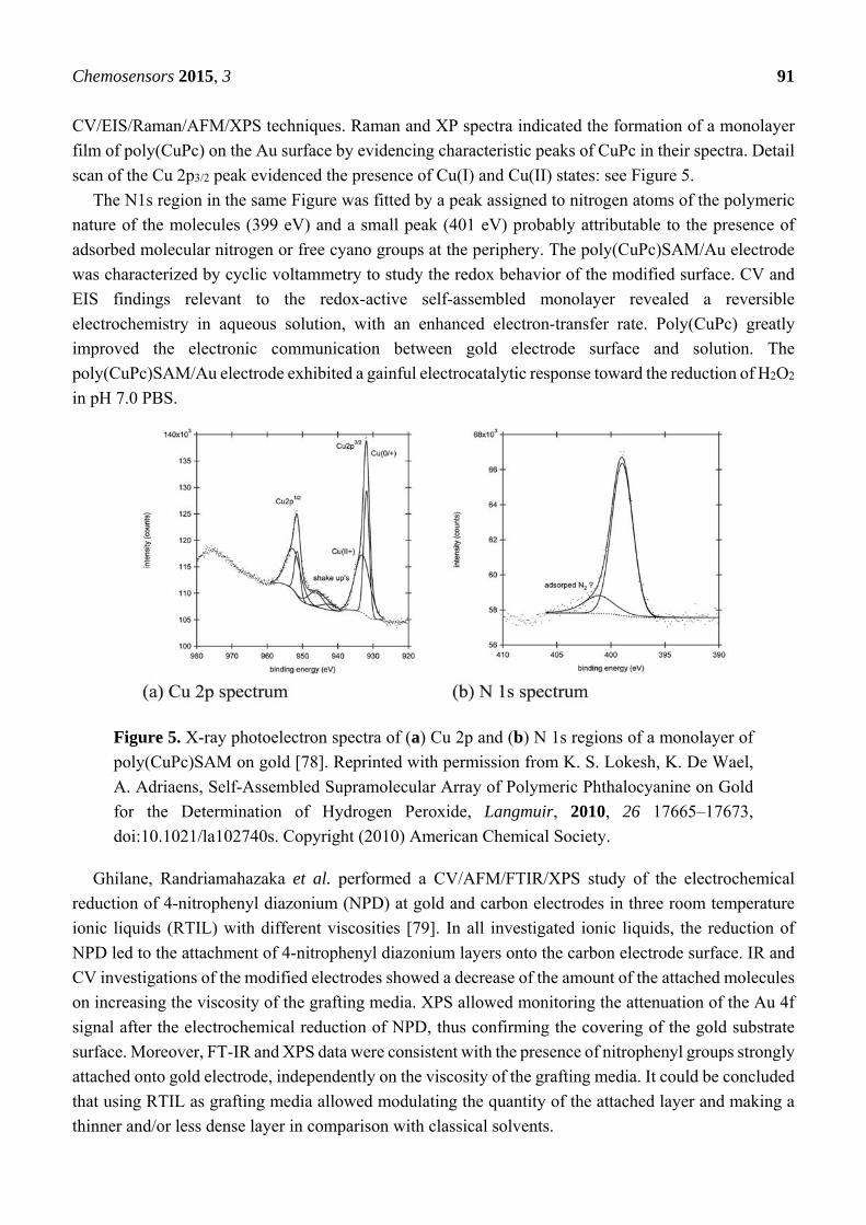
Adriaens et al. described the formation of a supramolecular self-assembled monolayer of polymeric
copper-phthalocyanine (poly(CuPc)SAM) onto a gold substrate [78]. The study was performed by
Chemosensors 2015, 3 91
CV/EIS/Raman/AFM/XPS techniques. Raman and XP spectra indicated the formation of a monolayer
film of poly(CuPc) on the Au surface by evidencing characteristic peaks of CuPc in their spectra. Detail
scan of the Cu 2p3/2 peak evidenced the presence of Cu(I) and Cu(II) states: see Figure 5.
The N1s region in the same Figure was fitted by a peak assigned to nitrogen atoms of the polymeric
nature of the molecules (399 eV) and a small peak (401 eV) probably attributable to the presence of
adsorbed molecular nitrogen or free cyano groups at the periphery. The poly(CuPc)SAM/Au electrode
was characterized by cyclic voltammetry to study the redox behavior of the modified surface. CV and
EIS findings relevant to the redox-active self-assembled monolayer revealed a reversible
electrochemistry in aqueous solution, with an enhanced electron-transfer rate. Poly(CuPc) greatly
improved the electronic communication between gold electrode surface and solution. The
poly(CuPc)SAM/Au electrode exhibited a gainful electrocatalytic response toward the reduction of H2O2
in pH 7.0 PBS.
Figure 5. X-ray photoelectron spectra of (a) Cu 2p and (b) N 1s regions of a monolayer of
poly(CuPc)SAM on gold [78]. Reprinted with permission from K. S. Lokesh, K. De Wael,
A. Adriaens, Self-Assembled Supramolecular Array of Polymeric Phthalocyanine on Gold
for the Determination of Hydrogen Peroxide, Langmuir, 2010, 26 17665–17673,
doi:10.1021/la102740s. Copyright (2010) American Chemical Society.
Ghilane, Randriamahazaka et al. performed a CV/AFM/FTIR/XPS study of the electrochemical
reduction of 4-nitrophenyl diazonium (NPD) at gold and carbon electrodes in three room temperature
ionic liquids (RTIL) with different viscosities [79]. In all investigated ionic liquids, the reduction of
NPD led to the attachment of 4-nitrophenyl diazonium layers onto the carbon electrode surface. IR and
CV investigations of the modified electrodes showed a decrease of the amount of the attached molecules
on increasing the viscosity of the grafting media. XPS allowed monitoring the attenuation of the Au 4f
signal after the electrochemical reduction of NPD, thus confirming the covering of the gold substrate
surface. Moreover, FT-IR and XPS data were consistent with the presence of nitrophenyl groups strongly
attached onto gold electrode, independently on the viscosity of the grafting media. It could be concluded
that using RTIL as grafting media allowed modulating the quantity of the attached layer and making a
thinner and/or less dense layer in comparison with classical solvents.
Chemosensors 2015, 3 92
Cabrera et al. described the modification of a palladium electrode by a self-assembled monolayer of
L-cysteine [80]. The CME was characterized by CV, EIS, FTIR and XPS. FTIR and XPS techniques
showed that L-cysteine was adsorbed on the Pd surface through the sulfur atom, while leaving the free
carboxylic acid and amino groups in the surface for conjugating biomolecules. XPS characterization
involved the acquisition of the Pd 3d, C 1s and S 2p regions. In particular, detail scans of the S 2p region
of the CME evidenced two doublets (SoS: 1.18 eV). The relevant S 2p3/2 peaks were assigned to the
thiolate formation on the surface through S–Pd bonds (162.1 eV) and to sulfonic groups of cysteic acid
(166.9 eV), thus suggesting that –OOCNH2CHCH2S–Pd was the adsorbed cysteine monolayer. The
electron transfer reaction of L-cysteine modified Pd electrode was probed by CV and EIS in Fe(CN)63−/4−
containing solutions. The CV measurements at bare and modified Pd electrodes revealed a
quasi-reversible voltammogram of the redox probe, indicating the promotion of electron transfer
between the redox couple and palladium electrode. Accordingly, EIS results at the CME were consistent
with an electron transfer resistance lower than at bare Pd electrodes.
Lee, Kim et al. developed an estrogen-sensing electrode for the EIS detection of estrogen bindings to
estrogen receptors covalently bonded to the electrode [81]. The sensor was prepared by modifying a gold
electrode with 3-mercaptopropionic acid (MPA). The carboxylic groups of the MPA/Au electrode were
activated by contact with a solution of 3-(3-dimethylaminopropyl)-1-ethylcarbodiimide
hydrochloride/N-hydroxysuccinimide (EDC/NHS) in a PBS. Dipping the EDC-NHS/MPA/Au in a
solution of estrogen-receptor α allowed a suitable covalent bonding to the receptor. XPS was used to
monitor the extent of each reaction step. The S 2p3/2 peak of the MPA/Au electrode was fitted by two
components, the first (162.1 eV) was attributed to S-Au bonds, the second (163.5 eV) to non-covalently
adsorbed free thiol groups on the Au electrode. In the C 1s region, the peak at 288.7 eV proved the
presence of carboxyl group of the MPA monolayer. The MPA modification with EDC and NHS was
also monitored by acquiring the N 1s region. The two peaks at 399.7 and 400.7 eV were assigned to
secondary amine and imine of EDC and, respectively, to the protonated tertiary amine of EDC. The
detection of estrogen hormone was evidenced by the observed EIS impedance change at the
receptor-modified electrode in the presence of the 10−6 M concentration of estrogen hormone in PBS
buffer solution.
Brunetti and Desimoni performed a CV/XPS investigation about the electropolymerization
of 8-hydroxyquinoline-5-sulfonic acid (HQSA) at glassy carbon electrodes in hydrochloric acid
solutions [82]. For a better understanding of the complex surface functionalization of the modified
electrode, another GCE (ox-GCE) was also prepared by using the same electropolymerization
parameters but in absence of HQSA. It is in fact known that potential cycling treatment can led to
oxygenated functionalities on carbonaceous surfaces. Survey and detail XPS scans were performed for
characterizing the two CMEs. The S 2ppeak (168.2 eV), assigned to sulfonic groups, confirmed the
presence of HQSA on the HQSA/GCE surface. As expected, the S 2p region was absent on the surface
of the ox-GCE. A deeper interpretation of the different XP regions of the two CMEs was postponed to
a more exhaustive XPS investigation (described below, after reference [90]). CV measurements allowed
comparing the electrochemical behavior of HQSA-GC, ox-GC and bare GC electrodes. The HQSA/GCE
showed improved electrochemical performances with respect to those of the other two electrodes. The
attractive features of the HQSA/GCE were evidenced by determining species of alimentary and
pharmaceutical interest such as dopamine, theophylline and tartrazine.
Chemosensors 2015, 3 93
Ganesh et al. prepared copper-hexacyanoferrate (CuHCF) and nickel-hexacyanoferrate (NiHCF)
modified ITO electrodes suitable for the electrocatalytic oxidation of alcohols in alkaline medium [83].
At first, CuHCF and NiHCF (in general, metal hexacyanoferrate, MHCF) were potentiodinamically
deposited on ITO electrodes. Then, both electrodes were cycled in aqueous NaOH solutions to convert
MHCF films to their corresponding M(OH)2 moieties. Both MHCF and M(OH)2 films were
characterized by SEM-EDAX and XPS. In the case of MHCF films, detail scans evidenced the presence
of Ni 2p3/2 (852 eV) or Cu 2p3/2 (931.7 eV) and Fe 2p3/2 (703 eV). Only Ni 2p and Cu 2p doublets were
unaltered by the potential cycling conversion to M(OH)2. The Fe 2p doublet progressively decreased up to
disappear with the number of cycle, revealing the cleavage of Fe–CN–Ni/Cu bond followed by dissolution
of iron as hydroxide species. The electroxidation ability of the two CMEs was investigated by CV. The
results proved that metal hydroxide modified electrodes display good catalytic performances and
stabilities for direct methanol or alcohols oxidation, along with low CO poisoning effect.
Millner et al. prepared organic thiol-modified glassy carbon electrodes (RSH/GCE) by potentiostatic
electroxidation in tetrabutylammonium hydroxide (Bu4NOH)/acetonitrile [84]. Adding Bu4NOH, a
strong deprotonating agent, allowed an easier oxidation/deposition of thiols onto the carbon surface. The
tested thiols were n-octanethiol, 3-(nitrobenzyl)mercaptan (NBM), cysteamine and N-acetylcysteamine.
The CMEs were characterized by CV and XPS. The surface composition of NBM/GCE before and after
modification was monitored by following the C 1s, N 1s, O 1s and S 2p photoelectron spectra. The
disappearance of N 1s signal assigned to the C–NO2 peak (404 eV), the increase of the N 1s signal
assigned to C–NH2/NHOH/NO peak (400 eV) and the unchanged S 2p signal (164 eV) proved that the
electrochemical reduction of nitrophenyl groups deposited during NBM oxidation was complete. NBM,
the model thiol used for exploring the possibility of thiols oxidation, proved capable of grafting the
carbon surface. In addition, the electrochemical results confirmed the stable binding of thiols and that
the surface modification occurred independently from RS• radicals formation and was explained by
nucleophilic addition of deprotonated thiols to the carbon surface.
Li, Sun et al. performed a CV/DPV/XPS/SEM/EDS and UV-Vis-NIR study about the
electrochemical properties of three fullerene-modified electrodes in aqueous solutions [85]. This was
allowed by drop-casting toluene solutions of the three isomers (C84-D2(IV), C84-Cs(a), and C84-C2(IV))
onto GC electrodes. Only the C84-C2(IV)/GC modified electrode showed a couple of redox peaks. CV
experiments allowed proving that those peaks were relevant to reduction/oxidation processes of the
C84-C2(IV) film. CV measurements at the CME in aqueous solutions of different salts proved that
cations, not anions, played an important role in the electrochemistry of the modified electrode. XPS
measurements were performed in the presence of KCl to prove that the K+ ions were incorporated into
the C2(IV) films upon reduction. The C 1s peak of the reduced film was fitted by components assigned
to negatively charged carbon atoms (283.4 ± 0.2 eV), indicating the reduction of C2(IV), to neutral
carbon atoms (284.8 ± 0.2 eV) and to positively charged carbon atoms (286.5 ± 0.2 eV). These findings,
together with the appearance of the K 2p photoelectron signal, allowed confirming that K+ ions were
incorporated in the C84-C2(IV) surface film upon reduction. Furthermore, interaction between C2(IV)
and guanine was investigated by DPV, UV-Vis-NIR and XPS. The results evidenced that guanine and
C2(IV) formed a charge-transfer complex due to their interaction, so that the reduction of C2(IV) was
blocked in the complex. The complex formation was also confirmed by the evolution of the N 1s signal

Chemosensors 2015, 3 94
before and after the interaction. Thus, the reoxidation peak current of C2(IV) decreased with the increase
of the guanine concentration.
S-H. Cheng et al. electrodeposited on a screen-printed carbon electrode (SPCE) a hybrid composite
consisting of a thin layer of the conducting poly(3,4-ethylenedioxythiophene) (PEDOT) polymer plus
gold nanoparticles [86]. The nanocomposite was characterized by UV/TEM/CV/DPV/FIA/XPS
techniques. The successful modification of the SPCE was checked by acquiring the C 1s, S 2p and Au
4f XPS regions. Assigning the S 2p3/2 peak (162.9 eV) to sulfur atoms in the thiophene ring, and the Au
4f7/2/4f5/2 doublet (82.8/86.5 eV) to Au(0), allowed confirming the successful modification by AuNP. The
electrochemical applications of the CME were explored by CV in deaerated cysteine-containing buffer
solutions. The results showed that the PEDOT-AuNP/SPCE was characterized by a favorable
electrocatalytic activity towards the oxidation of cysteine in the 2.0–8.0 pH range. The CME allowed
the separation of cysteine and glutathione oxidation peaks. Cysteine could be determined by DPV in the
presence of a high concentration of glutathione.
Kannan and John described a simple polymerization strategy for preparing AuNP-conducting polymer
films on GC and ITO surfaces by using 5-amino-2-mercapto-1,3,4-thiadiazole (AMT) capped gold
nanoparticles (AMT-AuNP) [87]. The study was performed by CV/EIS/AFM/FTIR/XPS techniques.
AFM results revealed the homogeneous distribution of AuNP in the modified film. The Au 4f XPS region
allowed proving the presence in the modified film of Au(0). The N 1s region was fitted by three peaks
assigned to imine nitrogen =NH (396.7 eV), secondary amine nitrogen –NH– (399.6 eV) and positively
charged nitrogen –N+H– (403.3 eV), thus confirming that the electropolymerization proceeded through
the oxidation of –NH2 groups present on the periphery of the AMT-AuNP. The CME electrocatalytic
performances were tested by studying the electroreduction of oxygen at pH 7.2. The AMT-AuNP film
showed an excellent electrocatalytic activity towards the reduction of oxygen at physiological pH,
suggesting the possibility of potential applications in energy storage systems and electrochemical
sensors.
Ma, Huang et al. performed a CV/SEM/TEM/FTIR/UV-Vis/XPS study aimed at developing a
one-step electrochemical synthesis of graphene/PANI composite film onto GCE and ITO electrodes [88].
XPS was used to characterize the chemical status of the surface film by examining the C 1s, O 1s, N 1s
and S 2p regions. The N 1s region was fitted by three different peaks assigned to benzenoid amine
(399.4 eV), quinoid amine (398.8 eV) and nitrogen cationic radical (N+•, 401.4 eV). The C 1s region was
fitted by five components assigned to C–C (284.5 eV), C–N (285.6 eV), C–O (286.6 eV), C=O (288.2 eV)
and O–C=O (290.2 eV) groups, thus proving the existence of few oxygen functionalities in the obtained
graphene of graphene/PANI, which were not evidenced by FTIR. In agreement with UV-Vis data, the
surface atomic S/N ratio of PANI confirmed its high doping level. The sensor was used to construct a
biosensor after entrapping onto its surface horseradish peroxidase. The CME exhibited a high catalytic
activity for the reduction of H2O2. Moreover, the graphene/PANI composite film could be directly used
as supercapacitor electrode.
Shi et al. performed a CV/SEM/TEM/EIS/XPS study aimed at characterizing some GCEs modified
by nitrogen doped multiwalled carbon nanotubes (N-MWCNT) decorated with SnO2, CeO2 or
SnO2+CeO2 nanoparticles [89]. TEM images revealed that SnO2 and CeO2 nanoparticles were
uniformly attached on the surface of the N-MWCNT. Detail XP scans of Sn 3d5/2/3d3/2 doublet of the
SnO2/N-MWCNT composite (486.4 eV/494.8 eV) confirmed the presence of SnO2. The Ce 3d5/2/3d3/2
Chemosensors 2015, 3 95
(882.8 eV/900.9 eV) detail scans of CeO2/N-MWCNT confirmed that cerium was mainly present as
Ce(IV). The N 1s region of N-MWCNT, SnO2/N-MWCNT and CeO2/N-MWCNT was investigated in
order of studying the interaction between nitrogen atoms and metallic oxides NPs. N 1s detail scans
of the three electrodes were fitted by four peaks assigned to the pyridine-like nitrogen (398.7 eV),
graphite-like nitrogen (401.0 eV), molecular N2 (404.3 eV) and nitrogen oxides adsorbed on the surface
of N-MWCNT (407.0 eV). Together with the results relevant to the Sn 3d and Ce 3d regions, these
findings supported the existence of strong interaction between metallic oxide NPs and nitrogen
atoms. CV experiments proved that N-MWCNT-based composites, as electrode materials, show a
better electrocatalytic activity for NO electroxidation than the conventional CNTs-based composites.
This suggested potential applications as a sensor for NO.
Yuan, Niu et al. grafted poly(4-vinylpyridine) (PV4P) to multiwalled carbon nanotubes by
in-situ free radical polymerization of the monomer [90]. The PV4P-g-MWCNT hybrids were loaded
with phosphotungstic acid (PW). The resulting material was characterized by SEM, TEM, FTIR and
XPS. SEM/TEM images showed that, after the grafting step, MWCNT were wrapped in a thick layer of
PV4P. MWCNT, PV4P-g-MWCNT and PW/PV4P-g-MWCNT hybrids were characterized by XPS.
The comparison of C 1s, N 1s and O 1s peaks relevant to the various hybrids samples confirmed
their chemical status after each preparation step. After PV4P was grafted onto MWCNT, a new C 1s
peak (286.2 eV), assigned to C–N bind of pyridine group at PV4P chains, added to those assigned to
hydroxyl (286.2 eV) and carbonyl (287.2 eV) carbons. The appearance of the W 4f signal in the
range from 33 to 40 eV proved the presence of PW in the PWs/PV4P-g-MWCNT hybrid. CV results
obtained at a PWs/PV4P-g-MWCNT/GCE showed that its electrochemical behavior followed a
four-one-electron surface-confined process of Keggin-type PWs. The electrochemical study proved
that PWs/PV4P-g-MWCNT hybrids are promising candidates for NO2− sensors in food safety and
medical diagnosis.
Desimoni et al. performed an XPS study aimed at characterizing the surface of glassy carbon
electrodes chemically modified with 8-hydroxyquinoline-5-sulfonic acid (HQSA/GCE) [23]. The work
completed a previous electrochemical investigation on the same CME [82]. Spectroscopic experiments
involved acquiring survey and detailed scans of C 1s, O 1s, Si 2p, Cl 2p, N 1s, and S 2p regions of an
HQSA powder standard sample as well as of GC electrodes cycled both in the presence and in the
absence of HQSA. The experimental value of the binding energy of the S 2p3/2 level of HQSA-modified
electrodes (167.6 eV) was found equal to that of the HQSA standard powder, thus confirming that HQSA
molecules were adsorbed on the surface of the HQSA/GC electrodes (see Figure 6) and that sulfur
maintained its chemical structure and properties. The C 1s spectrum of the CME was fitted by five peak
components associated to graphitic carbon (284.5 eV), CHx (285.0 eV), C–O (286.3 eV), C=O (287.7 eV)
and O=C–OH (289.0 eV). The experimental S/N and O/S ratios estimated for the HQSA standard were
slightly different from the theoretical ones, suggesting a limited degradation of the powder under
X-ray irradiation.
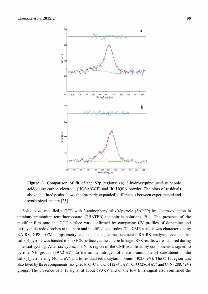
Chemosensors 2015, 3 96
Figure 6. Comparison of fit of the S2p regions: (a) 8-hydroxyquinoline-5-sulphonic
acid/glassy carbon electrode (HQSA/GCE) and (b) HQSA powder. The plots of residuals
above the fitted peaks shows the (properly expanded) differences between experimental and
synthesized spectra [23].
Solak et al. modified a GCE with 3-aminophenylcalix[4]pyrrole (3APCP) by electro-oxidation in
tetrabutylammonium-tetrafluoroborate (TBATFB)-acetonitrile solutions [91]. The presence of the
modifier film onto the GCE surface was confirmed by comparing CV profiles of dopamine and
ferricyanide redox probes at the bare and modified electrodes. The CME surface was characterized by
RAIRS, XPS, AFM, ellipsometry and contact angle measurements. RAIRS analysis revealed that
calix[4]pyrrole was bonded to the GCE surface via the etheric linkage. XPS results were acquired during
potential cycling. After six cycles, the N 1s region of the CME was fitted by components assigned to
pyrrole NH groups (397.2 eV), to the amine nitrogen of meso-p-aminophenyl substituent in the
calix[4]pyrrole ring (400.2 eV) and to residual tetrabutylammonium (402.0 eV). The C 1s region was
also fitted by three components, assigned to C–C and C–H (284.5 eV), C–O (286.4 eV) and C–N (288.7 eV)
groups. The presence of F 1s signal at about 690 eV and of the low B 1s signal also confirmed the
Chemosensors 2015, 3 97
complex formation between BF4− ions and the calix[4]pyrrole moieties. The stability of the 3APCP film
at the GC surface versus the applied potential was evaluated by a series of CV voltammograms acquired
in the presence of the of Fe(CN)63−/4− redox probe.
Ho et al. developed a template-free synthetic procedure for preparing a conducting
metal-doped polymer nanocomposite [92]. The study, performed by CV/TEM/EDX/FTIR/XPS
techniques, implied using PPY, HAuCl4 and 4-sulfocalix[4]arene as a reductant, oxidant and dopant,
respectively. TEM images showed that gold nanoparticles were uniformly distributed throughout the
nanocomposite material (gold particles-encapsulated polymer). The presence of 4-sulfocalix[4]arene in
the nanocomposite was confirmed by FTIR, EDX and XPS. XPS evidenced the presence of C, N, O, S
and Au in the nanocomposite. The S 2p (167.5 eV) and S 2s (230 eV) regions were assigned to
sulfate ions dopants of Calix-PPY and Au-Calix-PPY, while the Au 4f7/2/4f5/2 doublet (83.5/87.2 eV,
respectively) confirmed the presence of Au(0) on the Au-Calix-PPY nanocomposite surface.
CV experiments, performed at screen-printed carbon electrodes modified by drop coating the
nanocomposites, showed improved electrocatalytic properties of the CME towards the electro-oxidation
of formaldehyde and glucose.
Xu et al. prepared a potassium-modified graphene suspension to be used in nitrite-selective
sensors [93]. The suspension was characterized by CV/TEM/AFM/XPS techniques. AFM images of the
thickness of graphene sheet before and after K-modification allowed concluding that the introduction of
K cations and/or oxides and/or metals was assembled on a monolayer of graphene sheet. In agreement
with these results, XP spectra proved that potassium, absent in graphene (GR) samples, were present on
the surface of the K-modified GR. At low K-coverages, the K 2p3/2/2p1/2 doublet (293.2 eV/296.0 eV)
was assigned to K oxides and cations. At higher K-coverages, a second doublet (294.4 eV/297.1 eV)
was evidenced and assigned to metallic K. The C 1s region was fitted by contributions of C–C and C–H
bonding (284.6 eV) and of C–O, C=O and C–C=O bonding (at 285.9 eV, 287.0 eV, and 288.8 eV,
respectively). The intercalation of K species changed Fermi level and charge transfer, opening the band
gap of the GR. This improved the electrocatalytic activity of graphene in electrochemical systems. The
suspension of K-modified GR was drop casted on the surface of GCEs. The electrochemical behavior of
the K-modified-GR/GCE was explored by CV in PBS containing nitrite ions. The CME exhibited good
analytical performances. Responses were unaffected by high concentrations of a series of inorganic
anions, cations and saccharides. Promising performances were also assumed in biosensing, energy
conversion, biomedical, and other electronic systems applications.
Yuan, Niu et al. anchored nickel hexacyanoferrate nanoparticles (NiHCFNP) to multiwalled carbon
nanotubes with a grafted poly(4-vinylpyridine) (PV4P) linker for electrically switched ion
exchange [94]. TEM and FTIR findings confirmed the presence of highly dispersed NiHCFNP
on the surface of the NiHCFNP/PV4P-g-MWCNT composite. CVs experiments performed at a
NiHCFNP/PV4P-g-MWCNT/Au modified electrode evidenced its considerable capacity and stability as
ion exchanger. NiHCFNP could be reversibly switched between reduced and oxidized states depending
on the applied potential. A reversible uptake and release of Na+, K+ and Cs+ cations accompanied the
redox process, allowing maintaining the charge neutrality within the NiHCF matrix. XPS results
acquired after the CME exposition to high concentrations of Na+ ions confirmed its good selectivity for
Cs+ over Na+ ions. The stronger affinity of the composite towards Cs+ was consistent with CV data.
Chemosensors 2015, 3 98
Li et al. incorporated a water-soluble ionic liquid, 1-butyl-3-methylimidazolium tetrafluoroborate
([BMIM]BF4), into TiO2 nanoparticles to fabricate a hybrid film modified glassy carbon electrode [95].
The modification of GCEs was performed electrochemically, by dipping the GCE in a solution containing
[BMIM]BF4 and tetrabutyltitanate. The obtained nano-TiO2NP/[BMIM]BF4/GCE was characterized by
SEM and XPS. XPS survey scans evidenced the presence of C 1s, N 1s, Ti 2p, O 1s and F1s signals.
The Ti 2p (~ 458 eV) and O 1s (532.9 eV) signals were assigned to the nano-TiO2 matrix. The C 1s
(~285 eV), N 1s (~401 eV) and F 1s (~684 eV) peaks were attributed to [BMIM]BF4. XPS data allowed
estimating 4.1% and 2.3% atomic percentages of F and Ti, respectively. The CME was then applied to
study the electrochemical behavior of p-acetaminophen in slightly acidic PBS. CV results indicated that
the TiO2NP/[BMIM]BF4 hybrid film improved the redox reactions of p-acetaminophen in aqueous
medium, and that it was more efficient than TiO2NP/GCE and bare GCE to surface accumulate the
analyte, thus enhancing the sensor sensitivity. The sensor was successfully applied to the SWV analysis
of p-acetaminophen in urine samples.
Deng et al. developed an amperometric uric acid biosensor based on a nanobiocomposite derived
from thionine modified graphene oxide (T-GO) [96]. The T-GO composite was characterized by
CV/amperometry/TEM/AFM/UV-Vis and XPS techniques. UV-Vis spectra of the T-GO suspension
confirmed that thionine molecules were attached to GO. XPS allowed exploring their interactions.
Survey XPS scans of T-GO evidenced the signals of C, O, N and S. The C 1s region was fitted by four
peaks assigned to C–C (284.6 eV), C–O (286.7 eV), C=C (287.8 eV) and O–C=O (288.7 eV) carbons.
The N 1s region was fitted by two peaks assigned to amine groups (401.8 eV) and pyridine-N-oxides
(402.3 eV). The S 2p signal (169.1 eV) was assigned to S–N bond in thionine molecule. These findings
confirmed the noncovalent functionalization of graphene oxide by thionine. CV and amperometric
measurements proved that the composite, due to the synergistic effect between thionine and graphene
oxide, exhibited excellent performances towards H2O2 reduction. Uricase (UOx) modified T-GO/GC
electrodes were prepared by casting a UOx plus T-GO suspension onto the surface of GCEs. In the
presence of oxygen, the UOx/T-GO/GCE catalyzed the oxidation of UA to allantoin, with the
simultaneous production of H2O2. This H2O2 is reduced at the CME surface without interferences from
other electroactive compounds present at physiological levels (glucose, ascorbic acid, noradrenalin and
dopamine). The CME was applied to the analysis of UA in human blood and urine.
Abraham et al. performed an EIS/SEM/EDS/XPS/SIMS investigation aimed at improving the
performances of high-capacity lithium-ion(+)/graphite(−) cells by modifying the
Li1.2Ni0.15Mn0.55Co0.1O2-based positive electrode with alumina [97]. The modification modes were
(i) atomic layer deposition (ALD) of alumina and (ii) addition of nanoscale alumina powder. EIS data
showed an improvement of full-cell AC impedance by ALD coatings. EDS elemental maps proved that
all electrode constituents, including the binder, were covered by the alumina coating. C 1s, O 1s,
F 1s and Al 2p XP spectra were acquired for monitoring the changes induced on positive electrode
surfaces by the ALD process. The F1s intense peak at 687.8 eV was assigned to the PVDF binder (Solef®
PVDF is a fluorinated semi-crystalline thermoplastic which is obtained by polymerizing vinylidene
fluoride). The O1s peak at 529.6 eV was assigned to the Li1.2Ni0.15Mn0.55Co0.1O2 substrate and the weak
peak centered at 532 eV to a Li2CO3 impurity. Cycling produced a marked decrease of Al 2p and O 1s
regions of the positive electrode. An original equation was developed for estimating the thickness of the
alumina layer from XPS data. SIMS results proved that the alumina coating did not significantly alter
Chemosensors 2015, 3 99
the initial charge/discharge cycling characteristics of the cells. The modification was aimed at enhancing
the electronic conductivity within the electrode laminate, improving the surface stability of the coated
materials, or providing a physical barrier capable of suppressing detrimental chemical side reactions
between electrode surface and electrolyte. The overall results suggested that only very thin alumina
coatings could improve the cell performances while thicker coatings worsen capacity retention.
Chen et al. developed an amperometric glucose biosensor based on gold/platinum hybrid
functionalized zinc oxide nanorods (Pt–Au/ZnONR) and glucose oxidase (GOx)–lectin biospecific
interaction [98]. The CME was obtained by modifying a GCE in sequence with a gold/platinum hybrid
functionalized zinc oxide nanorods, a layer of porous gold nanocrystals (pAu) and concanavalin A
(ConA). At last glucose oxidase was immobilized on the ConA/pAu/Pt–Au/ZnONR/GCE by the
biospecific interaction between GOx and ConA. The Pt–Au/ZnONR composite was characterized by
TEM and XPS. XP detail spectra were relevant to the O 1s, Zn 2p, Au 4f and Pt 4f regions. The Zn
2p3/2/2p1/2 doublet (1021.1 eV/1044.9 eV) was assigned to ZnO. The Au 4f7/2/4f5/2 (83.6 eV/87.6 eV) and
the Pt 4f7/2/4f5/2 (70.8 eV/74.1 eV) doublets, assigned to the formation of Pt–Au hybrid on the surface of
ZnONR, confirmed the successful preparation of the Pt–Au/ZnONR nanocomposite. CV measurements,
performed in the presence of (Fe(CN)6)3−/4− after each stage of the GCE modification, indicated that
(i) pAu could act as tiny conduction centers assisting the electron transfer at the electrode surface and
(ii) that GOx/ConA/pAu/Pt–Au/ZnONR/GCE exhibited an excellent electrocatalytic activity toward
glucose. Amperometric measurements in PBS allowed estimating the promising sensor performances.
Guascito et al. prepared a composite nanomaterial electrodepositing copper nanoparticles (CuNP) on
an electrosynthesized film of poly-3-methylthiophene (P3MT) [99]. CuNP were electrodeposited by
pulsed potential SWV on both Pt and screen-printed electrodes. The composite film was characterized
by SEM and XPS. SEM images proved that pulse amplitude influenced the amount of the deposited
particles, but not the CuNP size. XP spectra relevant to C 1s, Cu 2p, S 2p, Cl 2p, N 1s, O 1s and Na 1s
regions were acquired and carefully analyzed both after electropolymerization of 3MT and after
electrodepositing CuNP on the P3MT. C 1s and S 2p signals revealed significantly different compositions
of the as-grown P3MT film and of the CuNP/P3MT composite. The analysis of the complex Cu 2p region
required the evaluation of the Auger parameters, which were estimated in the 1846.8–1847.1 eV and
1848.3–1848.8 eV ranges on considering the component of Cu 2p3/2 at lower and higher BE,
respectively. These results allowed excluding the formation of Cu(0) and confirmed the presence of
Cu(I) and Cu(II) in each sample. CV and FIA findings showed that the composite film could be applied
to the glucose electrochemical detection in flow systems. The response of the sensor to ascorbic acid,
uric acid, sorbitol, fructose and dopamine evidenced that it could be effectively used as an
electrochemical detector coupled to a chromatographic systems for the simultaneous detection of
biomolecules.
Narayanan et al. synthesized a nickel nanoparticles (NiNP) modified electrode in the ionic liquid
medium 1-ethyl-3-methylimidazolium ethyl sulfate (EMIMES) for application in biomolecules
determinations [100]. NiNP were electrochemically deposited on a polycysteine film (PCF) previously
electropolymerized on a paraffin wax impregnated graphite electrode (PIGE). The NiNP/PCF/PIGE was
characterized by FTIR, Raman, XPS, FESEM, EDS, EIS and CV techniques. The Ni-S bonds evidenced
by Raman spectroscopy were further confirmed by XPS survey and detail scans. In particular, the Ni
2p3/2/2p1/2 doublet (855.6 eV/873.3 eV) and the S 2p3/2 peak (162.5 eV) were in agreement with the
Chemosensors 2015, 3 100
values reported for NiS. The CV behavior of vitamin B6, L-tyrosine, L-tryptophan, vanillin, glucose and
H2O2 was studied at the bare PIGE, at the PCF/PIGE and at the NiNP/PCF/PIGE. This last electrode
allowed the best electrocatalytic performances. Good chronoamperometric responses obtained at the
NiNP/PCF/PIGE suggested it could be exploited in flow-systems application.
Khudaish et al. prepared a solid-state sensor based on a poly(4-aminodiphenylamine) (Padpa) film
deposited at a GCE doped with tris(2,2’-bipyridyl)Ru(II) complex (Padpa/Ru/GCE) [101]. The surface
of the CME was characterized by AFM and XPS. AFM images evidenced broken and fused Padpa–Ru
doped fiber structures as opposed to those of a native Padpa film, which appeared relatively more
uniform. The survey XP spectrum of Padpa showed the presence of carbon, oxygen, nitrogen and
ruthenium. The C 1s detail spectrum of the undoped Padpa sample was fitted by three peaks assigned to
C–C (283.93 eV), C–N (285.08 eV) and C=C (287.32 eV) bonds. After doping, the Ru 3d5/2/3d3/2 doublet
(280.3 eV/283.2 eV) partially overlapped the C 1s region. The N 1s spectrum of Padpa was also fitted
by three peaks assigned to –N= (398.9 eV), –NH2 (400 eV) and NH3+ (402.3 eV) nitrogens. The presence
of –N= groups confirmed that oxidation and deprotonation processes take place in the polymerization of
the Padpa. The electrochemical performances of the CME were tested by CV and DPV. After doping
with the Ru(II) complex, CV profiles evidenced reversible redox peaks assigned to the [Ru(bpy)3]3+/2+
couple. The CME was applied to the DPV simultaneous determination of Zn2+, Cd2+, Pb2+ and Cu2+ ions
in sea and ground water samples.
Pan et al. synthesized rod-like precursors of Co3O4 and Ag/Co3O4 composites with different Ag
contents via a co-precipitation method [102]. The composites were characterized by TGA/DTA, XRD,
TEM, FESEM and XPS. TGA and DTA allowed determining the optimum calcination temperature of
the AgNO3 and Co(NO3)2·6H2O precursors to obtain the final composites. XPS allowed estimating the
surface chemistry of the Ag/Co3O4 sample, containing 3% Ag. The Co 2p3/2/2p1/2 doublet
(779.6 eV/795.5 eV; SoS 15.9 eV) were consistent with Co3O4 reference data. The Ag 3d5/2/3d3/2
(367.95 eV/373.9 eV) doublet confirmed the presence of Ag. The experimental Ag/Co molar ratio was
very close to the theoretical value, further confirming the composition of the Ag/Co3O4 composite with
3% Ag. XRD patterns of Co3O4 and Ag/Co3O4 were also in good agreement with cubic spinel Co3O4
phase. The Co3O4/GCE and Ag/Co3O4/GCE were tested as sensors for p-nitrophenol reduction in basic
solution. At the two CME, the peak current was significantly higher than at a bare GCE. However, at
the Ag/Co3O4/GCE the peak current increase was also accompanied by a markedly peak potential
decrease. The Ag/Co3O4 composite with 4% Ag showed the highest electrocatalytic activity.
Guascito et al. developed a TeO2 nanowires modified platinum electrode (TeO2NW/Pt) by drop casting
TeO2NW, synthesized by thermal evaporation of Te(0) in oxygen atmosphere, on the substrate
surface [103]. The morphological and spectroscopic characterization of TeO2NW, as synthesized on Pt
foil, was performed by SEM, XRD and XPS. SEM images showed straight and smooth TeO2NW. Both
TeO2NW and a commercial TeO2 powder standard were analyzed by XPS. Detail scans of the Te 3d5/2
region of TeO2NW and TeO2 powder were fitted by peaks assigned to Te(IV) oxide (576.7 ± 0.1 eV) and
to Te mixed surface oxides (579.7 ± 0.1 and 579.4 ± 0.1 eV, respectively). Te(0) and Pt(0) were absent.
After cycling the TeO2NW/Pt in neutral PBS, XPS evidenced the presence of Te(0) and Te(IV). The
electrochemical characterization of the CME was performed by CV and CA in neutral PBS to investigate
the sensing properties of this material against H2O2. The results proved the catalytic activity
Chemosensors 2015, 3 101
toward hydrogen peroxide reduction and indicated that the TeO2NW/Pt electrode was suitable for sensing
applications.
Canevari et al. described the preparation of a hybrid material composed of disordered mesoporous
silica modified with multiwalled carbon nanotubes [104]. The material was characterized by N2
adsorption–desorption isotherms, XRD, FESEM, HRTEM, Raman and XPS techniques.
HRTEM images showed that MWCNT were embedded within the mesoporous silica matrix. XPS analysis
involved exploring the C 1s, O 1s and Si 2p regions. The C 1s region was fitted by peaks assigned to
graphitic carbons (284.7 eV), carbon present in alcohol, ether or C=N groups (286.0–286.3 eV), carboxyl
or ester groups (288.8–289.1 eV), carbonate groups (290.5–291.2 eV) and satellite peaks due to π–π*
transitions in aromatic rings (291.6 eV). The O 1s peak (532.8 eV) was assigned to carbonyl oxygen
atoms in esters, anhydrides and oxygen atoms in hydroxyl groups. The Si 2p region originated a signal
at 103.5 eV. CMEs were prepared by drop casting a mixture of SiO2/MWCNT (before and after an acid
functionalization) and Nafion® onto GCEs. CV measurements performed in neutral PBS in the presence
of the (Fe(CN)6)3−/4− couple allowed comparing the electrochemical performances of bare GCE and
SiO2/MWCNT/GCE before and after the acid treatment. The CME was applied to individual and
simultaneous determinations of dopamine, uric acid and acetaminophen in human urine samples.
In a subsequent study, Canevari et al. described synthesis, characterization and applications of a
mesoporous silica/multiwalled carbon nanotube (SiO2/MWCNT) hybrid material, subsequently
decorated with silver nanoparticles (AgNP) [105]. The AgNP/SiO2/MWCNT composite was characterized
by SEM, EDS, HRTEM and XPS. SEM images and EDS elemental mapping of Ag, C and Si showed a
homogeneous distribution of AgNP in the hybrid material. Detail XP spectra of the composite concerned
the Ag 3d, Si 2p, O 1s and C 1s regions. The Ag 3d region was fitted by two Ag 3d5/2/3d3/2 doublets
assigned to Ag(0) (374.4 eV/368.3 eV) and Ag-O species (379.7 eV/373.6 eV), likely due to the
formation of Si–O–Ag bonds. These findings were supported by the results of the fitting of O 1s and Si 2p
regions. The fitting of the C 1s region also supported the presence of functional groups suitable for anchoring
of the AgNP to the hybrid material. A thin film of AgNP/SiO2/MWCNT was deposited on GCEs. The
electrochemical performances of the AgNP/SiO2/MWCNT/GCE were explored by CV and SWV. In neutral
PBS, the sensor showed an excellent electrocatalytic response for the determination of hydroquinone and
catechol. Because of the absence of interference from resorcinol, 2,6-dichloroindophenol, phenol,
4-nitrophenol, or nitrite ions, the CME could be applied as a sensor for phenolic compounds in
environmental samples.
Guo et al. described the electrodeposition of a composite consisting of NiONP and PtNP onto
electrochemically reduced graphite oxide [106]. The NiONP/PtNP/ERGO film was characterized by
SEM/EDS, AFM, Raman and XPS techniques. AFM images allowed characterizing the various steps of
the preparation of the composite. Detail XP spectra were acquired in the C 1s region of GO and in the
C 1s, Pt 4f and Ni 2p regions of the composite. After the electrochemical reduction of GO to ERGO, the
C 1s peak assigned to C–C atoms (284.6 eV) increased significantly while those related to the oxidized
carbons (C–O, C=O and O=C–O) almost disappeared, thus indicating that oxygen-containing
functionalities were nearly completely absent in ERGO. The Pt 4f7/2/4f5/2 doublet of the
NiONP/PtNP/ERGO (70.9 eV/74.2 eV) was assigned to Pt(0) while the Ni 2p3/2/2p1/2 doublet
(854.8 eV/872.5 eV; SoS 17.7 eV) was consistent with NiO only. A GCE was electrochemically
modified by the NiONP/PtNP/ERGO composite. The electrochemical performances of bare GCE,
Chemosensors 2015, 3 102
ERGO/GCE, PtNP/ERGO/GCE and NiONP/PtNP/ERGO/GCE towards glucose oxidation in alkaline
medium were compared in NaOH solutions. The results proved that the combination of ERGO,
PtNP and NiONP led to the highest catalytic efficiency and that the small quantity of PtNP significantly
enhanced the catalytic effect of NiONP. The proposed CME was applied to glucose detection in real
human serum samples.
John et al. described the potentiodynamic formation of a gold nanoparticles film on glassy carbon
and indium tin oxide (ITO) electrodes by using aminophenyl (APh) diazonium cations grafted gold
nanoparticles (APh-AuNP) [107]. The APh-AuNP modified electrodes were characterized by CV, AFM,
EIS and XPS. EIS findings proved that the electron transfer reaction of the (Fe(CN)6)3−/4− couple at the
CME was faster than at bare GCE. AFM images showed mono dispersed spherical AuNP attached on the
surface of the ITO without undergoing aggregation. The XP survey scan evidenced the presence of C, N
and Au on the electrode surface. The Au 4f7/2/4f5/2 doublet (83.8 eV/87.5 eV; SoS 3.7 eV) proved the
presence of Au(0). The N 1s region was fitted by peaks assigned to –NH (396.7 eV), =NH (399.2 eV),
–N+H (400.2 eV) and azo bridges –N=N– (403.3 eV) groups on the APh-AuNP modified ITO substrate.
The C 1s region was fitted by three peaks, the one at 286 eV indicating the presence of C–N–bond on
the APh-AuNP film modified ITO substrate. CV and DPV measurements allowed verifying that the
APh-AuNP/GCE could be applied to the detection of ranitidine hydrochloride (an histamine H2 receptor
antagonist) in the presence of an excess of paracetamol (N-acetyl p-aminophenol). The CME was applied
to the detection of the concentration of ranitidine in commercial drugs.
Freire et al. prepared hybrid multilayer films composed by osmium metallopolymer
[Os(bpy)2(PVP)10Cl]Cl (Os-poly) and europium phosphomolybdate, K11[Eu(III)(PMo11O39)2]
(Eu(PMo11)2), by an electrostatic layer-by-layer self-assembly method [108]. Film growth, composition
and thickness were monitored by UV-Vis spectroscopy, XPS and SEM. The {Os-poly/Eu(PMo11)2}n
multilayer films (OMFn) were deposited on quartz slides for UV-Vis and photoluminescence
measurements, on glass slides for XPS and on glassy carbon and on ITO/Polyethylene glycol (ITO/PET)
electrodes for CV studies. SEM images of OMF20 showed a non-uniform distribution of polymeric
molecules along with the polyoxometalate (POM) clusters, and allowed measuring film thicknesses. The
exhaustive XPS characterization of the composites involved reliable fittings of the C 1s, Os 4f, O 1s,
P 2s, Mo 3d, 4f and Eu 3d. Peak positions (BEs) were reported together with the relevant FWHM. In
particular, the fitting of the Os 4f7/2/4f5/2 evidenced the presence of Os2+ (50.4 eV/53.2 eV) and Os3+
(52.0 eV/54.7 eV). The fitting of the Mo 3d5/2/3d3/2 region evidenced the presence of Mo6+
(253.8 eV/232.6 eV) and Mo5+ (234.2 eV/231.1 eV). The presence of 6.1% Mo5+ evidenced some
photoreduction of POMs under X-ray exposure. XPS findings were completed by surface atomic
percentages of the multilayer film. CV measurements obtained at hybrid films adsorbed onto a GCE
revealed three Mo-based reduction processes and one Os reduction process in the explored potential
range. Cyclic voltammograms of the (Fe(CN)6)3−/4− and (Ru(NH))63+/2+ electroactive probes on OMFn
modified electrodes revealed a redox mediation between film and probes and a Mo-based electrocatalytic
activity towards reduction of nitrite and iodate.
Guo et al. synthesized a series of highly dispersed bimetallic Pt:Pd alloy nanoparticles anchored on
RGO with the assistance of the 3-ethyl-1-vinylimidazolium tetrafluoroborate IL [109]. The composite
was prepared by ultrasonication of different H2PtCl6/PdCl2 mixtures in an IL-GO/ethylene glycol
suspension. The composite was characterized by SEM, EDS, TEM, XPS, Raman and XRD techniques.
Chemosensors 2015, 3 103
The overall results demonstrated that Pt:Pd alloy nanoparticles were uniformly dispersed on RGO. In
particular, after the reduction, XP spectra of the Pt(50%)Pd(50%)–IL–RGO composite evidenced a clear
diminution of the C 1s peak assigned to C–O bonds (286.6 eV) and an increase of the peak assigned to
C–C and C=C bonds (284.6 eV). Together with the results of the analysis of the Pt 4f and Pd 3d doublets
evidenced in the Pt(50%)Pd(50%)–IL–RGO composite, XPS proved the successful synthesis of the
composite. Glucose sensors where prepared by drop casting a composite/Nafion® ink on the surface of
GCEs. The electrochemical performances of the Pt(50%)Pd(50%)–IL–RGO/Nafion®/GCE were
explored by CV in sulfuric acid solutions. The comparison of the results of amperometric measurements
performed in the presence of different glucose concentrations evidenced the superior performances of
the proposed CME with respect to some typical enzyme-based glucose sensors, thus making the
proposed composite very promising for applications in direct detection of glucose in real human samples.
Scavetta et al. prepared a CME by coating ITO electrodes with a ferrocene-functionalized
poly(3,4-ethylenedioxythiophene):poly(styrene-4-sulfonate) (PEDOT:PSS) film [110]. The two-step
procedure implied the electrodeposition of azidomethyl functionalized 3,4-ethylenedioxythiophene
(PEDOT-N3) followed by copper-catalyzed azide–alkyne cycloaddition of ethynylferrocene. The
resulting PEDOT-Fc:PSS conducting polymer was characterized CV, AFM and XPS. Survey XPS scans
of PEDOT-N3:PSS evidenced the presence of the C 1s, O 1s, S 2p and N 1s regions. Authors reported
detailed results of the curve fitting (BEs and FWHMs). In particular, the S 2p region of the
PEDOT-N3:PSS coated ITO was fitted by peaks assigned to neutral sulfur of PEDOT
(163.7–164.9 eV), cationic S+ (165.3 eV) associated with the PEDOT backbone and the highly oxidized
SO3− (167.8–169.0 eV). In the case of the PEDOT-Fc:PSS polymer, the survey scan evidenced the
additional Fe 2p region. Together with the results relevant to the N 1s region, XPS proved the partial
transformation of the azide group into the 1,2,3-triazole unit bound to the terminal ferrocene head, and
the presence of Fe(II) and Fe(III) species. The electrocatalytic behavior of the coating was characterized
by the stable and highly reversible response of ferrocene. The linear scan voltammetry in LiClO4
solutions containing different amounts of dopamine proved that ferrocene could mediate the
electroxidation of the analyte.
Yang et al. developed by a templating approach a hollow CuO polyhedron-modified sensor for the
nonenzymatic glucose detection [111]. Morphology and structure of the composite were characterized
by FESEM, TEM, XRD, Raman and XPS techniques. XRD results evidenced the standard monoclinic
structure of the hollow CuO polyhedron particles. Their hollow interior and structure were also verified
by TEM. The purity and composition of the composite were further investigated by XPS. The Cu
2p3/2/2p1/2 doublet (933.5 eV/953.4 eV) was assigned to CuO. No spectral contribution of Cu2O was
evidenced. The O 1s region was fitted by two peaks assigned to CuO (529.3 eV) and to oxygen adsorbed
on the surface of CuO (531.3 eV). The enzyme-free amperometric sensor was prepared by casting a
Nafion®-impregnated CuO polyhedron suspension onto a GCE. CV and CA experiments allowed
exploring the electrocatalytic activity of the CuO-Nafion®/GCE towards the oxidation of glucose in
NaOH solutions. Interfering species such as sodium chloride, ascorbic acid and uric acid did not affect
the response of the sensor, which could then find applications as a nonenzymatic glucose sensor.
Chemosensors 2015, 3 104
Pan, Kang et al. developed a CME by electropolymerizing alizarin red S (ARS) onto a glassy carbon
electrode dipped in the room-temperature ionic liquid 1-butyl-3-methylimidazolium tetrafluoroborate
(BMIMBF4) [112]. The polymeric PARS/BMIMBF4 film was characterized by SEM, EDX, XPS, FTIR
and electrochemical techniques. SEM images showed that the electropolymerization of ARS in
BMIMBF4 led to a smoother and more compact morphology of the coating with respect to that obtained
in aqueous solution. EDX results showed that C, B, O, N, F, Si and S were the major elements of the
PARS film synthesized in BMIMBF4. In agreement with EDX results, XPS findings relevant to the
PARS film confirmed the presence of C, N and S on the composite surface. The comparison of the CV
profiles recorded at bare GCEs, PARS/GCEs and PARS/BMIMBF4/GCEs allowed proving that
BMIMBF4, owing to its good conductivity, was critical in accelerating electron transfers. The profitable
combination of the electrochemical activity of PARS and conductivity of BMIMBF4 allowed the DPV
determination of catechol by oxidation to O-quinone in neutral PBS as well as in real water samples.
4. Conclusions
The above papers offer a realistic and diversified panorama about the role played by XPS in the field
of electrochemical sensor preparation and characterization. However, some comments are necessary
about the interpretation of XPS results. It is evident that some papers report different BE values for the
same photoemitting level of the same atomic species in the same chemical state. Examples are the BE
values assigned to the Au 4f level of Au(0), or the C 1s BE values assigned to hydrocarbon
contamination. These discrepancies could be avoided by a careful selection of literature information.
Moreover, in the case of curve fittings of metal-like samples or metal-doped materials, asymmetric peak
lineshapes should replace the symmetric ones [113]. This is also the case of the C 1s peak lineshape of
graphitic carbons in conductive graphite, carbon nanotubes, and vitreous carbon [24,114,115]. As
evidenced in Figure 3, using a symmetrical peak 1 in place of a more correct asymmetrical peak should
significantly affect the height of peak 3 (a detailed discussion about how fitting asymmetric lineshapes
is reported in Appendix 3 of [16]).
Incorrect fittings could lead to somewhat unreliable conclusions. At last, FWHM and SoS values
used in curve fittings should always be reported (see [69] for a good example) but this is rarely done.
Canevari et al. [104] and Fernandes et al. [108] are appreciable examples of correctly reporting the
results of XPS curve fittings.
The reviewed papers are of sanitary, pharmacological, alimentary and environmental concern.
Tables 2 and 3 itemize the XPS-characterized sensors, which are suitable for food and environmental
analysis, respectively.
In conclusion, notwithstanding the above-mentioned comments, XPS always offers an unrivalled help
in controlling the composition of the ultimate surface layers of the sensor as well as in following the
evolution of surface functionalities during its preparation. Hopefully, the above described results could
induce more researchers to consider XPS as a key technique to achieve important surface information
for characterizing and optimizing the sensors.
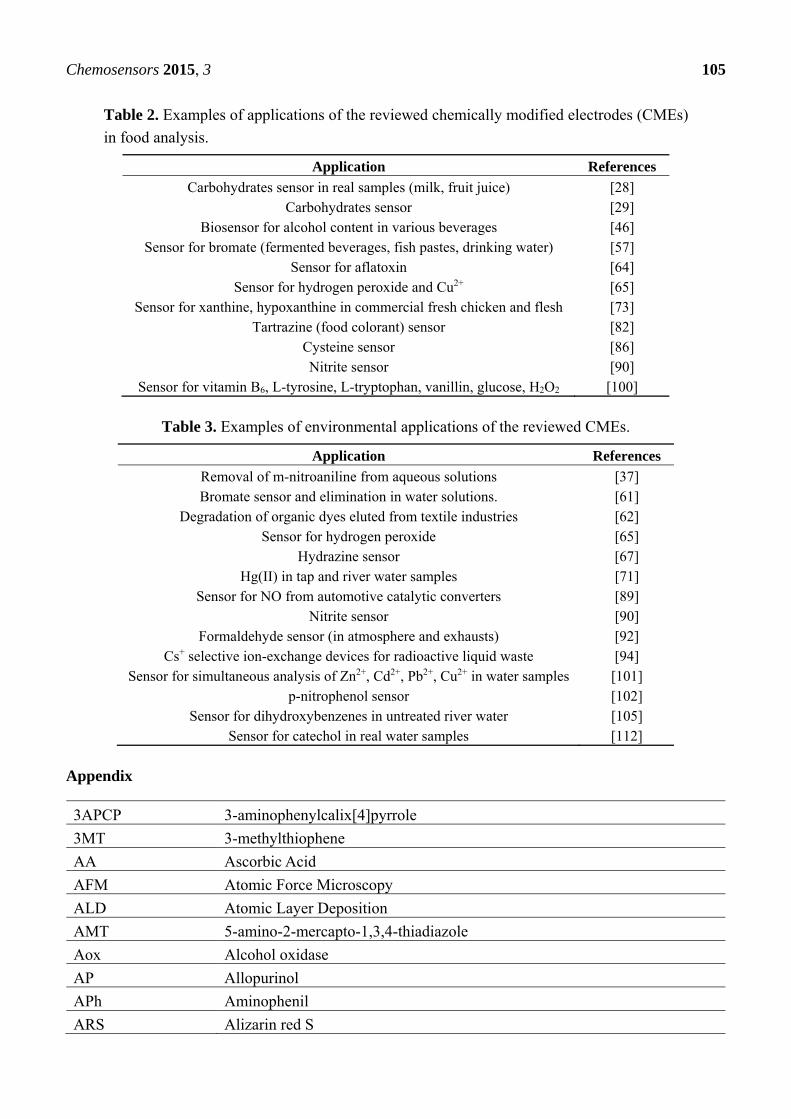
Chemosensors 2015, 3 105
Table 2. Examples of applications of the reviewed chemically modified electrodes (CMEs)
in food analysis.
Application References
Carbohydrates sensor in real samples (milk, fruit juice) [28] Carbohydrates sensor [29]
Biosensor for alcohol content in various beverages [46] Sensor for bromate (fermented beverages, fish pastes, drinking water) [57]
Sensor for aflatoxin [64] Sensor for hydrogen peroxide and Cu2+ [65]
Sensor for xanthine, hypoxanthine in commercial fresh chicken and flesh [73] Tartrazine (food colorant) sensor [82]
Cysteine sensor [86] Nitrite sensor [90]
Sensor for vitamin B6, L-tyrosine, L-tryptophan, vanillin, glucose, H2O2 [100]
Table 3. Examples of environmental applications of the reviewed CMEs.
Application References
Removal of m-nitroaniline from aqueous solutions [37] Bromate sensor and elimination in water solutions. [61]
Degradation of organic dyes eluted from textile industries [62] Sensor for hydrogen peroxide [65]
Hydrazine sensor [67] Hg(II) in tap and river water samples [71]
Sensor for NO from automotive catalytic converters [89] Nitrite sensor [90]
Formaldehyde sensor (in atmosphere and exhausts) [92] Cs+ selective ion-exchange devices for radioactive liquid waste [94]
Sensor for simultaneous analysis of Zn2+, Cd2+, Pb2+, Cu2+ in water samples [101] p-nitrophenol sensor [102]
Sensor for dihydroxybenzenes in untreated river water [105] Sensor for catechol in real water samples [112]
Appendix
3APCP 3-aminophenylcalix[4]pyrrole
3MT 3-methylthiophene
AA Ascorbic Acid
AFM Atomic Force Microscopy
ALD Atomic Layer Deposition
AMT 5-amino-2-mercapto-1,3,4-thiadiazole
Aox Alcohol oxidase
AP Allopurinol
APh Aminophenil
ARS Alizarin red S

Chemosensors 2015, 3 106
ATP Aminothiophenol ATR-FTIR Attenuated Total Reflectance-FTIR
BA Benzoic acid
BE Binding energy
BIPN 2-(4-nitrophenyl)-4,7-di(thiophene-2-yl)-1H-benzo[d]imidazole
BMIMBF4 1-butyl-3-methylimidazolium tetrafluoroborate
BMIM-Cl 1-butyl-3-methylimidazolium chloride
BSA Bovine Serum Albumin
Bupy-Cl 1-butylpyridinium chloride
CA Chronoamperometry
CDSI Cyclic Disulfide
CME Chemically Modified Electrode
CNT Carbon Nanotube
CV Cyclic Voltammetry
DA Dopamine
DMF Dymethylformamide
DMSO Dimethylsulfoxide
DPV Differential Pulse Voltammetry
DRS Diffuse Reflectance Spectroscopy
DTA Differential Thermal Analysis
EDAX Energy Dispersive X-ray Analysis
EDC 3-(3-dimethylaminopropyl)-1-ethylcarbodiimide hydrochloride
EDOT-N3 Azidomethyl functionalized 3,4-ethylenedioxythiophene
EDS Energy Dispersive X-ray Spectroscopy (also mentioned as EDX)
EELS Electron Energy Loss Spectroscopy
EIS Electrochemical Impedance Spectroscopy
EMIMES 1-ethyl-3-methylimidazolium ethyl sulfate
EQCM Electrochemical Quartz Crystal Microbalamce
ERGO Electrochemically Reduced Graphene Oxide
ES Electrode Surface
ESCA Electron Spectroscopy for Chemical Analysis
ESI-MS Electrospray Ionisation Mass Spectrometry
FESEM Field Emission SEM
FIA Flow Injection Analysis
FTIR Fourier Transform IR
FTO Fluorine-doped Tin Oxide
FWHM Full Width at Half Maximum
GA Glutaraldehyde
GCE Glassy Carbon Electrode
GO Graphite Oxide/Graphene Oxide
Chemosensors 2015, 3 107
GOD Glucose Oxidase
GR Graphene (also indicated as rGO – reduced GO)
HDA 1,6-hexanediamine
HNPs Heterodimer Nanoparticles
HOMO Highest Occupied Molecular Orbital
HOPG Highly Oriented Pyrolitic Graphite
HQ Hydroquinone
HQSA 8-hydroxyquinoline-5-sulphonic acid
HRTEM High Resolution TEM
KE Kinetic Energy
IL Ionic Liquid
IRRAS Infrared Reflection-Absorption Spectroscopy
ITO Indium Tin Oxide
LMM L-inner level-M-inner level-M-inner level Auger transition
LSV Linear Scan Voltammetry
LUMO Lowest Unoccupied Molecular Orbital
MB Methylene Blue
MHCF Metal hexacyanoferrate
MOR Methanol oxidation reaction
MPA 3-mercaptopropionic acid
μT Micro Tubes
MWCNT Multi-Walled CNT
NA Nafion®
Nap Naproxene
NBM 3-(nitrobenzyl)mercaptan
NC Nano Cubes
NCL Nano Clusters
NF Nano Fibers
NHS N-hydroxysuccinimide
NL Nano Leaves
NMR Nuclear Magnetic Resonance
NP Nano Particles
NPD 4-nitrophenyl diazonium
NR Nano Rod
NRed Neutral Red
NS Nano Seeds, Nano Sheets
NSSFs self-standing nanostructured silver fibres
NT Nano Tubes
OMF Osmium Multilayer Film
p(BCP) Poly Bromocresol Purple
Chemosensors 2015, 3 108
PAD Pulsed Amperometric Detection
Padpa poly(4-aminodiphenylamine)
PAMAM Poly(amidoamine)
PANI Poly-aniline
PAR Peaks Area Ratio
PB Prussian Blue
PBS Phosphate Buffer Solution
PC Photocatalytic
PCF Polycysteine
PDDA Poly(diallyldimethyl)ammonium chloride
PEC Photoelectrocatalytic
PEDOT Poly(3,4-ethylenedioxythiophene)
PET Polyethylene glycol
PIGE Paraffin wax Impregnated Graphite Electrode
PImox Overoxidized Polyimidazole
PMEL Poly(melamine)
PM-IRRAS Polarization Modulation Infrared Reflection-Absorption Spectroscopy
POM Polyoxometallates
PPUA poly(11-N-pyrrolylundecanoic) acid
PPY Polypyrrole
PSS Poly(styrene-4-sulfonate)
PUP Poly(N-undecylpyrrole)
PV4P Poly(4-vinylpyridine)
PVA Polyvinylalcohol
Py 1-aminopyrene
PW Phosphotungstic acid
RAIRS Reflection-Absorption Infrared Spectroscopy
RDE Rotating Disk Electrode
RTIL Room Temperature Ionic Liquid
rGO reduced Graphene Oxide
Rrms Root mean squared Roughness
SAED Selected-Area Electron Diffraction
SAM Self-assembled monolayer
SECM Scanning Electrochemical Microscopy
SEM Scanning Electron Microscopy
SHG Second Harmonic Generation
SIMS Secondary Ions Mass Spectrometry
SoS Spin Orbit Splitting
SPCE Screen Printed Carbon Electrode
SPE Screen Printed Electrode
Chemosensors 2015, 3 109
SWV Square Wave Voltammetry
TBAPF6 Tetrabutylammonium hexafluorophosphate
TBATFB Tetrabutylammonium tetrafluoroborate
TEM Trasmission Electron Microscopy
TGA Thermogravimetric Analysis
ThL Trametes hirsuta laccase
TMB 3, 3′, 5, 5′-tetramethylbenzidine
TNT Titanate Nanotubes
UA Uric Acid
UOx Uricase
UV-Vis Ultraviolet-Visible Spectroscopy
XP X-ray Photoelectron
XPS X-ray Photoelectron Spectroscopy
XRD X-ray Diffraction
XRF X-ray Fluorescence
Acknowledgments
Financial support by COFIN 2010–2011 (Programmi di Ricerca Scientifica di Rilevante Interesse
Nazionale, MIUR, 2010AXENJ8_002) is acknowledged.
Conflicts of Interest
The authors declare no conflict of interest.
References
1. Durst, R.A.; Bäumner, A.J.; Murray, R.W.; Buck, R.P.; Andrieux, C.P. Chemically modified
electrodes: Recommended terminology and definitions. Pure Appl. Chem. 1997, 69, 1317–1323.
2. Kutner, W.; Wang, J.; L’Her, M.; Buck, R.P. Analytical aspects of chemically modified electrodes:
Classification, critical evaluation and recommendations. Pure Appl. Chem. 1998, 70, 1301–1318.
3. Belding, S.R.; Campbell, F.W.; Dickinson, E.J.F.; Compton, R.G. Nanoparticle-modified
electrodes. Phys. Chem. Chem. Phys. 2010, 12, 11208–11221.
4. Oyama, M. Recent nanoarchitectures in metal nanoparticle-modified electrodes for electroanalysis.
Anal. Sci. 2010, 26, 1–12.
5. Radi, A.-E. Recent Updates of Chemically Modified Electrodes in Pharmaceutical Analysis.
Comb. Chem. High Throughput Screen. 2010, 13, 728–752.
6. Merkoçi, A. Nanoparticles-based strategies for DNA, protein and cell sensors. Biosens.
Bioelectron. 2010, 26, 1164–1177.
7. Toghill, K.E.; Compton, R.G. Metal Nanoparticle Modified Boron Doped Diamond Electrodes for
Use in Electroanalysis. Electroanalysis 2010, 22, 1947–1956.
8. Arduini, F.; Calvo, J.Q.; Palleschi, G.; Moscone, D.; Amine, A. Bismuth-modified electrodes for
lead detection. TrAC 2010, 29, 1295–1304.
Chemosensors 2015, 3 110
9. Rad, A.S.; Mirabi, A.; Binaian, E.; Tayebi, H. A Review on Glucose and Hydrogen Peroxide
Biosensor Based on Modified Electrode Included Silver Nanoparticles. Int. J. Electrochem. Sci.
2011, 8, 3671–3683.
10. Brownson, D.A.C.; Foster, C.W.; Banks, C.E. The electrochemical performance of graphene
modified electrodes: An analytical perspective. Analyst 2012, 137, 1815–1823.
11. Ciucu, A.A. Chemically Modified Electrodes in Biosensing. J. Biosens. Bioelectron. 2014, 5, 1–10.
12. Desimoni, E.; Brunetti, B. Glassy Carbon Electrodes Film-Modified with Acidic Functionalities.
A Review. Electroanalysis 2012, 24, 1481–1500.
13. Walcarius, A. Electrocatalysis, sensors and biosensors in analytical chemistry based on ordered
mesoporous and macroporous carbon-modified electrodes. TrAC 2012, 38, 79–97.
14. Doyle, R.L.; Godwin, I.J.; Brandon, M.P.; Lyons, M.E.G. Redox and electrochemical water
splitting catalytic properties of hydrated metal oxide modified electrodes. Phys. Chem. Chem.
Phys. 2013, 15, 13737–13783.
15. Martin, A.; Escarpa, A. Graphene: The cutting edge interaction between chemistry and
electrochemistry. TrAC 2014, 56, 13–26.
16. Briggs, D.; Seah, M.P. Practical Surface Analysis, 2nd ed.; J. Wiley & Sons: Chichester, UK,
1990; Volume 1.
17. Desimoni, E.; Zambonin, P.G. Spectroscopies for surface characterization. In Surface
Characterization of Advanced Polymers; Sabbatini, L., Zambonin, P.G., Eds.; VCH: Weinheim,
Germany, 1993; pp. 1–45.
18. Riviere, J.C.; Myhra, S. Handbook of Surface and Interface Analysis: Methods for
Problem-Solving, 2nd ed.; CRC Press: Boca Raton, FL, USA, 2009; pp. 1–64.
19. Hüfner, S. Photoelectron Spectroscopy: Principles and Applications, 3rd ed.; Springer: Berlin,
Germany, 2003.
20. Hofman, S. Auger- and X-Ray Photoelectron Spectroscopy in Materials Science: A User-Oriented
Guide; Springer, Berlin, Germany, 2012.
21. Van der Heide, P. X-Ray Photoelectron Spectroscopy: An Introduction to the Principles and
Practices; Wiley: Chichester, UK, 2012.
22. Bagus, P.S.; Ilton, E.S.; Nelin, C.J. The interpretation of XPS spectra: Insights into materials
properties. Surf. Sci. Rep. 2013, 68, 273–304.
23. Brunetti, B.; de Giglio, E.; Cafagna, D.; Desimoni, E. XPS analysis of glassy carbon electrodes
chemically modified with 8-hydroxyquinoline-5-sulphonic acid. Surf. Interface Anal. 2012, 44,
491–496.
24. Desimoni, E.; Salvi, A.M.; Casella, I.G.; Damiano, D. Controlled chemical oxidation of carbon
fibres: An XPS-XAES-SEM study. Surf. Interface Anal. 1993, 20, 909–918.
25. Beamson, G.; Briggs, D. High Resolution XPS of Organic Polymers; J. Wiley & Sons: Chichester,
UK, 1992.
26. Crist, B.V. Handbook of Monochromatic XPS Spectra, Polymers and Polymers Damagedby
X-Rays; J. Wiley & Sons: Chichester, UK, 2000.
27. Desimoni, E.; Malitesta, C.; Zambonin, P.G.; Riviere, J.C. An X-ray photoelectron spectroscopic
study of some cromium-oxygen systems. Surf. Interface Anal. 1988, 13, 173–179.
Chemosensors 2015, 3 111
28. Casella, I.G.; Desimoni, E.; Castaldi, T.R.I. Study of a nickel-catalysed glassy carbon
electrode for detection of carbohydrates in liquid chromatography and flow injection analysis.
Anal. Chim. Acta 1991, 248, 117–125.
29. Cataldi, T.R.I.; Casella, I.G.; Desimoni, E.; Rotunno, T. Co-based Glassy Carbon CME for
Constant-Potential Amperometric Detection of Carbohydrates in FIA and Liquid Chromatography.
Anal. Chim. Acta 1992, 270, 161–171.
30. Casella, I.G.; Desimoni, E. XPS, SEM and electrochemical characterization of a platinum-based
glassy carbon modified electrode. Electrocatalytic oxidation of ethanol in acidic medium.
Electroanalysis 1996, 8, 447–453.
31. Taouil, A.E.; Lallemand, F.; Melot, J.; Husson, J.; Hihn, J.; Lakard, B. Effects of polypyrrole
modified electrode functionalization on potentiometric pH responses. Synth. Met. 2010, 160,
1073–1080.
32. Li, S.; Yang, Y.; Yang, S.; Ye, S.; Fang, H.; Hu, J.; Li, Q. Electrochemical reduction and
voltammetric determination of pirarubicin at carboxyl ions implantation-modified indium tin oxide
electrode. Chin. J. Anal. Chem. 2011, 39, 990–993.
33. Xue, Y.; Zhao, H.; Wu, Z.; Li, X.; He, Y.; Yuan, Z. The comparison of different gold
nanoparticles/graphene nanosheets hybrid nanocomposites in electrochemical performance and the
construction of a sensitive uric acid electrochemical sensor with novel hybrid nanocomposites.
Biosens. Bioelectron. 2011, 29, 102–108.
34. Sundaram, S.; Annamalai, S.K. Selective immobilization of hydroquinone on carbon nanotube
modified electrode via phenol electro-oxidation method and its hydrazine electro-catalysis and
Escherichia coli antibacterial activity. Electrochim. Acta 2012, 62, 207–217.
35. Wei, L.; Lei, Y.; Fu, H.; Yao, J. Fullerene hollow microspheres prepared by bubble-templates as
sensitive and selective electrocatalytic sensor for biomolecules. ACS Appl. Mater. Interfaces 2012,
4, 1594–1600.
36. Valentini, F.; Roscioli, D.; Carbone, M.; Conte, V.; Floris, B.; Palleschi, G.; Flammini, R.;
Bauer, E.M.; Nasillo, G.; Caponetti, E. Oxidized graphene in ionic liquids for assembling
chemically modified electrodes: A structural and electrochemical characterization study.
Anal. Chem. 2012, 84, 5823–5831.
37. Ding, L.; Li, Q.; Zhou, D.; Cui, H.; Tang, R.; Zhai, J. Copolymerization of aniline with
m-nitroaniline and removal of m-nitroaniline from aqueous solutions using a polyaniline-modified
electrode: A comparative study. Electrochim. Acta 2012, 77, 302–308.
38. Demirci, S.; Emre, F.B.; Ekiz, F.; Oğuzkaya, F.; Timur, S.; Tanyeli, C.; Toppare, L.
Functionalization of poly-SNS-anchored carboxylic acid with Lys and PAMAM: Surface
modifications for biomolecule immobilization/stabilization and bio-sensing applications. Analyst
2012, 137, 4254–4261.
39. Yang, J.; Myoung, N.; Hong, H. Facile and controllable synthesis of Prussian blue on
chitosan-functionalized graphene nanosheets for the electrochemical detection of hydrogen
peroxide. Electrochim. Acta 2012, 81, 37–43.
40. Baskar, S.; Liao, C.; Chang, J.; Zen, J. Electrochemical synthesis of electroactive poly(melamine)
with mechanistic explanation and its applicability to functionalize carbon surface to prepare
nanotube–nanoparticles hybrid. Electrochim. Acta 2013, 88, 1–5.
Chemosensors 2015, 3 112
41. You, J.; Kim, D.; Kim, S.K.; Kim, M.; Han, H.S.; Jeon, S. Novel determination of hydrogen
peroxide by electrochemically reduced graphene oxide grafted with aminothiophenol–Pd
Nanoparticles. Sens. Actuators B Chem. 2013, 178, 450–457.
42. Raj, M.A.; John, S.A. Fabrication of electrochemically reduced graphene oxide films on glassy
carbon electrode by self-assembly method and their electrocatalytic application. J. Phys. Chem. C
2013, 117, 4326–4335.
43. Guo, K.; Chen, X.; Freguia, S.; Dolose, B.C. Spontaneous modification of carbon surface with
neutral red from its diazonium salts for bioelectrochemical systems. Biosens. Bioelectron. 2013,
47, 184–189.
44. Ghilane, J.; Hauquier, F.; Lacroix, J. Oxidative and stepwise grafting of dopamine inner-sphere
redox couple onto electrode material: Electron transfer activation of dopamine. Anal. Chem. 2013,
85, 11593–11601.
45. Raj, M.A.; John, S.A. Electrochemical determination of xanthine oxidase inhibitor drug in urate
lowering therapy using graphene nanosheets modified electrode. Electrochim. Acta 2014, 117,
360–366.
46. Soylemez, S.; Kanik, F.E.; Uzun, S.D.; Hacioglu, S.O.; Toppare, L. Development of an efficient
immobilization matrix based on a conducting polymer and functionalized multiwall carbon
nanotubes: Synthesis and its application to ethanol biosensors. J. Mater. Chem. B 2014, 2,
511–521.
47. Si, W.; Lei, W.; Han, Z.; Zhang, Y.; Hao, Q.; Xia, M. Electrochemical sensing of acetaminophen
based on poly(3,4-ethylenedioxythiophene)/graphene oxide composites. Sens. Actuators B Chem.
2014, 193, 823–829.
48. Yang, L.; Liu, D.; Huang, J.; You, T. Simultaneous determination of dopamine, ascorbic acid and
uric acid at electrochemically reduced graphene oxide modified electrode. Sens. Actuators B Chem.
2014, 193, 166–172.
49. Zong, X.; Kong, N.; Liu, J.; Yang, W.; Cao, M.; Gooding, J.J. The influence of graphene on the
electrical communication through organic layers on graphite and gold electrodes. Electroanalysis
2014, 26, 84–92.
50. Liu, X.; Zhang, L.; Wei, S.; Chen, S.; Ou, X.; Lu, Q. Overoxidized polyimidazole/graphene
oxide copolymer modified electrode for the simultaneous determination of ascorbic acid,
dopamine, uric acid, guanine and adenine. Biosens. Bioelectron. 2014, 57, 232–238.
51. Felix, S.; Kollu, P.; Raghupathy, B.P.C.; Jeong, S.K.; Grace, A.N. Electrocatalytic activity of Cu2O
nanocubes-based electrode for glucose oxidation. J. Chem. Sci. 2014, 126, 25–32.
52. Le Goff, A.; Moggia, F.; Debou, N.; Jegou, P.; Artero, V.; Fontecave, M.; Jousselme, B.;
Palacin, S. Facile and tunable functionalization of carbon nanotube electrodes with ferrocene by
covalent coupling and π-stacking interactions and their relevance to glucose bio-sensing.
J. Electroanal. Chem. 2010, 641, 57–63.
53. Sharifi, N.; Tajabadi, F.; Taghavinia, N. Nanostructured silver fibers: Facile synthesis based on
natural cellulose and application to graphite composite electrode for oxygen reduction. Int. J.
Hydrogen Energ. 2010, 35, 3258–3262.
Chemosensors 2015, 3 113
54. Che, X.; Yuan, R.; Chai, Y.; Li, J.; Song, Z.; Li, W. Amperometric glucose biosensor based on
Prussian blue–multiwall carbon nanotubes composite and hollow PtCo nanochains.
Electrochim. Acta 2010, 55, 5420–5427.
55. Guascito, M.R.; Chirizzi, D.; Picca, R.A.; Mazzotta, E.; Malitesta, C. Ag nanoparticles capped by
a nontoxic polymer: Electrochemical and spectroscopic characterization of a novel nanomaterial
for glucose detection. Mater. Sci. Eng. C 2011, 31, 606–611.
56. Guascito, M.R.; Chirizzi, D.; Malitesta, C.; Mazzotta, E.; Siciliano, M.; Siciliano, T.; Tepore, A.;
Turco, A. Low-potential sensitive H2O2 detection based on composite micro tubular Te
adsorbed on platinum electrode. Biosens. Bioelectron. 2011, 26, 3562–3569.
57. Zhou, D.; Ding, L.; Cui, H.; An, H.; Zhai, J.; Li, Q. Fabrication of high dispersion Pd/MWNTs
nanocomposite and its electrocatalytic performance for bromate determination. Chem. Eng. J.
2012, 200–202, 32–38.
58. Guascito, M.R.; Chirizzi, D.; Malitesta, C.; Siciliano, M.; Siciliano, T.; Tepore, A. Amperometric
non-enzymatic bimetallic glucose sensor based on platinum tellurium microtubes modified
electrode. Electrochem. Commun. 2012, 22, 45–48.
59. Devaraj, M.; Deivasigamani, R.K.; Jeyadevan, S. Enhancement of the electrochemical behavior of
CuO nanoleaves on MWCNT/GC composite film modified electrode for determination of
norfloxacin. Colloids Surf. B 2013, 102, 554–561.
60. Kim, H.J.; Kwon, S.; Kim, K. Electrochemically active cyclic disulfide-ended organic silane
linkage for preparation of multi-biofunctional electrode surfaces. Electrochem. Commun. 2012, 20,
52–55.
61. Li, Q.; Zhang, Q.; Ding, L.; Zhou, D.; Cui, H.; Wei, Z.; Zhai, J. Synthesis of Silver/multi-walled
carbon nanotubes composite and its application for electrocatalytic reduction of bromate.
Chem. Eng. J. 2013, 217, 28–33.
62. Chatchai, P.; Nosaka, A.Y.; Nosaka, Y. Photoelectrocatalytic performance of WO3/BiVO4 toward
the dye degradation. Electrochim. Acta 2013, 94, 314–319.
63. Tian, H.; Jia, M.; Zhang, M.; Hu, J. Nonenzymatic glucose sensor based on nickel ion
implanted-modified indium tin oxide electrode. Electrochim. Acta 2013, 96, 285–290.
64. Singh, J.; Roychoudhury, A.; Srivastava, M.; Solanki, P.R.; Lee, D.W.; Lee, S.H.; Malhotra, B.D.
A highly efficient rare earth metal oxide nanorods based platform for aflatoxin detection. J. Mater.
Chem. B 2013, 1, doi:10.1039/c3tb20690d.
65. Tanwar, S.; Ho, J.A.; Magi, E. Green synthesis and characterization of novel gold nanocomposites
for electrochemical sensing applications. Talanta 2013, 117, 352–358.
66. Fu, Y.; Liang, F.; Tian, H.; Hu, J. Nonenzymatic glucose sensor based on ITO electrode
modified with gold nanoparticles by ion implantation. Electrochim. Acta 2014, 120, 314–318.
67. Koçak, S.; Aslışen, B. Hydrazine oxidation at gold nanoparticles and poly(bromocresol purple)
carbon nanotube modified glassy carbon electrode. Sens. Actuators B Chem. 2014, 196, 610–618.
68. Kumar, D.R.; Manoj, D.; Santhanalakshmi, J. Au–ZnO bullet-like heterodimer nanoparticles:
Synthesis and use for enhanced nonenzymatic electrochemical determination of glucose. RSC Adv.
2014, 4, 8943–8952.
Chemosensors 2015, 3 114
69. Derouich, S.G.; Rinfray, C.; Izzet, G.; Pinson, J.; Gallet, J.; Kanoufi, F.; Proust, A.; Combellas, C.
Control of the grafting of hybrid polyoxometalates on metal and carbon surfaces: Toward
submonolayers. Langmuir 2014, 30, 2287–2296.
70. Piotrowski, P.; Pawłowska, J.; Pawłowski, J.; Więckowska, A.; Bilewicz, R.; Kaim, A.
Nanostructured films of in situ deprotected thioacetyl-functionalized C60-fullerenes on a gold
surface. J. Mater. Chem. A 2014, 2, 2353–2362.
71. Gong, J.; Zhou, T.; Song, D.; Zhang, L.; Hu, X. Stripping voltammetric detection of mercury(II)
based on a bimetallic Au-Pt inorganic-organic hybrid nanocomposite modified glassy carbon
electrode. Anal. Chem. 2010, 82, 567–573.
72. Melato, A.I.; Abrantes, L.M.; Botelho do Rego, A.M. Fe(CN)63− incorporation on
Poly(3,4-ethylenedioxythiophene) films: Preparation and X-ray photoelectron spectroscopy
characterization of the modified electrodes. Thin Solid Films 2010, 518, 1947–1952.
73. Kumar, A.S.; Swetha, P. Ru(DMSO)4Cl2 nano-aggregated Nafion membrane modified
electrode for simultaneous electrochemical detection of hypoxanthine, xanthine and uric acid.
J. Electroanal. Chem. 2010, 642, 135–142.
74. Wang, J.; Xu, M.; Zhao, R.; Chen, G. A highly sensitive H2O2 sensor based on zinc oxide nanorod
arrays film sensing interface. Analyst 2010, 135, 1992–1996.
75. Sosna, M.; Chrétien, J.-M.; Kilburn, J.D.; Bartlett, P.N. Monolayer anthracene and anthraquinone
modified electrodes as platforms for Trametes hirsuta laccase immobilization. Phys. Chem.
Chem. Phys. 2010, 12, 10018–10026.
76. Lin, L.; Huang, X.; Wang, L.; Tang, A. Synthesis, characterization and the electrocatalytic
application of prussian blue/titanate nanotubes nanocomposite. Solid State Sci. 2010, 12,
1764–1769.
77. Xing, L.; Jia, J.; Wang, Y.; Zhang, B.; Dong, S. Pt modified TiO2 nanotubes electrode: Preparation
and electrocatalytic application for methanol oxidation. Int. J. Hydrogen Energ. 2010, 35,
12169–12173.
78. Lokesh, K.S.; de Wael, K.; Adriaens, A. Self-Assembled Supramolecular Array of polymeric
phthalocyanine on gold for the determination of hydrogen peroxide. Langmuir 2010, 26,
17665–17673.
79. Fontaine, O.; Ghilane, J.; Martin, P.; Lacroix, J.-C.; Randriamahazaka, H. Ionic liquid viscosity
effects on the functionalization of electrode material through the electroreduction of diazonium.
Langmuir 2010, 26, 18542–18549.
80. Feliciano-Ramos, I.; Caban-Acevedo, M.; Scibioh, M.A.; Cabrera, C.R. Self-assembled
monolayers of L-cysteine on palladium electrodes. J. Electroanal. Chem. 2010, 650, 98–104.
81. Im, J.; Han, J.; Kim, B.K.; Han, J.H.; Park, T.S.; Hwang, S.; Cho, S.I.; Lee, W.; Kim, Y.
Electrochemical detection of estrogen hormone by immobilized estrogen receptor on Au electrode.
Surf. Coat. Technol. 2010, 205, S275–S278.
82. Brunetti, B.; Desimoni, E. A new voltammetric sensor based on a glassy carbon electrode
modified with 8-hydroxyquinoline-5-sulfonic acid. Electroanalysis 2011, 23, 1116–1122.
Chemosensors 2015, 3 115
83. Ganesh, V.; Maheswari, D.L.; Berchmans, S. Electrochemical behaviour of metal
hexacyanoferrate converted to metal hydroxide films immobilized on indium tin oxide
electrodes—Catalytic ability towards alcohol oxidation in alkaline medium. Electrochim. Acta
2011, 56, 1197–1207.
84. Pchelintsev, N.A.; Vakurov, A.; Hays, H.H.; Millner, P.A. Thiols deposition onto the surface of
glassy carbon electrodes mediated by electrical potential. Electrochim. Acta 2011, 56, 2696–2702.
85. Liu, L.; Wang, S.; Zhu, Z.; Li, M.; Sun, B. Electrochemistry of a C84-C2(IV)-Modified Electrode
in Aqueous Solutions and Its Interaction with Guanine. J. Phys. Chem. C 2011, 115, 5966–5973.
86. Hsiao, Y.; Su, W.; Cheng, J.; Cheng, S. Electrochemical determination of cysteine based on
conducting polymers/gold nanoparticles hybrid nanocomposites. Electrochim. Acta 2011, 56,
6887–6895.
87. Kannan, P.; John, S.A. Fabrication of conducting polymer-gold nanoparticles film on electrodes
using monolayer protected gold nanoparticles and its electrocatalytic application. Electrochim.
Acta 2011, 56, 7029–7037.
88. Feng, X.; Li, R.; Ma, Y.; Chen, R.; Shi, N.; Fan, Q.; Huang, W. One-Step Electrochemical
Synthesis of Graphene/Polyaniline Composite Film and Its Applications. Adv. Funct. Mater. 2011,
21, 2989–2996.
89. Zhang, R.; Li, L.; Chen, L.; Zhang, G.; Shi, K. N-doped carbon nanotubes synthesized in high
yield and decorated with CeO2 and SnO2 nanoparticles. J. Alloys Compd. 2011, 509, 8620–8624.
90. Yuan, J.; Jin, X.; Li, N.; Chen, J.; Miao, J.; Zhang, Q.; Niu, L.; Song, J. Large scale load of
phosphotungstic acid on multiwalled carbon nanotubes with a grafted poly(4-vinylpyridine) linker.
Electrochim. Acta 2011, 56, 10069–10076.
91. Taner, B.; Deveci, P.; Üstündağ, Z.; Keskin, S.; Özcan, E.; Solak, A.O. Modification of glassy
carbon electrode by the electrochemical oxidation of 3-aminophenylcalix[4]pyrrole in nonaqueous
media. Surf. Interface Anal. 2012, 44, 185–191.
92. Tanwar, S.; Chuang, M.-C.; Prasad, K.S.; Ho, J.-A.A. Template-free synthesis of an electroactive
Au-Calix-PPY nanocomposite for electrochemical sensor applications. Green Chem. 2012, 14,
799–808.
93. Li, X.; Kong, F.; Liu, J.; Liang, T.; Xu, J.; Chen, H. Synthesis of potassium-modified graphene
and its application in nitrite-selective sensing. Adv. Funct. Mater. 2012, 22, 1981–1988.
94. Li, N.; Li, Z.; Yuan, J.; Hu, J.; Miao, J.; Zhang, Q.; Niub, L.; Song, J. Nickel hexacyanoferrate
nanoparticles anchored to multiwalled carbon nanotubes with a grafted poly(4-vinylpyridine)
linker for electrically switched ion exchange. Electrochim. Acta 2012, 72, 150–156.
95. Wang, B.; Li, Y.; Qin, X.; Zhan, G.; Ma, M.; Li, C. Electrochemical fabrication of TiO2
nanoparticles/[BMIM]BF4 ionic liquid hybrid film electrode and its application in determination
of p-acetaminophen. Mat. Sci. Eng. C 2012, 32, 2280–2285.
96. Sun, Z.; Fu, H.; Deng, L.; Wang, J. Redox-active thionine–graphene oxide hybrid nanosheet:
One-pot, rapid synthesis, and application as a sensing platform for uric acid. Anal. Chim. Acta
2013, 761, 84–91.
97. Bettge, M.; Li, Y.; Sankaran, B.; Rago, N.D.; Spila, T.; Haasch, R.T.; Petrov, I.; Abraham, D.P.
Improving high-capacity Li1.2Ni0.15Mn0.55Co0.1O2-based lithium-ion cells by modifying the
positive electrode with alumina. J. Power Sources 2013, 233, 346–357.
Chemosensors 2015, 3 116
98. Zhang, J.; Wang, C.; Chen, S.; Yuan, D.; Zhong, X. Amperometric glucose biosensor based on
glucose oxidase–lectin biospecific interaction. Enzyme Microb. Technol. 2013, 52, 134–140.
99. Malitesta, C.; Guascito, M.R.; Mazzotta, E.; Siciliano, T.; Tepore, A. Copper nanoparticles/
poly-3-methylthiophene composite: Synthesis, characterization and catalytic application to
enzyme-less glucose detecting. Sens. Actuators B Chem. 2013, 184, 70–77.
100. Babu, R.S.; Prabhu, P.; Narayanan, S.S. Green synthesized nickel Nanoparticles modified
electrode in ionic liquid medium and its application towards determination of biomolecules.
Talanta 2013, 110, 135–143.
101. Khudaish, E.A.; Al-Hinaai, M.M.; Al-Harthi, S.H. A solid-state sensor based on
tris(2,2ꞌ-bipyridyl)ruthenium(II)/poly(4-aminodiphenylamine) modified electrode: Characterization
and applications. Sens. Actuators B Chem. 2013, 185, 478–487.
102. Pan, L.; Tang, J.; Wang, F. Synthesis and electrocatalytic performance for p-nitrophenol reduction
of rod-like Co3O4 and Ag/Co3O4 composites. Mater. Res. Bull. 2013, 48, 2648–2653.
103. Guascito, M.R.; Chirizzi, D.; Malitesta, C.; Siciliano, T.; Tepore, A. Te oxide nanowires as
advanced materials for amperometric nonenzymatic hydrogen peroxide sensing. Talanta 2013,
115, 863–869.
104. Canevari, T.C.; Raymundo-Pereira, P.A.; Landers, R.; Benvenutt, E.V.; Machado, S.A.S. Sol–gel
thin-film based mesoporous silica and carbon nanotubes for the determination of dopamine, uric
acid and paracetamol in urine. Talanta 2013, 116, 726–735.
105. Canevari, T.C.; Raymundo-Pereira, P.A.; Landers, R.; Machado, S.A.S. Direct synthesis of Ag
nanoparticles incorporated on a mesoporous hybrid material as a sensitive sensor for the
simultaneous determination of dihydroxybenzenes. Eur. J. Inorg. Chem. 2013, 2013, 5746–5754.
106. Li, M.; Bo, X.; Mu, Z.; Zhang, Y.; Guo, L. Electrodeposition of nickel oxide and platinum
nanoparticles on electrochemically reduced graphene oxide film as a nonenzymatic glucose sensor.
Sens. Actuators B Chem. 2014, 192, 261–268.
107. Kesavan, S.; Revin, S.B.; John, S.A. Potentiodynamic formation of gold nanoparticles film on
glassy carbon electrode using aminophenyl diazonium cations grafted gold nanoparticles:
Determination of histamine H2 receptor antagonist. Electrochim. Acta 2014, 119, 214–224.
108. Fernandes, D.M.; Vos, J.G.; Freire, C. Europium phosphomolybdate and osmium metallopolymer
multi-functional LbL films: Redox and electrocatalytic properties. J. Colloid Interface Sci. 2014,
420, 127–135.
109. Li, M.; Bo, X.; Zhang, Y.; Han, C.; Guo, L. One-pot ionic liquid-assisted synthesis of highly
dispersed PtPd nanoparticles/reduced graphene oxide composites for nonenzymatic glucose
detection. Biosens. Bioelectron. 2014, 56, 223–230.
110. Scavetta, E.; Mazzoni, R.; Mariani, F.; Margutta, R.G.; Bonfiglio, A.; Demelas, M.; Fiorilli, S.;
Marzocchi, M.; Fraboni, B. Dopamine amperometric detection at a ferrocene clicked PEDOT:PSS
coated electrode. J. Mater. Chem. B 2014, 2, 2861–2867.
111. Kong, C.; Tang, L.; Zhang, X.; Sun, S.; Yang, S.; Song, X.; Yang, Z. Templating synthesis of
hollow CuO polyhedron and its application for nonenzymatic glucose detection. J. Mater. Chem.
A 2014, 2, 7306–7312.
Chemosensors 2015, 3 117
112. Zhang, Q.; Pan, D.; Zhang, H.; Han, H.; Kang, Q. Development of a poly(alizarin red S)/ionic
liquid film modified electrode for voltammetric determination of catechol. Electrochim. Acta 2014,
133, 23–29.
113. Doniach, S.; Sunjic, M. Many-electron singularity in X-ray photoemission and X-ray line spectra
from metals. J. Phys. C 1970, 3, 285–291.
114. Yang, D.; Sacher, E. Carbon 1s X-ray Photoemission Line Shape Analysis of Highly Oriented
Pyrolytic Graphite: The Influence of Structural Damage on Peak Asymmetry. Langmuir 2006, 22,
860–862.
115. Desimoni, E.; Casella, G.I.; Cataldi, T.R.I.; Malitesta, C. A comparison of some asymmetrical
line shapes for XPS data analysis. J. Electron Spectrosc. Relat Phenom. 1989, 49, 247–261.
© 2015 by the authors; licensee MDPI, Basel, Switzerland. This article is an open access article
distributed under the terms and conditions of the Creative Commons Attribution license
(http://creativecommons.org/licenses/by/4.0/).