Embed Size (px)
Citation preview
BioMed Central
World Journal of Surgical Oncology
ss
Open AcceCase reportA rare case of primary mesenteric gastrointestinal stromal tumor with metastasis to the cervix uteriNupur Gupta*1, Suneeta Mittal1, Neena Lal1, Renu Misra2, Lalit Kumar3 and Sunita Bhalla4Address: 1Department of Obstetrics & Gynaecology, All India Institute of Medical Sciences, New Delhi, India, 2Department of Obstetrics & Gynaecology, Moolcahnd Khairati Hospital, New Delhi, India, 3Department of Medical Oncology, All India Institute of Medical Sciences, New Delhi, India and 4Department of Pathology, Sir Gangaram Hospital, New Delhi, India
Email: Nupur Gupta* - [email protected]; Suneeta Mittal - [email protected]; Neena Lal - [email protected]; Renu Misra - [email protected]; Lalit Kumar - [email protected]; Sunita Bhalla - [email protected]
* Corresponding author
AbstractBackground: Gastrointestinal stromal tumors are CD117 (C Kit) positive mesenchymalneoplasms, that may arise anywhere in the gastrointestinal tract. Their current therapy is imatinibmesylate before or after surgery.
Case presentation: We describe a case of 17-year-old female with metastasis to the cervix uteriof a primary mesenteric gastrointestinal tumor.
Conclusion: Surgery remains the mainstay of known curative treatment. The manifestations ofGIST are not restricted to the typical locations within the bowel; may have very unusual metastaticsites or infiltrations per continuitatem.
BackgroundGastrointestinal stromal tumor (GIST) is a rare mesenchy-mal tumor of the gastrointestinal tract with an incidenceof 10–20 cases per million populations of which almostone third are deemed malignant [1]. We report this rarecase of primary mesenteric tumor metastatic to the cervixthat was diagnosed to be a GIST on histopathologicalexamination.
Case presentationA 17 year old unmarried female presented with history ofmenorrhagia and passing tissue per vagina since 3months, low grade fever since 1 month and feeling ofmass per abdomen since 10 days. On examination, shehad a mass corresponding to 24 weeks size gravid uterusarising from pelvis, which was tender on palpation. There
was anemia. Four units of blood transfusion were given ina private clinic. Her routine liver and renal function testsand coagulation profile were normal. Ultrasound fol-lowed by contrast enhanced computerized tomography(CT) showed bilateral adnexal masses with a large soft tis-sue heterogeneous mass suggestive of sarcoma of theuterus and presence of enlarged retroperitoneal lymphnodes. Examination under anesthesia revealed the samemass with side to side restricted mobility, and a fleshy, fri-able and vascular growth protruding into vagina. Biopsyfrom the growth was initially misdiagnosed as a leiomy-osarcoma. Exploratory laparotomy was carried out as thepatient was continuously bleeding and staging was per-formed. Intraoperatively, there was a large 30 × 30 × 12cm friable mass with necrosis and hemorrhagic areas aris-ing from the mesentery of the transverse colon, with
Published: 29 November 2007
World Journal of Surgical Oncology 2007, 5:137 doi:10.1186/1477-7819-5-137
Received: 7 June 2007Accepted: 29 November 2007
This article is available from: http://www.wjso.com/content/5/1/137
© 2007 Gupta et al; licensee BioMed Central Ltd. This is an Open Access article distributed under the terms of the Creative Commons Attribution License (http://creativecommons.org/licenses/by/2.0), which permits unrestricted use, distribution, and reproduction in any medium, provided the original work is properly cited.
Page 1 of 4(page number not for citation purposes)
World Journal of Surgical Oncology 2007, 5:137 http://www.wjso.com/content/5/1/137
tumor deposits were present all over the pelvic perito-neum. A solid right adnexal mass, 10 × 4 cm was alsopresent. Resection of tumor with wide margin along withpanhysterectomy and infracolic omentectomy was done.Cut section of uterus, with bilateral fallopian tubes andovaries showed serosal deposits. Endocervix revealed a 4.5× 4 × 1 cm cauliflower like, exophytic, polypoidal vascularfriable growth lying at the lower end. Vagina was grosslylooking normal. Histopathological examination revealeda primary gastrointestinal tumor of mesentery with >5mitoses per 50 high power field and atypical spindle cellsarranged in fascicles. There were metastasis in the cervix,serosa and omentum. Immunohistochemical stainsshowed positivity for vimentin, smooth muscle actin,desmin and CD 117 (Figure 1) and were negative forcytokeratin, epithelial membrane antigen and CD 34.Molecular genetic analysis (KIT mutation analysis) wasnot done due to its unavailability at our institute. Postop-eratively, she received 6 units of packed RBC, fresh frozenplasma and platelet rich plasma each. She had a stormypostoperative period with fever, severe pain abdomen anddeveloped a recurrence after 4 weeks with huge mass perabdomen and faecal fistula from the surgical scar. On
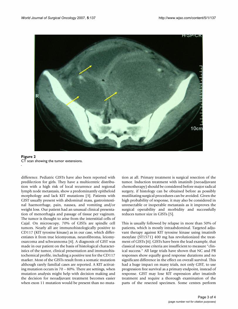
computerized tomography, there was a large heterogene-ous mass lesion in pelvis with areas of necrosis infiltratingsigmoid, iliopsoas with encasement of ureters causingbilateral hydroureteronephrosis. The mass was involvingthe iliopsoas muscle on left side and encasing the uretersbilaterally (Figure 2). Imatinib 400 mg qd was startedwhich showed a minimal improvement in her clinicalcondition. She continued to have a huge pelvic mass withpain, fever, urinary and bowel symptoms for which palli-ative care was administered. RECIST (Response Evalua-tion Criteria in evaluating Solid Tumors) criteria wereapplied for response assessment. At 4 weeks, only 16% ofthe lesions achieved partial response (PR) by RECIST and24% increased in size by a mean of 22.5% (progressivedisease, PD).
DiscussionGIST may present anywhere in the gastrointestinal tract,omentum, or mesentery. The most common sites are thestomach (60%), small intestine (15%), colon and rectum(5%), other abdominal organs including mesentery andomentum (5%) [2]. Usual age of presentation is around60 years, predominantly seen in Caucasians with little sex
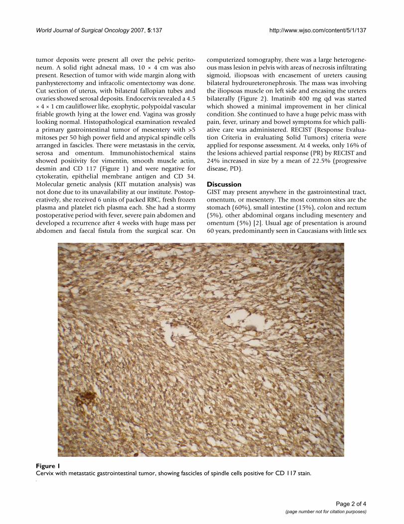
Cervix with metastatic gastrointestinal tumor, showing fascicles of spindle cells positive for CD 117 stainFigure 1Cervix with metastatic gastrointestinal tumor, showing fascicles of spindle cells positive for CD 117 stain.
Page 2 of 4(page number not for citation purposes)
World Journal of Surgical Oncology 2007, 5:137 http://www.wjso.com/content/5/1/137
difference. Pediatric GISTs have also been reported withpredilection for girls. They have a multicentric distribu-tion with a high risk of local recurrence and regionallymph node metastasis, show a predominantly epitheloidmorphology and lack KIT mutations [3]. Patients withGIST usually present with abdominal mass, gastrointesti-nal haemorrhage, pain, nausea, and vomiting and/orweight loss. Our patient had an unusual clinical presenta-tion of menorrhagia and passage of tissue per vaginum.The tumor is thought to arise from the interstitial cells ofCajal. On microscopy, 70% of GISTs are spindle celltumors. Nearly all are immunohistologically positive toCD117 (KIT tyrosine kinase) as in our case, which differ-entiates it from true leiomyomas, neurofibroma, leiomy-osarcoma and schwannoma [4]. A diagnosis of GIST wasmade in our patient on the basis of histological character-istics of the tumor, clinical presentation and immunohis-tochemical profile, including a positive test for the CD117marker. Most of the GISTs result from a somatic mutationalthough rarely familial cases are reported. A KIT activat-ing mutation occurs in 70 – 80%. There are settings, whenmutation analysis might help with decision making andthe decision for neoadjuvant treatment becomes easierwhen exon 11 mutation would be present than no muta-
tion at all. Primary treatment is surgical resection of thetumor. Induction treatment with imatinib (neoadjuvantchemotherapy) should be considered before major radicalsurgery, if histology can be obtained before as possiblymutilitating surgical procedures can be avoided. Given thehigh probability of response, it may also be considered inunresectable or inoperable metastasis as it improves thesurgical operability and morbidity and successfullyreduces tumor size in GISTs [5].
This is usually followed by relapse in more than 50% ofpatients, which is mostly intraabdominal. Targeted adju-vant therapy against KIT tyrosine kinase using imatinibmesylate (ST1571) 400 mg has revolutionized the treat-ment of GISTs [6]. GISTs have been the lead example, thatclassical response criteria are insufficient to measure "clin-ical success." All large trials have shown that NC and PRresponses show equally good response durations and nosignificant difference in the effect on overall survival. Thishad a huge impact on many trials, not only GIST, to useprogression free survival as a primary endpoint, instead ofresponse. GIST may lose KIT expression after imatinibtreatment and require a thorough examination of theparts of the resected specimen. Some centres perform
CT scan showing the tumor extensionsFigure 2CT scan showing the tumor extensions.
Page 3 of 4(page number not for citation purposes)
World Journal of Surgical Oncology 2007, 5:137 http://www.wjso.com/content/5/1/137
Publish with BioMed Central and every scientist can read your work free of charge
"BioMed Central will be the most significant development for disseminating the results of biomedical research in our lifetime."
Sir Paul Nurse, Cancer Research UK
Your research papers will be:
available free of charge to the entire biomedical community
peer reviewed and published immediately upon acceptance
cited in PubMed and archived on PubMed Central
yours — you keep the copyright
Submit your manuscript here:http://www.biomedcentral.com/info/publishing_adv.asp
BioMedcentral
monthly CT for response assessment until tumor progres-sion. Sometimes, false positive complete response (CR) isnoted on PET CT. Parameters recorded are largest dimen-sion, new lesions, and any new features and tumorresponse is assessed through RECIST criteria [7]. They area new set of tumor response criteria adopted by WHO, theNational Cancer Institute and the European Organisationfor Research and Treatment of Cancer. To our knowledge,this is the first case report of mesenteric GIST with metas-tasis to the uterine cervix in literature. GIST may alsopresent as a pelvic mass [8] or metastasize to the ovary [9-11].
ConclusionSurgery remains the mainstay of known curative treat-ment. Recently, the diagnosis and management of GISThas undergone a revolution with the emergence of CD117staining and the transforming oncogene (KIT mutation).Its specific targeted inhibition is greatly effective in treat-ing GIST. The manifestations of GIST are not restricted tothe typical locations within the bowel; may have very unu-sual metastatic sites or infiltrations per continuitatem.Thus, a multidisciplinary approach including a gynecolo-gist and a medical oncologist can improve the prognosisof patients with GIST.
Competing interestsThe author(s) declare that they have no competing inter-ests.
Authors' contributionsNG, literature search prepared the draft manuscript, SM,NL, RM helped in preparation of manuscript, LK, helpedin preparation of oncology part of the manuscript, SB con-tributed the pathological aspect of case. All authors readand approved the final manuscript.
AcknowledgementsWritten informed consent was obtained from patients father for publica-tion of this case report.
References1. Joensuu H, Kindblom LG: Gastrointestinal stromal tumors--a
review. Acta Orthop Scand Suppl 2004, 75(311):62-71.2. Miettinen M, Lasota J: Gastrointestinal stromal tumors: defini-
tion, clinical, histological, immunohistochemical, and molec-ular genetic features and differential diagnosis. Virchows Arch2001, 438:1-12.
3. Prakash S, Sarran L, Socci N, DeMatteo RP, Eisenstat J, Greco AM,Maki RG, Wexler LH, LaQuaglia MP, Besmer P, Antonescu CR: Gas-trointestinal Stromal Tumors in Children and Young Adults:A Clinicopathologic, Molecular, and Genomic Study of 15Cases and Review of the Literature. J Pediatr Hematol Oncol2005, 13:179-187.
4. Hirota S, Isozaki K, Moriyama Y, Hashimoto K, Nishida T, Ishiguro S,Kawano K, Hanada M, Kurata A, Takeda M, Muhammad Tunio G,Matsuzawa Y, Kanakura Y, Shinomura Y, Kitamura Y: Gain-of-func-tion mutations of c-kit in human gastrointestinal stromaltumors. Science 1998, 279:577-580.
5. Haller F, Detken S, Schulten HJ, Happel N, Gunawan B, Kuhlgatz J,Füzesi L: Surgical management after neoadjuvant imatinib
therapy in gastrointestinal stromal tumours (GISTs) withrespect to imatinib resistance caused by secondary KITmutations. Ann Surg Oncol 2007, 14:526-532.
6. Demetri GD, von Mehren M, Blanke CD, Van den Abbeele AD, Eisen-berg B, Roberts PJ, Heinrich MC, Tuveson DA, Singer S, Janicek M,Fletcher JA, Silverman SG, Silberman SL, Capdeville R, Kiese B, PengB, Dimitrijevic S, Druker BJ, Corless C, Fletcher CD, Joensuu H: Effi-cacy and safety of imatinib mesylate in advanced gastrointes-tinal stromal tumors. N Engl J Med 2002, 347:472-480.
7. Therasse P, Arbuck SG, Eisenhauer EA, Wanders J, Kaplan RS, Rubin-stein L, Verweij J, Van Glabbeke M, van Oosterom AT, Christian MC,Gwyther SG: New guidelines to evaluate the response totreatment in solid tumors. European Organization forResearch and Treatment of Cancer, National Cancer Insti-tute of the United States, National Cancer Institute of Can-ada. J Natl Cancer Inst 2000, 92:205-216.
8. Zighelboim I, Henao G, Kunda A, Gutierrez C, Edwards C: Gastroin-testinal stromal tumor presenting as a pelvic mass. GynecolOncol 2003, 91:630-635.
9. Carlomagno G, Beneduce P: A gastrointestinal stromal tumor(GIST) masquerading as an ovarian mass. World J Surg Oncol2004, 2:15.
10. Belics Z, Csapó Z, Szabó I, Pápay J, Szabó J, Papp Z: Large gastroin-testinal stromal tumor presenting as an ovarian tumor. Acase report. J Reprod Med 2003, 48:655-658.
11. Irving JA, Lerwill MF, Young RH: Gastrointestinal stromaltumors metastatic to the ovary: a report of five cases. Am JSurg Pathol 2005, 29:920-926.
Page 4 of 4(page number not for citation purposes)